Communications Chemie - Harvard University · constant). For the shorter probe incubation time, we...
5
German Edition: DOI: 10.1002/ange.201611729 Super-Resolution Microscopy International Edition: DOI: 10.1002/anie.201611729 Universal Super-Resolution Multiplexing by DNA Exchange Florian Schueder, MaximilianT. Strauss, David Hoerl, Joerg Schnitzbauer, Thomas Schlichthaerle, Sebastian Strauss, Peng Yin, Hartmann Harz, Heinrich Leonhardt, and Ralf Jungmann* Abstract: Super-resolution microscopy allows optical imaging below the classical diffraction limit of light with currently up to 20 ň higher spatial resolution. However, the detection of multiple targets (multiplexing) is still hard to implement and time-consuming to conduct. Here, we report a straightforward sequential multiplexing approach based on the fast exchange of DNA probes which enables efficient and rapid multiplexed target detection with common super-resolution techniques such as (d)STORM, STED, and SIM. We assay our approach using DNA origami nanostructures to quantitatively assess labeling, imaging, and washing efficiency. We furthermore demonstrate the applicability of our approach by imaging multiple protein targets in fixed cells. Super-resolution microscopy allows researchers to obtain images with currently up to 20 ň higher spatial resolution than the classical diffraction limit. [1] Although current techniques are already starting to transform research in the life scien- ces, [2] most implementations are still limited to the observa- tion of only a few molecular species in the same sample, so- called multiplexing. Exchange-PAINT, [3] a recent implemen- tation of the PAINT [4] concept (points accumulation in nanoscale topography) and extension of DNA-PAINT, [5] enables multiplexed super-resolution imaging by using tran- sient, programmable binding between dye-labeled “imager” strands and target-bound complementary “docking” strands during sequential imaging rounds. Although Exchange- PAINT allows spectrally unlimited multiplexing independent of different dye spectra (i.e. by using the same dye for each exchange round), imager strands are not fluorogenic, which firstly limits its applicability beyond total internal reflection (TIR) or oblique illumination away from the coverslip and secondly sets an upper limit for the achievable image speed. Recently, sequential labeling and imaging approaches have been devised for (d)STORM [6] ((direct) stochastic optical reconstruction microscopy), where a target is immunolabeled and imaged, followed by a fluorophore inactivation or quenching step. [7] This procedure is repeated sequentially for the acquisition of all remaining targets. Although these implementations allow spectrally unlimited multiplexing, the fluorophore quenching step followed by immunolabeling of the next target is time-intensive, which overall limits exper- imental throughput. Furthermore, relabeling and reimaging of targets from previous rounds is difficult to achieve. Recently, Exchange-PAINT was applied to STED [8] (stimu- lated emission depletion) microscopy. [9] To achieve this, the concentration of imager strands in Exchange-PAINT was increased to render most target strands “occupied” during image acquisition. While this allows for rapid probe exchange between sequential imaging rounds, it comes at the cost of potentially unoccupied target strands (as a result of the stochastic binding and unbinding of strands) and increased background fluorescence because of elevated concentrations of imager strands in solution, both ultimately limiting the achievable image resolution and quality. To overcome limitations of current sequential multiplex- ing approaches and translate DNA-based multiplexing to super-resolution techniques such as (d)STORM, STED, or SIM, we here describe a universal implementation using exchangeable DNA probes. We devised a procedure (Figure 1) that allows us to efficiently attach, image, and detach dye-modified DNA strands (“labeling” strands) to and from corresponding complementary handles coupled to different targets. To achieve this, we designed labeling strands that are optimized for stable binding during image acquisition but can still be efficiently removed from their targets using low-salinity washing buffer containing denaturing agents such as formamide. First, all target species (e.g. proteins P1 to Pn) are labeled with orthogonal DNA strands (e.g. using DNA- conjugated antibodies for proteins) in a one-pot reaction (Figure 1a). Then, buffer containing complementary labeling strands to targets P1 is introduced and DNA hybridization can occur (Figure 1 b). Next, the labeling buffer is exchanged by imaging buffer (optimized for dSTORM, STED, or SIM), which does not contain any unbound labeling strands, and image acquisition is performed (Figure 1c). Subsequently, the imaging buffer is exchanged by low-salinity washing buffer containing 30 % of the denaturing agent formamide (for more details see the Supporting information for experimental details), thereby facilitating the dissociation of the labeling strands from their targets by virtually “decreasing” the DNA [*] F. Schueder, M.T. Strauss, Dr. J. Schnitzbauer, T. Schlichthaerle, S. Strauss, Prof. R. Jungmann Faculty of Physics and Center for Nanoscience, LMU Munich Geschwister-Scholl-Platz 1, 80539 Munich (Germany) and Max Planck Institute of Biochemistry Am Klopferspitz 18, 82152 Martinsried (Germany) E-mail: [email protected] D. Hoerl, Dr. H. Harz, Prof. H. Leonhardt Department of Biology II and Center for Nanoscience, LMU Munich Grosshaderner Strasse 2, 82152 Martinsried (Germany) Prof. P. Yin Wyss Institute for Biologically Inspired Engineering and Department of Systems Biology, Harvard University 3 Blackfan Circle, Boston, MA 02115 (USA) Supporting information for this article can be found under: http://dx.doi.org/10.1002/anie.201611729. A ngewandte Chemi e Communications 1 Angew. Chem. Int. Ed. 2017, 56,1–5 # 2017 Wiley-VCH Verlag GmbH & Co. KGaA, Weinheim These are not the final page numbers! Ü Ü
Transcript of Communications Chemie - Harvard University · constant). For the shorter probe incubation time, we...
German Edition: DOI: 10.1002/ange.201611729Super-Resolution MicroscopyInternational Edition: DOI: 10.1002/anie.201611729
Universal Super-Resolution Multiplexing by DNA ExchangeFlorian Schueder, Maximilian T. Strauss, David Hoerl, Joerg Schnitzbauer,Thomas Schlichthaerle, Sebastian Strauss, Peng Yin, Hartmann Harz, Heinrich Leonhardt, andRalf Jungmann*
Abstract: Super-resolution microscopy allows optical imagingbelow the classical diffraction limit of light with currently up to20 � higher spatial resolution. However, the detection ofmultiple targets (multiplexing) is still hard to implement andtime-consuming to conduct. Here, we report a straightforwardsequential multiplexing approach based on the fast exchange ofDNA probes which enables efficient and rapid multiplexedtarget detection with common super-resolution techniques suchas (d)STORM, STED, and SIM. We assay our approach usingDNA origami nanostructures to quantitatively assess labeling,imaging, and washing efficiency. We furthermore demonstratethe applicability of our approach by imaging multiple proteintargets in fixed cells.
Super-resolution microscopy allows researchers to obtainimages with currently up to 20 � higher spatial resolution thanthe classical diffraction limit.[1] Although current techniquesare already starting to transform research in the life scien-ces,[2] most implementations are still limited to the observa-tion of only a few molecular species in the same sample, so-called multiplexing. Exchange-PAINT,[3] a recent implemen-tation of the PAINT[4] concept (points accumulation innanoscale topography) and extension of DNA-PAINT,[5]
enables multiplexed super-resolution imaging by using tran-sient, programmable binding between dye-labeled “imager”strands and target-bound complementary “docking” strandsduring sequential imaging rounds. Although Exchange-PAINT allows spectrally unlimited multiplexing independentof different dye spectra (i.e. by using the same dye for eachexchange round), imager strands are not fluorogenic, whichfirstly limits its applicability beyond total internal reflection(TIR) or oblique illumination away from the coverslip and
secondly sets an upper limit for the achievable image speed.Recently, sequential labeling and imaging approaches havebeen devised for (d)STORM[6] ((direct) stochastic opticalreconstruction microscopy), where a target is immunolabeledand imaged, followed by a fluorophore inactivation orquenching step.[7] This procedure is repeated sequentiallyfor the acquisition of all remaining targets. Although theseimplementations allow spectrally unlimited multiplexing, thefluorophore quenching step followed by immunolabeling ofthe next target is time-intensive, which overall limits exper-imental throughput. Furthermore, relabeling and reimagingof targets from previous rounds is difficult to achieve.Recently, Exchange-PAINT was applied to STED[8] (stimu-lated emission depletion) microscopy.[9] To achieve this, theconcentration of imager strands in Exchange-PAINT wasincreased to render most target strands “occupied” duringimage acquisition. While this allows for rapid probe exchangebetween sequential imaging rounds, it comes at the cost ofpotentially unoccupied target strands (as a result of thestochastic binding and unbinding of strands) and increasedbackground fluorescence because of elevated concentrationsof imager strands in solution, both ultimately limiting theachievable image resolution and quality.
To overcome limitations of current sequential multiplex-ing approaches and translate DNA-based multiplexing tosuper-resolution techniques such as (d)STORM, STED, orSIM, we here describe a universal implementation usingexchangeable DNA probes. We devised a procedure(Figure 1) that allows us to efficiently attach, image, anddetach dye-modified DNA strands (“labeling” strands) to andfrom corresponding complementary handles coupled todifferent targets. To achieve this, we designed labeling strandsthat are optimized for stable binding during image acquisitionbut can still be efficiently removed from their targets usinglow-salinity washing buffer containing denaturing agents suchas formamide. First, all target species (e.g. proteins P1 to Pn)are labeled with orthogonal DNA strands (e.g. using DNA-conjugated antibodies for proteins) in a one-pot reaction(Figure 1a). Then, buffer containing complementary labelingstrands to targets P1 is introduced and DNA hybridizationcan occur (Figure 1b). Next, the labeling buffer is exchangedby imaging buffer (optimized for dSTORM, STED, or SIM),which does not contain any unbound labeling strands, andimage acquisition is performed (Figure 1c). Subsequently, theimaging buffer is exchanged by low-salinity washing buffercontaining 30% of the denaturing agent formamide (for moredetails see the Supporting information for experimentaldetails), thereby facilitating the dissociation of the labelingstrands from their targets by virtually “decreasing” the DNA
[*] F. Schueder, M. T. Strauss, Dr. J. Schnitzbauer, T. Schlichthaerle,S. Strauss, Prof. R. JungmannFaculty of Physics and Center for Nanoscience, LMU MunichGeschwister-Scholl-Platz 1, 80539 Munich (Germany)andMax Planck Institute of BiochemistryAm Klopferspitz 18, 82152 Martinsried (Germany)E-mail: [email protected]
D. Hoerl, Dr. H. Harz, Prof. H. LeonhardtDepartment of Biology II and Center for Nanoscience, LMU MunichGrosshaderner Strasse 2, 82152 Martinsried (Germany)
Prof. P. YinWyss Institute for Biologically Inspired Engineering and Departmentof Systems Biology, Harvard University3 Blackfan Circle, Boston, MA 02115 (USA)
Supporting information for this article can be found under:http://dx.doi.org/10.1002/anie.201611729.
AngewandteChemieCommunications
1Angew. Chem. Int. Ed. 2017, 56, 1 – 5 � 2017 Wiley-VCH Verlag GmbH & Co. KGaA, Weinheim
These are not the final page numbers! � �
melting temperature.[10] This washing procedure is usuallyperformed for about 10 min, until all the labeling strands havedissociated. Finally, the washing buffer is replaced by hybrid-ization buffer and the whole procedure is repeated for all theremaining target species. In the resulting multiplexed super-resolution micrograph, a unique pseudocolor is assigned toeach imaging round (and, thus, each target). Most impor-tantly, multiplexing is not limited by distinct spectral colorsanymore, as the labeling strands for each exchange roundcarry the same spectral dye. The only limitation is the numberof orthogonal DNA sequences (as in Exchange-PAINT),which could easily reach hundreds.
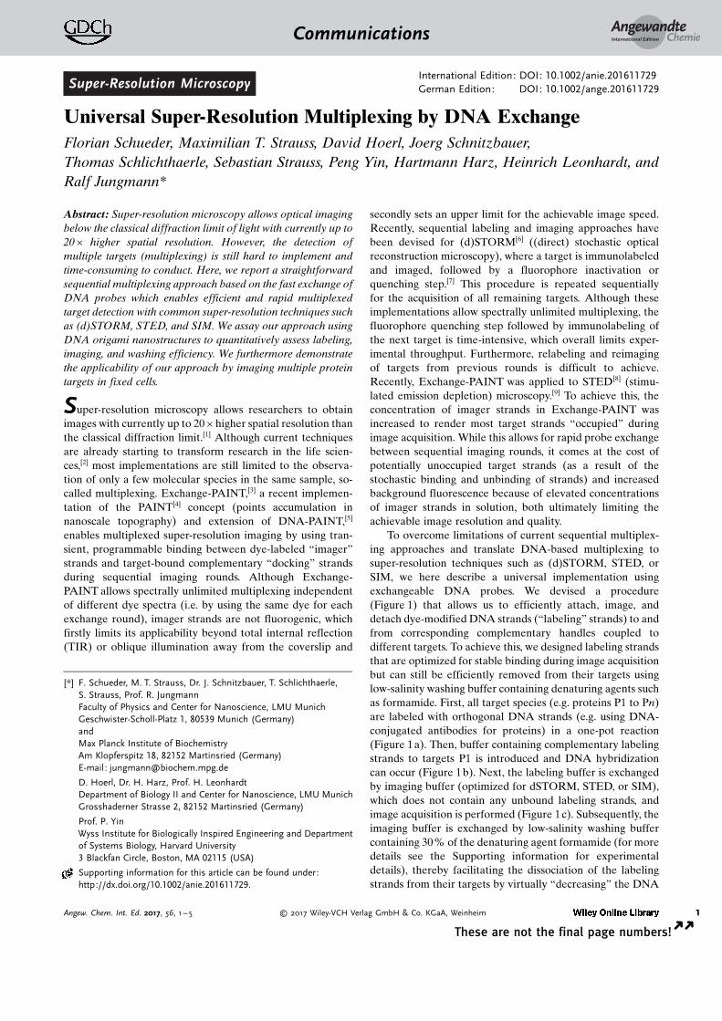
To demonstrate the feasibility of our approach, we usedself-assembled DNA origami[11] (Figure 2). We designed so-called six-helix-bundle (6HB) structures[12] carrying fourorthogonal single-stranded extensions on staple strands(which allows for four labeling and imaging rounds) atspecific positions[13] (Figure 2a). For Exchange-STED andExchange-dSTORM imaging, we arranged the sequences infour spots, approximately 113 nm apart (Figure 2 a). ForExchange-SIM, we opted for a structure displaying threespots spaced about 168 nm apart (Figure S2). Each spotconsists of six strands available for hybridization. Represen-tative images of the respective imaging rounds are shown inFigure 2b,c for Exchange-STED and Exchange-dSTORM,respectively (see Figures S3 and S4 for expanded views). Toassay the efficiency of our multiplexing approach, weinteractively analyzed approximately 100 structures in theExchange-STED and Exchange-dSTORM experiment. Forquantification of correct versus incorrect spots in eachlabeling and imaging round, it is important to note thatfalse negatives as well as false positives will lead to an “error”;however, these two “failure modes” have different rootcauses, and are thus important to distinguish. False positivesoccur when washing is inefficient, that is, labeling strands
have not dissociated from their respective targets. Falsenegatives occur when labeling or imaging is inefficient, that is,labeling strands have not hybridized to target strands or dyesare already bleached. The origami platform allows us touncover both failure modes independently and thus revealsany potential bias of our approach. We analyzed each spot ineach round separately to additionally assay for potentialbiases of different locations on the DNA origami structures.The results from our analysis show that on average about91% of spots are correct in the Exchange-STED experiments(Figure 2b) and 92 % in the Exchange-dSTORM experiments(Figure 2c).
To demonstrate that the order of Exchange rounds doesnot affect the experimental outcome, we varied the order forthe dSTORM experiments. We found that, indeed, theoutcome of the experiment is not affected by the order. Wenote, that in round 2 of the dSTORM experiment (Figure 2c),we do see a higher than expected number of false positives forspots 3 and 4 (70 % and 77% correct, respectively). Thispotentially suggests insufficient washing between rounds1 and 2. However, we also note that the expected numberof correct spots was restored in round 3. To assay theinfluence of different washing and hybridization times, weperformed additional experiments (Figure S5), where we firstdecreased the incubation time with the labeling strands from10 min to 1 min (keeping the washing times constant). Ina following experiment, we increased the washing time from2 � 3 min to 3 � 10 min (keeping the incubation time of 10 min
Figure 1. a) Targets 1�n are labeled with orthogonal approximately12 nucleotide long DNA sequences P1–Pn. b) Dye-modified “labeling”strands stably hybridize to complementary target strands P1.c) Acquisition is carried out in imaging buffer without free labelingstrands in solution. d) Imaging buffer is replaced by denaturingwashing buffer to facilitate the dissociation of labeling strands fromtargets P1. The labeling and washing procedure is repeated for allsubsequent targets. Note that the labeling strands are coupled to thesame spectral dye (e.g. Alexa 647) in each round, thus enablingspectrally unlimited multiplexing.
Figure 2. a) Illustration of the 6HB DNA origami “barcode”. Fourspots (with 6 binding sites each), spaced approximately 113 nm apart,can be “decorated” with up to four orthogonal target sequences each(colored in red, green, cyan, and magenta). b) Resulting super-resolution images of four rounds of Exchange-STED (top) withcorresponding statistical analysis (bottom). Histograms for eachround depict the percentage of correctly identified spots. (i) Statisticalanalysis showing the number of correct spots per structure inExchange-STED (14.6�0.7, mean � standard deviation). c) Corre-sponding results for Exchange-dSTORM. (ii) Correct spots per struc-ture in Exchange-dSTORM: 14.7�0.4 (mean � standard deviation).Scale bars: 200 nm.
AngewandteChemieCommunications
2 www.angewandte.org � 2017 Wiley-VCH Verlag GmbH & Co. KGaA, Weinheim Angew. Chem. Int. Ed. 2017, 56, 1 – 5� �
These are not the final page numbers!
constant). For the shorter probe incubation time, we detecta lower percentage of correctly labeled spots (true positives,see Figure S5). With longer washing times, we observea similar performance as with our standard conditions. Inconclusion, we note that our standard labeling and washingconditions (i.e. 10 min labeling, 2 � 3 min washing) shouldallow optimal results in exchange experiments. The statisticalanalysis of both Exchange-STED and -dSTORM experimentsfurther shows that no positional dependency on the DNAorigami structure was observed. There was also no biastowards false positives or negatives. Most importantly, there isalso no bias towards later washing or labeling rounds, thusindicating that our approach is viable for more extensivemultiplexing experiments (i.e beyond four rounds). Over fourlabeling and imaging rounds, we detected 14.6� 0.7 (mean �standard deviation) correct spots in Exchange-STED (Fig-ure 2b, (i)) and 14.7� 0.4 (mean � standard deviation)correct spots in Exchange-dSTORM (Figure 2c, (ii)) froma total of 16 spots. Detailed experimental conditions andimage processing specifics can be found in the SupportingInformation.
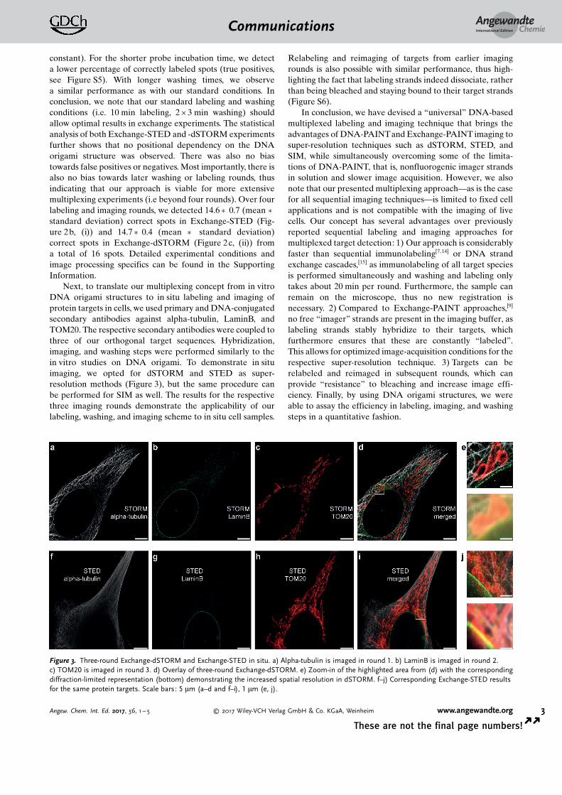
Next, to translate our multiplexing concept from in vitroDNA origami structures to in situ labeling and imaging ofprotein targets in cells, we used primary and DNA-conjugatedsecondary antibodies against alpha-tubulin, LaminB, andTOM20. The respective secondary antibodies were coupled tothree of our orthogonal target sequences. Hybridization,imaging, and washing steps were performed similarly to thein vitro studies on DNA origami. To demonstrate in situimaging, we opted for dSTORM and STED as super-resolution methods (Figure 3), but the same procedure canbe performed for SIM as well. The results for the respectivethree imaging rounds demonstrate the applicability of ourlabeling, washing, and imaging scheme to in situ cell samples.
Relabeling and reimaging of targets from earlier imagingrounds is also possible with similar performance, thus high-lighting the fact that labeling strands indeed dissociate, ratherthan being bleached and staying bound to their target strands(Figure S6).
In conclusion, we have devised a “universal” DNA-basedmultiplexed labeling and imaging technique that brings theadvantages of DNA-PAINTand Exchange-PAINT imaging tosuper-resolution techniques such as dSTORM, STED, andSIM, while simultaneously overcoming some of the limita-tions of DNA-PAINT, that is, nonfluorogenic imager strandsin solution and slower image acquisition. However, we alsonote that our presented multiplexing approach—as is the casefor all sequential imaging techniques—is limited to fixed cellapplications and is not compatible with the imaging of livecells. Our concept has several advantages over previouslyreported sequential labeling and imaging approaches formultiplexed target detection: 1) Our approach is considerablyfaster than sequential immunolabeling[7,14] or DNA strandexchange cascades,[15] as immunolabeling of all target speciesis performed simultaneously and washing and labeling onlytakes about 20 min per round. Furthermore, the sample canremain on the microscope, thus no new registration isnecessary. 2) Compared to Exchange-PAINT approaches,[9]
no free “imager” strands are present in the imaging buffer, aslabeling strands stably hybridize to their targets, whichfurthermore ensures that these are constantly “labeled”.This allows for optimized image-acquisition conditions for therespective super-resolution technique. 3) Targets can berelabeled and reimaged in subsequent rounds, which canprovide “resistance” to bleaching and increase image effi-ciency. Finally, by using DNA origami structures, we wereable to assay the efficiency in labeling, imaging, and washingsteps in a quantitative fashion.
Figure 3. Three-round Exchange-dSTORM and Exchange-STED in situ. a) Alpha-tubulin is imaged in round 1. b) LaminB is imaged in round 2.c) TOM20 is imaged in round 3. d) Overlay of three-round Exchange-dSTORM. e) Zoom-in of the highlighted area from (d) with the correspondingdiffraction-limited representation (bottom) demonstrating the increased spatial resolution in dSTORM. f–j) Corresponding Exchange-STED resultsfor the same protein targets. Scale bars: 5 mm (a–d and f–i), 1 mm (e, j).
AngewandteChemieCommunications
3Angew. Chem. Int. Ed. 2017, 56, 1 – 5 � 2017 Wiley-VCH Verlag GmbH & Co. KGaA, Weinheim www.angewandte.org
These are not the final page numbers! � �
Acknowledgements
We thank J. B. Woehrstein and S. S. Agasti for helpfuldiscussions and Y. Niyaz from Carl Zeiss MicroscopyGmbH for the use of their SIM microscope and dataacquisition support. This work was supported by the DFGthrough an Emmy Noether Fellowship (DFG JU 2957/1-1)and the SFB 1032 (Nanoagents for the spatiotemporal controlof molecular and cellular reactions), the ERC through anERC Starting Grant (MolMap, Grant agreement number680241), the Max Planck Society, the Max Planck Foundation,the Center for Nanoscience (CeNS), and the NanoinitiativeMunich (NIM). M.T.S. acknowledges support from theInternational Max Planck Research School for Molecularand Cellular Life Sciences (IMPRS-LS). T.S. acknowledgessupport from the DFG through the Graduate School ofQuantitative Biosciences Munich (QBM). P.Y. acknowledgessupport for from National Institute of Health (1-U01-MH106011-01 and 1R01EB018659-01).
Conflict of interest
Competing financial interest: A patent application has beenfiled. P.Y. and R.J. are cofounders of Ultivue, Inc., a startupcompany with interest in commercializing DNA-PAINTsuper-resolution technology.
Keywords: DNA nanotechnology · dSTORM · multiplexing ·SIM · STED
[1] S. W. Hell, S. J. Sahl, M. Bates, X. W. Zhuang, R. Heintzmann,M. J. Booth, J. Bewersdorf, G. Shtengel, H. Hess, P. Tinnefeld, A.Honigmann, S. Jakobs, I. Testa, L. Cognet, B. Lounis, H. Ewers,S. J. Davis, C. Eggeling, D. Klenerman, K. I. Willig, G. Vicido-mini, M. Castello, A. Diaspro, T. Cordes, J. Phys. D 2015, 48,443001.
[2] a) A. Szymborska, A. de Marco, N. Daigle, V. C. Cordes, J. A.Briggs, J. Ellenberg, Science 2013, 341, 655 – 658; b) K. Xu, G.
Zhong, X. Zhuang, Science 2013, 339, 452 – 456; c) A. N.Boettiger, B. Bintu, J. R. Moffitt, S. Wang, B. J. Beliveau, G.Fudenberg, M. Imakaev, L. A. Mirny, C. T. Wu, X. Zhuang,Nature 2016, 529, 418 – 422.
[3] R. Jungmann, M. S. Avendano, J. B. Woehrstein, M. Dai, W. M.Shih, P. Yin, Nat. Methods 2014, 11, 313 – 318.
[4] A. Sharonov, R. M. Hochstrasser, Proc. Natl. Acad. Sci. USA2006, 103, 18911 – 18916.
[5] R. Jungmann, C. Steinhauer, M. Scheible, A. Kuzyk, P.Tinnefeld, F. C. Simmel, Nano Lett. 2010, 10, 4756 – 4761.
[6] a) M. J. Rust, M. Bates, X. Zhuang, Nat. Methods 2006, 3, 793 –795; b) M. Heilemann, S. van de Linde, M. Schuttpelz, R.Kasper, B. Seefeldt, A. Mukherjee, P. Tinnefeld, M. Sauer,Angew. Chem. Int. Ed. 2008, 47, 6172 – 6176; Angew. Chem.2008, 120, 6266 – 6271.
[7] a) J. Tam, G. A. Cordier, J. S. Borbely, A. Sandoval Alvarez, M.Lakadamyali, PLoS One 2014, 9, e101772; b) C. C. Valley, S. Liu,D. S. Lidke, K. A. Lidke, PLoS One 2015, 10, e0123941.
[8] S. W. Hell, J. Wichmann, Opt. Lett. 1994, 19, 780 – 782.[9] S. Beater, P. Holzmeister, B. Lalkens, P. Tinnefeld, Opt. Express
2015, 23, 8630 – 8638.[10] a) B. L. McConaughy, C. D. Laird, B. J. McCarthy, Biochemistry
1969, 8, 3289 – 3295; b) R. D. Blake, S. G. Delcourt, NucleicAcids Res. 1996, 24, 2095 – 2103; c) R. Jungmann, T. Liedl, T. L.Sobey, W. Shih, F. C. Simmel, J. Am. Chem. Soc. 2008, 130,10062 – 10063.
[11] P. W. Rothemund, Nature 2006, 440, 297 – 302.[12] S. M. Douglas, J. J. Chou, W. M. Shih, Proc. Natl. Acad. Sci. USA
2007, 104, 6644 – 6648.[13] C. Lin, R. Jungmann, A. M. Leifer, C. Li, D. Levner, G. M.
Church, W. M. Shih, P. Yin, Nat. Chem. 2012, 4, 832 – 839.[14] M. J. Gerdes, C. J. Sevinsky, A. Sood, S. Adak, M. O. Bello, A.
Bordwell, A. Can, A. Corwin, S. Dinn, R. J. Filkins, D. Hollman,V. Kamath, S. Kaanumalle, K. Kenny, M. Larsen, M. Lazare, Q.Li, C. Lowes, C. C. McCulloch, E. McDonough, M. C. Montalto,Z. Pang, J. Rittscher, A. Santamaria-Pang, B. D. Sarachan, M. L.Seel, A. Seppo, K. Shaikh, Y. Sui, J. Zhang, F. Ginty, Proc. Natl.Acad. Sci. USA 2013, 110, 11982 – 11987.
[15] a) D. Y. Duose, R. M. Schweller, W. N. Hittelman, M. R. Diehl,Bioconjugate Chem. 2010, 21, 2327 – 2331; b) R. M. Schweller, J.Zimak, D. Y. Duose, A. A. Qutub, W. N. Hittelman, M. R. Diehl,Angew. Chem. Int. Ed. 2012, 51, 9292 – 9296; Angew. Chem.2012, 124, 9426 – 9430.
Manuscript received: December 1, 2016Final Article published: && &&, &&&&
AngewandteChemieCommunications
4 www.angewandte.org � 2017 Wiley-VCH Verlag GmbH & Co. KGaA, Weinheim Angew. Chem. Int. Ed. 2017, 56, 1 – 5� �
These are not the final page numbers!

Communications
Super-Resolution Microscopy
F. Schueder, M. T. Strauss, D. Hoerl,J. Schnitzbauer, T. Schlichthaerle,S. Strauss, P. Yin, H. Harz, H. Leonhardt,R. Jungmann* &&&&—&&&&
Universal Super-Resolution Multiplexingby DNA Exchange
Many happy returns : A straightforwardsequential multiplexing approach basedon the fast exchange of DNA probes hasbeen developed that enables efficient andrapid multiplexed target detection withcommon super-resolution techniquessuch as (d)STORM, STED, and SIM.
AngewandteChemieCommunications
5Angew. Chem. Int. Ed. 2017, 56, 1 – 5 � 2017 Wiley-VCH Verlag GmbH & Co. KGaA, Weinheim www.angewandte.org
These are not the final page numbers! � �