Common Pediatric Emergencies v1 · Common Pediatric Emergencies Chris Woleben MD, FAAP Asst....
22
1 1 Common Pediatric Emergencies Chris Woleben MD, FAAP Asst. Professor Emergency Medicine Pediatric Division VCU Medical Center 2 Objectives After this lecture, you should be able to: – recognize the most common injuries seen in the Pediatric Emergency Department – understand diagnostic and management plans for common pediatric illnesses 3 Pediatric Trauma Injuries are the most common cause of childhood death: – 40% of deaths children between ages 1-4 • 3 times more common than congenital anomalies – 70% of deaths for older children and adolescents • motor vehicle occupant injuries most common • pedestrian injuries (age 5 – 9) – drowning ranks second overall as cause for unintentional deaths • peaks in preschool and late teen years 4 Pediatric Trauma – homicide is leading cause of injury death for infants less than 1 year of age • Shaken Baby Syndrome • other forms of child abuse and neglect • asphyxiation and choking – homicide is second most common cause of death in adolescents • more than 80% involve use of firearm – suicide • rare before age ten but increasingly common • third leading cause of death ages 15-19 • males more successful than females 5 Pediatric Trauma Score 6 Pediatric Trauma Systematic Approach to All Patients: – follow the A, B, C, D, E’s of management – investigate mechanism of injury to see if injury is compatible with age and developmental status of the child
Transcript of Common Pediatric Emergencies v1 · Common Pediatric Emergencies Chris Woleben MD, FAAP Asst....

1
1
Common Pediatric Emergencies
Chris Woleben MD, FAAPAsst. Professor Emergency MedicinePediatric DivisionVCU Medical Center
2
ObjectivesAfter this lecture, you should be able to:– recognize the most common injuries seen
in the Pediatric Emergency Department– understand diagnostic and management
plans for common pediatric illnesses
3
Pediatric TraumaInjuries are the most common cause of childhood death:– 40% of deaths children between ages 1-4
• 3 times more common than congenital anomalies
– 70% of deaths for older children and adolescents• motor vehicle occupant injuries most common• pedestrian injuries (age 5 – 9)
– drowning ranks second overall as cause for unintentional deaths
• peaks in preschool and late teen years4
Pediatric Trauma– homicide is leading cause of injury death for
infants less than 1 year of age • Shaken Baby Syndrome• other forms of child abuse and neglect• asphyxiation and choking
– homicide is second most common cause of death in adolescents
• more than 80% involve use of firearm– suicide
• rare before age ten but increasingly common• third leading cause of death ages 15-19• males more successful than females
5
Pediatric Trauma Score
6
Pediatric TraumaSystematic Approach to All Patients:– follow the A, B, C, D, E’s of management– investigate mechanism of injury to see if
injury is compatible with age and developmental status of the child

2
7
Primary SurveyA = Airway / cervical spine control– assess airway patency:
• Patent / Conscious – Maintain position of comfort
• compromised – Position, suction, +/- oral airway
• Not Maintainable:– Oral endotracheal intubation– Bag-Valve-Mask ventilation
– maintain cervical spine in neutral position with manual immobilization
• head or facial trauma• suspicious mechanism of injury
8
Primary SurveyB = Breathing– assess:
• rate Work of Breathing• color Mental Status
– Adequate?• Administer High Flow Oxygen
– Inadequate?• Bag-Valve-Mask ventilation with 100%
O2• naso/orogastric tube• consider Intubation
9
Primary SurveyC = Circulation / Hemorrhage Control– Assess:
Heart rate Pulse qualityColor Skin signsMental status Direct pressure to bleeding
– If signs of shock are present:• obtain immediate vascular access (IV / IO)• isotonic fluid bolus (20cc/kg NS or LR)• baseline labs – VBG, Lactate, CBC• cardiac monitor• urinary catheter• consider blood transfusion 10
Primary SurveyD = Disability (Neurologic Status)– Assess
• Pupillary Function• Mental Status – AVPU
–A = Alert–V = Responsive to Voice–P = Responsive to Pain–U = Unresponsive
• Modified Glasgow Coma Score
11
Modified Glasgow Coma Scale
12
Primary SurveyE = Exposure– Remove clothing for complete exam– Prevent heat loss
• Warm blankets• Heat lamps• Radiant warmers• Warm IV fluid

3
13
Head Injury600,000 visits per year250,000 hospitalizations per year95,000 brain injuries per year– 4,000 deaths per year– 80% of children dying from trauma have a
significant head injuryPrimary Injury:– skull fracture, contusion, laceration, neuronal or
vascular injurySecondary Injury:– elevated ICP, cerebral hypoperfusion, hypoxia,
seizures, hyperthermiaPredisposing Risk Factors:– head is relatively large, brain is less myelinated,
skull thinner 14
Head InjuryClinical Findings:– hypotension (infants)– vomiting– seizures– bulging fontanelle– decreased LOC– motor deficits– posturing or paresis– pupillary changes
15
Head InjuryCushing’s Triad – sign of increased ICP:– Bradycardia– hypertension– Abnormal Respiratory Pattern
Treatment of increased ICP:– elevate head of bed– mild hyperventilation (PCO2 28-35)– Mannitol (osmotic agent); lasix– hypothermia– barbituate coma– surgical drainage
16
Head InjuryIndications for CT:– GCS < 15– GCS = 15 with features suggestive of significant
brain injury• prolonged LOC• amnesia• persistent vomiting• seizures• increasing headache• focal neurologic signs
– skull fracture– penetrating injury– suspected child abuse
17
Spinal Cord Trauma1,000 sustain cord injury per year– young child: C1 – C3 injured– older children: C5 – C7 injured
SCIWORA– spinal Cord Injury Without Radiographic
Abnormality– x-rays are normal– delayed onset of neurologic deficits (30
minutes – 4 days)– transient symptoms are common
Pseudo-Subluxation C2/C3 common– 40% of children <7 years old 18
Pseudo-Subluxation C2/C3

4
19
Abdominal TraumaSolid organs most commonly injured:– spleen
• LUQ contusion, lower rib pain• Left shoulder pain (Kehr’s sign)• LUQ tenderness and guarding
– liver• RUQ contusion, pain/guarding• elevated LFT’s
Duodenal injuries:• high speed or direct blows to upper abdomen
(bicycle handle bars, lap belt)• general abdominal tenderness, bilious vomiting 20
Abdominal TraumaIndications for CT:– suspected intra-abdominal injury and
stable vital signs– slowly declining hematocrit– physical exam unreliable due to neurologic
injury– hematuria– need for aggressive fluid resuscitation
without obvious source of hemorrhage
21
Pediatric Abdominal Emergencies
22
Case StudyA three week old child presents to ER withacute onset of irritability, bilious vomiting,and abdominal pain. Of the following, themost likely diagnosis is:
1. Intussusception2. Incarcerated Inguinal Hernia3. Malrotation with Volvulus4. Pyloric Stenosis5. Acute appendicitis
23
IntussusceptionSegment of bowel telescopes into more distal segment:– ileocolic most common– ileoilial– colocolic rare
Leading cause of intestinal obstruction in infants:– hypertrophied Peyer’s patches common lead-point– consider polyp, Meckel’s diverticulum, tumor in
older childrenTypical age range: 3mo – 12mo
24
Intussusception

5
25
IntussusceptionClinical Presentation:– crampy abdominal pain intermixed with
periods of lethargy– irritability / inconsolable crying– anorexia, vomiting– may feel sausage-like mass in RUQ– currant jelly stools typically late feature– sepsis-like presentation
26
IntussusceptionManagement:– IV hydration, lab-work (CBC, BMP, STBB)– nasogastric tube– plain films may reveal distended bowel with
air-fluid levels– air contrast barium enema often provides
both diagnosis and reduction– pediatric surgical consultation prior to
ACBE
27
Intussusception
28
Incarcerated Inguinal Hernia60% occur in first year of lifeOccur more often in males– usually involves ovary rather than intestine
in femalesMay lead to strangulation if not reduced within 24 hours– progressive edema of bowel wall by
venous and lymphatic obstruction– occlusion of arterial supply resulting in
necrosis of bowel and possibly perforation
29
Incarcerated Inguinal HerniaClinical Presentation:– irritability, crying– vomiting, abdominal distension– firm, discrete mass palpated at inguinal
ring (may extend into scrotum of boys)– easily confused with tense hydrocele in
boys• no mass will be felt in inguinal ring• hydroceles typically trans-illuminate
30
Hydrocoele
6
31
Incarcerated Inguinal HerniaAttempt manual reduction– sedate with morphine– older children placed in Trendelenburg
position (let gravity work for you!)– mild pressure exerted at inguinal ring in V-
shape with one hand; other hand squeezes gas or fluid out of incarcerated bowel into the abdominal cavity
– surgical reduction if unsuccessful
32
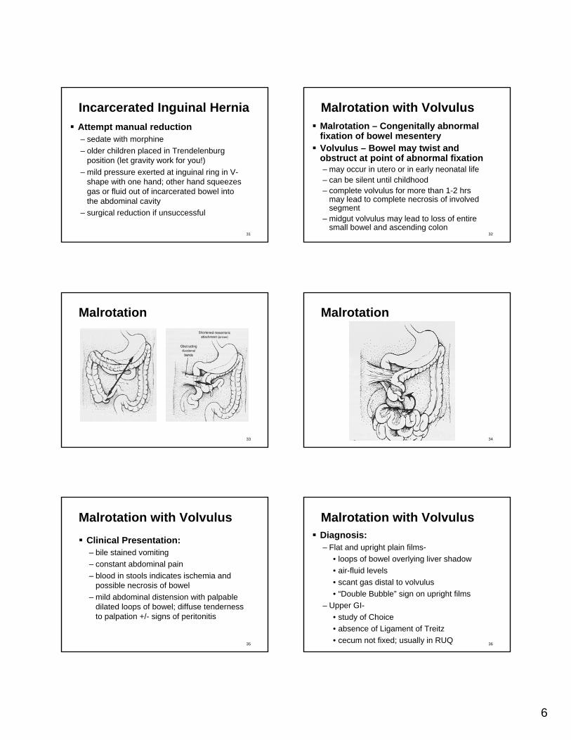
Malrotation with VolvulusMalrotation – Congenitally abnormal fixation of bowel mesenteryVolvulus – Bowel may twist and obstruct at point of abnormal fixation– may occur in utero or in early neonatal life– can be silent until childhood– complete volvulus for more than 1-2 hrs
may lead to complete necrosis of involved segment
– midgut volvulus may lead to loss of entire small bowel and ascending colon
33
Malrotation
34
Malrotation
35
Malrotation with VolvulusClinical Presentation:– bile stained vomiting– constant abdominal pain– blood in stools indicates ischemia and
possible necrosis of bowel– mild abdominal distension with palpable
dilated loops of bowel; diffuse tenderness to palpation +/- signs of peritonitis
36
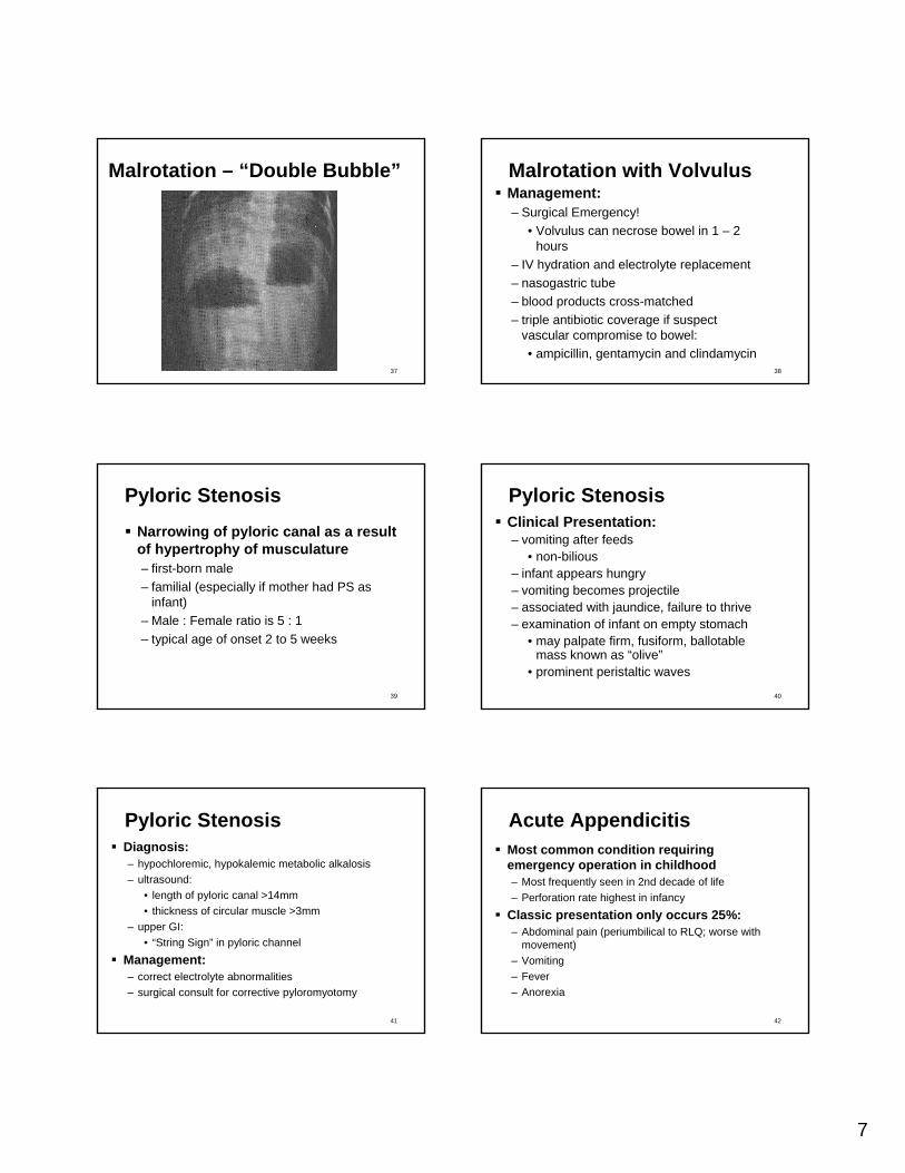
Malrotation with VolvulusDiagnosis:– Flat and upright plain films-
• loops of bowel overlying liver shadow• air-fluid levels• scant gas distal to volvulus• “Double Bubble” sign on upright films
– Upper GI-• study of Choice• absence of Ligament of Treitz• cecum not fixed; usually in RUQ
7
37
Malrotation – “Double Bubble”
38
Malrotation with VolvulusManagement:– Surgical Emergency!
• Volvulus can necrose bowel in 1 – 2 hours
– IV hydration and electrolyte replacement– nasogastric tube– blood products cross-matched– triple antibiotic coverage if suspect
vascular compromise to bowel:• ampicillin, gentamycin and clindamycin
39
Pyloric StenosisNarrowing of pyloric canal as a result of hypertrophy of musculature– first-born male– familial (especially if mother had PS as
infant)– Male : Female ratio is 5 : 1– typical age of onset 2 to 5 weeks
40
Pyloric StenosisClinical Presentation:– vomiting after feeds
• non-bilious– infant appears hungry– vomiting becomes projectile– associated with jaundice, failure to thrive– examination of infant on empty stomach
• may palpate firm, fusiform, ballotablemass known as “olive”
• prominent peristaltic waves
41
Pyloric StenosisDiagnosis:– hypochloremic, hypokalemic metabolic alkalosis– ultrasound:
• length of pyloric canal >14mm• thickness of circular muscle >3mm
– upper GI:• “String Sign” in pyloric channel
Management:– correct electrolyte abnormalities– surgical consult for corrective pyloromyotomy
42
Acute AppendicitisMost common condition requiring emergency operation in childhood– Most frequently seen in 2nd decade of life– Perforation rate highest in infancy
Classic presentation only occurs 25%:– Abdominal pain (periumbilical to RLQ; worse with
movement)– Vomiting– Fever– Anorexia

8
43
Acute AppendicitisClinical Signs:– fever– RLQ tenderness– involuntary guarding– rebound tenderness– irritability, lethargy (in infants)– hopping causes increased pain– psoas, obturator signs occur <30%– WBC usually 11,000 – 15,000– urinalysis: pyuria (30%)
44
AppendicitisDiagnosis:– clinical exam– plain films
• calcified fecalith (8-10%)• indistinct psoas margin with rightward scolisis
– ultrasound– CT with oral, rectal, IV contrast
Differential Diagnosis:– Gastroenteritis Constipation– Intussusception Ileus– Ectopic pregnancy PID / UTI– Mesenteric Adenitis Lower Lobe Pneumonia
45
Case Study - AnswerA three week old child presents to ER withacute onset of irritability, bilious vomiting,and abdominal pain. Of the following, themost likely diagnosis is:
1. Intussusception2. Incarcerated Inguinal Hernia3. Malrotation with Volvulus4. Pyloric Stenosis5. Acute appendicitis
46
Common Pediatric Orthopedic Emergencies
47
Salter Harris Classification
48
Buckle (Torus) FractureMetaphyseal fracture caused by compressive forces in a longitudinal planeBulge in metaphyseal region where cortex is weakest

9
49
Buckle (Torus) Fracture
50
Greenstick FractureMost common fracture due to a lateral bending forceFracture exists only on one side of cortex
51
Greenstick Fracture
52
Toddler’s FractureOccurs in 1-4 year oldsNon-displaced spiral fracture of the lower tibiaMake sure history is consistent with injury
53
Toddler’s Fracture
54
Clavicle FractureMost common pediatric fractureMost are greenstickMechanism: fall or blow to shoulderTreatment: figure-of-eight or sling
10
55
Supracondylar Fracture3-10 years old; males > femalesMechanism – fall on outstretched hand or flexed elbowComplications – brachial vessel, radial nerve damage– Volkmann’s Ischemia (5 P’s):
• pain, paresthesia, pallor, paralysis, pulselessness
56
Supracondylar Fracture
57
Slipped Capital Femoral Epiphysis
12-15 year oldsObese (90%)Males > FemalesBilateral 20-30%Child presents with limp, localizes pain to hip, groin or knee– examine hip in any child with knee pain
58
Slipped Capital Femoral Epiphysis
59
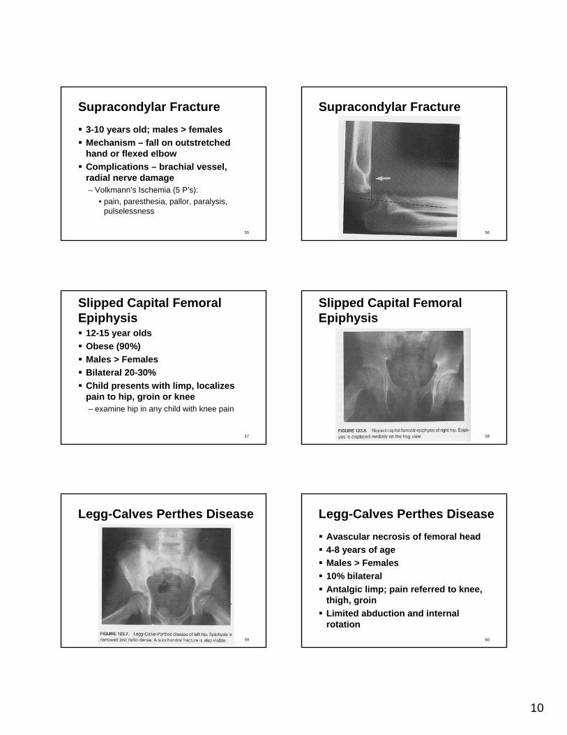
Legg-Calves Perthes Disease
60
Legg-Calves Perthes DiseaseAvascular necrosis of femoral head4-8 years of ageMales > Females10% bilateralAntalgic limp; pain referred to knee, thigh, groinLimited abduction and internal rotation

11
61
Septic ArthritisInfection within a joint spaceMost <5 years of ageMales > FemalesMechanism: hematogenous > spread from contiguous site, direct inoculation30% misdiagnosed as trauma90% monoarticular– Knee > Hip > Ankle > Shoulder > Wrist
Organisms– S. Aureus; Streptococcus; H. influenza; GNR – GBS (neonates); N. gonorrhea (sexually active) 62
Septic ArthritisPresentation:– limp, pain, erythema, swelling, warmth– decreased ROM– fever (absent in neonates, GC)– elevated WBC, ESR, CRP
• positive blood culture (50%)– x-rays reveal widened joint space, bone
destruction (late)– Synovial Fluid:
• WBC > 50,000 (>75% PMN’s)• positive gram stain (75%)• positive culture (65%)
63
Toxic Synovitis18 months – 12 years (peak age 2)Males > FemalesPain, limp, afebrileMinimal pain with ROMWBC, ESR, CRP normalSynovial fluid – turbid but sterileX-rays may reveal joint effusionTreatment – bed rest, NSAID’s
64
Febrile Infant
65
Fever in InfantBased on clinical signs and symptoms, it can be very hard to determine which infants develop neonatal septicemia:– temperature instability
• Rectal Temperature > 100.4 or < 96.8– change in feeding habits– seizure activity– respiratory distress– jaundice– loose Stools– no signs or symptoms 66
Fever in InfantsInfants aged 1-28 days are at higher risk of spontaneous bacterial infection– studies have consistently shown that 5-6
percent of infants less than 28 days old with fever >100.4 will have a serious bacterial infection:• most have UTI’s• some have bacteremia, pneumonia, or
meningitis• most common organisms:
–GBS, Listeria, gram negative organisms

12
67
Infant with FeverMandatory Work-up Includes:– CBC with manual differential– blood culture– urinalysis, urine culture– lumbar puncture– (+/-) CXR– empiric antibiotic coverage / admission
• Ampicillin and Tobramycin
68
Infant with FeverRSV and Influenza A/B should be sent during appropriate seasons– slightly decreased risk of having
spontaneous bacterial infection if positive– still complete septic work-up and admit
69
Infant with FeverInfants older than 28 days and less than 3 months of age with fever:– controversial
• at times decision is made on likelihood of adequate follow-up
70
Pediatric Ophthalmologic Emergencies
71
Periorbital CellulitisInfection is anterior to orbital septum<6 years of age (peaks at age 2)Unilateral lid swelling, erythema, tenderness, warmth, feverMost common organisms:– S. pneumoniae; S. aureus; Streptococcus;
H. influenzaeTreatment:– Nafcillin or Ceftriaxone; warm compresses
72
Periorbital Cellulitis

13
73
Orbital CellulitisInfection is posterior to orbital septumUsually due to complication of sinusitis (ethmoid)Swelling, erythema, tenderness, warmth, proptosis, loss of vision, ophthalmoplegiaComplications– meningitis, sepsis, cavernous sinus thrombosis
Treatment– Nafcillin, Ceftriaxone– +/- surgical drainage
74
Orbital Cellulitis
75
Corneal AbrasionUsually history of trauma to eye– common in infants with inconsolable crying due to
fingernails scratching surface of eyeSymptoms– pain when eye is open– tearing– blurry vision
Diagnosis – Fluorescein stain with wood’s lampTreatment – Bacitracin ophthalmic ointment
76
Corneal Abrasion
77
Pediatric Genitourinary Emergencies
78
Testicular TorsionMost common cause of acute painful scrotumPeak – age 14-17 yearsPreceding trauma in 5-6%50% recall similar pain which resolvedSpermatic cord twists and venous drainage is obstructed– testicular engorgement– arterial shutdown– tissue ischemia and eventual infarction (6 hours)

14
79
Testicular TorsionClinical Findings:– sudden onset of unilateral scrotal pain– followed by swelling, abdominal pain,
vomiting– testis is high riding and transverse in
position (Bell-clapper deformity)– cremasteric reflex is absent– epididymis is tender– lifting testis increases pain
80
Testicular Torsion
81
Testicular TorsionDiagnosis– doppler US to look for decreased blood
flowManagement– manual detorsion– surgical exploration and fixating orchiopexy
82
Torsion of the AppendixPeaks in pubertyPoint tenderness at upper pole of testis or epididymisVomiting“Blue Dot” sign– blue, pea-sized, tender nodule represents
ischemic appendage
83
Torsion of Appendix
84
Pediatric Respiratory Emergencies

15
85
Upper Airway DiseaseStridor:– externally audible sound produced by
turbulent flow through narrowed airway– acute vs. chronic-
• croup• epiglottitis• retropharyngeal, peritonsillar abscess• bacterial Tracheitis• foreign body aspiration• subglottic stenosis,
tracheo/laryngomalacia• anatomic variants 86
Remember Pousille’s Law?
87
CroupTypical age range: 6 – 36 monthsMales > Females (3:2)Fall / Winter predilectionCommon causes:– parainfluenza– RSV– adeonvirus– influenza
88
CroupClinical Presentation:– prodrome of URI symptoms, fever– development of barking, seal-like cough
and stridor• subglottic mucosal swelling and
secretions lead to narrowed airway– symptoms worse at night– “Steeple Sign” on plain film of neck
89
Croup – “Steeple Sign”
90
CroupManagement:– keep child calm (sitting in parent’s lap)– humidified air / saline– steroids
• Decadron 0.6 mg/kg IM or PO– Racemic Epinephrine nebulizer treatment
• decreases mucosal swelling and secretions
• must observe at least 4 hours after treatment given

16
91
Retropharyngeal AbscessRecent history of pharyngitis, otitismedia, or penetrating wound to posterior pharynxCellulitis and suppurative adenitis of lymph nodes in prevertebral fasciaPlain films reveal soft tissue swelling at level of C3-C4CT better delineates extent of infection
92
Retropharyngeal Abscess
93
Retropharyngeal AbscessClinical Presentation:– fever– difficulty swallowing, drooling– sore throat, hoarse voice– painful, stiff neck
Management:– secure stable airway– broad spectrum antibiotics
(Nafcillin/Clindamycin)– ENT consult for surgical I&D
94
EpiglottitisNot seen as frequently today– Haemophilus influenza type B vaccine
Other bacterial causes:– staphylococcus species– streptococcus species
Diagnosis:– “Thumbprint Sign” on lateral plain film of
neck
95
Epiglottitis – normal airway
96
Epiglottitis – Thumbprint Sign

17
97
Epiglottitis
98
EpiglottitisClinical Presentation:– sudden onset high fever– moderate to severe respiratory distress– stridor– drooling– toxic appearing child that sits in “tripod”
position
99
EpiglottitisManagement:– keep child as calm as possible– EMERGENT surgical consult to establish
definitive airway in operating room– Broad Spectrum antibiotic coverage
• Second or third generation cephalosporins
100
Bacterial Tracheitis
Bacterial complication of a viral URI– Staphylococcus aureus– Haemophilus influenza (non-typable)– streptococcal species
Pathophysiology:– swelling of tracheal mucosa below vocal
folds– thick purulent secretions may lead to
mucous plugging
101
Bacterial Tracheitis
Presentation similar to croup– more toxic appearing child– does not respond to typical croup
treatment– outside the typical age range for croup
Plain films of neck:– edema with an irregular border of the
subglottic tracheal mucosa (“subglotticmembrane”)
102
Subglottic Membrane

18
103
Bacterial TracheitisManagement:– assess and maintain patent airway– frequent suctioning if intubated– ENT consultation– Broad Spectrum antibiotic coverage
104
Foreign Body AspirationRecurrent wheezing or stridor that is unresponsive to usual therapy– afebrile– recurrent pneumonia in same location
Common items found:– coins, small toys– nuts or seeds– popcorn, small candy– beads, buttons, safety pins– balloons, latex gloves
105
Foreign Body AspirationDiagnosis:– plain films of the neck– PA and lateral chest xray
• radio-opaque FB• atelectasis• mediastinal deviation
– bilateral decubitus films– inspiratory and expiratory chest films
• hyper-inflation or air trapping on side of FB
106
Foreign Body Aspiration
107
Lower Airway IllnessesIntrathoracic Structures– mainstem bronchi, bronchial tree,
bronchiolesClinical Findings:– wheezing
• obstruction of intrathoracic airways• heard both during expiration and
inspiration– rales
• air trapping and atelectasis• Focal Consolidation
– diminished air movement 108
AsthmaReversible airway obstruction– bronchospasm of lower airway– swelling of airways and increased mucous
production (inflammation)

19
109
Asthma - TriggersAtopic Conditions– allergic rhinitis, eczema, chronic sinusitis
Allergen Exposures– cigarette smoke– pets– carpeting, ceiling fans
Viral Illnesses
110
Asthma - PresentationCoughWheezeShortness of breathChest tightness / painVomitingIncreased work of breathing– retractions, flaring
111
Asthma - ManagementBronchospasm:– beta-2 agonists
• Albuterol vs. Xopenex• Terbutaline, epinephrine
– anticholinergics• Atrovent
Inflammation– corticosteroids
112
BronchiolitisViral etiology– RSV, parainfluenza, adenovirus, rhinovirus
Symptoms:– cough– tachypnea– accessory muscle use– high pitched wheezing– fine inspiratory crackles and rhonchi– copious, thick nasal secretions
113
BronchiolitisManagement:– supportive care
• nasal saline spray / suctioning• adequate PO intake• ensure adequate oxygenation
– pharmacologic• Albuterol• racemic Epinephrine• steroids – consider if history of atopy exists
– infants <2 months old, history of prematurity• risk of developing apnea – generally admit and
observe 114
Wound Management

20
115
Wound ManagementGet a Good History:
• mechanism of injury• wounding object – mass, velocity, etc• environment in which injury occurred• time of injury• general health of patient• medications• allergies• immunization status
116
Wound ManagementPerform a Good Physical Exam:
• assessment of distal neurovascular function• assessment of tendon integrity• palpation of adjacent bony structures
– consider radiographs to look for fractures– surgical referral for all open fractures
• explore wound through full ROM and in position of injury
• evaluate for presence of foreign bodies– consider radiographs to look for foreign
bodies– remove all reactive foreign bodies
(vegetable materials, wood, organic materials, clothing)
117
Wound ManagementLocal cleansing and irrigation is important in almost all wounds– remove obvious dirt or foreign materials by
irrigation or gentle scrubbing– use of antiseptic solutions for wound cleansing is
controversial– saline irrigation is standard practice in treating
contaminated or dirty wounds• high pressure irrigation• 200-300 ml minimum volume• will cause tissue damage so not needed in
clean wounds
118
Wound ManagementHair– razor removal results in increased rate of wound
infections– use of electric clippers associated with lower rates
of infection– petroleum jelly can keep unruly hairs out of your
field– NEVER shave eyebrows
119
Wound ManagementDebridement– use to remove heavily contaminated tissue
or devitalized tissue– prevents bacterial growth– do not create too much tension in your
wound by debriding too large an area
120
Prophylactic Antibiotic UseIndications:– patient prone to development of infective
endocarditis– immuno-suppressed patients– soft tissue lacerations occurring in
previously lymphedematous tissue– wounds judged to be contaminated or dirty
by the clinician (especially in dependent areas)
– stellate lacerations with adjacent abrasions resulting from high impact

21
121
Prophylactic Antibiotic UseIndications:– delayed wound closing and repair
• between 6 to 18 hours after injury, bacteria colony counts reach potentially infective concentrations
• consider open management and delayed closure
– wounds contaminated by saliva, feces, vaginal fluids
• consider open management and delayed closure
– missile wounds– certain bite and crush wounds– foot wounds
122
Antibiotic UseBroad Spectrum– effective against Staphylococci, Streptococcus
and facultative organisms– Cephalexin (Kelfex) is inexpensive, well-tolerated,
effective choice– can also use Augmentin, Dicloxacillin– use of Erthromycin or Biaxin if PCN/Cephalosporin
sensitive
123
Animal BitesDog bites account for about 1% of ED visits:– wounds become infected slightly more
commonly than wounds in general– require meticulous wound preparation– loose sutures if needed– prophylactic antibiotics indicated for:
• dog bites to hand• bites older than 6 hours• bites that inflict puncture wound
124
Animal BitesCommon Organisms:– Staphylococcus, Streptococcus– Bacteroides species– anaerobic cocci– Pasteurella multicida
Best antibiotic choice: AugmentinSend wound culture if infected
125
Another reason not to have cats!
Cat bites have higher incidence of wound infections– do not close wounds if at all possible– meticulous irrigation– Pasteurella multocida is implicated in about
50% of cat bites
126
Rabies ProphylaxisSoap and water cleansing decrease transmission of rabiesActive and Passive Immunoprophylaxis– Human Rabies Immune Globulin
• Dose = 20 IU/kg• Infiltrate ½ dose around wound and other ½
dose at IM site distant from wound– Human Diploid Cell Vaccine
• Dose = 1ml IM• Give on days 1, 3, 7, 14, and 28
Contact Animal Control

22
127
Rabies Prophylaxis
128
Human BitesGenerally associated with higher rates of infection:– delay in seeking treatment– high impact mechanism of injury
• fist fights, sports injuries• increased tissue crushing and devitalization
Common pathogens:• Staphylococcus, Streptococcus (includes group
A Streptococcus)• Bacteroides species• anaerobic cocci• Eikenella corrodens
129
Tetanus ProphylaxisAll wounds carry risk of tetanus as a potential complication:– contaminated wounds (soil, feces)– devitalized tissue– deep puncture wounds
Tetanus Immune Globulin– dose = 250 IU IM– only give in patients who have not received at
least three previous doses of tetanus toxoid or whose immunization status is unknown
130
Tetanus Prophylaxis
131
Common Pediatric Emergencies:Questions???
Phone: 828-7400Email: [email protected]