Common adult fractures Axial skeleton (spine and...
62
Transcript of Common adult fractures Axial skeleton (spine and...


Objectives
The ability to demonstrate knowledge of the following:
Basic anatomy of the spine.
Initial assessment and treatment of spinal injuries at the field.
Principle of spinal stability.
Understanding of neurologic syndromes caused by spinal trauma.
Management of Cauda equina syndrome.

Spine Pathology Red Flag
Conditions
Beware of:
1) Cauda Equina/severe neurologic injury (perianal numbness, decreased rectal tone, loss of movement in the extremeties).
2) Tumour weakening the vertebrae (causing cord compression or vertebral fracture).
3) Infection weakening bone (causing disc/vertebral destruction or cord compression).
4) Traumatic Spine Fracture (causing vertebral angulation, pain, or neuro compromise).
Remember that spine fracture can occur without trauma.
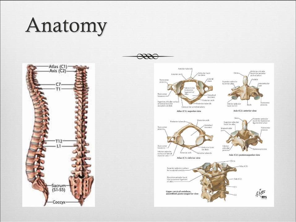
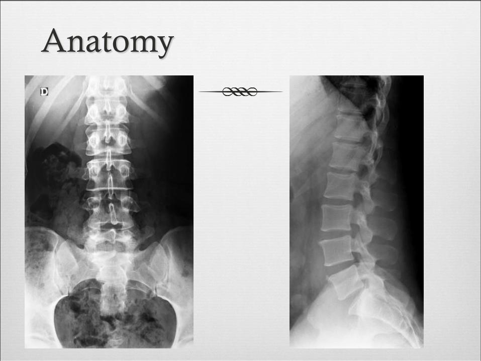
Anatomy

Anatomy

Anatomy

Anatomy

Anatomy
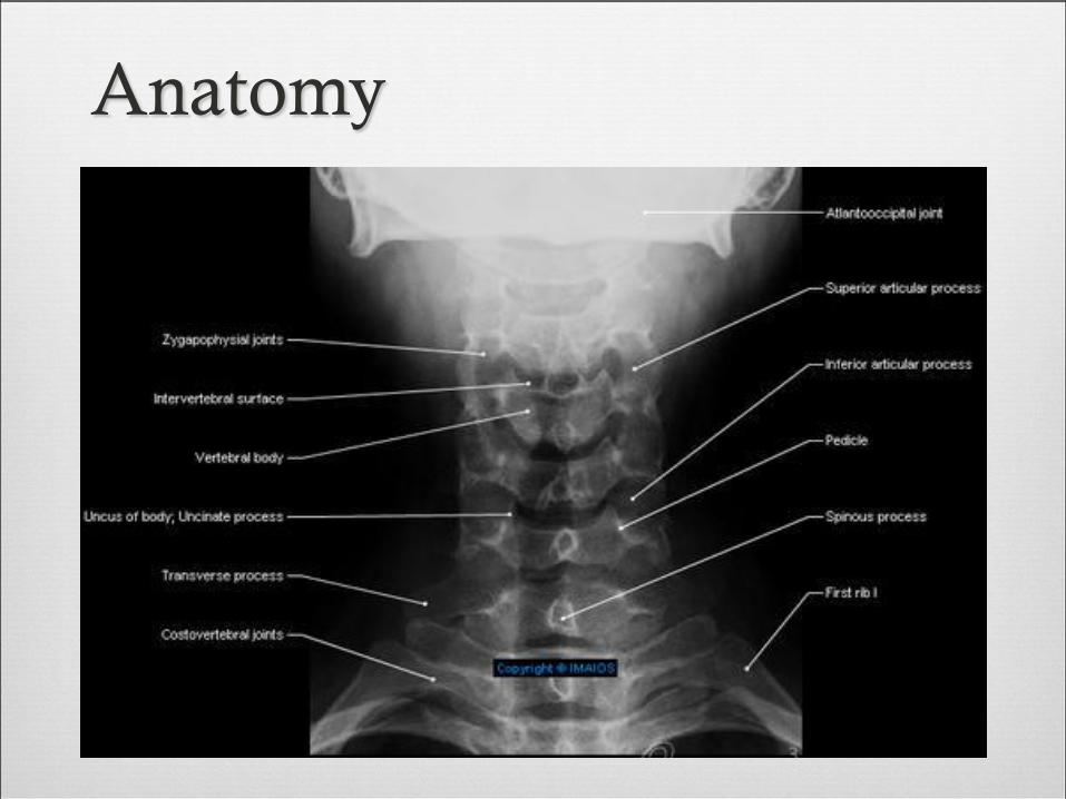
Anatomy

Anatomy

Anatomy

Anatomy

Anatomy
Anatomy

Anatomy

Anatomy

Epidemiology
56000 cases per year.
11000 new spinal cord injuries.
15-20% multiple non-contiguous levels.
10% involving the cervical spine.
90% involving thoracolumbar spine.
25% have neurologic deficit.
Age: mostly between 15-24 years.
Gender: mostly males (4:1).


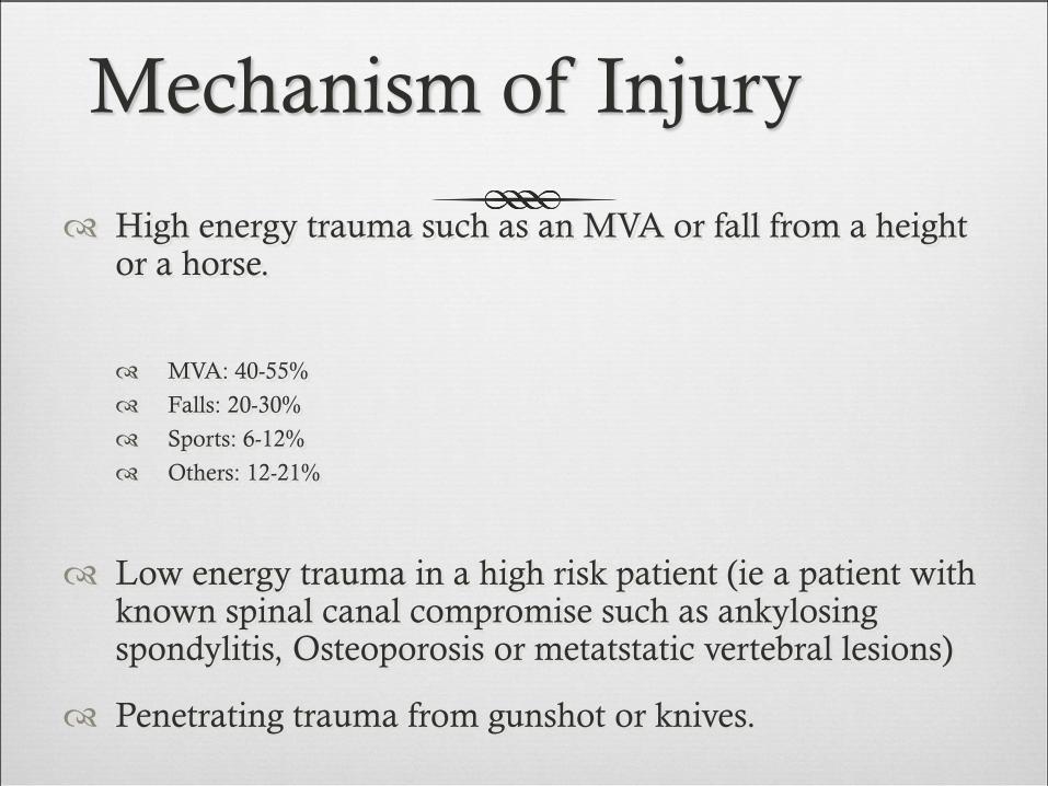
Mechanism of Injury
High energy trauma such as an MVA or fall from a height or a horse.
MVA: 40-55%
Falls: 20-30%
Sports: 6-12%
Others: 12-21%
Low energy trauma in a high risk patient (ie a patient with known spinal canal compromise such as ankylosing spondylitis, Osteoporosis or metatstatic vertebral lesions)
Penetrating trauma from gunshot or knives.


Spine stability
Cervical spine instability:
Compression fracture with 25% loss of height.
Angular displacement > 11 degrees.
Translation > 3.5mm.
Disc space separation >1.7mm.
Thoracic and lumbar spine: Denis three column.

The Three columns
Instability exists with
disruption of any two
of three columns.

Assessment
In cases of trauma, ABCDE’s must be assessed first and
treated appropriately.
Patients should be examined with spinal collar until spinal
pathology is excluded.
Careful log rolling keeping the head, neck and pelvis in
line should be done to examine the spine properly.

Assessment
Immobilization.
History:
Mechanism of injury:
compression, flexion, extension, distraction
Other injuries.
Seat belt.
Other causalities.
Physical examination:
Inspection, palpation.
Neurologic examination.

Immobilization
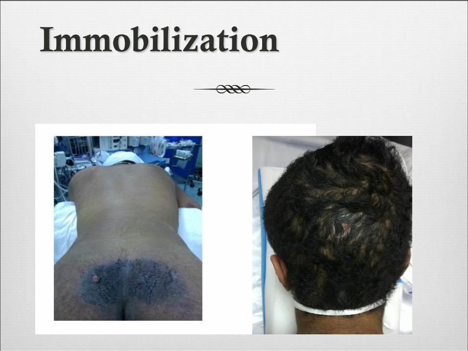
Immobilization

Immobilization

Neurologic
Muscle Test
Sensory exam
light touch, Sharp dull discrimination, Vibration sense, Proprioception
and two-point discrimination
Reflexes

Signs of Spinal Trauma
Apnea, lower cranial nerve injury VIII-XII (high C-spine).
Deformity of the spine or neck.
Tenderness on palpation along spinal processes.
Paralysis or muscle weakness (which spinal level).
Loss of sensation (which dermatones).
Loss of rectal tone.
Positive Babinski sign.
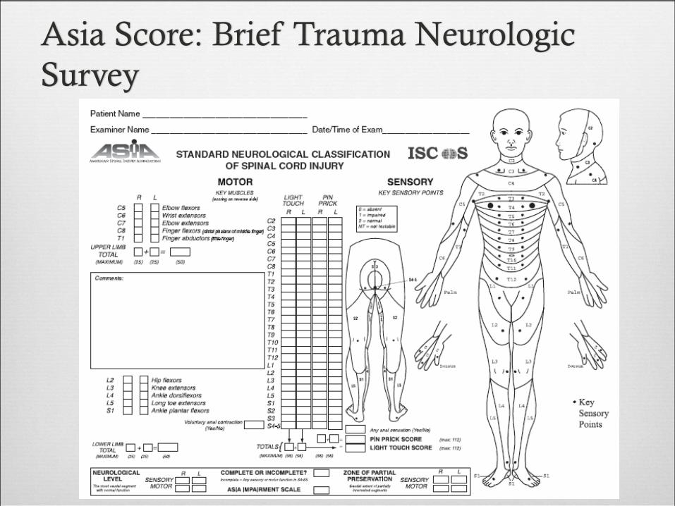
Asia Score: Brief Trauma Neurologic
Survey

Level of Cord Injury
determines level of function
Prognosis for
Recovery of spinal
Cord Injury:Poor prognosis for recovery if:
-pt arrives in shock
-pt cannot breath
-pt has a complete injury

Assessment
Severity of neurologic deficit
CompleteFlaccid paralysis below level of injury.
May involve diaphragm if injury above C5.
Sympathetic tone loss if fracture above T6.
Incomplete ? Any sensation.
? Sacral spairing.

Assessment
Severity of neurologic deficit
Incomplete
Central cord syndrome:
# Characterized by disproportionally (UL>LL).
# Mechanism: hyper-extension.
# Occur with or without fractures.
# Recovery: 50% regaining function.
# Prognosis is fair.
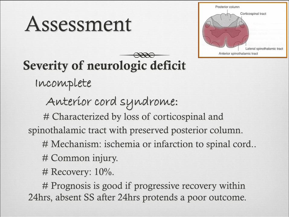
Assessment
Severity of neurologic deficit
Incomplete
Anterior cord syndrome:# Characterized by loss of corticospinal and
spinothalamic tract with preserved posterior column.
# Mechanism: ischemia or infarction to spinal cord..
# Common injury.
# Recovery: 10%.
# Prognosis is good if progressive recovery within
24hrs, absent SS after 24hrs protends a poor outcome.
Assessment
Severity of neurologic deficit
Incomplete
Brown-Sequard syndrome:# Characterized by hemicord injury with ipsilateral
paralysis, loss of proprioception and light touch, and
contralateral temperature and sharp pain loss.
# Prognosis is good, with over 90% regaining of bowel
and bladder function and ambulatory capacity.

Assessment
Severity of neurologic deficit
Incomplete
Conus Medullaris syndrome:
# Seen in T12-L1 injuries.
# Loss of voluntary bowel and bladder control with
preserved lumbar root function.
# Uncommon as pure lesion (mixed conus-cauda).
Assessment
Severity of neurologic deficit
Incomplete
Cauda Equina syndrome:# Saddle anesthesia, urinary retention and stool
incontinence.
# Usually due to large central disc herniation rather
than fracture.
Nerve root deficit: LMN
Spinal Shock
Transient loss of spinal reflexes.
Lasts 24-72 hours.
Neurogenic shock
Reduced tissue perfusion due to loss of sympathetic outflow
and un-apposed vagal tone.
Peripheral vasodilatation (hypotension and bradycardia).
Rx: fluid resuscitation and vasopressors.
X-rays:
Cervical: 3 views.
AP, lateral and open mouth.
Thoraco-lumbar: 2 views.
AP & lateral.
Flexion-Extension views.
CT: best for bony anatomy.
MRI: best to evaluate soft tissue.
Imaging
Depends on:
Level of injury.
Degree and morphology of injury: STABILITY
Presence of neurologic deficit.
Other factors.
Management of Spinal Injuries

Some general rules:
Stable injuries are usually treated conservatively.
Unstable injuries usually require surgery.
Neurologic compression requires decompression.

Specific Injuries

Cervical spine fractures
Descriptive: depends on mechanism of injury.
Flexion/extension.
Compression/distraction.
Shear.
Presence of subluxation/dislocation
SCI:
high fracture results in quadriplegia.
Low fracture results in paraplegia.

Cervical spine fractures
SCI:
high fracture results in quadriplegia.
Low fracture results in paraplegia.

Cervical spine fractures

Thoraco-Lumbar fractures
Spinal cord terminates at L1/2 disc in adult
L2/3 in a child
50% of injuries occur at Thoraco-lumbar junction.
Common fractures:
Wedge fracture (flexion/compression).
Burst (compression).
Chance (flexion/distraction).

Wedge fracture

Wedge fracture

Burst fracture
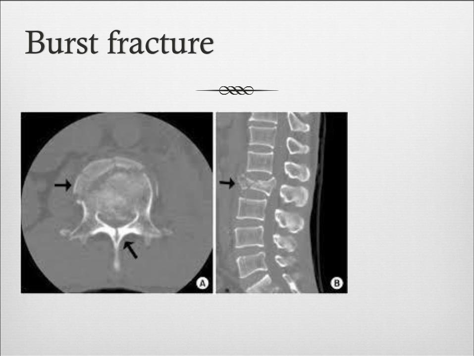
Burst fracture

Burst fracture
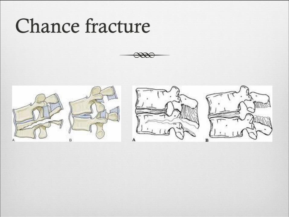
Chance fracture

Chance fracture
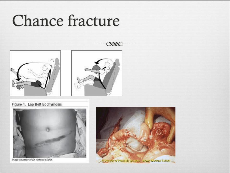
Chance fracture
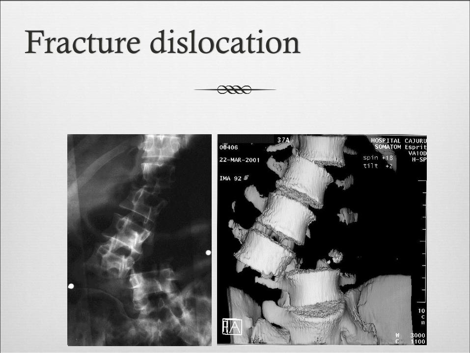
Fracture dislocation

Pathologic fractures
Low-energy fractures.
Usually due to infection or tumour.
Osteoporotic is common.
X-rays: “winking owl” sign for infection or tumour.

Pathologic fractures

Cauda Equina Syndrome
A surgical emergency.
Requires full neurologic examination including rectal
examination for anal tone.
Investigations: X-rays initially, but MRI is mandatory as X-
rays are usually unremarkable.
Treatment: Emergency decompression-usually discectomy
and wide laminectomy within 24 hours.
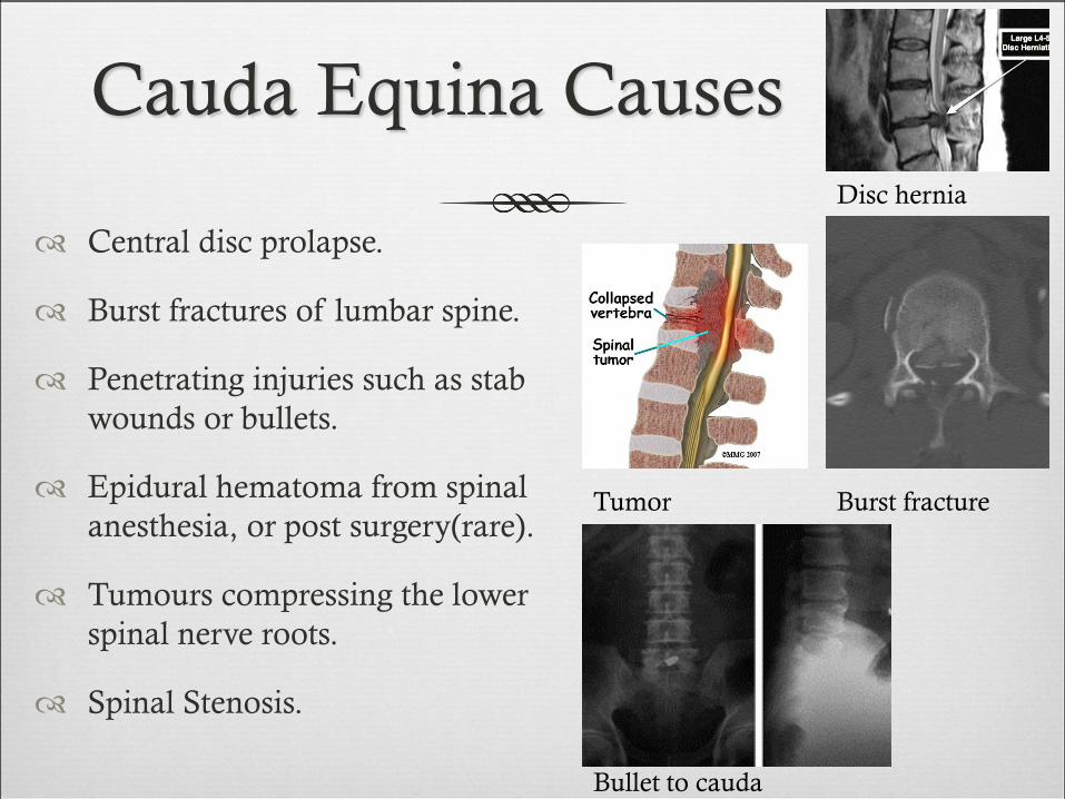
Cauda Equina Causes
Central disc prolapse.
Burst fractures of lumbar spine.
Penetrating injuries such as stab
wounds or bullets.
Epidural hematoma from spinal
anesthesia, or post surgery(rare).
Tumours compressing the lower
spinal nerve roots.
Spinal Stenosis.
Bullet to cauda
Disc hernia
Tumor Burst fracture

Cauda Equina Syndrome

Questions