
Combination Therapy of NSCLC Using Hsp90 Inhibitor …nanotheranosticlab.com/pubs/25. Molecular...
10
Combination Therapy of NSCLC Using Hsp90 Inhibitor and Doxorubicin Carrying Functional Nanoceria Shoukath Sulthana, Tuhina Banerjee, Jyothi Kallu, Sudershini Reddy Vuppala, Blaze Heckert, Shuguftha Naz, Tyler Shelby, Olivia Yambem, § and Santimukul Santra* ,† † Department of Chemistry, Kansas Polymer Research Center, Pittsburg State University, 1701 S. Broadway Street, Pittsburg, Kansas 66762, United States § Department of Neurology, College of Medicine, University of Tennessee Health Science Center, 855 Monroe Avenue, Memphis, Tennessee 38163, United States * S Supporting Information ABSTRACT: K-RAS driven non-small-cell lung cancer (NSCLC) represents a major cause of death among smokers. Recently, nanotechnology has introduced novel avenues for the diagnosis and personalized treatment options for cancer. Herein, we report a novel, multifunctional nanoceria platform loaded with a unique combination of two therapeutic drugs, doxorubicin (Doxo) and Hsp90 inhibitor ganetespib (GT), for the diagnosis and effective treatment of NSCLC. We hypothesize that the use of ganetespib synergizes and accelerates the therapeutic efficacy of Doxo via ROS production, while minimizing the potential cardiotoxicity of doxorubicin drug. Polyacrylic acid (PAA)-coated cerium oxide nanoparticles (PNC) were fabricated for the targeted combination therapy of lung cancers. Using “click” chemistry, the surface carboxylic acid groups of nanoceria were decorated with folic acid to target folate-receptor-overexpressing NSCLC. As a result of combination therapy, results showed more than 80% of NSCLC death within 48 h of incubation. These synergistic therapeutic effects were assessed via enhanced ROS, cytotoxicity, apoptosis, and migration assays. Overall, these results indicated that the targeted codelivery of Doxo and GT using nanoceria may offer an alternative combination therapy option for the treatment of undruggable NSCLC. KEYWORDS: combination therapy, NSCLC treatment, Hsp90 inhibitor, drug delivery, nanoceria, doxorubicin ■ INTRODUCTION Lung cancer is by far one of the leading cause of the mortality worldwide. 1,2 According to the American Cancer Society statistics, collectively 221 200 new cases of lung cancer and 158 040 deaths were estimated in the year 2015. Histologically, lung cancer is being classified into two major subtypes: small- cell lung cancer (SCLC) and non-small-cell lung cancer (NSCLC). Among both the subtypes, NSCLC by itself accounts for 85% of all the cases. 3 Oncogenic K-RAS, one of the major histologic subtype of NSCLC, accounts for more than 25% of the lung-cancer-related deaths. 4 Current existing therapies for K-RAS-driven NSCLC includes surgery, radiation therapy, and chemotherapy. One of the major limitation of all these therapies is severe toxicity, multidrug resistance, and poor survival outcomes. 5,6 Therefore, successful targeted therapy for K-RAS-driven NSCLC is considered to be clinically challeng- ing. Nanotechnology has opened up new avenues for more personalized treatment strategies for cancer therapy. The facile and reliable surface chemistry, better payloads makes these nanotechnology-based vehicles excellent candidates for drug delivery applications. 7−12 Nanoceria (NC), iron oxide nano- particles (IONPs), polymeric nanoparticles (PNPs), quantum dots (QDs), gold nanoparticles (AuNPs), and carbon nano- tubes (CNTs) have been used extensively for developing theranostic nanoplatforms. 13−18 Cerium oxide, a rare-earth metal has recently been shown to have diverse applications in cancer therapy due to unique redox property. 19−21 Several studies have examined the diverse role of antioxidant nanoceria as a therapeutic agent in cancer therapy because of its anti- invasive, radio protective, radio sensitizing, oxidant-mediated apoptosis, and antiangiogenic properties. 22−30 Targeted delivery of chemotherapeutic drug cocktails specifically to the tumor site is becoming a popular choice for clinicians for effective cancer treatments. The unique combination of drug-carrying nanoplatform allows for the inhibition of separate tumorigenesis pathways. This strategy Received: November 29, 2016 Revised: January 6, 2017 Accepted: January 12, 2017 Published: January 12, 2017 Article pubs.acs.org/molecularpharmaceutics © 2017 American Chemical Society 875 DOI: 10.1021/acs.molpharmaceut.6b01076 Mol. Pharmaceutics 2017, 14, 875−884
Transcript of Combination Therapy of NSCLC Using Hsp90 Inhibitor …nanotheranosticlab.com/pubs/25. Molecular...
Combination Therapy of NSCLC Using Hsp90 Inhibitor andDoxorubicin Carrying Functional NanoceriaShoukath Sulthana, Tuhina Banerjee, Jyothi Kallu, Sudershini Reddy Vuppala, Blaze Heckert,Shuguftha Naz, Tyler Shelby, Olivia Yambem,§ and Santimukul Santra*,†
†Department of Chemistry, Kansas Polymer Research Center, Pittsburg State University, 1701 S. Broadway Street, Pittsburg, Kansas66762, United States§Department of Neurology, College of Medicine, University of Tennessee Health Science Center, 855 Monroe Avenue, Memphis,Tennessee 38163, United States
*S Supporting Information
ABSTRACT: K-RAS driven non-small-cell lung cancer(NSCLC) represents a major cause of death among smokers.Recently, nanotechnology has introduced novel avenues forthe diagnosis and personalized treatment options for cancer.Herein, we report a novel, multifunctional nanoceria platformloaded with a unique combination of two therapeutic drugs,doxorubicin (Doxo) and Hsp90 inhibitor ganetespib (GT), forthe diagnosis and effective treatment of NSCLC. Wehypothesize that the use of ganetespib synergizes andaccelerates the therapeutic efficacy of Doxo via ROSproduction, while minimizing the potential cardiotoxicity ofdoxorubicin drug. Polyacrylic acid (PAA)-coated cerium oxidenanoparticles (PNC) were fabricated for the targeted combination therapy of lung cancers. Using “click” chemistry, the surfacecarboxylic acid groups of nanoceria were decorated with folic acid to target folate-receptor-overexpressing NSCLC. As a result ofcombination therapy, results showed more than 80% of NSCLC death within 48 h of incubation. These synergistic therapeuticeffects were assessed via enhanced ROS, cytotoxicity, apoptosis, and migration assays. Overall, these results indicated that thetargeted codelivery of Doxo and GT using nanoceria may offer an alternative combination therapy option for the treatment ofundruggable NSCLC.
KEYWORDS: combination therapy, NSCLC treatment, Hsp90 inhibitor, drug delivery, nanoceria, doxorubicin
■ INTRODUCTION
Lung cancer is by far one of the leading cause of the mortalityworldwide.1,2 According to the American Cancer Societystatistics, collectively 221 200 new cases of lung cancer and158 040 deaths were estimated in the year 2015. Histologically,lung cancer is being classified into two major subtypes: small-cell lung cancer (SCLC) and non-small-cell lung cancer(NSCLC). Among both the subtypes, NSCLC by itselfaccounts for 85% of all the cases.3 Oncogenic K-RAS, one ofthe major histologic subtype of NSCLC, accounts for morethan 25% of the lung-cancer-related deaths.4 Current existingtherapies for K-RAS-driven NSCLC includes surgery, radiationtherapy, and chemotherapy. One of the major limitation of allthese therapies is severe toxicity, multidrug resistance, and poorsurvival outcomes.5,6 Therefore, successful targeted therapy forK-RAS-driven NSCLC is considered to be clinically challeng-ing.Nanotechnology has opened up new avenues for more
personalized treatment strategies for cancer therapy. The facileand reliable surface chemistry, better payloads makes thesenanotechnology-based vehicles excellent candidates for drug
delivery applications.7−12 Nanoceria (NC), iron oxide nano-particles (IONPs), polymeric nanoparticles (PNPs), quantumdots (QDs), gold nanoparticles (AuNPs), and carbon nano-tubes (CNTs) have been used extensively for developingtheranostic nanoplatforms.13−18 Cerium oxide, a rare-earthmetal has recently been shown to have diverse applications incancer therapy due to unique redox property.19−21 Severalstudies have examined the diverse role of antioxidant nanoceriaas a therapeutic agent in cancer therapy because of its anti-invasive, radio protective, radio sensitizing, oxidant-mediatedapoptosis, and antiangiogenic properties.22−30
Targeted delivery of chemotherapeutic drug cocktailsspecifically to the tumor site is becoming a popular choicefor clinicians for effective cancer treatments. The uniquecombination of drug-carrying nanoplatform allows for theinhibition of separate tumorigenesis pathways. This strategy
Received: November 29, 2016Revised: January 6, 2017Accepted: January 12, 2017Published: January 12, 2017
Article
pubs.acs.org/molecularpharmaceutics
© 2017 American Chemical Society 875 DOI: 10.1021/acs.molpharmaceut.6b01076Mol. Pharmaceutics 2017, 14, 875−884

could overcome severe side effects, multidrug resistance(MDR) of traditional chemotherapy and thereby obtain highertherapeutic efficacy. Considering the importance of cancertherapy, a new, dual-drug-carrying (Doxo and GT) functionalnanoceria is proposed in this present study. Both drugs inhibitseparate target mechanisms that are critical for tumor growth.The classical chemotherapeutic drug Doxo was selected in thepresent study, which is responsible for DNA damage andgeneration of ROS via redox-cycling.31,32 To achieve synergistictreatment, GT, a second-generation Hsp90 inhibitor waschosen as another chemotherapeutic drug. Several studieshave highlighted Hsp90 as an important target for cancertherapy. Its interaction with several client proteins includingEGFR, RAF, and AKT leads to inhibition of multiple signalingcascades that are crucial for growth, differentiation, and survivalof tumors.33−38
Herein, active targeting of NSCLC is achieved via folateconjugation of nanoceria which minimizes the off-target effectsof the drug combination. Furthermore, the antioxidativeproperty of nanoceria in the normal cells further boostssurvival of healthy tissues and, therefore, minimizes side effects.Unique drug-cocktail-carrying nanoceria exhibits high anti-tumor activity for A549 cell line (NSCLC). Synergistictherapeutic effects are assessed via enhanced ROS, cytotoxicity,apoptosis, and migration assays as a result of codelivery of drugcocktail. Taken together, findings from the present studyprovide strong support for the clinical translation potential ofour designer nanoceria formulations.
■ RESULTS AND DISCUSSION
Synthesis and Characterizations of Poly(acrylic acid)(PAA)-Coated Cerium Oxide Nanoparticles (PNC). Thewater-based alkaline precipitation method was followed for thesynthesis of PAA-coated nanoceria (PNC), as previouslyreported.39 The resulting yellow colored PNC solution wascentrifuged to get rid of agglomerates and bigger size PNC and
finally purified using dialysis technique (MWCO 6−8 kDa) (1,Scheme 1).40 Next, modified solvent diffusion method was usedto encapsulate optical lipophilic DiI dye in PNC (encapsulationefficiency, EEDiI = 85%). In this case, the DiI dye-labeledcarboxylated PNC (2, Scheme 1) would be used as a negative-control optical nanoprobe and to evaluate cytotoxicity offormulated PNC. For optical imaging, this kind of lipophilicdye is important because of its high extinction coefficient (ε >125 000 cm−1 M−1) and higher fluorescence emission (λmax =565 nm). In order to conjugate receptor-targeting molecules,first, propargyl amine was conjugated on the surface of PNCusing standard EDC/NHS chemistry (3, Scheme 1). Briefly,carboxylic acid groups of PNC (2.0 × 10⁻³ mol) wereconjugated with propargylamine (15 × 10−3 mol) using EDC(15 × 10−3 mol) and NHS (15 × 10−3 mol) in the presence ofMES buffer (1×, pH = 6.0). This is an essential step to furtherconjugate our nanoparticles with azide-functionalized folic acidvia “click chemistry” to target folate receptor expressing lungcancer cells (4a−5a, Scheme 1). This “click” reaction wasperformed in the presence of CuI as catalyst in a slightly basicPBS solution (pH = 8.0) of propargylated PNC suspension (2mL, 1.5 × 10⁻³ mol) and azide-functionalized folic acid (Fol ∼N3, 2 × 10⁻² mol, see SI for detailed synthesis). The resultingfolate-decorated nanoceria (FNC, 1.2 × 10⁻³ mol, 4a−5a,Scheme 1) was purified using standard dialysis method(MWCO 6−8 kDa) and followed by DiI dye encapsulationas described earlier. The DiI dye-labeled FNC (4b, Scheme 1,1.0 × 10−3 mol, EEDiI = 83%) will be used for the targeteddelivery of drugs to the folate receptor overexpressing cancercells (positive nanoprobe). For therapeutic applications, Doxoand GT were encapsulated in FNC (5b−5d, Scheme 1, 1.0 ×10⁻³ mol, EEDoxo/GT = 75%) using solvent diffusion method.Finally, these nanoceria theranostics were purified using dialysistechnique against PBS (pH = 7.4) solution. All functionalnanoceria preparations were stored in 4 °C and found to be
Scheme 1. Formulation of Functional and Biocompatible FNC: (1) Synthesis of PAA Polymer-Coated Cerium OxideNanoparticles (PNC) and (2) DiI Dye-Encapsulating PNC; (3) Propargylated Nanoceria Was Synthesized Using CarbodiimideChemistry (4 and 5)a
a“Click” chemistry was used to synthesize FNC, whereas a combination of drugs encapsulated using a solvent diffusion method.
Molecular Pharmaceutics Article
DOI: 10.1021/acs.molpharmaceut.6b01076Mol. Pharmaceutics 2017, 14, 875−884
876

stable in PBS (pH = 7.4) and in serum for longer period oftime.Characterizations of PAA Polymer-Coated Functional
Nanoceria. The presence of the PAA coating on nanoceria isvery crucial to make our nanoplatform stable, biocompatibleand to efficiently encapsulate therapeutic drugs. Successful PAAcoating and the presence of carboxylic acid functionality onnanoceria was confirmed by FT-IR spectroscopy (see SI, FigureS1). The presence of a band at 1710 cm−1 in FT-IR confirmedthe presence of CO stretching, indicating the successfulcoating of PAA polymer on nanoceria. The hydrodynamicdiameter of synthesized PNC and FNC was found to be anaverage diameter of D = 57 ± 2 nm and D = 61 ± 3,respectively, measured through dynamic light-scattering experi-ment (Figure 1A and SI Figure S2). The overall surface chargeof functional nanoceria preparations were measured byperforming zeta-potential experiments and were found to be−32.9 mV and −25.2 mV for PNC and FNC (see SI, FigureS2), respectively. The successful encapsulation of optical dye
and drugs inside the PAA coating of nanoceria were confirmedby UV−vis and fluorescence experiments using a high-throughput plate reader. The absorbance spectrum (Figure1B) of the folate-decorated, DiI dye-encapsulating FNC 4confirmed for the presence of both DiI dye (λabs = 555 nm) andfolic acid (λabs = 352 nm). Similarly, the presence of Doxo inFNC (5b, Scheme 1) was confirmed by the UV−vis spectrum(λabs = 497 nm) as shown in Figure 1C. The effective folateconjugation and encapsulation of cargos were further confirmedby their corresponding fluorescence spectra (Fol: λem = 455nm; DiI: λem = 675 nm; Doxo: λem = 595 nm, Figure 1D,E,F).The fluorescence maxima and hydrodynamic diameters of thesefunctional nanoceria formulations remain unchanged (orminimal change) with a longer period of time, which isindicated for the synthesis of stable nanoceria formulations inphosphate buffer saline (PBS, pH = 7.4) and in serum.
Determination of Cytotoxicity of Functional Nano-ceria. A series of in vitro cytotoxicity experiments wereperformed to evaluate the therapeutic efficacy of our drug-
Figure 1. Characterizations of functional nanoceria. (A) Dynamic light-scattering experiment showed the formation of stable PNC with an averagediameter of 57 ± 2 nm. UV−vis absorption spectra of (B) DiI-encapsulating FNC and (C) Doxo-encapsulating FNC showed the presence of folicacid and Doxo. Fluorescence emission spectra of (D) FNC, (E) DiI dye-encapsulating FNC, and (F) Doxo-encapsulating FNC further confirms forthe successful synthesis.
Figure 2. Determination of cytotoxicity of the formulated functional nanoceria using MTT assay. (A) More than 70% of A549 cells were dead within24 h when incubated with the combination of drugs carrying nanoceria, whereas only 40% toxicity was observed with single drug delivered. However,more than 90% cells were found to be dead as a result of combination therapy after 48 h of incubation. (B) On the other hand, minimal toxicity wasobserved when functional nanoceria was incubated with H9c2 cells (FR−), suggesting for targeted delivery of drugs specifically to the tumor. A 1×PBS solution is used for the treatment of control cells. Average values of three measurements are depicted ± standard error.
Molecular Pharmaceutics Article
DOI: 10.1021/acs.molpharmaceut.6b01076Mol. Pharmaceutics 2017, 14, 875−884
877

encapsulated functional nanoceria using A549 (folate-positive)and H9c2 (folate-negative) cells. In these typical experiments,cells (2500 cells/well) in 96-well plate were incubated withvarious functional FNCs (35 μL, 1.0 × 10⁻³ mol) at 37 °C,followed by MTT incubation and finally data recorded atdifferent time points. Comparable cellular toxicity was observedwhen incubated with single-drug-encapsulating FNC (eitherGT or Doxo), and more than 40% cells were found to be deadafter 24 h of incubation. However, more than 70% cell deathoccurred within 24 h from the combination of drugsencapsulating nanoceria (FNC-Doxo-GT, 5d, 1.0 × 10⁻³ mol,Scheme 1), which became more severe when continued (morethan 90% cell death within 48 h of incubation, Figure 2A). Onthe other hand, functional nanoceria by itself (FNC) with nodrugs showed minimal toxicity to the cancer cells as well as tothe healthy cardiomyocytes, highlighting the biocompatibility ofour nanoceria-based drug delivery system. No or minimaltoxicity was observed when H9c2 cells (cardiomyocytes) wereincubated with drug-loaded FNC (Figure 2B). This result ispromising for reducing Doxo’s potential lethal cardiotoxicity.
Active targeting via FNC is one of the key aspect of ournanoplatform to achieve higher intracellular uptake of the drugswith minimal side effects to the healthy tissues. It is evident thatas a result of synergistic effect, GT accelerates the cytotoxicaction of Doxo, while minimizing Doxo’s cardiotoxicity.Therefore, the targeted codelivery of drug cocktails using ourfunctional nanoceria would offer an excellent combinationtherapy option for the treatment of undruggable NSCLC andother tumors.
Drug-Release Experiments of PAA Polymer-CoatedNanoceria. Next, drug-release studies were conducted toexamine the ability of our PAA-coated nanoceria to release drugin the presence of an appropriate external environment, forexample, low pH (tumor’s intracellular environment) andesterase enzyme (to indicate biodegradability). It is crucial todetermine timely release of cargos in order to evaluatetherapeutic efficacy of our functional FNC. A dialysis techniquewas used in order to perform the drug-release experiment at 37°C. Typically, 2 mL of Doxo-encapsulating FNC (1.0 × 10⁻³
mol) and 100 μL of porcine liver esterase (1 mM) were taken
Figure 3. Drug-release profiles of FNC-Doxo using dialysis method. Release of Doxo (λem = 594 nm) was observed with time in the presence of (A)esterase enzyme and (B) in PBS at pH 6.0. However, the release of Doxo was minimal in PBS at pH 7.4 (Red line, A and B). The released Doxoemission was measured using plate reader at λex = 497 nm. The results were recorded at λem = 594 nm and plotted in terms of % cumulative release.Average values of three measurements are depicted ± standard error.
Figure 4. Representative fluorescence microscopic images of the in vitro cellular imaging and drug delivery experiments. (A−D) Minimalinternalization was observed in the case of drug-loaded PNC, whereas (E−H) effective cellular internalization was observed with functional FNC(4b, Scheme 1). However, when drugs-loaded FNC (5d, Scheme 1) were incubated A549 cells, a substantial amount of cell death was observed (I−L). On the other hand, (M−P) minimal internalization was observed in healthy H9c2 cells, indicated for the targeted drug delivery. Nucleus stainedwith DAPI dye (blue).
Molecular Pharmaceutics Article
DOI: 10.1021/acs.molpharmaceut.6b01076Mol. Pharmaceutics 2017, 14, 875−884
878
in a dialysis bag (MWCO 6−8 kDa) and dialyzed against 200mL of DI water taken in a 250 mL beaker. In a timely fashion, 1mL of solution from the outer chamber (250 mL beaker) wastaken, and the fluorescence emission of this solution wasmeasured at 594 nm using a high-throughput plate reader. Asexpected, a minimal amount of Doxo was released at thebeginning of the dialysis; however, more than 50% of theencapsulating Doxo was released within 3 h of dialysis. In thiscase, the esterase enzyme was able to disrupt the PAA polymerfrom cerium oxide core, followed by the release of cargos.Within 6 h of incubation, more than 80% of the encapsulateddrug was released, as shown in Figure 3A. Similarly, a drug-release experiment was performed in the presence of acidicPBS-buffered solution (pH = 6.0) by substituting the esteraseenzyme. This experiment would demonstrate the drug-releasecapability of FNC inside the acidic microenvironment of thetumor. Within 12 h of incubation, more than 80% of theencapsulated drug was released, as shown in Figure 3B. In thiscase, the acidic pH hydrolyzes the coordinate bonding betweencerium oxide and the carbonyl groups of PAA polymer, whichresulted in PAA-coating disintegration and drug release. On theother hand, no or minimal drug release was observed inphysiological pH 7.4. These observations indicated the stabilityof our functional FNC in physiological pH. These experimentsfurther demonstrated that our functional nanoceria will delivertherapeutic drugs and imaging agents only in cancer cells,whereas they remain nontoxic to healthy tissues.Fluorescence Microscopic Studies: Imaging and
Treatment Monitoring of NSCLC. To evaluate the potentialbiomedical application of functional FNC, receptor-mediatedinternalization and selective therapeutic efficacy were assessedusing fluorescence microscopy. We hypothesized that func-tional FNC would target folate receptors and internalize intofolate receptor expressing tumors, while minimizing thecardiotoxicity of doxorubicin drug. In these in vitro experi-ments, lung cancer A549 cells were incubated with Doxo- andGT-encapsulating carboxylated nanoceria (nonfolate, PNC) for24 h. Results showed no or minimal internalizations of drug-encapsulating PNC into A549 cells (Figure 4A-4D). This is dueto the lack of interaction between carboxylated PNC and A549
cells. To validate effective internalization, optical dye DiI-encapsulating FNC (4b, Scheme 1) was incubated with A549cells for 24 h. Interestingly, cellular internalization of FNC wasobserved as the cytoplasm was found to be red fluorescence incolor due to the intracellular release of DiI dye (Figure 4E−H).To further demonstrate folate receptor-mediated internal-izations, in a typical experiment, A549 cells were preincubatedwith free folic acid for 6 h before they were incubated with DiI-FNC for 24 h. Results showed minimal internalizations of FNCinto the A549 cells (SI, Figure S3), indicating that the folatereceptors were saturated with free folic acid and, therefore,receptor-mediated internalizations were minimized. When GTand Doxo-loaded FNC (5d, 1.0 × 10⁻³ mol, Scheme 1) wereincubated with A549 cells for 24 h, tremendous change incellular morphology and cell death were observed (Figure 4I−L). These results demonstrated the effective targeting anddelivery of drugs effectively to cancer cells. These results werealso validated with cytotoxic assays as demonstrated earlier(Figure 3). To prove our hypothesis for targeted delivery, wehave considered folate-receptor-negative healthy cardiomyocytecells (H9c2 cells). Dual-drug-encapsulating FNC (5d, Scheme1) were incubated with H9c2 cells for 24 h and imaged underfluorescence microscope. As expected, results showed no orminimal internalizations into H9c2 cells, confirming tumor-targeting capability of our FNC, while minimizing toxicity tohealthy tissues (Figure 4M−P). Taken together, these micro-scopic results indicated for the tremendous possibility of ournewly designed drug-cocktail-encapsulating FNC to be used astargeted drug delivery system in clinical settings.
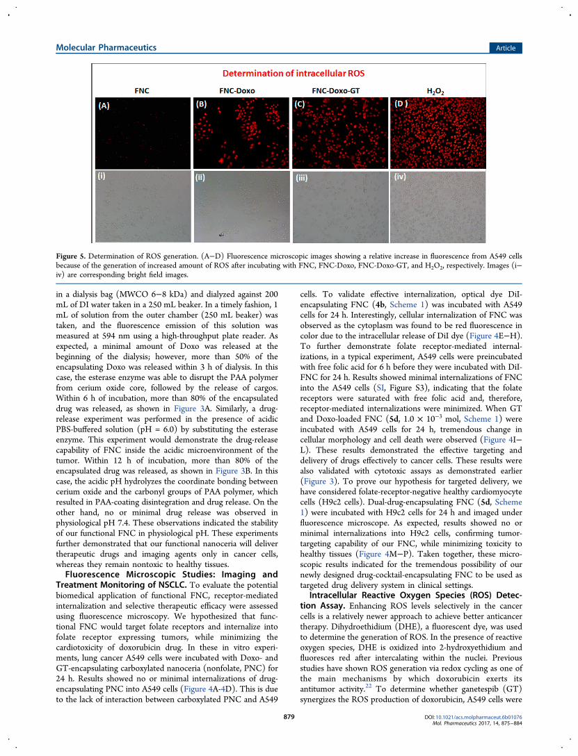
Intracellular Reactive Oxygen Species (ROS) Detec-tion Assay. Enhancing ROS levels selectively in the cancercells is a relatively newer approach to achieve better anticancertherapy. Dihydroethidium (DHE), a fluorescent dye, was usedto determine the generation of ROS. In the presence of reactiveoxygen species, DHE is oxidized into 2-hydroxyethidium andfluoresces red after intercalating within the nuclei. Previousstudies have shown ROS generation via redox cycling as one ofthe main mechanisms by which doxorubicin exerts itsantitumor activity.22 To determine whether ganetespib (GT)synergizes the ROS production of doxorubicin, A549 cells were
Figure 5. Determination of ROS generation. (A−D) Fluorescence microscopic images showing a relative increase in fluorescence from A549 cellsbecause of the generation of increased amount of ROS after incubating with FNC, FNC-Doxo, FNC-Doxo-GT, and H2O2, respectively. Images (i−iv) are corresponding bright field images.
Molecular Pharmaceutics Article
DOI: 10.1021/acs.molpharmaceut.6b01076Mol. Pharmaceutics 2017, 14, 875−884
879

treated with a combination of drugs, doxorubicin (Doxo), andGT-carrying FNC (5d, 1.0 × 10⁻³ mol, Scheme 1). In theseexperiments, four Petri dishes of A549 cells (10 000/well) weretreated with different functional FNCs and others: (1) FNCwithout any drug, (2) FNC with only doxorubicin (FNC-Doxo), (3) FNC with drug cocktail (FNC-Doxo-GT) and (4)H2O2 (positive control). After 6 h of treatment, cells werestained with highly cell permeable cytosolic probe dihydroethi-dium (DHE) for 30 min. Cells treated with FNC showedminimal red fluorescence due to production of limited ROSstress (Figure 5A-i). In contrast, FNC-Doxo elevated the ROSlevel significantly as indicated by the 4-fold higher fluorescenceintensity (Figure 5B-ii). This further demonstrated thedoxorubicin-induced cytotoxicity via redox-cycling. Next, toverify the synergistic effect of GT on Doxo’s ROS production,we have treated A549 cells with FNC-Doxo-GT (5d, 1.0 × 10⁻³
mol, Scheme 1). Interestingly, this led to the dramaticenhancement of ROS levels as indicated in Figure 5C-iii.This further validates our hypothesis that combination ofganetespib improves the therapeutic efficacy of doxorubicin bytriggering ROS generation. In addition, the numerical data offluorescent intensities from each fluorescence microscopicimages were calculated using the commercially available ImageJsoftware and the histogram profiles exhibited for the increasedlevels of ROS generation, as shown in Figure 6. One of the
mainstays in cancer therapy is to employ a combination ofdrugs to achieve better therapeutic outcome. Our presentfindings from the ROS assay provides a strong rationale ofexploring combination therapy for the treatment of NSCLCusing nanoceria.Detection of Apoptotic and Necrotic Events on
NSCLC. Having identified ROS production as a result offunctional FNC treatment, next we wanted to elucidate thecorrelation between ROS generation and apoptosis induction.To investigate the mechanism, in a typical experiment, A549cells were exposed to the FNC, FNC-Doxo, and FNC-Doxo-GT (1.0 × 10⁻³ mol) for 48 h. Additionally, to differentiatebetween early and late apoptotic, and necrotic cells, conven-tional fluorescence microscopic studies were performed aftertreatment with AnnexinV-FITC and ethidium homodimer, theapoptotic and necrotic cell staining dyes, respectively. Viableand healthy cells remain unstained, as shown in Figure 7A-i in
the case of treatment with FNC with no drug and due to theabsence of any cell death mechanism. Encapsulation ofdoxorubicin leads to early and late apoptosis, as exhibited byAnnexinV-FITC and ethidium homodimer staining pattern(major green spots for apoptotic cells and fewer red spots fornecrotic events, Figure 7B-ii). In contrast, upon combinationdrug treatment, A549 cells exhibited enhanced ethidiumhomodimer staining, indicated for cell death via necroticpathway (major red spots for necrotic cells: Figure 7C-iii).Quantitative analysis of these results are calculated usingImageJ software and presented in SI Figure S4. These furtherconfirm that GT treatment accelerates the apoptosis inductionof doxorubicin, leading to extensive loss in membrane integritythat eventually results in cell death via necrosis. On the basis ofthese results, it is evident that ganetespib provides superiortherapeutic efficacy by markedly enhancing the antitumoractivity of doxorubicin. Taken together, our results suggest thatcombination of ganetespib with classical chemotherapeutic drugdoxorubicin might provide a better synergistic antitumorresponse for the treatment of NSCLC. We also suggest thatthe development of functional nanoceria formulation withstrong evidence for better antitumor response would be anideal drug delivery system for the targeted combination therapyof NSCLC and other tumors.
Migration Assay. Metastasis is known to be a key featurefor the malignancy of NSCLC. To assess whether nanoceriacarrying drug cocktail can inhibit the migration of highlymetastatic A549 cell line, we performed transwell migrationassays. In these experiments, serum-starved A549 cells weretreated with functional FNC (1.0 × 10⁻³ mol) and seeded inthe upper invasion chamber, followed by incubation for 24 h.Our results showed that A549 cells migrated from the upper tothe lower chamber (feeder tray) containing medium with 10%FBS, when treated with FNC with no drugs (CTRL, Figure 8).However, when starved A549 cells were treated with nanoceriacarrying a combination of drugs, migration ability decreasedsignificantly, as reflected by the change in the fluorescenceintensity (FNC-Doxo-GT, Figure 8). Taken together, ourcurrent findings demonstrated that functional nanoceriacarrying doxorubicin and ganetespib might play an importantrole in preventing lung cancer metastasis.
■ EXPERIMENTAL SECTIONMaterials. Polyacrylic acid (PAA), 2-morpholinoethanesul-
fonic acid (MES), 1-ethyl-3-(3-(dimethylamino)-propylcarbodiimide hydrochloride (EDC), propargylamine(PA), N,N′-dimethyl sulfoxide (DMSO), 3-(4,5-dimethylth-iazol-2-yl)-2,5-diphenyltetrazolium bromide (MTT), chloro-propylamine, and N,N′-dimethylformamide (DMF) werepurchased from Sigma-Aldrich and used as received. Nearinfrared DiI dye and 4,6-diamidino-2-phenylindole (DAPI) dyewere purchased from Invitrogen. Cerium nitrate hexahydrate,N-hydroxy succinimide (NHS), tetrahydrofuran, acetonitrile,sodium azide, ammonium hydroxide, ethanol, folic acid,isopropanol, and MES sodium salt were purchased fromACROS organics and used without further purification. Dialysismembranes were received from spectrum laboratories.Dihydroethidium (DHE) was obtained from Cayman chemical,whereas H2O2 and para-formaldehyde were received fromelectron microscopy sciences. Fetal bovine serum (FBS) and5× Annexin V binding buffer were purchased from BDBiosciences, whereas ganetespib, isopropyl alcohol, apoptosisand necrosis quantification kit (FITC-Annexin V, Ethidium
Figure 6. Numerical data were calculated from corresponding ROSimages using ImageJ software. Results showed relatively higherfluorescence from the FNC-Doxo-GT-treated A549 cells because ofthe presence of increased amount of ROS, as expected. Average valuesof three measurements are depicted ± standard error.
Molecular Pharmaceutics Article
DOI: 10.1021/acs.molpharmaceut.6b01076Mol. Pharmaceutics 2017, 14, 875−884
880

homodimer III) were obtained from Biotium. Migration assaykit was purchased from Millipore. Rat cardiomyocytes (H9c2cells), A549 cells (NSCLC), DMEM, and F12K cell culturemedia were purchased from ATCC (U.S.A.).Synthesis of Poly(acrylic acid)-Coated Nanoceria 1
(PNC). Water-dispersible PNC were synthesized using apreviously reported water-based alkaline precipitation meth-od.39 Briefly, two different solutions were prepared: ceriumnitrate (0.901 g) in 2.5 mL of deionized (DI) water (solution1) and poly(acrylic acid) (0.905 g) in 10 mL of DI water(solution 2). Solution 1 was added to 30% ammoniumhydroxide solution (30 mL), stirred at room temperature,and followed by addition of solution 2. Color changed frombrown to dark brown within 5 min of stirring and became deepyellow after 24 h, which indicates the preparation of stablenanoceria. The reaction mixture was centrifuged three times(20 min each at 3000 rpm) to get rid of agglomerates andbigger size PNC. Finally, the product was purified by dialysistechnique using a dialysis bag of molecular weight cutoff(MWCO) 6−8K against DI water and finally with phosphate
buffer saline (PBS, pH = 7.4). The purified PNC (2.0 × 10⁻³
mol) was stored at 4 °C for further characterizations. Successfulcoating of PAA on nanoceria was characterized by FourierTransform Infra-Red (FT-IR) spectroscopy (SI, Figure S1),overall size and surface charge were measured using dynamiclight-scattering (DLS) technique (SI, Figure S2).
Synthesis of Propargylated PNC, 3: CarbodiimideChemistry. To synthesize propargylated PNC, four differentsolutions were prepared: (1) 3 mL PNC (2.0 × 10⁻³ mol) in 1mL of PBS, pH 7.4, (2) NHS (15 × 10−3 mol) in 200 μL ofMES buffer (0.1 M), pH = 6.0, (3) EDC (15 × 10−3 mol) in200 μL of MES buffer (0.1 M), pH = 6.0, and (4)propargylamine (15 × 10−3 mol) in 250 μL of DMSO. TheEDC solution was immediately added to PNC solution andfollowed by addition of NHS solution, after brief mixing. Thisreaction mixture was incubated for an additional 3 min beforedropwise addition of solution 4, at room temperature. Thereaction was continued for 4−6 h at room temperature. Thefinal alkynated PNC was purified against PBS (pH = 7.4) usingthe dialysis method (MWCO 6−8K) to get rid of unreactedreagents.
Synthesis of Folate-Conjugated PNC (FNC, 4a and 5a):“Click” Chemistry. To a solution of propargylated PNCsuspension (2 mL, 1.5 × 10⁻³ mol) in bicarbonate buffer (pH =8.0) and azide-functionalized folic acid (Fol ∼ N3, 2 × 10⁻²
mol, See SI for detailed synthesis) in DMF, 5 μL of CuI catalyst(1 × 10⁻³ mmol) in DMF was added and incubated on a tablemixer for overnight at room temperature. The resulting productwas purified using the dialysis technique. The purified FNC(1.2 × 10⁻³ mol) was stored at 4 °C for further character-izations.
Encapsulation of DiI Dye (2 and 4b): Solvent DiffusionMethod. To 1 mL of nanoceria (PNC or FNC) suspension,DiI dye (1 μL of 1 μM DiI dye in 100 μL of DMSO) was addeddropwise with continuous stirring. The resulting solution wasdialyzed against PBS (pH = 7.4) for 2 h. Successfulencapsulation of DiI was confirmed using UV−vis andfluorescence spectrophotometric analyses (Figure 1). Thepurified dye-labeled nanoceria suspensions (1.0 × 10⁻³ mol)were stored at 4 °C for further studies.
Figure 7. Detection of apoptotic and necrotic cell death by fluorescence microscopy using annexinV-FITC and ethidium homodimer III. (A)Healthy cells emitted almost no fluorescence representing early apoptotic cells. (B) Apoptotic cells emitted green fluorescence after treatment withFNC-Doxo. (C) Necrotic cells emitted red fluorescence when treated with FNC with combination of drugs. (i), (ii), and (iii) representscorresponding bright field images.
Figure 8. Assessment for the inhibition of migration of highlymetastatic A549 cells via migration assay. Control cells (green lines)incubated with FNC with no drug showed maximum invasion, whereasa combination of drug-carrying-FNC-treated A549 cells (red lines)showed minimal migration. The sample average value of threesuccessive measurements is presented in both CTRL and FNC-Doxo-GT experiments.
Molecular Pharmaceutics Article
DOI: 10.1021/acs.molpharmaceut.6b01076Mol. Pharmaceutics 2017, 14, 875−884
881

Coencapsulation of Doxorubicin (Doxo) and Ganetes-pib (GT). Using a similar solvent diffusion method, acombination of drugs or drug and dye were coencapsulated.Briefly, a solution of either GT and DiI or GT and Doxo (2 μMof drug in 100 μL DMSO) was slowly added to 1 mL ofvortexing FNC suspension, followed by overnight incubation at4 °C. The drug-encapsulating functional FNC suspensions weredialyzed against PBS (pH = 7.4) for 2 h. The purified functionalFNC suspension was stored at 4 °C for further studies.Characterizations of Functional Nanoceria. Fourier-
Transform Infra-Red Spectroscopy (FT-IR). PerkinElmer’sSpectrum Two FT-IR spectrometer was used for FT-IRmeasurement. PNC suspension was vacuum-dried on a Petridish to obtain the powder form. FT-IR spectra of PAA polymerand PNC were recorded and compared. The presence of FT-IRstretching band at 1710 cm−1 for the PAA polymer’s carboxylicacid carbonyl group confirmed for the formation of PAApolymer coating on PNC (SI, Figure S1).Dynamic Light-Scattering Experiments (DLS). The average
size distribution and surface charge of functional nanoceriawere obtained using the dynamic light-scattering technique.Malvern’s Nano-ZS90 zetasizer was used for these experiments.The average diameters of PNC and FNC were found to be57.66 and 61.65 nm, respectively. The zeta potentials of PNCand FNC were −32.9 and −25.2 mV, respectively (SI, FigureS2).Spectrophotometric Analysis. UV−vis and fluorescence
spectra of functional nanoceria were recorded using TECAN’sinfinite M200 PRO high throughput plate reader. Presence offolic acid in FNC was confirmed by UV−vis (λabs = 352 nm)and fluorescence emission spectra (λem = 455 nm). Successfulencapsulation of Doxo was determined by UV−vis (λabs = 497nm) and fluorescence emission spectra (λem = 595 nm). Finally,the presence of DiI dye was confirmed by UV−vis (λabs = 555nm) and fluorescence emission spectra (λem = 675 nm).Cytotoxicity Studies using MTT Assay. To determine the
potential time-dependent cytotoxicity, two different cell lines,lung carcinoma (A549 cells) and cardiomyocytes (H9c2 cells),were used. Cells were seeded in 96-well plates at a density of2500 cells per well and treated with various functional FNCs(1.0 × 10⁻³ mol) at 37 °C for different time points. Each wellwas washed thrice with 1× PBS and then incubated with 30 μLof 5 mM MTT solution. After 4−6 h of incubation, theresulting formazan crystals (purple color) were dissolved inacidic isopropyl alcohol (75 μL), and the absorbance at 570 nmwas recorded using TECAN’s microplate reader. These assayswere carried out in triplicate, and the results were reported inFigure 2.Drug-Release Studies via Dynamic Dialysis Technique.
The in vitro drug-release studies were carried out at 37 °Cusing esterase enzyme and at low pH. We have incubated 2 mLof functional nanoceria with a 100 μL of porcine liver esterase(1 mM) inside a dialysis bag of molecular weight cutoff 6000−8000 Da, and was placed in a PBS solution (200 mL, pH = 7.4).The amount of drug molecules released from the functionalFNC-Doxo (1.0 × 10⁻³ mol) into the PBS solution wasdetermined at regular time intervals by taking 1 mL aliquotsfrom the PBS solution and replacing the same with PBSsolution. Fluorescence emission was measured at 494 nm fordoxorubicin. The same procedure was followed with 100 μL ofacidic PBS buffer (pH 6.0) at 37 °C. A standard calibrationcurve was used to calculate the concentration of the drugreleased, and the cumulative drug release versus time was
calculated using the following eq (Figure 3). Cumulative release(%) = (guest)t/(guest)total × 100.
Fluorescence Imaging: Cellular Internalization andCancer Treatment. The lung carcinoma A549 cells wereseeded into 12-well plate and once cells become 75% confluent,they were treated with corresponding PNC-Doxo-GT, FNC-DiI and FNC-Doxo-GT (1.0 × 10⁻³ mol) for 24 h in ahumidified incubator (37 °C, 5% CO2). The cells were washedthrice with 1× PBS (pH = 7.4) and were fixed with 4%formaldehyde solution for 15 min at room temperature. Thecells were then washed twice with 1× PBS before treating with6-diamidino-2-phenylindole (DAPI, 5 mg/mL) dye for nucleistaining. Cells were washed with 1× PBS and optical imageswere taken using fluorescence microscope (Olympus IX73) andresults were shown in Figure 4A−L. For control experiment,H9c2 cells were treated with FNC-Doxo-GT (1.0 × 10⁻³ mol),and the results were shown in Figure 4M−P.
ROS Detection Assay. Cells were seeded into different Petri-dishes at a density of 10 000 cells per well and treated with 50μL of various nanoceria preparations (FNC, FNC-Doxo, FNC-Doxo-GT, H2O2, 1.0 × 10⁻³ mol). After 6 h of incubation at 37°C, cells were washed twice with 1× PBS. Then, 20 μL of DHEfluorescent probe was added to each well and incubated for 30min at room temperature, followed by washing the cells twicewith 1× PBS. Subsequently, cells were fixed with 1 mL of 4%paraformaldehyde. After fixation, cells were washed with 1×PBS, stored with 2 mL of PBS in each well, and the opticalimages were taken using fluorescence microscope (Figure 5).
Determination of ROS using ImageJ Software. Using ROSfluorescence images, the amount of ROS was quantified usingcommercial ImageJ software. A particular cell from each treatedFNCs condition was selected to get a stack of values for thearea, integrated density, and mean fluorescence of backgroundreadings. Using the CTCF formula, the corrected total cellfluorescence (CTCF) for each condition was calculated, andthe results were shown in Figure 6. CTCF = integrated density− (area of selected cell × mean fluorescence of backgroundreadings).
Apoptosis and Necrosis Assay. The apoptosis and necrosisassays were carried out using apoptosis and necrosisquantification kit obtained from Biotium and using fluorescencemicroscopy. First, A549 cells were seeded into three differentPetri-dishes at a density of 10 000 cells per well. Next, cellswere treated with different preparations (FNC, FNC-Doxo, andFNC-Doxo-GT, 1.0 × 10⁻³ mol) and incubated for 48 h. Cellswere washed thrice with 1× PBS and then fixed with 4%formaldehyde solution for 15 min at room temperature. Later,the cells were stained with two different dyes, 5 μL of FITC-annexin buffer, and 5 μL of ethidium dimer III and incubatedfor 15 min. After the cells were washed thrice with 1× PBS,cells were covered with 1× binding buffer. Multiplefluorescence images were taken using two filters: (1) FITCfor green fluorescence, representing apoptosis, and (2)ethidium homodimer III for red fluorescence, indicatingnecrosis (Figure 7).
Migration or Invasion Assay. Chemicon QCM 96-well cell-migration assay kit from Millipore was used to evaluate themigration of A549 cells in the presence of functionalnanoparticles. Briefly, serum-starved A549 cells were culturedfor 24 h. Next, A549 cells were harvested and treated with thefunctional nanoceria (FNC-Doxo-GT, 1.0 × 10⁻³ mol) andPBS (control). Following treatment, cells were seeded into theupper chamber (transwell, invasion chamber), coated with type
Molecular Pharmaceutics Article
DOI: 10.1021/acs.molpharmaceut.6b01076Mol. Pharmaceutics 2017, 14, 875−884
882

I collagen. The lower chamber (feeder tray) contained mediumwith 10% FBS, and the whole setup was placed in the incubatorfor an additional 24 h to allow migration. Then, the migratorycells were dislodged completely from the invasion chamber andincubated with cell detachment buffer for 30 min. Finally,diluted solution of CYQuant cell lysis buffer was added to stainthe migratory cells and was read on a fluorescent plate reader.Fluorescence intensity was measured at an emission wavelengthof 520 nm (Figure 8).
■ CONCLUSIONS
In conclusion, we have successfully synthesized functionalnanoceria as a drug-delivery vehicle with a unique drug cocktailfor the treatment of NSCLC. Folate-decorated nanoceria wereformulated for the targeted delivery of combination of drugs,Doxo and GT. Resulting functional nanoceria demonstratedexcellent drug payload as reflected by the encapsulation studies,enhanced stability, reduced systemic toxicity, and highertherapeutic efficacy. As a result of combination therapy, morethan 80% of NSCLC cells were dead within 48 h of incubation.Results indicated cotreatment with GT supplements theclassical chemotherapeutic efficacy of Doxo, as confirmed bycell-based experiments including MTT and apoptosis assays.Furthermore, ROS studies indicated that GT synergizes andenhances the therapeutic efficacy of Doxo via ROS production,while minimizing the potential cardiotoxicity of doxorubicindrug. Apoptosis and necrosis assays further indicated thesynergistic effect of GT on Doxo’s therapeutic action. Inaddition, a migration assay validated the important role of thiscombination therapy approach in preventing lung cancermetastasis. Therefore, our current approach of delivering GTand Doxo using functional nanoceria may offer a robustnanoplatform for the targeted treatment of clinically challeng-ing K-RAS driven NSCLC.
■ ASSOCIATED CONTENT
*S Supporting InformationThe Supporting Information is available free of charge on theACS Publications website at DOI: 10.1021/acs.molpharma-ceut.6b01076.
Synthesis and characterizations of folic acid derivatives;functional FNC; control experiments for cellular internal-izations; FT-IR spectra; size and ζ-potential studies(PDF)
■ AUTHOR INFORMATION
Corresponding Author*E-mail: [email protected].
ORCID
Santimukul Santra: 0000-0002-5047-5245NotesThe authors declare no competing financial interest.
■ ACKNOWLEDGMENTS
This work is supported by K-INBRE P20GM103418, ACS PRF56629-UNI7, and PSU polymer chemistry startup fund (allfunding to S.S.).
■ REFERENCES(1) Jemal, A.; Center, M. M.; DeSantis, C.; Ward, E. M. Globalpatterns of cancer incidence and mortality rates and trends. CancerEpidemiol., Biomarkers Prev. 2010, 19, 1893−1907.(2) Siegel, R.; Ma, J.; Zou, Z.; Jemal, A. Cancer statistics, 2014. Ca-Cancer J. Clin. 2014, 64, 9−29.(3) American Cancer Society. Cancer Facts & Figures 2016; AmericanCancer Society: Atlanta, GA, 2016.(4) Acquaviva, J.; Smith, D. L.; Sang, J.; Friedland, J. C.; He, S.;Sequeira, M.; Zhang, C.; Wada, Y.; Proia, D. A. Targeting KRAS-mutant non-small cell lung cancer with the Hsp90 inhibitorganetespib. Mol. Cancer Ther. 2012, 11, 2633−2643.(5) Ramalingam, S.; Belani, C. Systemic chemotherapy for advancednon-small cell lung cancer: recent advances and future directions.Oncologist 2008, 13 (Suppl1), 5−13.(6) Grossi, F.; Kubota, K.; Cappuzzo, F.; de Marinis, F.; Gridelli, C.;Aita, M.; Douillard, J. Y. Future scenarios for the treatment of non-small cell lung cancer: focus on taxane-containing regimens. Oncologist2010, 15, 1102−1112.(7) van Vlerken, L. E.; Amiji, M. M. Multi-functional polymericnanoparticles for tumour-targeted drug delivery. Expert Opin. DrugDelivery 2006, 3, 205−216.(8) Santra, S.; Kaittanis, C.; Perez, J. M. Aliphatic hyperbranchedpolyester: a new building block in the construction of multifunctionalnanoparticles and nanocomposites. Langmuir 2010, 26, 5364−5373.(9) Santra, S.; Kaittanis, C.; Grimm, J.; Perez, J. M. Drug/dye-loaded,multifunctional iron oxide nanoparticles for combined targeted cancertherapy and dual optical/magnetic resonance imaging. Small 2009, 5,1862−1868.(10) Singh, S. Nanomedicine-nanoscale drugs and delivery systems. J.Nanosci. Nanotechnol. 2010, 10, 7906−7918.(11) Santra, S.; Kaittanis, C.; Perez, J. M. Cytochrome cencapsulating theranostic nanoparticles: A novel bifunctional systemfor targeted delivery of therapeutic membrane-impermeable proteinsto tumors and imaging of cancer therapy. Mol. Pharmaceutics 2010, 7,1209−1222.(12) Singh, R.; Nalwa, H. S. Medical applications of nanoparticles inbiological imaging, cell labeling, antimicrobial agents, and anticancernanodrugs. J. Biomed. Nanotechnol. 2011, 7, 489−503.(13) Lin, W.; Huang, Y. W.; Zhou, X. D.; Ma, Y. Toxicity of ceriumoxide nanoparticles in human lung cancer cells. Int. J. Toxicol. 2006, 25,451−457.(14) Baker, C. H. Harnessing cerium oxide nanoparticles to protectnormal tissue from radiation damage. Transl. Cancer Res. 2013, 2,343−358.(15) McCarthy, J. R.; Perez, J. M.; Bruckner, C.; Weissleder, R.Polymeric nanoparticle preparation that eradicates tumors. Nano Lett.2005, 5, 2552−2556.(16) Santra, S.; Jativa, S. D.; Kaittanis, C.; Normand, G.; Grimm, J.;Perez, J. M. Gadolinium-encapsulating iron oxide nanoprobe asactivatable NMR/MRI contrast agent. ACS Nano 2012, 6, 7281−7294.(17) Qian, Y.; Qiu, M.; Wu, Q.; Tian, Y.; Zhang, Y.; Gu, N.; Li, S.;Xu, L.; Yin, R. Enhanced cytotoxic activity of cetuximab in EGFR-positive lung cancer by conjugating with gold nanoparticles. Sci. Rep.2014, 4, Article No. 7490.(18) Brown, S. D.; Nativo, P.; Smith, J. A.; Stirling, D.; Edwards, P.R.; Venugopal, B.; Flint, D. J.; Plumb, J. A.; Graham, D.; Wheate, N. J.Gold nanoparticles for the improved anticancer drug delivery of theactive component of oxaliplatin. J. Am. Chem. Soc. 2010, 132, 4678−4684.(19) Chen, J.; Patil, S.; Seal, S.; McGinnis, J. F. Rare earthnanoparticles prevent retinal degeneration induced by intracellularperoxides. Nat. Nanotechnol. 2006, 1, 142−150.(20) Tarnuzzer, R. W.; Colon, J.; Patil, S.; Seal, S. Vacancyengineered ceria nanostructures for protection from radiation-inducedcellular damage. Nano Lett. 2005, 5, 2573−2577.(21) Rzigalinski, B. A.; Bailey, D.; Chow, L.; Kuiry, S. C.; Patil, S.;Merchant, S.; Seal, S. Cerium oxide nanoparticles increase the lifespan
Molecular Pharmaceutics Article
DOI: 10.1021/acs.molpharmaceut.6b01076Mol. Pharmaceutics 2017, 14, 875−884
883

of cultured brain cells and protect against free radical and mechanicaltrauma. FASEB J. 2003, 17, A606.(22) Sack, M.; Alili, L.; Karaman, E.; Das, S.; Gupta, A.; Seal, S.;Brenneisen, P. Combination of conventional chemotherapeutics withredox-active cerium oxide nanoparticles-a novel aspect in cancertherapy. Mol. Cancer Ther. 2014, 13, 1740−1749.(23) Asati, A.; Santra, S.; Kaittanis, C.; Nath, S.; Perez, J. M. Oxidase-like activity of polymer-coated cerium oxide nanoparticles. Angew.Chem., Int. Ed. 2009, 48, 2308−2312.(24) Dowding, J. M.; Das, S.; Kumar, A.; Dosani, T.; McCormack, R.;Gupta, A.; Sayle, T. X.; Sayle, D. C.; von Kalm, L.; Seal, S.; Self, W. T.Cellular interaction and toxicity depend on physicochemical propertiesand surface modification of redox-active nanomaterials. ACS Nano2013, 7, 4855−4868.(25) Desmouliere, A.; Guyot, C.; Gabbiani, G. The stroma reactionmyofibroblast: a key player in the control of tumor cell behavior. Int. J.Dev. Biol. 2004, 48, 509−517.(26) Yuan, H.; Miao, J.; Du, Y. Z.; You, J.; Hu, F. Q.; Zeng, S.Cellular uptake of solid lipid nanoparticles and cytotoxicity ofencapsulated paclitaxel in A549 cancer cells. Int. J. Pharm. 2008,348, 137−145.(27) Nelson, M. E.; Loktionova, N. A.; Pegg, A. E.; Moschel, R. C. 2-Amino-O4-benzylpteridine derivatives: potent inactivators of O6-alkylguanine-DNA alkyltransferase. J. Med. Chem. 2004, 47, 3887−3891.(28) Weissleder, R.; Ntziachristos, V. Shedding light onto livemolecular targets. Nat. Med. 2003, 9, 123−128.(29) Lin, W.; Huang, Y.; Zhou, X.; Ma, Y. Toxicity of cerium oxidenanoparticles in human lung cancer cells. Int. J. Toxicol. 2006, 25,451−457.(30) Giri, S.; Karakoti, A.; Graham, R. P.; Maguire, J. L.; Reilly, C.M.; Seal, S.; Rattan, R.; Shridhar, V. Nanoceria: a rare-earthnanoparticle as a novel anti-angiogenic therapeutic agent in ovariancancer. PLoS One 2013, 8, e54578.(31) Minotti, G.; Menna, P.; Salvatorelli, E.; Cairo, G.; Gianni, L.Anthracyclines: molecular advances and pharmacologic developmentsin antitumor activity and cardiotoxicity. Pharmacol. Rev. 2004, 56,185−229.(32) Du, C.; Deng, D.; Shan, L.; Wan, S.; Cao, J.; Tian, J.; Achilefu,S.; Gu, Y. A pH-sensitive doxorubicin prodrug based on folate-conjugated BSA for tumor-targeted drug delivery. Biomaterials 2013,34, 3087−3097.(33) Hahn, J. S. The Hsp90 chaperone machinery: from structure todrug development. BMB Rep. 2009, 42, 623−630.(34) Whitesell, L.; Lindquist, S. L. HSP90 and the chaperoning ofcancer. Nat. Rev. Cancer 2005, 5, 761−772.(35) Trepel, J.; Mollapour, M.; Giaccone, G.; Neckers, L. Targetingthe dynamic HSP90 complex in cancer. Nat. Rev. Cancer 2010, 10,537−549.(36) Banerji, U. Heat shock protein 90 as a drug target: some like ithot. Clin. Cancer Res. 2009, 15, 9−14.(37) Neckers, L. Heat shock protein 90: the cancer chaperone. J.Biosci. 2007, 32, 517−530.(38) Xu, W.; Neckers, L. Targeting the molecular chaperone heatshock protein 90 provides a multifaceted effect on diverse cell signalingpathways of cancer cells. Clin. Cancer Res. 2007, 13, 1625−1629.(39) Perez, J. M.; Asati, A.; Nath, S.; Kaittanis, C. Synthesis ofbiocompatible dextran-coated nanoceria with pH-dependent antiox-idant properties. Small 2008, 4, 552−556.(40) Asati, A.; Santra, S.; Kaittanis, C.; Perez, J. M. Surface-charge-dependent cell localization and cytotoxicity of cerium oxide nano-particles. ACS Nano 2010, 4, 5321−5331.
Molecular Pharmaceutics Article
DOI: 10.1021/acs.molpharmaceut.6b01076Mol. Pharmaceutics 2017, 14, 875−884
884


![Research Paper HSP90 Inhibitor Encapsulated Photo ...synergistic. CI was calculated with Compusyn software based on Chou-Talalay method . [23, 24] Regarding apoptosis assay, PC3 cells](https://static.fdocuments.in/doc/165x107/60e4fea2389ac00bd571afd5/research-paper-hsp90-inhibitor-encapsulated-photo-synergistic-ci-was-calculated.jpg)


