COGNITIVE FUNCTIONS IN ABSTINENT ALCOHOL DEPENDENT …
121
COGNITIVE FUNCTIONS IN ABSTINENT ALCOHOL DEPENDENT MALES – A CROSS SECTIONAL STUDY Dissertation submitted to the TAMIL NADU DR. M. G. R. MEDICAL UNIVERSITY in part fulfillment of the requirements for M. D (PSYCHIATRY) BRANCH XVIII APRIL 2013 MADRAS MEDICAL COLLEGE
Transcript of COGNITIVE FUNCTIONS IN ABSTINENT ALCOHOL DEPENDENT …
COGNITIVE FUNCTIONS IN ABSTINENT ALCOHOL
DEPENDENT MALES – A CROSS SECTIONAL STUDY
Dissertation submitted to the TAMIL NADU DR. M. G. R. MEDICAL UNIVERSITY
in part fulfillment of the requirements for
M. D (PSYCHIATRY)
BRANCH XVIII
APRIL 2013
MADRAS MEDICAL COLLEGE

CERTIFICATE
This is to certify that the dissertation title, “Cognitive Functions In
Abstinent Alcohol Dependent Males – A Cross Sectional Study ”
submitted by Dr.V.Sujaritha, in partial fulfillment for the award of the
MD degree in Psychiatry by The Tamil Nadu Dr.M.G.R. Medical
University, Chennai, is a bonafide record of the work done by her in the
Institute of Mental Health, Rajiv Gandhi Government General Hospital
and Medical College during the academic years 2010-2013.
DEAN
Rajiv Gandhi Government General Hospital and Medical
College
DIRECTOR
Institute of Mental Health
Chennai

DECLARATION
I, Dr.V.Sujaritha, solemnly declare that the dissertation
titled , “ Cognitive Functions In Abstinent Alcohol Dependent
Males – A Cross Sectional Study ” has been prepared by me under
the guidance of Professor Dr. Jeyaprakash, M.D., D.P.M., Director,
Institute of Mental Health, Chennai. I also declare that this bonafide work
or a part of this work was not submitted by me or any other for any
award, degree, diploma to any other University board either in India or
abroad.
This is submitted to The Tamilnadu Dr. M. G. R. Medical
University, Chennai in partial fulfillment of the rules and regulation for
the award of M.D degree Branch – XVIII (Psychiatry) to be held in
April 2013.
Place : Chennai Dr. V.Sujaritha
Date :

ACKNOWLEDGEMENTS
I thank Prof. Dr. Kanagasabai M. D., Dean, Rajiv Gandhi
Government General Hospital and Medical College for permitting me to
conduct this study.
I thank Professor Dr. Jeyaprakash, M.D., D.P.M., Director,
Institute of Mental Health, Chennai for his encouragement, help and
guidance.
I thank Professor Dr. V.S.Krishnan, M.D., Deputy Superintendent,
Institute of Mental Health for her valuable guidance and help.
I thank my Guide, Dr. M. Malaiappan, M.D., Associate Professor,
Institute of Mental Health for his guidance and help.
My special thanks to Dr. Poornachandrika, Dr. Sujatha
Assistant Professors, Institute of Mental Health for their guidance and
suggestions.
My sincere thanks are due to all the Professors and Assistant
Professors of Institute of Mental Health for their encouragement and
support.

My sincere thanks to Mrs. Shanthi, Clinical
Psychologist, Institute of Mental Health for her valuable help.
I finally acknowledge and thank all my colleagues and the
participants of this study for their kind cooperation.

CONTENTS
S.No TOPIC PAGE No.
1 INTRODUCTION 1
2 REVIEW OF LITERATURE 4
3 AIM AND OBJECTIVES 32
4 MATERIALS AND METHODOLGY 34
5 DATA ANALYSIS AND RESULTS 41
6 DISCUSSION 68
7 CONCLUSION 75
8 LIMITATIONS 76
9 STRENGTHS 77
10 FUTURE DIRECTIONS 78
11 BIBLIOGRAPHY 79
12 APPENDIX 100

1
INTRODUCTION
Humans have drunk alcohol for atleast 12000 years and it was being
used in religious rituals in ancient cultures. Alcohol is an organic
compound in which the functional hydroxyl group is attached to a carbon
atom. Ethanol is the type of alcohol found in alcoholic beverages and its
chemical formula is CH3-CH2-OH.
The lifetime risk for alcohol use disorders is more than 15% for men and
between 8% and 10% for women, making alcoholism among the most
common psychiatric conditions observed in the western world. In India, the
estimated number of alcohol users in 2005 was 62.5 million, with 17.4% of
them being dependent users. (Ray R, national survey on extent, pattern and
trends of drug abuse in India, 2005)
The deleterious effects of alcohol on cognitive functioning were
reported as early as the 1880s separately by wernicke and by korsakoff,
followed by Hamilton, fisher and weschler. It was after the introduction of
clinical neuropsychological model by Fitzhugh and co- workers on
cognitive function in alcoholism which marked the beginning of systematic
research in this area.
(Fein G, Bachman L, Fisher S, et al, 1990)

2
Wide research has been done in clinically evident cognitive impairments
like those seen in korsakoff syndrome occurring due to thiamine
deficiency. But there are no large scale epidemiologic studies to establish
the prevalence of cognitive impairment in abstinent alcoholics which is not
evident during routine interviews.
The rate of abstinent alcoholics with cognitive impairments has been
reported in myriad studies. Most of the samples chosen for these studies
where from inpatient or out-patient treatment settings and had used
convenient samples.
(Fein G, Bachman L, Fisher S, et al, 1990)
Although studies show that cognitive deficits are reversible after prolonged
abstinence, residual deficits do exist for some patients.
Although cognitive deficits are reversible during sustained abstinence,
residual deficits persist in some patients for extended periods of time.
Because of the patient’s cognitive deficits, they find it difficult to continue
their treatment and participate in treatment and also indulge effectively in
their life. Assessing these functions as clinicians becomes essential as it
helaps us choose appropriate treatment and to time the treatment. With this
note it is also important to understand that not all alcoholics develop
cognitive impairment.

3
As a result it eventually instilled a need for determining cognitive
functions in alcohol dependent subjects during their abstinent period for
better treatment outcome and to choose appropriate treatment in them.
Hence this study is conducted among alcohol dependent males during their
abstinence period to assess their cognitive functions and to find the
correlation between duration of abstinence and cognitive functions.
4
REVIEW OF LITERATURE
Cognition:
“Cognition is what enables humans to function in everyday life:
personal, social and occupational. The ability to attend to things in a
selective and focused way, to concentrate over a period of time, to learn
new information and skills, to plan, determine strategies for actions and
execute them, to comprehend language and use verbal skills for
communication and self expression, to retain information and manipulate it
to solve complex problems are examples of mental processes that are
referred to as cognitive functions”.
(Dalal et al, 2010)
“Cognitive deficits may result in the inability to:
1. Pay attention
2. Process information quickly,
3. Remember and recall information,
4. Respond to information quickly,
5. Think critically, plan, organise and solve problems,
6. Initiate speech”

5
(Dalal et al, 2010)
“Cognitive domains:
1. Working memory
Working memory (WM) function is thought to be sustained by a
network of temporary memory systems. It plays a crucial role in many
cognitive tasks, such as reasoning, learning and understanding. It refers to
the ability to hold the stimuli ‘online’ for a short time, then either use it
directly after a short delay or process or manipulate it mentally to solve
cognitive and behavioural tasks. WM involves active rehearsing,
processing and manipulation of information. WM seems to depend on the
function of the prefrontal cortex ( Goldman-Rakic PS, 1994)
2. Executive function
Executive function (EF) refers to the ability to use abstract concepts,
to form an appropriate problem- solving test for the attainment of future
goals, to plan one’s actions, to work out strategies for problem- solving,
and to execute these with the self - monitoring of one’s mental and
physical processes. Executive skills are most important in delaying with
novel or complex situations. Physiologically, EF is linked to the cortical -
subcortical circuits and frontal lobes (Cummings JL, 1993)

6
3. Attention and information processing
Attention refers to the ability to identify relevant stimuli, focus on
these stimuli rather than others (selective attention), ability to perform a
task in the presence of distracting stimuli (focused attention), sustain focus
on the stimulus until it is processed (sustained attention or vigilance), and
allow for the transfer of the stimulus to the higher –level processes
(Trivedi, 2006)”
Alcohol dependence and cognitive deficits
Alcohol use disorders are characterised by the excessive
consumption of alcohol despite its interference with individual’s physical,
mental, interpersonal, and social wellbeing. These effects are mediated
through the brain, which undergoes changes in structure, function and
basic physiology. ( Margaret J.Rosenbloom and Adolf Pfefferbaum, 2008)
Cognitive deficits are common in alcohol dependence (Parsons,
1977) and may arise through direct toxic effects of alcohol or withdrawal,
associated deficiency of vitamins such as thiamine or due to cirrhosis of the
liver. The common cognitive deficit reported are deficits in problem
solving, verbal and non verbal abstraction, visuo - motor coordination,
learning and memory. (Tarter and Edwards, 1985; Parsons, 1998). These

7
findings are also supported by studies done by Noel et al 2001; Ratti et al
2002 which reports impaired abstract thinking, cognitive flexibility and
persistence, inhibition of competing response in patients after use of heavy
alcohol consumption. (S.J.C. Davies et al, 2005)
To support these findings there are various neuropsychological and
neuroimaging studies which supports the notion that substance dependence
is associated with dysfunctional neural circuits among which the prefrontal
cortex is a key component. Poorer performances on tests of working
memory and cognitive flexibility in users of alcohol have been linked to
the functioning of the dorsolateral prefrontal cortex. (Errico et al, 2002)
(A.Verdigo- Garcia et al,2005)
Structural and physiological changes in relevant brain areas in
chronic alcohol users add to the evidence that executive dysfunction is a
characteristic sequela of chronic heavy drinking. It was demonstrated that
chronic alcohol use causes atrophy of the frontal lobes by Kril et al, 1997
and Kubota et al, 2001. Studies also denote hypometabolism in the frontal
cortex, which is associated with specific neuropsychological deficits.
(Adams et al, 1993; Dao-Castellana et al, 1998; Demir et al,2002)

8
Evidence of structural and functional alterations in brain in chronic
users
There are certain areas in brain which are immune from the ill
effects of alcoholism. The regions of brain which are at risk include:
prefrontal cortex, subjacent white matter, cerebellar site and white matter
structure and tracts including the carpous callosum.
The results of MRI studies that compare patients with chronic alcoholism
to people without a history of excessive alcohol use typically find:
1. Smaller volumes of gray matter in the cerebral cortex, the folded
outer layer of the brain.
(Cardenas et al. 2005; Chanraud et al. 2007; Fein et al. 2002;
Gazdzinski et al. 2005b; Jernigan et al. 1991; Pfefferbaum et al.
1992)
2. Smaller volume of white matter lying beneath and beside cortical
gray matter in alcoholics than in nonalcoholics.
(Chanraud et al. 2007; Gazdzinski et al. 2005b; Pfefferbaum et al.
1992).
3. Older alcoholics show greater gray and white matter volume deficits
when compared with the age-matched control subjects than younger
alcoholics, especially in the frontal lobes even if the consumption of
alcohol is in equivalent amount as younger alcoholics. This indicates

9
that as people age their brain becomes more vulnerable to the effects
of excess alcohol consumption.
(Cardenas et al. 2005; Pfefferbaum et al. 1997, 1992)
Diffuse tensor imaging studies:
1.Reports of abnormally low anisotropy in regions of the corpus callosum
as well as in a white matter region above the cerebellum (centrum
semiovale) in both alcoholic men and women have occurred.
(Pfefferbaum et al. 2000 ,Pfefferbaum and Sullivan 2002).
2. DTI studies of corpus callosal microstructure by Pfefferbaum et al.
2006b found that an index of white matter tissue compromise (i.e.,
diffusivity) was strikingly higher in alcoholic men and women than in
control subjects and showed regionally nonspecific, substantial correlations
with macrostructural volume.
3.Studies using quantitative tractography shows signs of fiber tract
degradation, particularly of myelin, in frontal and superior brain regions of
alcoholics relative to controls (Pfefferbaum et al.)
There are only few studies to demonstrate the localised deficits in the brain
for the multiple behavioural deficits occurring in alcohol dependence.
( Chanraud et al. 2007).

10
This difficulty in finding associations between alcohol related deficits in
specific brain structures and specific cognitive functions has led to the
hypothesis that the mechanism underlying alcohol related cognitive
impairment may arise from the degradation of selective neural circuitry
connecting cortical sites rather than either specific damage at the site or
complete disconnection of white matter tracts connecting the cortical sites
(Sullivan and Pfefferbaum 2005).
Invitro culture models has suggested that chronic use of alcohol causes
inhibition of NMDA receptors and NMDA supersensitivity occurs during
withdrawal. And neurotoxicity occurs through NMDA receptors.
(Chandler et al, 1993a, 1993b)
Human studies have shown that the best indicators of brain damage are
recency and frequency of heavy drinking. These human studies also
support neurodegeneration during intoxication.
( Sullivan and Pfefferbaum, 2005)
At the cellular level alcohol induced brain damage is related to oxidative
stress from proinflammatory enzymes which are activated during ethanol
intoxication.
CREB, cAMP responsive element binding protein and NF-kB, nuclear
factor kB are transcription factors which regulate the gene expression.
11
CREB promote neuronal survival protecting neurons from excitotoxicity
and apoptosis and act as pro survival factors. NF-kB plays a role in
inflammatory and immune responses. In the presence of alcohol, there is
increased DNA binding of NF-Kb and decreased binding of CREB. The
imbalance between these transcription factors is a suggested mechanism
for ethanol induced brain damage.
(Lonze and Ginty, 2002; Mantamadiotis et al., 2002; O’Neill and
Kaltschmidt, 1997)
Another possible mechanism proposed is inhibition of ongoing genesis by
alcohol. As a result there is loss of brain / tissue volume or
neurodegeneration.
(Crews and Nixon, 2008)
In the study on binge model of alcohol dependence by Crews and Nixon,
2008 it has been found that even with one day of abstinence there is
increased cell proliferation in multiple brain regions. Majority of the
proliferating cells are the microglias. Another possible mechanism that
contributes to increased cell growth during abstinence is the response to
cell death, degeneration stimulated regeneration.

12
Another possible mechanism of regeneration during abstinence may be an
increase in pCREB transcription as it rebounds from prolonged
suppression, which increases plasticity and survival of neurons.
(Crews and Nixon,2008; Walton and Dragunow, 2000; Mabuchi et al.,
2001; Hara et al., 2003).
Cognitive functions during abstinence
Nearly 45% of alcohol dependent individuals have residual
cognitive deficits on neuropsychological testing after 3weeks of abstinence
and about 15% retain deficits after 1year of abstinence.
(Rourke and Loberg, 1996).
The most significant factor which determines the presence of cognitive
deficits in recovering alcoholics is the duration of abstinence.When this
time period is controlled for, different patterns of deficits emerge. Three
time periods have been described based on the duration of abstinence:
(George Fein et al, 1990)
1. Cognitive impairment during Acute Detoxification period
It is defined as the duration within 2 weeks of abstinence.
There has been a well-documented deleterious effect of alcohol on
attention, concentration, reaction time, motor coordination, and motor
speed, and judgment, problem-solving, learning, and short-term memory.

13
(Allen et al, 1971; Weingartner H et al, 1971; Page RD et al 1974; Clarke J
et al,1975; Tarter RE et al,1971; Farmer RH et al,1973; long JA et al,
1974)
But this wide range of impairments improves substantially with
detoxification. It is the residual deficits following detoxification that helps
to plan treatment.
(George Fein et al, 1990)
2. Cognitive impairment during Intermediate-term
It is defined as the duration from 2weeks of abstinence to 2 months of
abstinence. After detoxification, the overall composite IQ measured in
abstinent alcoholics to test the intellectual functioning, falls within the
normal range. The composite IQ is a measure of both crystallized and fluid
intelligence. The crystallized intelligence is a measure of learned verbal
skills and the fluid intelligence is a measure of visuospatial and problem
solving skills.
After the detoxification the crystallized intelligence is intact clinically and
it is the fluid intelligence skills which gets impaired. During a clinical
interview in such patients because of the intact crystallized intelligence no
apparent impairment is noted and it gives a mistaken impression that

14
patients’ cognitive function are intact as the medical interview tests only
the crystallized intelligence and not fluid intelligence.
(George Fein et al,1990)
This impairment in fluid intelligence i.e the visuo spatial processing and
problem solving skills has been demonstrated in various studies during the
intermediate abstinence period in recovering alcoholics as evidenced by
lower performance IQ test scores compared to verbal IQ scores.
(Fitzhugh LC et al, 1965; Loberg T et al 1980; Loberg T et al, 1986;
Klienknecht RA, 1972)
Recovering alcoholics also show impairments in other visuospatial and
constructional tasks which needs motor speed, motor coordination and
visual scanning and in copying complex design.
(Fitzhugh LC et al,1960; Loberg T et al,1980; Parsons OA, Leber WR,
198 1; Bergman H, Agren G, 1974;Grant I,1987; Sugerman A, Schneider
D,1976; Bertera JH 1978)
The poor performance on complex visuospatial and constructional tasks
reflect impairments in higher cognitive functions of perceptual analysis and
synthesis, in patients with intact visusensory perception (Ryan C, Butters
N,1983; Tarter RE,1975; Wilkinson DA, Carlen PL,1980)
The presence of motor deficits can influence and reduce the performance of
visuomotor abilities. Tarter and Jones concluded after examining the motor
15
functioning of abstinent alcoholics two and eight weeks after detoxification
that motor functioning becomes impaired after chronic alcohol abuse and
shorter the period of abuse the greater is the possibility for recovery of
motor deficits with abstinence.(Tarter RE, Jones B, 1971)
The outcome of this study implies that impaired motor functioning can
influence the neuropsychological test which requires motor component and
these deficits occur only in patients with long histories of alcohol abuse
and that the possibility of impaired visuomotor ability in intermediate
period of abstinence should be thought about in background of long history
of alcohol consumption.
The other tests in which the abstinent alcoholics perform poorly compared
to control group are on tests of problem-solving and abstracting abilities
which includes development of hypothesis, strategies for problem solving,
feedback monitoring and correction. Few studies in which tests used
involved familiar and overlearned concepts have failed to show any deficits
in verbal abstract reasoning.
(Fitzhugh LC, Fitzhugh KB, Reitan RM,1965)
When more challenging tests of verbal analogical reasoning are used,
abstinent alcoholics do perform substantially more poorly than controls.
(Jonsson C ,1962; Yohman JR, 1987)
16
There are large number of studies to establish the above finding and
indicates that about 75 % of intermediate term abstinent alcoholics perform
in the impaired range on categorie subtest of halstead - reitan battery test.
(Loberg T et al,1980; Long JA et al, 1974; Loberg T et al, 1986;
Klienknecht RA et al, 1972; Parson OA, 1981; Grandt et al, 1985; Jones et
al, 1971, 1971; Svanum 1986)
The majority of alcohol dependent population during their intermediate
abstinence also evidences deficits in complex spatial problem-solving task
and in tests that involves cognitive flexibility.
(Chelun et al, 1981)
Studies on learning and memory have not reported much deficits in these
functions.Tarter and Edwards report that learning and memory deficits
were elicited when patients were given challenging tasks and not during
the standard clinical tests.
(Tarter et al, 1985)
There are studies which have reported short-term-memory impairments and
learning deficits in both verbal and nonverbal tasks in these patients.
( Ryan et al,1980; Becker et al,1980; Brandt et al,1983; Cutting et al,1978
Ron et al,1980)
There are also studies which establish that short term memory tasks
improves relative to the length of abstinence.(Allen et al, 1971;
Weingartner, 1971)

17
It has been found that rather than using semantic strategies on verbal
learning tasks, alcoholic patients tend to use rote learning, which is a far
less efficient method. Butters and Brandt have also shown retrograde
memory impairments in alcoholism. It has to be noted that the impairments
in memory are not as conspicuous as are those in visuo-spatial, abstraction,
and problem solving abilities.
Ellenberg et al in 1970 compared rates of recovery of verbal versus
visuospatial learning abilities during alcohol abstinence and found that
visuospatial learning abilities were found to recover more slowly.
The verbal learning ability which is impaired during the detoxification
period, has been shown both to recover within the first two weeks of
abstinence and impaired after a month.
( Weingartner, 1971; Ryan et al,1980; Sharp et al,1977).
Weingartner and colleagues in 1971 found that abstinent alcoholics were
equivalent to non-alcoholic controls in their ability to remember a list of
words after a single presentation, but with repeated trials, the alcoholic
patients learned fewer additional words than did the controls.Ryan also
showed that abstinent alcoholics took substantially longer time than
controls to learn a word list, but when he provided the abstinent alcoholics
with mnemonic strategies for learning and remembering the words, they
did as well as the control groups.
18
(Ryan et al, 1980)
The implications from the above results is that recovering alcoholics have
particular difficulty in generating effective strategies for remembering
which may be due to their problems in organization on complex new tasks.
(George Fein et al, 1990)
3. Cognitive impairment during Long-term Abstinence (Greater Than
2 Months):
Cognitive functions like abstract reasoning, visuospatial ability, short-term
memory, and mental flexibility takes several years to recover. Age and
number of relapses are important factors that influence the extent of the
recovery.
Grant and co-workers have suggested using the terms"intermediate-
duration organic mental disorder" or "subacute organic mental disorder" to
characterize the slow recovery process associated with prolonged
abstinence.
(Grandt et al 1986, 1987)
Leber et al in 1981, examined learning and memory in control group and
two groups of alcoholics abstinent for 3 and 11 weeks, respectively and no
significant differences among the three groups were observed in verbal-
learning abilities. However, on a visuo spatial learning task and on memory
for designs, both the short-term-abstinent alcoholics and the long term
19
alcoholics performed poorly than controls and short term abstinent
alcoholics performed poorer than long term abstinent alcoholics. The same
results were inferred from a study done on visuo spatial memory by Fabian
and Parsons in 1983. Similarly for memory for designs task, the short-
term-abstinent alcoholics were impaired compared with the long term-
abstinent alcoholics.
Studies done on digit symbol substitution test, a test of mental speed on
short term abstinents, long term abstinents and controls inferred that
alcoholics perform poorly than controls though it was not statistically
significant. And performance of long term abstinents was better compared
to short-term-abstinent alcoholics.
(Ryan et al, 1980)
In a study done by Brandt and co-workers in 1983 who studied
prolonged- abstinent alcoholics (minimum of five years of abstinence) and
found that they perform at levels indistinguishable from those of controls.
A study on comparison of alcoholics with matched controls, it was found
that there were no significant difference between alcoholics and controls in
learning and memory but alcoholics performed poorly on verbal abilities,
abstracting, problem solving skills and perceptual motor abilities. In this
study they separated the alcoholics who maintained abstinence for 13
months and those who resumed drinking.They found that the abstainers

20
had improved in learning, memory, abstracting and problem-solving, and
verbal abilities, whereas the intermittent resumers had improved only in
verbal abilities. The alcoholics who maintained abstinence performed
significantly worse than controls on perceptual-motor tasks 13 months after
initial testing. These results show that alcoholics who resume drinking,
even at a reduced level, do not achieve the same cognitive function as their
abstinent peers and that even abstinent alcoholic do not fully recover in
their cognitive abilities after 13 months.
(Yohman et al, 1985)
As there were residual deficits even after prolonged abstinence, studies
were undertaken to find if other factors were responsible in influencing the
recovery of impairment. Goldman and colleagues in 1983 studied the effect
of age on the recovery of visuospatial impairments in abstinent alcoholics.
It was concluded that age itself was the critical variable in the failure of
recovery of these aspects of cognitive functioning.
With the findings from above study, further studies were done to analyse
the influence of age. Brandt and co-workers in 1983 studied younger (
mean age 42.2 years) and older (mean age 55.1 years) abstinent alcoholics
after seven years of abstinence and found that some cognitive changes may
not be reversible even in younger abstinent alcoholics .It was noted that
cognitive deficits did persist in the learning of new verbal associations
21
even in younger groups but short term memory and psychomotor
performance had returned to normal levels. This study implies that some of
the cognitive impairments associated with severe alcoholism may be
permanent, even in relatively young alcoholic persons.
Other studies which suggests that alcohol related cognitive impairment
attenuates over time after cessation of drinking are as follows: De soto et
al, 1985; Grant et al, 1987; Munro et al, 2000; Rourke and Grant et al,
1987; Munro et al, 2000; Reed et al, 1992.
Though there are numerous studies indicating recovery of cognitive
impairment during abstinence, studies to understand the residual deficits
after abstinence reveal that factors such as age, poor nutritionand medical
comorbidity seem to diminish the extent and prolong the time course of
recovery.
(Munro et al, 2000; Rourke and Grant, 1999; Lotfi and Meyer 1989;
Skinner et al, 1989, Adams and Grant, 1986; Edwin et al, 1999; Solomon
and Malloy, 1992)
The executive functioning may recover with the cessation of drinking
though systematic studies have been lacking (Zinn et al,2004). Abstraction
abilities, perceptual motor speed and spatial abilities show some recovery

22
within several months of abstinence, but short-term or working memory
has proved more resistant to recovery
(Mann et al, 1999; Rourke and Grant, 1999; Kish et al, 1980)
Although mild to moderate cognitive deficits have been documented in a
significant percentage of recovering alcoholics like visuo spatial abilities,
psychomotor speed, executive functions, such as working memory,
problem solving, temporal ordering, and response inhibition and gait and
balance it is said that functions tend to be impaired and not completely lost
in both alcoholic men and women.
(Fein et al. 1990; Moselhy et al. 2001; Nixon et al. 2002; OscarBerman
2000; OscarBerman and Marinkovic 2007; Sullivan et al 2000, Sullivan et
al. 2002b, Sullivan et al. 2000c).
Longitudinal neuropsychological studies report significantly better scores
on tests of working memory, visuo spatial abilities with abstinence from
alcohol. Some of the components of functional cognitive domains recover
faster and even completely than others, but atleast a measurable degree of
impairment during recovery typically accompanies prolonged sobriety.
This suggests that the changes observed with neuro imaging have
functional consequences in the form of cognitive impairment.

23
(Rosenbloom et al. 2004, Becker et al. 1983; Brandt et al. 1983; Mann et
al. 1999; Nixon and Glenn 1995; Parsons et al. 1987; Sullivan et al. 2000b)
O’Leary et al in 1977 demonstrated that within the first year of abstinence,
performance of alcohol-dependent patients in attention and executive
function, improved significantly which further adds the evidence of
cognitive recovery during abstinence.
In a more recent study, Fein et al. (2006) demonstrated that long-term
abstinent patients (average of 6.7 years) performed similarly to healthy
controls on a wide range of neuropsychological measures as already
discussed with impairment observed only with regard to deficits in the
spatial processing domain.
From the above evidence it can be inferred that cognitive impairment
improves with abstinence but the domain of cognitive functions which
improves and the domain of cognitive functions which remains impaired as
residual deficits is not clear. Moreover, the duration of cognitive recovery
after cessation of drinking is not clear. Some studies have shown partial
recovery with 14 to 20 days of abstinence whereas others have concluded
that cognition is relatively stable through early abstinence.
(Carlen et al, 1984; Eckardt et al, 1979; Mann et al, 1999; Unkenstein and
Bowden, 1991; Volkow et al, 1994).

24
Imaging evidence for reversal of structural changes during abstinence
With evidence from many neuropsychological studies on recovery of
cognitive functions during recovery, it becomes essential to corroborate
this evidence through imaging studies.
The brain structural abnormalities that had occurred due to chronic
use of alcohol are at least partially reversible with abstinence, through
remyelination, neurogenesis, or simple cellular revoluming, and this
reversible brain changes are accompanied by improvement in cognitive,
sensory, and motor functions.
This evidence has been proved even 20 years ago by Carlen and
colleagues (1986) using computerized tomography (CT), an X-ray based
brain imaging technique to demonstrate that the negative consequences of
chronic excessive alcohol use on the brain are mitigated to some extent by
maintaining sobriety.
Evidence from longitudinal MRI studies of alcoholics during short term
treatment–related abstinence (about 1 month), followed by continued
abstinence or relapse after discharge, have found that the cortical grey
matter, overall brain tissue and hippocampal structures increase in volume
in patients with short term abstinence. In patients who maintain abstinence
after discharge shows reduced volume of the third ventricle or a general
increase in brain volume that favors frontal and temporal lobes. In patients
25
who relapse show expansion of third ventricle, shrinkage of white matter
and loss of overall brain tissue.
(Pfefferbaum et al. 1995; Bartsch et al. 2007; Gazdzinski et al. 2005a;
Gazdzinski et al. 2008b ; Gazdzinski et al. 2005a; Cardenas et al. 2007)
Also there are studies which have established that the cortical white matter
volume may be particularly amenable to recovery with maintainance of
abstinence and vulnerable to decrease with continued drinking.
(Agartz et al. 2003; Meyerhoff 2005; O’Neill et al. 2001; Shear et al. 1994;
Pfefferbaum et al. 1995).
With the evidence of structural analysis demonstrating that improvement in
brain structure may be associated with cognitive impairment in recovering
alcohol dependent patients, next step is to identify the functions of brain
regions and to find any alterations during performance of any task for
which fMRI is used. fMRI is used to identify which regions of brain are
stimulated while performing a task and how alcoholics and control
participants differ in the systems activated While performing the task. The
findings from all of these studies are that alcoholics achieve normal levels
of performance but accomplish this by activating brain regions that are
different from controls. This implies an interesting finding that a

26
compensatory reorganization takes place on alcoholic patient’s brain to
enable them to perform at non-impaired levels.
( Margaret J.Rosenbloom and Adolf Pfefferbaum, 2008)
Impact of Cognitive deficits on treatment
Ultimately, identifying Cognitive impairment in patients with
substance use disorders becomes essential due to its impact on treatment.
Cognitive impairment contributes to poorer treatment outcomes: Decreased
treatment retention and less abstinence from substances of abuse.
(Aharonovich et al 2006; Aharonovich, nunes, and Hasin, 2003; Donovan,
Kivlahan, Kadden, and Hill; Fals-Stewart,1993; Fals-Stewart and
Schafer,1992)
Studies have shown that Cognitive dysfunction has been shown to have a
negative impact on “therapeutic mechanisms of change” like:
Less treatment adherence, less treatment engagement, less readiness to
change, lower self efficacy, decreased insight, increased denial of
addiction, greater reflection of impulsivity and negative impact on drink
refusal skill acquisition and aftercare treatment attendance.
(Bates, Pawlak, Tonigan and Buckman, 2006; Katz et al 2005; Blume,
Schmaling and Marlatt, 2005; Bates et al,2006; Horner, Harvey and

27
Denier, 1999; Shelton and Parsons, 1987; Rinn et al, 2002; Clark et al,
2006; Smith and McCrady, 1991; Persino et al 2011)
Among the cognitive impairment identified executive function deficits are
the most likely to affect rehabilitation success.
(Ihara et al, 2000)
When the residual cognitive impairment after detoxification typically
includes executive functions, learning and memory as well as visuospatial
processing and perceptual or motor integration as evidenced by the
following studies ( Noel et al,2001; Parsons, 1986; Rourke and Loberg,
1996; Sullivan et al, 2000), patient’s ability to use rehabilitative
information is likely to be compromised during this period
(Ihara et al, 2000; McCrady and Smith, 1986)
There are established studies to show that cognitive impairment affects
prognosis for treatment success and that moderate cognitive impairment
compromises the learning of treatment content.
(Parsons, 1983; Becer and Jaffe, 1984; Godding et al, 1992; Smith and
Mc Crady, 1991)
Also study by Miller in 1991 show that not only cognitive impairment of
executive functions in alcoholics has been associated with attrition from

28
rehabilitation as established in previous studies, it is also associated with
higher rates of relapse.
It can also cause social difficulties such as increased marital disruption and
employment failure all of which conspire towards poor treatment
outcomes. (Tuck and jackson, 1991; Moriyama et al, 2002)
Effect of repeated withdrawal on cognition
Chronic alcohol consumption leads to a prolonged inhibition of the N-
methyl-D-aspartate (NMDA) receptor. And during withdrawal there is a
rebound increase in glutamate release. Hence during abrupt cessation the
increased glutamate causes excitotoxicity leading to cell death.
(Lovinger, 1993; Tsai and Coyle, 1998)
Frontal lobes being rich in glutamatergic pathways , the glutamate
mediated excitotoxicity may affect the frontal lobes, and can result in
frontal lobe deficts. Though these mechanisms have been studied in
animals, there is less well established studies in humans for understanding
this as a mechanism of effect on brain due to repeated withdrawal. (Kril et
al., 1997)

29
Cognitive retraining in abstinent alcoholics
Cognitive retraining is useful in improving some of the cognitive
functions of detoxified abstinent alcoholics. The cognitive retraining target
focused, sustained, divided attention, information processing, planning and
reasoning.
Abstinence alone does not improve cognitive functions as evidenced
by studies showing residual deficits. However, when abstinence is
combined with cognitive retraining some of the fundamental cognitive
functions improve. It is also mentioned that cognitive retraining does not
have an impact on long term abstinence. The improvement occurs in as
brief a time as 6 weeks.
This improvement of cognition would have wide ranging implications for
the patient’s life. The patient’s functioning in vocational and family
spheres would improve. Improvement of speed of information processing
and memory would lead to a more efficient work performance. The patient
would be able to remember the commitments made to the family and
friends better. This would lead to a reduction in its interpersonal conflicts
and improve the quality of relationship at home and in the work place.
Another major gain of improving the cognitive functioning is that alcoholic
patients would become receptive to the psychotherapy and counselling. A
better understanding and memory of what is happening in the therapy
session would make the patient receptive to it and eventually benefit

30
from it.
The above findings are derived from a study done by Grace Mathai and co
workers in 1998 in a small group (about 8) of alcohol dependent patients.
Hence the results cannot be generalised to the entire group of study
population but these findings have to be kept in mind.
Indian studies on cognition in alcohol dependent patients:
Sabhesan et al, 1990: Compared 11 alcohol dependent head injured
patients continuing to consume alcohol, 11 alcohol dependent head injured
patients abstaining from alcohol, 11 non- alcoholic head injured patients
using PGI memory scale and found that the poorest performers were head
injured persistent alcohol abusers and abstinence was followed by a
welcome change.
Narang et al, 1991: Cognition was assessed using PGI battery of brain
dysfunction in 30 alcoholic patients and it was found that significant
relationship exist between cognitive impairment and duration of alcohol
use.
31
Saraswat et al, 2006: Compared 30 alcohol dependent patients and 15
controls using trail making and stroop test. It was found that patient group
performed poorly compared to controls and duration of abstinence over
past one year correlated with the performance of stroop test.

32
AIM AND OBJECTIVES
AIM: To assess the cognitive functions in abstinent alcohol dependent
males and to find its correlation with the duration of abstinence.
OBJECTIVES:
1. To assess the cognitive functions ( mental speed, sustained attention,
divided attention, verbal working memory, visual working memory,
planning, verbal learning and memory, logical memory, visuoconstructive
ability & visual memory and cognitive flexibility) in abstinent alcohol
dependent males and control subjects.
2. To compare the cognitive functions in abstinent alcohol dependent males
and control subjects.
3. To find any correlation between the duration of abstinence and cognitive
functions in the study group.

33
NULL HYPOTHESIS
1. There is no difference in mental speed between study and control
group.
2. There is no difference in sustained attention between study and
control group.
3. There is no difference in divided attention between study and control
group.
4. There is no difference in verbal working memory between study and
control group.
5. There is no difference in visual working memory between study and
control group.
6. There is no difference in planning between study and control group.
7. There is no difference in verbal learning and memory between study
and control group.
8. There is no difference in logical memory between study and control
group.
9. There is no difference in visuo constructive ability and visual
memory between study and control group.
10. There is no difference in cognitive flexibility between study and
control group.
11. There is no significant correlation with the duration of abstinence
and cognitive functions in the study group.

34
MATERIALS AND METHODOLOGY
1. A Semi structured performa for sociodemographic data.
2. Relevant clinical history from patients and informants
3. Subtests from NIMHANS neuropsychological battery (2004)
a) Digit symbol substitution test
b) Digit vigilance test
c) Triads test
d) Verbal N back test
e) Visual N back test
f) Tower of London test
g) Auditory verbal learning test
h) Logical memory test
i) Complex figure test
4. Trial making test: part A and part B
35
METHODOLOGY
The study was a cross sectional study done in Institute of Mental
Health, Chennai among alcohol dependent male patients who were
admitted in de-addiction ward and among those attending the review
clinics. The number of study group chosen based on inclusion and
exclusion criteria was 30. The control population were the staffs
working in IMH, Chennai and friends of patients attending IMH. The
number of control group selected based on inclusion and exclusion
criteria was 30. Informed consent was obtained from both groups prior
to the commencement of the study.
Inclusion criteria of study group:
1. Male patients from 18 to 50 years of age
2. Consent and cooperation for examination
3. Fulfilled ICD- 10 criteria for alcohol dependence syndrome, not in
withdrawal, without psychotic disorder
4. Completed detoxification
5. Abstinent from alcohol for 3weeks or more
6. >= 6 years of education

36
Exclusion criteria of study group:
1. < 18years or > 50years, females
2. Other comorbid Axis I disorders
3. History of head injury, medical illness, neurological illness
4. < 3 weeks of abstinence
5. Other drug dependence or abuse except tobacco
6. On any psychotropic medications except benzodiazepines,
anticraving drugs and disulfiram
7. Patients in withdrawal state or with psychotic disorder
8. < 6 years of education
Inclusion criteria of control group:
1. Age and education matched male controls
2. Consent and cooperation for examination
Exclusion criteria of control group:
1. <18years or >50years, females
2. History of head injury, medical illness, neurological illness
3. History of any drug dependence or abuse except tobacco
4. Axis I disorders
5. Not on any psychotropic medications

37
The interview and assessment were conducted in hospital during
admission of the patients and also in review clinics in a single sitting.
Minimum 3 weeks of abstinence was chosen because the severe cognitive
impairment due to the direct effect of alcohol and due to the immediate
withdrawal symptoms which may interfere with the patient’s test
performance. The neuropsychological assessment during this initial period
is of little value as this impairment will improve substantially with
detoxification. Patients with history of head injury, medical illness,
neurological illness, psychotic disorder, other substance dependence or
abuse, other comorbid axis I disorders were excluded as it may interfere
with the neuropsychological assessment. Patients aged more than 50years
were excluded to rule out the influence of age on cognition and patients
aged less than 18 years were as excluded as adolescent alcohol dependent
males have higher rate of comorbid psychiatric disturbances which might
influence the cognitive function independently.
NIMHANS Neuropsychological Battery (2004)
1. Digit Symbol Substitution Test
It is administered to test the mental speed. It also tests visuomotor
coordination, motor persistence and sustained attention. The test
consists of a sheet in which numbers 1 to 9 are randomly arranged in

38
4 rows of 25 squares each. The subject substitutes each number with
a symbol using a number symbol key given on top of the page.
2. Digit Vigilance Test
It tests the sustained attention. It consists of 1 to 9 randomly ordered
and placed with 30 digits per row and 50 rows totally. The subject
has to focus on the target digits 6 and 9 among other distracter digits.
3. Triads Test
It tests the divided attention. This combines a verbal triad task with a
tactual number identification task. Subjects are blindfolded. In verbal
triad task subject has to name the odd word from each group of 3
words and has about 16 word triads. In tactual number identification
task, a single or double digit Arabic numeral is written on subject’s
non dominant hand.
4. Verbal N Back Test
It tests the verbal working memory which is an executive function.
30 randomly ordered consonants are presented verbally. In 1 back
test subject responds whenever a consonant is repeated and in 2 back
test subject responds whenever a consonant is repeated after an
intervening consonant.
5. Visual N Back Test
It tests the visual working memory. Only the visual 1 back test
component is used in the study. It consisted of 36 cards each of

39
which had one black dot placed randomly. The subject was told to
respond whenever the location of the dot repeated itself.
6. Tower of London Test
The test evaluates the subject ability to plan and anticipate the results
of their actions to achieve a predetermined role which is an
executive function. The subject is presented with a goal state of
arrangement of the three balls on a board an instructed to make the
minimum number of moves to achieve a final goal. The time taken
to achieve the final goal is also noted.
7. Rey’s Auditory Verbal Learning Test
It tests the capacity to learn and remember verbal material. There are
two lists A & B with 15 different words in each list. Words in list A
are presented for 5 successive trials. Each trial consists of
presentation of 15 words immediately followed by recall.
Presentation of list B serves as interference. After a delay of 20
minutes words from list A are again recalled. Following delayed
recall recognition of words in list A is tested.
8. Logical Memory Test
The test consists of a short story with 21 facts. Immediate recall is
assessed after the story is read out and delayed recall is assessed
after a delay of 30 minutes. Number of facts remembered is noted.

40
9. Complex Figure Test
It tests the visuo constructive ability. Visuo constructive ability
requires attention, visuo spatial perception, visuo motor
coordination, planning and error correction abilities. It is tested using
Rey’s complex figure test and subjects were instructed to copy the
complex figure. The same test is also used to assess visual learning
and memory by drawing the complex figure 3 minutes after the copy
test and 30 minutes later.
10. Trail Making Test
It tests the attention, visual search, scanning, speed of processing,
mental flexibility and executive function. It consists of Part A and
Part B. Patients are instructed to connect the circles numbered from
1 to 25 in ascending order in Part A. In Part B, the sheet has circles
numbered from 1 to 13 and alphabets from A to L. Patients are
instructed to connect the circles in ascending order but alternating
between numbers and alphabets. The time taken to complete the both
the tasks is noted separately.
41
DATA ANALYSIS AND RESULTS
Cognitive functions were assessed in 30 study subjects and 30
control subjects. Chi square test was applied to compare the
sociodemographic details between study and control group. Student T- Test
was applied to compare the mean values between cases and control groups
for comparison of cognitive functions. Pearsons correlation coefficient was
applied to find any correlation between duration of abstinence and
cognitive functions in the study group. P value < 0.05 was kept significant.
The results are as follows:
Table 1.1
Description of Sociodemographic Details
Sociodemographic details
Group Total
P-Value Case Control
N=30 % N=30 % N %
Education 6 to 12yrs 20 66.7 18 60.0 38 63.3
0.592 > 12yrs 10 33.3 12 40.0 22 36.7
Marital Status Unmarried 4 13.3 9 30.0 13 21.7
0.117 Married 26 86.7 21 70.0 47 78.3
Occupation Unskilled 6 20.0 6 20.0 12 20.0
0.999 Skilled 24 80.0 24 80.0 48 80.0

42
Religion
Hinduism 21 70.0 27 90.0 48 80.0
0.201 Christianity 6 20.0 2 6.7 8 13.3
Islam 3 10.0 1 3.3 4 6.7
Income
< Rs.2000 6 20.0 5 16.7 11 18.3
0.562 Rs.2000 to Rs.5000
24 80.0 23 76.7 47 78.3
> Rs. 5000 0 0.0 2 6.7 2 3.3
Total 30 100.0 30 100.0 60 100.0
(N- number)
Table 1.2
Age distribution in both cases and controls
Group N Mean S.D P-Value
Age (in years) Case 30 39.330 7.092
0.860 Control 30 39.000 7.497
(N- number, S.D- standard deviation)
Chisquare test was applied to assess the proportions between cases and
controls. More than half of the cases and controls (About 66.7% of cases
and 60% of controls) had 6 to 12 years of education. Most of them were
married, about 86.7% of cases and 70% of controls were married. Most of
the cases and controls were skilled workers (80% each of both cases and
controls). 70% of cases and 90% of controls belonged to Hinduism by
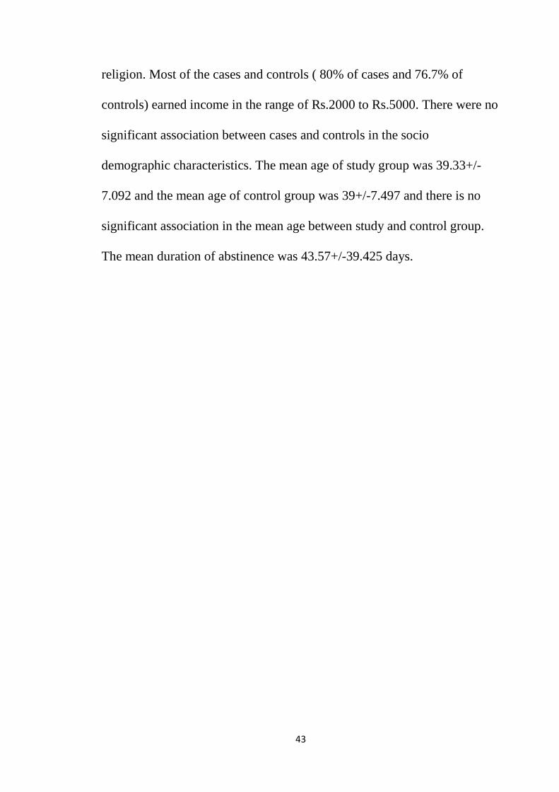
43
religion. Most of the cases and controls ( 80% of cases and 76.7% of
controls) earned income in the range of Rs.2000 to Rs.5000. There were no
significant association between cases and controls in the socio
demographic characteristics. The mean age of study group was 39.33+/-
7.092 and the mean age of control group was 39+/-7.497 and there is no
significant association in the mean age between study and control group.
The mean duration of abstinence was 43.57+/-39.425 days.

44
Comparison of Cognitive functions between cases and controls:
Table 2.1
Comparison of time taken (in seconds) for digit symbol substitution
test by T-test:
Digit Symbol Substitution Test
Group N Mean SD P-Value
Time Taken
(In Seconds)
Case 30 391.5 129.1 0.009*
Control 30 315.3 83.2
(N- number, S.D- standard deviation, *p< 0.05)
The time taken in seconds by cases and controls are mean= 391.5,
S.D= 129.1 and mean= 315.3, S.D= 83.2.
There is a significant difference between both groups in the time taken to
complete the task in digit symbol substitution test which is a measure of
mental speed, between cases and control groups when compared by by T-
test with p value <0.05.

45
Table 2.2
Comparison of number of errors in digit vigilance test by T-test:
Digit Vigilance Test Group N Mean S.D P-Value
No of Errors (Omission and Commission)
Case 30 4.670 1.729 0.213
Control 30 4.130 1.548
(N- number, S.D- standard deviation)
The number of errors made by cases and controls are mean= 4.670,
S.D= 1.729 and mean= 4.130, S.D= 1.548 respectively.
There is no significant difference between both groups in number of errors
in digit vigilance test which is a measure of sustained attention when
compared by T-test with p value >0.05.

46
Table 2.3
Comparison of number of errors in triads test by T- test:
Triad Test Group N Mean S.D P-Value
Errors (Word and Tactile)
Case 30 3.500 1.737 0.456
Control 30 3.170 1.704
(N- number, S.D- standard deviation)
The number of errors made by study and control group are mean= 3.5,
S.D= 1.737 and mean= 3.17, S.D= 1.704 respectively.
There is no significant difference between both groups in triads test (no. Of
errors), which is a measure of divided attention when compared by T- test
with p-value falling >0.05.

47
Table 2.4
Comparison of hits and errors in 1 back and 2 back tests respectively
by T- test:
Verbal N Back Group N Mean S.D P-Value
1 Back: Hits Case 30 8.070 1.337
0.041* Control 30 8.630 0.615
1 Back: Errors Case 30 1.200 1.606
0.020* Control 30 0.430 0.626
2 Back: Hits Case 30 5.070 1.721
0.001* Control 30 6.530 1.570
2 Back: Errors Case 30 5.030 2.266
0.033* Control 30 3.800 2.091
(N- number, S.D- standard deviation, * p<0.05)
There is a significant difference between both groups in all components of
verbal N back test ( both 1 back and 2 back) when compared by T- test
with p value <0.05 indicating poor verbal working memory in study group
compared with control group.

48
Table 2.5
Comparison of number of hits and errors in visual 1 back test by T-
test:
Visual N Back Group N Mean S.D P-Value
1 Back: Hits Case 30 7.530 1.106
0.541 Control 30 7.700 0.988
1 Back: Errors Case 30 2.370 1.426
0.770 Control 30 2.270 1.202
(N- number, S.D- standard deviation)
There is no significant difference in visual N back test, which is a measure
of visual working memory between both groups when compared by T-test
with p value falling >0.05.
49
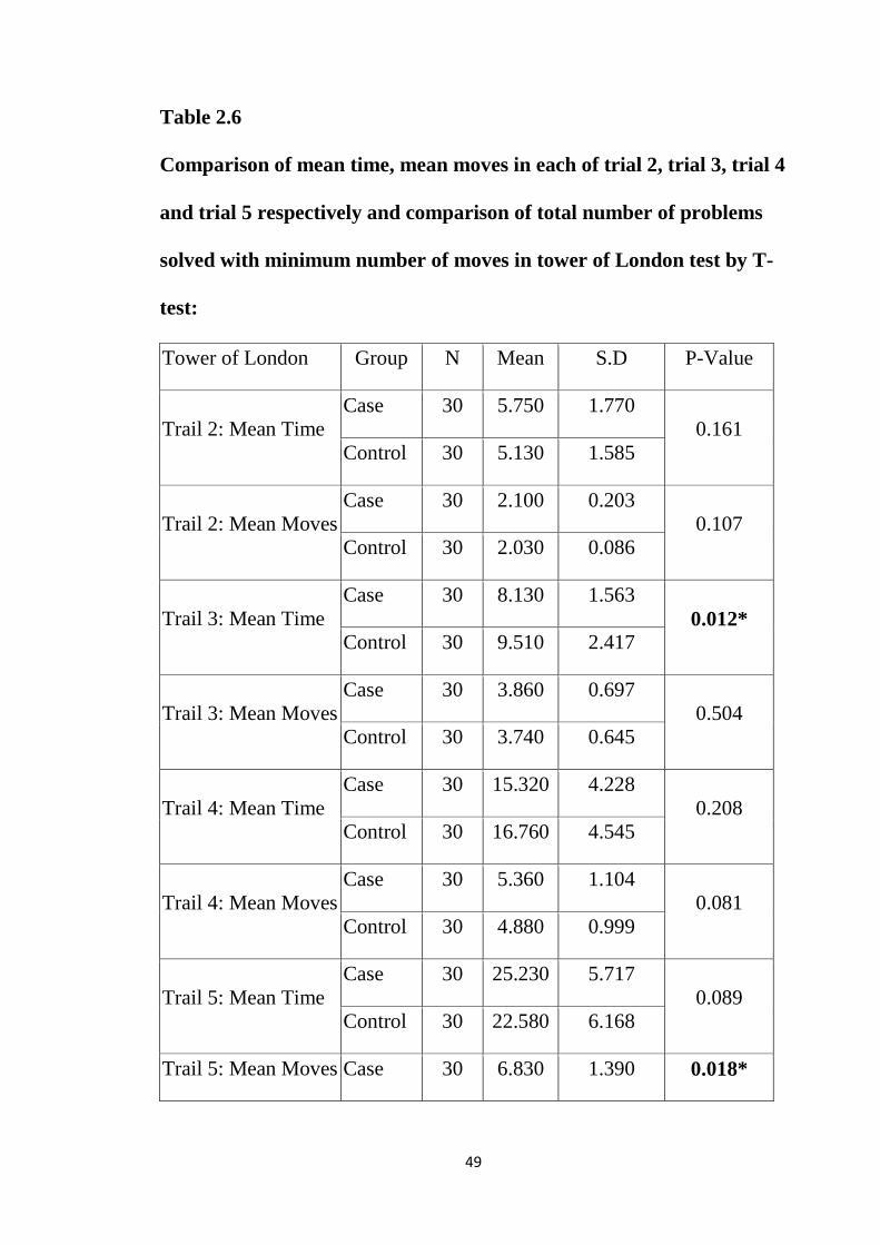
Table 2.6
Comparison of mean time, mean moves in each of trial 2, trial 3, trial 4
and trial 5 respectively and comparison of total number of problems
solved with minimum number of moves in tower of London test by T-
test:
Tower of London Group N Mean S.D P-Value
Trail 2: Mean Time Case 30 5.750 1.770
0.161 Control 30 5.130 1.585
Trail 2: Mean Moves Case 30 2.100 0.203
0.107 Control 30 2.030 0.086
Trail 3: Mean Time Case 30 8.130 1.563
0.012* Control 30 9.510 2.417
Trail 3: Mean Moves Case 30 3.860 0.697
0.504 Control 30 3.740 0.645
Trail 4: Mean Time Case 30 15.320 4.228
0.208 Control 30 16.760 4.545
Trail 4: Mean Moves Case 30 5.360 1.104
0.081 Control 30 4.880 0.999
Trail 5: Mean Time Case 30 25.230 5.717
0.089 Control 30 22.580 6.168
Trail 5: Mean Moves Case 30 6.830 1.390 0.018*
50
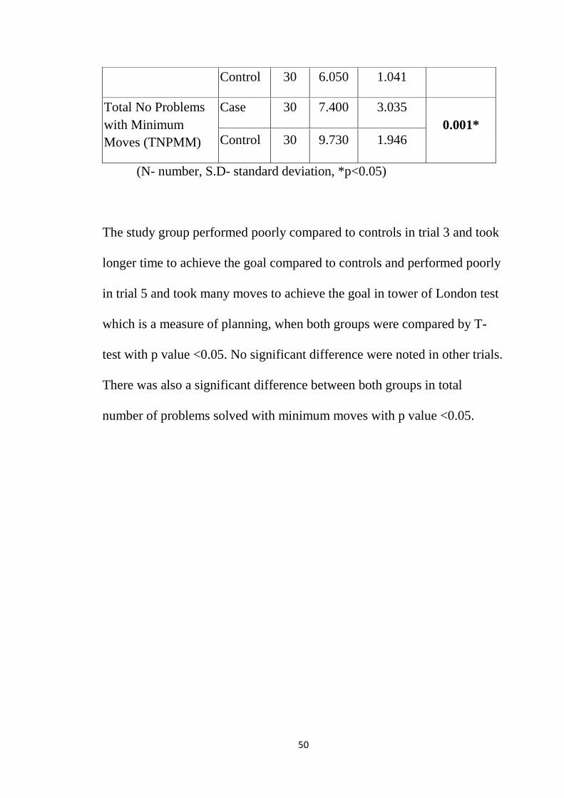
Control 30 6.050 1.041
Total No Problems with Minimum Moves (TNPMM)
Case 30 7.400 3.035 0.001*
Control 30 9.730 1.946
(N- number, S.D- standard deviation, *p<0.05)
The study group performed poorly compared to controls in trial 3 and took
longer time to achieve the goal compared to controls and performed poorly
in trial 5 and took many moves to achieve the goal in tower of London test
which is a measure of planning, when both groups were compared by T-
test with p value <0.05. No significant difference were noted in other trials.
There was also a significant difference between both groups in total
number of problems solved with minimum moves with p value <0.05.
51
Table 2.7
Comparison of number of words learned in trial 1, trial 5, number of
recognition hits, errors (omissions and false alarm) and number of
words recalled (immediate and delayed) by T-test:
Auditory Verbal Learning Test
Group N Mean S.D P-Value
Trail 1 Case 30 7.370 1.921
0.068 Control 30 8.370 2.236
Trail 5 Case 30 13.730 1.143
0.269 Control 30 14.030 0.928
Recognition Hits Case 30 14.630 0.556
0.026* Control 30 14.900 0.305
Omissions Case 30 0.370 0.556
0.026* Control 30 0.100 0.305
False Alarm Case 30 0.030 0.183
0.326 Control 30 0.000 0.000
Immediate Recall Case 30 14.030 1.159
0.009* Control 30 14.670 0.479
Delayed Recall Case 30 13.730 1.202
0.010* Control 30 14.430 0.774
52
(N- number, S.D- standard deviation, *p<0.05)
There was a significant difference between both groups in auditory verbal
learning test in both immediate and delayed recall of words and also
significant difference in number of recognition hits and omissions in both
groups when compared by T-test with p value falling <0.05. No significant
difference was noted in auditory verbal learning test in trial 1, trial 5, false
alarm with p value >0.05.

53
Table 2.8
Comparison of number of facts recalled (immediate and delayed) in
logical memory test by T- test:
Logical Memory Group N Mean S.D P-Value
Immediate Recall Case 30 11.730 1.311
0.204 Control 30 12.200 1.495
Delayed Recall Case 30 10.470 1.570
0.017* Control 30 11.400 1.354
(N- number, S.D- standard deviation, *p<0.05)
There was a significant difference between both groups in the delayed
recall in logical memory test with p value <0.05 when compared by T-test
but no significant difference were noted in the immediate recall of facts
with p value >0.05.
54
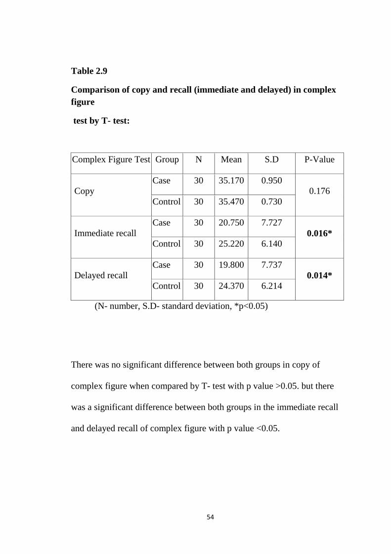
Table 2.9
Comparison of copy and recall (immediate and delayed) in complex figure
test by T- test:
Complex Figure Test Group N Mean S.D P-Value
Copy Case 30 35.170 0.950
0.176 Control 30 35.470 0.730
Immediate recall Case 30 20.750 7.727
0.016* Control 30 25.220 6.140
Delayed recall Case 30 19.800 7.737
0.014* Control 30 24.370 6.214
(N- number, S.D- standard deviation, *p<0.05)
There was no significant difference between both groups in copy of
complex figure when compared by T- test with p value >0.05. but there
was a significant difference between both groups in the immediate recall
and delayed recall of complex figure with p value <0.05.
55
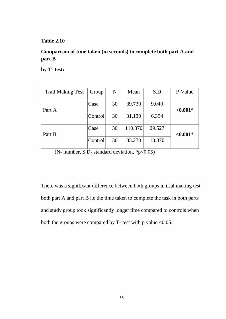
Table 2.10
Comparison of time taken (in seconds) to complete both part A and part B
by T- test:
Trail Making Test Group N Mean S.D P-Value
Part A Case 30 39.730 9.040
<0.001* Control 30 31.130 6.394
Part B Case 30 110.370 29.527
<0.001* Control 30 83.270 13.370
(N- number, S.D- standard deviation, *p<0.05)
There was a significant difference between both groups in trial making test
both part A and part B i.e the time taken to complete the task in both parts
and study group took significantly longer time compared to controls when
both the groups were compared by T- test with p value <0.05.
56
Correlation between Duration of abstinence and cognitive functions in the study group by Pearson’s correlation coefficient:
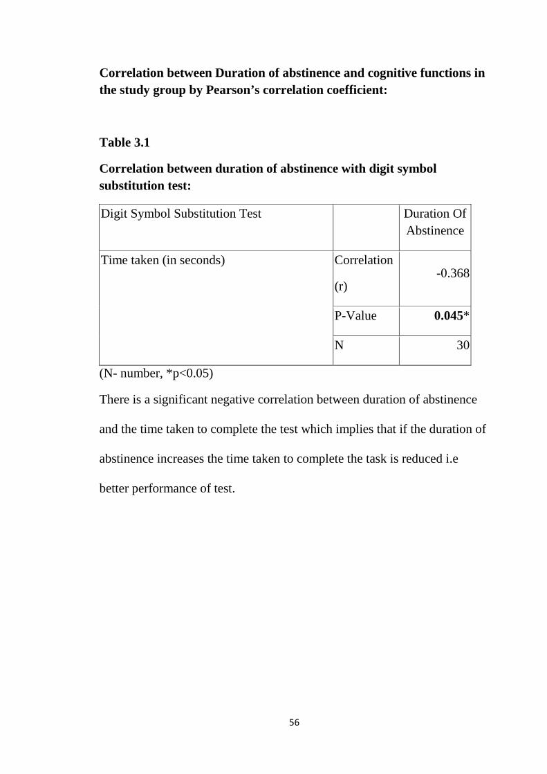
Table 3.1
Correlation between duration of abstinence with digit symbol substitution test:
Digit Symbol Substitution Test Duration Of Abstinence
Time taken (in seconds) Correlation
(r) -0.368
P-Value 0.045*
N 30
(N- number, *p<0.05)
There is a significant negative correlation between duration of abstinence
and the time taken to complete the test which implies that if the duration of
abstinence increases the time taken to complete the task is reduced i.e
better performance of test.

57
Table 3.2
Correlation between duration of abstinence with digit vigilance test:
Digit Vigilance Test Duration Of Abstinence
Errors (Omission and Commission) Correlation
( r ) -0.282
P-Value 0.131
N 30
No significant correlation was found between duration of abstinence and
the errors in alcohol dependent group with p value >0.05 which implies
that the duration of abstinence does not improve sustained attention.

58
Table 3.3
Correlation between duration of abstinence and errors in triad test:
Triad Test Duration Of Abstinence
Errors (Word and Tactile) Correlation
( r ) -0.359
P-Value 0.051
N 30
No significant correlation was found between the duration of abstinence
and errors in the triad test with p value > 0.05 which implies that the
duration of abstinence does not improve divided attention.

59
Table 3.4
Correlation between duration of abstinence and verbal N back test:
Verbal N Back Duration Of Abstinence
1 Back: Hits Correlation
( r ) 0.252
P-Value 0.180
N 30
1 Back: Errors Correlation
( r ) -0.200
P-Value 0.289
N 30
2 Back: Hits Correlation
( r ) 0.100
P-Value 0.601
N 30
2 Back: Errors Correlation
( r ) -0.028
P-Value 0.882
N 30

60
No significant correlation was found between the duration of abstinence
and the number of hits and errors in both 1 back and 2 back test which
implies that the duration of abstinence does not improve verbal working
memory.
Table 3.5
Correlation between duration of dependence with the visual 1 back test:
Visual N Back Duration Of Abstinence
1 Back: Hits Correlation
( r ) 0.448
P-Value 0.013
N 30
1 Back: Errors Correlation
( r ) -0.306
P-Value 0.100
N 30
(N- number, *p<0.05)
There is a significant positive correlation between the duration of
abstinence with the number of hits in visual 1 back test which implies that
as the duration of abstinence increases visual working memory improves.
61
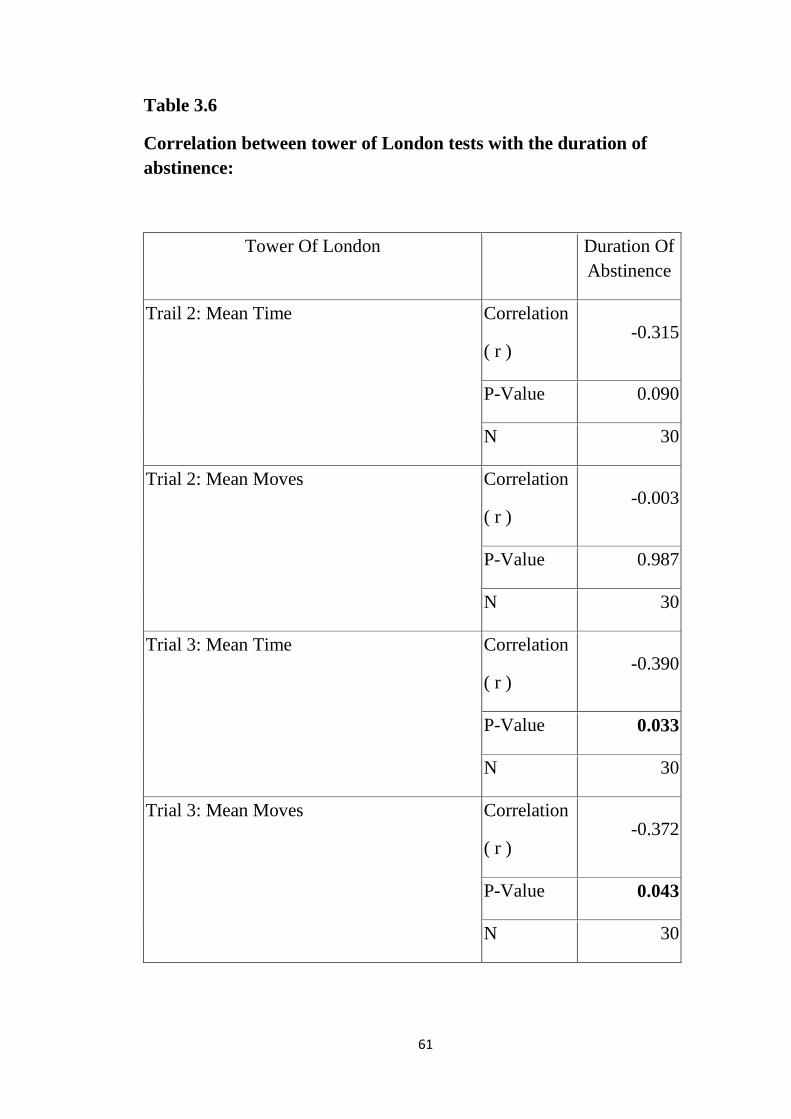
Table 3.6
Correlation between tower of London tests with the duration of abstinence:
Tower Of London Duration Of Abstinence
Trail 2: Mean Time Correlation
( r ) -0.315
P-Value 0.090
N 30
Trial 2: Mean Moves Correlation
( r ) -0.003
P-Value 0.987
N 30
Trial 3: Mean Time Correlation
( r ) -0.390
P-Value 0.033
N 30
Trial 3: Mean Moves Correlation
( r ) -0.372
P-Value 0.043
N 30
62
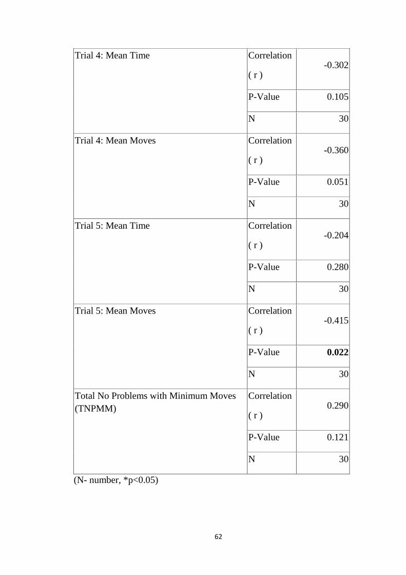
Trial 4: Mean Time Correlation
( r ) -0.302
P-Value 0.105
N 30
Trial 4: Mean Moves Correlation
( r ) -0.360
P-Value 0.051
N 30
Trial 5: Mean Time Correlation
( r ) -0.204
P-Value 0.280
N 30
Trial 5: Mean Moves Correlation
( r ) -0.415
P-Value 0.022
N 30
Total No Problems with Minimum Moves (TNPMM)
Correlation
( r ) 0.290
P-Value 0.121
N 30
(N- number, *p<0.05)
63
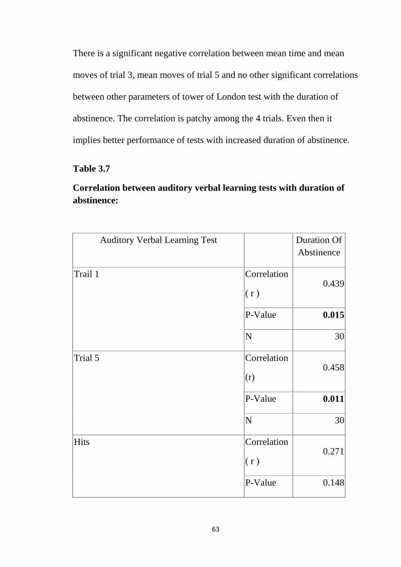
There is a significant negative correlation between mean time and mean
moves of trial 3, mean moves of trial 5 and no other significant correlations
between other parameters of tower of London test with the duration of
abstinence. The correlation is patchy among the 4 trials. Even then it
implies better performance of tests with increased duration of abstinence.
Table 3.7
Correlation between auditory verbal learning tests with duration of abstinence:
Auditory Verbal Learning Test Duration Of Abstinence
Trail 1 Correlation
( r ) 0.439
P-Value 0.015
N 30
Trial 5 Correlation
(r) 0.458
P-Value 0.011
N 30
Hits Correlation
( r ) 0.271
P-Value 0.148

64
N 30
Omission Correlation
( r ) -0.271
P-Value 0.148
N 30
False Alarm Correlation
( r ) -0.103
P-Value 0.587
N 30
Immediate Recall Correlation
( r ) 0.316
P-Value 0.089
N 30
Delayed Recall Correlation
( r ) 0.372
P-Value 0.043
N 30
There is a significant positive correlation between verbal learning of words
in trial 1 and trial 5 and also significant positive correlation between
delayed recall of words with the duration of abstinence. This implies that

65
verbal learning of words and delayed recall of words improves as the
duration of abstinence increases.
Table 3.8
Correlation between logical memory tests with the duration of abstinence:
Logical Memory Duration Of Abstinence
Immediate Recall Correlation
( r ) 0.285
P-Value 0.127
N 30
Delayed Recall Correlation
( r ) 0.391
P-Value 0.033
N 30
(N- number, *p<0.05)
There is a significant positive correlation between the duration of
abstinence and the delayed recall of facts and it implies that as the duration
of abstinence increases delayed recall of facts improves.

66
Table 3.9
Correlation between complex figure test and duration of abstinence:
Complex Figure Test Duration Of Abstinence
Copy Correlation
( r ) 0.261
P-Value 0.164
N 30
Immediate Recall Correlation
( r ) 0.513
P-Value 0.004
N 30
Delayed Recall Correlation
( r ) 0.531
P-Value 0.003
N 30
(N- number, *p<0.05)
There is significant positive correlation between immediate recall and
delayed recall of figure with the duration of abstinence and it implies that
as the duration of abstinence increases the performance in immediate and
delayed recall of figure improves.
67
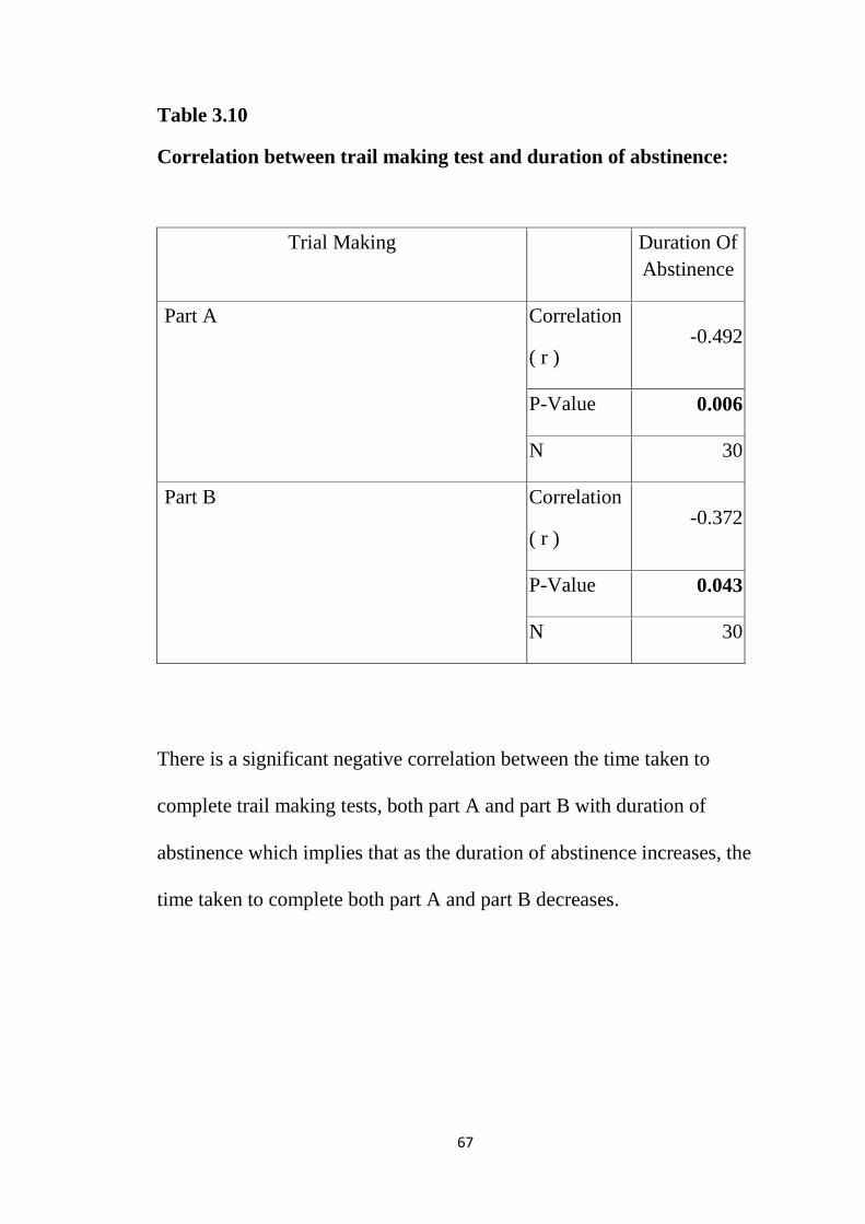
Table 3.10
Correlation between trail making test and duration of abstinence:
Trial Making Duration Of Abstinence
Part A Correlation
( r ) -0.492
P-Value 0.006
N 30
Part B Correlation
( r ) -0.372
P-Value 0.043
N 30
There is a significant negative correlation between the time taken to
complete trail making tests, both part A and part B with duration of
abstinence which implies that as the duration of abstinence increases, the
time taken to complete both part A and part B decreases.
68
DISCUSSION
The study shows that the abstinent alcohol dependent males perform
poorly when compared to control groups in mental speed, sustained
attention, verbal working memory, logical memory, verbal memory, visuo
constructive ability and executive functions (planning, speed of processing,
cognitive flexibility).
In our study it has been shown that study group’s performance on timed
tasks as in mental speed, trail making test, tower of London test was poorer
compared to controls which indicates frontal lobe dysfunction in the study
group.
These results have also been established in previous studies. In the study
of executive functioning early in abstinence from alcohol, Sandra Zinn et
al, 2004 has discussed that there is a greater discrepancy between alcohol
abusing patients and controls especially in timed tasks with a motor
component, visual perception elements and those which uses working
memory.
The same has also been established by S.J.C. Davies et al, 2005 in a
population of apparently clinically healthy abstinent alcohol-dependent
subjects where he found impaired frontal lobe function as evidenced by
69
poorer task performance on the Trail Making Test and digit symbol test of
the WAIS-R compared with control subjects which are again timed tasks.
Though the difference between study group and control group on digit
vigilance test and triad test which are tests for sustained attention and
divided attention was not significant, performance of study group was on
the lower side compared to control population.
Our study has shown impairment of verbal working memory in study
group compared to controls but no significant difference between both
groups in visual working memory. In a study by Zdrav Vestn on
neurocognitive assessment of alcoholic patients during recovery from
alcoholism, he illustrated that though both study and control group did not
differ significantly in both spatial and verbal memory, it was found that
alcohol abstainers had less accuracy during the task as the number of errors
made in this group was higher compared to controls. Similarly in our study
the number of errors made by the study group in verbal memory is
significantly higher and the number of errors in visual working memory
though not significant was higher compared to controls.
Our study has included tower of London test which tests exclusively the
ability to plan and it is a function of frontal lobe. The study group

70
performed fairly well though at a lower level compared with controls
except in trial 3, trial 5 and total number of problems solved with minimum
moves in which both group differed significantly. This indicates that
planning strategies of cases compared to controls are poorer. This issue has
also been discussed by George Fein et al, 1990 that intermediate term
abstinent alcoholics perform more poorly than non alcoholic persons on
tests of problem solving and abstraction abilities.
Our study group also showed significant impairment in immediate and
delayed recall of verbal material as well as visual memory when compared
with controls. It has been said that impairment in memory and learning in
abstinent alcoholics have been reported less frequently but they are now
receiving increasing attention (George Fein, 1990). Tarter and Edwards in
1985 report that learning and memory deficits were not observed when
standard clinical tests were employed but with a more challenging
laboratory tasks there were learning and memory deficits. Ryan C butters,
1980; becker JT et al, 1983; brandt J et al, 1983; ron MA et al, 1980 have
reported short term memory impairments and learning deficits in both
verbal and nonverbal tasks. There are also studies which report impaired
performance in verbal memory but not in non verbal memory task in
abstinent alcohol dependent subjects compared to controls similar to our
results.( S.J.C.Davies et al, 2005)

71
Our study also shows poor performance of study group in visual scanning,
attention, suppression of impulse, cognitive flexibility compared with
control group through trail making test. This result has been demonstrated
in number of studies. ( chelun GJ et al, 1981; S.J.C davies et al, 2005;
sarawat et al, 2006; K.mann et al,1999). In a study by saraswat et al, 2006
on executive functions in alcoholism it was shown that the alcohol
dependent group required a significantly longer time to complete both trail
making test part A and part. Poor performance on TMT part A suggests
impaired visual scanning and psychomotor speed, whereas significant
poorer performance between the alcohol dependent group and controls
group on TMT part B and part B minus part A indicate impaired cognitive
flexibility and set shifting. Even in our study the same findings have been
replicated with significant poor performance in both part A and part B
among the study group. This adds to the evidence of frontal lobe
dysfunction.
Earlier studies focussed on patients whose cognitive deficits were clinically
obvious, such as patients with Korsakoff’s syndrome or frank alcoholic
dementia (Brown et al., 1958), but subsequent studies (Loberg, 1980;
Eckhardt and Matarazzo, 1981; Moselhy et al., 2001) showed that
performance on the Trail B could be impaired in alcohol dependence

72
without any clinically obvious neurological deficits. Noel et al. (2001)
reported that ‘non-amnesic’ alcohol-dependent subjects were slower on
Trails A and B, and similar to our study, greater impairment was seen in
completing the Trail B. Noel et al. (2001) found that performance at easier
stages of tasks showed little or no impairment of executive functions and as
Trail B requires greater levels of flexibility and exploring planning ability
compared with Trail A, it appears that Trail B has sufficient complexity
compared with Trail A.
This study also tested for any correlation between duration of abstinence
and cognitive function in the study group by Pearson’s correlation
coefficient. Our results show that as the duration of abstinence increases
the performance on mental speed, visual memory and not verbal memory,
learning of verbal material and delayed recall of verbal material, delayed
recall of logical memory, immediate and delayed recall of complex figure
test (visual memory), visual scanning and cognitive flexibility also
improves.
According to saraswat et al 2006, there was a significant relationship
between the duration of abstinence and part C of stroop test. Studies on
cognitive recovery during abstinence are diverse and they give conflicting
results with studies demonstrating rapid, complete, partial recovery within

73
several weeks or months or years. (Kish et al., 1980; Leber et al., 1981;
Mann et al., 1999; Tracy and Bates, 1999; Drake et al., 1995; Fein et al.,
1990; Reed et al.,1992; Sullivan et al., 2000) There are also studies which
show residual or no cognitive impairment after a year or more of
abstinence (Brandt et al., 1983; Yohman et al.,1985; Schandler et al.,
1996). The impairment of visual memory among alcoholics studied among
recently detoxified (one month), intermediate- term abstinent (two years)
and long term abstinent (seven years) subjects showed improved memory
performance with increased duration of abstinence.
(Reed RJ et al, 1992; Tivis R et al, 1995)
According to certain tests alcoholics exhibited visuo spatial impairment
even when corrected for premorbid IQ and education.
(Beatty WW, 1996; Sullivan EV, 2000).
In a study by Leber WR, 1981, it was found that alcoholics in their 3 weeks
of abstinence performed significantly lesser than controls in drawing R-
OCF after observation similar to our study but no significant difference
was found between alcoholics in their 11 weeks of abstinence when
compared with controls. It also concludes that some recovery of visuo
spatial functioning may occur after 10 weeks of abstinence.

74
In 1981, leber et al studied learning and memory in controls and 2 groups
of alcoholics with 3 weeks and 11 weeks of abstinence respectively and
found no significant difference between the 3 groups in verbal learning
abilities but short term abstinent alcoholics performed poorly compared to
long term abstinents for visuo spatial learning tasks. Similarly ryan et al in
1980 compared short term and long term abstinents with controls for digit
substitution test and found that alcoholics performed poorly compared to
controls and long term abstinents performed better than short term
abstinents though not significantly similar to the findings of our study.
75
CONCLUSION
1. Abstinent alcohol dependent males perform poorly compared to the
controls in mental speed, sustained attention, logical memory, verbal
memory, visuo constructive ability and executive functions
(planning, speed of processing, cognitive flexibility, verbal working
memory).
2. As the duration of dependence increases study group performed
significantle better in mental speed, visual working memory, verbal
learning and memory, logical memory, visual memory, visual
scanning and cognitive flexibility.

76
LIMITATIONS
1. The role of drugs especially benzodiazepines on cognition have not
been considered. The assessment was made by the investigator
which introduces the interviewer bias.
2. The sample size is relatively small to generalise the findings in
alcohol dependent population.
3. Nutritional deficiency in the study population has not been studied
and hence the cognitive impairment due to korsakoff’s psychosis
cannot be ruled out.
4. Comorbid personality disorders have not been studied and its
influence on cognitive functions cannot be ruled out.
5. The history of previous relapses, previous detoxifications have not
been studied which has an influence on cognitive functions as per
studies.
6. Intelligence tests, tests for verbal fluency and comprehension has
not been assessed in this study.

77
STRENGTHS
1. To our knowledge there are only few Indian studies on cognitive
functions in abstinent alcohol dependent males and this study has
assessed multiple components of cognitive functions.
2. The study has included age and education matched control group
to minimise the possibility of confounding. It has also measured
the correlation of duration of abstinence with the cognitive
functions which has been extensively studied.
3. It has utilised NIMHANS neuropsychological battery (2004), a
validated Indian test to assess cognition.
4. This study may serve as an eye opener in understanding the
effects of alcohol on cognition.

78
FUTURE DIRECTIONS
1. A clearer picture of the cognitive impairment can be given when
longitudinal studies are undertaken rather than cross sectional
studies.
2. Though the cognition in alcohol dependence has been extensively
studied in the past three decades its importance is underestimated
clinically. With lots of background information on the effects of
cognitive impairment in the treatment and prognosis it becomes
essential to assess the cognition in alcohol dependent males to
provide them the appropriate treatment.
3. Assessment of cognition also indicates the clinician the need for
improvement of cognition in patients with cognitive impairment
even in patients who have normal cognition clinically.
4. Use of Wisconsin card sorting test will give a better picture of thr
executive functions.

79
BIBLIOGRAPHY
1.Adalsteinsson, e.; sullivan, e.v.; and pfefferbaum, a. Biochemical,
functional and microstructural magnetic resonance imaging (MRI). In
Liu, Y., and Lovinger, D.M., Eds.Methods in AlcoholRelated
Neuroscience Research. Boca Raton, FL: CRC Press, 2002, pp. 345–
372.
2.Adams KM, Gilman S, Koeppe RA, Kluin KJ, Brunberg JA,
Dede D, Berent S, Kroll PD (1993) Neuropsychological deficits
are correlated with frontal hypometabolism in positron emission
tomography studies of older alcoholic patients. Alcohol Clin Exp
Res 17:205–210.
3.Agartz, I., Momenan, R., Rawlings, R. R. et al. (1999) Hippocampal
volume in patients with alcohol dependence. Archives of General
Psychiatry 56, 356–363.
4.Allen RP, Faillace LA, Wagman A: Recovery time for alcoholics after
prolonged alcohol intoxication. Johns Hopkins Med J 1971; 128:158-
164
5.Anna kokkavec and simon f. Crowe: A comparison of
cognitive performance in binge versus regular chronic
alcohol misusers, alchol and alcoholism, vol.34, No. 4,
pp- 601 – 608,1999.
80
6. Antonio verdejo-garcía,1 antoine bechara,2,3 emily c. Recknor,3and
miguel pérez-garcía1; Executive dysfunction in substance dependent
individuals during drug use and abstinence: An examination of the
behavioral, cognitive and emotional correlates of addiction; Journal of
the International Neuropsychological Society (2006), 12, 405–415.
7.Arria, A. M. and Van Thiel, D. H. (1992) The epidemiology of alco- hol
related chronic disease. Alcohol, Health and Research World 16, 209–
216.
8.Arthur W. Blume, M.A.*.; J. Mark Davis, Ph.D.** & Karen B.
Schmaling, Ph.D; Neurocognitive Dysfunction in Dually-Diagnosed
Patients:A Potential Roadblock to Motivating Behavior Change;
Volume 31 (2), April – June 1999
9.Beatty WW, Tivis R, Stott HD, Nixon SJ, Parsons OA (2000)
Neuropsy- chological deficits in sober alcoholics: influences of
chronicity and
10.Becker JT, Butters N, Hermann A, et al: A comparison of the effects of
longterm alcohol abuse and aging on the performance of verbal and
nonverbal divided attention tasks. Alcoholism (NY) 1983; 7:213-219
11.Benton, A. L. and Hamsher, K.de S. (1976) Multilingual Aphasia
Examination. Iowa City, University of Iowa. (Manual Revised,1978)

81
12.Bloomer, c.w.; langleben, d.d.; and meyerhoff, D.J.Magnetic resonance
detects brainstem changes in chronic, active heavy drinkers. Psychiatry
Research 132:209–218, 2004
13.Bowden, S. C. and McCarter, R. J. (1993) Spatial memory in alcohol-
dependent subjects: using a push-button maze to test the principle of
equiavailability. Brain and Cognition 22, 51–62.
14.Brandt J, Butters N, Ryan C, et al: Cognitive loss and recovery in long-
term alcohol abusers. Arch Gen Psychiatry 1983; 40:435-442
15.Brown, E. C., Casey, A., Fisch, R. I. et al. (1958) Trail
making test as a screening advice for detection of
brain damage. Journal of Consulting Psychology 22,
469–473.
16.Mabuchi T, Kitagawa K, Kuwabara K et al. (2001)
Phosphorylation of cAMP response element-binding
protein in hippocampal neurons as a protective
response after exposure to glutamate in vitro and
ischemia in vivo. J Neurosci 21:9204–13.
17.Chandler LJ, Newsom H, Sumners C et al. (1993a) Chronic ethanol
exposure potentiates NMDA excitotoxicity in cerebral cortical neurons.
J Neurochem 60:1578–81.
18.Chandler LJ, Sumners C, Crews FT. (1993b) Ethanol inhibits NMDA
receptor-mediated excitotoxicity in rat primary neuronal cultures.

82
Alcohol Clin Exp Res 17:54–60.
19.Chanraud, s.; reynaud,m.;wessa,m.; et al. Diffusion tensor tractography
in mesencephalic bundles: Relation to mental flexibility in detoxified
alcohol dependent subjects. Neuropsychopharmacology 2009, Epub
ahead of print July 9, 2008. P
20.Chanraud, S.;Martelli, C.; Delain, F.; ET AL. Brain morphometry and
cognitive performance in detoxified alcohol dependents with preserved
psychosocial functioning. Neuropsychopharmacology 32:429–438,
2007. PMID: 17047671
21.Chelune GJ, Parker JB: Neuropsychological deficits associated with
chronic alcohol abuse. Clin Psychol Rev 1981; 1:181-195
22.Clarke J, Haughton H: A study of intellectual impairment and recovery
rates in heavy drinkers in Ireland. Br J Psychiatry 1975; 12:178-184
23.Claudia Bartels et. al;Recovery Of Hippocampus-Related Functions In
Chronic Alcoholics During Monitored Long-Term Abstinence; Alcohol
& Alcoholism Vol. 42, No. 2, pp. 92–102, 2007
24.D, Cardenas V, Chao LL, Rothlind J, Studholme C, Weiner MW.
Effects of heavy drinking binge drinking, and family history of
alcoholism on regional brain metabolites. Alcohol Clin Exp Res
2004;28:650–661.
25.Dao-Castellana MH, Samson Y, Legault F, Martinot JL,
Aubin HJ, Crouzel C, et al. (1998) Frontal dysfunction in

83
neurologically normal chronic alcoholic subjects: metabolic and
neuropsychological findings. Psychol Med.
26.Davies SJ, Pandit SA, Feeney A et al. (2005) Is there cognitive impair-
ment in clinically ‘healthy’ abstinent alcohol dependence? Alcohol
Alcohol 40:498–503.
27.Demir B, Ulug B, Lay Ergun E, Erbas B (2002) Regional cerebral
blood flow and neuropsychological functioning in early and late
onset alcoholism. Psychiatry Res 115:115–125.
28.Donovan DM, Kivlahan DR, Walker RD (1984) Clinical limitations
of neuropsychological testing in predicting treatment outcome
among al- coholics. Alcohol Clin Exp Res 8:470 – 475.
29.Drummond, D. C. (1990) The relationship Between
alcohol dependence and alcohol-related problems in
a clinical population. British Journal of Addiction
85, 357–366.
30.Durazzo TC, Gazdzinski S, Meyerhoff DJ. (2007) The neurobiological
and neurocognitive consequences of chronic cigarette smoking in
alcohol use disorders. Alcohol Alcohol 42:174–85.
31.Durazzo TC, Rothlind JC, Gazdzinski S et al. (2006) A comparison of
neurocognitive function in non-smoking and chronically smoking short-
term abstinent alcoholics. Alcohol 39:1–11.
32.Eckardt MJ, Parker ES, Noble EP, Pautler CP, Gottschalk LA

84
(1979) Changes in neuropsychological performance during
treatment for alco- holism. Biol Psychiatry 14:943–954.
33.Eckardt MJ, Stapleton JM, Rawlings RR, Davis EZ, Grodin DM
(1995) Neuropsychological functioning in detoxified alcoholics
between 18 and 35 years of age. Am J Psychiatry 152:45–52.
34.Eckhardt, M. J. and Matarazzo, J. D. (1981) Test-retest reliability of
Halstead Impairment Index in hospitalised alcoholic and
nonalcoholic males with mild to moderate neuropsychological
impairment. Journal of Clinical Neuropsychology 3, 257–259.
35.Fein G, Torres J, Price LJ et al. (2006) Cognitive performance in
long-term abstinent alcoholic individuals. Alcohol Clin Exp Res
30:1538–44
36.Fein, G., and Landman, B. Treated and treatment naive alcoholics come
from different populations. Alcohol 36:19–26, 2005.
37.Fein, G., Bachman, L., Fisher, S. et al. (1990) Cognitive impairments in
abstinent alcoholics. The Western Journal of Medicine 152,531–7.
38.Fein, G.; Di Sclafani, V.; Cardenas, V.A.; et al. Cortical gray matter
loss in treatment naive alcohol dependent individuals. Alcoholism:
Clinical and Experimental Research 26:558–564, 2002.
39.Fein, G.; Landman, B.; Tran,H.; et al. Brain atrophy in long term
abstinent alcoholics who demonstrate impairment on a simulated
gambling task. NeuroImage 32:1465–1471, 2006.

85
40.Fitzhugh LC, Fitzhugh KB, Reitan RM: Adaptive abilities and
intellectual functioning in hospitalized alcoholics. Q J Stud Alcohol
1960; 21:414-423
41. Frank Norton & Lavania Halay; The American Association of
Behavioral and Social Sciences Journal (The AABSS Journal, 2011,
Volume 15)
42.Glenn SW, Parsons OA, Sinha R et al. (1988) The effects of repeated
withdrawals from alcohol on the memory of male and female
alcoholics. Alcohol Alcohol 23:337–42.
43.Goldman MS, Williams DL, Klisz DK: Recoverability of psychological
functioning following alcohol abuse: Prolonged visual-spatial
dysfunction in older alcoholics.J Consult Clin Psychol 1983; 51:370-
378
44.Grant I, Adams KM, Reed R: Aging, abstinence, and medical risk
factors in the prediction of neuropsychologic deficit among long-term
alcoholics. Arch Gen Psychiatry 1984; 41:710-718
45.Harper C, Kril J. (1989) Patterns of neuronal loss in the cerebral cortex
in chronic alcoholic patients. J Neurol Sci 92:81–9.
46.HARPER, C. The neuropathology of alcohol specific brain damage, or
does alcohol damage the brain? Journal of Neuropathology and
Experimental Neurology 57:101–110, 1998.

86
47.Harper, C.G., AND Kril, J.J. Neuropathological changes in alcoholics.
InHunt W.A., and Nixon, S.J., Eds. Alcohol Induced Brain Damage.
NIAAA ResearchMonograph No. 22, Rockville, MD: National
Institutes of Health, 1993, pp, 39–69.
48.Harper, C.G., AND Kril, J.J. Neuropathology of alcoholism. Alcohol
and Alcoholism 25:207–216, 1990
49.Ihara H, Berrios GE, London M (2000) Group and case study
of the dysexecutive syndrome in alcoholism without amnesia. J
Neurol Neu- rosurg Psychiatry 68:731–737.
50.Impairment of Cognitive Abilities and Decision Making after
Chronic Use of Alcohol: The Impact of Multiple Detoxifications
in Patients With Chronic Alcoholism in Contrast to Patients With
Korsakoff’s Syndrome; J Neuropsychiatry Clin Neurosci 12:1, Winter
2000
51. Isolde Daig, Richard Mahlberg, Julia Stethin, Franziska Schroeder,
Jana Wrase,Nina Knoll, Tom Bschor, Guenter Esser, Andreas Heinz,
Thorsten Kienast; Decreased Verbal Learning but not Recognition
Performance in Alcohol-dependent Individuals During Early
Abstinence; Ir J Psych Med 2012; 29 (2): 96-101
52. Isolde Daig1et. al; Low effective organizational strategies in visual
memory performance of unmedicated alcoholics during early

87
abstinence; GMS Psycho-Social-Medicine 2010, Vol. 7, ISSN 1860-
5214
53. J.K. Trivedi: cognitive deficits in psychiatric disorders- current status,
Indian journal of psychiatry, 2006; 48:10-20.
54. Karl Mann*, Arthur Gunther, Friedhelm Stetter And Klaus Ackermann;
Rapid recovery from cognitive deficits in abstinent alcoholics: A
controlled test-retest study; Alcohol and Alcoholism Vol. 34, No. 4, pp.
567-574, 1999.
55.Katherine Tzambazis And Con Stough: alcohol impairs speed of
information processing and simple And choice reaction time and
differentially impairs Higher-order cognitive abilities, Alcohol &
Alcoholism Vol. 35, No. 2, pp. 197–201, 2000
56.Kleinknecht RA, Goldstein SG: Neuropsychological deficits associated
with alcoholism: A review and discussion. Q J Stud Alcohol 1972;
33:999-1019
57.Kopelman, M. D. (1995) The Korsakoff Syndrome. British Journal of
Psychiatry 166, 154–173.
58.Kubota,m.; nakazaki, s.;hirai, s.; et al. Alcohol consumption and frontal
lobe shrinkage: Study of 1432 non-alcoholic subjects. Journal of
Neurology, Neurosurgery, and Psychiatry 71:104–106, 2001.
59.Leber, W. R., Jenkins, R. L. and Parsons, O. A. (1981) Recovery of
visual-spatial learning and memory in chronic alcoholics. Journal of
88
Clinical Psychology 37, 192–197.
60.Leber, W. R., Parsons, O. A. and Nichols, N. (1985) Neuropsycholo-
gical test results are related to ratings of men alcoholics’ thera-peutic
progress: a replicated study. Journal of Studies on Alcohol 46(2),
116–121.
61.Lezak,M.D. Neuropsychological Assessment. 3rd ed. New York:
Oxford University Press, 1995.
62.Lilijana Šprah, Tatjana Novak; NEUROCOGNITIVE ASSESSMENT
OF ALCOHOL INPATIENTS DURING RECOVERY FROM
ALCOHOLISM; Zdrav Vestn 2008; 77: II-75–84
63.LUberg T, Miller WR: Personality, cognitive, and neuropsychological
correlates of harmful alcohol consumption: A cross-national
comparison of clinical samples.Ann NY Acad Sci 1986; 472:75-97
64.J. Rosenbloom, and Adolf Pfefferbaum, M.D;Magnetic Resonance
Imaging Of The Living Brain; Vol. 31, No. 4, 2008
65.Mann K, Gunther A, Stetter F, Ackermann K (1999) Rapid
recovery from cognitive deficits in abstinent alcoholics: a controlled
test-retest study. Alcohol Alcohol 34:567–574.
66.Mantamadiotis T, Lemberger T, Bleckmann SC et al. (2002) Disrup-
Margaret J. Rosenbloom, and Adolf Pfefferbaum, M.D
67.Mechanisms of Neurodegeneration and Regeneration
in Alcoholism Alvarez-Buylla A, Garcia-Verdugo JM.

89
(2002) Neurogenesis in adult subventricular zone. J
Neurosci 22:629–34.
68.Mega, M.S. & Cummings, J.L. 1994. Frontal-sub cortical circuits and
neuropsychiatric illness. Journal of Neuropsychiatry & Clinical
Neurosciences 6:358-70.
69.Meyerhoff, D.J. Brain spectroscopic imaging, morphometry, and
cognition in social drinkers and recovering alcoholics. Alcoholism:
Clinical and Experimental Research 29:153–154, 2005.
70.Moselhy, H.F.; Georgiou, G.; AND Kahn, A. Frontal lobe changes in
alcoholism: A review of the literature. Alcohol and Alcoholism 36:357–
368, 2001.
71.Munro, C. A., Saxton, J. and Butters, M. A. (2000) The neuropsycho-
72.Neuropsychological rehabilitation in alcoholics- a preminary
report:grace mathai, shobini l. Rao & p.s. gopinath: Indian journal of
psychiatry, 1998, 40(3), 280-288
73. Nirmal Saraswat, Sanjeev Ranja, Daya Ram; Set-shifting and selective
attentional impairment in alcoholish and its relation with drinking
variables; Indian Journal of psychiatry 2006;48:47-51.
74.Nixon, k., and crews, f.t. Temporally specific burst in cell proliferation
increases hippocampal neurogenesis in protracted abstinence from
alcohol. Journal of Neuroscience 24:9714–9722, 2004.

90
75.Nixon, S.J. Application of theoretical models to the study of alcohol
induced brain damage. InHunt,W., and Nixon, S.J., Eds. Alcohol
Induced Brain Damage. NIAAAMonograph 22. Rockville, MD:
National Institutes of Health, 1993, pp. 213–228.
76.Noel X, Van der Linden M, Schmidt N, Sferrazza R, Hanak C, Le
Bon O, et al. (2001) Supervisory attentional system in nonamnesic
alcoholic men. Arch Gen Psychiatry 58:1152–1158.
77.Noel, X., Van der Linden, M., Schmidt, N. et al. (2001) Supervisory
attentional system in nonamnesic alcoholic men. Archives of
General Psychiatry 58, 1152–1158.
78.O’neill, j.; cardenas, v.a.; andmeyerhoff, D.J. Effects of abstinence on
the brain: Quantitative magnetic resonance imaging and magnetic
resonance spectroscopic imaging in chronic alcohol abuse. Alcoholism:
Clinical and Experimental Research 25:1673–1682, 2001.
of equiavailability. Brain and Cognition 22, 51–62.
79.Oscarberman, M. Neuropsychological vulnerabilities in chronic
alcoholism. In Noronha, A., Eckardt, M.,Warren, K., Eds. Review of
NIAAA’s Neuroscience and Behavioral Research Portfolio. NIAAA
Research Monograph no. 34. Bethesda, MD: National Institutes of
Health, 2000, pp. 437–472.

91
80.Oscarberman,m., andmarinkovic, K. Alcohol: Effects on
neurobehavioral functions and the brain. Neuropsychology Review
17:239–257, 2007.
81.P.K. Dalal, T. Sivakumar: Cognitive psychiatry in India, Indian Journal
of psychiatry; 52, Supplement, Jan 2010.
82.Page RD, Linden JD: 'Reversible' organic brain syndrome in alcoholics
during abstinence-A psychometric evaluation. Q J Stud Alcohol 1974;
35:98-107
83.Parsons OA (1983) Cognitive dysfunction and recovery in
alcoholics. Subst Alcohol Actions Misuse 4:175–190.
84.Parsons OA (1986) Alcoholics’ neuropsychological impairment:
current findings and conclusions. Ann Behav Med 8:13–19.
85.Parsons OA, Nixon SJ. Neurobehavioral sequelae of alcoholism.
Neurol Clin 1993;11:205–218. [PubMed: 8441371]
86.Parsons OA. (1987) Intellectual impairment in alcoholics: persistent
issues. Acta Med Scand 717(Suppl) 33–46.
87.Parsons, O. A. (1977) Psychological deficits in alcoholics: facts and
fancies. Alcoholism: Clinical a n d Experimental Research 1,51–
56.
88.Parsons, O. A., Schaeffer, K. W. and Glenn, S. W. (1990) Does
neuropsychological test performance predict resumption of
drinking in posttreatment alcoholics? Addictive Behaviours 15, 297–
92
307.
89.Pfefferbaum, A., Rosenbloom, M., Crusan, K. et al. (1988)
Brain CT changes in alcoholics: effects of age and alcohol con-
sumption. Alcoholism: Clinical and Experimental Research 12, 81–
87.
90.Pfefferbaum, a., and sullivan, E.V. Diffusion MR imaging in
neuropsychiatry and aging. In Gillard, J.;Waldman, A.; and Barker, P.,
Eds. ClinicalMR Neuroimaging: Diffusion, Perfusion and
Spectroscopy. Cambridge: Cambridge University Press, 2005, pp. 558–
578.
91.Pfefferbaum, a., and sullivan, E.V. Microstructural but not
macrostructural disruption of white matter in women with chronic
alcoholism. NeuroImage 15:708–718, 2002.
92.Pfefferbaum, A.; Adalsteinsson, E.; and Sullivan, E.V. Dysmorphology
and micro structural degradation of the corpus callosum: Interaction of
age and alcoholism. Neurobiology of Aging 27:994–1009, 2006b.
93.Pfefferbaum, A.; Adalsteinsson, E.; BELL, R.L.; AND SULLIVAN,
E.V. Development and resolution of brain lesions caused by
pyrithiamineand dietary induced thiamine deficiency and alcohol
exposure in the alcohol preferring rat: A longitudinal magnetic
resonance imaging and spectroscopy study. Neuropsychopharmacology
32:1159–1177, 2007.
93
94.Pfefferbaum, A.; Adalsteinsson, E.; SOOD, R.; ET AL. Longitudinal
brain magnetic resonance imaging study of the alcohol preferring rat:
Part II: Effects of voluntary chronic alcohol consumption. Alcoholism:
Clinical and Experimental Research 30:1248–1261, 2006a.
95.Pfefferbaum, A.; Desmond, J.E.; Galloway, C.; ET AL. Reorganization
of frontal systems used by alcoholics for spatial working memory: An
fMRI study. NeuroImage 14:7–20, 2001.
96.Pfefferbaum, A.; LIM, K.O.; Zipursky, R.B.; ET AL. Brain gray and
white matter volume loss accelerates with aging in chronic alcoholics:
A quantitativeMRI study. Alcoholism: Clinical and Experimental
Research 16:1078–1089, 1992
97.Pfefferbaum, A.; Rosenbloom,M.J.; Rohlfing, T.; AND Sullivan, E.V.
Degradation of selective lateral and interhemispheric white matter
systems in alcoholism detected with quantitative fiber tracking.
Biological Psychiatry, 2008. In press.
98.Pfefferbaum, A.; Sullivan, E.V.; Mathalon, D.H.; ET AL. Longitudinal
changes in magnetic resonance imaging brain volumes in abstinent and
relapsed alcoholics. Alcoholism: Clinical and Experimental Research
19:1177–1191, 1995.
99.Pfefferbaum, A.; Sullivan, E.V.; Rosenbloom, M.J.; ET AL. A
controlled study of cortical gray matter and ventricular changes in

94
alcoholic men over a fiveyear interval. Archives of General Psychiatry
55:905–912, 1998.
100.Pfefferbaum, A.; Sullivan, E.V.;Hedehus,M.; ET AL. In vivo detection
and functional correlates of white matter microstructural disruption in
chronic alcoholism. Alcoholism: Clinical and Experimental Research
24:1214–1221, 2000.
101.Pfefferbaum, A.; Sullivan, E.V.;Mathalon, D.H.; AND LIM, K.O.
Frontal lobe volume loss observed with magnetic resonance imaging in
older chronic alcoholics. Alcoholism: Clinical and Experimental
Research 21:521–529, 1997.
102.Pfefferbaum, A.; Zahr, N.M.; Mayer, D.; ET AL. Ventricular
expansion in wildtypeWistar rats after alcohol exposure by vapor
chamber. Alcoholism: Clinical and Experimental Research 32:1459–
1467, 2008. ,pp. 217–242.
103.Ratti MT, Bo P, Giardini A, Soragna D (2002) Chronic alcoholism
and the frontal lobe: which executive functions are impaired?
Acta Neurol Scand 105:276 –281.
104.Ratti, M. T., Soragna, D., Sibilla, L. et al. (1999) Cognitive
impairment and cerebral atrophy in ‘heavy drinkers’. Progress in
Neuropsychopharmacology and Biological Psychiatry 23, 243–
258.

95
105.Rosenbloom MJ, Rohlfing T, O’Reilly AW et al. (2007) Improvement
in memory and static balance with abstinence in alcoholic men and
women: selective relations with change in brain structure.Psychiatry Res
155:91–102.
106.Rosenbloom, m.j.; rohlfing, T.; O’reilly, A.W.; ET AL. Improvement
in memory and static balance with abstinence in alcoholic men and
women: Selective relations with change in brain structure. Psychiatry
Research: Neuroimaging 155:91–102, 2007.
107.Rosenbloom,M.J.; Pfefferbaum, A.; AND Sullivan, E.V. Recovery of
short term memory and psychomotor speed but not postural stability
with long term sobriety in alcoholic women. Neuropsychology 18:589–
597, 2004.
108.Rosenbloom,m.j.; rohlfing, t.; o’reilly, a.w.; et al. Improvement in
memory and static balance with abstinence in alcoholic men and
women: Selective relations with change in brain structure. Psychiatry
Research: Neuroimaging 155:91–102, 2007.
109.Rosenbloom,m.j.; sassoon, s.a.; fama, r.; et al. Frontal callosal fiber
integrity selectively predicts coordinated psychomotor performance in
alcoholism. Brain Imaging and Behavior 2:74–83, 2008.
110.Rosenbloom,m.j.; sullivan, e.v.; and pfefferbaum, a. Using magnetic
resonance imaging and diffusion tensor imaging to assess brain damage
96
in alcoholics. Alcohol Research & Health 27:146–152, 2003. PMID:
15303625
111.Rourke SB, Grant I (1999) The interactive effects of age and
length of abstinence on the recovery of neuropsychological
functioning in chronic male alcoholics: a 2-year follow-up study. J Int
Neuropsychol Soc 5:234 –246.
112.Rourke SB, Loberg T (1996) Neurobehavioral correlates of
alcoholism, in Neuropsychological Assessment of Neuropsychiatric
Disorders (Grant I, Adams KM eds), 2nd ed, pp 423– 485.
Oxford University Press, New York.
113.Ryan C, Butters N: Cognitive deficits in alcoholics, chap 12, In Kissin
B,Begleiter H (Eds): The Pathogenesis of Alcoholism-Vol 7, Biological
Factors. New York, Plenum Press, 1983, pp 485-538
114.Ryan C: Learning and memory deficits in alcoholics. J Stud Alcohol
1980; 41:437-447
115. Sabine Loeber*, Theodora Duka, HelgaWelzel Márquez, Helmut
Nakovics , Andreas Heinz, Karl Mann and Herta Flor; Effects of
Repeated Withdrawal from Alcohol on Recovery of Cognitive
Impairment under Abstinence and Rate of Relapse; Alcohol and
Alcoholism Vol. 45, No. 6, pp. 541–547, 2010
116.Sandra Zinn, Roy Stein, and H. Scott Swartzwelder: Executive
Functioning Early in Abstinence From Alcohol: alcoholism-
97
vol.28,no.9,2004
117.Shear, p.k.; butters, n.; jernigan, t.l.; ET AL. Olfactory loss in
alcoholics: Correlations with cortical and subcorticalMRI indices.
Alcohol 9:247–255, 1992.
118.Simon j. C. Davies1, smita a. Pandit1, adrian feeney1, brian j.
Stevenson1, robert w. Kerwin2, david j. Nutt1, e. Jane
marshall3, stephen boddington4 and anne lingford-hughes1* Is
there cognitive impairment in clinically ‘healthy’ abstinent
alcohol dependence?;.Alcohol and alcoholism, vol.40,
no.6,pp.498-503,2005
119. Skinner HA, Allen BA. (1982) Alcohol dependence syndrome:
measurement and validation. J Abnorm Psychol 91:199–209.
120.Sullivan EV, Pfefferbaum A. (2005) Neurocircuitry in alcoholism: a
substrate of disruption and repair. Psychopharmacology
121.Sullivan, E.V. Human brain vulnerability to alcoholism: Evidence
from neuroimaging studies. In Noronha, A.; Eckardt,M.; andWarren,
K., Eds. Review of NIAAA’s Neuroscience and Behavioural Research
Portfolio. NIAAA ResearchMonograph no. 34. Bethesda,MD: National
Institutes of Health, 2000, pp. 473–508.
122. Sullivan, e.v., and pfefferbaum, A. Neurocircuitry in alcoholism: A
substrate of disruption and repair. Psychopharmacology 180:583–594,
2005. PMID: 15834536

98
123.Sullivan, e.v., and pfefferbaum, A. Neuroimaging of theWernicke
Korsakoff syndrome. Alcohol and Alcoholism, 2009. In press.
124.Sullivan, e.v.; deshmukh, a.; de rosa, E.; ET AL. Striatal and forebrain
nuclei volumes: Contribution to motor function and working memory
deficits in alcoholism. Biological Psychiatry 57:768–776, 2005..
125.Sullivan, e.v.; harding, a.j.; pentney, r.; ET AL. Disruption of
frontocerebellar circuitry and function in alcoholism. Alcoholism:
Clinical and Experimental Research 27:301–309, 2003a.
126.Sullivan, e.v.; lane, b.; deshmukh, a.; et al. In vivo mammillary body
volume deficits in amnesic and nonamnesic alcoholics. Alcoholism:
Clinical and Experimental Research 23:1629–1636, 1999. PMID:
10549995
127.Sullivan, e.v.; marsh, l.;mathalon, D.H.; ET AL. Relationship between
alcohol withdrawal seizures and temporal lobe white matter volume
deficits. Alcoholism: Clinical and Experimental Research; 20:348–354,
1996.
128.Sullivan, e.v.; Rosenbloom, m.j.; and Pfefferbaum, A. Pattern of motor
and cognitive deficits in detoxified alcoholic men. Alcoholism: Clinical
and Experimental Research 24:611–621, 2000c. PMID: 10832902
129.Sullivan, e.v.; rosenbloom,m.j.; lim, k.o.; and pfefferbaum, a.
Longitudinal changes in cognition, gait, and balance in abstinent and

99
relapsed alcoholic men: Relationships to changes in brain structure.
Neuropsychology 14:178–188, 2000b. PMID: 10791858
130.Tarter RE, Jones B: Motor impairment in chronic alcoholics. Dis Nerv
Syst 1971; 32:632-636
131.Unkenstein AE, Bowden SC (1991) Predicting the course of
neuropsy- chological status in recently abstinent alcoholics: a
pilot study. Clin Neuropsychol 5:24 –32.
132.Walton MR, Dragunow I. (2000) Is CREB a key to neuronal survival?
133.Weingartner H, Faillace LA, Markley HG: Verbal information
retention in alcoholics. Q J Stud Alcohol 1971; 32:293-303
134.Yohman, J. R., Parsons, O. A. and Leber, W. R. (1985) Lack of
recovery in male alcoholics’ neuropsychological performance one
year after treatment. Alcoholism: Clinical and Experimental Research
9, 114–117.
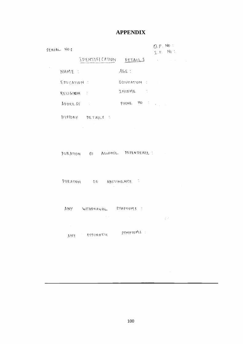
100
APPENDIX

101

102

103
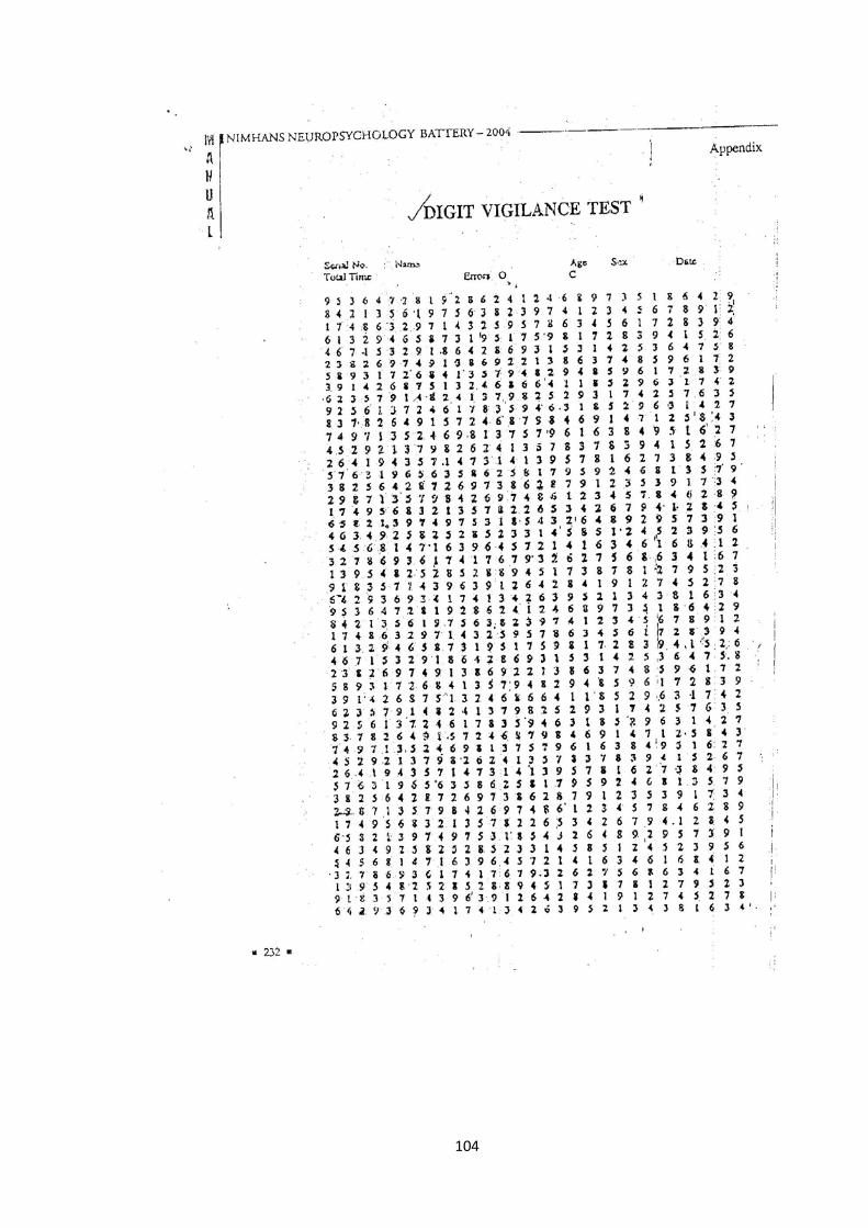
104

105

106

107

108

109

110
Trail making test- Part A:

111
Trail Making Test- Part B

112
Rey-Osterrieth Complex Figure Test:

113

114

115