Cocaine and Mefloquine-induced Acute Effects in Ventral ...
99
Brigham Young University Brigham Young University BYU ScholarsArchive BYU ScholarsArchive Theses and Dissertations 2009-12-10 Cocaine and Mefloquine-induced Acute Effects in Ventral Cocaine and Mefloquine-induced Acute Effects in Ventral Tegmental Area Dopamine and GABA Neurons Tegmental Area Dopamine and GABA Neurons David Wilbanks Allison Brigham Young University - Provo Follow this and additional works at: https://scholarsarchive.byu.edu/etd Part of the Neuroscience and Neurobiology Commons BYU ScholarsArchive Citation BYU ScholarsArchive Citation Allison, David Wilbanks, "Cocaine and Mefloquine-induced Acute Effects in Ventral Tegmental Area Dopamine and GABA Neurons" (2009). Theses and Dissertations. 2362. https://scholarsarchive.byu.edu/etd/2362 This Dissertation is brought to you for free and open access by BYU ScholarsArchive. It has been accepted for inclusion in Theses and Dissertations by an authorized administrator of BYU ScholarsArchive. For more information, please contact [email protected], [email protected].
Transcript of Cocaine and Mefloquine-induced Acute Effects in Ventral ...

Brigham Young University Brigham Young University
BYU ScholarsArchive BYU ScholarsArchive
Theses and Dissertations
2009-12-10
Cocaine and Mefloquine-induced Acute Effects in Ventral Cocaine and Mefloquine-induced Acute Effects in Ventral
Tegmental Area Dopamine and GABA Neurons Tegmental Area Dopamine and GABA Neurons
David Wilbanks Allison Brigham Young University - Provo
Follow this and additional works at: https://scholarsarchive.byu.edu/etd
Part of the Neuroscience and Neurobiology Commons
BYU ScholarsArchive Citation BYU ScholarsArchive Citation Allison, David Wilbanks, "Cocaine and Mefloquine-induced Acute Effects in Ventral Tegmental Area Dopamine and GABA Neurons" (2009). Theses and Dissertations. 2362. https://scholarsarchive.byu.edu/etd/2362
This Dissertation is brought to you for free and open access by BYU ScholarsArchive. It has been accepted for inclusion in Theses and Dissertations by an authorized administrator of BYU ScholarsArchive. For more information, please contact [email protected], [email protected].
Cocaine and Mefloquine-induced Acute Effects in Ventral Tegmental
Area Dopamine and GABA Neurons
David W. Allison
A dissertation submitted to the faculty of Brigham Young University
in partial fulfillment of the requirements for the degree of
Doctor of Philosophy
Neuroscience
Scott C. Steffensen, Chair Jeffrey G. Edwards
Sterling N. Sudweeks James D. Higley
Dixon J. Woodbury
Department of Physiology and Developmental Biology
Brigham Young University
April 2010
Copyright © 2010 David W. Allison
All Rights Reserved
ABSTRACT
COCAINE AND MEFLOQUINE-INDUCED ACUTE EFFECTS IN VENTRAL
TEGMENTAL AREA DOPAMINE AND GABA NEURONS
David W. Allison
Department Physiology and Developmental Biology
Doctor of Philosophy
The aim of the two studies presented here was to evaluate the effects of cocaine and mefloquine (MFQ) on γ-aminobutyric acid (GABA) and dopamine (DA) neurons in the ventral tegmental area (VTA). Cocaine: In vivo, lower doses of intravenous cocaine (0.25-0.5 mg/kg), or methamphetamine (METH), enhanced VTA GABA neuron firing rate via D2/D5 receptor activation. Higher cocaine doses (1.0-2.0 mg/kg) inhibited their firing rate. Cocaine and lidocaine inhibited the firing rate and spike discharges induced by stimulation of the internal capsule (ICPSDs) at dose levels 0.25-2 mg/kg (IC50 1.2 mg/kg), but neither DA nor METH reduced ICPSDs. In VTA GABA neurons in vitro, cocaine reduced (IC50 13 µM) current-evoked spikes and sodium currents in a use-dependent manner. In VTA DA neurons, cocaine reduced IPSCs (IC50 13 µM), increased IPSC paired-pulse facilitation, and decreased sIPSC frequency, without affecting mIPSC frequency or amplitude. These findings suggest cocaine reduces activity-dependent GABA release on DA neurons in the VTA, and that cocaine’s use-dependent blockade of VTA GABA neuron voltage-sensitive sodium channels (VSSCs) may synergize with its DAT inhibiting properties to enhance mesolimbic DA transmission implicated in cocaine reinforcement. Mefloquine: Mefloquine (MFQ) is an anti-malarial agent, Connexin-36 (Cx36) gap junction blocker, 5-HT3 antagonist, and calcium ionophore. Mounting evidence of a Cx36-mediated VTA GABA neuron syncytium suggests MFQ-related dysphoria may attribute to its gap junction blocking effects on VTA synaptic homeostasis. We observed that MFQ (25 µM) increased DA neuron spontaneous IPSC frequency 6 fold, and mIPSC 3 fold. Carbenoxolone (CBX, 100 µM) only increased sIPSC frequency 2 fold, and did not affect DA mIPSC frequency. Ondansetron did not mimic MFQ. Additionally, MFQ did not affect VTA DA evoked IPSC paired pulse ratio (PPR). However, Mefloquine did induce a 3.5 fold increase in bath-applied GABA current. Remarkably, MFQ did not affect VTA GABA neuron inhibition. At VTA DA neuron excitatory synapses MFQ increased sEPSC frequency in-part due to an increase in the AMPA/NMDA ratio. These finding suggest MFQ alters VTA synapses differentially depending on neuron and synapse type, and that these alterations appear to involve MFQ’s gap junction blocking and calcium ionophore actions.
Key words: GABA, VTA, dopamine, cocaine, mefloquine, 5HT3, mesolimbic, Connexin 36, gap junctions, electrical synapse, D2/D5.

ACKNOWLEDGMENTS
I begin as my life did, with my parents, Judge W. and Frieda H. Allison. They gave me the gift
of life, love, and the desire to live up to my potential. What has appeared to be a good set of
genes hasn’t hurt either.
My loving wife Linda gave me the time, encouragement, and never-ending supply of
forgiveness needed to make mistakes and prove to myself I could do this. This work is no
exception to all the good things in my life; it found its beginning in her.
In many ways I will always owe a debt of gratitude to Scott C. Steffensen for his unyielding
fortitude as a mentor and friend. His ability to transcend the X, Y, and Z axes has made all the
difference.
In addition I thank the members of my committee, Connie Provost, and C. Fernando Valenzuela
(University of New Mexico) for providing technical and spiritual guidance. I also thank the
Board of Directors of Brigham Young University, the tithe-payers of the Church of Jesus Christ
of Latter-day Saints, and the many donors to Brigham Young University for providing the
platform to conduct this research. I acknowledge the financial support of the United States
Government through the National Institute on Alcohol Abuse and Alcoholism (NIAAA).

iv
TABLE OF CONTENTS List of Figures ............................................................................................................................. vi
Chapter 1. .....................................................................................................................................1
Introduction ...........................................................................................................................1
The mesolimbic dopamine system ..................................................................................1
GABAergic transmission: A non-dopamine-dependent pathway of addiction...............2
Plasticity and the process of addiction ............................................................................3
Cocaine may act via a dopamine-dependent and non-dopamine-dependent pathway....4
Mefloquine as a pharmacological tool ............................................................................4
Objectives........................................................................................................................5
Figure...............................................................................................................................5
Chapter 2. Cocaine disinhibits dopamine neurons in the VTA via blockade of GABA neuron voltage-sensitive sodium channels .................................................................................................7
Introduction ...........................................................................................................................7
Methods .................................................................................................................................9
Animal Subjects ..............................................................................................................9
Single-unit Recordings in Anesthetized Rats ..................................................................9
Characterization of VTA GABA neurons in vivo .........................................................10
Single-unit recordings in vivo........................................................................................10
Drug Preparation and Administration in vivo................................................................11
Preparation of Brain Slices............................................................................................12
Whole-cell Recordings in vitro .....................................................................................12
Characterization of Neuron Types in vitro....................................................................14
Single-cell Quantitative RT-PCR..................................................................................15
Voltage Waveform Command.......................................................................................16
Statistical Analysis ........................................................................................................17
Results .................................................................................................................................18
v
Effects of Systemic Cocaine, Cocaine Methiodide, Dopamine, Lidocaine and Methamphetamine on VTA GABA Neuron Firing Rate in vivo ..................................19
Dopamine Pharmacology of Cocaine Effects on VTA GABA Neuron Firing Rate in vivo ................................................................................................................................19
Effects of Systemic Cocaine, Cocaine Methiodide, Dopamine, Lidocaine and Methamphetamine on VTA GABA Neuron Evoked Spikes in vivo.............................20
Dopamine Pharmacology of Cocaine Effects on VTA GABA Neuron Evoked Spikes in vivo ............................................................................................................................22
Expression of Select Genes in the Dopamine vs GABA Neurons in Mature Rats: Quantitative Single-cell RT-PCR..................................................................................22
Effects of Cocaine on Current-Evoked VTA GABA Neuron Spikes in vitro...............23
Effects of Cocaine on VTA GABA Neuron Sodium Currents in vitro.........................23
Effects of Cocaine on VTA DA Neuron Evoked and Spontaneous IPSCs in vitro ......25
Discussion ...........................................................................................................................27
Figures .................................................................................................................................34
Chapter 3. Mefloquine disrupts VTA synaptic activity via blockade of Cx36 gap junctions and disruption of calcium homeostasis ...............................................................................................41
Introduction .........................................................................................................................41
Methods ...............................................................................................................................43
Preparation of Brain Slices............................................................................................43
Whole-cell Recordings in vitro .....................................................................................44
Drug Preparation and Administration ...........................................................................46
Characterization of Neuron Types ................................................................................46
Statistical Analyses........................................................................................................47
Results .................................................................................................................................48
Time Course of Mefloquine Effects on VTA DA sIPSC Frequency ............................48
Mefloquine Effects on VTA DA sIPSC Frequency, Amplitude, and Inter-event Interval in WT and Cx36 Mice .....................................................................................49
Mefloquine Effects on VTA DA mIPSC Frequency, Amplitude, and Inter-event Interval in WT and Cx36 Mice......................................................................................51
Neither Presoaking nor Bath-application of Mefloquine Alters VTA DA Neuron Evoked IPSC Paired-pulse Ratio or Primary Current Amplitude .................................53
vi
Mefloquine Increases VTA DA Neuron GABA(A) Receptor Current .........................55
Mefloquine Does Not Affect VTA GABA Neuron sIPSC Frequency or Paired-pulse Ratio ..............................................................................................................................55
Mefloquine Increases VTA DA Neuron sEPSC Frequency in Wildtype and Cx36 KO Mice...............................................................................................................................56
Mefloquine Increases VTA DA Neuron mEPSC Frequency in WT Mice ...................57
Mefloquine Effects on VTA DA and GABA Neuron EPSC PPR and AMPAR to NMDAR Current ratios .................................................................................................58
MFQ Reduces VTA DA Neuron Firing Rate and Current-evoked Spiking .................59
Discussion ...........................................................................................................................61
Figures .................................................................................................................................66
Chapter 4. ...................................................................................................................................76
Conclusion.............................................................................................................................1
References ....................................................................................................................................79
Curriculum vitae...........................................................................................................................86

vii
LIST OF FIGURES Chapter 1
Figure 1: The Mesolimbic Dopamine Pathway ............................................................................6
Chapter 2
Figure 1: DA pharmacology of cocaine effects on VTA GABA neuron firing rate in vivo .....34
Figure 2: Cocaine inhibition of VTA GABA neuron ICPSDs in vivo: Time course and comparison to dopamine agonists, VSSC blockers and DAT inhibitors ..........................................35
Figure 3: Expression of TH and D2 receptors in dopamine versus GABA neurons...................36
Figure 4: Cocaine reduces current-evoked spiking of VTA GABA neurons in vitro .................37
Figure 5: Concentration-dependent and use-dependent effects of cocaine on sodium currents in VTA GABA neurons in vitro .......................................................................................................38
Figure 6: Cocaine reduces evoked and spontaneous GABA inhibitory synaptic transmission to VTA dopamine (DA) neurons in vitro .........................................................................................39
Chapter 3 Figure 1: Time course of Mefloquine effects VTA DA sIPSC frequency ..................................66
Figure 2: Mefloquine effects on VTA DA neuron sIPSC frequency, amplitude, and inter-event interval in WT and Cx36 KO mice ..............................................................................................67
Figure 3: Mefloquine effects on VTA DA mIPSC frequency, amplitude, and inter-event interval in WT and Cx36 KO mice ..............................................................................................68
Figure 4: Neither presoaking nor bath-application of Mefloquine alters VTA DA neuron evoked IPSC paired-pulse ratio or IPSC primary current amplitude ...........................................69
Figure 5: Mefloquine increases VTA DA neuron GABA(A) receptor current...........................70

viii
Figure 6: Mefloquine does not affect VTA GABA neuron sIPSC frequency or paired-pulse ratio...............................................................................................................................................71
Figure 7: Mefloquine increases VTA DA neuron sEPSC frequency in wildtype and Cx36 KO mice ..............................................................................................................................................72
Figure 8: Mefloquine increases VTA DA mEPSC frequency in WT mice ................................73
Figure 9: Mefloquine effects on VTA DA and GABA neuron EPSC PPR and AMPAR to NMDAR current ratio ..................................................................................................................74
Figure 10: MFQ reduces VTA DA neuron firing rate and current-evoked spiking....................75
1
CHAPTER 1
INTRODUCTION
The rationale for the cocaine and mefloquine research presented here is predicated on the
belief that advancement in the understanding of the brain mechanisms underlying the
recreational use and abuse potential of cocaine and other drugs of abuse will pave the way for
more effective treatment strategies that would save lives and resources throughout the world.
The mesolimbic dopamine system
The mesolimbic dopamine (DA) system (Figure 1) consists of projections from the
ventral tegmental area (VTA) to structures associated with the limbic system, primarily the
nucleus accumbens (NAcc). The NAcc (part of the ventral striatum) located in the ventral
forebrain. The can be divided anatomically and by the input it receives into the nucleus
accumbens core and the nucleus accumbens shell. This system has been implicated in the
rewarding effects of drugs of abuse (J. R. Blackburn et al., 1986; R. A. Wise and M. A. Bozarth,
1987; G. F. Koob, 1992; R. A. Wise, 1996), (D. L. McKinzie et al., 1999; R. C. Pierce and V.
Kumaresan, 2006). The VTA is a relatively amorphous midbrain structure that contains at least
three neuron types: the primary type or DA neurons that project to the NAcc, the secondary type
or γ-aminobutyric acid (GABA) neurons that may participate in local circuitry (acting to inhibit
DA neurons) or project to other brain regions, and a population of glutamatergic neurons (T.
Yamaguchi et al., 2007). The medial VTA running rostral to caudal seems hold the greatest
concentration of DA neurons. These DA neurons project to shell region of the nucleus
accumbens The shell portion of the nucleus accumbens appears to be more linked to drug
reward than the core (S. Ikemoto, 2007). Many drugs of abuse act in both the VTA and the
2
NAcc. Most rats and mice will self-administer (SA) cocaine (David et al., 2004, Rodd et al.,
2005), ethanol (E) (Gatto et al., 1994, Rodd-Henricks et al., 2000), nicotine (Museo and Wise,
1981, Laviolette and van der Kooy, 2003), cannabinoids (Zangen et al., 2006), and opiates (M.
A. Bozarth and R. A. Wise, 1981; H. Welzl et al., 1989; V. David and P. Cazala, 1994; D. P.
Devine and R. A. Wise, 1994) into the VTA. Taken together, these data suggest that DA
neurons in the VTA that project to the shell of the NAcc, and the GABA neurons that may
inhibit these DA neurons locally in the VTA, play an important role in mediating addiction to
various drugs of abuse.
GABAergic transmission: A non-dopamine-dependent pathway of addiction
Due to the evidence pointing to DA’s involvement in most drugs of abuse, a “Dopamine
Hypothesis” for reward and addiction has developed on the tenet that DA might be crucial for all
drug reward. The emerging view, however, is that DA is crucial for the rewarding effects of the
psychomotor stimulants such as cocaine and methamphetamine, and is important, but perhaps
not crucial, for the rewarding effects of benzodiazepines, opiates, nicotine, cannabis and ethanol.
The notion that DA-dependent mechanisms are the final common pathway in the processes
mediating drug or natural reward is perhaps too restrictive. It has been theorized that a non-DA-
dependent pathway may function in tandem or independently of this DA-dependent pathway.
We have previously identified a homogeneous population of GABA neurons in the VTA that are
linked by Connexin 36 (Cx36) gap junctions (GJs) (S. H. Stobbs et al., 2004; D. W. Allison et
al., 2006), and form part of a larger syncytium of GABA neurons in the reticular formation (M.
B. Lassen et al., 2007). We theorize that given their anatomical location, and previously
demonstrated role in cocaine (J. H. Ye et al., 1999; W. X. Shi et al., 2004), ethanol (M. Melis et
al., 2002; S. H. Stobbs et al., 2004; J. W. Theile et al., 2009) and opiate reward (H. Vargas-Perez

3
et al., 2009) that VTA GABA neurons are a possible candidate for a non-DA-dependent
addiction pathway.
Plasticity and the process of addiction
An important conceptual advance in understanding addiction is that the process of
addiction shares striking similarities with natural reward plasticity or reward learning and
memory. The newly emerging thought is that the basic mechanisms of reward and learning are
hijacked by drugs of abuse. The two neurotransmitter systems hijacked by drugs of abuse are
the DA system (previously summarized) and the glutamate (GLU) transmitter system, including
their intracellular and genomic targets (A. E. Kelley, 2004). Briefly put, maladaptive changes
such as drug-induced long-term potentiation (LTP) at GLU synapses on VTA DA neurons
translate into the long-term cellular and molecular alterations that form the physiological basis,
or substrates of addiction and addiction-related maladaptive behaviors (G. F. Koob and M. Le
Moal, 1997; J. D. Berke and S. E. Hyman, 2000; S. E. Hyman and R. C. Malenka, 2001; A. E.
Kelley and K. C. Berridge, 2002; S. E. Hyman et al., 2006). In addition to plasticity being
exhibited in the GLUergic and DAergic transmitter systems, plasticity at GABAergic synapses
has now been demonstrated in many brain areas including the VTA. Evidence suggests the
forms of plasticity in the GABAergic transmitter system are similar to those forms expressed in
the DA and GLU transmitter systems (for review F. S. Nugent and J. A. Kauer, 2008). The
adaptability of the GABAergic transmitter system, with its unique characteristic of being partly
regulated through gap junctions, could make it an especially vulnerable target for drugs of
abuse. Dividing these systems into completely separate and distinct pathways however, is
probably an oversimplification. Together they form an integrated system. The
interconnectedness of these transmitter systems underscores the vulnerability of the brain as a

4
whole to pharmacological insult. So while we refer to separate DA-dependent and non-DA-
dependent pathways for the sake of simplicity, they are interconnected, and share points of
convergence and divergence. It is the unique characteristic of its individual parts, and the
interconnected nature of the reward system that makes it such an important focus of addiction
researchers.
Cocaine may act via a dopamine-dependent and non-dopamine-dependent pathway
Cocaine, known as a dopamine transporter (DAT) blocker and local anesthetic, is widely
believed to exert its addictive influence via the DA-dependent pathway, by blocking DAT in the
NAcc leading to an increase in synaptic DA. Little attention has been paid to the question of
whether cocaine’s anesthetic properties contribute to its addictive liability as well. The few
studies that have examined this question have been unable to conclusively demonstrate that
lidocaine, an anesthetic similar to cocaine but without its DAT blocking properties, has any
addictive liability. For this reason, many researchers have ignored the anesthetic properties of
cocaine to the point of not controlling for this property in addiction-related studies. In the
following study we examined the possibility that cocaine’s anesthetic properties may bridge the
gap between the DA-dependent and non-DA-dependent pathways of addiction. This may occur
through cocaine decreasing VTA GABA neuron inhibition of VTA DA neurons via its anesthetic
actions which may lead to an effectual synergizing of the two properties to increase cocaine’s
addictive liability.
Mefloquine as a pharmacological tool
Mefloquine (MFQ) is a selective Cx36 GJ blocker commonly used to study electrical
synapses. MFQ is also used as an anti-malaria agent and reportedly has many adverse side
effects in humans ranging in severity from mild dysphoria to severe psychotic episodes or

5
seizure. In addition to data demonstrating the MFQ blockade of gap junctions, recent studies
have also observed non-specific MFQ effects not usually associated with gap junction blockade.
For us, the most interesting of these effects are the increase in inhibitory and excitatory synaptic
activity (S. J. Cruikshank et al., 2004; C. Zhou et al., 2006), which may stem in part from MFQ’s
ability to disrupt intracellular calcium (G. S. Dow et al., 2003; D. Caridha et al., 2008). In spite
of its many non-specific effects that raise questions regarding its suitability as an effective
pharmacological tool to study gap junctions, we reasoned that some of the heretofore labeled
“non-specific” effects of MFQ might actually be the result of blockade of electrical synapses.
The study of these “non-specific” effects in the VTA may shed light on the physiological
relevance of an electrically coupled network of VTA GABA neurons. Indeed, the research
presented here appears to show that MFQ is the pharmacological discrimination tool needed to
demonstrate the possible mechanism whereby Cx36 gap junctions facilitate VTA GABA neuron
inhibition of mesolimbic DA transmission.
Objectives
The overall objectives of the two studies detailed in this dissertation were: 1) Evaluate the
role of a specific class of VTA GABA neurons in mediating the rewarding properties of cocaine;
2) Determine the role Cx36 gap junction connected VTA GABA neurons play in regulating VTA
DA neuron activity, and how MFQ affects VTA synaptic activity.
6
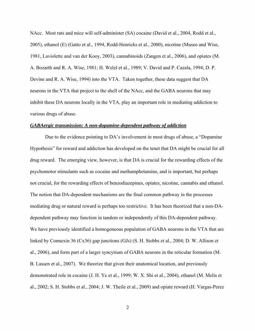
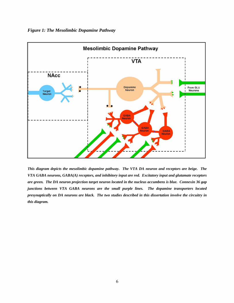
Figure 1: The Mesolimbic Dopamine Pathway
This diagram depicts the mesolimbic dopamine pathway. The VTA DA neuron and receptors are beige. The
VTA GABA neurons, GABA(A) receptors, and inhibitory input are red. Excitatory input and glutamate receptors
are green. The DA neuron projection target neuron located in the nucleus accumbens is blue. Connexin 36 gap
junctions between VTA GABA neurons are the small purple lines. The dopamine transporters located
presynaptically on DA neurons are black. The two studies described in this dissertation involve the circuitry in
this diagram.

7
CHAPTER 2
Cocaine Disinhibits Dopamine Neurons in the Ventral Tegmental Area via Use-
Dependent Blockade of GABA Neuron Voltage-Sensitive Sodium Channels
(The work presented in this chapter has been published under the same title in the
European Journal of Neuroscience 2008 Nov; 28(10):2028-40. The research diagramed in
Figures 1 and 2 of this chapter are not the work of the author, but have been included to maintain
continuity and context.)
INTRODUCTION
The mesocorticolimbic dopamine (DA) system originating in the ventral tegmental area
(VTA) and projecting to the nucleus accumbens (NAcc) has been implicated in motivated
behaviors, various types of reward, and in the habit-forming actions of addictive drugs including
cocaine (for review see (R. A. Wise, 2004)). The prevailing view is that cocaine’s locomotor
and reinforcing properties (D. C. S. Roberts et al., 1980; G. F. Koob et al., 1994) are mediated
primarily by enhancement of extracellular DA release (Y. L. Hurd et al., 1989; H. O. Pettit and J.
B. Justice, Jr., 1989, 1991; R. A. Wise et al., 1995; S. E. Hemby et al., 1997) via inhibition of the
DA transporter (DAT; (M. J. Kuhar et al., 1991; M. J. Kuhar, 1992; W. L. Woolverton and K. M.
Johnson, 1992)). Cocaine-induced cellular and molecular reshaping of this system may
contribute to learned reinforcement (for review see (S. Jones and A. Bonci, 2005)). The potency
of psychostimulants as positive reinforcers being correlated to their DAT binding affinity (M. C.
Ritz et al., 1987; J. Bergman et al., 1989; K. M. Wilcox et al., 1999), and cocaine’s high affinity
for the DAT (IC50 = 0.3-0.8 µM; (R. B. Rothman et al., 2001)), support this view.
8
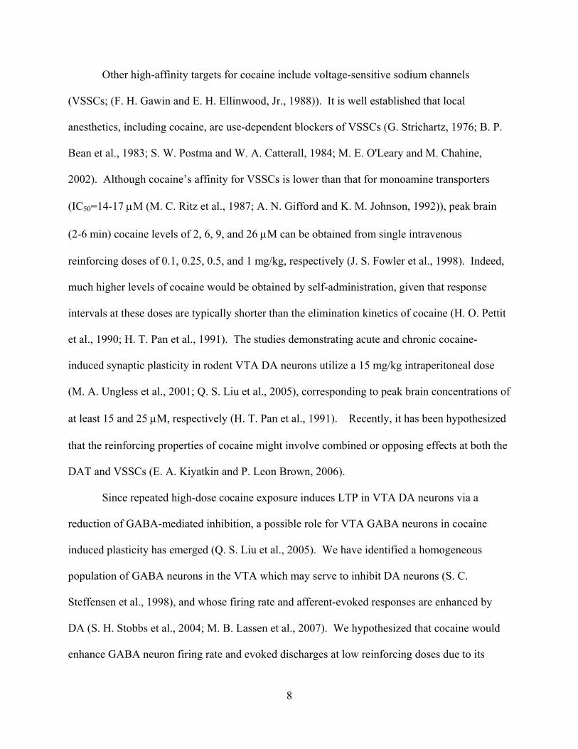
Other high-affinity targets for cocaine include voltage-sensitive sodium channels
(VSSCs; (F. H. Gawin and E. H. Ellinwood, Jr., 1988)). It is well established that local
anesthetics, including cocaine, are use-dependent blockers of VSSCs (G. Strichartz, 1976; B. P.
Bean et al., 1983; S. W. Postma and W. A. Catterall, 1984; M. E. O'Leary and M. Chahine,
2002). Although cocaine’s affinity for VSSCs is lower than that for monoamine transporters
(IC50=14-17 µM (M. C. Ritz et al., 1987; A. N. Gifford and K. M. Johnson, 1992)), peak brain
(2-6 min) cocaine levels of 2, 6, 9, and 26 µM can be obtained from single intravenous
reinforcing doses of 0.1, 0.25, 0.5, and 1 mg/kg, respectively (J. S. Fowler et al., 1998). Indeed,
much higher levels of cocaine would be obtained by self-administration, given that response
intervals at these doses are typically shorter than the elimination kinetics of cocaine (H. O. Pettit
et al., 1990; H. T. Pan et al., 1991). The studies demonstrating acute and chronic cocaine-
induced synaptic plasticity in rodent VTA DA neurons utilize a 15 mg/kg intraperitoneal dose
(M. A. Ungless et al., 2001; Q. S. Liu et al., 2005), corresponding to peak brain concentrations of
at least 15 and 25 µM, respectively (H. T. Pan et al., 1991). Recently, it has been hypothesized
that the reinforcing properties of cocaine might involve combined or opposing effects at both the
DAT and VSSCs (E. A. Kiyatkin and P. Leon Brown, 2006).
Since repeated high-dose cocaine exposure induces LTP in VTA DA neurons via a
reduction of GABA-mediated inhibition, a possible role for VTA GABA neurons in cocaine
induced plasticity has emerged (Q. S. Liu et al., 2005). We have identified a homogeneous
population of GABA neurons in the VTA which may serve to inhibit DA neurons (S. C.
Steffensen et al., 1998), and whose firing rate and afferent-evoked responses are enhanced by
DA (S. H. Stobbs et al., 2004; M. B. Lassen et al., 2007). We hypothesized that cocaine would
enhance GABA neuron firing rate and evoked discharges at low reinforcing doses due to its
9
DAT inhibiting properties, but at higher reinforcing doses its use-dependent VSSC blocking
effect would inhibit VTA GABA neurons leading to DA neuron disinhibition.
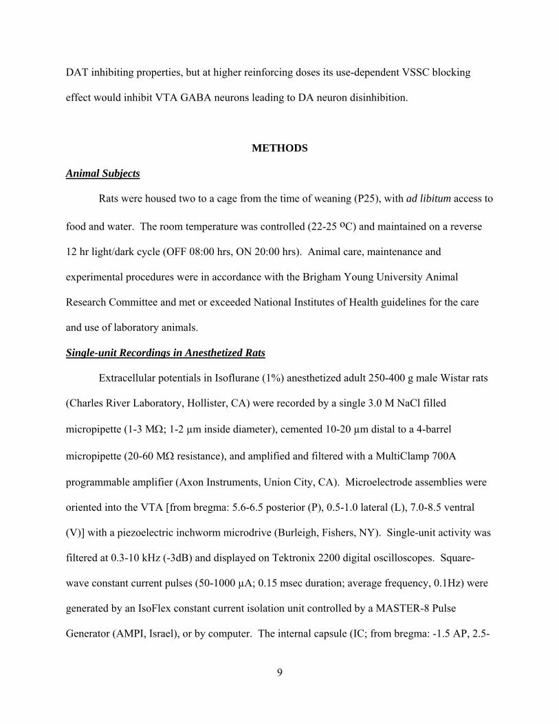
METHODS
Animal Subjects
Rats were housed two to a cage from the time of weaning (P25), with ad libitum access to
food and water. The room temperature was controlled (22-25 oC) and maintained on a reverse
12 hr light/dark cycle (OFF 08:00 hrs, ON 20:00 hrs). Animal care, maintenance and
experimental procedures were in accordance with the Brigham Young University Animal
Research Committee and met or exceeded National Institutes of Health guidelines for the care
and use of laboratory animals.
Single-unit Recordings in Anesthetized Rats
Extracellular potentials in Isoflurane (1%) anesthetized adult 250-400 g male Wistar rats
(Charles River Laboratory, Hollister, CA) were recorded by a single 3.0 M NaCl filled
micropipette (1-3 MΩ; 1-2 µm inside diameter), cemented 10-20 µm distal to a 4-barrel
micropipette (20-60 MΩ resistance), and amplified and filtered with a MultiClamp 700A
programmable amplifier (Axon Instruments, Union City, CA). Microelectrode assemblies were
oriented into the VTA [from bregma: 5.6-6.5 posterior (P), 0.5-1.0 lateral (L), 7.0-8.5 ventral
(V)] with a piezoelectric inchworm microdrive (Burleigh, Fishers, NY). Single-unit activity was
filtered at 0.3-10 kHz (-3dB) and displayed on Tektronix 2200 digital oscilloscopes. Square-
wave constant current pulses (50-1000 µA; 0.15 msec duration; average frequency, 0.1Hz) were
generated by an IsoFlex constant current isolation unit controlled by a MASTER-8 Pulse
Generator (AMPI, Israel), or by computer. The internal capsule (IC; from bregma: -1.5 AP, 2.5-
10
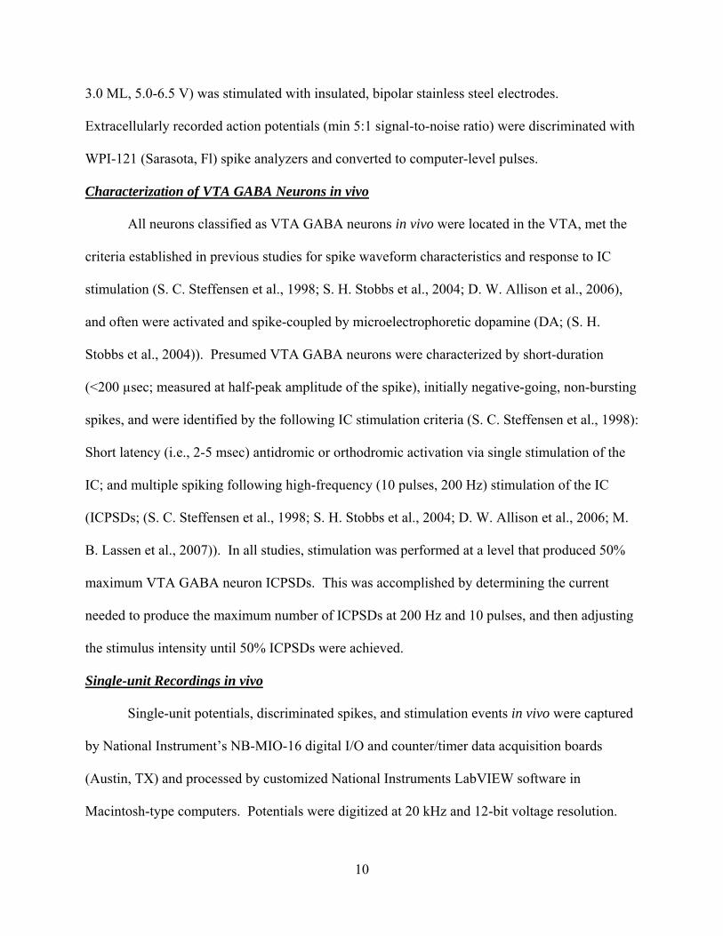
3.0 ML, 5.0-6.5 V) was stimulated with insulated, bipolar stainless steel electrodes.
Extracellularly recorded action potentials (min 5:1 signal-to-noise ratio) were discriminated with
WPI-121 (Sarasota, Fl) spike analyzers and converted to computer-level pulses.
Characterization of VTA GABA Neurons in vivo
All neurons classified as VTA GABA neurons in vivo were located in the VTA, met the
criteria established in previous studies for spike waveform characteristics and response to IC
stimulation (S. C. Steffensen et al., 1998; S. H. Stobbs et al., 2004; D. W. Allison et al., 2006),
and often were activated and spike-coupled by microelectrophoretic dopamine (DA; (S. H.
Stobbs et al., 2004)). Presumed VTA GABA neurons were characterized by short-duration
(<200 µsec; measured at half-peak amplitude of the spike), initially negative-going, non-bursting
spikes, and were identified by the following IC stimulation criteria (S. C. Steffensen et al., 1998):
Short latency (i.e., 2-5 msec) antidromic or orthodromic activation via single stimulation of the
IC; and multiple spiking following high-frequency (10 pulses, 200 Hz) stimulation of the IC
(ICPSDs; (S. C. Steffensen et al., 1998; S. H. Stobbs et al., 2004; D. W. Allison et al., 2006; M.
B. Lassen et al., 2007)). In all studies, stimulation was performed at a level that produced 50%
maximum VTA GABA neuron ICPSDs. This was accomplished by determining the current
needed to produce the maximum number of ICPSDs at 200 Hz and 10 pulses, and then adjusting
the stimulus intensity until 50% ICPSDs were achieved.
Single-unit Recordings in vivo
Single-unit potentials, discriminated spikes, and stimulation events in vivo were captured
by National Instrument’s NB-MIO-16 digital I/O and counter/timer data acquisition boards
(Austin, TX) and processed by customized National Instruments LabVIEW software in
Macintosh-type computers. Potentials were digitized at 20 kHz and 12-bit voltage resolution.

11
For single-unit activity, all spikes were captured by computer and time stamped. Spontaneous
firing rates were determined on- and off-line by calculating the number of events over a 5 min
epoch, typically 5 min before and at specific intervals after drug injection. Peri-stimulus and
interval-spike histograms were generated off-line using IGOR Pro (WaveMetrics, Lake Oswego,
OR) analysis of the time-stamped data. The duration (msec) and extent (#events/bin) of post-
stimulus permutation of ICPSDs was determined by rectangular integration at specific time
points on the peri-stimulus spike histogram using IGOR Pro analysis software. The minimum
bin width for peri-stimulus spike histograms was 1.0 msec and the number of bins was 1000.
These parameters allow for detection of all phases of pre- and post-stimulus spike activity.
Drug Preparation and Administration in vivo
Cocaine hydrochloride, cocaine methiodide, DA, lidocaine hydrochloride, and
methamphetamine hydrochloride were dissolved in 0.9% saline and administered intravenously
through an indwelling jugular catheter. Given the transient duration of effect of cocaine and
lidocaine on VTA GABA neuron firing rate and ICPSDs, dose-response studies were performed
for these two drugs, as well as for cocaine methiodide, in the same rats with a 40 min interval
between each dose and by randomizing the sequence of dose levels, while dose-response studies
for amphetamine, whose effects on firing rate were more prolonged, were performed in separate
rats. For in situ microelectrophoretic application of drugs in the VTA, DA (10 mM), eticlopride
hydrochloride (1.0 mM), SCH23390 hydrochloride (1.0 mM), SKF38393 hydrochloride (1.0
mM), and quinpirole hydrochloride (1 mM) were dissolved in distilled water and iontophoresed
by current injection (25-100 nA) which was regulated by Medical Systems BH-2 iontophoretic
pump and balance unit modules. For systemic drug studies on VTA GABA neuron responses,
all drugs including cocaine were administered intravenously through a jugular catheter. For
12
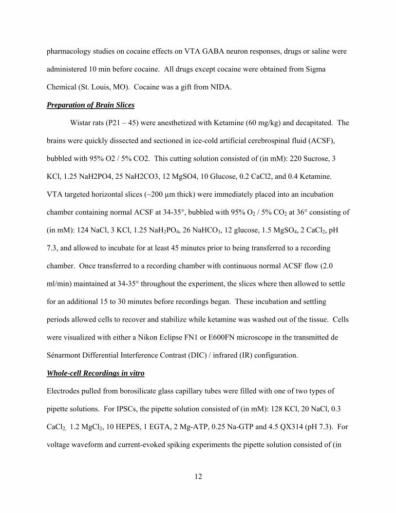
pharmacology studies on cocaine effects on VTA GABA neuron responses, drugs or saline were
administered 10 min before cocaine. All drugs except cocaine were obtained from Sigma
Chemical (St. Louis, MO). Cocaine was a gift from NIDA.
Preparation of Brain Slices
Wistar rats (P21 – 45) were anesthetized with Ketamine (60 mg/kg) and decapitated. The
brains were quickly dissected and sectioned in ice-cold artificial cerebrospinal fluid (ACSF),
bubbled with 95% O2 / 5% CO2. This cutting solution consisted of (in mM): 220 Sucrose, 3
KCl, 1.25 NaH2PO4, 25 NaH2CO3, 12 MgSO4, 10 Glucose, 0.2 CaCl2, and 0.4 Ketamine.
VTA targeted horizontal slices (~200 µm thick) were immediately placed into an incubation
chamber containing normal ACSF at 34-35°, bubbled with 95% O2 / 5% CO2 at 36° consisting of
(in mM): 124 NaCl, 3 KCl, 1.25 NaH2PO4, 26 NaHCO3, 12 glucose, 1.5 MgSO4, 2 CaCl2, pH
7.3, and allowed to incubate for at least 45 minutes prior to being transferred to a recording
chamber. Once transferred to a recording chamber with continuous normal ACSF flow (2.0
ml/min) maintained at 34-35° throughout the experiment, the slices where then allowed to settle
for an additional 15 to 30 minutes before recordings began. These incubation and settling
periods allowed cells to recover and stabilize while ketamine was washed out of the tissue. Cells
were visualized with either a Nikon Eclipse FN1 or E600FN microscope in the transmitted de
Sénarmont Differential Interference Contrast (DIC) / infrared (IR) configuration.
Whole-cell Recordings in vitro
Electrodes pulled from borosilicate glass capillary tubes were filled with one of two types of
pipette solutions. For IPSCs, the pipette solution consisted of (in mM): 128 KCl, 20 NaCl, 0.3
CaCl2, 1.2 MgCl2, 10 HEPES, 1 EGTA, 2 Mg-ATP, 0.25 Na-GTP and 4.5 QX314 (pH 7.3). For
voltage waveform and current-evoked spiking experiments the pipette solution consisted of (in
13
mM): 115 K-Gluconate, 9 NaCl, 25 KCl, 10 HEPES, 0.2 EGTA, 1.2 MgCl2, 3 Na-ATP, 1 Na-
GTP, and had resistances of 2-4 MΩ. Series resistance (Ra) typically 10 to 20 MΩ, and input
resistance (Rm) typically 300 to 400 MΩ, were continuously monitored with a 10 mV voltage
step delivered at 0.1 Hz throughout each experiment and only experiments that maintained stable
Ra and Rm (less than 15% change) were included in this study. IPSCs were filtered at 2 kHz
while voltage waveform-generated currents and current-drive spikes were filtered at 6 kHz using
an Axon Instruments Multiclamp 700A or 700B amplifier and digitized at 5-20 kHz,
respectively, using an Axon 1440A digitizer, and collected and analyzed using pClamp10 and
Igor Pro (Wavemetrics: Oswego, OR) software packages. Evoked and spontaneous IPSCs were
recorded in the presence of 100 µM D-L 2-amino-5-phosphonopentanoic acid (APV), 30 µM 6-
cyano-23-dihydroxy-7-nitro-quinoxaline (CNQX), and 100 nM eticlopride to block NMDA,
AMPA, and DA D2-mediated synaptic currents (A. Bonci and J. T. Williams, 1997),
respectively. Miniature IPSCs (mIPSCs) were isolated from all other spontaneous IPSCs by
addition of 0.5 µM TTX. To evoke IPSCs, cells were stimulated at 0.1 Hz with a stainless steel-
platinum/iridium concentric bipolar stimulating electrode placed ~100 µm rostral to the
recording electrode. Evoked IPSCs were inward at the holding potential of -70 mV and were
completely blocked by picrotoxin (100 µM). Evoked IPSC amplitudes were calculated by taking
the difference between the 1.0 msec window around the peak and the 5.0 msec baseline window
immediately preceding the stimulation artifact. Spontaneous IPSC activity amplitude and
frequency were calculated the same for both sIPSCs and mIPSCs; the average amplitude or
frequency during a 2 min period 8-10 min following drug were normalized to the average
amplitude or frequency from a 2 min window prior to drug.
Characterization of Neuron Types in vitro
14
Traditionally, neurons have been classified as either primary (DAergic) and secondary
(GABAergic) based on electrophysiological and pharmacological properties (A. A. Grace and S.
P. Onn, 1989; S. W. Johnson and R. A. North, 1992; D. L. Cameron et al., 1997; M. A. Ungless
et al., 2004). Despite extensive research, no single best electrophysiological characteristic has
been identified to conclusively distinguish VTA DA neurons from other neurons in the VTA.
The most widely accepted electrophysiological method to distinguish DA neurons from non-DA
neurons has been the presence of a non-cation specific inward rectifying current (Ih). However,
not all Ih(+) neurons stain positive for tyrosine hydroxylase (TH), a molecular marker specific to
DA neurons, while Ih(-) cells appear to stain negative for TH (S. W. Johnson and R. A. North,
1992; D. W. Allison et al., 2006; E. B. Margolis et al., 2006). The combination of several
electrophysiological characteristics, depending on experiment type, were used to distinguish
putative DA from putative non-DA neurons in this study. Specifically: neurons that exhibited a
modest non-cation specific inward rectifying current (Ih) in combination with spike
accommodation, low input resistance, higher spike threshold, and shorter spike duration were
assumed to be DA neurons. Neurons that exhibited a complete lack of Ih in combination with
high input resistance, lack of spike accommodation, and low spike threshold were assumed to be
non-dopminergic (or putative GABA neurons) (S. W. Johnson and R. A. North, 1992; D. W.
Allison et al., 2006; E. B. Margolis et al., 2006). A recent anatomical study has demonstrated the
presence of glutamatergic neurons in the VTA and surrounding areas (T. Yamaguchi et al.,
2007). However, this subpopulation of VTA cells remains uncharacterized and the highest
concentration of vesicular transport-2 mRNA is in the rostral medial aspects of the VTA, with
the lowest density in the parabrachial pigmental area (PBP). Most of the in vitro recordings in
this study were conducted in the PBP in order to maintain uniformity with previous work. To

15
provide additional assurance that the characterization criteria described above effectively
discriminates VTA DA neurons from non-DA neurons (putative GABA neurons), we performed
a separate set of experiments to link these electrophysiological criteria to the presence or absence
of tyrosine hydroxylase (TH) mRNA, a marker for DA neurons. This was accomplished using
quantitative real-time PCR.
Single-cell Quantitative RT-PCR
Following electrophysiological characterization, putative VTA GABA neurons and
putative DA neurons in mature rats were aspirated under visual observation by application of
suction attached to the recording pipette, and were immediately added to a reverse transcription
(RT) reaction mixture. The iScript cDNA synthesis kit (Biorad) was used for a total volume of
10 µl per reaction. Reactions were run at 25°C for 10 min, 42°C for 60 min, and 95°C for 5 min
in a PTC-200 thermal cycler (MJ Research Inc., Watertown MA). Reactions were then stored at -
20°C until running the PCR. A preamplification round of multiplex PCR was performed by
adding iTAQ Supermix with ROX (Biorad) and a cocktail of primers to the completed RT
reaction, for a final volume of 50 µL. The reactions were held at 94°C for 30 seconds then cycled
20 times. Each cycle consisted of: 92°C for 15 seconds, 60°C for 20 seconds, and 72°C for 30
seconds. One µl samples of the initial multiplex PCR were then used as substrate for each
reaction in the subsequent real-time quantitative PCR. Real-Time quantitative PCR using gene
specific primers with FAM-TAMRA TaqMan® probes (Applied Biosystems; TH plus primer:
CTTCCAGTACAAGCACGGTGAA, TH minus primer: AGCGTGACATATA-
CCTCCTTCCA, and TH probe: CCCCATGTGGAATACACAGCGGAAGAG; D2 plus primer:
CGCAGAAAGCTCTCCCAGCAGA, D2 minus primer:
GACTGGTGGGATGTTGCAATCACA and D2 probe: CCATTGTTCTCGGTGTGTTCA; 18s
16
plus primer: GTGCATGGCCGTTCTTAGTTG, minus 18s primer:
GCCACTTGTCCCTCTAAGAAGTTG, and 18s probe:
TGGAGCGATTTGTCTGGTTAATTCCGATAAC) were performed using the iTaq Supermix
with ROX (Bio-Rad) with an iCycler IQ (Bio-Rad) real-time PCR System. Samples were
amplified in triplicate, together with a negative control for each subunit (an ACSF-only
aspiration taken from the brain slice recording chamber when the cells were aspirated). The
amplification protocol was 50°C for 2 minutes, 95°C for 5 minutes, then 50 cycles of 95°C for
15 seconds, 60°C for 20 seconds, and 72°C for 30 seconds. Cycle threshold (Ct) values were
calculated automatically by the iCycler IQ software, with threshold values set between 5 and 20.
Relative fold expression was calculated using the 2-∆∆CT method as described in (K. J. Livak and
T. D. Schmittgen, 2001).
Voltage Waveform Command
In order to mimic in VTA GABA neurons recorded in vitro the frequency and underlying
ionic currents linked to high frequency spontaneous spiking and current-evoked postsynaptic
discharges elicited from VTA GABA neurons recorded in vivo a “typical” train of high
frequency current-evoked spikes was used as a “voltage waveform command.” The voltage
waveform command was derived from a recording of current-evoked spikes obtained in current
clamp mode with a spiking frequency of 172 Hz evoked at 250 pA. This voltage waveform
command was used due to the length of time required to generate a voltage-clamp protocol from
a recorded current-clamp waveform for each neuron (R. Llinas et al., 1982; M. T. Do and B. P.
Bean, 2003; A. Enomoto et al., 2006). The resulting data obtained in voltage-clamp mode was a
train of whole-current events with inward TTX-sensitive (0.5 µM) currents that followed the
voltage waveform command closely. In order to subtract passive membrane and capacitative
17
currents from the ionic current trace acquired from the voltage waveform (baseline -55 mV) the
same waveform was applied sub-threshold (-120 mV), and the resulting current trace was
subtracted from the current trace acquired at -55 mV. Three measurements were then obtained
from the remaining current trace: 1) The amplitude of the first inward current event; 2) The
average of all, or total inward current; and 3) The average total inward current of the last third
(25 current events), or plateau phase of the trace. Currents are presented as percent of control 8
minutes following cocaine application to the ACSF. Due to space clamp problems common to
slice preparations, no measurements of inward current kinetics were made. The voltage
waveform command protocol utilized in this study was designed to clamp frequency, while
imitating natural spike dynamics. We found that when Ra and Rm remained stable, and the time
between the applications of the voltage waveform command protocol was not less than 2
minutes, waveform-generated current amplitudes remained constant and exhibited little sign of
run-down.
Statistical Analyses
For in vivo and in vitro studies, the results for control and drug treatment groups were
derived from calculations performed on VTA GABA neuron spontaneous firing rate, ICPSDs,
current-evoked spikes, sodium currents, DA neuron evoked and spontaneous IPSCs, and mean
relative fold expression differences of TH and D2 mRNA. A paired two sample for means t test
was performed to determine statistical significance for all measures. Welch’s correction was
applied to RT PCR data due to unequal variances. All results, except for paired pulse, in text are
presented as the mean ± SEM and the t distribution variance for a two-tailed level of confidence
of 95%. Paired pulse results are presented as one-tailed 95% confidence interval. Results in
Figures are presented as mean ± SEM; P < 0.05 was taken as indicating statistical significance.

18
Analysis software included Microsoft Excel and Igor Pro (Wavemetrics, Oswego, OR) Stat Pak.
Figures were constructed with Igor Pro software.
RESULTS
Effects of Systemic Cocaine, Cocaine Methiodide, Dopamine, Lidocaine and
Methamphetamine on VTA GABA Neuron Firing Rate in vivo
Fifty three VTA GABA neurons were tested in vivo for their sensitivity to intravenous
administration of cocaine, cocaine methiodide, DA, lidocaine or methamphetamine. Initially, we
hypothesized that reinforcing doses of cocaine would enhance VTA GABA neuron firing rate
similar to microelectrophoretic DA (S. H. Stobbs et al., 2004), given its DAT inhibiting
properties. However, unexpectedly, cocaine (0.25-2 mg/kg) had somewhat variable, but
significant dose-dependent effects on firing rate. Cocaine significantly increased VTA GABA
neuron firing at doses at or below 0.5 mg/kg and significantly decreased firing rate at doses
above 0.5 mg/kg (Figure 1A,B, 0.25 mg/kg: +56 ± 6.1 %, P<0.01, t(2,7)=2.3; 0.5 mg/kg: +68 ±
6.9 %, P<0.01, t(2,9)=2.2; 1.0 mg/kg: -18 ± 8 %, P<0.05, t(2,24)=2.0; 2.0 mg/kg: -31 ± 7.2 %,
P<0.05, t(2,7)=2.3; mean saline firing rate = 31.4 ± 2.84 Hz; n=25), compared to saline control. In
order to determine if cocaine’s well-known peripheral cardiovascular pressor effects (C. W.
Schindler et al., 1995) or temperature effects (E. A. Kiyatkin and P. Leon Brown, 2006) might
have contributed to cocaine effects on VTA GABA neuron firing rate, we evaluated the effects
of intravenous DA, which has well-known pressor effects, and cocaine methiodide, which does
not cross the blood-brain barrier, but has cocaine’s peripheral temperature and blood pressure
effects (D. A. Shriver and J. P. Long, 1971; E. A. Kiyatkin and P. Leon Brown, 2006).
Intravenous administration of DA (3 mg/kg) increased mean arterial pressure 223%, respiratory

19
rate, and induced piloerection, but had no effect on VTA GABA neuron firing rate (P>0.05,
t(2,4)=2.7; n=5). Intravenous administration of cocaine methiodide did not significantly alter
firing rate at any dose level tested (Figure 1B; 0.25 mg/kg: -1 ± 9 %, P>0.24, t(2,4)=2.7; 0.5
mg/kg: -2.2 ± 1.3 %, P>0.05, t(2,4)=2.7; 1.0 mg/kg: +5.5 ± 4.5 %, P>0. 05, t(2,4)=2.7; 2.0 mg/kg: -
1.3 ± 1.3 %, P>0.05, t(2,4)=2.7; mean saline firing rate = 26.3 ± 4.73; n=5), compared to saline
control. Intravenous administration of lidocaine, a VSSC blocker with negligible DAT activity,
significantly decreased VTA GABA neuron firing rate at all doses tested (Figure 1B; 0.25
mg/kg: -18 ± 5.8 %, P<0.05, t(2,8)=2.3; 0.5 mg/kg: -26%, P<0.05, t(2,6)=2.4; 1.0 mg/kg: -32 ± 6 %,
P<0.05, t(2,8)=2.3; 2.0 mg/kg: -38 ± 5.8 %, P<0.01, t(2,6)=2.4; mean saline firing rate = 26.3 ± 4.73
Hz; n=9), compared to saline control. Intravenous administration of methamphetamine, a DAT
inhibitor with negligible VSSC activity, significantly increased VTA GABA neuron firing rate at
all dose levels tested (Figure 1B; 0.25 mg/kg: +45 ± 11.6 %, P<0.05, t(2,4)=2.7; 0.5 mg/kg: +92 ±
22 %, P<0.05, t(2,4)=2.7; 1.0 mg/kg: +140 ± 32 %, P<0.05, t(2,4)=2.7; 2.0 mg/kg: +176 ± 28 %,
P<0.05, t(2,4)=2.7; mean saline firing rate = 22.2 ± 3.93 Hz; n=9), compared to saline control.
Dopamine Pharmacology of Cocaine Effects on VTA GABA Neuron Firing Rate in vivo
Of the forty eight VTA GABA neurons tested in vivo for sensitivity to
microelectrophoretically applied DA, 46% of them were activated by DA (S. H. Stobbs et al.,
2004; M. B. Lassen et al., 2007). We evaluated the DA pharmacology of 22 of these neurons.
Microelectrophoretic application of DA (Figure 1C; + 50 nA; P<0.001, t(2,21)=5.0; n=22), or the
D2 agonist quinpirole (+50 nA; P<0.001, t(2,14)=4.31; n=15; data not shown) significantly
increased the firing rate of VTA GABA neurons 162 % and 140 %, compared to saline control
(mean saline firing rate = 27.3 ± 4.1 Hz; + 50 nA ejection current). Neither the D1 agonist
SKF38393 nor the D1 antagonist SCH23390 (+ 50 nA; P>0.05, n=4) significantly altered the
20
firing rate of VTA GABA neurons, compared to saline control (data not shown). In situ
microelectrophoretic administration of eticlopride blocked DA activation of firing rate, and iv
administration of eticlopride (1.0 mg/kg) blocked low-dose (0.5 mg/kg) iv cocaine activation, but
not higher dose iv cocaine inhibition, of VTA GABA neuron firing rate P<0.05; n=6; Figure
1C).
Effects of Systemic Cocaine, Cocaine Methiodide, Dopamine, Lidocaine and
Methamphetamine on VTA GABA Neuron Evoked Spikes in vivo
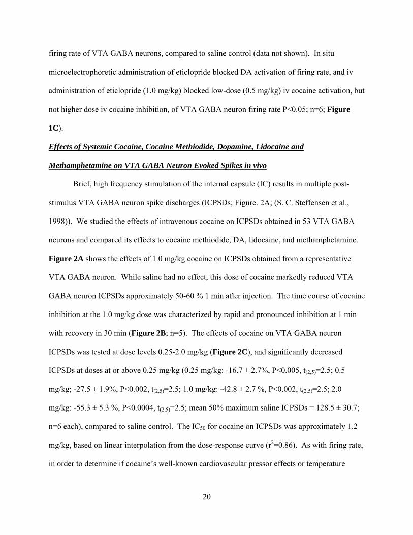
Brief, high frequency stimulation of the internal capsule (IC) results in multiple post-
stimulus VTA GABA neuron spike discharges (ICPSDs; Figure. 2A; (S. C. Steffensen et al.,
1998)). We studied the effects of intravenous cocaine on ICPSDs obtained in 53 VTA GABA
neurons and compared its effects to cocaine methiodide, DA, lidocaine, and methamphetamine.
Figure 2A shows the effects of 1.0 mg/kg cocaine on ICPSDs obtained from a representative
VTA GABA neuron. While saline had no effect, this dose of cocaine markedly reduced VTA
GABA neuron ICPSDs approximately 50-60 % 1 min after injection. The time course of cocaine
inhibition at the 1.0 mg/kg dose was characterized by rapid and pronounced inhibition at 1 min
with recovery in 30 min (Figure 2B; n=5). The effects of cocaine on VTA GABA neuron
ICPSDs was tested at dose levels 0.25-2.0 mg/kg (Figure 2C), and significantly decreased
ICPSDs at doses at or above 0.25 mg/kg (0.25 mg/kg: -16.7 ± 2.7%, P<0.005, t(2,5)=2.5; 0.5
mg/kg; -27.5 ± 1.9%, P<0.002, t(2,5)=2.5; 1.0 mg/kg: -42.8 ± 2.7 %, P<0.002, t(2,5)=2.5; 2.0
mg/kg: -55.3 ± 5.3 %, P<0.0004, t(2,5)=2.5; mean 50% maximum saline ICPSDs = 128.5 ± 30.7;
n=6 each), compared to saline control. The IC50 for cocaine on ICPSDs was approximately 1.2
mg/kg, based on linear interpolation from the dose-response curve (r2=0.86). As with firing rate,
in order to determine if cocaine’s well-known cardiovascular pressor effects or temperature
21
effects might contribute to cocaine reduction of VTA GABA neuron ICPSDs, we evaluated the
effects of intravenous DA and cocaine methiodide. Intravenous administration of DA (3 mg/kg)
had no effect on VTA GABA neuron ICPSDs (P>0.05, n=4). Similarly, intravenous
administration of cocaine methiodide did not significantly affect ICPSDs at any dose level tested
(Figure 2C; 0.25 mg/kg: -2 ± 2 %, P<0.24, t(2,5)=2.5; 0.5 mg/kg: -11 ± 2.7 % P<0.18, t(2,5)=2.5;
1.0 mg/kg: -6 ± 1.7 %, P<0.105, t(2,5)=2.5; 2.0 mg/kg: -10.1 ± 3.5 %, P<0.11, t(2,5)=2.5; mean
50% maximum saline ICPSDs = 121.5 ± 4.2; n=6), compared to saline control. Since local DA
markedly enhanced firing rate (Figure 1; (S. H. Stobbs et al., 2004)), we evaluated the effects of
local application of DA on ICPSDs. Compared to saline control, microelectrophoretic
application of DA (+ 50 nA) did not significantly alter ICPSDs (P>0.5; mean 50% maximum
saline ICPSDs = 60.6 ± 4.4; n=21 each). Intravenous administration of lidocaine markedly
decreased ICPSDs at all doses tested (Figure 2C; 0.25 mg/kg: -18.3 ± 2.4 %, P<0.005, t(2,5)=2.5;
0.5 mg/kg: -25.6 ± 1.7 %, P<0.0002, t(2,5)=2.5; 1.0 mg/kg: -41.9 ± 2 %, P<0.0001, t(2,5)=2.5; 2.0
mg/kg: -62.6 ± 3.1 %, P<0.00003, t(2,5)=2.5; mean 50% maximum saline ICPSDs = 136.9 ± 14.3;
n=6), compared to saline control. The IC50 for lidocaine on ICPSDs was approximately 1.2
mg/kg, based on linear interpolation from the dose-response curve (r2=0.91). Intravenous
administration of methamphetamine did not significantly alter VTA GABA neuron ICPSDs
(Figure 2C; 0.25 mg/kg: -10 ± 5 %, P>0.05, t(2,4)=2.7; 0.5 mg/kg: -5 ± 6.2 %, P>0.05, t(2,3)=3.1;
1.0 mg/kg: -8 ± 5.2 %, P>0.05, t(2,3)=3.1; mean 50% maximum saline ICPSDs = 74.9 ± 9.5;
n=15), compared to saline control.
Dopamine Pharmacology of Cocaine Effects on VTA GABA Neuron Evoked Spikes in vivo
In order to evaluate the role of DA in mediating cocaine inhibition of VTA GABA
ICPSDs we tested the effects of select DA receptor subtype antagonists on cocaine inhibition of

22
VTA GABA neuron ICPSDs at the 1.0 mg/kg dose level 1 min after injection, and compared to
iv saline (Figure 2D). Intravenous administration of the D1/D5 DA receptor antagonist
SCH23390 (1.0 mg/kg) had no effect on VTA GABA neuron ICPSDs (104.2 ± 7.9 %; P>0.05;
n=5), and did not alter cocaine’s ability to reduce ICPSDs at the 1.0 mg/kg dose level (56.2 ± 4.0
%) when administered 10 min after injection of SCH23390. Similarly, iv administration of the
D2/D3 antagonist eticlopride (1.0 mg/kg) had no effect on VTA GABA neuron ICPSDs (102.7 ±
6.6 %; P>0.05; n=5), and did not alter cocaine’s ability to reduce ICPSDs (51 ± 2.1 %) when
administered 10 min after injection of eticlopride.
Expression of Select Genes in Dopamine vs GABA Neurons in Mature Rats: Quantitative
Single-cell RT-PCR
In order to further evaluate the role of D2 receptors in DA activation of VTA GABA
neurons, we evaluated the quantitative expression of D2 receptor gene products in these neurons.
In addition, in order to validate the electrophysiological criteria used to distinguish VTA
neurons, we compared the expression of TH in VTA GABA and DA neurons. Figure 3
summarizes the differences between VTA DA and GABA neurons for the expression of TH and
D2 mRNA transcripts in adult male rats. Tyrosine hydroxylase expression in DA neurons was
significantly greater than GABA neurons (P=0.0002, t(1,23)=4.5; mean DA neuron TH expression
= 6.12 ± 0.77 and mean GABA neuron TH expression = 0.07 ± 0.01; n=33). The cycle threshold
for 18s, the housekeeping gene that was used for the quantification of the relative expression of
each of the gene products, was not significantly different between DA and GABA neurons
(P=0.35, t(1,31)=0.95; mean DA cell 18s CT = 12.53 ± 0.18 and mean GABA cell 18s CT = 12.87
± 0.34). The expression of D2 was significantly greater in DA neurons than GABA neurons
(P=0.019; mean DA cell D2 expression = 3.39 ± 0.59 and mean GABA cell D2 expression =
23
0.26 ± 0.03; n=29). The cycle threshold for 18s was not significantly different between groups
(P=0.82, t(1,27)=0.23; mean DA cell 18s CT = 12.53 ± 0.18 and mean GABA cell 18s CT = 12.63
± 0.35).
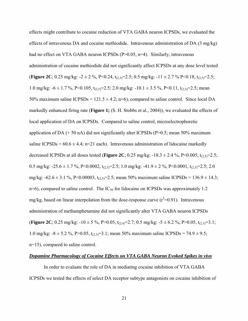
Effects of Cocaine on Current-Evoked VTA GABA Neuron Spikes in vitro
Based on in vivo studies, cocaine did not appear to be acting through DA to reduce VTA
GABA neuron firing rate or ICPSDs. Thus, we tested the effect of cocaine on VTA GABA
neuron current-evoked spiking in vitro to determine whether cocaine might reduce spiking
similar to ICPSDs in vivo. VTA GABA neurons displayed an average spiking frequency of 22.8
± 9.9 Hz at threshold (25 pA) and 180 ± 49.7 Hz at the maximum current step (400 pA). Not
surprisingly, 50 µM cocaine markedly reduced spiking at current levels 100-400 pA (85.7 ± 13.8
Hz, P < 0.05, t(2,4)=2.8 at 400 pA; Figure 4AB). Since cocaine reduced current-evoked spikes in
vitro similar to ICPSDs in vivo we postulated that cocaine was blocking ICPSDs, current-evoked
spikes, and reducing firing rate via its VSSC blocking properties. To provide evidence to
support this assumption, we next examined cocaine’s effects of VTA GABA neuron sodium
currents.
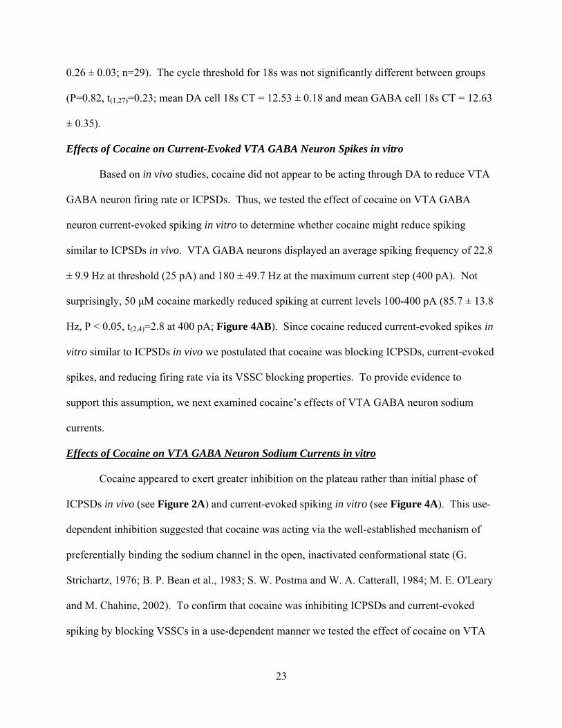
Effects of Cocaine on VTA GABA Neuron Sodium Currents in vitro
Cocaine appeared to exert greater inhibition on the plateau rather than initial phase of
ICPSDs in vivo (see Figure 2A) and current-evoked spiking in vitro (see Figure 4A). This use-
dependent inhibition suggested that cocaine was acting via the well-established mechanism of
preferentially binding the sodium channel in the open, inactivated conformational state (G.
Strichartz, 1976; B. P. Bean et al., 1983; S. W. Postma and W. A. Catterall, 1984; M. E. O'Leary
and M. Chahine, 2002). To confirm that cocaine was inhibiting ICPSDs and current-evoked
spiking by blocking VSSCs in a use-dependent manner we tested the effect of cocaine on VTA

24
GABA neuron whole-currents elicited using a voltage waveform command protocol (R. Llinas et
al., 1982; M. T. Do and B. P. Bean, 2003; A. Enomoto et al., 2006). The voltage waveform
command protocol was created from a typical voltage trace obtained in current clamp mode
(Figure 5Ai) of a VTA GABA neuron driven to fire at 172 Hz, a frequency consistent with what
we have previously determined for ICPSDs in vivo (M. B. Lassen et al., 2007) at 50% maximum
stimulus level, and at sub-max current levels for spikes driven in current-clamp mode. This
current-clamp derived voltage waveform command protocol has the advantage of clamping both
spike dynamics and frequency, compared to a sequence of square waves. All inward current
events obtained using the voltage waveform command were abolished by TTX (0.5 µM),
indicating that they were sodium currents. Figure 5Aii-iv illustrates the effects of cocaine (7
µM) and TTX (0.5 µM) on sodium currents evoked by the voltage waveform command in a
typical VTA GABA neuron. We postulated that if cocaine was blocking VSSCs in a use-
dependent manner, suggested by the effect observed on current-evoked spikes, cocaine would
have its greatest effect during the plateau phase of the waveform trace. We therefore measured
cocaine’s effect on the amplitude of the first inward current event, the mean of all or total inward
current, and the mean inward current of the last 25 events- the plateau phase of the trace (Figure
5B,C). The superimposed traces of the first current event and last 5 current events in panel
Figure 5B taken from the raw traces in Figure 5A illustrate this. Cocaine was tested at
concentration levels 3, 7, 15 and 50 µM and significantly reduced VTA GABA neuron TTX-
sensitive sodium currents in a concentration-dependent manner, exerting its greatest VSSC-
blocking effect at each dose on the plateau phase of the trace (percent reduction of plateau phase
inward current: 3 µM, 25.8 ± 2.9%, P < 0.001, t(2,4)=2.7; 7 µM, 36.6 ± 3.3%, P < 0.01, t(2,3)=3.1;
15 µM, 69.1 ± 4.6%, P < 0.001, t(2,3)=3.1; 50 µM, 76.9 ± 4.5%; P < 0.0001, t(2,5)=2.5; Figure
25
5C). Cocaine’s IC50 on sodium currents in VTA GABA neurons was calculated to be 13 µM,
using a least squares fit line to the points generated from 3, 7, 15 µM, (r2 = 0.90, y = -3.1x +
90.4).
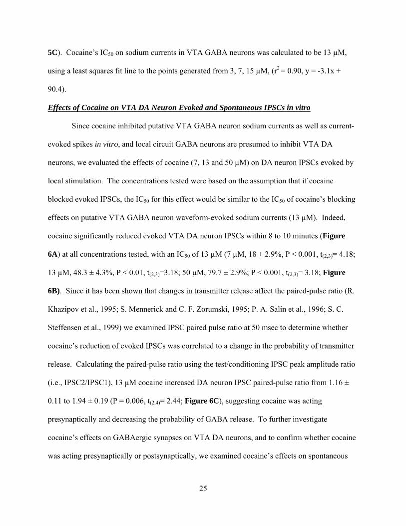
Effects of Cocaine on VTA DA Neuron Evoked and Spontaneous IPSCs in vitro
Since cocaine inhibited putative VTA GABA neuron sodium currents as well as current-
evoked spikes in vitro, and local circuit GABA neurons are presumed to inhibit VTA DA
neurons, we evaluated the effects of cocaine (7, 13 and 50 µM) on DA neuron IPSCs evoked by
local stimulation. The concentrations tested were based on the assumption that if cocaine
blocked evoked IPSCs, the IC50 for this effect would be similar to the IC50 of cocaine’s blocking
effects on putative VTA GABA neuron waveform-evoked sodium currents (13 µM). Indeed,
cocaine significantly reduced evoked VTA DA neuron IPSCs within 8 to 10 minutes (Figure
6A) at all concentrations tested, with an IC50 of 13 µM (7 µM, 18 ± 2.9%, P < 0.001, t(2,3)= 4.18;
13 µM, 48.3 ± 4.3%, P < 0.01, t(2,3)=3.18; 50 µM, 79.7 ± 2.9%; P < 0.001, t(2,3)= 3.18; Figure
6B). Since it has been shown that changes in transmitter release affect the paired-pulse ratio (R.
Khazipov et al., 1995; S. Mennerick and C. F. Zorumski, 1995; P. A. Salin et al., 1996; S. C.
Steffensen et al., 1999) we examined IPSC paired pulse ratio at 50 msec to determine whether
cocaine’s reduction of evoked IPSCs was correlated to a change in the probability of transmitter
release. Calculating the paired-pulse ratio using the test/conditioning IPSC peak amplitude ratio
(i.e., IPSC2/IPSC1), 13 µM cocaine increased DA neuron IPSC paired-pulse ratio from 1.16 ±
0.11 to 1.94 ± 0.19 (P = 0.006, t(2,4)= 2.44; Figure 6C), suggesting cocaine was acting
presynaptically and decreasing the probability of GABA release. To further investigate
cocaine’s effects on GABAergic synapses on VTA DA neurons, and to confirm whether cocaine
was acting presynaptically or postsynaptically, we examined cocaine’s effects on spontaneous

26
action potential-dependent GABAergic IPSCs (sIPSCs). Cocaine (13 µM) reduced sIPSC
frequency 33.7 ± 7.2% (P= 0.018, t(2,3)= 3.18; Figure 6D), but did not significantly affect sIPSC
amplitude (control, 37 ± 10.2 pA; n=4; cocaine, 40.4 ± 9.1 pA; n=4; p=0.31; Figure 6D).
Finally, in order to determine whether cocaine was reducing the action potential-dependent or
independent components of sIPSC activity, or both, we sought to isolate this effect by examining
the action potential-independent, TTX-insensitive or miniature IPSC (mIPSC) component alone.
Cocaine did not significantly affect mIPSC frequency or amplitude (frequency: control, 2.78 ±
0.48 Hz; n=5; cocaine, 2.61 ± 0.46 Hz; n=5; p=0.3; amplitude: control, 33.8 ± 2.3 pA; n=5;
cocaine, 33.4 ± 2.7 pA; n=5; p=0.88; Figure 6E), suggesting that cocaine was acting
presynaptically as a VSSC blocker to reduce the action potential-dependent component of
spontaneous IPSC activity.
27
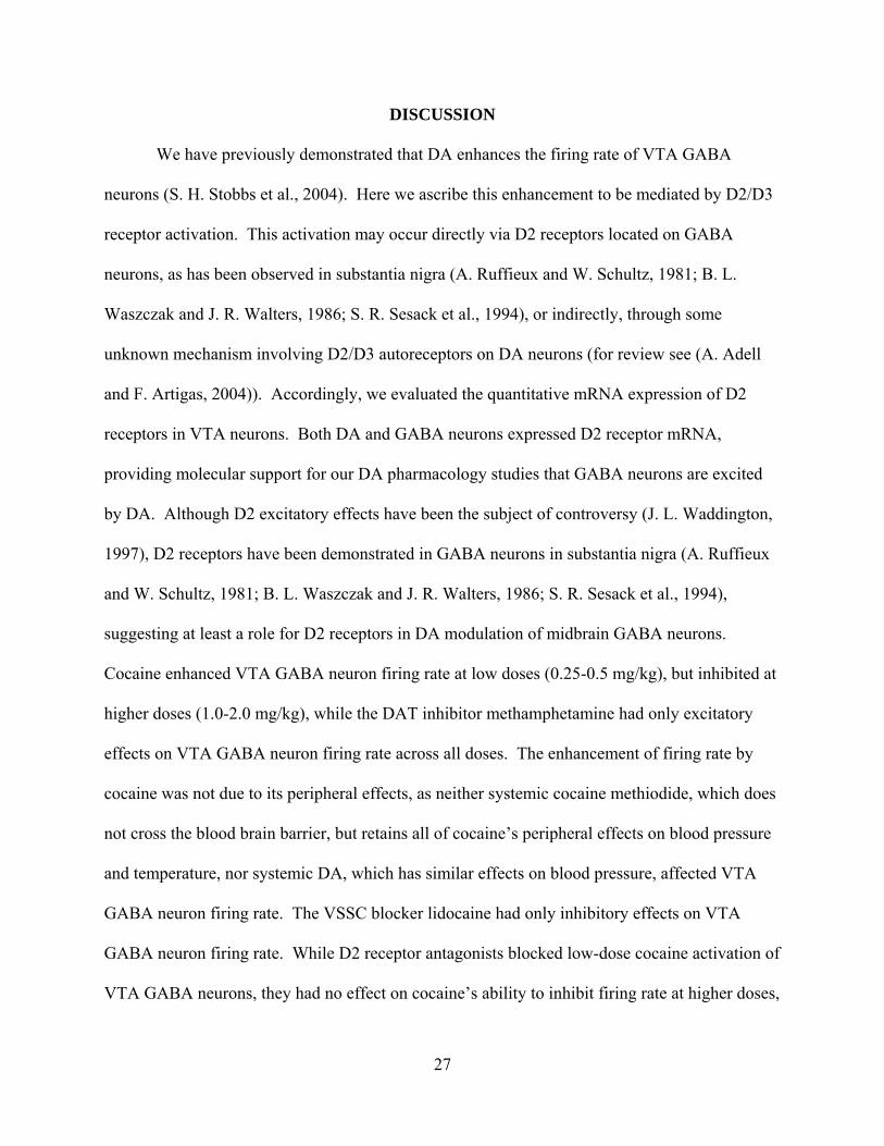
DISCUSSION
We have previously demonstrated that DA enhances the firing rate of VTA GABA
neurons (S. H. Stobbs et al., 2004). Here we ascribe this enhancement to be mediated by D2/D3
receptor activation. This activation may occur directly via D2 receptors located on GABA
neurons, as has been observed in substantia nigra (A. Ruffieux and W. Schultz, 1981; B. L.
Waszczak and J. R. Walters, 1986; S. R. Sesack et al., 1994), or indirectly, through some
unknown mechanism involving D2/D3 autoreceptors on DA neurons (for review see (A. Adell
and F. Artigas, 2004)). Accordingly, we evaluated the quantitative mRNA expression of D2
receptors in VTA neurons. Both DA and GABA neurons expressed D2 receptor mRNA,
providing molecular support for our DA pharmacology studies that GABA neurons are excited
by DA. Although D2 excitatory effects have been the subject of controversy (J. L. Waddington,
1997), D2 receptors have been demonstrated in GABA neurons in substantia nigra (A. Ruffieux
and W. Schultz, 1981; B. L. Waszczak and J. R. Walters, 1986; S. R. Sesack et al., 1994),
suggesting at least a role for D2 receptors in DA modulation of midbrain GABA neurons.
Cocaine enhanced VTA GABA neuron firing rate at low doses (0.25-0.5 mg/kg), but inhibited at
higher doses (1.0-2.0 mg/kg), while the DAT inhibitor methamphetamine had only excitatory
effects on VTA GABA neuron firing rate across all doses. The enhancement of firing rate by
cocaine was not due to its peripheral effects, as neither systemic cocaine methiodide, which does
not cross the blood brain barrier, but retains all of cocaine’s peripheral effects on blood pressure
and temperature, nor systemic DA, which has similar effects on blood pressure, affected VTA
GABA neuron firing rate. The VSSC blocker lidocaine had only inhibitory effects on VTA
GABA neuron firing rate. While D2 receptor antagonists blocked low-dose cocaine activation of
VTA GABA neurons, they had no effect on cocaine’s ability to inhibit firing rate at higher doses,

28
suggesting that cocaine enhances VTA GABA neuron firing rate at low doses via its central
effects on DA release and D2 receptor activation by its DAT inhibiting properties, but at higher
doses it inhibits firing rate by its central VSSC blocking properties.
Cocaine significantly reduced ICPSDs, a putative physiological index of electrical
coupling (D. W. Allison et al., 2006) that is sensitive to NMDA agonists and antagonists (S. C.
Steffensen et al., 1998; S. H. Stobbs et al., 2004), ethanol (S. H. Stobbs et al., 2004), and
connexin-36 gap junction (GJ) blockers (D. W. Allison et al., 2006). The number of ICPSDs is a
monotonic function of frequency, stimulus intensity, and pulse number (M. B. Lassen et al.,
2007), rats will self-stimulate the IC, and the number of ICPSDs is directly proportional to
responding for brain stimulation reward (M. B. Lassen et al., 2007). Neither local DA nor
systemic methamphetamine had any significant effect on ICPSDs, evincing a lack of
involvement of DA in cocaine reduction of ICPSDs, and distinguishing them mechanistically
from spontaneous activity. Cocaine methiodide had no effect on ICPSDs, suggesting that
cocaine’s peripheral actions were not responsible for its inhibitory effects. In support of the lack
of DA involvement in cocaine’s effects on ICPSDs, systemic administration of D1/D5 or D2/D3
antagonists had no effect on cocaine reduction of ICPSDs. Lidocaine markedly reduced
ICPSDs, nearly identical to cocaine. Thus, while ICPSDs depend on activation of NMDA
receptors (which may induce oscillations by acting as current amplifiers due to their voltage
dependence (A. S. Kuznetsov et al., 2006)), and appear to require electrical synaptic connectivity
(S. H. Stobbs et al., 2004; D. W. Allison et al., 2006), cocaine appears to be reducing ICPSDs via
block of VSSCs, and does not appear to affect them through DA neurotransmission.

29
While the characterization of VTA neurons in vitro is problematic, it seems VTA GABA
and DA neurons each possess unique electrophysiological characteristics that may aid in
distinguishing them (for overview see (E. B. Margolis et al., 2006)). In an effort to reconcile
these electrophysiological characteristics to a post hoc test that might aid in distinguishing
GABA from DA neurons, we examined the expression of TH mRNA in a separate set of adult
male rats. Surprisingly, TH mRNA was detected in both putative DA (Ih positive, low input
resistance, spike accomodating) and putative GABA (Ih negative, high input resistance, no spike
accommodation) neurons in the VTA. However, TH was expressed 143X more in putative DA
than putative GABA neurons, suggesting this small amount of TH mRNA detected in GABA
neurons might simply be contamination-related background noise associated with the harvesting
of cells from the slice. There is evidence, however, that TH protein is present in GABA neurons
(R. Klink et al., 2001; V. G. Olson and E. J. Nestler, 2007). In spite of the possible disparity
between detection of mRNA transcripts and the presence of encoded protein, and some persisting
vagaries regarding how the VTA is defined anatomically (S. Ikemoto, 2007), this RT PCR data
supports the idea that VTA neurons can be distinguished somewhat reliably based on certain
electophysiological characteristics in some areas of the VTA.
Based on cocaine’s reduction of ICPSDs and firing rate in vivo, and current evoked
spikes in vitro, we performed voltage-clamp experiments in the midbrain slice preparation to
evaluate cocaine’s effects on sodium current in VTA GABA neurons. Since VTA GABA
neurons lack spike accommodation, very high-frequency (~ 200 Hz) sodium currents could be
studied in vitro that might emulate the high-frequency spiking obtained in current clamp in vitro
(i.e., current-evoked spiking) and in vivo (i.e., ICPSDs). A voltage waveform command (R.
Llinas et al., 1982; M. T. Do and B. P. Bean, 2003; A. Enomoto et al., 2006) was created from a
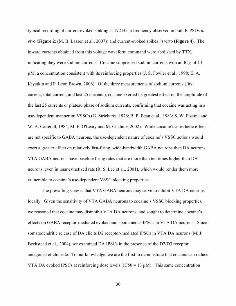
30
typical recording of current-evoked spiking at 172 Hz, a frequency observed in both ICPSDs in
vivo (Figure 2; (M. B. Lassen et al., 2007)) and current-evoked spikes in vitro (Figure 4). The
inward currents obtained from this voltage waveform command were abolished by TTX,
indicating they were sodium currents. Cocaine suppressed sodium currents with an IC50 of 13
µM, a concentration consistent with its reinforcing properties (J. S. Fowler et al., 1998; E. A.
Kiyatkin and P. Leon Brown, 2006). Of the three measurements of sodium currents (first
current, total current, and last 25 currents), cocaine exerted its greatest effect on the amplitude of
the last 25 currents or plateau phase of sodium currents, confirming that cocaine was acting in a
use-dependent manner on VSSCs (G. Strichartz, 1976; B. P. Bean et al., 1983; S. W. Postma and
W. A. Catterall, 1984; M. E. O'Leary and M. Chahine, 2002). While cocaine’s anesthetic effects
are not specific to GABA neurons, the use-dependent nature of cocaine’s VSSC actions would
exert a greater effect on relatively fast-firing, wide-bandwidth GABA neurons than DA neurons.
VTA GABA neurons have baseline firing rates that are more than ten times higher than DA
neurons, even in unanesthetized rats (R. S. Lee et al., 2001), which would render them more
vulnerable to cocaine’s use-dependent VSSC blocking properties.
The prevailing view is that VTA GABA neurons may serve to inhibit VTA DA neurons
locally. Given the sensitivity of VTA GABA neurons to cocaine’s VSSC blocking properties,
we reasoned that cocaine may disinhibit VTA DA neurons, and sought to determine cocaine’s
effects on GABA receptor-mediated evoked and spontaneous IPSCs in VTA DA neurons. Since
somatodendritic release of DA elicits D2 receptor-mediated IPSCs in VTA DA neurons (M. J.
Beckstead et al., 2004), we examined DA IPSCs in the presence of the D2/D3 receptor
antagonist eticlopride. To our knowledge, we are the first to demonstrate that cocaine can reduce
VTA DA evoked IPSCs at reinforcing dose levels (IC50 = 13 µM). This same concentration

31
significantly increased the IPSC paired-pulse ratio, suggesting cocaine acts to decrease the
probability of GABA release (R. Khazipov et al., 1995; S. Mennerick and C. F. Zorumski, 1995;
P. A. Salin et al., 1996; S. C. Steffensen et al., 1999). Additionally, 13 µM cocaine reduced
action potential-dependent spontaneous IPSC (sIPSC) frequency, but not amplitude, and did not
reduce TTX-insensitive action potential-independent miniature IPSC (mIPSC) frequency or
amplitude. Taken together, these observations support the idea that cocaine is acting
presynaptically through its VSSC blocking properties on VTA GABA neurons to reduce activity-
dependent GABA release in the VTA. This concept is supported by the fact that lidocaine (500
µM) is used to pharmacologically isolate mIPSCs (M. Melis et al., 2002). Given that acute
intravenous injections of cocaine at 1.0 mg/kg or higher can lead to extracellular cocaine
concentrations greater than 25 µM in the brain (H. T. Pan et al., 1991), it is somewhat surprising
that lidocaine has not been used as a control in in vitro studies wherein cellular and molecular
changes in VTA DA neurons have been attributed to high doses of cocaine (P. W. Kalivas and J.
E. Alesdatter, 1993; M. A. Ungless et al., 2001; F. Sarti et al., 2007).
Conceivably, the effects of cocaine in the mesolimbic system are concentration-
dependent: a low-dose cocaine effect that enhances DA levels via DAT inhibition at the
terminals of DA neurons, while moderate to higher dose level effects of cocaine may be
mediated through both its DAT-inhibiting and VSSC-inhibiting properties. These actions would
affect the midbrain regions containing DA neuron cell bodies, and their projection areas such as
the nucleus accumbens, with cocaine's VSSC-inhibiting properties preferentially affecting fast-
firing GABA neurons. In support of the data of this study, and our hypothesis that DA neurons
may not be as sensitive as GABA neurons to cocaine's VSSC-blocking effects, Shi et al. (2004)
demonstrated that cocaine (1.0 mg/kg) enhancement of DA neuron firing rate, bursting activity,
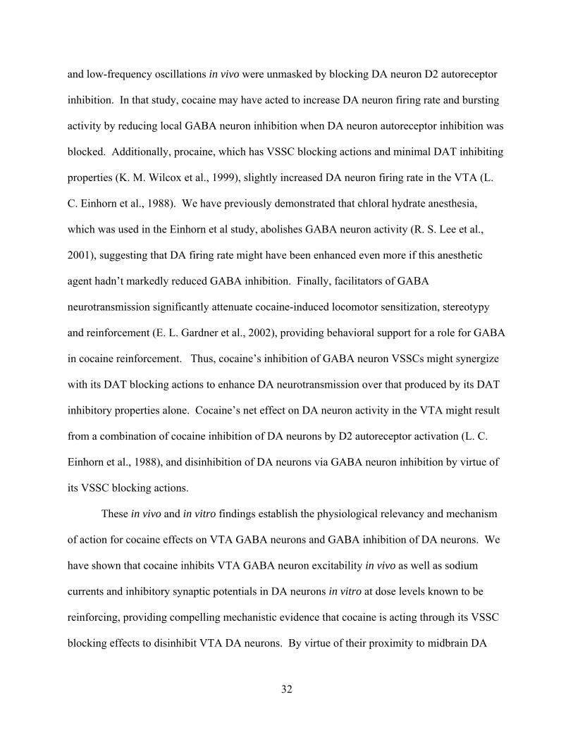
32
and low-frequency oscillations in vivo were unmasked by blocking DA neuron D2 autoreceptor
inhibition. In that study, cocaine may have acted to increase DA neuron firing rate and bursting
activity by reducing local GABA neuron inhibition when DA neuron autoreceptor inhibition was
blocked. Additionally, procaine, which has VSSC blocking actions and minimal DAT inhibiting
properties (K. M. Wilcox et al., 1999), slightly increased DA neuron firing rate in the VTA (L.
C. Einhorn et al., 1988). We have previously demonstrated that chloral hydrate anesthesia,
which was used in the Einhorn et al study, abolishes GABA neuron activity (R. S. Lee et al.,
2001), suggesting that DA firing rate might have been enhanced even more if this anesthetic
agent hadn’t markedly reduced GABA inhibition. Finally, facilitators of GABA
neurotransmission significantly attenuate cocaine-induced locomotor sensitization, stereotypy
and reinforcement (E. L. Gardner et al., 2002), providing behavioral support for a role for GABA
in cocaine reinforcement. Thus, cocaine’s inhibition of GABA neuron VSSCs might synergize
with its DAT blocking actions to enhance DA neurotransmission over that produced by its DAT
inhibitory properties alone. Cocaine’s net effect on DA neuron activity in the VTA might result
from a combination of cocaine inhibition of DA neurons by D2 autoreceptor activation (L. C.
Einhorn et al., 1988), and disinhibition of DA neurons via GABA neuron inhibition by virtue of
its VSSC blocking actions.
These in vivo and in vitro findings establish the physiological relevancy and mechanism
of action for cocaine effects on VTA GABA neurons and GABA inhibition of DA neurons. We
have shown that cocaine inhibits VTA GABA neuron excitability in vivo as well as sodium
currents and inhibitory synaptic potentials in DA neurons in vitro at dose levels known to be
reinforcing, providing compelling mechanistic evidence that cocaine is acting through its VSSC
blocking effects to disinhibit VTA DA neurons. By virtue of their proximity to midbrain DA
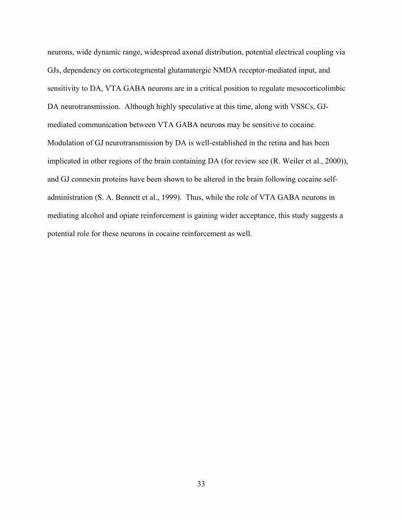
33
neurons, wide dynamic range, widespread axonal distribution, potential electrical coupling via
GJs, dependency on corticotegmental glutamatergic NMDA receptor-mediated input, and
sensitivity to DA, VTA GABA neurons are in a critical position to regulate mesocorticolimbic
DA neurotransmission. Although highly speculative at this time, along with VSSCs, GJ-
mediated communication between VTA GABA neurons may be sensitive to cocaine.
Modulation of GJ neurotransmission by DA is well-established in the retina and has been
implicated in other regions of the brain containing DA (for review see (R. Weiler et al., 2000)),
and GJ connexin proteins have been shown to be altered in the brain following cocaine self-
administration (S. A. Bennett et al., 1999). Thus, while the role of VTA GABA neurons in
mediating alcohol and opiate reinforcement is gaining wider acceptance, this study suggests a
potential role for these neurons in cocaine reinforcement as well.
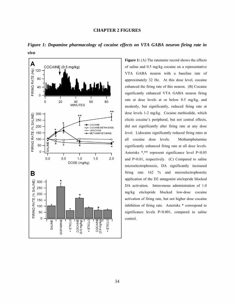
34
CHAPTER 2 FIGURES
Figure 1: Dopamine pharmacology of cocaine effects on VTA GABA neuron firing rate in
vivo Figure 1: (A) The ratemeter record shows the effects
of saline and 0.5 mg/kg cocaine on a representative
VTA GABA neuron with a baseline rate of
approximately 32 Hz. At this dose level, cocaine
enhanced the firing rate of this neuron. (B) Cocaine
significantly enhanced VTA GABA neuron firing
rate at dose levels at or below 0.5 mg/kg, and
modestly, but significantly, reduced firing rate at
dose levels 1-2 mg/kg. Cocaine methiodide, which
elicits cocaine’s peripheral, but not central effects,
did not significantly alter firing rate at any dose
level. Lidocaine significantly reduced firing rates at
all cocaine dose levels. Methamphetamine
significantly enhanced firing rate at all dose levels.
Asterisks *,** represent significance level P<0.05
and P<0.01, respectively. (C) Compared to saline
microelectrophoresis, DA significantly increased
firing rate 162 % and microelectrophoretic
application of the D2 antagonist eticlopride blocked
DA activation. Intravenous administration of 1.0
mg/kg eticlopride blocked low-dose cocaine
activation of firing rate, but not higher dose cocaine
inhibition of firing rate. Asterisks * correspond to
significance levels P<0.001, compared to saline
control.
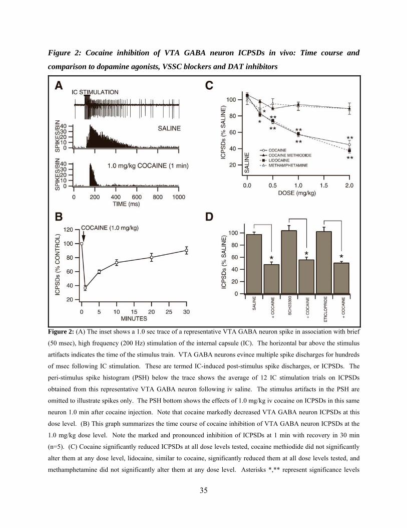
35
Figure 2: Cocaine inhibition of VTA GABA neuron ICPSDs in vivo: Time course and
comparison to dopamine agonists, VSSC blockers and DAT inhibitors
Figure 2: (A) The inset shows a 1.0 sec trace of a representative VTA GABA neuron spike in association with brief
(50 msec), high frequency (200 Hz) stimulation of the internal capsule (IC). The horizontal bar above the stimulus
artifacts indicates the time of the stimulus train. VTA GABA neurons evince multiple spike discharges for hundreds
of msec following IC stimulation. These are termed IC-induced post-stimulus spike discharges, or ICPSDs. The
peri-stimulus spike histogram (PSH) below the trace shows the average of 12 IC stimulation trials on ICPSDs
obtained from this representative VTA GABA neuron following iv saline. The stimulus artifacts in the PSH are
omitted to illustrate spikes only. The PSH bottom shows the effects of 1.0 mg/kg iv cocaine on ICPSDs in this same
neuron 1.0 min after cocaine injection. Note that cocaine markedly decreased VTA GABA neuron ICPSDs at this
dose level. (B) This graph summarizes the time course of cocaine inhibition of VTA GABA neuron ICPSDs at the
1.0 mg/kg dose level. Note the marked and pronounced inhibition of ICPSDs at 1 min with recovery in 30 min
(n=5). (C) Cocaine significantly reduced ICPSDs at all dose levels tested, cocaine methiodide did not significantly
alter them at any dose level, lidocaine, similar to cocaine, significantly reduced them at all dose levels tested, and
methamphetamine did not significantly alter them at any dose level. Asterisks *,** represent significance levels
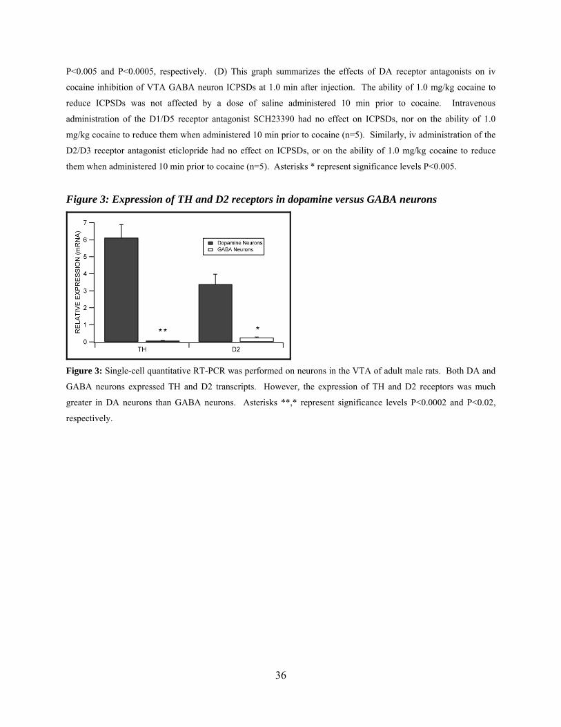
36
P<0.005 and P<0.0005, respectively. (D) This graph summarizes the effects of DA receptor antagonists on iv
cocaine inhibition of VTA GABA neuron ICPSDs at 1.0 min after injection. The ability of 1.0 mg/kg cocaine to
reduce ICPSDs was not affected by a dose of saline administered 10 min prior to cocaine. Intravenous
administration of the D1/D5 receptor antagonist SCH23390 had no effect on ICPSDs, nor on the ability of 1.0
mg/kg cocaine to reduce them when administered 10 min prior to cocaine (n=5). Similarly, iv administration of the
D2/D3 receptor antagonist eticlopride had no effect on ICPSDs, or on the ability of 1.0 mg/kg cocaine to reduce
them when administered 10 min prior to cocaine (n=5). Asterisks * represent significance levels P<0.005.
Figure 3: Expression of TH and D2 receptors in dopamine versus GABA neurons
Figure 3: Single-cell quantitative RT-PCR was performed on neurons in the VTA of adult male rats. Both DA and
GABA neurons expressed TH and D2 transcripts. However, the expression of TH and D2 receptors was much
greater in DA neurons than GABA neurons. Asterisks **,* represent significance levels P<0.0002 and P<0.02,
respectively.

37
Figure 4: Cocaine reduces current-evoked spiking of VTA GABA neurons in vitro
Figure 4: (A) These are representative current-clamp traces of
current-evoked VTA GABA neuron spikes recorded in the
horizontal midbrain slice preparation. Note the lack of spike
accommodation characteristic of VTA GABA neurons (D. W.
Allison et al., 2006). Superfusion of cocaine (50 µM) markedly
reduced spiking at this level of depolarization (250 pA; darker
line of the current steps that were performed). (B) This graph
summarizes the effects of 50 µM cocaine on the frequency of
current-evoked spiking of VTA GABA neurons at all current
steps tested (25-400 pA steps shown in A). Superfusion of
cocaine significantly reduced spiking at current steps 100-400
pA in all cells tested. Asterisk * indicates significance level
P<0.03 at each point.
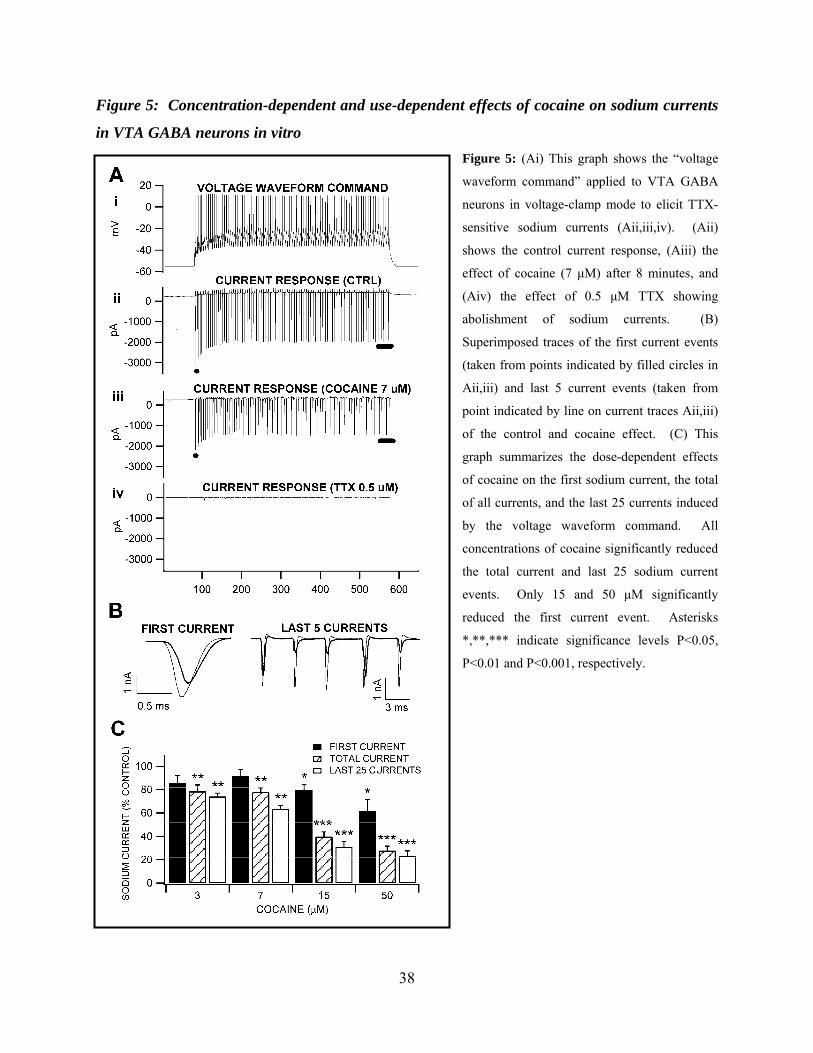
38
Figure 5: Concentration-dependent and use-dependent effects of cocaine on sodium currents
in VTA GABA neurons in vitro Figure 5: (Ai) This graph shows the “voltage
waveform command” applied to VTA GABA
neurons in voltage-clamp mode to elicit TTX-
sensitive sodium currents (Aii,iii,iv). (Aii)
shows the control current response, (Aiii) the
effect of cocaine (7 µM) after 8 minutes, and
(Aiv) the effect of 0.5 µM TTX showing
abolishment of sodium currents. (B)
Superimposed traces of the first current events
(taken from points indicated by filled circles in
Aii,iii) and last 5 current events (taken from
point indicated by line on current traces Aii,iii)
of the control and cocaine effect. (C) This
graph summarizes the dose-dependent effects
of cocaine on the first sodium current, the total
of all currents, and the last 25 currents induced
by the voltage waveform command. All
concentrations of cocaine significantly reduced
the total current and last 25 sodium current
events. Only 15 and 50 µM significantly
reduced the first current event. Asterisks
*,**,*** indicate significance levels P<0.05,
P<0.01 and P<0.001, respectively.
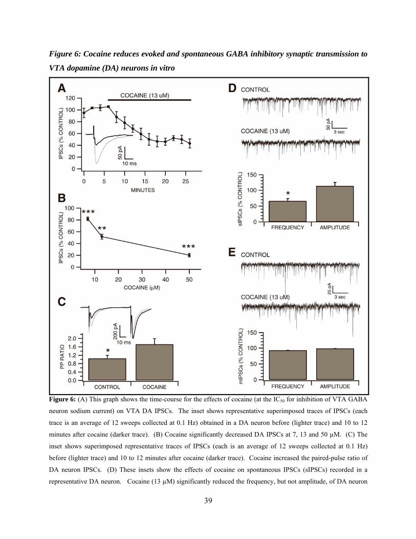
39
Figure 6: Cocaine reduces evoked and spontaneous GABA inhibitory synaptic transmission to
VTA dopamine (DA) neurons in vitro
Figure 6: (A) This graph shows the time-course for the effects of cocaine (at the IC50 for inhibition of VTA GABA
neuron sodium current) on VTA DA IPSCs. The inset shows representative superimposed traces of IPSCs (each
trace is an average of 12 sweeps collected at 0.1 Hz) obtained in a DA neuron before (lighter trace) and 10 to 12
minutes after cocaine (darker trace). (B) Cocaine significantly decreased DA IPSCs at 7, 13 and 50 µM. (C) The
inset shows superimposed representative traces of IPSCs (each is an average of 12 sweeps collected at 0.1 Hz)
before (lighter trace) and 10 to 12 minutes after cocaine (darker trace). Cocaine increased the paired-pulse ratio of
DA neuron IPSCs. (D) These insets show the effects of cocaine on spontaneous IPSCs (sIPSCs) recorded in a
representative DA neuron. Cocaine (13 µM) significantly reduced the frequency, but not amplitude, of DA neuron

40
sIPSCs. (E) These insets show the effects of cocaine on miniature IPSCs (mIPSCs) recorded in a representative DA
neuron after treatment with TTX. Cocaine (13 µM) had no effect on frequency or amplitude of TTX-insensitive
miniature IPSCs. Asterisks *,**,*** indicate significance levels P<0.05, P<0.01 and P<0.001, respectively.
41
CHAPTER 3
Mefloquine Disrupts Ventral Tegmental Area Inhibitory and Excitatory Synaptic
Activity via the Blockade of Connexin 36 Gap Junctions and Disruption of
Intracellular Calcium Homeostasis
INTRODUCTION
The mesolimbic dopamine (DA) pathway is composed of DA neurons located in the
ventral tegmental area (VTA) of the midbrain that project to the dorsal striatum, hippocampus,
amygdala, and prefrontal cortex. This diffuse modulatory pathway is involved in attention to
rewarding stimuli, learned behavior, and drug addiction (for review, (H. L. Fields et al., 2007; S.
Ikemoto, 2007). It is believed that VTA DA neurons are inhibited by local GABA interneurons,
and projection GABA neurons from other brain regions. There is evidence suggesting this DA
pathway contains reciprocal excitatory, DAergic, and GABAergic communication with other
brain regions (D. B. Carr and S. R. Sesack, 2000a, b; N. Omelchenko and S. R. Sesack, 2005,
2006; E. B. Margolis et al., 2008). Recent evidence suggests VTA GABA neurons express
Connexin 36 (Cx36) gap junctions (GJs), dye couple (S. C. Steffensen et al., 1998; D. W. Allison
et al., 2006), and form part of a larger syncytium of GABA neurons in the reticular formation
that are linked by Cx36 electrical synapses (M. B. Lassen et al., 2007). Acute intoxicating doses
of ethanol and GJ antagonists, including the Cx36-selective antagonist mefloquine, suppress
electrical coupling between VTA GABA neurons in vivo (S. H. Stobbs et al., 2004).
Studying Cx36 GJ’s is made difficult by the lack of drugs that act specifically and
exclusively on GJs. The most promising drugs are mefloquine (a Cx36 selective GJ blocker,
MFQ) and carbenoxolone (CBX), both of which have non-GJ blocking affects (for review G. R.
42
Juszczak and A. H. Swiergiel, 2009). But despite their shortcomings, MFQ and CBX can be
utilized to determine the neurophysiology of GJs if used in correctly designed and executed
experiments that control for non-specific effects.
Mefloquine (MFQ) serves as a potent anti-malarial prophylaxis and treatment. It is well
known that it may act as a neurotoxin due to a potential to cross the blood-brain barrier and
accumulate to high concentrations (G. S. Dow et al., 2004). MFQ is linked to adverse side-
effects ranging in severity from mild dysphoria to severe psychotic episodes or seizure (E.
Wooltorton, 2002; T. M. Tran et al., 2006; P. Schlagenhauf et al., 2009). While MFQ’s adverse
side-effects have not been attributed to any particular drug effect, its ability to affect synaptic
activity suggests its neuropsychiatric side-effects derive from one, or a combination of its
pharmacological actions. Owing to DA’s involvement in synaptic transmission, mood, and
addiction, we hypothesized some of MFQ’s side-effects may result from MFQ’s blockade of
Cx36 GJs in the VTA and point to the possible role for Cx36 GJs in regulation of DA.
Specifically, we hypothesized that MFQ’s blockade of VTA Cx36 GJs might disrupt the network
activity of electrically-coupled VTA GABA neurons, thereby altering inhibition of VTA DA
neurons resulting in a change in DA neuron activity. To that end, we investigated the effects of
MFQ and CBX on VTA DA and GABA neuron inhibition and excitation in both wildtype and
Cx36 KO mice. We hypothesized that Cx36 GJs would play a significant role in regulating
mesolimbic DA transmission.
43
METHODS
Preparation of Brain Slices
The care and use of animals and experimental protocols were approved by the
Institutional Animal Care and Use Committee (IACUC) of Brigham Young University. Two
different mouse models (age P21 – 35) were used in this study. A glutamic acid decarboxylase-
67 (GAD67) linked to green fluorescent protein (GFP) knock-in mouse created on a CD1
background (N. Tamamaki et al., 2003), and a Cx36 KO mouse created on a C57BL6/129SvEv
hybrid background, originally created by Dr. David Paul at Harvard Medical School (H.
Arumugam et al., 2008) and donated to us by Dr. Marla Feller (University of California, San
Diego, USA). The GAD67-GFP mice (also referred to as “WT”) afforded us the possibility of
positively identifying and recording from GAD67 GABA neurons in the VTA. The Cx36 KO
mice provided a comparison against mefloquine’s Cx36 GJ blocking effects on VTA dopamine
(DA) neurons in GAD67-GFP mice. Midbrain slices were obtained as previously described (S.
C. Steffensen et al., 2008). Briefly, mice were anesthetized with Ketamine (60 mg/kg),
decapitated, and brains were quickly dissected and sectioned into 200 µm thick horizontal slices
in ice-cold artificial cerebrospinal fluid (ACSF), bubbled with 95% O2 / 5% CO2. This cutting
solution consisted of (in mM): 220 Sucrose, 3 KCl, 1.25 NaH2PO4, 25 NaH2CO3, 12 MgSO4,
10 Glucose, 0.2 CaCl2, and 0.4 Ketamine. These VTA-targeted horizontal slices were
immediately placed into an incubation chamber containing normal ACSF at 34-35°, bubbled
with 95% O2 / 5% CO2 at 36° consisting of (in mM): 124 NaCl, 3 KCl, 1.25 NaH2PO4, 26
NaHCO3, 12 glucose, 1.5 MgSO4, 2 CaCl2, pH 7.3, and allowed to incubate for at least 45
minutes prior to being transferred to a recording chamber. Once transferred to a recording
chamber with continuous normal ACSF flow (2.0 ml/min) maintained at 34-35° throughout the

44
experiment, the slices where then allowed to settle for an additional 15 minutes before recordings
began. Cells were visualized with either a Nikon Eclipse FN1 or E600FN microscope in the
transmitted de Sénarmont Differential Interference Contrast (DIC) / infrared (IR) configuration.
Whole-cell Recordings in vitro
Electrodes pulled from borosilicate glass capillary tubes were filled with one of two types
of pipette solutions consisting of either (in mM): 128 KCl, 20 NaCl, 0.3 CaCl2, 1.2 MgCl2, 10
HEPES, 1 EGTA, 2 Mg-ATP, 0.25 Na-GTP and 4.5 QX314 (pH 7.3) for IPSCs, and 123 K-
Gluconate, 8 NaCl, 10 HEPES, 0.2 EGTA, 2 Mg-ATP, 2 Na3-GTP, and 4.5 QX314 (pH 7.3) for
EPSCs and firing rate experiments. Pipettes had tip resistances of 2.5 - 4 MΩ, and series
resistances typically ranging from 7 to 15 MΩ. Access resistance (Ra, typically 100 to 300 MΩ)
and membrane resistance (Rm), were continuously monitored with a 5 mV voltage step delivered
with each sweep (0.1 Hz) throughout the experiment. Only experiments that maintained stable
Ra and Rm (less than 15% change) were included in this study. Voltage clamp recordings were
filtered at 2 kHz while current-drive spikes were filtered at 6 kHz with an Axon Instruments
Multiclamp 700B amplifier and digitized at 5 to 20 kHz respectively using an Axon 1440A
digitizer. pClampv10, Mini Analysis (Synaptsoft: Decatur, GA), and Igor Pro (Wavemetrics:
Oswego, OR) software packages software were utilized for data collection and analysis. Evoked
and spontaneous IPSCs (sIPSCs) were recorded in the presence of D- 2-amino-5-
phosphonopentanoic acid (D-APV, 50 µM), 6-cyano-23-dihydroxy-7-nitro-quinoxaline (CNQX,
30 µM), and eticlopride (100 nM) to block NMDA, AMPA, and DA D2-mediated synaptic
currents respectively. Spontaneous and evoked EPSCs were recorded in the presence of
bicuculline (10 µM), a concentration that eliminated all spontaneous inhibitory activity.
Miniature IPSCs and EPSCs were isolated from all other spontaneous activity by addition of

45
lidocaine (500 µM). To evoke IPSCs and EPSCs, cells were stimulated at 0.1 Hz with a stainless
steel-platinum/iridium concentric bipolar stimulating electrode placed ~100 µm rostral to the
recording electrode. All evoked and spontaneous inhibitory and excitatory currents (except
while examining AMPA to NMDA ratio) were inward at the holding potential of -70 mV and
were completely blocked by bicuculline and APV, respectively.
Evoked current amplitudes were calculated by taking the difference between the 1.0 msec
window around the peak and the 5.0 msec baseline window immediately preceding the
stimulation artifact. The paired-pulse ratio was calculated by averaging traces from 2 minutes of
data and then dividing the average secondary by the average primary. For spontaneous synaptic
currents, the amplitude threshold was set to 5 times RMS, while area threshold was adjusted for
excitatory and inhibitory currents respectively. Spontaneous and mini currents were then
automatically detected and verified and corrected by hand. The AMPA to NMDA ratio was
determined by first obtaining robust evoked EPSCs at -70 mV and then slowly stepping the
holding potential to +30 mV. At +30 mV, a combined AMPA/NMDA current response was
recorded for 2 min, then D-APV (50 µM) was applied. Once the NMDA current was completely
diminished, the AMPA current response was recorded. Posthoc subtraction of the pure AMPA
current from the combined AMPA/NMDA current yielded pure NMDA current. Each 2 minute
recording period was averaged. The ratio was calculated as the peak average AMPA current
amplitude of divided by the peak average NMDA current amplitude. Peak GABA(A) current
(30 second bins) was acquired in the presence of the GABA(B) receptor antagonist CGP 35348
(100 µM) and calculated by comparing the average of 1 minute of control and test current at
peak. Firing rate was calculated by comparing 5 minutes control and test data.
46
Drug Preparation and Administration
In vivo: MFQ (5mg MFQ, 800 µl distilled H2O, 200 µl Tween 80) was administered via
an intra-peritoneal injection at 30mg/kg. In vitro: All drugs except D-APV, CNQX, CGP 35348
, and bicuculline were prepared fresh. MFQ was prepared 1mg/mL water to obtain a 25 mM
stock solution. In all experiments (except Figure 4) wherein brain slices were presoaked in
MFQ, carbenoxolone (CBX), ondansetron (ODS), or mecamylamine hydrochloride (MEC),
these drugs were added to the slice incubation chamber 10 minutes after slicing and allowed to
presoak 60 to 90 minutes, then continuously perfused throughout the experiment. There are two
advantages to this method of drug application over obtaining a viable recording and then
applying drugs acutely and observing the effect progress over. First, any postsynaptic MFQ
effects due to disruption of neuronal calcium levels are not washed out by pipette solution.
Second, given that long quality recording periods can be problematical using the whole-cell
patchclamp technique, presoaking enables long drug application times which more closely mimic
the chronic exposure to MFQ in humans. For time course of MFQ effects (Fig 1) brain slices
where allowed to settle after cutting for 45 minutes before recordings began.
Characterization of Neuron Types
In vitro: GAD67-GFP (wildtype) and Cx36 KO mice the combination of two
electrophysiological characteristics, were used to identify putative DA neurons in this study.
Specifically: neurons located in the VTA that exhibited a modest non-cation specific inward
rectifying current (Ih) in combination with low input resistance were assumed to be DA neurons
(S. W. Johnson and R. A. North, 1992; D. W. Allison et al., 2006; E. B. Margolis et al., 2006).
In GAD GFP mice, GABA neurons were identified with the aid of fluorescence microscopy.
Only neurons located in the VTA that exhibited robust GFP fluorescence were considered
47
GABAergic (N. Tamamaki et al., 2003). In vivo: all neurons classified as VTA GABA neurons
were located in the VTA and met the criteria established in previous studies for spike waveform
characteristics and response to IC stimulation (S. C. Steffensen et al., 1998; S. H. Stobbs et al.,
2004; D. W. Allison et al., 2006)
Statistical Analyses
All results are presented as mean raw values and percent control ± SEM. Results
between groups were compared using a two-tailed unpaired t test or ANOVA. Experiments
relying on variance in time or current were analyzed using ANOVA with post hoc t-test at
individual points. Statistical significance required ≥95% level of confidence (P≤0.05). Analysis
software included Microsoft Excel, and Igor Pro (Wavemetrics, Oswego, OR) Stat Pak. Figures
were constructed with Igor Pro software.

48
RESULTS
Time Course of Mefloquine Effects VTA DA sIPSC Frequency
In recent studies mefloquine (MFQ) was shown to quickly increase sIPSCs in substantia
nigra pars compacta (SNc) dopamine (DA) neurons (EC50 ≈ 3µM) (C. Zhou et al., 2006) and
EPSPs in neocortical interneurons (IC50 ≈ 15µM)(S. J. Cruikshank et al., 2004). However, the
maximum effect times of MFQ differed in each study. We first determined the time required for
25 µM MFQ to reach maximum effect on VTA DA neuron sIPSC frequency, and whether there
was a significant difference between WT and Cx36 mice in the time course of its effects. In
Figure 1 MFQ (25 µM) was bath-applied in ACSF containing D-APV (50 µM) and CNQX (30
µM) and the frequency of VTA DA neuron sIPSCs was calculated at 15 minute intervals. MFQ
increased sIPSC frequency incrementally and reached maximum effect at ≥60 minutes in
wildtype. In wildtype mice, baseline sIPSC frequency increased from 7.3 ± 1.7 Hz (t=0) to: 8.4
± 0.9 Hz (15 min); 18.9 ± 3.5 Hz (30 min); 19.2 ± 1.4 Hz (45 min); 59.8 ± 4.3 Hz (60 min); an
increase of 772.3 ± 27.8% at the ≥60 minute time point. These sIPSCs were completely
eliminated by bicuculline (10 µM, data not shown).
In Cx36 KO mice, MFQ also increased sIPSC frequency incrementally as in WT mice
(Figure 1) and reached maximum effect at ≥60 minutes. However, in Cx36 KO mice the
baseline frequency only increased from 7.3 ± 1.0 Hz (t=0) to: 7.5 ± 0.9 Hz (15 min); 13.7 ± 1.6
Hz (30 min); 21.9 ± 3.7 Hz (45 min); 32.2 ± 6.5 Hz (60 min); an increase of 441.4 ± 20.4% at the
≥60 minute time point.. The only point at which WT and Cx36 KO data differed significantly
was at the ≥60 minute time point where MFQ increased WT mouse sIPSC frequency 331% in
over Cx36 KO mice (ANOVA F=8.03, P=0.0001, DF=46).
49
Mefloquine Effects on VTA DA sIPSC Frequency, Amplitude, and Inter-event Interval in WT
and Cx36 KO Mice
Having established the time course of the full-effect of MFQ on GABA(A) receptor-
mediated DA neuron sIPSC frequency in WT and Cx36 KO mice, we next compared the effects
of presoaked MFQ to other presoaked drugs on VTA DA neuron sIPSC frequency in wildtype
and Cx36 KO mice. Because MFQ has effects other than being a selective Cx36 gap junction
(GJ) blocker (such as the Ca2+ effect previously mentioned), we compared its effects to
carbenoxolone (CBX, a less potent GJ blocker than MFQ) in WT and KO mice. We reasoned
that if MFQ’s GJ blocking properties played a role in increasing sIPSC frequency, a less
selective gap junction blocker such as CBX, which has different non-specific effects, would
increase sIPSC frequency in similar to MFQ in wildtype mice, but perhaps not at all in Cx36 KO
mice. As shown in Figure 2C, in WT mice MFQ (25 µM) increased VTA DA neuron sIPSC
frequency from 7.7 ± 0.8 Hz to 40.9 ± 2.7 Hz (533 ± 35.4% of control, P=0.0001, MFQ n=28,
control n=30). Carbenoxolone (100 µM) increased sIPSC frequency in WT mice from baseline
to 16.2 ± 2.2 Hz (210.7 ± 28.3% of control, P= 0.0001, CBX n=9, Fig 2C). Only MFQ increased
DA sIPSC average amplitude significantly (134 ± 13.8%, P<0.023; Figure 2C) suggesting a
possible postsynaptic effect. MFQ and CBX both shifted the cumulative inter-event interval
distribution plot to the left (Figure 2B), suggesting increased activity-dependent, and or non-
activity dependent GABA release.
In Cx36 KO mice 25 µM MFQ increased DA neuron sIPSC frequency from 8.9 ± 1.7 Hz
to 27.9 ± 3.7 Hz (311.4 ± 41.1% of control, P=0.0003, control n=10, MFQ n=8 Figure 2F),
while CBX slightly reduced sIPSC frequency to 7.9 ± 0.6 Hz (88.5 ± 7.3% of control, P=0.64,
CBX n=7, Figure 2F). As with WT mice, MFQ shifted the cumulative sIPSC inter-event

50
distribution plot markedly to the left in Cx36 KO mice (Figure 2E) indicating increased GABA
release. This MFQ induced increase in Cx36 KO mouse VTA DA neuron sIPSC frequency was
58% of the total increase seen in WT mice (Figure 2C). This difference, in addition to the lack
of CBX effect on sIPSC frequency in Cx36 KO mice (Figure 2E, F), reinforces the idea that the
MFQ-induced increase in VTA DA neuron sIPSC frequency in WT mice was partly due to
MFQ’s GJ blocking properties, and partly due to MFQ’s disruption of intracellular calcium (C.
Zhou et al., 2006). CBX decreased DA sIPSC average amplitude significantly (decrease of 59.1
± 3.6%, P<0.003; Figure 2H).
Although we doubted MFQ was acting as a GABA(A) receptor antagonist (A. J.
Thompson et al., 2007), the possibility still existed it was increasing DA sIPSCs via 5HT3
receptor antagonism (A. J. Thompson and S. C. Lummis, 2008). To this end we also examined
the specific 5HT3 receptor antagonist odansetron (ODS) in WT mice. Contrary to MFQ’s
potentiation of sIPSCs, ODS markedly reduced VTA DA neuron sIPSC frequency from baseline
to 2.0 Hz (decrease of 74 ± 7.1%, P=0.0006, ODS n=9, data only shown in bar graph, Fig 2C).
A recent study by Zhou et al (2006) hypothesized that along with MFQ’s potent calcium-
mediated effects it was also increasing sIPSC frequency by inhibition of cholinesterase (J. J.
McArdle et al., 2006), thereby increasing GABA release via excess acetylcholine activation of
presynaptic nAChRs. In that study, Zhou et al found pre-application of mecamylamine
hydrochloride (MEC, a non- α7nAChR antagonist), which reduced baseline sIPSCs frequency by
itself, also reduced the MFQ increase of DA sIPSC frequency ~70%. For this reason, we also
examined the effect of presoaking slices with MFQ in the presence of MEC. Mefloquine + MEC
presoaked together increased DA neuron sIPSC frequency similar to, and not significantly
different from MFQ alone (488 ± 47.3% of control, MFQ + MEC n=6, P=0.58, Fig 2C), and

51
shifted the cumulative inter-event interval plot to the left similar to MFQ alone (Fig 2B). As
neither ODS or MEC mimicked MFQ in WT mice, we did not examine these drugs in Cx36 KO
mice.
It has been reported in rats that the co-activation of mGluR1 and NMDA glutamate
receptors may increase sIPSC frequency, transforming dispersed GABAergic input on midbrain
SNc DA neurons into a rhythmic pattern of sIPSCs frequency from 4 to 22 Hz (N. Berretta et al.,
2001). Investigators in that study attributed this sIPSC frequency increase and onset of rhythmic
sIPSC activity to increased electrical coupling of local GABA neurons connected via GJs. The
present study demonstrates a marked increase in sIPSC frequency in part due to the blockade of
Cx36 GJs in the presence of AMPA and NMDA receptor antagonists. As expected, we did not
detect rhythmic sIPSC activity in either control or MFQ treated VTA DA neurons (data not
shown), demonstrating increased sIPSC frequency and rhythmicity are not related.
Mefloquine Effects on VTA DA mIPSC Frequency, Amplitude, and Inter-event Interval in
WT and Cx36 KO Mice
Spontaneous postsynaptic currents are a combination of activity-dependent (spike-
dependent) and non-activity-dependent transmitter release. In order to determine what portion of
MFQ and CBX-induced increase in sIPSC frequency was due to non-activity-dependent GABA
release, we examined the effect of MFQ and CBX on WT and Cx36 KO mouse VTA DA neuron
non-activity-dependent IPSCs or mini IPSC (mIPSC) frequency by adding lidocaine (500 uM) to
the ACSF. In WT mice, MFQ (25 µM) increased VTA DA neuron mIPSC frequency from 7.6 ±
1.2 Hz to 21.9 ± 2.7 Hz (289.8 ± 35.5% of control, P<0.0001, control n=13, MFQ n=6, Figure
3C), had no significant affect on amplitude, but did significantly shift the mIPSC cumulative
inter-event interval distribution plot to the left (Figure 3B), suggesting MFQ was acting
52
presynaptically to increase non-activity-dependent GABA release. Conversely, CBX (100 µM)
had no significant affect on frequency, but significantly reduced mIPSC amplitude from 30.7 ±
6.5 pA to 13.8 ± 0.7 pA (-55.2 ± 2.3%, P<0.027, CBX n=11, Figure 3B, C).
In Cx36 KO mice, MFQ (25 µM) increased VTA DA neuron mIPSC frequency from 8.1
± 1.8 to 29.0 ± 2.15 Hz (358.3 ± 43.0% of control, P=0.004, control n=4, MFQ n=5) and did not
affect amplitude (Figure 3F). Similar to WT mice, Cx36 KO mouse mIPSC cumulative inter-
event interval distribution plot was shifted to the left by MFQ (Figure 3E). We did not examine
CBX effects on DA mIPSC frequency in Cx36 KO mice since it did not alter mIPSC frequency
in WT mice.
The sIPSC and mIPSC data presented demonstrates MFQ and CBX were increasing
activity-dependent and non-activity dependent GABA release in WT mice, but only non-activity
dependent GABA release in Cx36 KO mice. This difference in effects between strains could
possibly be explained by two separate mechanisms. We reasoned MFQ could be increasing the
frequency of non-activity-dependent presynaptic GABA release in WT and Cx36 KO mice via
MFQ’s presynaptic Ca2+ effects (G. S. Dow et al., 2003; C. Zhou et al., 2006; D. Caridha et al.,
2008), and increasing activity-dependent presynaptic GABA release via both Ca2+ effects and
selective blockade of Cx36 GJs (S. J. Cruikshank et al., 2004). The credibility of this scenario is
bolstered by the results obtained from CBX, a drug which blocks GJs but does not increase
intracellular Ca2+. Similar to MFQ, CBX increased DA neuron sIPSC frequency in WT. But
unlike MFQ, it did not increase sIPSCs in Cx36 KO mice or increase mIPSCs in either mouse
model.
53
Neither Presoaking nor Bath-application of Mefloquine Alters VTA DA Neuron Evoked IPSC
Paired Pulse Ratio or IPSC Primary Current Amplitude
Because an increase in mIPSC frequency is generally associated with an increase in
probability of transmitter release (R. C. Malenka and R. A. Nicoll, 1999), we next attempted to
more firmly establish that MFQ was increasing VTA DA neuron sIPSC frequency, in part
through a presynaptic mechanism, by examining MFQ’s effect on VTA DA neuron evoked IPSC
(eIPSC) paired pulse ratio (PPR). A decrease in the PPR is commonly associated with an
increase in probability of release at excitatory and inhibitory synapses in the hippocampus (R.
Khazipov et al., 1995; S. Mennerick and C. F. Zorumski, 1995; D. Debanne et al., 1996; S. C.
Steffensen et al., 1999), and the VTA (M. Melis et al., 2002). Contrary to what we expected,
presoaking slices with MFQ (25 µM) did not significantly alter VTA DA neuron eIPSC PPR in
WT mice (Control 0.65 ± 0.03, n=10; MFQ 0.71 ± 0.06, n=8, P=0.41, Figure 4A, C). To
confirm that inhibitory synapses on VTA DA neurons obey the rule of exhibiting PPD during an
increase in probability of release as observed by others (M. Melis et al., 2002; J. W. Theile et al.,
2008), we conducted a separate set of experiments without MFQ in which extracellular Ca2+ was
increased from 2 mM to 4 mM while monitoring primary IPSC amplitude, PPR, and sIPSC
frequency. Indeed, as has been demonstrated ad nauseam (D. Debanne et al., 1996; L. E.
Dobrunz and C. F. Stevens, 1997; M. O. Kravchenko et al., 2006), this increase of extracellular
Ca2+ increased the primary IPSC amplitude 127.8 ± 7.2% of control (P=0.05), decreased the PPR
31 ± 2.1% (P=0.05), and increased sIPSC frequency from 7.0 ± 0.8 Hz to 15.1 ± 0.6 Hz (increase
of 114%, P=0.04, data not shown). We reasoned MFQ’s robust ability to disrupt neuronal
calcium stores (G. S. Dow et al., 2003; D. Caridha et al., 2008) might facilitate an increase in
GABA release, but prevent the ability to measure this calcium-linked phenomenon via

54
established methods that rely on normal calcium-linked ready-releasable transmitter vesicle pool
regulation (L. E. Dobrunz and C. F. Stevens, 1997).
Because MFQ’s ability to increase intracellular Ca2+ and block GJs may both contribute
to increased sIPSC frequency, we reasoned that we might detect a change in the PPR ratio as
MFQ begins to take effect at some time point from zero to 45 minutes. To explore this
possibility we added mefloquine to ACSF containing D-APV and CNQX only after achieving a
stable VTA DA neuron whole-cell recording, rather than presoaking the slice in MFQ (25 µM).
Evoked IPSC primary current amplitude, PPR, and sIPSC frequency where measured
continuously for up to 45 min. The average DA neuron evoked IPSC amplitude did not
significantly change throughout these experiments (98.8 ± 6.5% of control, P=0.85, n=5, Figure
4B), and there was no significant change in the PPR (Control 0.7 ± 0.1, MFQ 0.7 ± 0.1, P=0.8,
Figure 4B, C). However, the average VTA DA neuron sIPSC frequency following 45 minutes
of MFQ perfusion was increased from 6.9 ± 1.4Hz to 14.8 ± 2.5Hz (219.7 ± 37.3% of control,
P=0.02, Figure 4D, E). This MFQ-induced increase in sIPSC frequency obtained while
continuously recording up to 45 minutes was not significantly different from the increase elicited
by presoaked MFQ at the same time point diagramed in Figure 1.

55
Mefloquine Increases VTA DA Neuron GABA(A) Receptor Current
Given that MFQ increased the frequency and amplitude of activity-dependent VTA DA
sIPSCs in WT mice (Figure 2C), we next examined the possibility of a postsynaptic component
of MFQ effect. Bath application of GABA (100 µM) for 2.5 minutes in WT VTA DA neurons
increased peak GABA current in MFQ presoaked slices from -51.7 ± 17.0pA to -211.4 ± 27.9pA
(408.5 ± 54.0% of control, P=0.001, control n=6, MFQ n=10, Figure 5A, B), suggesting that in
addition to an increase in GABA release presynaptically via Cx36 GJ blockade and possibly
disruption of Ca2+ homeostasis, MFQ was inducing a form of GABAergic postsynaptic LTP at
VTA DA neuron inhibitory synapses. This GABAergic LTP could possibly involve the increase
in number and or efficiency of postsynaptic GABA receptors on VTA DA neurons. Ca2+-
dependent long-term potentiation (LTP) at GABAergic synapses has been demonstrated in the
hippocampus, (H. A. McLean et al., 1996; J. Kang et al., 1998; O. Caillard et al., 1999) as well
as other brain regions including the cerebellum (M. Kano, 1994; S. Y. Kawaguchi and T. Hirano,
2007), neocortex (Y. Komatsu and Y. Yoshimura, 2000), and the VTA (F. S. Nugent et al.,
2007) . To our knowledge the present study is the first to demonstrate postsynaptic LTP at
GABA(A) receptors on VTA DA neurons.
Mefloquine Does Not Affect VTA GABA Neuron sIPSC Frequency or eIPSC Paired-pulse
Ratio
Mefloquine’s increase of VTA DA neuron sIPSC frequency is remarkable. We also
examined MFQ’s effect on VTA GABA neuron sIPSC frequency, as well as eIPSC paired-pulse
ratio in WT mice. Identification of VTA GABA neurons was aided by the use of GAD67-GFP
mice (referred to as WT mice, N. Tamamaki et al., 2003). With the use of fluorescence
microscopy, GAD67-GFP neurons could be easily identified. MFQ (25 µM) increased slightly,
56
but not significantly, sIPSC baseline frequency in VTA GABAergic neurons from 11.0 ± 1.4 Hz
to 13.7 ± 1.9 Hz (124% of control n=17; MFQ n= 21, P=0.29, Figure 6 A, C). Mefloquine did
not significantly alter sIPSC average amplitude or inter-event distribution plots compared to
control (Figure 6 B, C). VTA GABA neuron control PPR was not changed in slices presoaked
with 25 µM MFQ (control 1.0 ± 0.07, n=8; MFQ 0.84 ± 0.1, n=6, P=0.3, Figure 6 D, E). These
finding demonstrate that mefloquine’s marked increase in VTA DA neuron sIPSC frequency was
not a universal phenomenon common to all inhibitory synapses in the midbrain.
Mefloquine Increases VTA DA Neuron sEPSC Frequency in Wildtype and Cx36 KO Mice
When comparing experiments where slices were presoaked D-APV + CNQX + MFQ
(Figure 1) verses MFQ alone (Figure 2C), we observed that presoaking slices with D-APV +
CNQX + MFQ yielded a slight, but significant, increase in average sIPSC frequency over
presoaking with MFQ alone in WT, but not Cx36 KO mice. This significant increase from 40.9
to 59.8 Hz (increase of 31.4 ± 2.3%, P<0.04) suggested a possible role for GJs in regulating
excitatory synaptic activity in mouse VTA. Indeed, Cruikshank et al (2004) observed a MFQ
(25 µM) increased sEPSPs in rat neocortex interneurons. In order to determine whether GJs play
a role in regulating excitatory synaptic activity in mouse VTA, we examined MFQ effects on
VTA DA neuron sEPSC frequency in wildtype and Cx36 KO mice, taking the same
pharmacological approach as with VTA DA neuron sIPSCs; mainly examining the possibility
that carbenoxolone (CBX) and odansetron (ODS) might mimic MFQ effects on sEPSC
frequency. The reasoning for the use of these drugs was previously discussed. Briefly stated,
CBX mimicking MFQ in wildtype but not Cx36 KO mice would portent the role for Cx36 GJs in
regulating excitatory synaptic activity. In WT mice, VTA DA neuron sEPSC frequency was
increased from 5.7 ± 0.6 Hz to 18.5 ± 3.0 Hz (324.5 ± 52.9% of control, control n=16, MFQ
57
n=15, P=0.0002, Figure 7A, C) by MFQ (25 µM). CBX (3.9 ± 1.4 Hz, n=4, P=0.2, Figure 7A,
C) did not mimic MFQ, suggesting that GJs are not involved in regulating GLUergic input onto
VTA DA neurons. ODS did not mimic MFQ’s effects (4.9 ± 1.0 Hz, n=6, P=0.5, Figure 7C).
We did not test the effects of CBX or ODS in KO mice, because they did not mimic MFQ in WT
mice. In Cx36 KO mice, sEPSC frequency was increased from 6.8 ± 1.6Hz to 23.1 ± 4.2Hz
(332.6 ± 59.9% of control, control n=6, MFQ n=10, P=0.01, Figure 7D, F). The difference in
sEPSC frequency increase between WT and KO mice was not significant (P=0.3, Figure 7C, F).
Mefloquine’s leftward shift of the sEPSC inter-event interval distribution plot was about equal in
WT and KO mice as well (Figure 7B, E). Taken together, the similarity in MFQ effect on VTA
DA neuron sEPSC frequency in WT and Cx36 KO mice suggests GJs do not play a role in
regulating GLUergic input onto VTA DA neurons.
Mefloquine Increases VTA DA Neuron mEPSC Frequency in WT Mice
We next examined what portion of MFQ effects on VTA DA neuron sEPSC frequency
were due to an increase in activity-dependent and non-activity-dependent GLU release and
whether we should search for a postsynaptic component, we examined presoaked MFQ’s effects
on mEPSC frequency. Since CBX did not increase sEPSC frequency, suggesting the lack of GJ
involvement in MFQ’s effects on sEPSC, we conducted these experiments in WT mice only.
Mini EPSCs were obtained by adding lidocaine (500 µM) to the ACSF. Mefloquine
significantly increased mEPSC frequency from 6.7 ± 1.5 Hz to 16.9 ± 3.4 Hz (253.1 ± 51.4% of
control, n=8; MFQ n=7, P=0.015, Figure 8A, C). This increase in mEPSC frequency was
accompanied by a leftward shift in the inter-event-interval distribution plot (Figure 8B). The
increase in mIPSCs was ~82% of total MFQ-induced sEPSC frequency increase. This difference
58
(while not significant) did hint at the remaining ~20% of MFQ increase could be due to an
increase in activity-dependent GLU release alone and or a postsynaptic mechanism.
Mefloquine Effects on VTA DA and GABA Neuron EPSC PPR and AMPAR to NMDAR
Current Ratios
As mentioned previously, an increase in mIPSC frequency is generally associated with an
increase in probability of transmitter release (R. C. Malenka and R. A. Nicoll, 1999). We tested
whether the MFQ-induced increase in mEPSC frequency was accompanied by EPSC paired-
pulse depression in WT mice. Although excitatory synapses on DA neurons in the VTA have
been shown to express PPD in association with an increase in probability of glutamate release
(A. Bonci and R. C. Malenka, 1999), 25 µM MFQ did not increase the PPR in evoked EPSCs
(control PPR 0.89 ± 0.04, n=10; MFQ PPR 0.88 ± 0.05, n=10, P=0.88; Figure 9A, B). Owing to
observations in other studies that MFQ’s disruption of intracellular calcium may lead to
alterations in synaptic activity (G. S. Dow et al., 2003; C. Zhou et al., 2006; D. Caridha et al.,
2008), as well as the evidence in the this study suggesting a potent MFQ effect on intracellular
calcium regulation, we reasoned that as with the IPSC PPR, MFQ’s disruption of intracellular
calcium might increase non-activity-dependent transmitter release but somehow negate the
ability to measure the increase in probability of transmitter release through the paired-pulse
paradigm.
Given MFQ’s potent calcium affects, and the role calcium plays in affecting plasticity at
excitatory synapses (for review R. C. Malenka and R. A. Nicoll, 1999) we examined the
possibility of postsynaptic modification at VTA DA neurons. Presoaking slices with 25 µM
MFQ increased the AMPA to NMDA current ratio of 0.40 ± 0.06 (n=5) to 0.67 ± 0.05 (n=5,
P=0.01, Figure 9C, D).
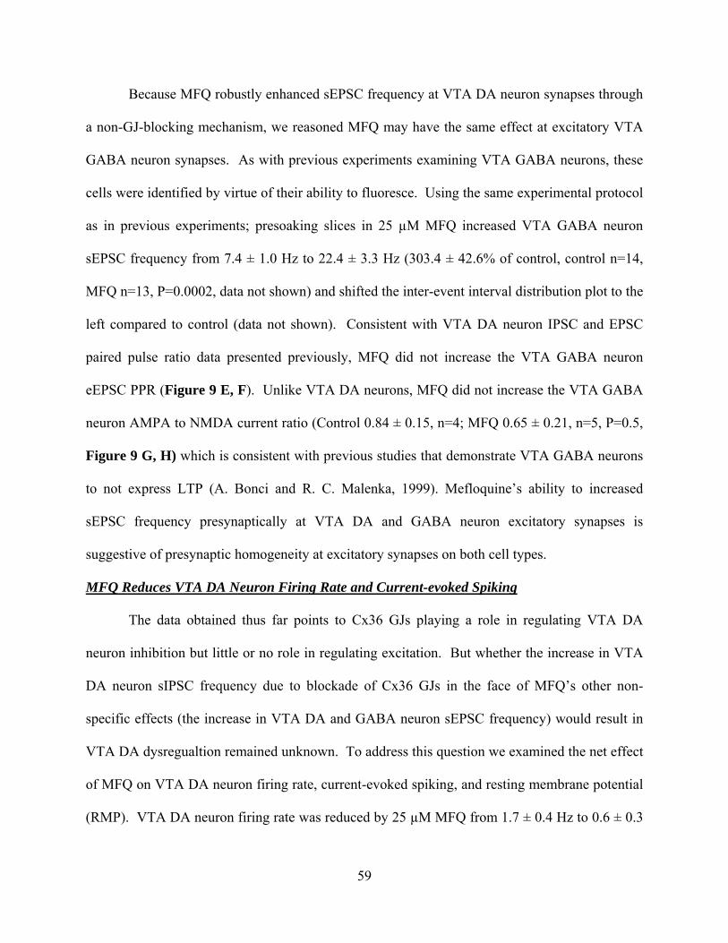
59
Because MFQ robustly enhanced sEPSC frequency at VTA DA neuron synapses through
a non-GJ-blocking mechanism, we reasoned MFQ may have the same effect at excitatory VTA
GABA neuron synapses. As with previous experiments examining VTA GABA neurons, these
cells were identified by virtue of their ability to fluoresce. Using the same experimental protocol
as in previous experiments; presoaking slices in 25 µM MFQ increased VTA GABA neuron
sEPSC frequency from 7.4 ± 1.0 Hz to 22.4 ± 3.3 Hz (303.4 ± 42.6% of control, control n=14,
MFQ n=13, P=0.0002, data not shown) and shifted the inter-event interval distribution plot to the
left compared to control (data not shown). Consistent with VTA DA neuron IPSC and EPSC
paired pulse ratio data presented previously, MFQ did not increase the VTA GABA neuron
eEPSC PPR (Figure 9 E, F). Unlike VTA DA neurons, MFQ did not increase the VTA GABA
neuron AMPA to NMDA current ratio (Control 0.84 ± 0.15, n=4; MFQ 0.65 ± 0.21, n=5, P=0.5,
Figure 9 G, H) which is consistent with previous studies that demonstrate VTA GABA neurons
to not express LTP (A. Bonci and R. C. Malenka, 1999). Mefloquine’s ability to increased
sEPSC frequency presynaptically at VTA DA and GABA neuron excitatory synapses is
suggestive of presynaptic homogeneity at excitatory synapses on both cell types.
MFQ Reduces VTA DA Neuron Firing Rate and Current-evoked Spiking
The data obtained thus far points to Cx36 GJs playing a role in regulating VTA DA
neuron inhibition but little or no role in regulating excitation. But whether the increase in VTA
DA neuron sIPSC frequency due to blockade of Cx36 GJs in the face of MFQ’s other non-
specific effects (the increase in VTA DA and GABA neuron sEPSC frequency) would result in
VTA DA dysregualtion remained unknown. To address this question we examined the net effect
of MFQ on VTA DA neuron firing rate, current-evoked spiking, and resting membrane potential
(RMP). VTA DA neuron firing rate was reduced by 25 µM MFQ from 1.7 ± 0.4 Hz to 0.6 ± 0.3

60
Hz (32.8 ± 17.0% of control, control n=11, MFQ=9, P=0.036, Figure 10B). Current-evoked
spiking was also reduced significantly at all currents greater than 125pA (P=0.001, Figure 10D).
Interestingly, there was no significant difference in VTA DA neuron RPM between control and
MFQ treated slices (Control -47.1 ± 2.3mV, MFQ 47.25 ± 3.8mV, P=0.97).
61
DISCUSSION
The aim of this study was to determine the role Cx36 GJs play in regulating synaptic
activity in the VTA in mice. We were able to indentify VTA GABA neurons through the use of
GAD67-GFP mice, while DA neurons in GAD67-GFP and Cx36 KO mice were identified by
previously tested electrophysiological characteristics. The selective Cx36 GJ-blocker
mefloquine (MFQ) and the less selective GJ-blocker carbenoxolone, in conjunction with the use
of Cx36 KO mice, where utilized to determine the role Cx36 GJs play in VTA synaptic activity.
Mefloquine’s presynaptic increase in VTA DA sIPSCs in WT mice can be divided into two
components: an increase in non-activity-dependent GABA release, most-likely attributed to
MFQ’s ability to increase intracellular calcium, and an increase in activity-dependent GABA
release that we attribute to MFQ’s selective blockade of Cx36 GJs. This is evidenced by the fact
that MFQ increased VTA DA neuron sIPSC frequency ~533% in WT mice, but only ~310% in
Cx36 KO mice. Additionally, mIPSCs (a measure of non-activity-dependent GABA release)
were increased ~300% in both WT and Cx36 KO mice, but carbenoxolone (CBX), a less specific
GJ blocker then MFQ, only increased sIPSC frequency in wildtype mice and did not affect
minis. In addition to this increase in presynaptic GABA release, MFQ also affected postsynaptic
changes on VTA DA neurons by increasing the number and or function of GABA(A) receptors.
Of great interest was our discovery that MFQ did not increase VTA GABA neuron sIPSCs,
suggesting VTA GABA and DA neuron inhibitory synapses are not physiologically
homogenous, and that Cx36 GJs are not involved in regulating VTA GABA neuron inhibition.
The differential effect of MFQ on DA and GABA neuron sIPSCs establishes the possibility that
the negative side-effects of MFQ seen in humans may be related to dysregulation of VTA DA
neuron inhibition.
62
MFQ increased sEPSCs markedly in VTA DA and GABA neurons, a similar
phenomenon to that observed by Cruikshank et al (2004) in rat neocortex. We could not
attribute this increase to MFQ’s GJ blocking properties. Mefloquine’s actions at both VTA DA
and GABA excitatory synapses were due to an increase in presynaptic GLU release. However,
MFQ appeared to only have postsynaptic effects, in the form of an increase in AMPAR to
NMDAR receptor mediated current ratio, in DA neurons. This increase in AMPAR to NMDAR
ratio may involve the increase in number and or function of AMPARs or the decrease in number
and or function of NMDARs, or some combination of the two. These findings validate
observations by Bonci et al (1999), that only VTA DA neurons are capable of this excitatory
postsynaptic plasticity.
We demonstrated the overall net physiologic effect of MFQ was to decrease in VTA DA
neuron firing rate. This net effect of MFQ on VTA synaptic activity demonstrates the
importance of GJs in regulating VTA synaptic activity and poses a scenario in which midbrain
synaptic activity is disrupted sufficiently to provide the physiologic framework to explain many
of MFQ’s adverse side effects in humans. Alterations at these important synapses may lead to
downstream behavioral changes (J. L. Gaiarsa et al., 2002; F. S. Nugent and J. A. Kauer, 2008).
Typically, an increase in the probability of transmitter release would be accompanied by
an increase in the PPR. This was not the case in this study with eIPSCs and eEPSCs. We
attribute the absence of PPD in DA and GABA neurons to the possibility that MFQ’s potent
disruption of intracellular calcium homeostasis prevented the ability to detect this calcium-linked
phenomenon. It is believed that the entry of calcium through presynaptic voltage-sensitive

63
calcium channels leads to the release of transmitter via the fusion of synaptic vesicles from a
readily-releasable pool of vesicles (D. Debanne et al., 1996; C. Rosenmund and C. F. Stevens,
1996; L. E. Dobrunz and C. F. Stevens, 1997). The synaptic machinery regulating the number
and fate of vesicles in this readily-releasable pool are regulated in part by calcium levels within
the presynaptic bouton. The paired-pulse test paradigm relies partly on the constant kinetics of
the replenishment of this readily-releasable pool, and the role calcium plays in replenishing this
pool. We theorize that MFQ’s potent ability to release calcium from internal stores and allow it
to enter from the external environment (G. S. Dow et al., 2003; C. Zhou et al., 2006; D. Caridha
et al., 2008) would alter all calcium-related processes in the presynaptic bouton, including the
replenishment kinetics of the readily-releasable pool, decreasing the odds of a depleted pool in
the face of increased probability of release. Additional evidence suggesting MFQ treated
synapses violate traditionally accepted measures of synaptic activity is our observation that bath-
applied MFQ did not increase the amplitude of the primary IPSC in conjunction with an increase
in mIPSC frequency or increase in the AMPAR to NMDAR current ratio.
We are not alone in the observation that MFQ increases inhibition of midbrain DA
neurons, or the idea that disruption of calcium regulation is involved. Zhou et al (2006) found
that in acutely dissociated brain slices, 3 µM MFQ induced a small increase in sIPSC frequency
in midbrain DA neurons. At 25 µM, we were unable to substantiate Zhous et al’s claims that
MFQ’s increase in midbrain DA neuron sIPSC frequency was facilitated through cholinesterase
inhibition, but we did provide support for the role calcium plays. A possible explanation for the
difference in concentrations of MFQ in these studies may be that effectual concentrations of drug
are lower in whole-brain tissue as a result of diffusion through brain slices. The difficulty of
MFQ’s ability to penetrate brain slice tissue is made more difficult by its propensity to bind
64
tightly to phospholipid membranes. We tested the effects of odansetron as a control against
MFQ’s reported 5HT3 receptor antagonism, to eliminate the possibility the increase in sIPSC and
sEPSC frequency could be due to this MFQ effect. MFQ’s effects on sIPSCs and sEPSCs were
not mimicked by odansetron, but odansetron suppressed both sIPSC and sEPSC frequency,
presenting another possible non-specific drug effect of MFQ that could contribute to its side
effects reported in humans.
Electrical synapses (composed of Cx36 GJs) connecting inhibitory interneurons are
common in certain areas of the brain (for review B. W. Connors and M. A. Long, 2004),
including the midbrain. We have previously shown VTA GABA neurons express Cx36-
mediated electrical synapses (S. C. Steffensen et al., 1998), dye couple, can be blocked by MFQ
(D. W. Allison et al., 2006), and appear to form part of a larger syncytium of GABA neurons in
the reticular formation (M. B. Lassen et al., 2007). While there is evidence to suggest the current
sharing and synchronizing ability of electrical synapses can be modulated (C. E. Landisman and
B. W. Connors, 2005), to our knowledge we are the first to provide evidence of electrical
synapses regulating interneuron inhibition of the primary neuron populations they target. A
possible explanation for this data is that when MFQ blocks electrical synapses the electrically-
coupled VTA GABA network becomes uncoupled, releasing each cell from the resistive network
load. The net result being individual GABA neuron activity is no longer dampened, resulting in
increased GABA release onto target neurons. Under this model, these electrical synapses may
serve to synchronize spiking and propagate action potentials when the network is tightly coupled,
or serve as resistive loads that decrease synchrony and dampen overall network activity when
loosely coupled (R. W. Joyner et al., 1983; R. W. Joyner et al., 1984). The findings of this study

65
strengthen the argument that GJs serve as critical modulators of brain activity and targets for the
clinical treatment of many disorders involving the mesolimbic DA system.
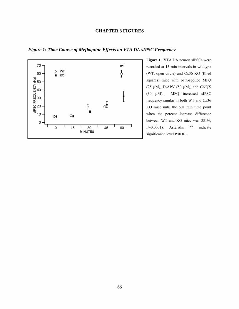
66
CHAPTER 3 FIGURES
Figure 1: Time Course of Mefloquine Effects on VTA DA sIPSC Frequency
Figure 1: VTA DA neuron sIPSCs were
recorded at 15 min intervals in wildtype
(WT, open circle) and Cx36 KO (filled
squares) mice with bath-applied MFQ
(25 µM), D-APV (50 µM), and CNQX
(30 µM). MFQ increased sIPSC
frequency similar in both WT and Cx36
KO mice until the 60+ min time point
when the percent increase difference
between WT and KO mice was 331%,
P=0.0001). Asterisks ** indicate
significance level P<0.01.
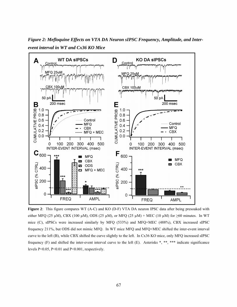
67
Figure 2: Mefloquine Effects on VTA DA Neuron sIPSC Frequency, Amplitude, and Inter-
event interval in WT and Cx36 KO Mice
Figure 2: This figure compares WT (A-C) and KO (D-F) VTA DA neuron IPSC data after being presoaked with
either MFQ (25 µM), CBX (100 µM), ODS (25 µM), or MFQ (25 µM) + MEC (10 µM) for ≥60 minutes. In WT
mice (C), sIPSCs were increased similarly by MFQ (533%) and MFQ+MEC (488%), CBX increased sIPSC
frequency 211%, but ODS did not mimic MFQ. In WT mice MFQ and MFQ+MEC shifted the inter-event interval
curve to the left (B), while CBX shifted the curve slightly to the left. In Cx36 KO mice, only MFQ increased sIPSC
frequency (F) and shifted the inter-event interval curve to the left (E). Asterisks *, **, *** indicate significance
levels P<0.05, P<0.01 and P<0.001, respectively.
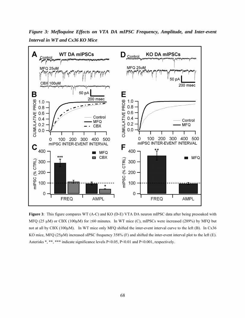
68
Figure 3: Mefloquine Effects on VTA DA mIPSC Frequency, Amplitude, and Inter-event
Interval in WT and Cx36 KO Mice
Figure 3: This figure compares WT (A-C) and KO (D-E) VTA DA neuron mIPSC data after being presoaked with
MFQ (25 µM) or CBX (100µM) for ≥60 minutes. In WT mice (C), mIPSCs were increased (289%) by MFQ but
not at all by CBX (100µM). In WT mice only MFQ shifted the inter-event interval curve to the left (B). In Cx36
KO mice, MFQ (25µM) increased sIPSC frequency 358% (F) and shifted the inter-event interval plot to the left (E).
Asterisks *, **, *** indicate significance levels P<0.05, P<0.01 and P<0.001, respectively.
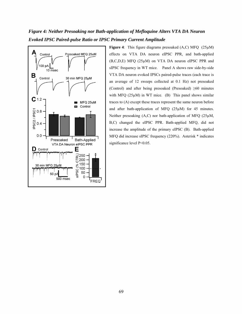
69
Figure 4: Neither Presoaking nor Bath-application of Mefloquine Alters VTA DA Neuron
Evoked IPSC Paired-pulse Ratio or IPSC Primary Current Amplitude Figure 4: This figure diagrams presoaked (A,C) MFQ (25µM)
effects on VTA DA neuron eIPSC PPR, and bath-applied
(B,C,D,E) MFQ (25µM) on VTA DA neuron eIPSC PPR and
sIPSC frequency in WT mice. Panel A shows raw side-by-side
VTA DA neuron evoked IPSCs paired-pulse traces (each trace is
an average of 12 sweeps collected at 0.1 Hz) not presoaked
(Control) and after being presoaked (Presoaked) ≥60 minutes
with MFQ (25µM) in WT mice. (B) This panel shows similar
traces to (A) except these traces represent the same neuron before
and after bath-application of MFQ (25µM) for 45 minutes.
Neither presoaking (A,C) nor bath-application of MFQ (25µM,
B,C) changed the eIPSC PPR. Bath-applied MFQ, did not
increase the amplitude of the primary eIPSC (B). Bath-applied
MFQ did increase sIPSC frequency (220%). Asterisk * indicates
significance level P<0.05.
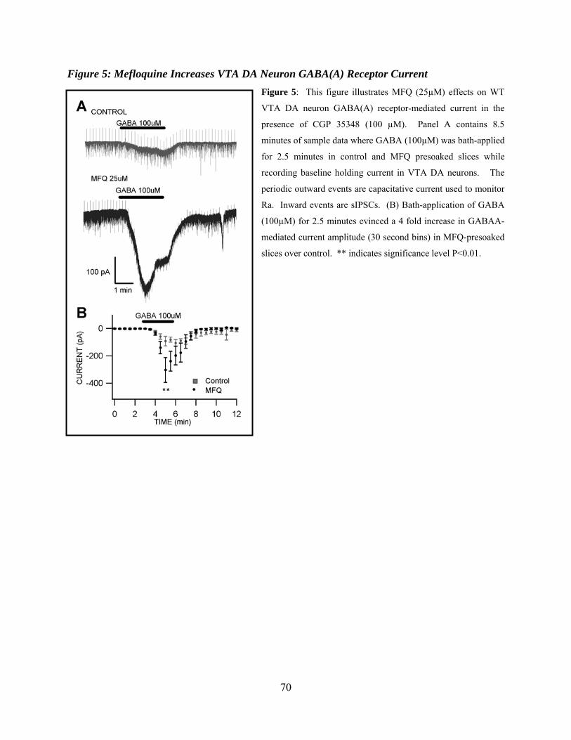
70
Figure 5: Mefloquine Increases VTA DA Neuron GABA(A) Receptor Current Figure 5: This figure illustrates MFQ (25µM) effects on WT
VTA DA neuron GABA(A) receptor-mediated current in the
presence of CGP 35348 (100 µM). Panel A contains 8.5
minutes of sample data where GABA (100µM) was bath-applied
for 2.5 minutes in control and MFQ presoaked slices while
recording baseline holding current in VTA DA neurons. The
periodic outward events are capacitative current used to monitor
Ra. Inward events are sIPSCs. (B) Bath-application of GABA
(100µM) for 2.5 minutes evinced a 4 fold increase in GABAA-
mediated current amplitude (30 second bins) in MFQ-presoaked
slices over control. ** indicates significance level P<0.01.
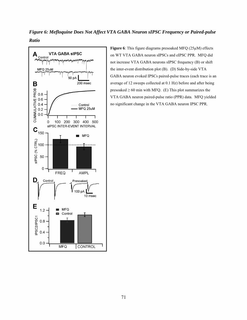
71
Figure 6: Mefloquine Does Not Affect VTA GABA Neuron sIPSC Frequency or Paired-pulse
Ratio Figure 6: This figure diagrams presoaked MFQ (25µM) effects
on WT VTA GABA neuron sIPSCs and eIPSC PPR. MFQ did
not increase VTA GABA neurons sIPSC frequency (B) or shift
the inter-event distribution plot (B). (D) Side-by-side VTA
GABA neuron evoked IPSCs paired-pulse traces (each trace is an
average of 12 sweeps collected at 0.1 Hz) before and after being
presoaked ≥ 60 min with MFQ. (E) This plot summarizes the
VTA GABA neuron paired-pulse ratio (PPR) data. MFQ yielded
no significant change in the VTA GABA neuron IPSC PPR.
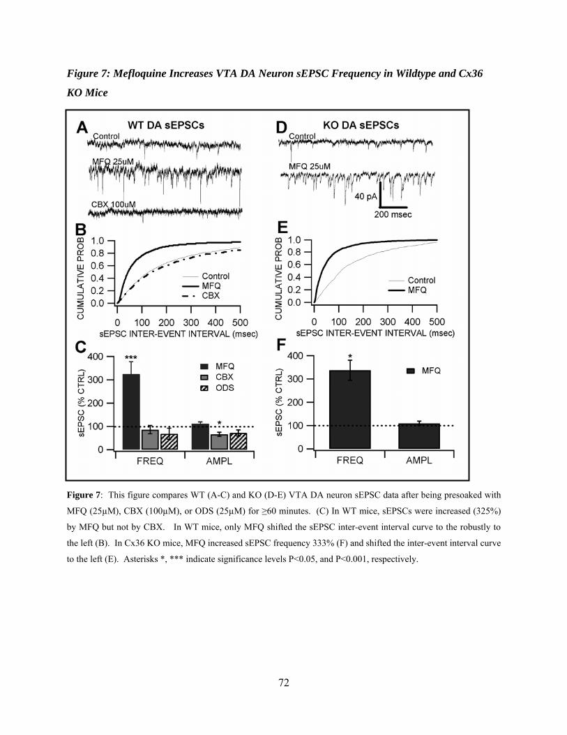
72
Figure 7: Mefloquine Increases VTA DA Neuron sEPSC Frequency in Wildtype and Cx36
KO Mice
Figure 7: This figure compares WT (A-C) and KO (D-E) VTA DA neuron sEPSC data after being presoaked with
MFQ (25µM), CBX (100µM), or ODS (25µM) for ≥60 minutes. (C) In WT mice, sEPSCs were increased (325%)
by MFQ but not by CBX. In WT mice, only MFQ shifted the sEPSC inter-event interval curve to the robustly to
the left (B). In Cx36 KO mice, MFQ increased sEPSC frequency 333% (F) and shifted the inter-event interval curve
to the left (E). Asterisks *, *** indicate significance levels P<0.05, and P<0.001, respectively.
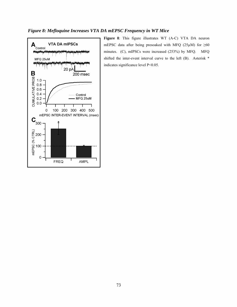
73
Figure 8: Mefloquine Increases VTA DA mEPSC Frequency in WT Mice Figure 8: This figure illustrates WT (A-C) VTA DA neuron
mEPSC data after being presoaked with MFQ (25µM) for ≥60
minutes. (C), mIPSCs were increased (253%) by MFQ. MFQ
shifted the inter-event interval curve to the left (B). Asterisk *
indicates significance level P<0.05.
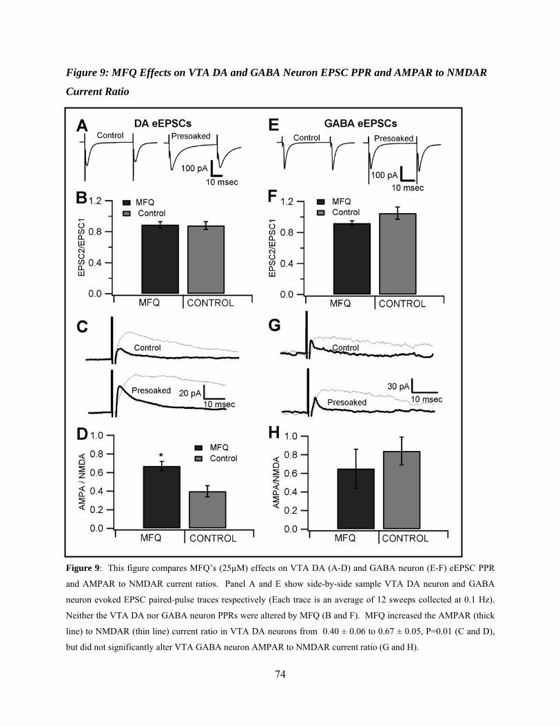
74
Figure 9: MFQ Effects on VTA DA and GABA Neuron EPSC PPR and AMPAR to NMDAR
Current Ratio
Figure 9: This figure compares MFQ’s (25µM) effects on VTA DA (A-D) and GABA neuron (E-F) eEPSC PPR
and AMPAR to NMDAR current ratios. Panel A and E show side-by-side sample VTA DA neuron and GABA
neuron evoked EPSC paired-pulse traces respectively (Each trace is an average of 12 sweeps collected at 0.1 Hz).
Neither the VTA DA nor GABA neuron PPRs were altered by MFQ (B and F). MFQ increased the AMPAR (thick
line) to NMDAR (thin line) current ratio in VTA DA neurons from 0.40 ± 0.06 to 0.67 ± 0.05, P=0.01 (C and D),
but did not significantly alter VTA GABA neuron AMPAR to NMDAR current ratio (G and H).
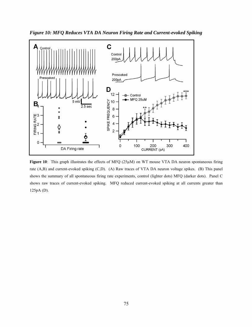
75
Figure 10: MFQ Reduces VTA DA Neuron Firing Rate and Current-evoked Spiking
Figure 10: This graph illustrates the effects of MFQ (25µM) on WT mouse VTA DA neuron spontaneous firing
rate (A,B) and current-evoked spiking (C,D). (A) Raw traces of VTA DA neuron voltage spikes. (B) This panel
shows the summary of all spontaneous firing rate experiments, control (lighter dots) MFQ (darker dots). Panel C
shows raw traces of current-evoked spiking. MFQ reduced current-evoked spiking at all currents greater than
125pA (D).

76
CHAPTER 4
CONCLUSION
The rationale for the cocaine and mefloquine research presented here is predicated on the
belief that advancement in the understanding of the brain mechanisms underlying the
recreational use and abuse potential of cocaine, alcohol and other drugs of abuse will pave the
way for more effective treatment strategies that would save lives and resources throughout the
world. Specifically, a better understanding of the role Cx36 GJ connected VTA GABA neurons
play in mediating cocaine addiction, and regulation of the mesolimbic dopamine (DA) pathway
in the brain. To what end does the research presented here further the aim of that rationale, and
what implications toward future work does it hold? Taking these two studies as one body of
research, they provide compelling evidence for a non-DA-mediated pathway of cocaine
addiction discussed previously. They also provide support for electrically coupled GABA
neurons as key regulators of mesolimbic DA activity. In addition these studies present
electrically coupled GABA neurons as possible candidates for a separate but parallel non-DA-
dependent pathway of addiction for other drugs of abuse such as ethanol, heroin, and nicotine.
The in vivo and in vitro findings in Chapter 2 provide evidence for cocaine’s anesthetic
actions to reinforce its DAT blocking properties, and support for the idea of a non-DA-dependent
mechanism of cocaine reinforcement and reward. In vivo, cocaine and lidocaine both markedly
reduced VTA GABA neuron firing rate and internal capsule driven postsynaptic discharges
(ICPSDs), showing cocaine’s ability to block a putative measure of electrical coupling. In vitro,
cocaine reduced current-driven spikes in VTA GABA neurons via its block of VSSCs leading to
a marked decreased VTA GABA neuron inhibition of VTA DA neurons; demonstrating

77
cocaine’s addictive liability may be greatly increased if its DAT blocking and anesthetic actions
can combine to simultaneously increase DA in the NAcc by blocking DAT and increase DA
release by acting to disinhibit DA neurons in the VTA. This study underscores cocaine’s
dangers as an illicit compound, provides direct evidence of VTA GABA neuron regulation of
mesolimbic DA, and highlights the important role electrical connectivity of VTA GABA neurons
plays in regulating VTA DA neurons.
It should be noted that any research examining the role that electrically-coupled VTA
GABA neurons play in addiction and regulation of the mesolimbic DA system encounters the
same stumbling blocks: the lack of specific GJ blockers and the inability to definitively indentify
GABA neurons in vitro. The study presented in Chapter 3 attempts to overcome these stumbling
blocks by combining the most selective, albeit non-specific, GJ blocking drugs, mefloquine
(MFQ) and carbenoxolone (CBX), with the use of GAD67-GFP and Cx36 KO mouse models.
Some studies have relied exclusively on the use of Cx36 KO mice to study electrical synapses.
By utilizing wildtype and KO mice in conjunction with more than one GJ blocking drug we
avoid many of the pitfalls associated with using only KO mice.
The integral role that electrically-connected VTA GABA neurons play in regulating VTA
DA neurons was addressed in Chapter 3. This research demonstrated that inhibition of VTA DA
neurons could be increased significantly by decreasing the electrical connectivity of VTA GABA
neurons. To our knowledge we are the first to demonstrate GJs playing a critical role in
regulating a well-established physiologically relevant activity in the brain such as excitatory and
inhibitory synaptic activity. With this data, and other evidence suggesting electrical coupling
may play a role in many phenomena such as: rhythmic activity (N. Berretta et al., 2001; M. A.
Long et al., 2002; M. A. Long et al., 2005); propagation of slow calcium waves (L. R. Wolszon

78
et al., 1994); learning and memory; epilepsy (V. M. Nemani and D. K. Binder, 2005); etc (for
review, B. W. Connors and M. A. Long, 2004), we are loath to envision GJs as anything less
than critical brain machinery. This work supports the belief that by virtue of their dynamic range
(high discharge rate and short refractory period), widespread distribution to structures implicated
in drug reward, and their electrical network properties, VTA GABA neurons are a prime target
and focal point for addiction research.
79
REFERENCES
Adell A, Artigas F (2004) The somatodendritic release of dopamine in the ventral tegmental area
and its regulation by afferent transmitter systems. Neurosci Biobehav Rev 28:415-431. Allison DW, Ohran AJ, Stobbs SH, Mameli M, Valenzuela CF, Sudweeks SN, Ray AP,
Henriksen SH, Steffensen SC (2006) Connexin-36 gap junctions mediate electrical coupling between ventral tegmental area GABA neurons. Synapse 60:20-31.
Arumugam H, Denisova JV, Neve RL, Corriveau RA, Belousov AB (2008) Use of calcium imaging for analysis of neuronal gap junction coupling. Neurosci Lett 445:26-30.
Bean BP, Cohen CJ, Tsien RW (1983) Lidocaine block of cardiac sodium channels. J Gen Physiol 81:613-642.
Beckstead MJ, Grandy DK, Wickman K, Williams JT (2004) Vesicular dopamine release elicits an inhibitory postsynaptic current in midbrain dopamine neurons. Neuron 42:939-946.
Bennett SA, Arnold JM, Chen J, Stenger J, Paul DL, Roberts DC (1999) Long-term changes in connexin32 gap junction protein and mRNA expression following cocaine self-administration in rats. Eur J Neurosci 11:3329-3338.
Bergman J, Madras BK, Johnson SE, Spealman RD (1989) Effects of cocaine and related drugs in nonhuman primates. III. Self-administration by squirrel monkeys. J Pharmacol Exp Ther 251:150-155.
Berke JD, Hyman SE (2000) Addiction, dopamine, and the molecular mechanisms of memory. Neuron 25:515-532.
Berretta N, Paolucci E, Bernardi G, Mercuri NB (2001) Glutamate receptor stimulation induces a persistent rhythmicity of the GABAergic inputs to rat midbrain dopaminergic neurons. Eur J Neurosci 14:777-784.
Blackburn JR, Phillips AG, Jakubovic A, Fibiger HC (1986) Increased dopamine metabolism in the nucleus accumbens and striatum following consumption of a nutritive meal but not a palatable non-nutritive saccharin solution. Pharmacology, biochemistry, and behavior 25:1095-1100.
Bonci A, Williams JT (1997) Increased probability of GABA release during withdrawal from morphine. J Neurosci 17:796-803.
Bonci A, Malenka RC (1999) Properties and plasticity of excitatory synapses on dopaminergic and GABAergic cells in the ventral tegmental area. J Neurosci 19:3723-3730.
Bozarth MA, Wise RA (1981) Intracranial self-administration of morphine into the ventral tegmental area. Life Sci 28:551-555.
Caillard O, Ben-Ari Y, Gaiarsa JL (1999) Long-term potentiation of GABAergic synaptic transmission in neonatal rat hippocampus. J Physiol 518 ( Pt 1):109-119.
Cameron DL, Wessendorf MW, Williams JT (1997) A subset of ventral tegmental area neurons is inhibited by dopamine, 5-hydroxytryptamine and opioids. Neuroscience 77:155-166.
Caridha D, Yourick D, Cabezas M, Wolf L, Hudson TH, Dow GS (2008) Mefloquine-induced disruption of calcium homeostasis in mammalian cells is similar to that induced by ionomycin. Antimicrob Agents Chemother 52:684-693.
Carr DB, Sesack SR (2000a) GABA-containing neurons in the rat ventral tegmental area project to the prefrontal cortex. Synapse 38:114-123.
Carr DB, Sesack SR (2000b) Projections from the rat prefrontal cortex to the ventral tegmental area: target specificity in the synaptic associations with mesoaccumbens and mesocortical neurons. J Neurosci 20:3864-3873.
80
Connors BW, Long MA (2004) Electrical synapses in the mammalian brain. Annu Rev Neurosci 27:393-418.
Cruikshank SJ, Hopperstad M, Younger M, Connors BW, Spray DC, Srinivas M (2004) Potent block of Cx36 and Cx50 gap junction channels by mefloquine. Proc Natl Acad Sci U S A 101:12364-12369.
David V, Cazala P (1994) A comparative study of self-administration of morphine into the amygdala and the ventral tegmental area in mice. Behav Brain Res 65:205-211.
Debanne D, Guerineau NC, Gahwiler BH, Thompson SM (1996) Paired-pulse facilitation and depression at unitary synapses in rat hippocampus: quantal fluctuation affects subsequent release. J Physiol 491 ( Pt 1):163-176.
Devine DP, Wise RA (1994) Self-administration of morphine, DAMGO, and DPDPE into the ventral tegmental area of rats. J Neurosci 14:1978-1984.
Do MT, Bean BP (2003) Subthreshold sodium currents and pacemaking of subthalamic neurons: modulation by slow inactivation. Neuron 39:109-120.
Dobrunz LE, Stevens CF (1997) Heterogeneity of release probability, facilitation, and depletion at central synapses. Neuron 18:995-1008.
Dow GS, Hudson TH, Vahey M, Koenig ML (2003) The acute neurotoxicity of mefloquine may be mediated through a disruption of calcium homeostasis and ER function in vitro. Malar J 2:14.
Dow GS, Koenig ML, Wolf L, Gerena L, Lopez-Sanchez M, Hudson TH, Bhattacharjee AK (2004) The antimalarial potential of 4-quinolinecarbinolamines may be limited due to neurotoxicity and cross-resistance in mefloquine-resistant Plasmodium falciparum strains. Antimicrob Agents Chemother 48:2624-2632.
Einhorn LC, Johansen PA, White FJ (1988) Electrophysiological effects of cocaine in the mesoaccumbens dopamine system: studies in the ventral tegmental area. J Neurosci 8:100-112.
Enomoto A, Han JM, Hsiao CF, Wu N, Chandler SH (2006) Participation of sodium currents in burst generation and control of membrane excitability in mesencephalic trigeminal neurons. J Neurosci 26:3412-3422.
Fields HL, Hjelmstad GO, Margolis EB, Nicola SM (2007) Ventral tegmental area neurons in learned appetitive behavior and positive reinforcement. Annu Rev Neurosci 30:289-316.
Fowler JS, Volkow ND, Logan J, Gatley SJ, Pappas N, King P, Ding YS, Wang GJ (1998) Measuring dopamine transporter occupancy by cocaine in vivo: radiotracer considerations. Synapse 28:111-116.
Gaiarsa JL, Caillard O, Ben-Ari Y (2002) Long-term plasticity at GABAergic and glycinergic synapses: mechanisms and functional significance. Trends Neurosci 25:564-570.
Gardner EL, Schiffer WK, Horan BA, Highfield D, Dewey SL, Brodie JD, Ashby CR, Jr. (2002) Gamma-vinyl GABA, an irreversible inhibitor of GABA transaminase, alters the acquisition and expression of cocaine-induced sensitization in male rats. Synapse 46:240-250.
Gawin FH, Ellinwood EH, Jr. (1988) Cocaine and other stimulants. Actions, abuse, and treatment. N Engl J Med 318:1173-1182.
Gifford AN, Johnson KM (1992) Comparison of the role of local anesthetic properties with dopamine uptake blockade in the inhibition of striatal and nucleus accumbens [3H]acetylcholine release by cocaine. J Pharmacol Exp Ther 263:757-761.
81
Grace AA, Onn SP (1989) Morphology and electrophysiological properties of immunocytochemically identified rat dopamine neurons recorded in vitro. J Neurosci 9:3463-3481.
Hemby SE, Co C, Koves TR, Smith JE, Dworkin SI (1997) Differences in extracellular dopamine concentrations in the nucleus accumbens during response-dependent and response-independent cocaine administration in the rat. Psychopharmacology (Berl) 133:7-16.
Hurd YL, Weiss F, Koob GF, And NE, Ungerstedt U (1989) Cocaine reinforcement and extracellular dopamine overflow in rat nucleus accumbens: an in vivo microdialysis study. Brain Res 498:199-203.
Hyman SE, Malenka RC (2001) Addiction and the brain: the neurobiology of compulsion and its persistence. Nat Rev Neurosci 2:695-703.
Hyman SE, Malenka RC, Nestler EJ (2006) Neural Mechanisms of Addiction: The Role of Reward-Related Learning and Memory. Annu Rev Neurosci.
Ikemoto S (2007) Dopamine reward circuitry: Two projection systems from the ventral midbrain to the nucleus accumbens-olfactory tubercle complex. Brain Res Rev 56:27-78.
Johnson SW, North RA (1992) Two types of neurone in the rat ventral tegmental area and their synaptic inputs. J Physiol (Lond) 450:455-468.
Jones S, Bonci A (2005) Synaptic plasticity and drug addiction. Curr Opin Pharmacol 5:20-25. Joyner RW, Picone J, Veenstra R, Rawling D (1983) Propagation through electrically coupled
cells. Effects of regional changes in membrane properties. Circ Res 53:526-534. Joyner RW, Veenstra R, Rawling D, Chorro A (1984) Propagation through electrically coupled
cells. Effects of a resistive barrier. Biophys J 45:1017-1025. Juszczak GR, Swiergiel AH (2009) Properties of gap junction blockers and their behavioural,
cognitive and electrophysiological effects: animal and human studies. Prog Neuropsychopharmacol Biol Psychiatry 33:181-198.
Kalivas PW, Alesdatter JE (1993) Involvement of N-methyl-D-aspartate receptor stimulation in the ventral tegmental area and amygdala in behavioral sensitization to cocaine. J Pharmacol Exp Ther 267:486-495.
Kang J, Jiang L, Goldman SA, Nedergaard M (1998) Astrocyte-mediated potentiation of inhibitory synaptic transmission. Nat Neurosci 1:683-692.
Kano M (1994) Calcium-induced long-lasting potentiation of GABAergic currents in cerebellar Purkinje cells. Jpn J Physiol 44 Suppl 2:S131-136.
Kawaguchi SY, Hirano T (2007) Sustained structural change of GABA(A) receptor-associated protein underlies long-term potentiation at inhibitory synapses on a cerebellar Purkinje neuron. J Neurosci 27:6788-6799.
Kelley AE (2004) Memory and addiction: shared neural circuitry and molecular mechanisms. Neuron 44:161-179.
Kelley AE, Berridge KC (2002) The neuroscience of natural rewards: relevance to addictive drugs. J Neurosci 22:3306-3311.
Khazipov R, Congar P, Ben-Ari Y (1995) Hippocampal CA1 lacunosum-moleculare interneurons: modulation of monosynaptic GABAergic IPSCs by presynaptic GABAB receptors. J Neurophysiol 74:2126-2137.
Kiyatkin EA, Leon Brown P (2006) The role of peripheral and central sodium channels in mediating brain temperature fluctuations induced by intravenous cocaine. Brain Res 1117:38-53.
82
Klink R, Exaerde AK, Zoli M, Changeux JP (2001) Molecular and physiological diversity of nicotinic acetylcholine receptors in the midbrain dopaminergic nuclei. J Neurosci 21:1542-1463.
Komatsu Y, Yoshimura Y (2000) Activity-dependent maintenance of long-term potentiation at visual cortical inhibitory synapses. J Neurosci 20:7539-7546.
Koob GF (1992) Dopamine, addiction and reward. Semin Neurosci 4:139-148. Koob GF, Le Moal M (1997) Drug abuse: hedonic homeostatic dysregulation. Science 278:52-
58. Koob GF, Caine B, Markou A, Pulvirenti L, Weiss F (1994) Role for the mesocortical dopamine
system in the motivating effects of cocaine. NIDA Res Monogr 145:1-18. Kravchenko MO, Moskalyuk AO, Fedulova SA, Veselovsky NS (2006) Calcium-dependent
changes of paired-pulse modulation at single GABAergic synapses. Neurosci Lett 395:133-137.
Kuhar MJ (1992) Molecular pharmacology of cocaine: a dopamine hypothesis and its implications. Ciba Found Symp 166:81-89; discussion 89-95.
Kuhar MJ, Ritz MC, Boja JW (1991) The dopamine hypothesis of the reinforcing properties of cocaine. Trends Neurosci 14:299-302.
Kuznetsov AS, Kopell NJ, Wilson CJ (2006) Transient high-frequency firing in a coupled-oscillator model of the mesencephalic dopaminergic neuron. J Neurophysiol 95:932-947.
Landisman CE, Connors BW (2005) Long-term modulation of electrical synapses in the mammalian thalamus. Science 310:1809-1813.
Lassen MB, Brown JE, Stobbs SH, Gunderson SH, Maes L, Valenzuela CF, Ray AP, Henriksen SJ, Steffensen SC (2007) Brain stimulation reward is integrated by a network of electrically coupled GABA neurons. Brain Res 1156:46-58.
Lee RS, Steffensen SC, Henriksen SJ (2001) Discharge profiles of ventral tegmental area GABA neurons during movement, anesthesia, and the sleep-wake cycle. J Neurosci 21:1757-1766.
Liu QS, Pu L, Poo MM (2005) Repeated cocaine exposure in vivo facilitates LTP induction in midbrain dopamine neurons. Nature 437:1027-1031.
Livak KJ, Schmittgen TD (2001) Analysis of relative gene expression data using real-time quantitative PCR and the 2(-Delta Delta C(T)) Method. Methods 25:402-408.
Llinas R, Sugimori M, Simon SM (1982) Transmission by presynaptic spike-like depolarization in the squid giant synapse. Proc Natl Acad Sci U S A 79:2415-2419.
Long MA, Deans MR, Paul DL, Connors BW (2002) Rhythmicity without synchrony in the electrically uncoupled inferior olive. J Neurosci 22:10898-10905.
Long MA, Jutras MJ, Connors BW, Burwell RD (2005) Electrical synapses coordinate activity in the suprachiasmatic nucleus. Nat Neurosci 8:61-66.
Malenka RC, Nicoll RA (1999) Long-term potentiation--a decade of progress? Science 285:1870-1874.
Margolis EB, Lock H, Hjelmstad GO, Fields HL (2006) The ventral tegmental area revisited: is there an electrophysiological marker for dopaminergic neurons? J Physiol 577:907-924.
Margolis EB, Mitchell JM, Ishikawa J, Hjelmstad GO, Fields HL (2008) Midbrain dopamine neurons: projection target determines action potential duration and dopamine D(2) receptor inhibition. J Neurosci 28:8908-8913.

83
McArdle JJ, Sellin LC, Coakley KM, Potian JG, Hognason K (2006) Mefloquine selectively increases asynchronous acetylcholine release from motor nerve terminals. Neuropharmacology 50:345-353.
McKinzie DL, Rodd-Henricks ZA, Dagon CT, Murphy JM, McBride WJ (1999) Cocaine is self-administered into the shell region of the nucleus accumbens in Wistar rats. 877:788-791.
McLean HA, Caillard O, Ben-Ari Y, Gaiarsa JL (1996) Bidirectional plasticity expressed by GABAergic synapses in the neonatal rat hippocampus. J Physiol 496 ( Pt 2):471-477.
Melis M, Camarini R, Ungless MA, Bonci A (2002) Long-lasting potentiation of GABAergic synapses in dopamine neurons after a single in vivo ethanol exposure. J Neurosci 22:2074-2082.
Mennerick S, Zorumski CF (1995) Paired-pulse modulation of fast excitatory synaptic currents in microcultures of rat hippocampal neurons. J Physiol 488 ( Pt 1):85-101.
Nemani VM, Binder DK (2005) Emerging role of gap junctions in epilepsy. Histol Histopathol 20:253-259.
Nugent FS, Kauer JA (2008) LTP of GABAergic synapses in the ventral tegmental area and beyond. J Physiol 586:1487-1493.
Nugent FS, Penick EC, Kauer JA (2007) Opioids block long-term potentiation of inhibitory synapses. Nature 446:1086-1090.
O'Leary ME, Chahine M (2002) Cocaine binds to a common site on open and inactivated human heart (Na(v)1.5) sodium channels. J Physiol 541:701-716.
Olson VG, Nestler EJ (2007) Topographical organization of GABAergic neurons within the ventral tegmental area of the rat. Synapse 61:87-95.
Omelchenko N, Sesack SR (2005) Laterodorsal tegmental projections to identified cell populations in the rat ventral tegmental area. J Comp Neurol 483:217-235.
Omelchenko N, Sesack SR (2006) Cholinergic axons in the rat ventral tegmental area synapse preferentially onto mesoaccumbens dopamine neurons. J Comp Neurol 494:863-875.
Pan HT, Menacherry S, Justice JB, Jr. (1991) Differences in the pharmacokinetics of cocaine in naive and cocaine-experienced rats. J Neurochem 56:1299-1306.
Pettit HO, Justice JB, Jr. (1989) Dopamine in the nucleus accumbens during cocaine self-administration as studied by in vivo microdialysis. Pharmacol Biochem Behav 34:899-904.
Pettit HO, Justice JB, Jr. (1991) Effect of dose on cocaine self-administration behavior and dopamine levels in the nucleus accumbens. Brain Res 539:94-102.
Pettit HO, Pan HT, Parsons LH, Justice JB, Jr. (1990) Extracellular concentrations of cocaine and dopamine are enhanced during chronic cocaine administration. J Neurochem 55:798-804.
Pierce RC, Kumaresan V (2006) The mesolimbic dopamine system: the final common pathway for the reinforcing effect of drugs of abuse? Neurosci Biobehav Rev 30:215-238.
Postma SW, Catterall WA (1984) Inhibition of binding of [3H]batrachotoxinin A 20-alpha-benzoate to sodium channels by local anesthetics. Mol Pharmacol 25:219-227.
Ritz MC, Lamb RJ, Goldberg SR, Kuhar MJ (1987) Cocaine receptors on dopamine transporters are related to self-administration of cocaine. Science 237:1219-1223.
Roberts DCS, Koob GF, Klonoff P, Fibiger HC (1980) Extinction and recovery of cocaine self-administration following intraventricular 6-hydroxydopamine lesions of the nucleus accumbens. Pharmacol Biochem Behav 24:881-887.

84
Rosenmund C, Stevens CF (1996) Definition of the readily releasable pool of vesicles at hippocampal synapses. Neuron 16:1197-1207.
Rothman RB, Baumann MH, Dersch CM, Romero DV, Rice KC, Carroll FI, Partilla JS (2001) Amphetamine-type central nervous system stimulants release norepinephrine more potently than they release dopamine and serotonin. Synapse 39:32-41.
Ruffieux A, Schultz W (1981) Influence of dopamine on pars reticulata neurones of substantia nigra. J Physiol (Paris) 77:63-69.
Salin PA, Malenka RC, Nicoll RA (1996) Cyclic AMP mediates a presynaptic form of LTP at cerebellar parallel fiber synapses. Neuron 16:797-803.
Sarti F, Borgland SL, Kharazia VN, Bonci A (2007) Acute cocaine exposure alters spine density and long-term potentiation in the ventral tegmental area. Eur J Neurosci 26:749-756.
Schindler CW, Tella SR, Erzouki HK, Goldberg SR (1995) Pharmacological mechanisms in cocaine's cardiovascular effects. Drug Alcohol Depend 37:183-191.
Schlagenhauf P, Johnson R, Schwartz E, Nothdurft HD, Steffen R (2009) Evaluation of mood profiles during malaria chemoprophylaxis: a randomized, double-blind, four-arm study. J Travel Med 16:42-45.
Sesack SR, Aoki C, Pickel VM (1994) Ultrastructural localization of D2 receptor-like immunoreactivity in midbrain dopamine neurons and their targets. J Neurosci 14:88-106.
Shi WX, Pun CL, Zhou Y (2004) Psychostimulants induce low-frequency oscillations in the firing activity of dopamine neurons. Neuropsychopharmacology 29:2160-2167.
Shriver DA, Long JP (1971) A pharmacologic comparison of some quaternary derivatives of cocaine. Arch Int Pharmacodyn Ther 189:198-208.
Steffensen SC, Henriksen SJ, Wilson MC (1999) Transgenic rescue of SNAP-25 restores dopamine-modulated synaptic transmission in the coloboma mutant. Brain Res 847:186-195.
Steffensen SC, Svingos AL, Pickel VM, Henriksen SJ (1998) Electrophysiological characterization of GABAergic neurons in the ventral tegmental area. J Neurosci 18:8003-8015.
Steffensen SC, Taylor SR, Horton ML, Barber EN, Lyle LT, Stobbs SH, Allison DW (2008) Cocaine disinhibits dopamine neurons in the ventral tegmental area via use-dependent blockade of GABA neuron voltage-sensitive sodium channels. Eur J Neurosci 28:2028-2040.
Stobbs SH, Ohran AJ, Lassen MB, Allison DW, Brown JE, Steffensen SC (2004) Ethanol suppression of ventral tegmental area GABA neuron electrical transmission involves NMDA receptors. J Pharmacol Exp Ther 311:282-289.
Strichartz G (1976) Molecular mechanisms of nerve block by local anesthetics. Anesthesiology 45:421-441.
Tamamaki N, Yanagawa Y, Tomioka R, Miyazaki J, Obata K, Kaneko T (2003) Green fluorescent protein expression and colocalization with calretinin, parvalbumin, and somatostatin in the GAD67-GFP knock-in mouse. J Comp Neurol 467:60-79.
Theile JW, Morikawa H, Gonzales RA, Morrisett RA (2008) Ethanol enhances GABAergic transmission onto dopamine neurons in the ventral tegmental area of the rat. Alcohol Clin Exp Res 32:1040-1048.
Theile JW, Morikawa H, Gonzales RA, Morrisett RA (2009) Role of 5-hydroxytryptamine2C receptors in Ca2+-dependent ethanol potentiation of GABA release onto ventral tegmental area dopamine neurons. J Pharmacol Exp Ther 329:625-633.
85
Thompson AJ, Lummis SC (2008) Antimalarial drugs inhibit human 5-HT(3) and GABA(A) but not GABA(C) receptors. Br J Pharmacol 153:1686-1696.
Thompson AJ, Lochner M, Lummis SC (2007) The antimalarial drugs quinine, chloroquine and mefloquine are antagonists at 5-HT3 receptors. Br J Pharmacol 151:666-677.
Tran TM, Browning J, Dell ML (2006) Psychosis with paranoid delusions after a therapeutic dose of mefloquine: a case report. Malar J 5:74.
Ungless MA, Magill PJ, Bolam JP (2004) Uniform inhibition of dopamine neurons in the ventral tegmental area by aversive stimuli. Science 303:2040-2042.
Ungless MA, Whistler JL, Malenka RC, Bonci A (2001) Single cocaine exposure in vivo induces long-term potentiation in dopamine neurons. Nature 411:583-587.
Vargas-Perez H, Kee RT, Walton CH, Hansen DM, Razavi R, Clarke L, Bufalino MR, Allison DW, Steffensen SC, van der Kooy D (2009) Ventral tegmental area BDNF induces an opiate-dependent-like reward state in naive rats. Science 324:1732-1734.
Waddington JL (1997) Do D2-like dopamine receptors mediate neuronal excitation or inhibition: some functional-behavioural implications. Nihon Shinkei Seishin Yakurigaku Zasshi 17:111-112.
Waszczak BL, Walters JR (1986) Endogenous dopamine can modulate inhibition of substantia nigra pars reticulata neurons elicited by GABA iontophoresis or striatal stimulation. J Neurosci 6:120-126.
Weiler R, Pottek M, He S, Vaney DI (2000) Modulation of coupling between retinal horizontal cells by retinoic acid and endogenous dopamine. Brain Res Brain Res Rev 32:121-129.
Welzl H, Kuhn G, Huston JP (1989) Self-administration of small amounts of morphine through glass micropipettes into the ventral tegmental area of the rat. Neuropharmacology 28:1017-1023.
Wilcox KM, Paul IA, Woolverton WL (1999) Comparison between dopamine transporter affinity and self-administration potency of local anesthetics in rhesus monkeys. Eur J Pharmacol 367:175-181.
Wise RA (1996) Addictive drugs and brain stimulation reward. Annu Rev Neurosci 19:319-340. Wise RA (2004) Dopamine, learning and motivation. Nat Rev Neurosci 5:483-494. Wise RA, Bozarth MA (1987) A psychostimulant theory of addiction. Psychol Rev 94. Wise RA, Newton P, Leeb K, Burnette B, Pocock D, Justice JB, Jr. (1995) Fluctuations in
nucleus accumbens dopamine concentration during intravenous cocaine self-administration in rats. Psychopharmacology (Berl) 120:10-20.
Wolszon LR, Rehder V, Kater SB, Macagno ER (1994) Calcium wave fronts that cross gap junctions may signal neuronal death during development. J Neurosci 14:3437-3448.
Wooltorton E (2002) Mefloquine: contraindicated in patients with mood, psychotic or seizure disorders. CMAJ 167:1147.
Woolverton WL, Johnson KM (1992) Neurobiology of cocaine abuse. Trends Pharmacol Sci 13:193-200.
Yamaguchi T, Sheen W, Morales M (2007) Glutamatergic neurons are present in the rat ventral tegmental area. Eur J Neurosci 25:106-118.
Ye JH, Ren J, Krnjevic K, Liu PL, McArdle JJ (1999) Cocaine and lidocaine have additive inhibitory effects on the GABAA current of acutely dissociated hippocampal pyramidal neurons. Brain Res 821:26-32.
Zhou C, Xiao C, McArdle JJ, Ye JH (2006) Mefloquine enhances nigral gamma-aminobutyric acid release via inhibition of cholinesterase. J Pharmacol Exp Ther 317:1155-1160.
86
Curriculum vitae
David W. Allison
Personal Data Graduate Student, PhD Candidate (Neuroscience)
Department of Psychology and Neuroscience Center Brigham Young University, 1290 SWKT Provo, Utah 84602 (801) 422-6089 [email protected]
Education
2010 1997
PhD Neuroscience, Brigham Young University, Utah BS in Biochemistry/ Cell Biology, University of California San Diego
Professional Experience
2006-2010 2001-2006 1998-2001
Awards
Graduate Student, (PhD candidate- Neuroscience) Faculty Research Associate, Brigham Young University Research Technician II, The Scripps Research Institute 2009/2010 Brigham Young University Graduate Student Fellowship Award- “Cocaine disinhibits VTA dopamine neurons via its anesthetic effects”
Publications Refereed Journal Articles
Allison, D.W., Ellefsen, K.L., Askew, C.E., Wilcox, B.S., Sandoval, S.S., Hansen, D.M., Yanagawa, Y., Kaneke, T., Steffensen, S.C. Cocaine and Mefloquine-induced Acute Effects in Ventral Tegmental Area Dopamine and GABA Neurons. J Neurosci (in submission). Vargas-Perez H., Ting-A-Kee R., Walton C.H., Hansen D.M., Razavi R., Clarke L., Bufalino M.R., Allison D.W., Steffensen S.C., van der Kooy D. Ventral Tegmental Aream BDNF Induces Opiate-Dependent-Like Reward in Naïve Rats. Science. 2009 May 28. Hales, K.H., Bradley, K.D., Allison, D.W., Taylor S.R., Hansen, D.M., Yorgason, J.T., Walton, C.H., Sudweeks, S.N. and Steffensen, S.C. Physiological and molecular adaptation of the GABA neurons in the ventral tegmental area to chronic alcohol consumption. Alcohol Clin Exp Res. 2008 Dec (in press) Steffensen, S.C., Taylor, S.R., Horton, M.L., Barber, E.N., Lyle, L.T., Stobbs, S.H. and Allison, D.W. Cocaine disinhibits dopamine neurons in the ventral tegmental area via use-
87
dependent blockade of GABA neuron voltage-sensitive sodium channels. Eur J Neurosci. 2008 Nov; 28(10):2028-40. Steffensen, S.C., Hales, K., Jones, M., Allison, D.W. Dehydroepiandrosterone sulfate and estrone sulfate reduce GABA recurrent inhibition in the hippocampus via muscarinic receptors. Hippocampus (2006) 16(12):1080-90 Allison, D.W., Ohran, A.L., Stobbs, S.H., Mameli, M., Valenzuela, F., Sudweeks, S.N., Ray, A.P., Henriksen, S.J. and Steffensen, S.C. Involvement of Connexin-36 Gap Junctions in Ventral Tegmental Area GABA neuron excitability and electrical coupling. Synapse, In Press (2006) Stobbs, S.H., Ohran, A.L., Lassen, M.B., Allison, D.W., Brown, J.E. and Steffensen, S.C., Ethanol suppression of ventral tegmental area GABA neuron electrical transmission involves NMDA receptors. J Pharmacol Exp Ther. 311 (2004) 282-289 Krucker T., Siggins G.R., McNamara R.K., Lindsley K.A., Dao A., Allison D.W., De Lecea L, Lovenberg TW, Sutcliffe JG, Gerendasy D.D. Targeted disruption of RC3 reveals a calmodulin-based mechanism for regulating metaplasticity in the hippocampus. Journal of Neuroscience. (2002) Jul 1; 22(13): 5525-35 Dissertation (in preparation) Allison, D.W. Cocaine and Mefloquine-induced Acute Effects in Ventral Tegmental Area Dopamine and GABA Neurons. Submitted to the faculty of Brigham Young University, Provo Utah 2009.
Abstracts Allison, D. W., Askew, C. E., Ellefsen, K. L., Hamaker, A. N., Steffensen S. C. Ethanol effects on midbrain dopamine neurons in connexin 36 knock out mice. Soc. Neurosci. Absts 2009 233.50 Steffensen, S. C., Bradley, K. D., Allison, D. W., Hansen, D. M., Wilcox, J. D., Foote, M., Hoyt, B., Yorgason, J. T. The role of connexin-36 gap junctions in alcohol intoxication and reward. Soc. Neurosci. Absts 2009 233.60 Allison, D.W., Hansen, D.M., Wilcox, J.D., Yorgason, J.T., Steffensen, S.C. Effects of dopamine receptor antagonists on ethanol-induced modulation of dopamine release in the nucleus accumbens. Alcoholism: Clin. Exp. Res. (2009) 33(6) 037 Steffensen, S. C., Flemming, D. E., Taylor, S. R., Hansen, D.M., Walton, C.H., Allison, D. W. Cocaine disinhibits dopamine neurons in the ventral tegmental area via use-dependent blockade of GABA neuron voltage-sensitive sodium channels. Soc. Neurosci. Absts 2008 661.20

88
Allison, D.W., Taylor, S.R., Askew, C.E., Anderson, E., Partridge, L.D., Valenzuela, C.F., Steffensen, S. C. Pregnenolone sulfate enhances GABA inhibition in the CA1 subfield of the hippocampus. Soc. Neurosci. Absts 2008 589.1 Steffensen, S. C., Allison, D.W., Yorgason, J.T., Hansen, D.M. The retina as a window on alcohol effects on mesolimbic dopamine neurotransmission. Alcoholism: Clin. Exp. Res. (2008) 32(6) 076 Steffensen, S. C., Hales, K., Bradely, K., Trika, A., Hansen, D.M., Yorgason, J.T., Walton, C.H., Thomas, S. J., Taylor, S. R., Askew, C.E., Allison, D.W. Physiological and molecular adaptation of ventral tegmental area GABA neurons to chronic alcohol. Alcoholism: Clin. Exp. Res. (2008) 32(6) 064 Criado, J.R., Lassen, M.B., Allison, D.W., Hansen, D.M., Layton, S.P., Steffensen, S. C. Opiod effects on the dentate gyrus responses are mediated by effects on mu-opioid receptor containing GABA neurons in the ventral tegmental area. Soc. Neurosci. Absts 2007 611.10 Steffensen, S. C., Allison, D.W., Hales, K., Mickelsen, R., Maes, L., Valenzuela, C.F., Effects of acute and chronic ethanol on the excitability and electrical coupling of GABA neurons in the ventral tegmental area. Soc. Neurosci. Absts 2007 611.13 Allison, D.W., Mickelsen, R., Taylor, S. R., Lassen, M. B., Flemming, D. E., Steffensen, S. C., Thomas, S. J. Cocaine reduces the excitability and electrical coupling of VTA GABA neurons via its sodium channel blocking actions. Soc. Neurosci. Absts 2007 912.11 Hales, K., Allison, D.W., Valenzuela, C.F., Maes, L., Steffensen, S. C. Effects of acute and chronic ethanol on the excitability and electrical coupling of GABA neurons in the ventral tegmental area. Alcoholism: Clin. Exp. Res. (2007) 31(6) 538 Allison D.W., Mickelsen, R.S., Steffensen, S. C., Effects of ethanol on the excitability and electrical coupling of GABA neurons in the midbrain. Alcoholism: Clin. Exp. Res. (2007) 31(6) 543 Steffensen, S.C., Barber, E.N., Horton, M.L., Hansen, D.M., Mickelsen, R., Rogers, D.C., Clark, T. and Allison, D.W. Contigent and non-contingent effects of cocaine on the discharge activity and electrical coupling of ventral tegmental area GABA neurons. Soc. Neurosci. Absts 2006 32: 691.2
Allison, D.W., Horton, M.L., Sudweeks, S.N., Valenzuela, F.C. and Steffensen, S.C., Ethanol modulates electrical coupling between GABA neurons in the ventral tegmental area. Soc. Neurosci. Absts (2006) 32: 292.10
Steffensen, S.C., Horton, M.L., and Allison, D.W. Ethanol influences T-type calcium currents and electrical coupling between GABA neurons in the ventral tegmental area. Alcoholism: Clin. Exp. Res. (2006) 30(6) 182A
89
Allison, D.W., Hawes, M.E., Horton, M.L., Stobbs, S.H., Sudweeks, S.N., Steffensen, S.C. Ethanol modulates electrical coupling between GABA neurons in the ventral tegmental area. Soc. Neurosci. Absts. (2005) Steffensen S.C., Ohran A.J., Bevan A., Haws M.E., Allison, D.W., and Stobbs S.H. Ethanol modulates electrical coupling between GABA neurons in the ventral tegmental area. Alcoholism. Clin. Exp. Res. (2005) 29(5) 93A Steffensen S.C., Ohran A.L., Stobbs S.H., Allison D.W., Lassen M.B., Ray A.P., Henriksen S.J. Ventral tegmental area GABA neurons form a network of dopamine-sensitive electrical synapses. Soc. Neurosci. Absts. (2004) 30: 272.9 Steffensen, S.C., Stobbs, S.H., Allison, D.W., Lassen, M.B., Brown, J.E., Henriksen, S.J. Ventral tegmental area GABA neurons form a network of dopamine-sensitive electrical synapses: Role in brain stimulation reward. Soc. Neurosci. Absts. (2003) 29: 679.4 Steffensen, S.C., Stobbs, S.H., Allison, D.W., Brown, J.E., Lassen, M.B., Lee, R.S., and Henriksen, S.J. Differential adaptation of ventral tegmental area GABA neurons by ethanol and barbiturates. Alcoholism: Clin. Exp. Res. (2003) 27(5) 56A Krucker, T., Mosley, A., Allison, D.W., Siggins, G.R. Methamphetamine treatment modifies synaptic transmission and plasticity in hippocampal slices from wild type and transgenic mice with cerebral expression of HIV-1 coat protein GP120. Soc. Neurosci. Absts. (2002) 808.18 Meyer, E.P., Allison, D.W., Staufenbiel, M., Siggins, G.R., Krucker T. Three-dimensional reconstruction of early changes in microvascular architecture and morphology in APP23 transgenic mice with neural overexpression of mutant amyloid precursor protein. Soc. Neurosci. Absts. (2001) 321.14 Meyer, E.P., Allison, D.W., Campbell, I.L., Siggins, G.R., Krucker, T. The microvasculature of transgenic mice with glial expression of interleukin 6 shows extensive morphological changes as revealed by vascular corrosion casts. Soc. Neurosci. Absts. (2000) 27.9 Krucker, T., Allison, D.W, Staufenbiel, M., Sommer, B., Sturchler-Pierrat, C., Siggins, G.R. Changes in excitabililty and long-term plasticity in hippocampal CA1 of transgenic mice with neuronal overexpression of mutant amyloid precursor protein. Soc. Neurosci. Absts. (2000) 275.15 Manuscripts in preparation Allison, D.W., Ellefsen, K., Askew, C.L., Steffensen, S.C. Connexin 36 Gap Junctions Play an Integral Role in Regulating Ventral Tegmental Area Synaptic Activity. December 2009 Membership in Professional Societies
90
Society for Neuroscience (Member from 2001-present)
Teaching Experience 2001-present Psychology 370: Sensation and Perception
Brigham Young University Department of Psychology
Winter 2009 2001-2003
Neuroscience 205: Neurobiology Brigham Young University Neuroscience Center Neuroscience 480: Advanced Neuroscience Brigham Young University Neuroscience Center
2001-present Neuroscience 481: Advanced Neuroscience Laboratory Brigham Young University Neuroscience Center
2001-present Neuroscience 449R: Research Brigham Young University Neuroscience Center
Licenses held
Amateur radio license, KF7BDU Community professional service
Utah State Science Fair Judge CUSEF 2006-2008