Co-operation of TLR4 and raft proteins in LPS-induced pro ... · Co-operation of TLR4 and raft...
25
REVIEW Co-operation of TLR4 and raft proteins in LPS-induced pro-inflammatory signaling Agnieszka Plo ´ciennikowska • Aneta Hromada-Judycka • Kinga Borze ˛cka • Katarzyna Kwiatkowska Received: 14 July 2014 / Revised: 1 October 2014 / Accepted: 13 October 2014 / Published online: 22 October 2014 Ó The Author(s) 2014. This article is published with open access at Springerlink.com Abstract Toll-like receptor 4 (TLR4) is activated by lipopolysaccharide (LPS), a component of Gram-negative bacteria to induce production of pro-inflammatory media- tors aiming at eradication of the bacteria. Dysregulation of the host responses to LPS can lead to a systemic inflam- matory condition named sepsis. In a typical scenario, activation of TLR4 is preceded by binding of LPS to CD14 protein anchored in cholesterol- and sphingolipid-rich microdomains of the plasma membrane called rafts. CD14 then transfers the LPS to the TLR4/MD-2 complex which dimerizes and triggers MyD88- and TRIF-dependent pro- duction of pro-inflammatory cytokines and type I interferons. The TRIF-dependent signaling is linked with endocytosis of the activated TLR4, which is controlled by CD14. In addition to CD14, other raft proteins like Lyn tyrosine kinase of the Src family, acid sphingomyelinase, CD44, Hsp70, and CD36 participate in the TLR4 signaling triggered by LPS and non-microbial endogenous ligands. In this review, we summarize the current state of the knowledge on the involvement of rafts in TLR4 signaling, with an emphasis on how the raft proteins regulate the TLR4 signaling pathways. CD14-bearing rafts, and possi- bly CD36-rich rafts, are believed to be preferred sites of the assembly of a multimolecular complex which mediates the endocytosis of activated TLR4. Keywords Lipopolysaccharide Á Toll-like receptor 4 Á Plasma membrane rafts Á CD14 Á Lyn tyrosine kinase Á Acid sphingomyelinase Á CD36 Introduction Toll-like receptors (TLRs) recognize a variety of micro- bial structural components called pathogen-associated molecular patterns (PAMPs). Upon recognition of the PAMPs, TLRs trigger production of pro-inflammatory mediators helping to eradicate infection. Until now, thir- teen TLRs have been identified and described in mammals, twelve of which are expressed in mice and ten in humans. The discovery of the role of TLRs has greatly advanced the field of innate immunology and was hon- ored with the Nobel Prize to Jules Hoffmann and Bruce Beutler in 2011. The Beutler’s group has revealed that TLR4 is activated by lipopolysaccharide (LPS, endo- toxin), a component of the outer membrane of Gram- negative bacteria. LPS is anchored in the bacterial membrane by up to seven acyl chains composing so- called lipid A which is bound to an oligosaccharide core and a highly variable polysaccharide chain named O- antigen. Lipid A is the most evolutionarily conserved part of LPS responsible for its pro-inflammatory activity. The maximal potency to trigger inflammation is shown by LPS with a bis-phosphorylated lipid A composed of six saturated acyl chains. The pro-inflammatory action of LPS is crucial for curbing bacterial infections, but excessive host responses to LPS can lead to systemic inflammatory conditions—sepsis, severe sepsis, and fatal septic shock. The incidence of severe sepsis in the European Union has been estimated at 90.4 cases per 100,000 population. In the United States, severe sepsis causes approximately 215,000 deaths per year (nearly as many as lung, colorectal, and breast cancers together). The mortality of severe sepsis reaches 30–50 % world- wide and the absence of efficient therapies makes studies on the molecular mechanisms of activation of cells by A. Plo ´ciennikowska Á A. Hromada-Judycka Á K. Borze ˛cka Á K. Kwiatkowska (&) Laboratory of Molecular Membrane Biology, Nencki Institute of Experimental Biology, 3 Pasteur St., 02-093 Warsaw, Poland e-mail: [email protected] Cell. Mol. Life Sci. (2015) 72:557–581 DOI 10.1007/s00018-014-1762-5 Cellular and Molecular Life Sciences 123
Transcript of Co-operation of TLR4 and raft proteins in LPS-induced pro ... · Co-operation of TLR4 and raft...

REVIEW
Co-operation of TLR4 and raft proteins in LPS-inducedpro-inflammatory signaling
Agnieszka Płociennikowska • Aneta Hromada-Judycka •
Kinga Borzecka • Katarzyna Kwiatkowska
Received: 14 July 2014 / Revised: 1 October 2014 / Accepted: 13 October 2014 / Published online: 22 October 2014
� The Author(s) 2014. This article is published with open access at Springerlink.com
Abstract Toll-like receptor 4 (TLR4) is activated by
lipopolysaccharide (LPS), a component of Gram-negative
bacteria to induce production of pro-inflammatory media-
tors aiming at eradication of the bacteria. Dysregulation of
the host responses to LPS can lead to a systemic inflam-
matory condition named sepsis. In a typical scenario,
activation of TLR4 is preceded by binding of LPS to CD14
protein anchored in cholesterol- and sphingolipid-rich
microdomains of the plasma membrane called rafts. CD14
then transfers the LPS to the TLR4/MD-2 complex which
dimerizes and triggers MyD88- and TRIF-dependent pro-
duction of pro-inflammatory cytokines and type I
interferons. The TRIF-dependent signaling is linked with
endocytosis of the activated TLR4, which is controlled by
CD14. In addition to CD14, other raft proteins like Lyn
tyrosine kinase of the Src family, acid sphingomyelinase,
CD44, Hsp70, and CD36 participate in the TLR4 signaling
triggered by LPS and non-microbial endogenous ligands.
In this review, we summarize the current state of the
knowledge on the involvement of rafts in TLR4 signaling,
with an emphasis on how the raft proteins regulate the
TLR4 signaling pathways. CD14-bearing rafts, and possi-
bly CD36-rich rafts, are believed to be preferred sites of the
assembly of a multimolecular complex which mediates the
endocytosis of activated TLR4.
Keywords Lipopolysaccharide � Toll-like receptor 4 �Plasma membrane rafts � CD14 � Lyn tyrosine kinase �Acid sphingomyelinase � CD36
Introduction
Toll-like receptors (TLRs) recognize a variety of micro-
bial structural components called pathogen-associated
molecular patterns (PAMPs). Upon recognition of the
PAMPs, TLRs trigger production of pro-inflammatory
mediators helping to eradicate infection. Until now, thir-
teen TLRs have been identified and described in
mammals, twelve of which are expressed in mice and ten
in humans. The discovery of the role of TLRs has greatly
advanced the field of innate immunology and was hon-
ored with the Nobel Prize to Jules Hoffmann and Bruce
Beutler in 2011. The Beutler’s group has revealed that
TLR4 is activated by lipopolysaccharide (LPS, endo-
toxin), a component of the outer membrane of Gram-
negative bacteria. LPS is anchored in the bacterial
membrane by up to seven acyl chains composing so-
called lipid A which is bound to an oligosaccharide core
and a highly variable polysaccharide chain named O-
antigen. Lipid A is the most evolutionarily conserved part
of LPS responsible for its pro-inflammatory activity. The
maximal potency to trigger inflammation is shown by
LPS with a bis-phosphorylated lipid A composed of six
saturated acyl chains. The pro-inflammatory action of
LPS is crucial for curbing bacterial infections, but
excessive host responses to LPS can lead to systemic
inflammatory conditions—sepsis, severe sepsis, and fatal
septic shock. The incidence of severe sepsis in the
European Union has been estimated at 90.4 cases per
100,000 population. In the United States, severe sepsis
causes approximately 215,000 deaths per year (nearly as
many as lung, colorectal, and breast cancers together).
The mortality of severe sepsis reaches 30–50 % world-
wide and the absence of efficient therapies makes studies
on the molecular mechanisms of activation of cells by
A. Płociennikowska � A. Hromada-Judycka � K. Borzecka �K. Kwiatkowska (&)
Laboratory of Molecular Membrane Biology, Nencki Institute of
Experimental Biology, 3 Pasteur St., 02-093 Warsaw, Poland
e-mail: [email protected]
Cell. Mol. Life Sci. (2015) 72:557–581
DOI 10.1007/s00018-014-1762-5 Cellular and Molecular Life Sciences
123

LPS of utmost importance. Furthermore, the pro-inflam-
matory activity of TLR4 is linked with pathological
responses to endogenous ligands in autoimmune disorders
and chronic inflammatory conditions accompanying
development of atherosclerosis, neurodegenerative dis-
eases, and others [1–3], which fuels interest in TLR4
signaling.
Activation of TLR4 by LPS
Since the identification of TLR4 as the LPS receptor in
1998, it has long been assumed to trigger all the
responses to LPS [4, 5]. The receptor is expressed in
myeloid lineage cells and some non-immune cells, like
intestinal epithelial cells and endothelial cells. It is a
single-spanning transmembrane protein with an extracel-
lular domain composed of 22 leucine-rich repeats
conferring a horseshoe-like shape on the protein, found
typical for TLRs by crystallography studies [6–8]. A
transmembrane helix of 21 amino acids links the TLR4
ectodomain with the endodomain of about 200 amino
acids, which contains a conserved region called the Toll/
IL-1 receptor (TIR) domain. The TIR domain is critical
for signal transduction and is also present in adaptor
proteins of TLRs [9].
Fig. 1 Activation of TLR4 by LPS. LBP facilitates transfer of LPS
monomers to CD14 with the help of LBP and CD14 subsequently shifts
the endotoxin to TLR4/MD-2 complex. Dimerization of the receptor
complex induces the assembly of TIRAP, MyD88, and IRAK kinases in
a myddosome at the TIR domain of TLR4 inducing a signaling pathway
leading to production of pro-inflammatory cytokines. After endocytosis,
TRAM and TRIF associate with TLR4 triggering a signaling pathway
which controls production of type I interferons and some other
cytokines. The presence of leucine-rich repeats in CD14 and TLR4 is
marked by ellipses. The molecules are drawn not to scale
558 A. Płociennikowska et al.
123
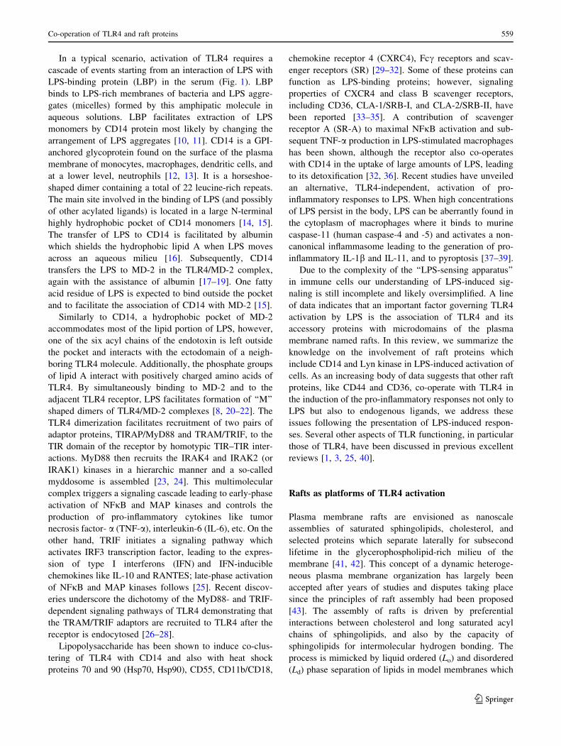
In a typical scenario, activation of TLR4 requires a
cascade of events starting from an interaction of LPS with
LPS-binding protein (LBP) in the serum (Fig. 1). LBP
binds to LPS-rich membranes of bacteria and LPS aggre-
gates (micelles) formed by this amphipatic molecule in
aqueous solutions. LBP facilitates extraction of LPS
monomers by CD14 protein most likely by changing the
arrangement of LPS aggregates [10, 11]. CD14 is a GPI-
anchored glycoprotein found on the surface of the plasma
membrane of monocytes, macrophages, dendritic cells, and
at a lower level, neutrophils [12, 13]. It is a horseshoe-
shaped dimer containing a total of 22 leucine-rich repeats.
The main site involved in the binding of LPS (and possibly
of other acylated ligands) is located in a large N-terminal
highly hydrophobic pocket of CD14 monomers [14, 15].
The transfer of LPS to CD14 is facilitated by albumin
which shields the hydrophobic lipid A when LPS moves
across an aqueous milieu [16]. Subsequently, CD14
transfers the LPS to MD-2 in the TLR4/MD-2 complex,
again with the assistance of albumin [17–19]. One fatty
acid residue of LPS is expected to bind outside the pocket
and to facilitate the association of CD14 with MD-2 [15].
Similarly to CD14, a hydrophobic pocket of MD-2
accommodates most of the lipid portion of LPS, however,
one of the six acyl chains of the endotoxin is left outside
the pocket and interacts with the ectodomain of a neigh-
boring TLR4 molecule. Additionally, the phosphate groups
of lipid A interact with positively charged amino acids of
TLR4. By simultaneously binding to MD-2 and to the
adjacent TLR4 receptor, LPS facilitates formation of ‘‘M’’
shaped dimers of TLR4/MD-2 complexes [8, 20–22]. The
TLR4 dimerization facilitates recruitment of two pairs of
adaptor proteins, TIRAP/MyD88 and TRAM/TRIF, to the
TIR domain of the receptor by homotypic TIR–TIR inter-
actions. MyD88 then recruits the IRAK4 and IRAK2 (or
IRAK1) kinases in a hierarchic manner and a so-called
myddosome is assembled [23, 24]. This multimolecular
complex triggers a signaling cascade leading to early-phase
activation of NFjB and MAP kinases and controls the
production of pro-inflammatory cytokines like tumor
necrosis factor- a (TNF-a), interleukin-6 (IL-6), etc. On the
other hand, TRIF initiates a signaling pathway which
activates IRF3 transcription factor, leading to the expres-
sion of type I interferons (IFN) and IFN-inducible
chemokines like IL-10 and RANTES; late-phase activation
of NFjB and MAP kinases follows [25]. Recent discov-
eries underscore the dichotomy of the MyD88- and TRIF-
dependent signaling pathways of TLR4 demonstrating that
the TRAM/TRIF adaptors are recruited to TLR4 after the
receptor is endocytosed [26–28].
Lipopolysaccharide has been shown to induce co-clus-
tering of TLR4 with CD14 and also with heat shock
proteins 70 and 90 (Hsp70, Hsp90), CD55, CD11b/CD18,
chemokine receptor 4 (CXRC4), Fcc receptors and scav-
enger receptors (SR) [29–32]. Some of these proteins can
function as LPS-binding proteins; however, signaling
properties of CXCR4 and class B scavenger receptors,
including CD36, CLA-1/SRB-I, and CLA-2/SRB-II, have
been reported [33–35]. A contribution of scavenger
receptor A (SR-A) to maximal NFjB activation and sub-
sequent TNF-a production in LPS-stimulated macrophages
has been shown, although the receptor also co-operates
with CD14 in the uptake of large amounts of LPS, leading
to its detoxification [32, 36]. Recent studies have unveiled
an alternative, TLR4-independent, activation of pro-
inflammatory responses to LPS. When high concentrations
of LPS persist in the body, LPS can be aberrantly found in
the cytoplasm of macrophages where it binds to murine
caspase-11 (human caspase-4 and -5) and activates a non-
canonical inflammasome leading to the generation of pro-
inflammatory IL-1b and IL-11, and to pyroptosis [37–39].
Due to the complexity of the ‘‘LPS-sensing apparatus’’
in immune cells our understanding of LPS-induced sig-
naling is still incomplete and likely oversimplified. A line
of data indicates that an important factor governing TLR4
activation by LPS is the association of TLR4 and its
accessory proteins with microdomains of the plasma
membrane named rafts. In this review, we summarize the
knowledge on the involvement of raft proteins which
include CD14 and Lyn kinase in LPS-induced activation of
cells. As an increasing body of data suggests that other raft
proteins, like CD44 and CD36, co-operate with TLR4 in
the induction of the pro-inflammatory responses not only to
LPS but also to endogenous ligands, we address these
issues following the presentation of LPS-induced respon-
ses. Several other aspects of TLR functioning, in particular
those of TLR4, have been discussed in previous excellent
reviews [1, 3, 25, 40].
Rafts as platforms of TLR4 activation
Plasma membrane rafts are envisioned as nanoscale
assemblies of saturated sphingolipids, cholesterol, and
selected proteins which separate laterally for subsecond
lifetime in the glycerophospholipid-rich milieu of the
membrane [41, 42]. This concept of a dynamic heteroge-
neous plasma membrane organization has largely been
accepted after years of studies and disputes taking place
since the principles of raft assembly had been proposed
[43]. The assembly of rafts is driven by preferential
interactions between cholesterol and long saturated acyl
chains of sphingolipids, and also by the capacity of
sphingolipids for intermolecular hydrogen bonding. The
process is mimicked by liquid ordered (Lo) and disordered
(Ld) phase separation of lipids in model membranes which
Co-operation of TLR4 and raft proteins 559
123

depends solely on cholesterol-sphingolipid interactions
[42, 44–46]. However, the composition of the plasma
membrane is far more complicated and multifarious lipid–
lipid, lipid–protein, and protein–protein interactions affect
the formation of nanoscopic raft domains and facilitate
their clustering into more stable functional platforms at
physiological conditions [47–49]. The inherent propensity
of the plasma membrane components to form nanoscopic
rafts has been demonstrated in studies using vesicles/
spheres of the plasma membrane obtained by osmotic
swelling or chemically induced vesiculation of cells. In
these plasma membrane fragments, separation of Lo and Ld
phases accompanied by accumulation of selected mem-
brane proteins in the ordered phase can be observed [47,
49–51].
Several mechanisms facilitate the association of proteins
with sphingolipid/cholesterol assemblies. Due to the high
content of saturated acyl chains of sphingolipids in such
assemblies, the thickness of the lipid bilayer and lipid
packing increase locally in comparison with the sur-
rounding bilayer composed mainly of unsaturated
glycerophospholipids. This, in turn, creates conditions at
which insertion of proteins modified with saturated lipids
into those domains rather than into the membrane bulk is
energetically favorable. For these reasons, in the outer
leaflet of rafts, proteins with glycosylphosphatidylinositol
(GPI) anchor, like CD14, are accumulated [52]. The inner
leaflet of rafts preferentially accommodates proteins mod-
ified by palmitoylation, which include tyrosine kinases of
the Src family and Ga subunits of trimeric G proteins [53].
Reciprocally, palmitoylated proteins can facilitate the
assembly of rafts [54]. There are also some transmembrane
proteins, mostly palmitoylated ones, that are intrinsically
targeted to rafts, as exemplified by CD44 and CD36
involved in TLR4 signaling (see below) and Cbp/PAG and
NTAL multipurpose adaptor proteins [55–59]. In fact, the
presence of palmitoylation, the length and the amino acid
sequence of the protein transmembrane domain, and the
oligomerization status of the protein are now recognized as
essential factors controlling the partition of transmembrane
proteins to rafts [60, 61]. It has also been proposed that
cholesterol- and sphingolipid-rich shells adjacent to the
transmembrane domains of proteins facilitate their associ-
ation with rafts [62]. In contrast, a transmembrane domain
coupled to an unsaturated phosphatidylethanolamine can
exclude the protein from rafts [49]. A combination of
lipid–lipid and lipid–protein interactions in living cells is
likely to give rise to plasma membrane rafts of different
protein composition [49].
Raft occurrence is manifested upon cell stimulation,
when they merge into larger platforms and facilitate
interactions of some receptors with their proximal signal-
ing molecules. This mode of action is common to
immunoreceptors, including T cell receptor (TCR), Fccreceptor IIa (FccRIIa), B-cell receptor (BCR), and Fcereceptor I (FceRI), which trigger signaling cascades after
phosphorylation by raft-anchored tyrosine kinases of the
Src family [57, 63–65]. Raft-based platforms also function
in cell polarization and membrane trafficking from the
Golgi apparatus to the plasma membrane, and during
endocytosis (see [66, 67] for review). All these events are
relevant to LPS-induced activation of macrophages draw-
ing attention to rafts as potential sites of LPS interaction
with CD14 and TLR4.
The plasma membrane rafts share lipid composition
with caveolae, flask-shaped invaginations of the plasma
membrane stabilized by caveolin 1–3 proteins. Contribu-
tion of caveolae to macrophage functioning is unclear,
although depletion of caveolin-1 inhibited phagocytosis of
Escherichia coli, decreased amounts of CD14, CD36, and
TLR4, and reduced cytokine production in macrophages
and cav1-/- mice exposed to the bacteria [68].
Ample data indicate that TLR4 and accessory proteins
can associate with plasma membrane rafts and the TLR4-
raft association is stimulated by LPS (Table 1). Such
results have been obtained in studies based on density
gradient centrifugation of Triton X-100 cell lysates yield-
ing detergent-resistant membrane (DRM) fraction [29, 31,
69]. This approach is based on model membrane studies
indicating that regions of the plasma membrane rich in
saturated lipids and cholesterol are insoluble in non-ionic
detergents, like Triton X-100, due to the tight packing of
the lipids [70]. DRM fragments can be subsequently sep-
arated from other membrane and cytosol components based
on their low density due to high lipid content. Ample
studies have indicated that the protein and lipid composi-
tion of the DRM fraction isolated in density gradients is
variable and depends on the protocol used, in particular the
detergent type and its concentration, temperature, and
duration of cell solubilization [71–73]. One possible reason
of this DRM variability is selective extraction of proteins
depending on the strength of their association with native
rafts in the membrane or, conversely, incorporation of non-
raft proteins during membrane solubilization [74, 75].
Thus, although isolation and characterization of the DRM
fraction is a useful approach for raft analysis, it should be
born in mind that the DRM fraction is not identical with
rafts of the intact plasma membrane. It has been hypothe-
sized, however, that the different composition of isolated
DRMs can actually reflect the inherent variability of pro-
tein concentration affecting lipid order in native plasma
membrane rafts [49].
Due to technical limitations of detergent-based bio-
chemical approaches, microscopic techniques are
especially useful for examining native rafts in living cells.
In recent years, studies on rafts have benefited
560 A. Płociennikowska et al.
123

Ta
ble
1S
tud
ies
sup
po
rtin
gth
ein
vo
lvem
ent
of
pla
sma
mem
bra
ne
raft
sin
TL
R4
-tri
gg
ered
sig
nal
ing
Pro
tein
san
dli
pid
sd
etec
ted
inra
fts
LP
S
chem
oty
pe
Cel
lsT
ech
niq
ue
Lip
idre
late
dev
ents
,co
mm
ents
Ref
eren
ces
Co
nst
itu
tiv
ely
pre
sen
t:C
D1
4,
CD
55
,C
D4
7,
CD
32
,C
D6
4;
Aft
erL
PS
stim
ula
tio
n:
TL
R4
,C
D1
1b
/CD
18
,
CD
16
aC
D3
6,
CD
81
;ex
clu
sio
no
fC
D4
7
LP
Sfr
om
Sa
lmo
nel
la
min
nes
ota
a
Hu
man
blo
od
mo
no
cyte
sF
RE
Tb
etw
een
CD
14
and
the
oth
erp
rote
ins;
Su
cro
seg
rad
ien
tce
ntr
ifu
gat
ion
of
0.5
%T
X-1
00
cell
lysa
tes
Ap
pli
cati
on
of
pro
py
l-C
Do
rn
yst
atin
red
uce
sC
D1
4/
CD
11
bco
-lo
cali
zati
on
ind
uce
db
yL
PS
;
Ex
og
eno
us
lon
g-c
hai
nce
ram
ide
ind
uce
sco
-
clu
ster
ing
of
CD
14
wit
ho
ther
cell
surf
ace
rece
pto
rsw
ith
exce
pti
on
of
TL
R4
[29]
Co
nst
itu
tiv
ely
pre
sen
t:C
D1
4,
GM
1,
Hsp
70
,
Hsp
90
;
Aft
erL
PS
stim
ula
tio
n:
TL
R4
,G
DF
5,
CX
CR
4,
My
D8
8,
JNK
ReL
PS
59
5
fro
m
Sa
lmo
nel
la
min
nes
ota
;
ReL
PS
fro
m
Esc
her
ich
ia
coli
Mo
no
Mac
-6(m
atu
re
hu
man
mo
no
cyte
cell
lin
e)b;
CH
Oex
pre
ssin
gh
um
an
CD
14
and
hu
man
TL
R4
c;
Hu
man
blo
od
mo
no
cyte
sc,d
Su
cro
seg
rad
ien
tce
ntr
ifu
gat
ion
of
1%
TX
-10
0ce
llly
sate
s;
FR
ET
bet
wee
nG
M1
and
ind
icat
edp
rote
ins
Ap
pli
cati
on
of
mbC
Do
rn
yst
atin
inh
ibit
sas
soci
atio
n
of
CD
14
,T
LR
4,
Hsp
70
,H
sp9
0,
CX
CR
4w
ith
raft
frac
tio
ns
and
inh
ibit
sT
NF
-ap
rod
uct
ion
[29,
31
,
19
0]
Aft
erL
PS
stim
ula
tio
n:
TL
R4
ReL
PS
59
5
fro
m
Sa
lmo
nel
la
min
nes
ota
Hu
man
blo
od
mo
no
cyte
sF
RA
PA
pp
lica
tio
no
fm
bCD
pre
ven
tsim
mo
bil
izat
ion
of
TL
R4
inth
ep
lasm
am
emb
ran
ein
du
ced
by
LP
S
[31,
77
]e
Co
nst
itu
tiv
ely
pre
sen
t:C
D1
4,fl
oti
llin
,C
D5
5;
Aft
erL
PS
stim
ula
tio
n:
acti
ve
Cd
c42
and
p3
8;
excl
usi
on
of
Rac
sLP
Sfr
om
Esc
her
ich
ia
coli
01
11
:B4
Hu
man
blo
od
neu
tro
ph
ils
Su
cro
seg
rad
ien
tce
ntr
ifu
gat
ion
of
1%
TX
-10
0ce
llly
sate
s
Ap
pli
cati
on
of
mbC
Du
pre
gu
late
sC
dc4
2an
dp
38
acti
vit
yan
din
du
ces
acti
np
oly
mer
izat
ion
bu
t
inh
ibit
ssu
bse
qu
ent
LP
S-i
nd
uce
dC
dc4
2an
dp
38
acti
vat
ion
[20
0]
Aft
erL
PS
stim
ula
tio
n:
CD
14
,E
RK
,p
38
LP
Sa
Raw
26
4.7
Su
cro
seg
rad
ien
tce
ntr
ifu
gat
ion
of
1%
TX
-10
0ce
llly
sate
s
mbC
Dan
dn
yst
atin
do
no
tin
hib
itT
NF
-ap
rod
uct
ion
and
ER
Kac
tiv
atio
nin
du
ced
by
LP
Sb
ut
hav
e
stim
ula
tory
effe
ctth
emse
lves
[11
7]
Co
nst
itu
tiv
ely
pre
sen
t:C
D1
4,
flo
till
in-1
,
GM
1,
smal
lam
ou
nts
of
CD
9,
CD
81
;
Aft
erL
PS
stim
ula
tio
n:
TL
R4
(sm
all
amo
un
ts),
enri
chm
ent
of
CD
14
,C
D9
,
CD
81
sLP
Sfr
om
Esc
her
ich
ia
coli
05
5:B
5
Bo
ne
mar
row
-der
ived
mac
rop
hag
eso
fm
ice
Su
cro
seg
rad
ien
tce
ntr
ifu
gat
ion
of
1%
TX
-10
0ce
llly
sate
s
Ap
pli
cati
on
of
pro
py
l-C
Din
hib
its
TN
F-a
ind
uce
d
by
LP
S
[20
1]
Co
nst
itu
tiv
ely
pre
sen
t,en
rich
edaf
ter
LP
S
stim
ula
tio
n:
CD
14
,H
sp7
0,
Hsp
90
,L
yn
,
Hck
,F
gr,
CD
44
,ac
idsp
hin
go
my
elin
ase,
gp
91
(ph
ox
);
Aft
erL
PS
stim
ula
tio
n:
sev
eral
pro
tein
s
inv
olv
edin
pro
tein
ub
iqu
itin
atio
n,
pro
teas
om
esu
bu
nit
s,ac
tiv
e
(ph
osp
ho
ryla
ted
)E
RK
and
ME
K;
Lac
ko
fT
LR
4
sLP
Sfr
om
Esc
her
ich
ia
coli
01
11
:B4
Raw
26
4.7
Su
cro
seg
rad
ien
tce
ntr
ifu
gat
ion
of
1%
TX
-10
0ce
llly
sate
s,
pro
teo
mic
anal
ysi
so
f
gra
die
nt
frac
tio
ns
Pro
teas
om
e-m
edia
ted
reg
ula
tio
no
fE
RK
acti
vit
yin
raft
frac
tio
ns
mbC
Din
du
ces
ER
Kac
tiv
atio
nb
ut
atte
nu
ates
sub
seq
uen
tL
PS
-in
du
ced
acti
vat
ion
of
the
kin
ase.
Ny
stat
inal
soin
hib
its
ER
Kac
tiv
atio
nin
LP
S-
stim
ula
ted
cell
s
[84]
Co-operation of TLR4 and raft proteins 561
123

Ta
ble
1co
nti
nu
ed
Pro
tein
san
dli
pid
sd
etec
ted
inra
fts
LP
S
chem
oty
pe
Cel
lsT
ech
niq
ue
Lip
idre
late
dev
ents
,co
mm
ents
Ref
eren
ces
Co
nst
itu
tiv
ely
pre
sen
t:C
D1
4;
Aft
erL
PS
stim
ula
tio
n:
TL
R4
,H
sp7
0
sLP
Sfr
om
Esc
her
ich
ia
coli
01
11
:B4
TH
P1
mo
no
cyte
s
dif
fere
nti
ated
by
PM
A
Su
cro
seg
rad
ien
tce
ntr
ifu
gat
ion
of
1%
TX
-10
0ce
llly
sate
s
Imip
ram
ine
use
das
AS
Mas
ein
hib
ito
rd
imin
ish
es
TL
R4
and
Hsp
70
recr
uit
men
tto
raft
s,E
RK
,p
38
and
JNK
acti
vat
ion
and
TN
F-a
pro
du
ctio
n
ind
uce
db
yL
PS
.A
llth
ein
hib
ito
ryev
ents
rev
erse
db
yex
og
eno
us
C2-c
eram
ide
[83]
Co
nst
itu
tiv
ely
pre
sen
t:C
D1
4,
flo
till
in-1
,
cav
eoli
n-1
,T
LR
4
LP
Sa
fro
m
Sa
lmo
nel
la
typ
him
uri
um
Per
ito
nel
mac
rop
hag
esan
d
bo
ne
mar
row
-der
ived
mac
rop
hag
eso
fA
bca
1-
M/-
Mm
ice
Op
tip
rep
gra
die
nt
cen
trif
ug
atio
no
fso
nic
ated
mem
bra
ne
frac
tio
n(n
od
eter
gen
tu
sed
)
AB
CA
1
defi
cien
cyin
crea
ses
par
titi
on
of
TL
R4
to
raft
frac
tio
n,
acti
vit
yo
fN
Fj
Ban
dM
AP
kin
ases
,p
rod
uct
ion
of
pro
-in
flam
mat
ory
cyto
kin
esin
LP
S-s
tim
ula
ted
cell
s;
mb
CD
dec
reas
esT
NF
-a,
IL-6
and
IL-1
2p
40
pro
du
ctio
n
[80,
20
2]
En
rich
edaf
ter
LP
Sst
imu
lati
on
:T
LR
4,
TR
IF,
My
D8
8
sLP
Sfr
om
Esc
her
ich
ia
coli
01
11
:B4
Raw
26
4.7
;
HE
K2
93
Tan
dB
a/F
3
(in
terl
euk
in-3
-dep
end
ent
mu
rin
ep
ro-3
cell
lin
e)ex
pre
ssin
gT
LR
/
MD
-2
Su
cro
seg
rad
ien
tce
ntr
ifu
gat
ion
of
1–
1.5
%T
X-1
00
cell
lysa
tes;
Co
-lo
cali
zati
on
of
TL
R4
and
GM
1
Lau
ric
acid
mim
ick
sL
PS
ind
uci
ng
dim
eriz
atio
no
f
TL
R4
inra
ftfr
acti
on
s;
DH
Ain
hib
its
LP
S-
and
lau
ric
acid
-in
du
ced
asso
ciat
ion
of
TL
R4
wit
hra
fts;
Ny
stat
inan
dm
bCD
dec
reas
ed
imer
izat
ion
of
TL
R4
inra
ftfr
acti
on
,p
rev
ents
NFjB
acti
vat
ion
and
targ
etg
ene
exp
ress
ion
ind
uce
db
yL
PS
and
lau
ric
acid
[81,
82
]
Pro
pyl
-CD
2-h
yd
rox
yp
rop
yl-b-
cycl
od
extr
in,
mb
CD
met
hy
l-b
-cy
clo
dex
trin
,T
X-1
00
Tri
ton
X-1
00
,A
bca
1-
M/-
Mh
om
ozy
go
us
mac
rop
hag
e-sp
ecifi
cA
TP
-bin
din
gca
sset
tetr
ansp
ort
erA
1
kn
ock
ou
tm
ice
aO
rig
ino
rch
emo
typ
eo
fL
PS
no
tsp
ecifi
edb
Cel
lsu
sed
for
DR
Mis
ola
tio
nc
Cel
lsu
sed
for
FR
ET
stu
die
sd
Cel
lsu
sed
toan
aly
zeT
NF
-ap
rod
uct
ion
eS
imil
ard
ata
sho
wn
on
TL
R2
-raf
tas
soci
atio
nst
imu
late
db
yli
po
teic
ho
icac
id
562 A. Płociennikowska et al.
123

exceptionally from the development of super-resolution
microscopy [76] which is yet to be employed to studies on
the involvement of plasma membrane rafts in LPS-trig-
gered signaling. Thus far, in support of the biochemical
data, measurements of the fluorescence resonance energy
transfer (FRET) between the constitutive raft components
GM1 or CD14 and selected plasma membrane proteins
have indicated that LPS induces the assembly of a raft-
associated multimolecular complex composed of TLR4
and other proteins potentially involved in LPS recognition
[29, 69]. In line with these data, LPS has been shown to
reduce the lateral mobility of TLR4 in the plane of the
plasma membrane. This LPS-induced confined diffusion of
TLR4, revealed by measurements of fluorescence recovery
after photobleaching (FRAP), has been ascribed to TLR4
trapping within plasma membrane rafts [31, 77].
Taking into account that raft assembly is driven by
interactions of sphingolipids and cholesterol, the observed
disturbances in TLR4 signaling following changes of the
cellular level of these lipids favor the idea of raft
involvement in LPS-induced inflammatory responses
(Table 1). Extraction or sequestration of cholesterol with
cyclodextrin or nystatin has been shown to disturb clus-
tering of TLR4 and accessory proteins in rafts and to
inhibit LPS-induced TNF-a production [29, 69, 77]. On the
other hand, a deficiency of ATP-binding cassette trans-
porters A1 or G1, linked with cholesterol elevation and an
apparent increase of raft content in macrophages, enhanced
the partition of TLR4 to raft fractions and augmented pro-
inflammatory signaling [78–80]. Similarly, the pro-
inflammatory effect of an exposure of RAW264 cells to
saturated fatty acids [81, 82] can be interpreted as a result
of enhanced raft assembly.
It is noteworthy that the majority of data supporting
LPS-induced accumulation of TLR4 in rafts and the
assembly of a raft-based multimolecular complex con-
taining TLR4 were obtained by microscopic and
biochemical studies of monocytes and cells of established
monocyte lines [29, 31, 69, 83]. In contrast, a recent pro-
teomic analysis of the DRM fraction isolated from
RAW264 macrophage-like cells indicated a lack of TLR4
in this raft-derived fraction, regardless of LPS stimulation
[84]. Those data suggest that in macrophages the associa-
tion of TLR4 with rafts can be dynamic and/or too weak to
allow its preservation during fractionation of Triton X-100
cell lysates over density gradients. In accordance, studies
on the co-localization of TLR4 and CD14 in J774 macro-
phage-like cells showed that the proteins co-localized
transiently and their coincidence was confined to lamellae
of LPS-stimulated cells [85]. Despite the failure to detect
TLR4 in the DRM fraction of RAW264 cells, the proteo-
mic analysis identified several dozen proteins which were
either enriched or recruited to this fraction after 5 and
30 min of LPS stimulation [84]. The list of proteins enri-
ched in the DRM after LPS stimulation includes CD14,
CD44, Src family tyrosine kinases Lyn, Hck, and Fgr,
Hsp70, Hsp90, acid sphingomyelinase, and NADPH oxi-
dase subunit gp91phox, supporting other ample data on the
involvement of these proteins in TLR4 signaling.
Participation of CD14 in LPS binding and signal
transduction
CD14 is more than LPS-binding protein
The role of CD14 as a key component of LPS-induced
inflammatory responses was indicated by studies on
transgenic mice expressing human CD14 and mice defi-
cient in CD14. The transgenic mice were hypersensitive to
LPS while mice devoid of CD14 did not develop septic
shock or accumulate pro-inflammatory cytokines in the
blood following exposure to E. coli or intraperitoneal
injection of LPS at a dose lethal to wild-type mice [86–88].
This protective effect of CD14 deficiency reflects a fatal
role of this protein in exaggerating the inflammatory
response in the course of systemic septic shock, although
during local infection, the CD14 involvement in combating
invading bacteria can be beneficial, as discussed by Zanoni
and Granucci [89].
CD14 has long been considered mainly as a molecule
which concentrates and delivers LPS to TLR4/MD-2
facilitating TLR4 activation [17, 90]. Originally, however,
CD14 was envisioned as a pattern recognition receptor
[91], but a lack of a transmembrane and cytoplasmic
domain called the signaling role of CD14 into question.
The importance of the membrane localization of CD14 for
LPS-induced signaling was also negated by the fact that a
soluble CD14 (sCD14) devoid of the GPI moiety could
substitute for membrane CD14 (mCD14) endowing cells
that normally do not express CD14 or express it at a very
low level, like endothelial and epithelial cells, as well as
Cd14-/- macrophages, with the ability to produce TNF-aand some other pro-inflammatory cytokines in response to
LPS stimulation [87, 92, 93].
Indications of a more complex role of CD14 in LPS-
induced responses have come from a series of studies
performed by the Goyert’s and Beutler’s groups. The for-
mer one found that some genes, like IP-10, that are now
known to be TRIF-dependent, were minimally expressed in
macrophages isolated from Cd14-/- mice and stimulated
with K235 LPS of E. coli. Simultaneously, expression of
genes encoding TNF-a and IL-1b could be induced in a
CD14-independent manner [94]. Studies of Beutler and co-
authors have unraveled a general picture of a disparate
requirement for CD14 to trigger the MyD88-dependent and
Co-operation of TLR4 and raft proteins 563
123

TRIF-dependent signaling pathways of TLR4. In addition,
the involvement of CD14 was found to differently deter-
mine TLR4 responses to so-called smooth (s) and rough
(r) chemotypes of LPS [88]. rLPS is produced by some
Gram-negative bacteria, especially Enterobacteriaceae
with mutations in genes involved in the O-chain synthesis.
Therefore, it is devoid of the O-polysaccharide chain and
can bear incomplete core oligosaccharides in contrast to the
sugar-linked smooth (s) LPS produced, e.g., by most E. coli
strains including the K235 strain. In studies on sLPS and
rLPS signaling requirements, N-ethyl-N-nitrosourea-muta-
ted mice were used bearing a recessive Heedless mutation
identified as a premature stop codon in Cd14 gene [88].
The lack of CD14 expression abolished the TRIF-depen-
dent pathway regardless of the LPS chemotype used for
cell stimulation. Thus, macrophages isolated from Heed-
less homozygotes failed to produce type I IFN as a
consequence of a lack of IRF3 activation and did not dis-
play induction of IFN-inducible genes in response to sLPS
or lipid A. Accordingly, no type I IFN was found in the
blood of Heedless mutant mice injected with sLPS or rLPS.
In summary, both sLPS and rLPS seemed to share a
common requirement for CD14 participation in triggering
TRIF-dependent signaling (Fig. 2a–c). This important
finding was addressed in further studies on the CD14 sig-
naling abilities discussed below.
There was, however, a clear distinction between the
ability of sLPS and rLPS to induce TNF-a production in
the absence of CD14. Both mice and ex vivo macrophages
bearing the Heedless mutation failed to produce TNF-a in
response to sLPS but retained the ability to generate TNF-aas a result of the activation of NFjB and MAP kinases after
exposure to rLPS or lipid A. The data suggested that
MyD88-dependent signaling can be generated by TLR4/
MD-2 alone in response to rLPS but triggering this sig-
naling pathway by sLPS requires CD14 (Fig. 2b, c). Such a
strict requirement of CD14 for sLPS-induced TNF-a pro-
duction can be typical for lower doses of sLPS, as parallel
ex vivo studies performed on Cd14-/- macrophages by the
Goyert’s group indicated that at concentrations equal or
higher than 100 ng/ml sLPS displayed some potency for
TNF-a induction. This potency was higher for rLPS [95].
Although both groups interpreted their data differently,
both sets of results in fact seem to indicate that CD14
participation ameliorates the differences between the abil-
ity of rLPS and sLPS to activate TLR4. This assumption is
supported by recent in vivo studies in which rLPS and
sLPS elicited nearly similar accumulation of pro-inflam-
matory cytokines in the serum of mice injected with 1 mg/
ml of the endotoxin [96]. These data leave unresolved the
question of the meaning of the ability of rLPS to activate
cells in a CD14-independent manner. It is conceivable that
rLPS can induce production of pro-inflammatory cytokines
in CD14-negative cells which otherwise can benefit also
from the assistance of sCD14, as discussed above. In
agreement with this assumption, it was found that mast
cells, which do not express CD14, produce IL-6 when
stimulated with rLPS but not sLPS [93].
Subsequent studies performed on RAW264 cells
exposed to antibodies blocking the LPS binding to CD14
showed that the requirement for CD14 participation in
LPS-induced signaling vary depending on the concentra-
tion of both sLPS and rLPS. At low doses of LPS (\10 ng/
ml), CD14 is crucial for the production of TNF-a and
RANTES (used to gauge MyD88- and TRIF-dependent
signaling, respectively) induced by either LPS chemotype
[97] (Fig. 2d). These data are consistent with earlier find-
ings showing that participation of CD14 markedly
increases responsiveness of cells to low concentrations of
rLPS or sLPS [90, 95]. At higher doses, sLPS induces
moderate production of TNF-a also without the CD14
participation (Fig. 2e), resembling results of earlier studies
of the Goyert’s group [94, 95]. Notably, rLPS in these
conditions activates the moderate production of both TNF-
a and RANTES (Fig. 2f). In addition, an assistance of LBP
was indispensable to induce maximal generation of these
cytokines in response to sLPS but not to higher doses of
rLPS [97, 98]. In summary, it is clear that rLPS relies on
CD14 assistance to a lower extent than does sLPS in
activating TLR4, and of the two signaling pathways trig-
gered by TLR4, the involvement of CD14 is especially
important for the TRIF-dependent one.
Questions arise as to the molecular mechanism of CD14
(and LBP)-independent activation of cells by LPS. In the
absence of CD14, albumin can bind LPS monomers with-
out the assistance of LBP and deliver them to TLR4/MD-2
[99]. Alternatively, LPS could be incorporated into rafts of
the plasma membrane and subsequently bind to the
receptor complex. The membrane incorporation could be
facilitated by the lack of the O-chain and thus higher
hydrophobicity of rLPS in comparison with sLPS [93].
Integration of LPS into the plasma membrane could also be
potentiated by the aggregated state of rLPS [100]. The
incorporation of rLPS into membranes can induce profound
changes of the membrane raft organization, as revealed by
studies on the interaction of ReLPS (the shortest form of
rLPS also used in [88, 97]) with model membranes per-
formed by solid-state NMR spectroscopy. When mixed
with DEPE/sphingomyelin/cholesterol liposomes, a ternary
lipid mixture in which the sphingomyelin/cholesterol-rich
Lo phase co-exists with the DEPE-rich Ld phase, ReLPS
induced coalescence and expansion of the Lo phase [101].
These data correspond with the results of microscopic
studies on LPS organization in giant liposomes composed
of polar lipids isolated from E. coli. In these membranes,
rLPS formed micron-sized gel-like microdomains while
564 A. Płociennikowska et al.
123

sLPS formed small clusters about 380 nm in diameter
[102]. A line of other studies performed on model mem-
brane also indicated that sLPS can spontaneously
incorporate into membranes. The preferred sites of sLPS
binding and incorporation were sphingomyelin/cholesterol-
rich domains formed in DOPC/sphingomyelin/cholesterol
liposomes [103, 104]. These findings suggest the impor-
tance of a direct interaction of rLPS and sLPS with plasma
membrane rafts. In the case of rLPS, its incorporation
could efficiently induce coalescence of rafts, possibly
inducing TLR4 dimerization and pro-inflammatory sig-
naling. The coalescence of nanoscale rafts into more stable,
larger raft domains is fundamental for their functioning in
the plasma membrane. It causes co-patching of raft proteins
and lipids into functional signaling platforms with simul-
taneous exclusion of non-raft proteins [42].
It should be noted that in model membrane studies
discussed above relatively high LPS concentrations (e.g., in
the range of lg/ml [103]) were used to reveal changes of
membrane organization. Stimulation of cells with high
doses of LPS could also facilitate a direct action of LPS,
particularly rLPS, on the plasma membrane. This
assumption could explain the activation of TRIF signaling
by 100–1,000 ng/ml rLPS in RAW264 cells even when the
binding of rLPS to CD14 was inhibited, and the lack of
such activation by 10–100 ng/ml rLPS or lipid A in CD14-
deficient macrophages bearing the Heedless mutation [88,
97] (Fig. 2c vs. f). These data suggest that, when present in
higher concentrations, rLPS can trigger TRIF-dependent
signaling bypassing the requirement for the binding to
CD14 which is otherwise required for the internalization of
TLR4/LPS leading to TRIF recruitment, as discussed in the
next section.
CD14 participates in internalization of TLR4/LPS
Both sLPS and, to a lower extent, rLPS rely on CD14
assistance to activate the TRIF-dependent pathway of
TLR4 (Fig. 2). Why is the CD14 participation required for
activation of this pathway?
The importance of CD14 for the initiation of the TRIF-
dependent pathway has been linked with internalization of
TLR4 which is essential for this signaling cascade. The
link between the endocytosis of LPS-activated TLR4 and
the subsequent TRIF-mediated signaling has been shown
by a line of data. It has been demonstrated that the surface
level of the receptor decreases in LPS-stimulated macro-
phages, monocytes, and CD14/TLR4/MD-2-transfected
Ba/F3 cells. TLR4 co-localized with markers of early/
sorting endosomes and its clearance from the cell surface
Fig. 2 Participation of CD14 in TLR4 signaling pathways triggered
by sLPS and rLPS. a In the presence of CD14, sLPS and rLPS
activate TLR4 and trigger MyD88- and TRIF-dependent pathways
with similar intensity. b, c Studies performed on macrophages bearing
the Heedless mutation of Cd14 have indicated that sLPS requires
CD14 to activate TLR4 (b), while rLPS can induce TRIF-dependent
signaling of TLR4 in CD14-deficient cells (c). d–f Other studies
suggest that the requirement of CD14 for activation of TLR4 varies
depending on the concentration of the endotoxin. At low concentra-
tions, sLPS or rLPS are unable to activate TLR4 without the
involvement of CD14 (d). At higher doses of sLPS, CD14 is
dispensable for initiation of MyD88-dependent pathway of TLR4,
although the production of TNF-a is submaximal in these conditions
(e). When present in relatively high concentrations, rLPS can induce
submaximal activation of both signaling pathways of TLR4 without
binding to CD14 (f)
Co-operation of TLR4 and raft proteins 565
123

was inhibited by dynasore, an inhibitor of the GTPase
dynamin controlling the pinching-off of endocytic vesicles
[26, 27, 105]. Concomitantly, dynasore abolished the
TRIF-dependent signaling indicating that endocytosis of
TLR4/LPS is important for this signaling pathway of TLR4
[27, 36]. The endocytosis of TLR4 is supposed to follow
MyD88-dependent signaling originating from the plasma
membrane [27, 105, 106] and the switch from the plasma
membrane MyD88-based to the TRIF-dependent endo-
somal signaling of TLR4 could be controlled by
phosphatidylinositol 4,5-bisphosphate [PI(4,5)P2] turnover
in the plasma membrane. The drop of PI(4,5)P2 level in the
membrane of endosomes was proposed to facilitate the
disassembly of the TIRAP/MyD88 signaling complex and
association of TRAM/TRIF adaptors [27, 107]. In support
of this thesis, the MyD88-dependent activation of NFjB
was enhanced in HEK293-CD14/TLR4/MD-2 transfec-
tants when the endocytosis of TLR4 was disrupted [26].
Overaccumulation of PI(4,5)P2 in dendritic cells as a result
of inactivation of the 110d isoform of class I phosphati-
dylinositol 3-kinase (PI3-kinase), which phosphorylates
PI(4,5)P2 to phosphatidylinositol 3,4,5-trisphosphate
[PI(3,4,5)P3], was one of the means of achieving this goal
[106].
A line of data indicates that CD14 controls the inter-
nalization of LPS-activated TLR4. Indeed, studies
performed on bone marrow-derived macrophages and
dendritic cells confirmed a significant time-dependent
decrease of the cell surface level of TLR4 in wild type
but not in Cd14-/- cells exposed to 1 lg/ml sLPS [28].
Simultaneously, the CD14-deficient cells failed to trigger
TRIF-dependent signaling in response to sLPS, in agree-
ment with results of earlier studies discussed above [88,
94, 97]. It has been established that the clearance of
CD14 and TLR4 from the cell surface, and subsequent
IRF3 activation, and type I IFN production all require the
activity of Syk kinase. Syk binds to proteins containing
the immunoreceptor tyrosine-based activation motif
(ITAM) and contributes to the downstream activation of
phospholipase Cc2 (PLCc2) [28]. Furthermore, PLCc2,
but not PLCc1, was found to account for inositol tris-
phosphate (IP3) generation and subsequent release of
Ca2? from intracellular stores required for TLR4 endo-
cytosis and IRF3 activation in RAW264 cells and bone
marrow-derived murine macrophages [108]. These data
support the idea that CD14 controls TLR4/LPS macr-
opinocytosis by activating ITAM-mediated events which
lead to Syk-PLCc2-dependent internalization of TLR4
[28] (Fig. 3a). This proposed chain of events is indepen-
dent of the Src kinase-PLCc2 axis found to control NFAT
activation in dendritic cells only [109, 110]. The data on
TLR4 endocytosis confirmed a biphasic scenario of TLR4
activation with the second phase being dependent on the
CD14-controlled internalization of the LPS/CD14/TLR4
complex. It is noteworthy that macropinocytosis of LPS/
CD14/TLR4 can overlap the CD14- and SR-A-mediated
uptake of LPS leading to is detoxification. This non-sig-
naling uptake of LPS does not involve TLR4 and in fact
can compete with the TLR4/LPS uptake which contrib-
utes to TLR4 signaling [36, 111].
The endosomal origin of the TRIF-dependent signaling
of TLR4 has been verified by the effects exerted on the
signaling by phagocytosis of E. coli by macrophages and
dendritic cells. The phagocytosis was expected to bypass
the lack of CD14 in knockout cells, force TLR4 internal-
ization and eventually induce generation of the TRIF-
dependent cascade. The prediction turned out to be correct
for dendritic cells only. In CD14-deficient macrophages,
the phagocytosis of E. coli failed to restore endocytosis of
TLR4 and TRIF signaling [28]. The authors ascribed those
contrasting results to a more ‘‘permissive’’ nature of den-
dritic cells facilitating TLR4 uptake in the absence of
CD14 (Fig. 3b).
Some studies questioned the need of TLR4 internaliza-
tion for triggering the TRIF-dependent pathway during
phagocytosis of E. coli and instead ascribed the signaling
abilities to an intracellular pool of TLR4. This pool of
TLR4 resides in Rab11-positive recycling endosomes and
can be transported to the E. coli-containing phagosomes,
thereby triggering the TRIF-dependent pathway. The
authors suggested that the internal pool of TLR4 can
induce the TRIF-dependent cascade without previously
being engaged in MyD88-dependent signaling in the
plasma membrane, provided LPS reaches the endosomal
compartment [112]. These data do not explain why the
phagocytosis of E. coli by Cd14-/- macrophages, which
proceeded without concomitant TLR4 uptake, did not
restore TRIF-dependent signaling [28] thereby suggesting
that TLR4 delivered to phagosomes from Rab11-bearing
endosomes can only amplify TRIF-dependent signaling
triggered by internalized TLR4.
Beside phagocytosis of E. coli in dendritic cells, one
more CD14-independent pathway of TLR4/LPS uptake
leading to TRIF activation can be considered. It has
recently been found that endocytosis of LPS-containing
liposomes can proceed unassisted by CD14 in clathrin-
coated vesicles (Fig. 3c). Endocytosis of LPS liposomes
led to IRF3 activation and RANTES production in mac-
rophages without a concomitant TNF-a and IL-6 release
[113]. Such endocytic pathway could also explain the
CD14-independent activation of RANTES production
induced at higher doses of rLPS (see Fig. 2f). An
involvement of clathrin-coated endocytosis in the inter-
nalization of LPS and TLR4/LPS was indicated by electron
microscopy and immunofluorescence studies [26, 114].
About ten times more LPS is internalized in non-coated
566 A. Płociennikowska et al.
123
than in coated vesicles [114], yet drugs inferring with
clathrin-dependent endocytosis significantly inhibit TRIF-
dependent signaling in LPS-stimulated cells [36, 115].
Further studies are required to reveal whether CD14 affects
clathrin-mediated uptake of TLR4/LPS, which factors
direct TLR4/LPS for macropinocytosis or clathrin-medi-
ated endocytosis, and whether the pathway of TLR4
internalization modulates its signaling, as suggested for
receptor tyrosine kinases [116].
Is raft localization of CD14 crucial for its involvement
in LPS-triggered events?
The relevance of the raft association of CD14 to its
involvement in TLR4 signaling in monocytes/macro-
phages, key players in inflammatory reaction, has long
puzzled researchers. Circumstantial evidence supporting
such dependence came from cell fractionation studies
showing enrichment of CD14 in the raft-originating DRM
fraction of LPS-stimulated RAW264 cells [84, 117]. A
more direct approach to this issue relied on a comparison of
cell activation by LPS mediated by GPI-anchored and
chimeric transmembrane forms of CD14. For this purpose,
CD14 was fused to the transmembrane plus cytoplasmic
fragment of either LDL receptor or tissue factor and
expressed in THP-1 cells [114, 118, 119]. The CD14-tissue
factor chimera localized to the Triton X-100-soluble frac-
tion, yet it induced NFjB activation, p38 phosphorylation
and IL-8 and TNF-a production in a similar manner as its
wild-type raft-anchored counterpart [118]. Of note, the
MyD88- and TRIF-dependent signaling could not be dis-
tinguished at the time when those studies were carried out.
It seems, therefore, that the apparent dispensability of GPI
anchoring for CD14 functioning can be revisited in view of
our present understanding of the distinct requirements for
the involvement of CD14 in the two signaling pathways of
TLR4.
Raft localization of CD14 can be critical for macr-
opinocytosis of LPS-activated TLR4. The CD14-mediated
macropinocytosis of the LPS/CD14/TLR4 complex relies
on the formation of large non-coated vesicles and involves
Syk kinase activity, as described above [28]. A recent
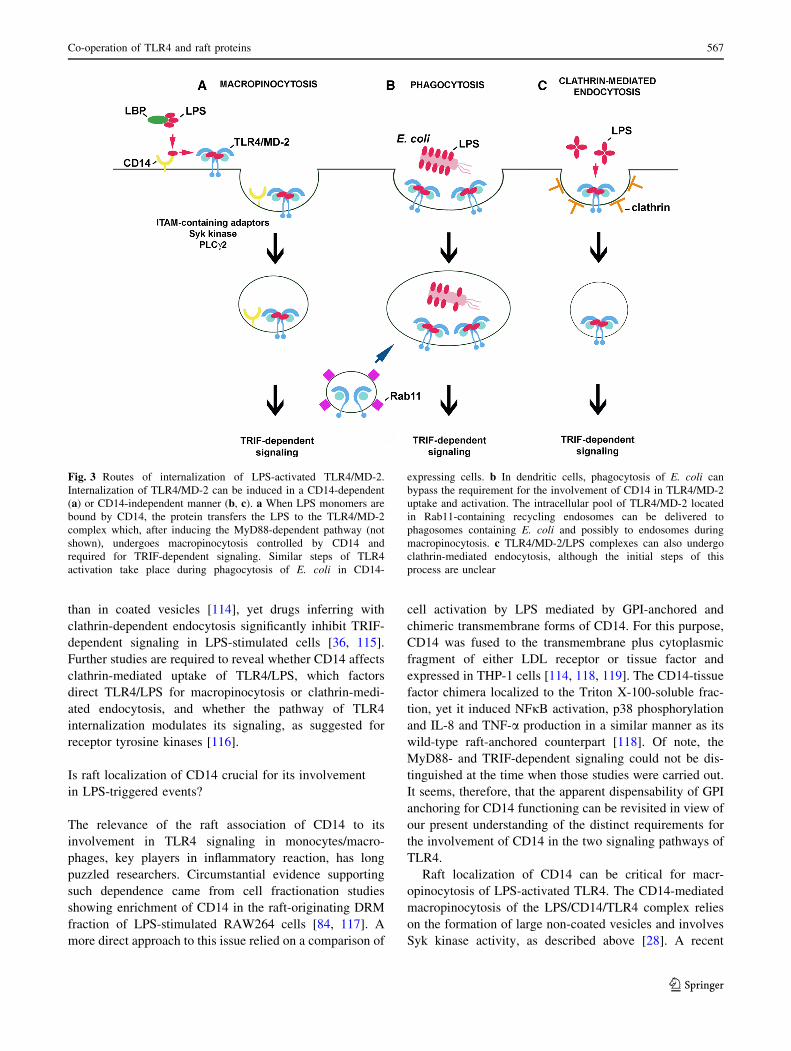
Fig. 3 Routes of internalization of LPS-activated TLR4/MD-2.
Internalization of TLR4/MD-2 can be induced in a CD14-dependent
(a) or CD14-independent manner (b, c). a When LPS monomers are
bound by CD14, the protein transfers the LPS to the TLR4/MD-2
complex which, after inducing the MyD88-dependent pathway (not
shown), undergoes macropinocytosis controlled by CD14 and
required for TRIF-dependent signaling. Similar steps of TLR4
activation take place during phagocytosis of E. coli in CD14-
expressing cells. b In dendritic cells, phagocytosis of E. coli can
bypass the requirement for the involvement of CD14 in TLR4/MD-2
uptake and activation. The intracellular pool of TLR4/MD-2 located
in Rab11-containing recycling endosomes can be delivered to
phagosomes containing E. coli and possibly to endosomes during
macropinocytosis. c TLR4/MD-2/LPS complexes can also undergo
clathrin-mediated endocytosis, although the initial steps of this
process are unclear
Co-operation of TLR4 and raft proteins 567
123

discovery of Syk-mediated internalization of CD36, a
scavenger receptor localized in rafts (and caveolae), has
paved the way for consideration of how CD14 could
trigger the LPS/CD14/TLR4 internalization in macro-
phages. Similarly to CD36, CD14 could facilitate the
assembly of a multimolecular complex composed of tet-
raspanins, b1 and b2 integrins, and ITAM-bearing Fccreceptors [120]. Notably, FRET analysis indicated that
tetraspanin CD81, integrin b2 (CD11/CD18) and Fccreceptors gather in the vicinity of CD14 and TLR4 in
LPS-stimulated human monocytes [29, 31, 121] (Table 1).
Subsequent binding of Syk to ITAMs, both likely to be
phosphorylated by Src family kinases, would then trigger a
cascade of phosphorylations of a variety of adaptor and
scaffolding proteins, and recruitment of lipid kinases and
PLCc2 leading to the activation of Rho GTPases and
WASP/Scar proteins. This chain of events could control
local actin polymerization providing the driving force for
internalization of LPS/CD14/TLR4, as has been deci-
phered for Fcc receptor-mediated phagocytosis. Taking
into consideration that Fcc receptors are functionally
connected with TLR4 and associate with rafts in LPS-
stimulated cells [29, 121, 122] and that phosphorylation of
their ITAMs by Src family kinases in the rafts is well
established [64, 65], the CD14-bearing rafts would be
preferred sites of the assembly of the multimolecular
complex mediating the internalization of LPS/CD14/
TLR4. Induction of raft reorganization by high doses of
rLPS, as found in model membrane studies [101], could
trigger a similar chain of events leading to TLR4 inter-
nalization without prior binding of rLPS to CD14 (see
Fig. 2f). A potential caveat of this model that needs to be
addressed is the postulated lack of an involvement of Src
family kinases in TLR4 internalization inferred from the
application of Src inhibitor-1 [28].
In addition to the CD14-controlled macropinocytosis of
LPS-activated TLR4, another role is ascribed to CD14
exclusively in dendritic cells. The CD14-dependent NFAT
activation in these cells provides a most clear indication
that the raft localization of CD14 is critical for its func-
tioning in LPS-stimulated cells. Upon LPS stimulation of
dendritic cells CD14 triggers an influx of Ca2? leading to
calcineurin-mediated activation of NFAT independently
of TLR4. Eventually, production of IL-2, prostaglandin E2
as well as apoptosis of the cells occurs [109, 110]. To
fulfill this function, CD14 needs to be membrane
anchored as sCD14 does not support NFAT activation. It
was found that mCD14 activates Src kinases and PLCc2
leading to IP3 generation and an influx of extracellular
Ca2? to the cytoplasm. Cholesterol depletion abolished
this Ca2? signaling, suggesting that raft integrity is crucial
for the co-operation of CD14 with raft-anchored Src
kinases [109].
Involvement of Lyn in LPS-induced TLR4 signaling
pathways
Tyrosine kinases of the Src family in LPS-induced
signaling
Toll-like receptor 4 signaling relies on cascades of protein
serine–threonine phosphorylation and polyubiquitination
events. Activation of this receptor also triggers protein
tyrosine phosphorylation catalyzed by multiple protein
tyrosine kinases including Bruton’s tyrosine kinase [123,
124], Syk kinase [28, 125], and kinases of the Src family
[126–128]. Of note, the activity of all these kinases is
crucial for the signaling by raft-associated receptors. The
most thoroughly characterized examples of that come from
studies on the involvement of Src family kinases and Syk
kinase in the signaling cascades of receptors containing
ITAM signaling motifs, e.g., FccRIIa, FceRI, and TCR [64,
65, 129].
In terms of TLR4 activation, pretreatment of human
monocytes and macrophages with herbimycin A or geni-
stein, broad-spectrum inhibitors of tyrosine kinases, or with
PP1, an inhibitor of Src family kinases, reduced LPS-
induced production of several cytokines like TNF-a, IL-1a,
IL-6, IL-10, and IP-10, and prevented the activation of
MAP kinases and NFjB [126, 127, 130, 131]. In contrast,
macrophages isolated form hck-/-fgr-/-lyn-/- triple
knockout mice released normal or even increased amounts
of TNF-a, IL-1, IL-6, and NO and showed no impairment
of the activation of MAP kinases and NFjB [132]. Double-
deficient hck-/-fgr-/- mice displayed an increased resis-
tance to endotoxic shock which was ascribed, however, to
defective integrin signaling and consequent reduced neu-
trophil migration into the tissue rather than to a direct
effect of the lack of Hck and Fgr activities on cytokine
production [133]. The discrepancies between the effects of
drug application and the knockout of the tyrosine kinase
genes on the pro-inflammatory reaction of cells are likely
to be due to the fact that the Src family of protein tyrosine
kinases comprises nine members: Src, Lyn, Hck, Fgr, Fyn,
Yes, Lck, Ylk, and Blk, of which the first six are known to
be expressed in macrophages [131]. All these kinases share
a common domain structure, with the N-terminal domain
undergoing myristoylation and palmitoylation, the latter
facilitating anchoring of the kinase in plasma membrane
rafts. The kinases also contain the SH3 and SH2 domains,
the catalytic domain, and a short C-terminal tail controlling
their conformation and enzymatic activity [134]. It has
been suggested that the apparent failure to detect changes
of LPS-induced responses in the knockout mice resulted
from a compensation of the absence of some of the Src
kinases by other family members. Indeed, short-term
adenoviral overexpresion and siRNA knockdown studies
568 A. Płociennikowska et al.
123

have indicated that the Hck kinase activity controls the
production of TNF-a and IL-6 induced by LPS in human
macrophages. The kinase affects the activity of AP-1
transcription factor without influencing the activity of
MAP kinases or NFjB [128]. LPS also triggers association
of Hck with Vav, a Rho family guanine nucleotide
exchange factor (RhoGEF) [135] involved in TNF-a pro-
duction, as discussed below. In addition, Src kinase has
been reported to act as a downstream effector of LPS-
induced actin cytoskeleton rearrangements [136]. The
participation of Lyn kinase in LPS-triggered TLR4 sig-
naling is supported by the most extensive line of data.
A positive role of Lyn in TLR4 signaling pathways
First indications on the involvement of Lyn kinase in LPS-
induced signaling came from studies on CD14 protein.
CD14 immunoprecipitated from human monocytes was
found to be associated with Lyn kinase. The activity of the
kinase increased shortly (1–5 min) after stimulation of the
cells with 1 ng/ml sLPS [126]. Other studies on human
monocytes revealed that within 1–5 min of stimulation
with 10 ng/ml of ReLPS, TLR4 underwent tyrosine-phos-
phorylation [137]. Rapid Lyn activation was also observed
in LPS- or taxol-treated mouse peritoneal macrophages
[138] and more recently in human macrophages [128].
When expressed in HEK293 cells together with human
TLR4 and MD-2, Lyn kinase co-immunoprecipitated with
the receptor even in the absence of CD14. Recruitment of
Lyn to TLR4 was triggered within 1 min of LPS stimula-
tion of the cells with a maximal response at 15 min. Within
the same time frame tyrosine phosphorylation of TLR4 was
observed in the TLR4/MD-2-expressing HEK293 regard-
less of CD14 presence [127]. Those data suggested,
although not proved, that Lyn kinase could be responsible
for the phosphorylation of tyrosine reside(s) of TLR4.
Crucially, further studies using HEK293 cells transfected
with a constitutively active human CD4-TLR4 chimera
indicated that TLR4 tyrosine phosphorylation was required
for TLR4-induced signaling. Thus, mutation of tyrosine
residues Y674A and Y680A in the TIR domain of TLR4
inhibited the recruitment of MyD88 and activation of
IRAK-1 by the constitutively active form of the receptor.
Furthermore, activation of NFjB, phosphorylation of p38
and JNK kinases, and RANTES production were also
strongly suppressed, indicating that tyrosine phosphoryla-
tion of TLR4 is prerequisite for both the MyD88- and
TRIF-dependent pathways. Similar suppression of TLR4
phosphorylation and signaling was found for P714H human
and P712H murine TLR4, known as mutant receptors
unresponsive to LPS [127]. Taken together, the data indi-
cated the importance of protein tyrosine phosphorylation
for TLR4 signaling and suggested a positive role of Lyn
activity in this process (Fig. 4). The CD14/Lyn association
and activation of CD14-associated kinase by LPS, found by
Stefanova et al. [126], pointed to the importance of raft
localization of Lyn in LPS-triggered signaling of TLR4. To
our knowledge, however, this subject has not been
addressed directly in any further studies.
In accordance with its plasma membrane localization,
Lyn kinase was recently found to bind TRAF6 in response
to LPS. TRAF6 is an E3 ubiquitin ligase which transiently
associates with the myddosome and controls downstream
steps of TLR4 signaling cascades affecting the TAK-1
kinase activity after catalyzing its own polyubiquitination
via Lys63-linked chains [139]. The involvement of Lyn in
LPS-induced TRAF6 activity has been verified with the use
of mast cells from lyn-/- mice [140]. Remarkably, a low-
level binding of TAK-1 to TRAF6, ubiquitination of
TRAF6, and TAK-1 phosphorylation occurred in the
lyn-/- mast cells, which were not found in wild-type cells.
However, stimulation of the Lyn-deficient mast cells with
LPS did not increase the association of TRAF6 with TAK-
1, and subsequent ubiquitination of TRAF6 and phos-
phorylation of TAK-1 were also inhibited. As a result of
the impairment of the TAK-1 activation also phosphory-
lation of ERK1/2, p38 and JNK kinases and NFjB
activation were inhibited in the Lyn-deficient mast cells
[140]. This scenario resembled the impaired signaling of
non-phosphorylated mutants of TLR4 which correlated
with their low basal association with MyD88 that was not
changed upon LPS treatment in contrast to the LPS-
induced MyD88 recruitment by wild-type TLR4 [127]. It
seems, therefore, that Lyn can modulate the TLR4/MyD88
and TRAF6/TAK-1 association/disassociation cycles
required for optimal TLR4 signaling.
Lyn kinase has been found to associate with TRAF6 in
LPS-stimulated mast cells [140]; however, the mechanism
of this interaction has not been resolved. An indication on
how the TRAF6 and Lyn kinase interaction could be reg-
ulated came from studies on TRAF6 functioning in LPS-
stimulated human lung microvascular endothelial cells. In
these cells, LPS binding to TLR4 triggers a cascade of
tyrosine phosphorylations catalyzed by kinases of the Src
family contributing to the disruption of the endothelial
barrier integrity. In the endothelial cells, LPS stimulates the
association of TRAF6 with Src and Fyn, but not with Lyn
kinase [141], suggesting that identity of the Src family
kinase engaged in TLR4 signaling can be cell type-
dependent. The binding site for the Src kinases has been
mapped to a proline-rich region of TRAF6. The assembly
of the TRAF6-Src(Fyn) kinase complex requires the kinase
enzymatic activity, while silencing of the TRAF6 gene
precludes activation of the Src kinases, which does not
allow distinguishing which enzyme acts upstream of which
[142]. In addition, during the association of TRAF6 with
Co-operation of TLR4 and raft proteins 569
123

Src(Fyn), each protein can serve as a substrate for the
catalytic activity of the other and their ubiquitination/
phosphorylation is catalyzed [142]. If a similar relation
holds for the TRAF6-Lyn interaction, Lyn would phos-
phorylate TRAF6 and stimulate the downstream TRAF6
signaling in LPS-treated cells while TRAF6 would catalyze
Lys63-mediated ubiquitination of Lyn. Whether this poly-
ubiquitination of Src family kinases affects their activity or
association with TRAF6 remains unknown. It has been
established that Lys48-linked ubiquitination and subsequent
proteasomal degradation of Lyn controls its level in cells
[143].
Recently, a newly discovered macrophage scaffolding
protein Themis2 was found to bind Lyn kinase in LPS-
stimulated RAW264 cells [144]. Analysis of the Themis2
role has provided evidence for the action of fine-tuning
mechanisms of LPS-induced signaling. It was found that
Themis2 underwent tyrosine phosphorylation on Tyr660 in
LPS-stimulated cells and when phosphorylated could bind
SH2 domain of Lyn kinase, however, the possibility of
phosphorylation of Themis2 by Lyn has not been addres-
sed. On the other hand, a proline-rich sequence of Themis2
binds constitutively the RhoGEF Vav. Therefore, cluster-
ing of Lyn and Vav at Themis2 could facilitate Vav
phosphorylation by Lyn leading to stimulation of its GEF
activity. The Themis2-Lyn-Vav axis has been found to
stimulate selectively ERK and p38 activity but not that of
JNK, NFjB, or IRF3. Eventually, the signaling events
involving Themis2 and Lyn upregulate the production of
TNF-a but not IL-6 or Cox2 in LPS-stimulated cells [144].
Some data indicate that not only plasma membrane-
associated Lyn but also its intracellular fraction could be
engaged in the signaling in LPS-stimulated cells. Lyn
kinase was found to associate constitutively with MD-2 and
to mediate its LPS-induced phosphorylation in the endo-
plasmic reticulum/endosomes of HEK293 expressing
TLR4/MD-2 which suggested the MD-2 phosphorylation
could be triggered after LPS internalization [145]. A key
unresolved question regarding the proposed MD-2 phos-
phorylation by Lyn is how MD-2, apparently enclosed in
the vesicle lumen, could be accessed by the kinase.
To summarize, stimulation of cells by LPS induces rapid
activation of Lyn which can be involved in the initial steps
of LPS-induced signaling by phosphorylating TLR4. The
link between the Lyn and CD14 involvement in LPS-
induced signaling, suggested by studies of Stefanova et al.
[126] deserves further attention in view of the signaling
properties of CD14 discussed in the previous chapter. Lyn
kinase also associates with (and phosphorylates ?) TRAF6
and Themis2, downstream TLR4 signaling proteins, and
Fig. 4 Participation of Lyn in
TLR4 signaling. Lyn kinase
either associates constitutively
with proteins or associates with,
and phosphorylates, indicated
proteins after stimulation of cell
with LPS. These events regulate
positively or negatively TLR4-
induced signaling (left and right
side of the figure, respectively).
Arrows indicate domains of Lyn
most likely involved in binding
of indicated proteins. The
molecules are drawn not to scale
570 A. Płociennikowska et al.
123
can phosphorylate endosomal MD-2, probably only after
LPS internalization. With the exception of the MD-2
phosphorylation, Lyn is likely to interact with other pro-
teins and catalyze phosphorylation of its substrates at the
plasma membrane. In all the cases mentioned above, Lyn
activity stimulates the respective events of LPS-induced
signaling pathways (Fig. 4, left). This, however, is not the
only mode of Lyn participation in the signaling.
Lyn kinase as a negative regulator of TLR4 signaling
pathway
Lyn kinase is unique among the Src family kinases as it
exerts both positive and negative regulatory action toward
diverse signaling pathways [143, 146]. It has also been
reported to act as a negative regulator of TLR4 signaling
[147]. The studies were sparked by the finding that bone
marrow-derived macrophages isolated from lyn-/- mice
and stimulated with ReLPS produced more IL-6, TNF-a,
and IFN-a/b than their wild-type counterparts. In agreement
with those ex vivo studies, increased amounts of TNF-a, IL-
6, and IFN-a/b were also found in the serum of lyn-/- mice
injected with ReLPS. Those results indicated that Lyn is
involved in the downregulation of both the MyD88- and
TRIF-dependent pathways of TLR4. In search for the
mechanism of the suppressory action of Lyn it was estab-
lished that PI3-kinase and Akt kinase could be downstream
targets of Lyn. It was inferred that the activity of PI3-kinase
in LPS-stimulated cells led to phosphorylation of PI(4,5)P2
to PI(3,4,5)P3 in the plasma membrane and subsequent
activation of Akt kinase. The latter was suggested to be
responsible for the down-regulation of p38 phosphorylation
eventually leading to decreased production of cytokines
[147]. These data put PI3-kinase in a position of a negative
regulator of TLR4 signaling (Fig. 4, right). Corroborating
these results, wortmannin, a PI3-kinase inhibitor, aug-
mented TNF-a and IL-6 production while a lack of SHIP1
phosphatase which dephosphorylates PI(3,4,5)P3 had an
opposite effect. Recently, a contribution of PI3-kinase(s) to
negative regulation of TLR4 pro-inflammatory signaling
through the Akt-mTOR-Foxo1 signaling axis has been
shown [148].
The association of Lyn with PI3-kinase(s) was described
a decade earlier in LPS-stimulated human monocytes
[149]. The enzymes co-immunoprecipitated and became
activated in a coordinated manner within minutes of LPS
action. Also, the level of PI(3,4,5)P3 increased markedly in
LPS-treated cells, as found by analyzing P32-labeled lipid
extracts by HPLC. Of note neutralizing CD14 with a spe-
cific antibody abrogated this LPS-induced PI(3,4,5)P3
production which indicated that CD14 and Lyn, two raft-
enriched proteins, regulate PI3-kinase involvement and
PI(3,4,5)P3 production in LPS-induced signaling.
There is a possible functional link between the CD14
involvement in LPS-induced signaling and PI(3,4,5)P3
production, as both control the endocytosis of TLR4.
Expression of a kinase-dead p110d isoform of the catalytic
domain of PI3-kinase in mice inhibited LPS-induced
endocytosis of TLR4 in bone marrow-derived dendritic
cells [106]. The p110d defect downregulated TRIF-
dependent signaling and led to lower IFN-a, IP-10, and
RANTES production with concomitant augmentation of
MyD88/TIRAP-mediated signaling detected both in the
mice and in the bone marrow-derived dendritic cells. The
inhibition on TRIF-dependent signaling caused by the lack
of the activity of p110d isoform of PI3-kinase resembled
that caused by the lack of CD14 in cells [28] but not the
effects seen in Lyn-deficient macrophages where both
MyD88- and TRIF-dependent signaling pathways of TLR4
were upregulated [147]. This indicates that proteins other
than PI3-kinase p110d co-operate with Lyn to regulate
negatively both signaling pathways of TLR4.
Indeed, Lyn can exert its negative regulatory function by
affecting many other proteins beside PI3-kinases. The list
includes Cbp/PAG, a raft protein which is phosphorylated
by Lyn and binds Csk kinase responsible for inactivation of
Src family kinases [129]. Lyn also has a unique ability to
phosphorylate the immunoreceptor tyrosine-base inhibitory
motif (ITIM) of plasma membrane receptors such as
FccRIIb, paired immunoglobulin-like receptor B (PIR-B),
signal regulatory protein-a (SIRPa), and CD22. These
phosphorylated proteins recruit inhibitory phosphatases,
such as protein phosphatase SHP-1/2 and SHIP1, a phos-
phatase of PI(3,4,5)P3 [150]. The latter enzyme does not
seem to contribute to the Lyn-dependent negative regula-
tion of TLR4 signaling [147] while other possibilities have
not been examined in this context. However, the inhibitory
role of FccRIIb in TLR4-mediated responses was indicated
[151].
In summary, the involvement of Lyn in LPS-induced
signaling has both a positive role and generates a negative
loop switching off the signaling (Fig. 4). Such a dual role
of Lyn has been established previously for BCR and FceRI
signaling in mast cells. The positive versus negative action
of Lyn in FceRI-triggered signaling can depend on the
intensity of stimulation of the receptor [152]. On the other
hand, during activation of BCR, the two functions of Lyn
need to be finely balanced and factors which determine the
final outcome of Lyn activity are ill-defined, as both Lyn
knockout mice and Lynup/up mice expressing constitutively
active Lyn develop autoimmune diseases related to B-cell
dysfunction [146]. Of special interest is that the positive
role of Lyn detected in TLR4 signaling in mast cells con-
trasted with its negative role in TLR4 signaling in
macrophages, since both series of experiments were per-
formed on cells isolated form Lyn-deficient mice [140,
Co-operation of TLR4 and raft proteins 571
123

147]. Furthermore, in macrophages, a triple knockout of
Hck, Fgr, and Lyn facilitated production of some cytokines
[132]. These data suggest that the final outcome of the Lyn
involvement in TLR4-triggered signaling could be cell
type-specific and depend, e.g., on the receptors complexing
with TLR4, like CD14 or CD36 (see below) or Fccreceptors [29, 31, 153]. Taking into account that macro-
phages express CD14 at a high level while mast cells are
CD14-deficient, it is tempting to speculate that if raft-
residing CD14 is engaged in LPS-induced signaling, the
negative regulatory function of Lyn rather than its indis-
pensability for TLR4 signaling is emphasized. It is also
plausible that Lyn regulates positively or negatively dis-
tinct components of TLR4 signaling pathways in a single
cell type, resembling the Lyn involvement in BCR sig-
naling [146]. Thus, Lyn could provide a subtle regulation
of the amplitude and duration of the pro-inflammatory
signaling triggered by LPS.
Involvement of acid sphingomyelinase and other
enzymes of sphingomyelin cycle in LPS-induced
signaling
The accumulation of acid sphingomyelinase (ASMase) in
the DRM fraction of RAW264 cells within 5–30 min of
LPS treatment [84] points to the importance of sphingo-
myelin turnover in LPS-induced signaling. About half of
the cellular pool of sphingomyelin is that in the plasma
membrane, 80–90 % of which resides in the outer leaflet of
the membrane and is enriched in rafts [154, 155]. Under the
action of sphingomyelinases, sphingomyelin is hydrolyzed
yielding ceramide, a multifaceted lipid which significantly
changes the organization of the plasma membrane, affects
the activity of several intracellular enzymes and is a pre-
cursor of other bioactive lipids, like ceramide-1-phosphate
and sphingosine-1-phosphate [154, 156]. The generation of
ceramide in the outer leaflet of the plasma membrane is
linked with the activity of ASMase which translocates to
the cell surface from intracellular compartments in
response to diverse stimuli. Hydrolysis of sphingomyelin in
the inner leaflet of the membrane is attributed to the
activity of neutral sphingomyelinase (NSMase) [156–158].
ASMase and NSMase are activated rapidly in J774 and
THP-1 cells under the action of LPS [83, 159, 160]. The
production of ceramide by sphingomyelin hydrolysis in
LPS-treated cells is also rapid, within minutes [160, 161],
and is distinct from the TLR4-dependent de novo ceramide
synthesis leading to prolonged ceramide accumulation
observed in chronic diseases [162].
The data on the role of ASMase and ceramide in LPS-
induced signaling are contradictory. Early studies indicated
some similarities between LPS and exogenous ceramides,
including long-chain ones, or ceramide generated in cells
upon treatment with bacterial sphingomyelinase, in
inducing the assembly of multimolecular complexes in the
plasma membrane, activation of MAP kinases and pro-
duction of selected cytokines in various cells, including
RAW264 cells and murine macrophages [29, 161, 163,
164]. Furthermore, a positive role of endogenous ceramide
generated by ASMase in the pro-inflammatory responses of
cells to LPS was indicated. The ceramide was required for
LPS-induced recruitment of TLR4 to rafts, activation
of MAP kinases and TNF-a production in differentiated
THP-1 cells, as all these events were inhibited by imipra-
mine, a drug causing ASMase degradation, and were
reversed by exogenous C2-ceramide [83]. In agreement,
inhibition of ASMase by the SMA-7 drug prevented NFjB
activation and release of pro-inflammatory mediators in
THP-1 cells [159]. Recently, a positive influence of
ASMase-generated ceramide on IL-6 production by RAW
264 cells after prolonged stimulation by low doses of LPS
(1 ng/ml) and palmitic acid was found [165].
In contrast, studies performed on ASMase knockout
mouse or murine macrophages and J774 cells after ASM-
ase or NSMase silencing indicated that ceramide generated
in LPS-stimulated cells downregulates the production of
TNF-a and other cytokines [160, 166]. In accordance,
infection of the ASMase-deficient mice with Gram-nega-
tive bacterium Pseudomonas aeruginosa resulted in a
roughly tenfold higher release of pro-inflammatory cyto-
kine IL-1b than in wild-type mice, and their death [167].
Exogenous C8-ceramide was found to inhibit production of
pro-inflammatory cytokines, TNF-a, IL-6, and IL-12p40,
by murine macrophages and pro-asthmatic IL-5, IL-10, and
IL-13 by mast cells [168]. Accordingly, in a murine model
of LPS- or Staphylococcus aureus-induced corneal
inflammation, topical delivery of low doses of C6-ceramide
in liposome formulation had a therapeutic effect supporting
the anti-inflammatory properties of ceramide [169].
Ceramide generated in the outer leaflet of the plasma
membrane can be converted back to sphingomyelin by
sphingomyelin synthases 2 (SMS2) located in plasma
membrane rafts/caveolae [156]. An impact of the SMS2
activity in the plasma membrane on pro-inflammatory
signaling is yet to be proven unequivocally. Recent data
indicate that in SMS2-deficient macrophages, enriched in
ceramide and depleted in sphingomyelin, NFjB activation
was attenuated and LPS-induced lung injury reduced [170,
171]. In SMS2-deficient mice, the serum level of IL-6 and
TNF-a was lower compared to their wild-type counterparts
[172]. These data underscore the anti-inflammatory out-
come of changing the sphingomyelin/ceramide balance in
the plasma membrane in favor of ceramide.
The molecular mechanisms of the action of the ASM-
ase-generated ceramide in LPS-stimulated cells are not
572 A. Płociennikowska et al.
123
completely understood and their revealing is hindered by
the fact that exogenous short-chain ceramides do not nec-
essarily mimic the action of endogenous ceramides
acylated with long-chain fatty acids which are produced by
ASMase. It is known that the exofacial ceramide generated
by ASMase dramatically alters the biophysical properties
of the plasma membrane [154]. Ceramide molecules sep-
arate laterally into domains, thereby displacing cholesterol
form rafts [173] and affecting the lateral distribution of
plasma membrane proteins. For example, ceramide was
found to facilitate clustering of FccRII and CD95 in rafts
[157, 174]. Following this scenario, one can assume that
ceramide generated by ASMase in LPS-stimulated cells
promotes mobilization of TLR4 to rafts and facilitates the
CD14-TLR4 interaction leading to activation of pro-
inflammatory signaling. Accordingly, binding of LPS to
CD14 was found to be required for ceramide generation in
THP-1 cells [83]. The ability of ceramide to induce
redistribution of plasma membrane proteins most likely
accounts for the observed recruitment of selected cell
surface proteins toward CD14 creating conditions for
activation of selected signaling pathways in cells exposed
to exogenous ceramides [29, 163].
The above scenario is in contrast with reports indicating
a negative influence of ASMase-generated ceramide on the
production of pro-inflammatory mediators, like TNF-a[160, 166]. The ceramide inhibited maturation of TNF-a at
the stage of the cleavage of TNF-a precursor to its active
17-kDa soluble form by the TNF-a converting enzyme
(TACE) [166], which also associated with rafts [175]. The
ceramide negatively regulated TACE activity, which was
tentatively linked with changes of TACE partitioning to
rafts in ASMase-deficient cells. In addition, ASMase-gen-
erated ceramide could also affect cellular trafficking of the
TNF-a precursor [166]. The mechanisms of the negative
influence of ceramide on the production of other cytokines
in LPS-stimulated cells are unknown, albeit inhibition of
the LPS-induced activity of NFjB and AP-1 by exogenous
C-2 ceramide has been reported in RAW264 cells [176].
Possibly, the impact of the ASMase-generated ceramide on
the associations of TLR4 with rafts also needs further
studies. The shift of the balanced from sphingomyelin
toward ceramide in the plasma membrane seems to nega-
tively affect the association of TNF-a receptor with rafts
[170]. Bearing in mind the positive effect of ceramide on
the association of FccRII and CD95 with rafts, the lipid
seems to be able to both facilitate and hinder the partition
of various receptors to rafts. In accordance, recent studies
on transferrin receptor ligated with transferrin indicated
that ASMase-generated ceramide inhibited receptor parti-
tioning to rafts and facilitated its endocytosis by clathrin-
coated pits [177]. The latter is in agreement with the ability
of exofacial ceramide to induce inward curvature of the
plasma membrane and subsequent budding of endocytic
vesicles [178]. Taking into account, the dynamic associa-
tion of TLR4 with rafts followed by its endocytosis in LPS-
stimulated cells, one can expect a complex impact of
sphingomyelin/ceramide turnover on the receptor translo-
cations in the plasma membrane which could change in the
course of TLR4 activation. Such complexity could account
for the discrepant effects of the interference with the
turnover of sphingomyelin in LPS-stimulated cells.
Proteasome as newly identified raft component involved
in LPS signaling
A recent proteomic analysis has revealed that following
LPS action, numerous proteins involved in protein ubiq-
uitination and several proteasome subunits are recruited to
the DRM fraction of RAW264 cells [84]. Furthermore,
p105 protein was identified as a proteasomal substrate in
the DRM fraction of LPS-stimulated cells. Degradation of
this protein releases Tpl2 kinase which phosphorylates
MEK and leads to ERK activation, and both phosphory-
lated MEK and ERK kinases were found to be confined to
the DRM fraction of RAW264 cells [84, 117]. Thus, these
studies suggest that the plasma membrane rafts are sites of
the activity of proteasomes linked to ERK activation in
LPS-stimulated cells. It has also been shown that LPS
alters the composition of proteasomes, and that proteaso-
mal degradation of several signaling proteins regulates
both MyD88- and TRIF-dependent generation of pro-
inflammatory mediators [179], with the proteolysis of IjB
by 26 S proteasome being the best characterized.
Remarkably, 26 S proteasome is activated by LPS in the
DRM fraction of RAW264 cells [84]. Proteasome activity
is a novel function linked to rafts and further studies are
required to reveal how proteasomes associate with the
plasma membrane and are activated in LPS-stimulated
cells.
Involvement of CD44 and Hsp70 in TLR4 signaling
triggered by LPS and non-microbial ligands
CD44, the hyaluronan receptor, is another raft protein
likely to co-operate with TLR4. This co-operation regulates
the pro-inflammatory responses induced by LPS and also
those observed in the absence of microbial stimuli. During
trauma fragments of hyaluronan are released from the
extracellular matrix and, after binding to CD44, induce
expression of pro-inflammatory genes in a TLR4/MD-2-
dependent manner. Only some of these genes are those
activated by LPS as well, as shown in vivo by studying skin
samples of sterile-injured mice [180]. In those studies,
Co-operation of TLR4 and raft proteins 573
123

CD44 was identified as an important intermediate in the
hyaluronan-induced TLR4 activation. However, in the
presence of microbial PAMPs, CD44 negatively regulates
cytokine production: macrophages derived form Cd44-/-
mice and exposed to LPS or ligands for TLR2, TLR3,
TLR6, TLR8, or TLR9 produced more cytokines compared
with Cd44?/? cells [181]. In agreement with those
molecular data, CD44-deficient mice were more suscepti-
ble to LPS-induced shock [182, 183]. The protective role of
CD44 against excessive inflammatory responses to LPS
was ascribed to expression of negative regulators of TLR4
signaling, IRAK-M, Toll-interacting protein, and AD20
[182, 183]. Hyaluronan also acts through TLR4 to inhibit
TLR3-dependent inflammation [184].
Hyaluronan is one of alarmins or DAMPs (damage-
associated molecular patterns), endogenous molecules
released from injured or dead cells and activating pattern
recognition receptors for production of pro-inflammatory
mediators in a similar manner as PAMPs do. This class of
molecules also includes heat shock proteins [185]. After
exposure of cells to a stress even without LPS stimulation,
cytoplasmic Hsp70 incorporates into the plasma membrane
in the raft region judging from its partition to the DRM
fraction. The protein spans the membrane exposing only a
small portion of its C-terminus to the cell exterior and is
released in the membrane-bound form by exocytosis or
membrane shedding. When reintroduced into J774 cell
culture such membrane-associated Hsp70 induces produc-
tion of TNF-a [186]. Soluble Hsp70 can also associate
with/incorporate in the plasma membrane of macrophages
with a preference toward the DRM fraction and stimulate
phagocytosis [187], but its ability for TNF-a induction is
much weaker than that of the membrane-bound form [186].
The Hsp70-bearing membranes used in those studies were
prepared from eukaryotic HepG2 cells; therefore, the pro-
inflammatory activity observed can safely be ascribed to
Hsp70 itself rather than to contaminating microbial com-
ponents, as suggested earlier [188]. Notably, the relation
between Hsp70 and TLR4 activity was not addressed in the
above studies. However, preincubation of THP-1 cells with
Hsp70 reduced the NFjB activation by subsequent LPS
treatment, an effect known as endotoxin tolerance, which
indicates a cross-talk between the signaling cascades trig-
gered by the DAMP and PAMP [189].
The participation of Hsp70 in LPS-triggered pro-
inflammatory responses is supported by a diversified line of
data. In LPS-stimulated cells, Hsp70 and Hsp90 were
among the few proteins found in close proximity to GM1
and TLR4 in the plasma membrane by FRET analysis, and
accumulated together with TLR4 in the DRM fraction [33,
69, 190]. LPS and the febrile-range temperature that
accompanies first days of sepsis induced Hsp70 expression
and its release outside RAW264 cells. Concomitant
exposure of mice to febrile-range hyperthermia and intra-
tracheal administration of LPS also caused an elevation of
Hsp70 in a cell-free lung lavage greater than hyperthermia
or LPS alone [191]. It has been also demonstrated that
Hsp70 and Hsp90 bind LPS [33]. The interaction of Hsp70
and Hsp90 with LPS, and possibly also with lipopeptides,
is probably mediated by the hydrophobic interactions that
otherwise facilitate the interactions of the Hsp with their
substrate proteins. Thus, it seems likely that Hsp70 acts as
an LPS/lipoprotein acceptor donating them to TLR4 or
TLR2 and inducing production of pro-inflammatory
mediators after incorporation into plasma membrane rafts.
It has been also hypothesized that rafts can serve as plat-
form which govern phagocytosis triggered by Hsp70 [187],
leaving an open question of a possible contribution of this
process to the pro-inflammatory activity of Hsp70.
CD36, a newly discovered player in TLR4 signaling
triggered by non-microbial ligands
CD36, a class B scavenger receptor, is a plasma membrane
protein of an unusual hairpin-like structure with both its N-
and C-termini directed toward the cytoplasm and con-
nected by a large extracellular ligand-binding loop. The
both cytoplasmic tails of CD36 are palmitoylated, which
modification facilitates the association of CD36 with rafts
isolated as the DRM fraction [55, 56]. Those early studies
indicated also that CD36 associates with Lyn kinase in
rafts, and this interaction has recently been proven to be
pivotal for controlling TLR signaling triggered by ligands
of endogenous origin [153]. After recognition of oxidized
low-density lipoprotein (oxLDL) or b-amyloid, CD36
undergoes phosphorylation on C-terminal Tyr436 and
associates with Lyn, thereby inducing formation of a het-
erotrimeric complex of CD36, TLR4, and TLR6. Once
clustered, TLR4/TLR6 trigger signaling pathways which
engage MyD88 and TRIF, activate inflammasome and
induce production of an array of pro-inflammatory medi-
ators. Signaling from the CD36/TLR4/TLR6 complex
requires dynamin-mediated endocytosis. This is in agree-
ment with studies showing that CD36-mediated
endocytosis provides the major route for the oxLDL uptake
by human and mouse macrophages [120, 192]. The
molecular mechanisms of CD36-induced internalization
are divergent but the prevailing one has been characterized
by Heit et al. [120], and discussed above as a possible
model of CD14-mediated uptake of TLR4/LPS. The
apparent link between CD36 activity and endocytosis
which controls signaling of the TLR4/TLR6 complex
recalls the participation of CD14 in endocytosis of TLR4
which governs TRIF-dependent signaling in LPS-stimu-
lated cells. The mechanism of the cross-talk among CD36,
574 A. Płociennikowska et al.
123

Lyn, TLR4, and TLR6 remains unknown; however, the
involvement of CD36 and Lyn indicates a raft-based reg-
ulation of these events [153]. Of note, that newly detected
link between CD36 and TLR4/TLR6 activity was descri-
bed in microglia and monocytes/macrophages activated not
by microbial PAMPs but by endogenous ligands [153, 193]
whose accumulation is a hallmark of atherosclerosis and
Alzheimer’s disease.
CD36 also participates in the recognition of microbial
PAMPs. It is a multipurpose protein which binds and
mediates internalization of Gram-positive and Gram-neg-
ative bacteria as well as LPS [35, 194, 195]. Human CD36
can activate JNK1/2 kinase and IL-8 production in cells
exposed to LPS or E. coli independently of TLR4 [195].
Therefore, the action of LPS as a possible contaminant in
oxLDL samples needs to be considered when studying the
pro-inflammatory activity of oxLDL, as indicated recently
by Kannan et al. [196]. A line of data indicates that the
protein functions also as a co-receptor which delivers other
bacterial components to TLRs. CD36 serves as an acceptor
of lipoteichoic acid and diacylated peptides and subse-
quently associates with TLR2/TLR6 complex, likely using
rafts as regulators of the CD36–TLR2–TLR6 proximity
[197]. A complex of CD36, TLR2/TLR1, and CD11/CD18
likely formed in rafts has been shown to mediate pro-
inflammatory responses to the tetra- or penta-acylated LPS
chemotypes which do not activate TLR4 [198]. The pro-
inflammatory reaction to these ligands can be further
upregulated by the presence of oxLDL [199]. Taken
together, the data indicate that CD36 binds distinct ‘‘self-’’
and ‘‘non-self’’ ligands and contributes to activation of
diverse TLRs and other co-receptors. Both the raft locali-
zation and the endocytic activity of CD36 seem crucial for
its involvement in TLR signaling pointing to similarities
between functioning of CD36 and CD14.
Concluding remarks
Ample data point to the importance of raft integrity for pro-
inflammatory TLR4 signaling. These include the distur-
bances of the signaling caused by manipulation of the
cholesterol level in the plasma membrane or by interfer-
ence in the sphingomyelin/ceramide balance. Furthermore,
several cell surface proteins accumulated in plasma mem-
brane rafts, like CD14, CD44, and CD36 participate in the
recognition of LPS and non-microbial components which
subsequently activate TLR4. Others, like Lyn kinase
anchored in the inner leaflet of the rafts, are involved in
TLR4-triggered signaling cascades. Surprisingly, the
importance of the raft localization of these proteins for
their involvement in TLR4 signaling has only rarely been
addressed. A common feature of the CD14 and CD36
engagement in TLR4-induced signaling is their contribu-
tion to the internalization of activated TLR4 and TLR4/
TLR6. This suggests that the endocytosis of the receptors
could be a major phenomenon governed by raft-associated
proteins during stimulation of cells with LPS or non-
microbial components activating TLR4. Rafts can serve as
platforms clustering together a group of proteins, like
ITAM-containing proteins, Lyn and Syk kinases, and
PLCc2 controlling the uptake of TLR4 to trigger the TRIF-
dependent signaling pathway.
Acknowledgments This work was supported by Grant 2013/08/A/
NZ3/00850 from the National Science Centre, Poland. We gratefully
thank Prof. Andrzej Sobota, Nencki Institute of Experimental Biol-
ogy, Warsaw, for support and discussion. We also thank Prof. Jan
Fronk, Warsaw University, and anonymous reviewers for valuable
comments on the manuscript.
Open Access This article is distributed under the terms of the
Creative Commons Attribution License which permits any use, dis-
tribution, and reproduction in any medium, provided the original
author(s) and the source are credited.
References
1. O’Neill LA, Bryant CE, Doyle SL (2009) Therapeutic targeting
of Toll-like receptors for infectious and inflammatory diseases
and cancer. Pharmacol Rev 61:177–197
2. den Dekker WK, Cheng C, Pasterkamp G, Duckers HJ (2010)
Toll like receptor 4 in atherosclerosis and plaque destabilization.
Atherosclerosis 209:314–320
3. Trotta T, Porro C, Calvello R, Panaro MA (2014) Biological role
of Toll-like receptor-4 in the brain. J Neuroimmunol 268:1–12
4. Poltorak A, He X, Smirnova I, Liu MY, Van Huffel C, Du X,
Birdwell D, Alejos E, Silva M, Galanos C, Freudenburg M,
Ricciardi-Castagnoli P, Layton B, Beutler B (1998) Defective
LPS signaling in C3H/HeJ and C57BL/10ScCr mice: mutations
in Tlr4 gene. Science 282:2085–2088
5. Beutler B, Du X, Poltorak A (2001) Identification of Toll-like
receptor 4 (Tlr4) as the sole conduit for LPS signal transduction:
genetic and evolutionary studies. J Endotoxin Res 7:277–280
6. Liu L, Botos I, Wang Y, Leonard JN, Shiloach J, Segal DM,
Davies DR (2008) Structural basis of Toll-like receptor 3 sig-
naling with double-stranded RNA. Science 320:379–381
7. Kang JY, Nan X, Jin MS, Youn S-J, Ryu YH, Mah S, Han SH,
Lee H, Paik SG, Lee JO (2009) Recognition of lipopeptide
patterns by Toll-like receptor 2-Toll-like receptor 6 heterodimer.
Immunity 31:873–884
8. Park BS, Song DH, Kim HM, Choi BS, Lee H, Lee JO (2009)
The structural basis of lipopolysaccharide recognition by the
TLR4-MD-2 complex. Nature 458:1191–1195
9. Xu Y, Tao X, Shen B, Horng T, Medzhitov R, Manley JL, Tong
L (2000) Structural basis for signal transduction by the toll/
interleukin-1 receptor domains. Nature 408:111–115
10. Yu B, Wright SD (1996) Catalytic properties of lipopolysac-
charide (LPS) binding protein. Transfer of LPS to soluble CD14.
J Biol Chem 271:4100–4105
11. Iovine N, Eastvold J, Elsbach P, Weiss JP, Gioannini TL (2002)
The carboxyl-terminal domain of closely related endotoxin-
binding proteins determines the target of protein-lipopoly-
saccharide complexes. J Biol Chem 277:7970–7978
Co-operation of TLR4 and raft proteins 575
123
12. Goyert SM, Ferrero E, Rettig WJ, Yenamandra AK, Obata F,
Beau MML (1988) The CD14 monocyte differentiation antigen
maps to a region encoding growth factors and receptors. Science
239:497–500
13. Simmons DL, Tan S, Tenen DG, Nicholson-Weller A, Seed B
(1989) Monocyte antigen CD14 is a phospholipid anchored
membrane protein. Blood 73:284–289
14. Kim J-I, Lee CJ, Jin MS, Lee CH, Paik SG, Lee H, Lee JO
(2005) Crystal structure of CD14 and its implications for lipo-
polysaccharide signaling. J Biol Chem 280:11347–11351
15. Kelley SL, Lukk T, Nair SK, Tapping RI (2013) The crystal
structure of human soluble CD14 reveals a bent solenoid with a
hydrophobic amino-terminal pocket. J Immunol 190:1304–1311
16. Gioannini TL, Zhang D, Teghanemt A, Weiss JP (2002) An
essential role for albumin in the interaction of endotoxin with
lipopolysaccharide-binding protein and sCD14 and resultant cell
activation. J Biol Chem 277:47818–47825
17. Da Silva Correia J, Soldau K, Christen U, Tobias PS, Ulevitch
RJ (2001) Lipopolysaccharide is in close proximity to each of
the proteins in its membrane receptor complex transfer from
CD14 to TLR4 and MD-2. J Biol Chem 276:21129–21135
18. Gioannini TL, Teghanemt A, Zhang D, Coussens NP, Dockst-
ader W, Ramaswamy S, Weiss JP (2004) Isolation of an
endotoxin-MD-2 complex that produces Toll-like receptor
4-dependent cell activation at picomolar concentrations. Proc
Natl Acad Sci USA 101:4186–4191
19. Gioannini TL, Teghanemt A, Zhang D, Levis EN, Weiss JP
(2005) Monomeric endotoxin:protein complexes are essential
for TLR4-dependent cell activation. J Endotoxin Res
11:117–123
20. Nagai Y, Akashi S, Nagafuku M, Ogata M, Iwakura Y, Akira S,
Kitamura T, Kosugi A, Kimoto M, Miyake K (2002) Essential
role of MD-2 in LPS responsiveness and TLR4 distribution. Nat
Immunol 3:667–672
21. Ohto U, Fukase K, Miyake K, Satow Y (2007) Crystal structures
of human MD-2 and its complex with antiendotoxic lipid IVa.
Science 316:1632–1634
22. Resman N, Vasl J, Oblak A, Pristovsek P, Gioannini TL, Weiss
JP, Jerala R (2009) Essential roles of hydrophobic residues in
both MD-2 and Toll-like receptor 4 in activation by endotoxin.
J Biol Chem 284:15052–15060
23. Motshwene PG, Moncrieffe MC, Grossmann JG, Kao C, Ayal-
uru M, Sandercock AM, Carol V, Robinson C, Latz E, Gay NJ
(2009) An oligomeric signaling platform formed by the Toll-like
receptor signal transducers MyD88 and IRAK-4. J Biol Chem
284:25404–25411
24. Lin S-C, Lo Y-C, Wu H (2010) Helical assembly in the MyD88-
IRAK4-IRAK2 complex in TLR/IL-1R signalling. Nature
465:885–890
25. Kawai T, Akira S (2011) Toll-like receptors and their crosstalk
with other innate receptors in infection and immunity. Immunity
34:637–650
26. Husebye H, Halaas O, Stenmark H, Tunheim G, Sandanger O,
Bogen B, Brech A, Latz E, Espevik T (2006) Endocytic path-
ways regulate Toll-like receptor 4 signaling and link innate and
adaptive immunity. EMBO J 25:683–692
27. Kagan JC, Su T, Horng T, Chow A, Akira S, Medzhitov R
(2008) TRAM couples endocytosis of Toll-like receptor 4 to the
induction of interferon-b. Nat Immunol 9:361–368
28. Zanoni I, Ostuni R, Marek LR, Barresi S, Barbalat R, Barton
GM, Granucci F, Kagan JC (2011) CD14 controls the LPS-
induced endocytosis of Toll-like receptor 4. Cell 147:868–880
29. Pfeiffer A, Bottcher A, Orso E, Kapinsky M, Nagy P, Bodnar A,
Spreitzer I, Liebisch G, Drobnik W, Gempel K, Horn M, Holmer
S, Hartung T, Multhoff G, Schutz G, Schindler H, Ulmer AJ,
Heine H, Stelter F, Schutt C, Rothe G, Szollosi J, Damjanovich
S, Schmitz G (2001) Lipopolysaccharide and ceramide docking
to CD14 provokes ligand-specific receptor clustering in rafts.
Eur J Immunol 31:3153–3164
30. Heine H, El-Samalouti VT, Notzel C, Pfeiffer A, Lentschat A,
Kusumoto S, Schmitz G, Hamann L, Ulmer AJ (2003) CD55/
decay accelerating factor is part of the lipopolysaccharide-
induced receptor complex. Eur J Immunol 33:1399–1408
31. Triantafilou M, Brandenburg K, Kusumoto S, Fukase K, Mackie
A, Seydel U, Triantafilou K (2004) Combinational clustering of
receptors following stimulation by bacterial products determines
lipopolysaccharide responses. Biochem J 381:527–536
32. Yu H, Ha T, Liu L, Wang X, Gao M, Kelley J, Kao R, Williams
D, Li C (2012) Scavenger receptor A (SR-A) is required for
LPS-induced TLR4 mediated NF-jB activation in macrophages.
Biochim Biophys Acta 1823:1192–1198
33. Triantafilou K, Triantafilou M, Dedrick RL (2001) A CD14-
independent LPS receptor cluster. Nat Immunol 2:338–345
34. Triantafilou M, Lepper PM, Briault CD, Ahmed MA, Dmo-
chowski JM, Schumann C, Triantafilou K (2008) Chemokine
receptor 4 (CXCR4) is part of the lipopolysaccharide ‘‘sensing
apparatus’’. Eur J Immunol 38:192–203
35. Baranova IN, Vishnyakova TG, Bocharov AV, Lee-
lahavanichkul A, Kurlander R, Chen Z, Souza AC, Yuen PS,
Star RA, Csako G, Patterson AP, Eggerman TL (2012) Class B
scavenger receptor types I and II and CD36 mediate bacterial
recognition and proinflammatory signaling induced by Esche-
richia coli, lipopolysaccharide, and cytosolic chaperonin 60.
J Immunol 188:1371–1380
36. Czerkies M, Borzecka K, Zdioruk MI, Płociennikowska A,
Sobota A, Kwiatkowska K (2013) An interplay between scav-
enger receptor A and CD14 during activation of J774 cells by
high concentrations of LPS. Immunobiology 218:1217–1226
37. Hagar JA, Powell DA, Aachoui Y, Ernst RK, Miao EA (2013)
Cytoplasmic LPS activates caspase-11: implications in TLR4-
independent endotoxic shock. Science 341:1250–1253
38. Kayagaki N, Wong MT, Stowe IB, Ramani SR, Gonzalez LC,
Akashi-Takamura S, Miyake K, Zhang J, Lee WP, Muszynski
A, Forsberg LS, Carlson RW, Dixit VM (2013) Noncanonical
inflammasome activation by intracellular LPS independent of
TLR4. Science 341:1246–1249
39. Shi J, Zhao Y, Wang Y, Gao W, Ding J, Li P, Hu L, Shao F
(2014) Inflammatory caspases are innate immune receptors for
intracellular LPS. Nature 514:187–192
40. Coll RC, O’Neill LAJ (2010) New insights into the regulation of
signalling by Toll-like receptors and nod-like receptors. J Innate
Immun 2:406–421
41. Hancock JF (2006) Lipid rafts: contentious only from simplistic
standpoints. Nat Rev Mol Cell Biol 7:456–462
42. Lingwood D, Simons K (2010) Lipid rafts as a membrane-
organizing principle. Science 327:46–50
43. Simons K, Ikonen E (1997) Functional rafts in cell membranes.
Nature 387:569–572
44. Brown RE (1998) Sphingolipid organization in biomembranes:
what physical studies of model membranes reveal. J Cell Sci
111:1–9
45. Ahmed SN, Brown DA, London E (1997) On the origin of
sphingolipid/cholesterol-rich detergent-insoluble cell membranes:
physiological concentrations of cholesterol and sphingolipid induce
formation of a detergent-insoluble, liquid-ordered lipid phase in
model membranes. Biochemistry 36:10944–10953
46. Veiga MP, Arrondo JL, Goni FM, Alonso A, Marsh D (2001)
Interaction of cholesterol with sphingomyelin in mixed
membranes containing phosphatidylcholine, studied by spin-
label ESR and IR spectroscopies. A possible stabilization of gel-
phase sphingolipid domains by cholesterol. Biochemistry
40:2614–2622
576 A. Płociennikowska et al.
123

47. Lingwood D, Ries J, Schwille P, Simons K (2008) Plasma
membranes are poised for activation of raft phase coalescence at
physiological temperature. Proc Natl Acad Sci USA
105:10005–10010
48. Kaiser H-J, Lingwood D, Levental I, Sampaio JL, Kalvodova L,
Rajendran L, Simons K (2009) Order of lipid phases in model
and plasma membranes. Proc Natl Acad Sci USA
106:16645–16650
49. Levental I, Grzybek M, Simons K (2011) Raft domains of
variable properties and compositions in plasma membrane ves-
icles. Proc Natl Acad Sci USA 108:11411–11416
50. Baumgart T, Hammond AT, Sengupta P, Hess ST, Holowka
DA, Baird BA, Webb WW (2007) Large-scale fluid/fluid phase
separation of proteins and lipids in giant plasma membrane
vesicles. Proc Natl Acad Sci USA 104:3165–3170
51. Sezgin E, Kaiser HJ, Baumgart T, Schwille P, Simons K, Lev-
ental I (2012) Elucidating membrane structure and protein
behavior using giant plasma membrane vesicles. Nat Protoc
7:1042–1051
52. Benting J, Rietveld A, Ansorge I, Simons K (1999) Acyl and
alkyl chain length of GPI-anchors is critical for raft association
in vitro. FEBS Lett 462:47–50
53. Resh MD (2006) Palmitoylation of ligands, receptors, and
intracellular signaling molecules. Sci STKE 359:re14
54. Łach A, Grzybek M, Heger E, Korycka J, Wolny M, Kubiak J,
Kolondra A, Bogusławska DM, Augoff K, Majkowski M,
Podkalicka J, Kaczor J, Stefanko A, Kuliczkowski K, Sikorski
AF (2012) Palmitoylation of MPP1 (membrane-palmitoylated
protein 1)/p55 is crucial for lateral membrane organization in
erythroid cells. J Biol Chem 287:18974–118984
55. Dorahy DJ, Lincz LF, Meldrum CJ, Burns GF (1996) Bio-
chemical isolation of a membrane microdomain from resting
platelets highly enriched in the plasma membrane glycoprotein
CD36. Biochem J 319:67–72
56. Tao N, Wagner SJ, Lublin DM (1996) CD36 is palmitoylated on
both N- and C-terminal cytoplasmic tails. J Biol Chem
271:22315–22320
57. Brdicka T, Imrich M, Angelisova P, Brdickova N, Horvath O,
Spicka J, Hilgert I, Luskova P, Draber P, Novak P, Engels N,
Wienands J, Simeoni L, Osterreicher J, Aguado E, Malissen M,
Schraven B, Horejsı V (2002) Non-T cell activation linker
(NTAL): a transmembrane adaptor protein involved in immu-
noreceptor signaling. J Exp Med 196:1617–1626
58. Thankamony SP, Knudson W (2006) Acylation of CD44 and its
association with lipid rafts are required for receptor and hyalu-
ronan endocytosis. J Biol Chem 281:34601–34609
59. Hrdinka M, Horejsi V (2013) PAG: a multipurpose transmem-
brane adaptor protein. Oncogene 33:4881–4892
60. Levental I, Lingwood D, Grzybek M, Coskun U, Simons K
(2010) Palmitoylation regulates raft affinity for the majority
of integral raft proteins. Proc Natl Acad Sci USA 107:
22050–22054
61. Diaz-Rohrer BB, Levental KR, Simons K, Levental I (2014)
Membrane raft association is a determinant of plasma membrane
localization. Proc Natl Acad Sci USA 111:8500–8505
62. Anderson RGW, Jacobson K (2002) A role for lipid shells in
targeting proteins to caveolae, rafts, and other lipid domains.
Science 296:1821–1825
63. Janes PW, Ley SC, Magee AI (1999) Aggregation of lipid rafts
accompanies signaling via the T cell antigen receptor. J Cell
Biol 147:447–461
64. Sheets ED, Holowka D, Baird B (1999) Critical role for cho-
lesterol in Lyn-mediated tyrosine phosphorylation of FceRI and
their association with detergent-resistant membranes. J Cell Biol
145:877–887
65. Kwiatkowska K, Frey J, Sobota A (2003) Phosphorylation of
FccRIIA is required for the receptor-induced actin rearrange-
ment and capping: the role of membrane rafts. J Cell Sci
116:537–550
66. Lajoie P, Nabi IR (2010) Lipid rafts, caveolae, and their endo-
cytosis. Int Rev Cell Mol Biol 282:135–163
67. Cao X, Surma MA, Simons K (2012) Polarized sorting and
trafficking in epithelial cells. Cell Res 22:793–800
68. Tsai TH, Chen SF, Huang TY, Tzeng CF, Chiang AS, Kou YR,
Lee TS, Shyue SK (2011) Impaired Cd14 and CD36 expression,
bacterial clearance, and Toll-like receptor 4-Myd88 signaling in
caveolin-1-deleted macrophages and mice. Shock 35:92–99
69. Triantafilou M, Miyake K, Golenbock DT, Triantafilou K (2002)
Mediators of innate immune recognition of bacteria concentrate
in lipid rafts and facilitate lipopolysaccharide-induced cell
activation. J Cell Sci 115:2603–2611
70. London E, Brown DA (2000) Insolubility of lipids in Triton X-100:
physical origin and relationship to sphingolipid/cholesterol mem-
brane domains (rafts). Biochim Biophys Acta 1508:182–195
71. Pike LJ (2004) Lipid rafts: heterogeneity on the high seas.
Biochem J 378:281–292
72. Schuck S, Honsho M, Ekroos K, Shevchenko A, Simons K
(2003) Resistance of cell membranes to different detergents.
Proc Natl Acad Sci USA 100:5795–5800
73. Otahal P, Angelisova P, Hrdinka M, Brdicka T, Novak P, Drbal
K, Horejsı V (2010) A new type of membrane raft-like micro-
domains and their possible involvement in TCR signaling.
J Immunol 184:3689–3696
74. Heerklotz H (2002) Triton promotes domain formation in lipid
raft mixtures. Biophys J 83:2693–2701
75. Korzeniowski M, Kwiatkowska K, Sobota A (2003) Insights
into the association of FccRII and TCR with detergent-resistant
membrane domains: isolation of the domains in detergent-free
density gradients facilitates membrane fragment reconstitution.
Biochemistry 42:5358–5367
76. Owen DM, Gaus K (2013) Imaging lipid domains in cell
membranes: the advent of super-resolution fluorescence
microscopy. Front Plant Sci 4:503
77. Triantafilou M, Morath S, Mackie A, Hartung T, Triantafilou K
(2004) Lateral diffusion of Toll-like receptors reveals that they
are transiently confined within lipid rafts on the plasma mem-
brane. J Cell Sci 117:4007–4014
78. Koseki M, Hirano K, Masuda D, Ikegami C, Tanaka M, Ota A,
Sandoval JC, Nakagawa-Toyama Y, Sato SB, Kobayashi T,
Shimada Y, Ohno-Iwashita Y, Matsuura F, Shimomura I, Ya-
mashita S (2007) Increased lipid rafts and accelerated
lipopolysaccharide-induced tumor necrosis factor-a secretion in
Abca1-deficient macrophages. J Lipid Res 48:299–306
79. Yvan-Charvet L, Welch C, Pagler TA, Ranalletta M, Lamkanfi
M, Han S, Ishibashi M, Li R, Wang N, Tall AR (2008) Increased
inflammatory gene expression in ABC transporter-deficient
macrophages: free cholesterol accumulation, increased signaling
via Toll-like receptors, and neutrophil infiltration of athero-
sclerotic lesions. Circulation 118:1837–1847
80. Zhu X, Owen JS, Wilson MD, Li H, Griffiths GL, Thomas MJ,
Hiltbold EM, Fessler MB, Parks JS (2010) Macrophage ABCA1
reduces MyD88-dependent Toll-like receptor trafficking to lipid rafts
by reduction of lipid raft cholesterol. J Lipid Res 51:3196–3206
81. Wong SW, Kwon MJ, Choi AM, Kim HP, Nakahira K, Hwang
DH (2009) Fatty acids modulate Toll-like receptor 4 activation
through regulation of receptor dimerization and recruitment into
lipid rafts in a reactive oxygen species-dependent manner. J Biol
Chem 284:27384–27392
82. Huang S, Rutkowsky JM, Snodgrass RG, Ono-Moore KD,
Schneider DA, Newman JW, Adams SH, Hwang DH (2012)
Co-operation of TLR4 and raft proteins 577
123

Saturated fatty acids activate TLR-mediated proinflammatory
signaling pathways. J Lipid Res 53:2002–2013
83. Cuschieri J, Bulger E, Billgrin J, Garcia I, Maier RV (2007)
Acid sphingomyelinase is required for lipid Raft TLR4 complex
formation. Surg Infect (Larchmt) 8:91–106
84. Dhungana S, Merrick BA, Tomer KB, Fessler MB (2009) Quan-
titative proteomics analysis of macrophage rafts reveals
compartmentalized activation of the proteasome and of protea-
some-mediated ERK activation in response to lipopolysaccharide.
Mol Cell Proteomics 8:201–213
85. Kleveta G, Borzecka K, Zdioruk M, Czerkies M, Kuberczyk H,
Sybirna N, Sobota A, Kwiatkowska K (2012) LPS induces
phosphorylation of actin-regulatory proteins leading to actin
reassembly and macrophage motility. J Cell Biochem 113:80–92
86. Ferrero E, Jiao D, Tsuberi BZ, Tesio L, Rong GW, Haziot A,
Goyert SM (1993) Transgenic mice expressing human CD14 are
hypersensitive to lipopolysaccharide. Proc Natl Acad Sci USA
90:2380–2384
87. Haziot A, Ferrero E, Kontgen F, Hijiya N, Yamamoto S, Silver
J, Stewart CL, Goyert SM (1996) Resistance to endotoxin shock
and reduced dissemination of gram-negative bacteria in CD14-
deficient mice. Immunity 4:407–414
88. Jiang Z, Georgel P, Du X, Shamel L, Sovath S, Mudd S, Huber
M, Kalis C, Keck S, Galanos C, Freudenberg M, Beutler B
(2005) CD14 is required for MyD88-independent LPS signaling.
Nat Immunol 6:565–570
89. Zanoni I, Granucci F (2013) Role of CD14 in host protection
against infections and in metabolism regulation. Front Cell
Infect Microbiol 3:32
90. Fitzgerald KA, Rowe DC, Golenbock DT (2004) Endotoxin
recognition and signal transduction by the TLR4/MD2-complex.
Microbes Infect 6:1361–1367
91. Pugin J, Heumann ID, Tomasz A, Kravchenko VV, Akamatsu
Y, Nishijima M, Glauser MP, Tobias PS, Ulevitch RJ (1994)
CD14 is a pattern recognition receptor. Immunity 1:509–516
92. Pugin J, Schurer-Maly CC, Leturcq D, Leturcq D, Moriarty A,
Ulevitch RJ, Tobias PS (1993) Lipopolysaccharide activation of
human endothelial and epithelial cells is mediated by lipopoly-
saccharide-binding protein and soluble CD14. Proc Natl Acad
Sci USA 90:2744–2748
93. Huber M, Kalis C, Keck S, Jiang Z, Georgel P, Du X, Shamel L,
Sovath S, Mudd S, Beutler B, Galanos C, Freudenberg MA
(2006) R-form LPS, the master key to the activation of TLR4/
MD-2-positive cells. Eur J Immunol 36:701–711
94. Perera PY, Vogel SN, Detore GR, Haziot A, Goyert SM (1997)
CD14-dependent and CD14-independent signaling pathways in
murine macrophages from normal and CD14 knockout mice
stimulated with lipopolysaccharide or taxol. J Immunol
158:4422–4429
95. Gangloff SC, Zahringer U, Blondin C, Guenounou M, Silver J,
Goyert SM (2005) Influence of CD14 on ligand interactions
between lipopolysaccharide and its receptor complex. J Immu-
nol 175:3940–3945
96. Zanoni I, Bodio C, Broggi A, Ostuni R, Caccia M, Collini M,
Venkatesh A, Spreafico R, Capuano G, Granucci F (2012)
Similarities and differences of innate immune responses elicited
by smooth and rough LPS. Immunol Lett 142:41–47
97. Borzecka K, Płociennikowska A, Bjorkelund H, Sobota A,
Kwiatkowska K (2013) CD14 mediates binding of high doses of
LPS but is dispensable for TNF-a production. Mediators Inflam
2013:824919
98. Pupo E, Lindner B, Brade H, Schromm AB (2013) Intact rough-
and smooth-form lipopolysaccharides from Escherichia coli
separated by preparative gel electrophoresis exhibit differen-
tial biologic activity in human macrophages. FEBS J 280:
1095–1111
99. Esparza GA, Teghanemt A, Zhang D, Gioannini TL, Weiss JP
(2012) Endotoxin:albumin complexes transfer endotoxin
monomers to MD-2 resulting in activation of TLR4. Innate
Immun 18:478–491
100. Mueller M, Lindner B, Kusumoto S, Fukase K, Schromm AB,
Seydel U (2004) Aggregates are the biologically active units of
endotoxin. J Biol Chem 279:26307–26313
101. Nomura K, Maeda M, Sugase K, Kusumoto S (2011) Lipo-
polysaccharide induces raft domain expansion in membrane
composed of a phospholipid-cholesterol-sphingomyelin ternary
system. Innate Immun 17:256–268
102. Kubiak J, Brewer J, Hansen S, Bagatolli LA (2011) Lipid lateral
organization on giant unilamellar vesicles containing lipopoly-
saccharides. Biophys J 100:978–986
103. Alam JM, Yamazaki M (2011) Spontaneous insertion of lipo-
polysaccharide into lipid membranes from aqueous solution.
Chem Phys Lipids 164:166–174
104. Ciesielski F, Davis B, Rittig M, Bonev BB, O’Shea P (2012)
Receptor-independent interaction of bacterial lipopolysaccha-
ride with lipid and lymphocyte membranes; the role of
cholesterol. PLoS ONE 7:e38677
105. Tanimura N, Saitoh S, Matsumoto F, Akashi-Takamura S, Mi-
yake K (2008) Roles for LPS-dependent interaction and
relocation of TLR4 and TRAM in TRIF-signaling. Biochem
Biophys Res Commun 368:94–99
106. Aksoy E, Taboubi S, Torres D, Delbauve S, Hachani A,
Whitehead MA, Pearce WP, Berenjeno IM, Nock G, Filloux A,
Beyaert R, Flamand V, Vanhaesebroeck B (2012) The p110disoform of the kinase PI(3)K controls the subcellular compart-
mentalization of TLR4 signaling and protects from endotoxic
shock. Nat Immunol 13:1045–1054
107. Kagan JC, Medzhitov R (2006) Phosphoinositide-mediated
adaptor recruitment controls Toll-like receptor signaling. Cell
125:943–955
108. Chiang C-Y, Veckman V, Limmer K, David M (2012) Phos-
pholipase Cc-2 and intracellular calcium are required for
lipopolysaccharide-induced Toll-like receptor 4 (TLR4) endo-
cytosis and interferon regulatory factor 3 (IRF3) activation.
J Biol Chem 287:3704–3709
109. Zanoni I, Ostuni R, Capuano G, Collini M, Caccia M, Ronchi
AE, Rocchetti M, Mingozzi F, Foti M, Chirico G, Costa B, Zaza
A, Ricciardi-Castagnoli P, Granucci F (2009) CD14 regulates
the dendritic cell life cycle after LPS exposure through NFAT
activation. Nature 460:264–268
110. Zanoni I, Ostuni R, Barresi S, Di Gioia M, Broggi A, Costa B,
Marzi R, Granucci F (2012) CD14 and NFAT mediate lipopo-
lysaccharide-induced skin edema formation in mice. J Clin
Invest 122:1747–1757
111. Dunzendorfer S, Lee H-K, Soldau K, Tobias PS (2004) TLR4 is
the signaling but not the lipopolysaccharide uptake receptor.
J Immunol 173:1166–1170
112. Husebye H, Aune MH, Stenvik J, Samstad E, Skjeldal F, Halaas
O, Nilsen NJ, Stemark H, Latz E, Lein E, Mollens TE, Bakke O,
Espevik T (2010) The Rab11a GTPase controls Toll-like
receptor 4-induced activation of interferon regulatory factor-3
on phagosomes. Immunity 33:583–596
113. Watanabe S, Kumazawa Y, Inoue J (2013) Liposomal lipo-
polysaccharide initiates TRIF-dependent signaling pathway
independent of CD14. PLoS ONE 8:e60078
114. Kitchens RL, Wang P, Munford RS (1998) Bacterial lipopoly-
saccharide can enter monocytes via two CD14-dependent
pathways. J Immunol 161:5534–5545
115. Wang Y, Yang Y, Liu X, Wang N, Cao H, Lu Y, Zhou H, Zhneg
J (2012) Inhibition of clathrin/dynamin-dependent internaliza-
tion interferes with LPS-mediated TRAM-TRIF-dependent
signaling pathway. Cell Immunol 274:121–129
578 A. Płociennikowska et al.
123

116. Miaczynska M (2013) Effects of membrane trafficking on sig-
naling by receptor tyrosine kinases. Cold Spring Harb Perspect
Biol 5:a009035
117. Olsson S, Sundler R (2006) The role of lipid rafts in LPS-
induced signaling in a macrophage cell line. Mol Immunol
43:607–612
118. Pugin J, Kravchenko VV, Lee JD, Kline L, Ulevitch RJ, Tobias
PS (1998) Cell activation mediated by glycosylpho-
sphatidylinositol-anchored or transmembrane forms of CD14.
Infect Immun 66:1174–1180
119. Poussin C, Foti M, Carpentier JL, Pugin J (1998) CD14-
dependent endotoxin internalization via a macropinocytic path-
way. J Biol Chem 273:20285–20291
120. Heit B, Kim H, Cosıo G, Castano D, Collins R, Lowell CA, Kain
KC, Trimble WS, Grinstein S (2013) Multimolecular signaling
complexes enable Syk-mediated signaling of CD36 internali-
zation. Dev Cell 24:372–383
121. Shang L, Daubeuf B, Triantafilou M, Olden R, Depis F, Raby
AC, Herren S, Dos Santos A, Malinge P, Dunn-Siegrist I,
Benmkaddem S, Geinoz A, Magistrelli G, Rousseau F, Buatois
V, Salgado-Pires S, Reith W, Monteiro R, Pugin J, Leger O,
Ferlin W, Kosco-Vilbois M, Triantafilou K, Elson G (2014)
Selective antibody intervention of Toll-like receptor 4 activation
through Fcc receptor tethering. J Biol Chem 289:15309–15318
122. Rittirsch D, Flierl MA, Day DE, Nadeau BA, Zetoune FS,
Sarma JV, Werner CM, Wanner GA, Simmen HP, Huber-Lang
MS, Ward PA (2009) Cross-talk between TLR4 and Fccreceptor III (CD16) pathways. PLoS Pathog 5:e1000464
123. Horwood NJ, Mahon T, McDaid JP, Campbell J, Mano H,
Brennan FM, Webster D, Foxwell BMJ (2003) Bruton’s tyrosine
kinase is required for lipopolysaccharide-induced tumor necrosis
factor alpha production. J Exp Med 197:1603–1611
124. Krupa A, Fudala R, Florence JM, Tucker T, Allen TC, Standi-
ford TJ, Luchowski R, Fol M, Rahman M, Gryczynski Z,
Gryczynski I, Kurdowska AK (2013) Bruton’s tyrosine kinase
mediates FccRIIa/Toll-like receptor-4 receptor crosstalk in
human neutrophils. Am J Respir Cell Mol Biol 48:240–249
125. Lin YC, Huang DY, Chu CL, Lin YL, Lin WW (2013) The
tyrosine kinase Syk differentially regulates Toll-like receptor
signaling downstream of the adaptor molecules TRAF6 and
TRAF3. Sci Signal 6:ra71
126. Stefanova I, Corcoran ML, Horak EM, Wahl LM, Bolen JB,
Horak ID (1993) Lipopolysaccharide induces activation of
CD14-associated protein tyrosine kinase p53/56lyn. J Biol Chem
268:20725–20728
127. Medvedev AE, Piao W, Shoenfelt J, Rhee SH, Chen H, Basu S,
Wahl LM, Fenton MJ, Vogel SN (2007) Role of TLR4 tyrosine
phosphorylation in signal transduction and endotoxin tolerance.
J Biol Chem 282:16042–16053
128. Smolinska MJ, Page TH, Urbaniak AM, Mutch BE, Horwood
NJ (2011) Hck tyrosine kinase regulates TLR4-induced TNF
and IL-6 production via AP-1. J Immunol 187:6043–6051
129. Brdicka T, Pavlistova D, Leo A, Bruyns E, Korınek V, Angel-
isova P, Scherer J, Shevchenko A, Hilgert I, Cerny J, Drbal K,
Kuramitsu Y, Kornacker B, Horejsı V, Schraven B (2000)
Phosphoprotein associated with glycosphingolipid-enriched
microdomains (PAG), a novel ubiquitously expressed trans-
membrane adaptor protein, binds the protein tyrosine kinase Csk
and is involved in regulation of T cell activation. J Exp Med
191:1591–1604
130. Beaty CD, Franklin TL, Uehara Y, Wilson CB (1994) Lipopo-
lysaccharide-induced cytokine production in human monocytes:
role of tyrosine phosphorylation in transmembrane signal
transduction. Eur J Immunol 24:1278–1284
131. Smolinska MJ, Horwood NJ, Page TH, Smallie T, Foxwell BM
(2008) Chemical inhibition of Src family kinases affects major
LPS-activated pathways in primary human macrophages. Mol
Immunol 45:990–1000
132. Meng F, Lowell CA (1997) Lipopolysaccharide (LPS)-induced
macrophage activation and signal transduction in the absence
of Src-family kinases Hck, Fgr, and Lyn. J Exp Med
185:1661–1670
133. Lowell CA, Berton G (1998) Resistance to endotoxic shock and
reduced neutrophil migration in mice deficient for the Src-family
kinases Hck and Fgr. Proc Natl Acad Sci USA 95:7580–7584
134. Ingley E (2008) Src family kinases: regulation of their activities,
levels and identification of new pathways. Biochim Biophys
Acta 1784:56–65
135. English BK, Orlicek SL, Mei Z, Meals EA (1997) Bacterial LPS
and IFN-c trigger the tyrosine phosphorylation of vav in mac-
rophages: evidence for involvement of the hck tyrosine kinase.
J Leukoc Biol 62:859–864
136. Kim JY, Lee YG, Kim MY, Byeon SE, Rhee MH, Park J, Katz
DR, Chain BM, Cho JY (2010) Src-mediated regulation of
inflammatory responses by actin polymerization. Biochem
Pharmacol 79:431–443
137. Chen L-Y, Zuraw BL, Zhao M, Liu FT, Huang S, Pan ZK
(2003) Involvement of protein tyrosine kinase in Toll-like
receptor 4-mediated NF-kappa B activation in human peripheral
blood monocytes. Am J Physiol Lung Cell Mol Physiol
284:L607–L613
138. Henricson BE, Carboni JM, Burkhardt AL, Vogel SN (1995)
LPS and Taxol activate Lyn kinase autophosphorylation in Lpsn,
but not in Lpsd, macrophages. Mol Med 1:428–435
139. Jiang Z, Ninomiya-Tsuji J, Qian Y, Matsumoto K, Li X (2002)
Interleukin-1 (IL-1) receptor-associated kinase-dependent IL-1-
induced signaling complexes phosphorylate TAK1 and TAB 2 at
the plasma membrane and activate TAK1 in the cytosol. Mol
Cell Biol 22:7158–7167
140. Avila M, Martinez-Juarez A, Ibarra-Sanchez A, Gonzalez-Es-
pinosa C (2012) Lyn kinase controls TLR4-dependent IKK and
MAPK activation modulating the activity of TRAF-6/TAK-1
protein complex in mast cells. Innate Immun 18:648–660
141. Gong P, Angelini DJ, Yang S, Xia G, Cross AS, Mann D,
Bannerman DD, Vogel SN, Goldblum SE (2008) TLR4 sig-
naling is coupled to SRC family kinase activation, tyrosine
phosphorylation of zonula adherens proteins, and opening of the
paracellular pathway in human lung microvascular endothelia.
J Biol Chem 283:13437–13449
142. Liu A, Gong P, Hyun SW, Wang KZ, Cates EA, Perkins D,
Bannerman DD, Puche AC, Toshchakov VY, Fang S, Auron PE,
Vogel SN, Goldblum SE (2012) TRAF6 protein couples Toll-
like receptor 4 signaling to Src family kinase activation and
opening of paracellular pathway in human lung microvascular
endothelia. J Biol Chem 287:16132–16145
143. Ingley E (2012) Functions of the Lyn tyrosine kinase in health
and disease. Cell Commun Signal 10:21
144. Peirce MJ, Brook M, Morrice N, Snelgrove R, Begum S, Lan-
francotti A, Notley C, Hussell T, Cope AP, Wait R (2010)
Themis2/ICB1 is a signaling scaffold that selectively regulates
macrophage Toll-like receptor signaling and cytokine produc-
tion. PLoS ONE 5:e11465
145. Gray P, Dagvadorj J, Michelsen KS, Brikos C, Rentsendorj A,
Town T, Crother TR, Arditi M (2011) Myeloid differentiation
factor-2 interacts with Lyn kinase and is tyrosine phosphorylated
following lipopolysaccharide-induced activation of the TLR4
signaling pathway. J Immunol 187:4331–4337
146. Xu Y, Harder KW, Huntington ND, Hibbs ML, Tarlinton DM
(2005) Lyn tyrosine kinase: accentuating the positive and the
negative. Immunity 22:9–18
147. Keck S, Freudenberg M, Huber M (2010) Activation of murine
macrophages via TLR2 and TLR4 is negatively regulated by a
Co-operation of TLR4 and raft proteins 579
123

Lyn/PI3K module and promoted by SHIP1. J Immunol
184:5809–5818
148. Brown J, Wang H, Suttles J, Graves DT, Martin M (2011)
Mammalian target of rapamycin complex 2 (mTORC2) nega-
tively regulates Toll-like receptor 4-mediated inflammatory
response via FoxO1. J Biol Chem 286:44295–44305
149. Herrera-Velit P, Reiner NE (1996) Bacterial lipopolysaccharide
induces the association and coordinate activation of p53/56lyn
and phosphatidylinositol 3-kinase in human monocytes.
J Immunol 156:1157–1165
150. Scapini P, Pereira S, Zhang H, Lowell CA (2009) Multiple roles
of Lyn kinase in myeloid cell signaling and function. Immunol
Rev 228:23–40
151. Wenink MH, Santegoets KC, Roelofs MF, Huijbens R, Koenen
HJ, van Beek R, Joosten I, Meyer-Wentrup F, Mathsson L,
Ronnelid J, Adema GJ, Bonvini E, Koenig S, van den Berg WB,
van Riel PL, Radstake TR (2009) The inhibitory Fc cIIb receptor
dampens TLR4-mediated immune responses and is selectively
up-regulated on dendritic cells from rheumatoid arthritis patients
with quiescent disease. J Immunol 183:4509–4520
152. Xiao W, Nishimoto H, Hong H, Kitaura J, Nunomura S, Maeda-
Yamamoto M, Kawakami Y, Lowell CA, Ra C, Kawakami T
(2005) Positive and negative regulation of mast cell activation
by Lyn via the FceRI. J Immunol 175:6885–6892
153. Stewart CR, Stuart LM, Wilkinson K, van Gils JM, Deng J, Halle
A, Rayner KJ, Boyer L, Zhong R, Frazier WA, Lacy-Hulbert A,
El Khoury J, Golenbock DT, Moore KJ (2010) CD36 ligands
promote sterile inflammation through assembly of a Toll-like
receptor 4 and 6 heterodimer. Nat Immunol 11:155–161
154. van Blitterswijk WJ, van der Luit AH, Veldman RJ, Verheij M,
Borst J (2003) Ceramide: second messenger or modulator of
membrane structure and dynamics? Biochem 369:199–211
155. Pike LJ, Han X, Chung KN, Gross RW (2002) Lipid rafts are
enriched in arachidonic acid and plasmenylethanolamine and
their composition is independent of caveolin-1 expression: a
quantitative electrospray ionization/mass spectrometric analysis.
Biochemistry 41:2075–2088
156. Milhas D, Clarke CJ, Hannun YA (2010) Sphingomyelin
metabolism at the plasma membrane: implications for bioactive
sphingolipids. FEBS Lett 584:1887–1894
157. Abdel Shakor AB, Kwiatkowska K, Sobota A (2004) Cell surface
ceramide generation precedes and controls FccRII clustering and
phosphorylation in rafts. J Biol Chem 279:36778–36787
158. Grassme H, Jendrossek V, Bock J, Riehle A, Gulbins E (2002)
Ceramide-rich membrane rafts mediate CD40 clustering.
J Immunol 168:298–307
159. Sakata A, Ochiai T, Shimeno H, Hikishima S, Yokomatsu T,
Shibuya S, Toda A, Eyanagi R, Soeda S (2007) Acid sphingo-
myelinase inhibition suppresses lipopolysaccharide-mediated
release of inflammatory cytokines from macrophages and pro-
tects against disease pathology in dextran sulphate sodium-
induced colitis in mice. Immunology 122:54–64
160. Jozefowski S, Czerkies M, Łukasik A, Bielawska A, Bielawski
J, Kwiatkowska K, Sobota A (2010) Ceramide and ceramide
1-phosphate are negative regulators of TNF-a production
induced by lipopolysaccharide. J Immunol 185:6960–6973
161. MacKichan ML, DeFranco AL (1999) Role of ceramide in
lipopolysaccharide (LPS)-induced signaling. LPS increases
ceramide rather than acting as a structural homolog. J Biol
Chem 274:1767–1775
162. Schilling JD, Machkovech HM, He L, Sidhu R, Fujiwara H,
Weber K, Ory DS, Schaffer JE (2013) Palmitate and lipopoly-
saccharide trigger synergistic ceramide production in primary
macrophages. J Biol Chem 288:2923–2932
163. Medvedev AE, Blanco JC, Qureshi N, Vogel SN (1999) Limited
role of ceramide in lipopolysaccharide-mediated mitogen-
activated protein kinase activation, transcription factor induc-
tion, and cytokine release. J Biol Chem 274:9342–9350
164. Fischer H, Ellstrom P, Ekstrom K, Gustafsson L, Gustafsson M,
Svanborg C (2007) Ceramide as a TLR4 agonist; a putative
signalling intermediate between sphingolipid receptors for
microbial ligands and TLR4. Cell Microbiol 9:1239–1251
165. Jin J, Zhang X, Lu Z, Perry DM, Li Y, Russo SB, Cowart LA,
Hannun YA, Huang Y (2013) Acid sphingomyelinase plays a
key role in palmitic acid-amplified inflammatory signaling
triggered by lipopolysaccharide at low concentrations in mac-
rophages. Am J Physiol Endocrinol Metab 305:E853–E867
166. Rozenova KA, Deevska GM, Karakashian AA, Nikolova-Ka-
rakashian MN (2010) Studies on the role of acid
sphingomyelinase and ceramide in the regulation of tumor
necrosis factor alpha (TNFa)-converting enzyme activity and
TNFa secretion in macrophages. J Biol Chem 285:21103–21113
167. Grassme H, Jendrossek V, Riehle A, von Kurthy G, Berger J,
Schwarz H, Weller M, Kolesnick R, Gulbins E (2003) Host
defense against Pseudomonas aeruginosa requires ceramide-rich
membrane rafts. Nat Med 9:322–330
168. Chiba N, Masuda A, Yoshikai Y, Matsuguchi T (2007) Cera-
mide inhibits LPS-induced production of IL-5, IL-10, and IL-13
from mast cells. J Cell Physiol 213:126–136
169. Sun Y, Fox T, Adhikary G, Kester M, Pearlman E (2008)
Inhibition of corneal inflammation by liposomal delivery of
short-chain, C-6 ceramide. J Leukoc Biol 83:1512–1521
170. Hailemariam TK, Huan C, Liu J, Li Z, Roman C, Kalbfeisch M,Bui HH, Peake DA, Kuo MS, Cao G, Wadgaonkar R, Jiang XC
(2008) Sphingomyelin synthase 2 deficiency attenuates NFjB
activation. Arterioscler Thromb Vasc Biol 28:1519–1526
171. Gowda S, Yeang C, Wadgaonkar S, Anjum F, Grinkina N,
Cutaia M, Jiang XC, Wadgaonkar R (2011) Sphingomyelin
synthase 2 (SMS2) deficiency attenuates LPS-induces lung
injury. Am J Physiol Lung Cell Mol Physiol 300:L430–L440
172. Liu J, Huan C, Chakraborty M, Zhang H, Lu D, Kuo MS, Cao G,
Jiang XC (2009) Macrophage sphingomyelin synthase 2 defi-
ciency decreases atherosclerosis in mice. Circ Res 105:295–303
173. Yu C, Alterman M, Dobrowsky RT (2005) Ceramide displaces
cholesterol from lipid rafts and decreases the association of the
cholesterol binding protein caveolin-1. J Lipid Res 46:1678–1691
174. Grassme H, Jekle A, Riehle A, Schwarz H, Berger J, Sandhoff
K, Kolesnick R, Gulbins E (2001) CD95 signaling via ceramide-
rich membrane rafts. J Biol Chem 276:20589–20596
175. Tellier E, Canault M, Rebsomen L, Bonardo B, Juhan-Vague I,
Nalbone G, Peiretti F (2006) The shedding activity of ADAM17
is sequestered in lipid rafts. Exp Cell Res 312:3969–3980
176. Hsu YW, Chi KM, Huang WC, Lin WW (2001) Ceramide
inhibits lipopolysaccharide-mediated nitric oxide synthase and
cyclooxygenase-2 induction in macrophages: effects on protein
kinases and transcription factors. J Immunol 166:5388–5397
177. Abdel Shakor AB, Atia MM, Kwiatkowska K, Sobota A (2012)
Cell surface ceramide controls translocation of transferrin
receptor to clathrin-coated pits. Cell Signal 24:677–684
178. Li R, Blanchette-Mackie EJ, Ladisch S (1999) Induction of
endocytic vesicles by exogenous C(6)-ceramide. J Biol Chem
274:21121–21127
179. Reis J, Guan XQ, Kisselev AF, Papasian CJ, Qureshi AA,
Morrison DC, Van Way CW 3rd, Vogel SN, Qureshi N (2011)
LPS-induced formation of immunoproteasomes: TNF-a and
nitric oxide production are regulated by altered composition of
proteasome-active sites. Cell Biochem Biophys 60:77–88
180. Taylor KR, Yamasaki K, Radek KA, Di Nardo A, Goodarzi H,
Golenbock D, Beutler B, Gallo RL (2007) Recognition of hya-
luronan released in sterile injury involves a unique receptor
complex dependent on Toll-like receptor 4, CD44, and MD-2.
J Biol Chem 282:18265–18275
580 A. Płociennikowska et al.
123
181. Kawana H, Karaki H, Higashi M, Miyazaki M, Hilberg F, Ki-
tagawa M, Harigaya K (2008) CD44 suppresses TLR-mediated
inflammation. J Immunol 180:4235–4245
182. Liang J, Jiang D, Griffith J, Yu S, Fan J, Zhao X, Bucala R,
Noble PW (2007) CD44 is a negative regulator of acute pul-
monary inflammation and lipopolysaccharide-TLR signaling in
mouse macrophages. J Immunol 178:2469–2475
183. Muto J, Yamasaki K, Taylor KR, Gallo RL (2009) Engagement
of CD44 by hyaluronan suppresses TLR4 signaling and the
septic response to LPS. Mol Immunol 47:449–456
184. Kim MY, Muto J, Gallo RL (2013) Hyaluronic acid oligosac-
charides suppress TLR3-dependent cytokine expression in a
TLR4-dependent manner. PLoS ONE 8:e72421
185. Saıd-Sadier N, Ojcius DM (2012) Alarmins, inflammasomes and
immunity. Biomed J 35:437–449
186. Vega VL, Rodrıguez-Silva M, Frey T, Gehrmann M, Diaz JC,
Steinem C, Multhoff G, Arispe N, De Maio A (2008) Hsp70
translocates into the plasma membrane after stress and is
released into the extracellular environment in a membrane-
associated form that activates macrophages. J Immunol
180:4299–4307
187. Wang R, Kovalchin JT, Muhlenkamp P, Chandawarkar RY
(2006) Exogenous heat shock protein 70 binds macrophage lipid
raft microdomain and stimulates phagocytosis, processing, and
MHC-II presentation of antigens. Blood 107:1636–1642
188. Tsan M-F, Gao B (2009) Heat shock proteins and immune
system. J Leukoc Biol 85:905–910
189. Aneja R, Odoms K, Dunsmore K, Shanley TP, Wong HR (2006)
Extracellular heat shock protein-70 induces endotoxin tolerance
in THP-1 cells. J Immunol 177:7184–7192
190. Triantafilou M, Triantafilou K (2004) Heat-shock protein 70 and
heat-shock protein 90 associate with Toll-like receptor 4 in
response to bacterial lipopolysaccharide. Biochem Soc Trans
32:636–639
191. Gupta A, Cooper ZA, Tulapurkar ME, Potla R, Maity T, Hasday
JD, Singh IS (2013) Toll-like receptor agonists and febrile range
hyperthermia synergize to induce heat shock protein 70 expres-
sion and extracellular release. J Biol Chem 288:2756–2766
192. Nozaki S, Kashiwagi H, Yamashita S, Nakagawa T, Kostner B,
Tomiyama Y, Nakata A, Ishigami M, Miyagawa J, Kameda-
Takemura K, Kurata Y, Matsuzawa Y (1995) Reduced uptake of
oxidized low density lipoproteins in monocyte-derived macro-
phages from CD36-deficient subjects. J Clin Invest 96:1859–1865
193. Chavez-Sanchez L, Garza-Reyes MG, Espinosa-Luna JE, Cha-
vez-Rueda K, Legorreta-Haquet MV, Blanco-Favela F (2014)
The role of TLR2, TLR4 and CD36 in macrophage activation
and foam cell formation in response to oxLDL in humans. Hum
Immunol 75:322–329
194. Stuart LM, Deng J, Silver JM, Takahashi K, Tseng AA, Hen-
nessy EJ, Ezekowitz RA, Moore KJ (2005) Response to
Staphylococcus aureus requires CD36-mediated phagocytosis
triggered by the COOH-terminal cytoplasmic domain. J Cell
Biol 170:477–485
195. Baranova IN, Kurlander R, Bocharov AV, Vishnyakova TG,
Chen Z, Remaley AT, Csako G, Patterson AP, Eggerman TL
(2008) Role of human CD36 in bacterial recognition, phago-
cytosis, and pathogen-induced JNK-mediated signaling.
J Immunol 181:7147–7156
196. Kannan Y, Sundaram K, Aluganti Narasimhulu C, Parthasarathy
S, Wewers MD (2012) Oxidatively modified low density lipo-
protein (LDL) inhibits TLR2 and TLR4 cytokine responses in
human monocytes but not in macrophages. J BiolChem
287:23479–23488
197. Triantafilou M, Gamper FGJ, Haston RM, Mouratis MA, Morath
S, Hartung T, Triantafilou K (2006) Membrane sorting of Toll-
like receptor (TLR)-2/6 and TLR2/1 heterodimers at the cell
surface determines heterotypic associations with CD36 and
intracellular targeting. J Biol Chem 281:31002–31011
198. Triantafilou M, Gamper FGJ, Lepper PM, Mouratis MA,
Schumann C, Harokopakis E, Schifferle RE, Hajishengallis G,
Triantafilou K (2007) Lipopolysaccharides from atherosclerosis-
associated bacteria antagonize TLR4, induce formation of
TLR2/1/CD36 complexes in lipid rafts and trigger TLR2-
induced inflammatory responses in human vascular endothelial
cells. Cell Microbiol 9:2030–2039
199. Bzowska M, Nogiec A, Skrzeczynska-Moncznik J, Mickowska
B, Guzik K, Pryjma J (2012) Oxidized LDLs inhibit TLR-
induced IL-10 production by monocytes: a new aspect of
pathogen-accelerated atherosclerosis. Inflammation 35:1567–
1584
200. Fessler MB, Arndt PG, Frasch SC, Lieber JG, Johnson CA,
Murphy RC, Nick JA, Bratton DL, Malcolm KC, Worthen GS
(2004) Lipid rafts regulate lipopolysaccharide-induced activa-
tion of Cdc42 and inflammatory functions of the human
neutrophil. J Biol Chem 279:39989–39998
201. Suzuki M, Tachibana I, Takeda Y, He P, Minami S, Iwasaki T,
Kida H, Goya S, Kijima T, Yoshida M, Kumagai T, Osaki T,
Kawase I (2009) Tetraspanin CD9 negatively regulates lipopo-
lysaccharide-induced macrophage activation and lung
inflammation. J Immunol 182:6485–6493
202. Zhu X, Lee JY, Timmins JM, Brown JM, Boudyguina E, Mulya
A, Gebre AK, Willingham MC, Hiltbold EM, Mishra N, Maeda
N, Parks JS (2008) Increased cellular free cholesterol in mac-
rophage-specific Abca1 knock-out mice enhances pro-
inflammatory response. J Biol Chem 283:22930–22941
Co-operation of TLR4 and raft proteins 581
123