Title Page Curcumin analog L48H37 prevents LPS-induced...
42
JPET #222570 1 Title Page Curcumin analog L48H37 prevents LPS-induced TLR4 signaling pathway activation and sepsis via targeting MD2 Yi Wang, Xiaoou Shan, Yuanrong Dai, Lili Jiang, Gaozhi Chen, Yali Zhang, Zhe Wang, Lili Dong, Jianzhang Wu, Guilong Guo, Guang Liang Chemical Biology Research Center, School of Pharmaceutical Sciences, Wenzhou Medical University, Wenzhou, Zhejiang, China (YW, GC, YZ, ZW, JW, GL) Department of Paediatrics, the Second Affiliated Hospital of Wenzhou Medical University, Wenzhou, Zhejiang, China (XS, LJ, LD) Department of Respiratory Medicine, the First Affiliated Hospital of Wenzhou Medical University, Wenzhou, Zhejiang, China (YD) Department of Oncological Surgery, the First Affiliated Hospital of Wenzhou Medical University, Wenzhou, Zhejiang, China (GG) This article has not been copyedited and formatted. The final version may differ from this version. JPET Fast Forward. Published on April 10, 2015 as DOI: 10.1124/jpet.115.222570 at ASPET Journals on May 29, 2018 jpet.aspetjournals.org Downloaded from
Transcript of Title Page Curcumin analog L48H37 prevents LPS-induced...

JPET #222570
1
Title Page
Curcumin analog L48H37 prevents LPS-induced TLR4 signaling
pathway activation and sepsis via targeting MD2
Yi Wang, Xiaoou Shan, Yuanrong Dai, Lili Jiang, Gaozhi Chen, Yali Zhang, Zhe Wang, Lili Dong,
Jianzhang Wu, Guilong Guo, Guang Liang
Chemical Biology Research Center, School of Pharmaceutical Sciences, Wenzhou Medical
University, Wenzhou, Zhejiang, China (YW, GC, YZ, ZW, JW, GL)
Department of Paediatrics, the Second Affiliated Hospital of Wenzhou Medical University, Wenzhou,
Zhejiang, China (XS, LJ, LD)
Department of Respiratory Medicine, the First Affiliated Hospital of Wenzhou Medical University,
Wenzhou, Zhejiang, China (YD)
Department of Oncological Surgery, the First Affiliated Hospital of Wenzhou Medical University,
Wenzhou, Zhejiang, China (GG)
This article has not been copyedited and formatted. The final version may differ from this version.JPET Fast Forward. Published on April 10, 2015 as DOI: 10.1124/jpet.115.222570
at ASPE
T Journals on M
ay 29, 2018jpet.aspetjournals.org
Dow
nloaded from

JPET #222570
2
Running Title Page
Running title: MD2 as a therapeutic target of inflammatory disorders
* Corresponding authors:
Guang Liang, Ph.D.,
Address: Chemical Biology Research Center, School of Pharmaceutical Sciences, Wenzhou Medical
University, Wenzhou, Zhejiang, 325035, China;
Tel/Fax: +86-577-86699892;
E-mail: [email protected];
Guilong Guo, Ph.D.,
Address: Department of Oncological Surgery, the First Affiliated Hospital of Wenzhou Medical
University, Wenzhou, Zhejiang, China
E-mail: [email protected]
Text:
Number of Text pages: 30
Number of tables: none
Number of Figures: 7
Number of References: 33
Number of Words in Abstract: 194
Number of Words in Introduction: 581
Number of Words in Discussion: 1448
This article has not been copyedited and formatted. The final version may differ from this version.JPET Fast Forward. Published on April 10, 2015 as DOI: 10.1124/jpet.115.222570
at ASPE
T Journals on M
ay 29, 2018jpet.aspetjournals.org
Dow
nloaded from
JPET #222570
3
Non-standard abbreviations:
L48H37, 1-ethyl-3,5-bis(3,4,5-trimethoxybenzylidene)piperidin-4-one; SIRS, systemic
inflammatory response syndrome; LBP, LPS-binding protein; MD2, myeloid differentiation 2;
MPMs, Mouse primary peritoneal macrophages; PBMCs, peripheral blood mononuclear cells.
Recommended section assignment: Inflammation, Immunopharmacology, and Asthma
This article has not been copyedited and formatted. The final version may differ from this version.JPET Fast Forward. Published on April 10, 2015 as DOI: 10.1124/jpet.115.222570
at ASPE
T Journals on M
ay 29, 2018jpet.aspetjournals.org
Dow
nloaded from

JPET #222570
4
Abstract
Endotoxin-induced acute inflammatory diseases such as sepsis, mediated by excessive production
of various pro-inflammatory cytokines, remain the leading cause of mortality in critically ill patients.
Lipopolysaccharide (LPS), the characteristic endotoxin found in the outer membrane of
Gram-negative bacteria, can induce the innate immunity system and through the myeloid
differentiation 2 (MD-2) and toll-like receptor 4 (TLR4) complex, increase the production
of inflammatory mediators. Our previous studies have found that a curcumin analog L48H37 was
able to inhibit LPS-induced inflammation, particularly TNF-α and IL-6 production and gene
expression in mouse macrophages. In this study, a series of biochemical experiments demonstrate
L48H37 specifically targets MD-2 and inhibits the interaction and signaling transduction of
LPS-TLR4/MD-2. L48H37 binds to the hydrophobic region of MD-2 pocket and forms hydrogen
bond interactions with Arg90 and Tyr102. Subsequently, L48H37 was shown to suppress LPS-induced
MAPK phosphorylation and NF-κB activation in macrophages; it also dose-dependently inhibits the
cytokine expression in macrophages and human peripheral blood mononuclear cells stimulated by
LPS. In LPS-induced septic mice, both pre-treatment and treatment with L48H37 significantly
improved survival and protected lung injury. Taken together, this work identified a new MD2
specific inhibitor, L48H37, as a potential candidate in the treatment of sepsis.
.
This article has not been copyedited and formatted. The final version may differ from this version.JPET Fast Forward. Published on April 10, 2015 as DOI: 10.1124/jpet.115.222570
at ASPE
T Journals on M
ay 29, 2018jpet.aspetjournals.org
Dow
nloaded from
JPET #222570
5
Introduction
Sepsis, or systemic inflammatory response syndrome (SIRS)�is a severe condition marked by
an overwhelming immune response to a serious infection, and results in the excessive production of
various pro-inflammatory cytokines and cellular injury. In critically ill patients, sepsis is the leading
cause of mortality with hospital mortality rates between 15-30% (Gaieski and Goyal, 2013) and is
responsible globally for millions of deaths each year (Balk, 2014). Bacteria are the most common
culprits in infections that develop into sepsis, particularly Gram-negative bacteria due to
lipopolysaccharide (LPS), which is the major glycolipid found in its outer membrane. Following
infection with Gram-negative bacteria, the host is exposed to microbial LPS through ancillary
proteins, such as LPS-binding protein (LBP) and CD14, which transports the microbial LPS to
specific target cells and a surface receptor complex composed of Toll-like receptor 4 (TLR4) and
myeloid differentiation 2 (MD2). Subsequent TLR4 activation leads to the recruitment of myeloid
differentiation primary-response gene 88 (MyD88), activating downstream NF-κB and MAPK
pathways. It is well known that LPS from Gram-negative bacteria is a potent stimulant of the
immune response (Rossol et al., 2011), and the activation of the NF-κB and MAPK pathways
induces the up-regulation and increased expression of pro-inflammatory genes, contributing to
multiple organ dysfunction and SIRS (Park et al., 2012; Cighetti et al., 2014).
Interestingly, rather than TLR4, it is MD2 that recognizes the lipid A moiety of LPS, and MD2
is absolutely required to trigger LPS-induced TLR4 activity and primarily responsible for
determining the specificity of different LPS chemotypes (Park et al., 2012). Since MD2 plays a
critical role in LPS recognition, increasing studies reveal that MD2 can be the potential therapeutic
target of acute inflammatory disorders including sepsis. So far, several MD2 inhibitors have been
reported (Figure 1A). For example, Peluso et al. showed that a chalcone derivative, xanthohumol,
This article has not been copyedited and formatted. The final version may differ from this version.JPET Fast Forward. Published on April 10, 2015 as DOI: 10.1124/jpet.115.222570
at ASPE
T Journals on M
ay 29, 2018jpet.aspetjournals.org
Dow
nloaded from
JPET #222570
6
competitively displaced LPS from MD2, which inhibited LPS-induced TLR4 activity (Peluso et al.,
2010). Two natural compounds caffeic acid phenethyl ester and JSH, containing the same moiety
3-(4-hydroxyphenyl) acrylaldehyde in the structures, were also found to inhibit LPS-induced TLR4
activation partly by interfering LPS binding to MD2 (Roh et al., 2011; Kim et al., 2013). Curcumin,
isolated from the natural spice turmeric, is a pleiotropic molecule that exhibits many
pharmacological effects, including anti-inflammation, anti-tumor, anti-oxidation, and cardiovascular
protection (Prasad et al., 2014). It has been shown that curcumin is able to regulate a variety of
molecular targets in cells (Hasima and Aggarwal, 2012). With the moiety 3-(4-hydroxyphenyl)
acrylaldehyde in its structure, curcumin has been shown to bind at submicromolar affinity to MD2,
competing with LPS for the same binding site and resulting in the pharmacological outcomes in
suppression of the inflammation caused by LPS (Gradisar et al., 2007).
In the past decade, our lab has been engaged in the medicinal chemistry, turning to natural
products, such as curcumin, in efforts to discover new anti-inflammatory drugs. Our lab and others
observed that due to curcumin’s poor stability under physiological conditions, its clinical
application of curcumin was extremely limited (Joe et al., 2004). Therefore, in efforts to increase the
stability of curcumin, we designed and synthesized in our previous studies a series of
mono-carbonyl curcumin analogs without the β-diketone moiety of curcumin, which showed
enhanced chemical stability (Figure 1B) (Wu et al., 2013). Among these curcumin analogs,
1-ethyl-3,5-bis(3,4,5-trimethoxybenzylidene)piperidin-4-one (L48H37, Figure 1B), exhibited high
chemical stability and strong anti-inflammatory ability. The aim of the present study is to find the
molecular target of L48H37 as well as the underlying mechanism of its anti-inflammatory actions.
Based on the structural similarity, we hypothesized that L48H37 exerted its anti-inflammatory
actions by directly targeting MD2. This study identified L48H37 as a novel and specific MD2
This article has not been copyedited and formatted. The final version may differ from this version.JPET Fast Forward. Published on April 10, 2015 as DOI: 10.1124/jpet.115.222570
at ASPE
T Journals on M
ay 29, 2018jpet.aspetjournals.org
Dow
nloaded from
JPET #222570
7
inhibitor that can directly bind to MD2, block the LPS-TLR4/MD2 signaling activation, and
suppress the expression of inflammatory cytokine in vitro and endotoxin-induced septic shock in
vivo.
This article has not been copyedited and formatted. The final version may differ from this version.JPET Fast Forward. Published on April 10, 2015 as DOI: 10.1124/jpet.115.222570
at ASPE
T Journals on M
ay 29, 2018jpet.aspetjournals.org
Dow
nloaded from
JPET #222570
8
Materials and Methods
Cells, Materials and Reagents
Mouse primary peritoneal macrophages (MPMs) were prepared and cultured from C57BL/6
mice using the method described in our previous paper (Pan et al., 2013). Human peripheral blood
mononuclear cells (PBMCs) was purified as described by Goodall et al. (Goodall et al., 2014).
Briefly, the whole blood was overlayered on Ficoll Hypaque (GE Healthcare, Buckinghamshire, UK)
at a 2:1 ratio of blood to Ficoll before separation via centrifugation at 1600 rpm without braking for
30 min at room temperature. After the layers were separated, the PBMC layer was directly removed
from above the Ficoll layer and washed three times with PBS. Collected PBMCs were resuspended
in RPMI for further analysis. Curcumin, LPS (from Salmonella typhosa), TLR2 agonist Pam3CK,
and LPS-FITC (from E. coli 055:B5) were purchased from Sigma Chemical Co. (St. Louis, MO).
Antibodies against p-p38, p38, p-JNK, JNK, p-ERK, ERK, IκBα, and β-actin were purchased from
Santa Cruz Biotechnology (Santa Cruz, CA). Recombinant human TLR4 protein was purchased
from Sino Biological Inc. (Beijing, China). Anti-human MD2 antibody and mouse TNF-α and
IL-6 ELISA kits were obtained from eBioscience (Bioscience, San Diego, CA).
Synthesis of L48H37
The procedure for synthesis of compound L48H37 is briefly described as follows. The
compounds 1-ethylpiperidin-4-one (2 mmol) and 3,4,5-trimethoxybenzaldehyde (4 mmol) were
dissolved in ethanol and catalyzed by NaOH at 5�8�. Silica gel TLC was used to monitor the
reactions and at the end of the reaction, distilled water was added into the reaction mixture to
precipitate the product. Further purification was accomplished through using column
chromatography with PE/EA. The chemical structure of synthetic L48H37 was well characterized
This article has not been copyedited and formatted. The final version may differ from this version.JPET Fast Forward. Published on April 10, 2015 as DOI: 10.1124/jpet.115.222570
at ASPE
T Journals on M
ay 29, 2018jpet.aspetjournals.org
Dow
nloaded from
JPET #222570
9
by 1HNMR and ESI-MS. Before use in biological experiments, HPLC was employed to determine
the purity (99.12%). In in vitro experiments, L48H37 was dissolved in DMSO solution with DMSO
as a vehicle control. In in vivo studies, L48H37 was first dissolved in water with macrogol 15
hydroxystearate (a nonionic solubilizer for injection from BASF). The concentration of L48H37 in
the water solution was 2 mg/mL, while the concentration of solubilizer was ranged 7.5% in final
solution. A 7.5% solubilizer/water solution was used as the vehicle control.
Protein Expression and Purification of MD2 proteins
Recombinant human MD2 (rhMD2, residue 17-160, PDB ID: 2E59) cDNA was synthesized
from Invitrogen (Shanghai, China). The synthesized fragment was then ligated into pET28a vector
(Invitrogen, Carlsbad, CA). The rhMD2 mutations R90A and Y102A were introduced into the
pET28a vector by PCR-based mutagenesis (The primers for mutations were listed in supplementary
data). The three expression vectors were cloned into E. coli BL21(DE3). 1 mM of IPTG was added
to induce protein expression, and the culture was allowed to incubate at 28� for 8 h. After
extraction from E. coli cells using a combination of lysozyme and sonication, the inclusion bodies
were harvested and dissolved with 50 mM Tris-HCl, 0.6 M NaCl and 8 M urea (pH 8.0), and kept at
room temperature for 12 h. Centrifugation at 12,000 rpm for 30 mins removed any residual
insoluble matter, and the supernatant was filtered through a 0.22 μM filter (Millipore, MA). The
diluted supernatant was applied to Ni-IDA SepharoseTM 6 Fast Flow (General Electric Company,
Fairfield, CT) according to the manufacturer’s instruction. Refolding of rhMD2, rhMD2/R90A,
rhMD2/Y102A peptides were carried out by gradient dialysis against 6 M�0.5 M urea in 50 mM
Tris-HCl and 0.6 NaCl (pH 8.0) at 4�. Bradford method was used to assess the concentration of
purified protein.
This article has not been copyedited and formatted. The final version may differ from this version.JPET Fast Forward. Published on April 10, 2015 as DOI: 10.1124/jpet.115.222570
at ASPE
T Journals on M
ay 29, 2018jpet.aspetjournals.org
Dow
nloaded from

JPET #222570
10
Animals
Male C57BL/6 (B6) mice (6–8 weeks age) were obtained from the Animal Center of Wenzhou
Medical University (Wenzhou, China). Animal experiments were performed in accordance with the
Guide for the Care and Use of Laboratory Animals. And all animal experimental procedures were
approved by the Wenzhou Medical University Animal Policy and Welfare Committee
(wydw2013-0042).
UV-visible absorption spectra of curcumin and L48H37
Absorbance readings from 250 to 600 nm were taken using a SpectraMax M5 (Molecular
Devices, Sunnyvale, CA). A stock solution of 1 mM curcumin or L48H37 was prepared and diluted
by phosphate buffer (pH 7.4) to a final concentration of 20 μM. The UV absorption spectra was
collected for over 25 min at 5 min intervals at 25 °C. All spectral measurements were carried out in
a 1 cm path-length quartz cuvette.
Co-immunoprecipitation assay
Mouse peritoneal macrophages (MPMs) were obtained as previous described (Pan et al., 2013).
Before treatment, MPMs were cultured in 60-mm plates and incubated overnight at 37�. After
overnight incubation, MPMs were pretreated with L48H37 (10 μM) or vehicle control (DMSO) for
30 min and then incubated with LPS (1 μg/mL) for 5 min. Total cells were lysed in an extraction
buffer (containing mammalian protein extraction reagent supplemented with protease and
phosphatase inhibitor cocktails) and centrifuged at 12,000 rpm for 10min at 4�. Anti-MD2
This article has not been copyedited and formatted. The final version may differ from this version.JPET Fast Forward. Published on April 10, 2015 as DOI: 10.1124/jpet.115.222570
at ASPE
T Journals on M
ay 29, 2018jpet.aspetjournals.org
Dow
nloaded from
JPET #222570
11
antibody was then added into 400 µg of protein and gently shaken at 4� overnight. The
immunocomplex was collected with protein A+G agarose, and the precipitates were washed five
times with ice-cold PBS. Finally, proteins were released by boiling in sample buffer and analyzed
through Western blotting with anti TLR-4 antibody.
Flow Cytometric Analysis
Cellular binding of fluorescein isothiocyanate-labeled LPS (LPS-FITC, from E.coli 055:B5,
Sigma, St. Louis, MO) was measured as described previously (Roh et al., 2011). Briefly,
HUVEC304 cells (1×10⁵) were incubated with LPS-FITC (50 μg/mL) for 30 min with or without
the presence of L48H37 (0.1, 1, or 10 μM). After washing, the cells with bound LPS-FITC were
analyzed by flow cytometry.
LPS binding assay
Anti-human MD2 antibody (eBioscience, San Diego, CA) was coated to a 96-well plate
overnight at 4°C in 10 mM Tris-HCl buffer (pH 7.5). The plate was washed with PBST and blocked
with 3 % BSA for 1.5 h at room temperature. rhMD2, rhMD2/R90A, or rhMD2/Y102A (4 µg/mL,
respectively) in 10 mM Tris–HCl buffer (pH 7.5) were added to a pre-coated plate and incubated for
1.5 h at room temperature. After washing with PBST, biotin-labeled LPS (InvivoGen, San Diego,
CA) was incubated for 1 h at room temperature with or without the presence of compound L48H37
(1 µM). After further washing, streptavidin-conjugated horseradish peroxidase (Beyotime, Shanghai,
China) was added for 1 h at room temperature. The horseradish peroxidase activity was determined
using TMB substrate solution (eBioscience, San Diego, CA). The optical density of each well was
measured at 450 nm.
This article has not been copyedited and formatted. The final version may differ from this version.JPET Fast Forward. Published on April 10, 2015 as DOI: 10.1124/jpet.115.222570
at ASPE
T Journals on M
ay 29, 2018jpet.aspetjournals.org
Dow
nloaded from
JPET #222570
12
Fluorescence measurements of Competition Displacement
Fluorescence measurements were performed with a SpectraMax M5 (Molecular Devices,
Sunnyvale, CA). Briefly, 1,1’-Bis(anilino)-4,4’-bis(naphthalene)-8,8’-disulfonate (bis-ANS, 5 μM)
and rhMD2 protein (5 nM) were mixed in PBS (pH 7.4) and incubated until fluorescence values
following excitation at 385 nm stabilized. Non-fluorescent L48H37 (at 2.5, 5, 10, 20, 30 μM) was
then treated for 5 min, and the relative fluorescence units (RFUs) emitted at 430-590 nm were
measured.
Docking of L48H37 to the MD2 structural model
Docking simulation of L48H37 with MD2 protein (PDB ID: 2E56) was carried out with the
program Tripos molecular modeling packages Sybyl-2.0 (Tripos, St. Louis, MO). The
ligand–receptor complex went through energy minimization using the Tripos force field and
Gasteiger-Hückel electrostatic charges, using the protocol previously indicated. To allow for
flexible docking and production of over 100 structures, the ligand-binding groove on MD2 was kept
fixed, while all torsible bonds of L48H37 were kept free. Final docked conformations were
clustered within the tolerance of 1 Å root-mean-square deviation.
Surface Plasmon Resonance Analysis
The binding affinity of L48H37 was determined using a ProteOn XPR36 Protein Interaction
Array system (Bio-Rad Laboratories, Hercules, CA) with a HTE sensor chip (ProteOn™,
#176-5033). Briefly, rhMD2, rhTLR4, rhMD2/R90A, or rhMD2/Y102A (in acetate acid buffer pH
5.5) was loaded to the sensor, which was activated with 10 mM NiSO4, and the L48H37 samples (at
This article has not been copyedited and formatted. The final version may differ from this version.JPET Fast Forward. Published on April 10, 2015 as DOI: 10.1124/jpet.115.222570
at ASPE
T Journals on M
ay 29, 2018jpet.aspetjournals.org
Dow
nloaded from

JPET #222570
13
100, 50, 25, 12.5 and 6.25 μM) were prepared with running buffer (PBS, 0.1% SDS, 5% DMSO).
Sensor and sample plates were placed on the instrument. The L48H37 samples were then captured
in the first flow cells, while the second flow cell was left as a blank. Five concentrations were
simultaneously injected at a flow rate of 30 μM/min for 120 s of association phase, followed with
120 s of dissociation phase at 25 �. The final graphs represent the difference the duplex or
quadruplex sensorgrams and the blank sensorgrams. Data analysis was done using the ProteOn
manager software, and the KD was calculated by aligning the kinetic data from various
concentrations of L48H37 to the 1:1 Langmuir binding model.
Western Blot
Collected cells were lysated, and 30 mg of lysates were separated by 10% SDS-PAGE and
electrotransferred onto a nitrocellulose membrane. Following pre-incubation for 1 h at room
temperature in Tris-buffered saline (pH 7.6) with 0.05% Tween 20 and 5% non-fat milk, each
membrane was then incubated with specific antibodies. Following incubation with a secondary
antibody conjugated with horseradish peroxidase, the membrane was visualized using enhanced
chemiluminescence reagents (Bio-Rad, Hercules, CA, USA), and the immunoreactive bands were
then detected. The protein levels were analyzed using ImageJ software version 1.38e (NIH,
Bethesda, MD) and normalized to their respective control.
Assay of cellular NF-κB p-65 translocation
Using a Cellular NF-κB p-65 Translocation Kit (Beyotime Biotech, Nantong, China), the cells
were immunofluorescence-labeled in accordance with the manufacturer’s instructions. Briefly,
cultured MPMs were pretreated L48H37 (10 μM) or the vehicle control (DMSO) for 2 h, and then
This article has not been copyedited and formatted. The final version may differ from this version.JPET Fast Forward. Published on April 10, 2015 as DOI: 10.1124/jpet.115.222570
at ASPE
T Journals on M
ay 29, 2018jpet.aspetjournals.org
Dow
nloaded from

JPET #222570
14
stimulated with LPS (0.5 μg/mL) for 1 h. After 1 h of treatment, the cells were incubated with p65
antibody and Cy3 fluorescein-conjugated secondary antibody, and nuclei were stained with DAPI.
The images (200×) were obtained by fluorescence microscope. The experiment was repeated
independently three times, obtaining similar results, and the results were quantified.
ELISA
ELISA kits (Bioscience, San Diego, CA) were used to measure the protein levels of TNF-α and
IL-6 in the culture medium. The total amount of cytokines in the cell medium was normalized to the
total amount of protein in the viable cell pellet. The experiments were performed in triplicate.
RNA extraction and real-time quantitative polymerase chain
Cells were homogenized in TRIzol (Invitrogen, Carlsbad, CA) according to the manufacturer’s
protocol for extraction of RNA. A two-step M-MLV Platinum SYBR Green qPCR SuperMix-UDG
kit (Invitrogen, Carlsbad, CA) was used for both reverse transcription and quantitative PCR, and
Eppendorf’s Mastercycler realplex detection system (Eppendorf, Hamburg, Germany) was used for
q-PCR analysis. The primers of genes used are shown as following: mouse TNF-α forward 5’-
TCTCATTCCTGCTTGTGGCAG-3’ and reverse 5’- TCCACTTGGTGGTTTGCTACG-3’; mouse
IL-6 forward 5’-CCAAGAGGTGAGTGCTTCCC-3’ and reverse 5’-
CTGTTGTTCAGACTCTCTCCCT-3’; mouse IL-1β forward 5’-ACTCCTTAGTCCTCGGCCA-3’
and reverse 5’-CCATCAGAGGCAAGGAGGAA-3’; mouse IL-10 forward
5’-GGTTGCCAAGCCTTATCGGA-3’ and reverse 5’-ACCTGCTCCACTGCCTTGCT-3’; mouse
COX-2 forward 5’-TGGTGCCTGGTCTGATGATG-3’ and reverse
5’-GTGGTAACCGCTCAGGTGTTG-3’; mouse iNOS forward
This article has not been copyedited and formatted. The final version may differ from this version.JPET Fast Forward. Published on April 10, 2015 as DOI: 10.1124/jpet.115.222570
at ASPE
T Journals on M
ay 29, 2018jpet.aspetjournals.org
Dow
nloaded from

JPET #222570
15
5’-CAGCTGGGCTGTACAAACCTT-3’ and reverse 5’-CATTGGAAGTGAAGCGTTTCG-3’; mouse
β-actin forward 5’-TGCACCACCAACTGCTTAG-3’ and reverse
5’-GGATGCAGGGATGATGTTC-3’; human TNF-α forward 5’-CCCAGGGACCTCTCTCTAATC-3’
and reverse 5’-ATGGGCTACAGGCTTGTCACT-3’; human IL-6 forward
5’-GCACTGGCAGAAAACAACCT-3’ and reverse 5’-TCAAACTCCAAAAGACCAGTGA-3’;
human β-actin forward 5’-CCTGGCACCCAGCACAAT-3’ and reverse
5’-GCCGATCCACACGGAGTACT-3’. All primers were synthesized and purchased from Invitrogen
(Invitrogen, Shanghai, China). The gene expression levels were normalized to the amount of
β-actin.
Treatment of mice with L48H37 in endotoxic mouse model
Male C57BL/6 mice weighing 18-22 g were injected with 200 μL of LPS (at 20 mg/kg,
intravenous and through tail vein) 15 min before (for treatment) or after (for prevention)
intravenous (IV) injection of L48H37 (at 10 mg/kg, IV and through tail vein), respectively. LPS was
used for IV injection in a 0.9% saline. Body weight change and mortality were recorded for 7 days.
In another experiment, male C57BL/6 mice weighing 18-22 g were injected with 200 μL of LPS (at
20 mg/kg, i.v., and through tail vein) 15 min after i.v. injection of L48H37 (at 10 mg/kg, i.v. and
through tail vein). Two or eight hours after LPS injection, mice were anesthetized with diethyl ether
and sacrificed. Lung samples were harvested and fixed in 10% formalin for 24 h. The
formalin-fixed lung samples were then embedded in paraffin, sectioned, stained with hematoxylin
and eosin (H&E), and observed at 200× under a light microscope. In the above in vivo studies, mice
in both the vehicle control group and LPS alone group received 100 uL solution of 7.5% macrogol
15 hydroxystearate in water, and mice in vehicle control group also received 200 μL saline.
This article has not been copyedited and formatted. The final version may differ from this version.JPET Fast Forward. Published on April 10, 2015 as DOI: 10.1124/jpet.115.222570
at ASPE
T Journals on M
ay 29, 2018jpet.aspetjournals.org
Dow
nloaded from

JPET #222570
16
Statistical analysis
All values are represented as means ± SEM from three independent experiments. Data analysis,
including statistic analysis with the student’s t-test or one-way ANOVA, was done using GraphPad
Prism 5.0. A p-value <0.05 was considered to be statistical significant.
This article has not been copyedited and formatted. The final version may differ from this version.JPET Fast Forward. Published on April 10, 2015 as DOI: 10.1124/jpet.115.222570
at ASPE
T Journals on M
ay 29, 2018jpet.aspetjournals.org
Dow
nloaded from
JPET #222570
17
Results
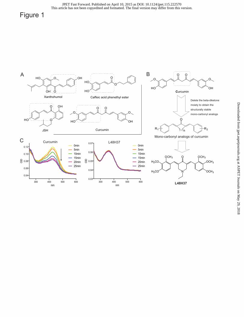
The chemical stability of L48H37 was improved in vitro
The chemical stability of L48H37 and curcumin was tested using an absorption spectrum assay.
Figure 1C showed that the UV-visible absorption spectrum of curcumin displayed a significant peak
with a maximum absorption of close to 425 nm. However, the intensity of curcumin’s absorption
spectrum significantly decreased over time in the phosphate buffer (pH 7.4). In contrast, L48H37
showed no degradation under the same conditions (Figure 1C), suggesting that a chemical
modification to L48H37 significantly increased curcumin’s stability and attenuated in vitro
degradation.
L48H37 blocks the interaction between MD2 and LPS
We first determined the effect of L48H37 on LPS-induced TLR4/MD2 complex conformation
by immunoprecipitation assay. As shown in Figure 2A, the complex of TLR4/MD2 profoundly
increased in LPS-stimulated macrophages, while treatment with L48H37 significantly inhibited
LPS-induced TLR4/MD2 complex. It is unclear whether L48H37 directly affects the interaction of
LPS-MD2 or that of TLR4-MD2. Since MD2 is mainly located in the cell membrane, where LPS
can interact with MD2, we further tested if L48H37 is able to reduce the LPS-MD2 binding in cell
surface. MPMs, with or without L48H37, were incubated with FITC-marked LPS (FITC-LPS) and
then subjected to flow cytometry analysis. Figure 2B shows that FITC-LPS binds to the cell surface
with an MFI of 17.4, and treatment with L48H37 dose-dependently reduced the interaction of
FITC-LPS with the receptor on the cell surface. To validate the effects of L48H37 on the interaction
between LPS and MD2, we established a biotin-streptavidin-based ELISA system at the molecular
This article has not been copyedited and formatted. The final version may differ from this version.JPET Fast Forward. Published on April 10, 2015 as DOI: 10.1124/jpet.115.222570
at ASPE
T Journals on M
ay 29, 2018jpet.aspetjournals.org
Dow
nloaded from

JPET #222570
18
level. The results in Figure 2C show that biotin-marked LPS (Biotin-LPS) was able to bind to
recombinant human MD2 (rhMD2) protein in the plates, while co-incubation with L48H37
significantly blocked the interaction of Biotin-LPS and rhMD2.
L48H37 directly binds to MD2 protein,
The direct interaction of L48H37 and rhMD2 protein was determined using fluorescence
spectroscopy and SPR assay. As shown in Figure 2D, fluorescence values of bis-ANS, a fluorescent
probe used to map the hydrophobic binding sites in proteins, were markedly enhanced upon binding
to cell-free rhMD2 protein, while incubation with L48H37 dose-dependently decreased the
fluorescence intensity of bis-ANS, suggesting that L48H37 competitively binds to rhMD2. Next,
the SPR experiments showed no interaction between L48H37 and recombinant human TLR4
proteins (Figure 2E), while Figure 2F exhibited that L48H37 directly binds rhMD2 protein in a
dose-dependent manner and with a very high affinity (KD value = 0.0000113M). These data indicate
that L48H37 is a novel and MD2-specific inhibitor.
L48H37 acts on Arg90 and Tyr102 residues in MD2 protein pocket
We further predict the underlying binding mode of L48H37 in MD2 protein using a molecular
simulation of L48H37-MD2 complex. As shown in Figure 3A, L48H37 was fitted into the
hydrophobic pocket of MD2, interacting the residues including Tyr102, Phe121, Leu61, Cys133, and
Arg90 in the most energetically favorable configuration (Figure 3A). The whole molecule of
L48H37 is buried inside the lipid-binding pocket and overlaps to a large extent with the binding
sites of LPS, indicating the structural mechanism behind L48H37’s observed competitive inhibition
of LPS. The computer-assisted simulation also show that two amino residues Arg90 and Tyr102 are
This article has not been copyedited and formatted. The final version may differ from this version.JPET Fast Forward. Published on April 10, 2015 as DOI: 10.1124/jpet.115.222570
at ASPE
T Journals on M
ay 29, 2018jpet.aspetjournals.org
Dow
nloaded from
JPET #222570
19
most likely to form hydrogen bonds with L48H37 (Figure 3A). Thus, in order to confirm the
importance of Arg90 and Tyr102 in L48H37 binding to rhMD2, two new rhMD2 mutations,
rhMD2R90A or rhMD2Y102A, were prepared respectively. SPR assay indicated that L48H37 no longer
binds to these two mutations (Figure 3B and 3C), and the ELISA method also found that L48H37
could not inhibit the binding of biotin-LPS with either rhMD2R90A or rhMD2Y102A (Figure 3D and
3E). These results present the possible binding sites of L48H37 in the MD2 protein pocket, which
we believe will be helpful in the design of new MD2 inhibitors.
L48H37 inhibited LPS-induced MAPKs and NF-κB activation in macrophages
We then determined the effects of L48H37 on LPS-activated downstream signaling in
TLR4/MD2 cascade, including the representative MAPKs pathway and the transcriptional factor
NF-κB. MAPK family consists of ERK, p38, and JNK. Figure 4A shows that all of the three
pathways were activated by LPS stimulation in MPMs, while the LPS-induced phosphorylations of
ERK, p38 and JNK were markedly decreased following pre-treatment with L48H37 in a
dose-dependent manner. After IκB degradation, NF-κB p65 translocates from the cytoplasm to the
nucleus, binds to the target promoters, and induces transcription. Using Western blotting, we first
evaluated the effect of L48H37 on IκB degradation in total cell protein extracts. LPS exposure for 1
h induced an 84% degradation of IκB, while pre-treatment with L48H37 reversed LPS-induced IκB
degradation in MPMs in a dose-dependent manner (Figure 4B). Consequently, as shown in Figure
4C, LPS stimulation could increase NF-κB p65 nuclear translocation (red point in blue nucleus),
while in L48H37 pre-treated cells, LPS-induced nuclear levels of p65 were significantly decreased.
L48H37 strongly inhibits LPS-induced inflammatory cytokine expression in macrophages
This article has not been copyedited and formatted. The final version may differ from this version.JPET Fast Forward. Published on April 10, 2015 as DOI: 10.1124/jpet.115.222570
at ASPE
T Journals on M
ay 29, 2018jpet.aspetjournals.org
Dow
nloaded from

JPET #222570
20
MPMs and human PBMCs were used to examine the anti-inflammatory activity of L48H37.
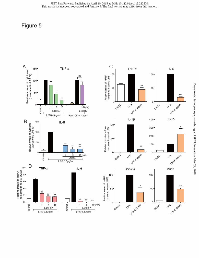
As shown in Figure 5A–B, the LPS-induced increases in TNF-α and IL-6 levels were
dose-dependently inhibited by L48H37 in MPMs. Here, a TLR2 agonist Pam3CK was used as a
comparison. Interestingly, although 0.1 μg/ml Pam3CK significantly induced TNF-α
overexpression in MPMs, L48H37 could not inhibit the inflammatory response induced by
Pam3CK (Figure 5A). Since MD2 is not required in TLR2 signaling pathway activation, the fact
that L48H37 failed to fight TLR2-related inflammation validates the specificity of L48H37 as a
MD2 inhibitor. The anti-inflammatory activity of L48H37 was also observed at the mRNA level.
MPMs treated with LPS (0.5 μg/mL) for 6 h were examined through real-time quantitative PCR for
the expression of pro-inflammatory genes in the presence or absence of L48H37. As shown in
Figure 5C, L48H37 at 10 μM potently inhibited LPS-induced up-regulation of TNF-α (54.7%,
p<0.01), IL-6 (82.3%, p<0.01), IL-1β (91.2%, p<0.01), cycloxygenase-2 (COX-2, 57.5%, p<0.05),
and inducible nitric oxide synthase (iNOS, 50.9%, p<0.01) transcripts in MPMs. As expected,
L48H37 up-regulated the expression of the anti-inflammatory cytokine IL-10. Furthermore, similar
results were observed in human PBMCs, and L48H37 also significantly and dose-dependently
suppressed LPS-increased TNF-α and IL-6 expression (Figure 5D).
L48H37 effectively protects mice from LPS-induced septic shock and lung injury
Male C57BL/6 mice were injected with LPS (i.v., 20 mg/kg) in the presence or absence of
L48H37 pre-treatment (i.v.), and the survival rates were monitored for seven days. Figure 6A
showed that animals treated with LPS alone all died within 48 h. In contrast, treatment with
L48H37 at 10 mg/kg either 15 min prior to LPS injection (prevention group) or 15 min after LPS
injection (treatment group) significantly improved the survival rates compared to that of the control
This article has not been copyedited and formatted. The final version may differ from this version.JPET Fast Forward. Published on April 10, 2015 as DOI: 10.1124/jpet.115.222570
at ASPE
T Journals on M
ay 29, 2018jpet.aspetjournals.org
Dow
nloaded from

JPET #222570
21
group (p<0.01 in both groups vs. LPS group). Also, the weight lost in both groups improved slowly
2-7 days after LPS injection (Figure 6B).
We also examined the beneficial effects of L48H37 on lung injury in LPS-treated mice. Two or
eight hours after administration with LPS (20 mg/kg, i.v.), histopathological changes in the lungs of
C57BL/6 mice were observed using H&E staining. L48H37 pre-treatment at 10 mg/kg significantly
improved pulmonary damage and amended the LPS-injured tissue structure of pulmonary lobules
(Figure 6C). These data demonstrate the anti-inflammatory effects of L48H37 in septic mice.
This article has not been copyedited and formatted. The final version may differ from this version.JPET Fast Forward. Published on April 10, 2015 as DOI: 10.1124/jpet.115.222570
at ASPE
T Journals on M
ay 29, 2018jpet.aspetjournals.org
Dow
nloaded from
JPET #222570
22
Discussion
Sepsis can be caused by trauma, infection or burns and can lead to septic shock and organ
failure. Despite there being more than 30 pharmaceutical candidates for the treatment of sepsis
currently in the developmental stage, most of these treatments have failed due to the complicated
nature of sepsis, and sepsis remains as the most common cause of death in intensive care units
(King et al., 2014). Xigris was used to treat sepsis as a recombinant human-activated protein C that
attenuates the development of organ failure due to sepsis. However, no significant improvement
was observed in clinical uses, and in 2012, use of Xigris was suspended (Opal et al., 2014).
Although statins have some non-specific anti-inflammatory effects, they are currently not being
considered as therapeutic options for sepsis (Gazzerro et al., 2012; Ou et al., 2014). Therefore, there
is an urgent need to find novel and effective therapeutic approaches for sepsis.
One potential approach to treating and preventing septic shock and its associated diseases is
the intervention of the TLR/MD2-mediated inflammatory response (Savva and Roger, 2013). LPS
is presented to TLR4/MD2 complex via the LPS-binding protein and CD14 (Park and Lee, 2013).
MD2 recognizes the lipid A domain of LPS, leading to the formation of the TLR4/MD2/LPS
complex and activation of the downstream cellular response (Park et al., 2012; Oblak and Jerala,
2014). Both TLR4 and MD2 are essential for the LPS-induced inflammatory response and sepsis.
Both TLR4-/- and MD2-/- mice fail to respond to LPS and survive endotoxic shock (Duan et al.,
2014). Therefore, TLR4 and MD2 are proposed as potential targets for the therapy that neutralizes
the toxic effects of endotoxin.
The growth in our understanding of the structure and function of the TLR4/MD2 complex has
provided a new direction in the development of new drug targets in the treatment and prevention of
This article has not been copyedited and formatted. The final version may differ from this version.JPET Fast Forward. Published on April 10, 2015 as DOI: 10.1124/jpet.115.222570
at ASPE
T Journals on M
ay 29, 2018jpet.aspetjournals.org
Dow
nloaded from

JPET #222570
23
sepsis (Peri and Calabrese, 2014). Due to the seemingly higher importance of TLR4, researchers
paid more attention to TLR4 than MD2 in the past decades (Wittebole et al., 2010; Svajger et al.,
2013). However, the clinical trials of TAK-242, a TLR4 specific inhibitor, have failed to treat severe
sepsis and related respiratory disease in patients (Rice et al., 2010). In addition, blocking TLR can
lead to severe side effects, ‘inappropriate’ immune responses such as allergic Th2 responses, or
immunological tolerance (Ishii et al., 2006; Nakamoto and Kanai, 2014). On the other hand, a series
of MD2 antagonists with lipid A structure (mimics LPS) targeting the MD2 protein, such as fatty
acid chain-containing E5531 (Bryant et al., 2007), CRX-526 (Lin et al., 2013), and eritoran
(Rallabhandi et al., 2012), have been evaluated in clinical and pre-clinical studies. Unfortunately,
the most studied one, eritoran, has failed in phase III clinical trial in 2011 due to no significant
improvement in eritoran-treated patients compared to the placebo group (Barochia et al., 2011).
Recently, some natural active compounds that do not contain the structure of lipid A or fatty
acids have been found to be able to target MD2 directly (Figure 1A). These small molecules, such
as xanthohumol (Peluso et al., 2010), CAPE (Kim et al., 2013), JSH,(Roh et al., 2011) and
curcumin (Gradisar et al., 2007), bind directly to the MD2 pocket, and block the TLR4/MD2’s
recognition of LPS, resulting in the prevention of pro-inflammatory signaling and septic shock.
Although their specificities for targeting other proteins remain to be defined, these natural
compounds (1-4) provide us the important structural information for the design and discovery of
new synthetic MD2 inhibitors. As shown in Figure 1A, the structures of the MD2 inhibitors share
the same 3-(4-hydroxyphenyl) acrylaldehyde skeleton. Thus, it is hypothesized that our new
synthetic compound L48H37, which shows excellent anti-inflammatory activity and contains the
structure of 3-(4-hydroxyphenyl) acrylaldehyde, may target MD2 and serve as an anti-sepsis
candidate.
This article has not been copyedited and formatted. The final version may differ from this version.JPET Fast Forward. Published on April 10, 2015 as DOI: 10.1124/jpet.115.222570
at ASPE
T Journals on M
ay 29, 2018jpet.aspetjournals.org
Dow
nloaded from
JPET #222570
24
Hence, the interaction between L48H37 and MD2 was investigated at both cell-free molecular
and cellular levels. Fluorescence spectroscopy and SPR assay demonstrated that L48H37 was able
to dose-dependently bind to rhMD2 protein (Figure 2D and 2E). The interaction of L48H37 with
MD2 remarkably affected the LPS binding to rhMD2 (Figure 2C), suggesting that the binding site
for L48H37 in MD2 pocket overlaps that for LPS, which is also consistent with the molecular
docking results (Figure 3A). At the cellular level, flow cytometry (Figure 2B) and
immunoprecipitation (Figure 2A) revealed the inhibitory effects of L48H37 on LPS-MD2
interactions and MD2-TLR4 complex formation, respectively. Interestingly, our data also showed
that L48H37 is a specific MD2 inhibitor, since L48H37 could not inhibit the Pam3CK-induced
TLR2 activation, which shares the MAPKs/NF-kB-involved pro-inflammatory signaling pathway
with TLR4 (Figure 5A), but is independent on MD2. There, this study demonstrates that MD2 is a
molecular target of L48H37 and that L48H37 can down-regulate TLR4 activation and inflammatory
gene expression, as well as attenuate LPS-induced sepsis, by interrupting the association of LPS
with MD2 (Figure 6).
Molecular modeling of the crystal structure of MD2 provided further support for the binding of
L48H37 to MD2. The X-ray diffraction-based structural information and exact binding mechanism
for non-lipid compounds binding to MD2 protein are still unclear. The MD2-binding sites of
non-lipid compounds have been predicted by computer-assisted simulation, and the Cys133 in the
MD2 binding pocket is considered a molecular target of several natural inhibitors. Small-molecule
inhibitors with α,β-unsaturated ketones are capable of forming covalent bonds with Cys133 via a
Michael-type reaction. JTT-705 (Mancek-Keber et al., 2009) and CAPE (Kim et al., 2013) have
been predicted to covalently bind Cys133 residue and showed an irreversible inhibition against MD2.
However, the α,β-unsaturated ketone-containing curcumin interacts with MD2 via a non-covalent
This article has not been copyedited and formatted. The final version may differ from this version.JPET Fast Forward. Published on April 10, 2015 as DOI: 10.1124/jpet.115.222570
at ASPE
T Journals on M
ay 29, 2018jpet.aspetjournals.org
Dow
nloaded from

JPET #222570
25
mechanism, supported by studies showing that it can be removed from the complex bound to MD2
by chloroform extraction and that it can still inhibit LPS from binding to the mutant MD2
Cys133Phe in the same manner as the wild-type (Gradisar et al., 2007). In addition, some residues
Lys122, Tyr102, Gly123, Ser120, Lys130, and Phe126 in MD2 pocket were predicted to play a possible
role in the interaction between MD2 protein and natural small-molecule inhibitors such as JSH (Roh
et al., 2011), taxanes (Resman et al., 2008), and xanthohumol (Peluso et al., 2010). In this study, we
found the possible binding mechanism of L48H37-MD2 using the molecular docking method. The
results indicated that the binding site for L48H37 in the MD2 pocket overlapped that for LPS, rather
than TLR4 (Figure 3A), which is also evidenced by the experimental data at the molecular and
cellular levels. Using further molecular dynamics, we showed that L48H37 may form hydrogen
bonds with two key residues, Arg90 and Tyr102, which also play a role in the binding of LPS (Figure
3A). To validate this prediction, we replaced these two amino residues Arg90 and Tyr102 with Ala in
rhMD2 mutations. As expected, the SPR analysis and ELISA showed that L48H37 could not
interact the rhMD2 mutations any more, indicating that Arg90 and Tyr102 play a critical role in
L48H37-MD2 interactions. Although the Tyr102 residue has been predicted to be of importance in
isoxanthohumol-MD2 interactions, the authors failed to demonstrate the possible hydrogen bond
formation with Tyr102 (Peluso et al., 2010). In addition, this is the first time that Arg90 has been
highlighted as an important molecular target for MD2 inhibitors. Thus, the results of this study
provide the important structural information and understanding of the amino residue sites that
support the use and further design of MD2 inhibitors as anti-inflammatory agents.
L48H37’s inhibition of MD2 resulted in a series of anti-inflammatory activities in
macrophages. MAPKs and NF-κB have been demonstrated as the main mediators in the
LPS-TLR4/MD2 pro-inflammatory signaling pathway. L48H37 prevented TLR4-mediated MAPKs
This article has not been copyedited and formatted. The final version may differ from this version.JPET Fast Forward. Published on April 10, 2015 as DOI: 10.1124/jpet.115.222570
at ASPE
T Journals on M
ay 29, 2018jpet.aspetjournals.org
Dow
nloaded from

JPET #222570
26
and NF-κB activation in LPS-stimulated macrophage, as evidenced by a dose-dependent decrease in
the levels of ERK/p38/JNK phosphorylation, IκB degradation, and p65 translocation (Figure 4).
Figure 5 further showed the inhibitory effects of L48H37 on LPS-induced inflammatory cytokine
overexpression in both mouse MPMs and human PBMCs. In vivo, either pre-treatment or
post-treatment with L48H37 significantly increased survival in the LPS-induced septic mice (Figure
6A). Lung histological changes in the LPS-injected mice were also suppressed by L48H37
pre-treatment (Figure 6C). These results validated the potential of the MD2-targeting L48H37 as a
therapeutic agent in both the prevention and treatment of acute inflammatory diseases.
Collectively, our data reveal that MD2 is the anti-inflammatory target of novel compound
L48H37 and can lead to the blockage of LPS-TLR4/MD2 complex formation and decrease of
downstream signal activation and inflammatory mediator expression. A schematic for the protection
of L48H37 from LPS-induced sepsis is illustrated in Figure 7. Arg90 and Tyr102 in the MD2 protein
play an important role in L48H37’s interaction with MD2 via two hydrogen bonds. In vivo, L48H37
improved survival and protected lungs against LPS-induced injury in septic mice. This study
suggests that MD2 is an important therapeutic target against inflammatory disorders and proves that
a new MD2 inhibitor, L48H37, can be developed as a potential agent in the treatment of sepsis.
This article has not been copyedited and formatted. The final version may differ from this version.JPET Fast Forward. Published on April 10, 2015 as DOI: 10.1124/jpet.115.222570
at ASPE
T Journals on M
ay 29, 2018jpet.aspetjournals.org
Dow
nloaded from
JPET #222570
27
Authorship Contributions
Participated in research design: Yi Wang, Guang Liang, Xiaoou Shan, Guilong Guo
Conducted experiments: Yi Wang, Lili Jiang, Gaozhi Chen, Yali Zhang, Zhe Wang
Contributed new reagents or analytic tools: Lili Dong, Jianzhang Wu
Performed data analysis: Yi Wang, Yuanrong Dai, Guang Liang
Wrote or contributed to the writing of the manuscript: Yi Wang, Lili Jiang, Guang Liang
This article has not been copyedited and formatted. The final version may differ from this version.JPET Fast Forward. Published on April 10, 2015 as DOI: 10.1124/jpet.115.222570
at ASPE
T Journals on M
ay 29, 2018jpet.aspetjournals.org
Dow
nloaded from
JPET #222570
28
References Balk RA (2014) Systemic inflammatory response syndrome (SIRS): where did it come from and is it still
relevant today? Virulence 5:20-26. Barochia A, Solomon S, Cui X, Natanson C and Eichacker PQ (2011) Eritoran tetrasodium (E5564) treatment
for sepsis: review of preclinical and clinical studies. Expert Opin Drug Metab Toxicol 7:479-494. Bryant CE, Ouellette A, Lohmann K, Vandenplas M, Moore JN, Maskell DJ and Farnfield BA (2007) The
cellular Toll-like receptor 4 antagonist E5531 can act as an agonist in horse whole blood. Vet
Immunol Immunopathol 116:182-189. Cighetti R, Ciaramelli C, Sestito SE, Zanoni I, Kubik L, Arda-Freire A, Calabrese V, Granucci F, Jerala R,
Martin-Santamaria S, Jimenez-Barbero J and Peri F (2014) Modulation of CD14 and TLR4.MD-2 activities by a synthetic lipid A mimetic. Chembiochem : a European journal of chemical biology
15:250-258. Duan G, Zhu J, Xu J and Liu Y (2014) Targeting myeloid differentiation 2 for treatment of sepsis. Front Biosci
(Landmark Ed) 19:904-915. Gaieski DF and Goyal M (2013) What is sepsis? What is severe sepsis? What is septic shock? Searching for
objective definitions among the winds of doctrines and wild theories. Expert review of anti-infective
therapy 11:867-871. Gazzerro P, Proto MC, Gangemi G, Malfitano AM, Ciaglia E, Pisanti S, Santoro A, Laezza C and Bifulco M
(2012) Pharmacological actions of statins: a critical appraisal in the management of cancer. Pharmacological reviews 64:102-146.
Goodall KJ, Poon IK, Phipps S and Hulett MD (2014) Soluble Heparan Sulfate Fragments Generated by Heparanase Trigger the Release of Pro-Inflammatory Cytokines through TLR-4. PLoS One
9:e109596. Gradisar H, Keber MM, Pristovsek P and Jerala R (2007) MD-2 as the target of curcumin in the inhibition of
response to LPS. J Leukoc Biol 82:968-974. Hasima N and Aggarwal BB (2012) Cancer-linked targets modulated by curcumin. Int J Biochem Mol Biol
3:328-351. Ishii KJ, Uematsu S and Akira S (2006) 'Toll' gates for future immunotherapy. Curr Pharm Des
12:4135-4142. Joe B, Vijaykumar M and Lokesh B (2004) Biological properties of curcumin-cellular and molecular
mechanisms of action. Critical reviews in food science and nutrition 44:97-111. Kim SY, Koo JE, Seo YJ, Tyagi N, Jeong E, Choi J, Lim KM, Park ZY and Lee JY (2013) Suppression of
Toll-like receptor 4 activation by caffeic acid phenethyl ester is mediated by interference of LPS binding to MD2. Br J Pharmacol 168:1933-1945.
King EG, Bauza GJ, Mella JR and Remick DG (2014) Pathophysiologic mechanisms in septic shock. Lab
Invest 94:4-12. Lin M, Yiu WH, Li RX, Wu HJ, Wong DW, Chan LY, Leung JC, Lai KN and Tang SC (2013) The TLR4
antagonist CRX-526 protects against advanced diabetic nephropathy. Kidney Int 83:887-900. Mancek-Keber M, Gradisar H, Inigo Pestana M, Martinez de Tejada G and Jerala R (2009) Free thiol group of
MD-2 as the target for inhibition of the lipopolysaccharide-induced cell activation. J Biol Chem
284:19493-19500. Nakamoto N and Kanai T (2014) Role of toll-like receptors in immune activation and tolerance in the liver.
This article has not been copyedited and formatted. The final version may differ from this version.JPET Fast Forward. Published on April 10, 2015 as DOI: 10.1124/jpet.115.222570
at ASPE
T Journals on M
ay 29, 2018jpet.aspetjournals.org
Dow
nloaded from

JPET #222570
29
Front Immunol 5:221. Oblak A and Jerala R (2014) The molecular mechanism of species-specific recognition of lipopolysaccharides
by the MD-2/TLR4 receptor complex. Mol Immunol.Opal SM, Dellinger RP, Vincent JL, Masur H and Angus DC (2014) The next generation of sepsis clinical trial
designs: what is next after the demise of recombinant human activated protein C?*. Crit Care Med
42:1714-1721. Ou SY, Chu H, Chao PW, Ou SM, Lee YJ, Kuo SC, Li SY, Shih CJ and Chen YT (2014) Effect of the use of
low and high potency statins and sepsis outcomes. Intensive care medicine 40:1509-1517. Pan Y, Zhu G, Wang Y, Cai L, Cai Y, Hu J, Li Y, Yan Y, Wang Z, Li X, Wei T and Liang G (2013) Attenuation
of high-glucose-induced inflammatory response by a novel curcumin derivative B06 contributes to its protection from diabetic pathogenic changes in rat kidney and heart. J Nutr Biochem 24:146-155.
Park BS and Lee JO (2013) Recognition of lipopolysaccharide pattern by TLR4 complexes. Exp Mol Med
45:e66. Park SH, Kim ND, Jung JK, Lee CK, Han SB and Kim Y (2012) Myeloid differentiation 2 as a therapeutic
target of inflammatory disorders. Pharmacol Ther 133:291-298. Peluso MR, Miranda CL, Hobbs DJ, Proteau RR and Stevens JF (2010) Xanthohumol and related prenylated
flavonoids inhibit inflammatory cytokine production in LPS-activated THP-1 monocytes: structure-activity relationships and in silico binding to myeloid differentiation protein-2 (MD-2). Planta Med 76:1536-1543.
Peri F and Calabrese V (2014) Toll-like receptor 4 (TLR4) modulation by synthetic and natural compounds: an update. J Med Chem 57:3612-3622.
Prasad S, Gupta SC, Tyagi AK and Aggarwal BB (2014) Curcumin, a component of golden spice: From bedside to bench and back. Biotechnology advances 32:1053-1064.
Rallabhandi P, Phillips RL, Boukhvalova MS, Pletneva LM, Shirey KA, Gioannini TL, Weiss JP, Chow JC, Hawkins LD, Vogel SN and Blanco JC (2012) Respiratory syncytial virus fusion protein-induced toll-like receptor 4 (TLR4) signaling is inhibited by the TLR4 antagonists Rhodobacter sphaeroides lipopolysaccharide and eritoran (E5564) and requires direct interaction with MD-2. mBio 3.
Resman N, Gradisar H, Vasl J, Keber MM, Pristovsek P and Jerala R (2008) Taxanes inhibit human TLR4 signaling by binding to MD-2. FEBS Lett 582:3929-3934.
Rice TW, Wheeler AP, Bernard GR, Vincent JL, Angus DC, Aikawa N, Demeyer I, Sainati S, Amlot N, Cao C, Ii M, Matsuda H, Mouri K and Cohen J (2010) A randomized, double-blind, placebo-controlled trial of TAK-242 for the treatment of severe sepsis. Crit Care Med 38:1685-1694.
Roh E, Lee HS, Kwak JA, Hong JT, Nam SY, Jung SH, Lee JY, Kim ND, Han SB and Kim Y (2011) MD-2 as the target of nonlipid chalcone in the inhibition of endotoxin LPS-induced TLR4 activity. J Infect Dis
203:1012-1020. Rossol M, Heine H, Meusch U, Quandt D, Klein C, Sweet MJ and Hauschildt S (2011) LPS-induced cytokine
production in human monocytes and macrophages. Critical reviews in immunology 31:379-446. Savva A and Roger T (2013) Targeting toll-like receptors: promising therapeutic strategies for the
management of sepsis-associated pathology and infectious diseases. Front Immunol 4:387. Svajger U, Brus B, Turk S, Sova M, Hodnik V, Anderluh G and Gobec S (2013) Novel toll-like receptor 4
(TLR4) antagonists identified by structure- and ligand-based virtual screening. Eur J Med Chem
70:393-399. Wittebole X, Castanares-Zapatero D and Laterre PF (2010) Toll-like receptor 4 modulation as a strategy to
treat sepsis. Mediators Inflamm 2010:568396. Wu J, Zhang Y, Cai Y, Wang J, Weng B, Tang Q, Chen X, Pan Z, Liang G and Yang S (2013) Discovery and
This article has not been copyedited and formatted. The final version may differ from this version.JPET Fast Forward. Published on April 10, 2015 as DOI: 10.1124/jpet.115.222570
at ASPE
T Journals on M
ay 29, 2018jpet.aspetjournals.org
Dow
nloaded from
JPET #222570
30
evaluation of piperid-4-one-containing mono-carbonyl analogs of curcumin as anti-inflammatory agents. Bioorg Med Chem 21:3058-3065.
Footnotes
Yi Wang and Xiaoou Shan contributed equally to this paper.
This study was supported by the National Natural Science Funding of China [Grants 81472307
21272179], High-level Innovative Talent Funding of Zhejiang Department of Health (GL), Zhejiang
Natural Science Funding [Grant LQ14H310003], and Zhejiang College Students’ Science and
Technology Innovation Activities Program [Grant 2014R413071].
This article has not been copyedited and formatted. The final version may differ from this version.JPET Fast Forward. Published on April 10, 2015 as DOI: 10.1124/jpet.115.222570
at ASPE
T Journals on M
ay 29, 2018jpet.aspetjournals.org
Dow
nloaded from
JPET #222570
31
Figure Legends
Figure 1. Design, synthesis and stability assay of curcumin analogue L48H37. (A) Structures of
current MD2 inhibitors; (B) Design and synthesis of curcumin analog L48H37; (C) UV-visible
absorption spectra of curcumin and L48H37. Curcumin or L48H37 were dissolved in phosphate
buffer (pH 7.4) to a final concentration of 20 μM. Absorbance readings were taken from 250 to 600
nm using a spectraMax M5. The UV absorption spectra were collected for over 25 min at 5 min
intervals at 25�.
Figure 2. Antagonistic effect of L48H37 on LPS binding to MD2. (A) Co-immunoprecipitation
of MD2. MPMs were pretreated with L48H37 (10 μM) or DMSO for 30 min and then incubated
with LPS (1 μg/mL) for 5 min. Cells were lysed and the total protein was collected. 400 μg of the
total protein were incubated with beads and anti-MD2 antibody overnight at 4�. The
immunoprecipitated proteins and precipitated MD2 proteins, were resolved by SDS-PAGE and
detected using anti-TLR4 antibody. The column figures represent the mean optical density ratio of
three independent experiments (** p<0.01). (B) Flow cytometric analysis. HUVEC304 cells were
incubated with media alone (Ctrl), L48H37 (10 μM), LPS-FITC (50 μg/mL), LPS-FITC (50 μg/mL)
plus L48H37 (0.1, 1, and 10 μM), respectively. These cells were subjected to flow cytometry
analysis, in which the values for the median fluorescence intensity (MFI) were also provided; (C) In
vitro assays for LPS binding to MD2. rhMD2 antibody was coated to a 96-well at 4°C overnight.
rhMD2 (4 µg/mL) in 10 mM Tris-HCl buffer was added to the pre-coated plate for 1.5 h at room
temperature. After washing with PBST, biotin-labeled LPS was added to the plate with or without
the presence of L48H37 (1.5 μM). LPS ability to bind to rhMD2 was determined using ELISA,
This article has not been copyedited and formatted. The final version may differ from this version.JPET Fast Forward. Published on April 10, 2015 as DOI: 10.1124/jpet.115.222570
at ASPE
T Journals on M
ay 29, 2018jpet.aspetjournals.org
Dow
nloaded from

JPET #222570
32
represented by absorbance values at 450 nm (A450). Data are mean values (±SEM) of 3 separate
experiments, each performed in duplication. *p<0.05, **p<0.01 vs. buffer alone-added group; (D)
Fluorescence measurements. bis-ANS (5 μM) was pre-incubated with rhMD2 (5 nM) to reach stable
fluorescence values under excitation at 380 nM and to reach stable relative fluorescence units
(RFUs) emitted at 430–590 nm under excitation at 385 nm. Non-fluorescent L48H37 (at 2.5, 5, 10,
20, 30 μM) was then treated for 5 min, and the relative fluorescence units (RFUs) emitted at
430-590 nm were measured; (E) SPR analysis showed that L48H37 could not directly bind to
rhTLR4 protein. (F) The binding affinity of L48H37 with rhMD2 was determined using a SPR
assay.
Figure 3. Antagonistic mechanism of L48H37 on LPS binding to MD2. (A) Molecular docking
of L48H37 with rhMD2 (PDB ID: 2E56) was analyzed with the Sybyl-2.0 molecular modeling
software from Tripos. hTLR4 and rhMD2 are shown in white and green, respectively; (B-C)
Surface Plasmon resonance analysis. rhMD2R90A or rhMD2Y102A was biotinylated with biotin, and
L48H37 was diluted to 100, 50, 25, 12.5, or 6.25 μM. The binding affinity of L48H37 was
determined using a FortéBio Octet Red equipped with a super streptavidin (SSA) sensor; (D-E) In
vitro assay for LPS binding to MD2 variants. rhMD2 antibody was coated on a 96-well at 4°C
overnight. rhMD2R90A or rhMD2Y102A (4 µg/mL) in 10 mM Tris-HCl buffer was added to the
pre-coated plate for 1.5 h at room temperature. After washing with PBST, biotin-labeled LPS was
added to the plate with or without the L48H37 treatment (1.5 μM). LPS binding to rhMD2 was
determined by ELISA and represented by absorbance values at 450 nm (A450). Data are mean values
(±SEM) of 3 separate experiments, each performed in duplication.
This article has not been copyedited and formatted. The final version may differ from this version.JPET Fast Forward. Published on April 10, 2015 as DOI: 10.1124/jpet.115.222570
at ASPE
T Journals on M
ay 29, 2018jpet.aspetjournals.org
Dow
nloaded from
JPET #222570
33
Figure 4. L48H37 inhibited LPS-induced MAPK phosphorylation and NF-κB activation. (A-B)
MPMs were pretreated with the vehicle control (DMSO) or L48H37 (1, 2.5, 5, or 10 μM) for 2 h
followed by incubation with LPS (0.5 μg/mL) for 1 h. The protein levels of p-ERK, ERK, p-p38,
p38, p-JNK, JNK, I-κB were examined by Western blot. The column figures represent the mean
optical density ratio of three independent experiments. * p<0.05, ** p<0.01, vs. the LPS-treated
group; (C) Cultured MPMs were pretreated with L48H37 (10 μM) or vehicle control (DMSO) for 2
h, and then stimulated with LPS (0.5 μg/mL). After 1 h of treatment, the cells were incubated with
p65 antibody and Cy3 fluorescein-conjugated secondary antibody (red), and the nuclei were stained
with DAPI (blue). The images (200×) were obtained by fluorescence microscope and overlay.
Similar results were obtained for three independent experiments. The column figure for the p65
translocation represents the mean optical density ratio in three independent experiments. * p<0.05,
** p<0.01, vs. LPS-treated group.
Figure 5. L48H37 inhibited LPS-induced inflammatory cytokine expression in mouse
macrophages and human PBMCs. (A-B) MPMs were pretreated with the vehicle control (DMSO)
or L48H37 (1, 5, or 10 μM) for 2 h followed by incubation with LPS (0.5 μg/mL) or Pam3CK (0.1
μg/mL) for 22 h. The protein levels of TNF-α (A) and IL-6 (B) in the culture medium were
measured by ELISA. The total amount of cytokines in the cell medium was normalized to the total
amount of protein in the viable cell pellet. The results are expressed as a percentage of the
LPS-alone group (solid dark bar). Each bar represents mean ± SEM of 3-5 independent
experiments. * p<0.05, ** p<0.01, v.s. LPS-treated group. (C) MPMs were pretreated with vehicle
control (DMSO) or L48H37 (10 μM) for 2 h followed by incubation with LPS (0.5 μg/mL) for 6 h.
The mRNA levels of inflammatory cytokines, including TNF-α, IL-6, IL-1β, IL-10, COX-2, and
This article has not been copyedited and formatted. The final version may differ from this version.JPET Fast Forward. Published on April 10, 2015 as DOI: 10.1124/jpet.115.222570
at ASPE
T Journals on M
ay 29, 2018jpet.aspetjournals.org
Dow
nloaded from
JPET #222570
34
iNOS were quantified by RT-qPCR. The mRNA values were normalized to the internal control
β-actin mRNA and are expressed as a percentage of the vales for the LPS control. Each bar
represents mean ± SEM of 3-5 independent experiments. * p<0.05, ** p<0.01, vs. LPS-treated
group. (D) Human PBMCs were pretreated with the vehicle control (DMSO) or L48H37 (1, 5, or 10
μM) for 2 h followed by incubation with LPS (0.5 μg/mL) for 6 h. The mRNA levels of
inflammatory cytokines, including TNF-α and IL-6 were quantified by RT-qPCR. The mRNA
values were normalized to the internal control β-actin mRNA and are expressed as a ratio of the
LPS-alone group (solid dark bar). Each bar represents mean ± SEM of 3-5 independent
experiments. * p<0.05, ** p<0.01, vs. LPS-treated group.
Figure 6. L48H37 improved survival and lung injury of mice subjected to a lethal dose of LPS.
C57BL/6 mice (n=10/group) were treated with 10 mg/kg L48H37 15 min before or after injection of
20 mg/kg LPS (i.v.). Survival rates (A) and body weight (B) were recorded for 7 days after LPS
injection at the interval of 12 h. ** p<0.01 vs. LPS-treated group. (C) C57BL/6 mice (n=10/group)
were treated with 10 mg/kg L48H37 15 min before injection of 20 mg/kg LPS (i.v.). Two or eight
hours after LPS injection, five mice were anesthetized with diethyl ether and sacrificed, respectively.
Lung histopathological analysis was performed using H&E staining as described in Materials and
Methods. The representative images are shown.
Figure 7. Proposed model of signaling pathway involved in L48H37 prevented LPS-induced
TLR4 signaling pathway activation and sepsis.
This article has not been copyedited and formatted. The final version may differ from this version.JPET Fast Forward. Published on April 10, 2015 as DOI: 10.1124/jpet.115.222570
at ASPE
T Journals on M
ay 29, 2018jpet.aspetjournals.org
Dow
nloaded from

JPET #222570
35
This article has not been copyedited and formatted. The final version may differ from this version.JPET Fast Forward. Published on April 10, 2015 as DOI: 10.1124/jpet.115.222570
at ASPE
T Journals on M
ay 29, 2018jpet.aspetjournals.org
Dow
nloaded from
O
HO
OH
O
O
OHOHO
OH
O OO
HO OH
O
O
OHO
HO
JSH Curcumin
Xanthohumol Caffeic acid phenethyl ester
Figure 1
N
OH3CO
OCH3
H3CO
OCH3
OCH3
OCH3
L48H37
O OO
HO OH
O
Curcumin
O
n R2R1
Mono-carbonyl analogs of curcumin
Delete the beta-diketone
moiety to obtain the
structurally stable
mono-carbonyl analogs
300 400 500 600
0.04
0.06
0.08
0.10
0.12 0min5min10min15min20min25min
nm
OD
300 400 500 6000.03
0.04
0.05
0.06
0.070min5min10min15min20min25min
nm
OD
Curcumin L48H37
A B
C
This article has not been copyedited and formatted. The final version may differ from this version.JPET Fast Forward. Published on April 10, 2015 as DOI: 10.1124/jpet.115.222570
at ASPE
T Journals on M
ay 29, 2018jpet.aspetjournals.org
Dow
nloaded from
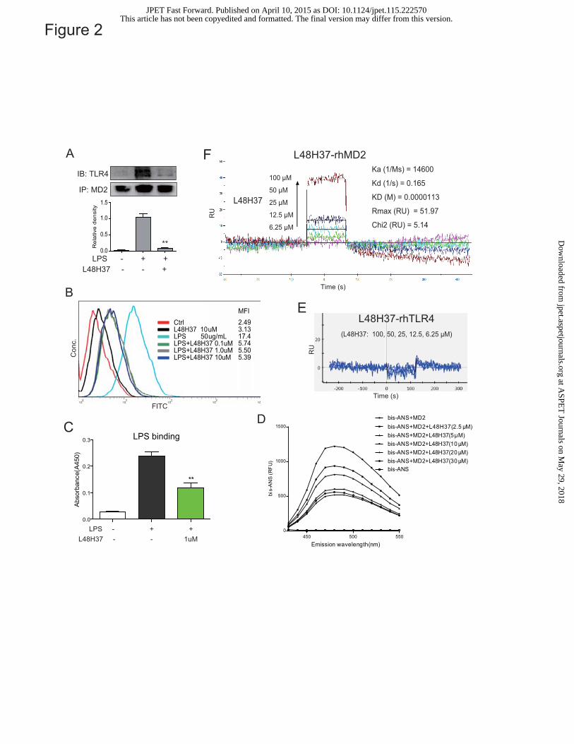
Figure 2
450 500 5500
500
1000
1500
bis-ANS+MD2bis-ANS+MD2+L48H37(2.5 μM)bis-ANS+MD2+L48H37(5μM)bis-ANS+MD2+L48H37(10 μM)bis-ANS+MD2+L48H37(20 μM)bis-ANS+MD2+L48H37(30 μM)bis-ANS
Emission wavelength(nm)
bis-
AN
S(R
FU
)
DLPS binding
0.0
0.1
0.2
0.3
**
Abs
orba
nce(
A45
0)
LPS - + + L48H37 - - 1uM
C
B
CtrlL48H37 10 uMLPS 50 ug/mLLPS+L48H37 0.1uMLPS+L48H37 1.0uMLPS+L48H37 10uM
MFI2.493.1317.45.745.505.39C
onc.
FITC
RU
Time (s)
EL48H37-rhTLR4
Time (s)
L48H37
100 μM
50 μM
25 μM
12.5 μM
6.25 μM
Ka (1/Ms) = 14600
Kd (1/s) = 0.165
KD (M) = 0.0000113
Rmax (RU) = 51.97
Chi2 (RU) = 5.14
RU
F L48H37-rhMD2
(L48H37: 100, 50, 25, 12.5, 6.25 μM)
AIB: TLR4
IP: MD2
LPS - + +L48H37 - - +
0.0
0.5
1.0
1.5
Rel
ativ
e de
nsity
**
This article has not been copyedited and formatted. The final version may differ from this version.JPET Fast Forward. Published on April 10, 2015 as DOI: 10.1124/jpet.115.222570
at ASPE
T Journals on M
ay 29, 2018jpet.aspetjournals.org
Dow
nloaded from

Biotin-LPS - + + L48H37 - - 1uM
100 μM50 μM25 μM12.5 μM6.25 μM
L48H37
100 μM50 μM25 μM12.5 μM6.25 μM
L48H37
L48H37-MD2R90A binding
L48H37-MD2Y102A binding
Figure 3
A
B D
EC
0.0
0.2
0.4
0.6
0.8
1.0
0.0
0.5
1.0
1.5
Abso
rban
ce(A
450)
Abso
rban
ce(A
450)
LPS-MD2R90A binding
LPS-MD2Y102A binding
Biotin-LPS - + + L48H37 - - 1uM
This article has not been copyedited and formatted. The final version may differ from this version.JPET Fast Forward. Published on April 10, 2015 as DOI: 10.1124/jpet.115.222570
at ASPE
T Journals on M
ay 29, 2018jpet.aspetjournals.org
Dow
nloaded from

LPS (0.5ug/ml) - + + + + +
p-ERK1/2
ERK2
p-p38
p38
L48H37 (uM) - - 1.0 2.5 5 10
p-JNK
JNK
0
50
100
**
P65
tran
sloc
atio
nre
late
d to
DAP
I (%
)
DMSO LPS LPS+L48H37
A
BC
Figure 4
LPS (0.5ug/ml) - + + + + L48H37 (uM) - - 2.5 5.0 10
I-κB
GAPDH
0
50
100
*
****
Dens
ity (c
ompa
red
to L
PS %
)
LPSDMSO
LPS+L48H37
0.0
0.5
1.0
p-ERK/ERK p-P38/P38 p-JNK/JNK
rela
tive
desi
tty
LPS (0.5ug/ml) - + + + + + - + + + + + - + + + + +L48H37 (uM) - - 1.0 2.5 5 10 - - 1.0 2.5 5 10 - - 1.0 2.5 5 10
*
**
**
**
**
**
****
**
This article has not been copyedited and formatted. The final version may differ from this version.JPET Fast Forward. Published on April 10, 2015 as DOI: 10.1124/jpet.115.222570
at ASPE
T Journals on M
ay 29, 2018jpet.aspetjournals.org
Dow
nloaded from
B
Figure 5
0
50
100
150 IL-6
1 5 10
LPS 0.5μg/ml
DM
SO
**
(μM)
**
L48H37
Rela
tive
amou
nt o
f cy
tokin
es(c
ompa
red
to L
PS %
)
**
0
2
4
6
8
10 TNF-α
1 5 10
LPS 0.5μg/ml
DM
SO
****
(μM)
**
L48H37
** ** ** 1 5 10
LPS 0.5μg/ml
DM
SO L48H37
IL-6
C
D
Rela
tive
amou
nt o
f m
RNA
com
pare
d to
con
trol D
MSO
DMSOLP
S
LPS+L48
H370
50
100
TNF-α
Relat
ive
amou
nt o
f mR
NAco
mpar
ed to
cont
rol L
PSDMSO
LPS
LPS+L48
H370
50
100
IL-6
**
DMSOLP
S
LPS+L48
H370
50
100
IL-1
**
DMSOLP
S0
100
200
300
400 IL-10
*
DMSOLP
S0
50
100
COX-2
*
DMSOLP
S
LPS+L48
H370
50
100
iNOS
**
β
Relat
ive
amou
nt o
f mR
NAco
mpar
ed to
cont
rol L
PSRe
lativ
e am
ount
of
mRNA
comp
ared
to co
ntro
l LPS
0
50
100
150 TNF-α
1 5 10 10
LPS 0.5μg/ml
DM
SO
**
(μM)
**
L48H37
ns
L48H37
Pam3CK 0.1μg/ml
Rel
ativ
e am
ount
of
cyto
kine
s(c
ompa
red
to L
PS
%)
A
LPS+L48
H37
LPS+L48
H37
This article has not been copyedited and formatted. The final version may differ from this version.JPET Fast Forward. Published on April 10, 2015 as DOI: 10.1124/jpet.115.222570
at ASPE
T Journals on M
ay 29, 2018jpet.aspetjournals.org
Dow
nloaded from

A B
Figure 6
0 20 40 60 80 100 120 140 1600
20
40
60
80
100
Vehicle+LPS(20mg/kg)
**
L48H37(10mg/kg)+LPS(20mg/kg)
LPS(20mg/kg)+L48H37(10mg/kg)(Prevention)
(Treatment)
**
Hours
Sur
viva
l Rat
e
0 12 24 36 48 60 72 84 96 108 120 132 144
156
168
0
5
10
15
20
25
Vehicle+LPS(20mg/kg)
L48H37(10mg/kg)+LPS(20mg/kg)
LPS(20mg/kg)+L48H37(10mg/kg)(Prevention)
(Treatment)
Hours
Bod
y W
eigh
t(g)
Vihecle LPS LPS + L48H37
2h
8h
C
50uM
50uM50uM 50uM
50uM50uM
This article has not been copyedited and formatted. The final version may differ from this version.JPET Fast Forward. Published on April 10, 2015 as DOI: 10.1124/jpet.115.222570
at ASPE
T Journals on M
ay 29, 2018jpet.aspetjournals.org
Dow
nloaded from

NFκBP65
IκBα
MyD88
PDegradationMAPKs
P
MD-2
TLR4
TLR4
CD14
IKKβ
Pro-inflammatory cytokine mRNA
Cytokine over-expression
LPS L48H37
Promotor cytokine genes
Figure 7This article has not been copyedited and formatted. The final version may differ from this version.
JPET Fast Forward. Published on April 10, 2015 as DOI: 10.1124/jpet.115.222570 at A
SPET
Journals on May 29, 2018
jpet.aspetjournals.orgD
ownloaded from


![gguo...ò ' ! LPS LBP LPS Bacteria LPS mCD 14 MONOCYTE TNF-A mCD14 ± f_f[jZggucj_p_ilhjfZdjhnZ]h\ - ©magZ_lªebihihebkZoZjb^ EIK ò ' ! LPS LBP LPS Bacteria LPS LBP LPS mCD 14 …](https://static.fdocuments.in/doc/165x107/60e7d4891f692c03dd4a8287/-lps-lbp-lps-bacteria-lps-mcd-14-monocyte-tnf-a-mcd14-ffjzggucjpilhjfzdjhnzh.jpg)