CMV Infection in Immunocompromised
22
Chapter 3 Management of CMV-Associated Diseases in Immunocompromised Patients A.L. Corona-Nakamura and M.J. Arias-Merino Additional information is available at the end of the chapter http://dx.doi.org/10.5772/56141 1. Introduction Among the great advances that have been achieved in infectious diseases has been on the management of cytomegalovirus (CMV) infection and disease. This chapter describe an overview of the clinical manifestations of CMV diseases that are in immunocompromised patients, including patients with human immunodeficiency virus infection / Acquired Immunodeficiency Syndrome (HIV / AIDS), organ transplant recipients, bone marrow transplant recipients, and individuals receiving immunosuppressive therapy or chemotherapeutic agents. We also present the conditions for the development of CMV disease in these patients. In the overall population, the seroprevalence of CMV (IgG) is 30 to 100%. CMV disease is a major cause of death in bone marrow and organ transplant recipients and persons with AIDS. In adult patients with cancer and leukemia (except T cell leukemia) who have not undergone transplantation, the frequency of CMV disease is lower than 3%, but mortality can reach 82% [1-5]. The direct clinical effects of CMV are CMV viral syndrome and end-organ diseases. The indirect effects include superinfections caused by bacteria (eg: Listeria or Pseudomonas), fungi (eg: Aspergillus, Pneumocystis jiroveci, Cryptococcus) or other viruses (herpes zoster, Epstein Barr virus) [6]. © 2013 Corona-Nakamura and Arias-Merino; licensee InTech. This is an open access article distributed under the terms of the Creative Commons Attribution License (http://creativecommons.org/licenses/by/3.0), which permits unrestricted use, distribution, and reproduction in any medium, provided the original work is properly cited.
-
Upload
irfanaishaq -
Category
Documents
-
view
22 -
download
3
Transcript of CMV Infection in Immunocompromised

Chapter 3
Management ofCMV-Associated Diseasesin Immunocompromised Patients
A.L. Corona-Nakamura andM.J. Arias-Merino
Additional information is available at the end of the chapter
http://dx.doi.org/10.5772/56141
1. Introduction
Among the great advances that have been achieved in infectious diseases has been on themanagement of cytomegalovirus (CMV) infection and disease.
This chapter describe an overview of the clinical manifestations of CMV diseases that are inimmunocompromised patients, including patients with human immunodeficiency virusinfection / Acquired Immunodeficiency Syndrome (HIV / AIDS), organ transplant recipients,bone marrow transplant recipients, and individuals receiving immunosuppressive therapy orchemotherapeutic agents. We also present the conditions for the development of CMV diseasein these patients.
In the overall population, the seroprevalence of CMV (IgG) is 30 to 100%. CMV disease isa major cause of death in bone marrow and organ transplant recipients and persons withAIDS. In adult patients with cancer and leukemia (except T cell leukemia) who have notundergone transplantation, the frequency of CMV disease is lower than 3%, but mortalitycan reach 82% [1-5].
The direct clinical effects of CMV are CMV viral syndrome and end-organ diseases. Theindirect effects include superinfections caused by bacteria (eg: Listeria or Pseudomonas), fungi(eg: Aspergillus, Pneumocystis jiroveci, Cryptococcus) or other viruses (herpes zoster, Epstein Barrvirus) [6].
© 2013 Corona-Nakamura and Arias-Merino; licensee InTech. This is an open access article distributed underthe terms of the Creative Commons Attribution License (http://creativecommons.org/licenses/by/3.0), whichpermits unrestricted use, distribution, and reproduction in any medium, provided the original work isproperly cited.
2. Terminology
CMV Infection is defined as the detection of the CMV virus by antibodies in blood or thedetection of this virus by polymerase chain reaction (PCR), or antigens in any body fluid ortissue specimen, but the infected patient not show any clinical symptoms caused by the virus[2,4,7].
CMV Disease is the presence of CMV infection, evident as:
CMV Syndrome, a clinical condition characterized by fever ≥ 101 ºF (≥ 38.3ºC) at least twicewithin 7 days, muscle pain, leukopenia ≤ 3500/μl, neutropenia ≤ 1,500/μl, atypical lymphocy‐tosis ≥ 5% and/or thrombocytopenia < 100,000/μl [7] or…
CMV Disease with Organ and Tissue involvement. Clinical presentations [3,4,6] includepneumonitis, gastrointestinal disease (e.g., gastritis, colitis, esophageal ulcers), hepatitis,pancreatitis, nephritis, cystitis, myocarditis, retinitis, central nervous system disease (e.g.,meningitis, polyradiculitis, encephalitis, transverse myelitis, Guillain-Barré Syndrome,peripheral neuropathy), thrombocytopenia, hemolytic anemia, adrenalitis, disseminateddisease [2].
Primary infection is defined as the detection of CMV infection in an individual previouslyfound to be CMV seronegative. In the case transplanted patients, when the recipient with CMVseronegative (IgG and IgM) receives blood products or a graft from a donor CMV seropositiveIgG (D+/R-). The appearance of de novo specific antibodies in a seronegative patient may alsobe acceptable for the diagnosis of CMV [2,7].
Secondary (Reactivation) infection occurs with the reactivation of endogenous latent CMV, ina CMV seropositive patient, who has (cancer, chronic lymphocytic leukemia, solid organtransplantation, or bone marrow transplantation) with diminished immunity after immuno‐suppressive therapy or a patient with HIV. The recipient before transplantation is seropositivefor CMV (IgG) and the donor is seronegative to CMV (IgG and IgM) (D-/R+) [2,4,8,9]. Reacti‐vation or reinfection again initiates an IgM response. The IgG appears within a few weeks ofthe IgM rise (4).
Superinfection can occur when the recipient receives a graft or blood products from a donorwho is CMV seropositive with different strain of CMV (D+/R+) [2].
Preemptive therapy consists on to monitor weekly by CMV blood PCR to immunocompro‐mised patients and if the test becomes positive, they will be treated with antiviral, irrespectiveof clinical symptoms. This type of therapy is used in patients with solid organ transplant(specifically with serotypes D+/R +, D-/ R+ and D-/R-), hemopoietic stem cell transplantation,and patient with chronic lymphocytic leukemia who received alemtuzumab, each group ofpatients has specific guidelines for this type of therapy. Following a study in which CMV DNAwas found in 83% of liver transplant recipients at a mean of 13 days before the onset ofsymptomatic CMV infection, it has become apparent that the preemptive therapy decreasesthe morbidity and mortality of CMV infection [2].
Manifestations of Cytomegalovirus Infection42

Prophylactic therapy involves administration of oral valganciclovir or intravenous ganciclovirat-risk patients, such as patients with CMV IgG serostatus negative (D+/R-) or when therecipients need anti-rejection therapy, such as anti-thymocyte globulin (ATG) or anti-lym‐phocyte globulins (ALGs) [2].
3. The role of immunosuppression
3.1. CMV disease in transplantation recipients
3.1.1. Hemopoietic Stem Cell Transplantation (HSCT)
CMV infections may be more frequently caused by reactivation of the virus in the recipientrather than a primary infection. Approximately 30% of seronegative recipients with seropos‐itive stem cell donors (D+/R-) develop primary CMV infection, whereas reactivation occurs inabout 80% who were seropositive before transplantation [8,10].
According the guidelines by Tomblyn et al 2009, HSCT recipients at risk for post transplantCMV disease (all CMV-seropositive HSCT recipients, and all CMV-seronegative recipientswith a CMV seropositive donor) should have a CMV disease prevention program from thetime of transplantation until at least 100 days after HSCT, using prophylaxis or preemptivetreatment for allogeneic recipients.
A preemptive strategy against CMV replication (<100 days post-HSCT):
1. To all allogeneic HSCT recipients with evidence of CMV infection for CMV DNA, andthis strategy is preferred over prophylaxis therapy for D+/R-. Administer induction doses:Valganciclovir 900 mg twice daily or ganciclovir I.V. 5mg/kg every 12 hours for 7-14 days.Maintenance doses: for another 3-4 weeks until the test is negative or resolution ofsymptoms.
2. CMV seropositive autologous HSCT recipients with high risk for CMV replication ordisease, for example patients who had total body irradiation, and patients who havereceived alemtuzumab within 6 months prior to HSCT. Administer induction doses:Valganciclovir 900 mg twice daily or ganciclovir I.V. 5mg/kg every 12 hours for 7days.Maintenance doses: for another 3-4 weeks until the test is negative or resolution ofsymptoms. Note: Continue screening for CMV reactivation and re-treat if screening testsbecome positive after discontinuation of therapy [11].
Preemptive therapy > 100 days post-HSCT for:
1. Allogeneic HSCT recipients.
2. All patients receiving steroids for graft-versus-host disease (GVHD), steroid use, low CD4counts <50/mm3, and use of grafts from CMV-seronegative donors in CMV-seropositiverecipients.
Management of CMV-Associated Diseases in Immunocompromised Patientshttp://dx.doi.org/10.5772/56141
43
Administer induction doses: Valganciclovir 900 mg twice daily or ganciclovir I.V. 5mg/kgevery 12 hours for 7-14 days.
Maintenance doses: for another 3-4 weeks until the test is negative or resolution of symp‐toms [11].
Prophylactic therapy can be recommended for all allogeneic recipients (from engraftment to100 days after HSCT), this therapy is not recommended for seropositive autologous recipients,except the patient is at high risk as recipients unrelated, patient with human leucocyte antigen(HLA) system-mismatched or in patients who used the alemtuzumab and are candidates forHSCT. The induction: valganciclovir 450 mg twice daily for 5-7 days. Maintenance: Daily untilday 100 after HSCT [8,11,12].
Before the introduction of specific prophylaxis, the risk of CMV disease was reported to be upto 58% in of the allogeneic stem cell transplant seropositive recipients, the clinical presentationmore likely was pneumonia with mortality to 94 % [2,7,8,13]. The incidence of CMV pneumoniaafter autologous bone marrow transplantation and peripheral blood SCT ranges from 1% to6% [8,14].
Gastrointestinal disease is the most common disease, after CMV pneumonia, which can escapeblood-based surveillance by PCR in approximately 25 % of patients. There is presently noconsensus on how to use molecular methods to diagnose CMV gastrointestinal and pneumoniadisease because there are no data on what level of CMV DNA in brochoalveolar lavage (BAL)fluid or tissue that correlates best with CMV disease. The gastrointestinal disease is treatedwith antiviral alone. The treatment of CMV pneumonia includes the antiviral and intravenousimmunoglobulin. CMV retinitis and encephalitis are rare complications [8,12].
3.1.2. Late CMV disease in HSCT patients
Late CMV disease (after 100 days) occurs in 15% to 20% of seropositive allograft recipients,and it occurs between months 4 and 12 after HSCT, with a mortality rate of 46%. Risk factorsfor late CMV disease include CMV infection during the first 3 months after transplantation,chronic graft-versus-host disease (GVHD), CD4 counts less 50 per mm3, and undetectableCMV-specific T-cell immunity [8,12].
3.1.3. Solid Organ Transplant (SOT)
CMV infection is most common during the first 3 to 12 weeks after transplantation, this is becausein this period is more intense immunosuppression to prevent rejection [2].
There is a high risk of CMV disease when a seronegative receptor receives an organ from aseropositive individual [Donor+/Recipient- (D+/R-)]. Up to 85% of SOT recipients with CMVD+/R- serologic status develop primary CMV disease, with the prophylactic therapy reducingCMV disease to 22% [2].
Other high risk factors are biologic agents used for induction therapy or rejection treatment.These include T lymphocyte (OKT3) monoclonal antibody, ATG, ALGs, or high doses ofcorticosteroids [2,7].
Manifestations of Cytomegalovirus Infection44

There is an Intermediate risk of CMV disease with D+/R+ or D-/R+ combinations and a lowrisk when the donor and recipient are CMV seronegative [2,7].
In kidney transplant patients, 8-18 % will have CMV infection. The clinical presentations maybe asymptomatic, fever or affect the transplanted organ, as a glomerulopathy or nephritis[2,7,15].
Amongst liver transplant patients, 29% present CMV infection manifesting as CMV hepa‐titis [2,6].
Amongst heart transplant patients, 25% present with CMV infection manifesting as myocar‐ditis [2,7]. Of the patients transplanted kidney-pancreas, 50% will present CMV infectionusually affecting the transplanted pancreas [2,7].
22% of patients with transplanted small bowel will have CMV infection affecting the trans‐planted bowel [2,7].
Around 39% of patients with heart-lung transplants can be expected to have CMV infection,usually affecting the lung causing pneumonitis [2,7].
3.1.4. Late CMV disease in solid organ transplanted patients
Antiviral prophylaxis is highly effective in preventing CMV disease in transplanted recip‐ients, particularly in D+/R- patients. However, late-onset CMV disease may occur after100 days or several years after transplantation, coinciding with discontinuation of antivi‐ral prophylaxis. Among kidney and kidney-pancreas transplant recipients, late-onsetCMV disease was documented in 47% of D+/R- patients, 12% of D+/R+ patients, 7% ofD-/R+ patients, and 4% of D-/R- patients [16]. One study reported that up to 27% of high-risk (CMV D+/R-) liver and kidney transplantation recipients who received oral ganciclo‐vir prophylaxis for 3 months developed late-onset CMV disease after the completion ofantiviral prophylaxis. CMV retinitis and CMV colitis tend to be later manifestations ofdisease or a clinical presentation atypical [2,16].
In a systematic review, CMV disease occurred in 2.6% and 9.9% of SOT recipients receivingvalganciclovir as preemptive therapy and prophylaxis, respectively. In patients receivingvalganciclovir prophylaxis, the incidence of early-onset (≤ 90 days posttransplant) CMVdisease was 0.8% and 1.2% in all patients (D+/R+, D-/R+) and D+/R- patients, respectively. Inthe prophylactic group, the incidence of late-onset (>90 days posttransplant) CMV disease roseup to 8.9 % and 17.7 % in all patients and D+/R-, respectively. Ninety-two percent of the patientswith CMV disease in the prophylactic group were late-onset disease. No patients developedlate-onset CMV disease in preemptive group. Late-onset CMV disease is a complicationobserved uniquely with valganciclovir prophylaxis, particularly in D+/R- patients, but not withpreemptive therapy [17].
The rejection rate was 10.8% in SOT recipients who receiving preemptive therapy. The overallrejection was 17.6% in the prophylactic studies. Fifteen patients (3.9%) of 380 patients inpreemptive group had graft loss. In prophylactic studies the graft loss rate was 2.5%. Thepatients who receiving preemptive therapy, 28.5% developed opportunistic infections. In
Management of CMV-Associated Diseases in Immunocompromised Patientshttp://dx.doi.org/10.5772/56141
45
contrast, prophylactic studies reported the proportion of patients with opportunistic infectionswas 7.8%. The mortality was 8.2% from four preemptive studies, and 4.4% in prophylacticstudies [17].
3.1.5. Recurrent CMV disease
Recurrent CMV disease may occur in up to 25% of SOTR (2). Predictive factors include thetype of organ transplant, CMV DNA in plasma at day 21, negative CMV IgG serostatus D+/R-at start of treatment and therapy for acute rejection (18). The rate of recurrent CMV disease forlung transplant recipients was 38.5%, for kidney 14.6%, for heart 11.8%, and for liver transplantrecipients was 0%. The yearly risk of recurrent CMV disease was 24.4% for patients withpersistent CMV DNAemia in plasma at day 21 versus 8.8% for those eradicated at day 21 [18].
CMV recurrence may be related to incomplete suppression of viral replication or the durationof treatment (often 2-4 weeks) may have been insufficient. Some authors suggest treatment for3 months for pneumonitis, retinitis and gastrointestinal CMV disease. Plasma levels of CMVDNA should influence the therapy duration [2]. Weekly monitoring until eradication isrecommended [18].
3.2. CMV disease in patients with HIV/AIDS
CMV infection was one of the most important opportunistic infection in HIV-infected patientsbefore the introduction of the highly active antiretroviral therapy. Approximately 40% of HIV-infected patients with advanced disease suffered from one of several manifestations of CMVinfection during their life. Colitis is the second most common presentation of CMV diseaseafter CMV retinitis (4). It is related to the degree of T-cell impairment, being most common inpatients with CD4+ T-cell counts bellow 50-100 cells/μl [3,19].
3.3. CMV disease in patients with rheumatic diseases
The incidence of CMV in rheumatic patients was 50% for systemic lupus erythematosus (SLE),10% for dermatomyositis, 8.8% for microscopic polyangitis, and less than 5% for rheumatoidarthritis, rheumatoid vasculitis, Behcet´s disease, Chung-Strauss syndrome. The mortalityrates CMV disease were 20-75% rheumatological disease depending on the type. The feverwas the most common symptom, respiratory symptoms were the second most common,followed by gastrointestinal symptoms. Visual disturbance was observed in one patient [20].
CMV infection was most common among patients under strong immunosupressive thera‐py (eg: 500-1000 mg pulsed methylprednisolone per day, 60-100 mg oral prednisolone, orintravenous or oral cyclophosphamide within a year before CMV diagnosis [19]. The effectof corticosteroid involves derangement of T lymphocyte and monocyte/macrophagefunctions, and blockade of the production of cytokines such as TNF-α. Cyclophosphamidesuppresses lymphocyte proliferation and function which increasing the risk of CMVreactivation and replication [1,20-22].
Manifestations of Cytomegalovirus Infection46
3.4. CMV disease in patients with haematological malignancies and solid tumours
CMV disease is potentiated by drugs that cause profound cell-mediated immunosuppression,such as fludarabine (which depresses CD4 T-lymphocytes), high-dose cyclophosphamide,high-dose of steroids and granulocyte transfusions from donors who have CMV disease, andwith the use of metotrexate, cyclosporine, alemtuzumab (anti-CD52 MoAb) and rituximab(anti-CD20 MoAb). The mortality rate among the patients with leukemia, myelodysplasticsyndrome or lymphoma was 82%, and the 63% of the fatal cases was due to, relapse ofleukemia, refractory leukemia, or that these patients were in accelerated or blast phase [1,23].
In 2001, serious CMV disease, (primarily pneumonia) was found at autopsy in 17%-75% ofpatients dying with T cell leukemia. Mortality was higher among patients who had lympho‐penia [1].
3.4.1. Guidelines on the management of CMV reactivation in patients with chronic lymphocyticleukemia treated with alemtuzumab
Chronic lymphocytic leukemia (CLL) is a disease of progressive with an accumulation of clonalB lymphocytes in peripheral blood, marrow, and lymphoid organs. This is generally incurable,except the patients who receive an allogeneic cell transplant, and it is the most common formof adult leukemia in Western countries. Patients with CLL have impaired humoral and cellularimmunity [24-26]. Current treatments for patients with CLL include monoclonal antibodies(eg. rituximab and alemtuzumab) among others [9,27].
Alentuzumab is a recombinant humanized, anti-CD52 monoclonal antibody with significantactivity in CLL, including frudarabine-refractory disease. CD52 is a glycoprotein of unknownfunction that is expressed on the surfaces of normal and malignant B and T lymphocytes.Binding of alemtuzumab to CD52 on lymphocytes induces complement-dependent cytotox‐icity, antibody-dependent cell-mediated cytotoxicity (which results in a rapid and profoundreduction of lymphocytes, and this produces viral replication and reactivation CMV) anddirect cytotoxicity (likely apoptotic cell death) [9,24,25,27-29].
Viral infections often are presented at the third week after the initiation of alemtuzumab, whichcoincides with the nadir in T-cell numbers. The CMV reactivation is the most commonopportunistic infection observed in alemtuzumab-treated patients and it is observed at thebeginning of the 4 - 6 weeks of alemtuzumab [30]. O´Brien et al estimated the incidence of CMVreactivation ranges from 4 to 30 %. This incidence typically refers to symptomatic CMVinfection [9,24,28,31,32]. CMV pneumonitis was reported 0.8 %, and CMV-related death 0.2 %.CMV reactivation which frequently presents as fever of unknown origin or respiratorysymptoms [9,24,28,31,33].
Updated management guidelines for using alemtuzumab in CLL.
Among the recommendations on the use of alemtuzumab in the patient with CLL, is moni‐toring for opportunistic infections, such as CMV reactivation, theses management guidelinesare referred by Osterborg A et al, 2009, and O'Brien et al 2006 for monitoring and treating ofCMV reactivation, such as:
Management of CMV-Associated Diseases in Immunocompromised Patientshttp://dx.doi.org/10.5772/56141
47

1. Baseline CMV serology prior to therapy of the patient
2. If the fever unresponsive to antibacterial agents and test not available should be presumedto be CMV reactivation and the alemtuzumab should discontinue and antiviral therapyshould start [9,24,25,27,28,31]
3. Monitoring CMV reactivation by weekly PCR during therapy, and every 2 weeks for 6weeks after alemtuzumab discontinuation [34-36].
If the CMV PCR two consecutive positive results obtained 1 week apart, it should startpreemptive therapy with intravenous ganciclovir or oral valganciclovir or when CMVreactivation becomes symptomatic or viremia increase, alemtuzumab therapy should beinterrupted and anti-CMV therapy to be started (Figure 1) [9,24,25,28].
The antiviral is administrated 900 mg twice daily for 21 days, or continue with the maintenancedose 900 mg twice daily until the CMV PCR is negative or until you have 2 consecutive negativeresults [9,24,25,28].
The pre-emptive treatment prevents the occurrence of potentially life-threatening infec‐tious diseases, and the initiation of anti-CMV treatment avoids the interruption of alem‐tuzumab [31].
Another modality is the anti-CMV prophylaxis in CLL patients receiving alemtuzumab, is withvalganciclovir 450 mg twice daily. The prophylaxis is administrated entire duration ofAlemtuzumab therapy and until 2 months after end the therapy and the frequency of CMVPCR is every 2 weeks. The valganciclovir prophylaxis may be used in patients with elevatedrisk for CMV reactivation [9, 28]. Patients on prophylactic valganciclovir had a lower rate ofCMV activation compared with valacyclovir (3% vs 24%) among patients being treated withan alemtuzumab-based regimen [26,32]
3.5. CMV infection in patients with inflammatory bowel disease
CMV disease is seen in patients under treatment with azathioprine alone or with 5-aminosa‐licylic acid, steroids, and/or infliximab, or 6-mercaptopurine, or leukocytapheresis. Crohndisease (CD) was underlying disease in 77% of cases possibly because immunosuppression ismore common in CD compared to Ulcerative colitis (UC) [6].
4. Clinical presentations of CMV disease
4.1. CMV Pneumonia (CMVp)
“CMVp” is defined as the occurrence of clinical and radiographic evidence of pneumonia, inassociation with the isolation of CMV in BAL, or lung-tissue specimens or with the identifi‐cation of CMV in lung tissue by histopathology, immunohistochemistry or PCR [1].
CMVp represents a major cause of morbidity and mortality in highly immunosuppressedpatients, the clinical presentation resembles Pneumocystis jiroveci pneumonia (PCP), the
Manifestations of Cytomegalovirus Infection48

presence of extrapulmonary CMV disease could suggest the diagnosis of CMV pneumonia[37]. The symptoms are fever, nonproductive cough, dyspnea, or worsening dyspnea thatprogresses to hypoxemia, and necessitates assisted mechanical ventilation [6]. It can includeextrapulmonary CMV disease (gastrointestinal or retinitis) [37]. The signs can include normalbreath sounds at auscultation or basal crepitations [6].
On chest radiograph the infiltrates are usually bilateral and may be interstitial and diffuse(figure 2), or nodular, or alveolar and occasionally small pleural effusions [37]. The mostcommon manifestations of CMVp on conventional radiographs are parenchymal consolida‐tion and multiple nodules measuring less 5 mm in diameter [38].
In patients having AIDS, the most frequent finding was dense consolidation and mass-likeopacities. The most frequent computed tomography (CT) pattern in immunocompromisedpatients without AIDS was ground-glass opacities which were bilateral patchy, diffusedistribution. Other findings included poorly-defined small nodules and consolidation.Interlobular septal thickening and pleural effusion [38,39].
Coinfections were other potentially life-threatening infections that occurred within 90 days ofthe episode of CMVp. These can contribute to death in patients with fatal CMVp [23].
Running Title
9
Basal status CMV Weekly monitoring with PCR or pp65 antigen assay
CMV positive
Asymptomatic CMV
reactivation
Symptomatic CMV
reactivation
Preemptive therapy
CMV positive
PCR or pp65 antigen assay
Treatment for
CMV infection
for 14-21
days:
Ganciclovir
(I.V. 5mg/kg
every 12
hours or
Oral
Valganciclovir
900 mg every
12 hours;
Foscarnet can be added
Unresponsive to antibiotics
therapy
Fever unknown
origen
Empirical treatment
Prophylaxis Valganciclovir 900 mg daily
PCR, polymerase chain reaction; IV, via intravenous.
Figure 1. Guidelines on the Management in Cytomegalovirus Monitoring for Patients with Chronic Lymphocytic Leu‐kemia Treated with Alemtuzumab
Management of CMV-Associated Diseases in Immunocompromised Patientshttp://dx.doi.org/10.5772/56141
49

HSCT and lung transplant recipients who develop CMVp or infection have an increased riskfor subsequent invasive aspergillosis [23].
In allogeneic bone marrow transplant recipients, the incidence of CMVp is 20-35% and themortality is up to 50% [40]. IV ganciclovir is given concurrently with immune serum globulinor hyperimmune globulin. In autologous bone marrow transplant recipients, the incidence ofCMVp is 2% [9,23]. The mortality rate from CMVp in patients with HSCT was 100% [23].
Figure 2. Bilateral interstitial pneumonia caused by CMV in a renal transplant recipient
In solid organ transplantation, the incidence of CMVp is 17 to 90 % [23].
In adults with leukemia, the frequency of CMVp was 0.4%, 2.2%, 2.3%, and 2.5% in patientswith myelodysplastic syndrome, acute myelogenous leukemia (AML), chronic myelogenousleukemia (CML), and acute lymphocytic leukemia (ALL) respectively and 8.8% and 11% inpatients with chronic lymphocytic leukemia (CLL) and lymphoblastic lymphoma. The medianduration of time from the diagnosis of leukemia to the occurrence of CMVp ranged from 6months and 9 months in patients with AML and ALL, respectively, to 25 months and 54 monthsin patients with CML, and CLL respectively [1,23].
The CMVp among patients with leukemia, lymphoma and myelodysplastic syndrome, themortality rate was 57%, and the death occurred 15 (2-36) days after onset of illness. Amongpatients treated before the occurrence of respiratory failure, the mortality rate was 48%. Whentherapy was initiated after the occurrence of respiratory failure that required mechanicalventilation, the mortality rate was 57-100 % [1,23].
Chemaly et al. [23] observed that, the incidence of CMVp among adults with lymphoma was0.6-1.2%. In the 92% of the patients, chemotherapy had been administered to the patients within6 months before the onset of CMVp. Essentially, these patients with lymphoma were treatedwith rituximab or alemtuzumab [23].
Manifestations of Cytomegalovirus Infection50

A study of cancer patients receiving chemotherapy placed the incidence of CMVp below 3%,in patients with head and neck cancers, nasopharyngeal cancer (NPC), hypopharyngeal cancer(HPC), lung cancer, lymphoma and rectal cancer. The chemotherapy regimen used wascisplatin, 5-FU, fluorouracil, leucovorin and etoposide. As the incidence is low, prophylactictherapy was not recommended [5].
Cascio et al. [6] reported that the 85% of patients with inflammatory bowel disease whodeveloped CMVp were on treatment with thiopurines (azathioprine and 6-mercaptopurine)when they developed CMVp. The mean length of treatment with azathioprine before theappearance of respiratory symptoms was 19 months, with 6-mercaptopurine was 18 months,infliximab was 10 days to 3 weeks, and cyclosporine was 3 days. These patients had hemato‐logical findings such as pancytopenia, lymphopenia, neutropenia, leucopenia, severe anemia,hemophagocytic lymphohistiocytosis or thrombocytopenia. Symptoms lasted from 2 days to1 month [6,23].
4.2. CMV gastrointestinal (GI) diseases
Symptoms range from low-grade fever, weight loss, anorexia, abdominal pain, and bloodydiarrhea to a fulminant colitis. In HIV patients can have present esophageal ulcer, esophagitis,gastritis, duodenitis, jejunal and ileal perforation, peritonitis secondary, odynophagia, andbowel obstruction. GI CMV disease is estimated to affect about 20% of adults with AIDS, andit can be involved all parts of the gastrointestinal tract, but the colon and esophagus are themost common sites [4, 41]. CMV infection of the endothelial cells and ensuing vasculitis mayplay a role in the development of thrombosis, local ischemia and ulceration of the gastroin‐testinal mucosa [41].
4.2.1. CMV colitis
Refers to the presence of the virus in the colon in sites of inflamed tissue. Within patients withsevere ulcerative colitis (UC), CMV disease may occur more commonly in patients over age55, and in patients treated with steroids. Steroids produce suppression of CMV-specific T-cellfunction. Infliximab has not been associated with an increased risk of CMV in patients withinflammatory bowel disease (IBD) [4].
The prevalence of CMV colitis in resected IBD specimens ranged from 0 to 22% [39], the prevalenceassessed using CMV DNA in colon biopsy was 81% in UC patients, and 66% prevalence in Crohn´s disease patients [3]. Domenech et al, showed a prevalence of colonic CMV of 32% in patientswith steroid-refractory UC [10].
CMV colitis has occurred primarily in patients with pre-existing UC, with documented diseasefor as long as 20-30 years [3]. Another theory was that CMV was an innocent bystander in IBDcolitis. This may reflect infection with nonpathogenic genotypes. The challenge is differenti‐ating the innocent bystanders from the pathogenic strains, so most patients are treated withantivirals, as the possible cost of delaying antiviral therapy is colectomy or even death.Refractory IBD colitis have been associated with CMV inclusions bodies, and these patientshave a colectomy rate of 62% and a mortality rate of 44% [3].
Management of CMV-Associated Diseases in Immunocompromised Patientshttp://dx.doi.org/10.5772/56141
51
CMV reactivation exacerbates disease severity in those with active intestinal inflammation.Patients with IBD have impaired NK cell activity and defects in mucosal immunity, which mayenhance susceptibility to CMV reactivation). Patients described as “steroid-refractory” showCMV detectable by immunohistochemistry (IHC) in 20%-40% of both endoscopic biopsies andcolectomy specimens. CMV DNA was detectable in the colon of up to 60% of patients in thesame study [4].
CMV colitis is rare in patients with Crohn´s disease or mild-moderate ulcerative colitis. Inpatients with severe and/or steroid-refractory ulcerative colitis, the possibility of a concurrentCMV infection causing or worsening the colitis is considered, especially when patients are onimmunosuppressive medications. Local reactivation of CMV can be detected in activelyinflamed colonic tissue in about 30% of cases [3].
CMV has tropism for dysplastic colonic tissue (adenomas and adenocarcinomas) and may playa significant role in cancer progression. The association of CMV infection with dysplasiaprogression in IBD patients increases the risk of developing colorectal cancer [3].
The diagnosis includes:
Endoscopic findings comprise patchy erythema, exudates, microerosions, edematous mu‐cosa, or deep ulcers and pseudotumor [42]. These findings can be very difficult to distin‐guish from severe IBD colitis [39]. CMV colitis may exclusively affect the right colon inup to 30% of cases [3].
CMV antigenemia is being supplanted by leukocyte CMV PCR. A “cut off” level of viremiafor distinguishing infection from disease is required for CMV colitis in patients with IBD [4].Higher CMV viral loads correlate with symptomatic disease [3]. Most studies in patients withIBD have reported a correlation between identification of CMV by PCR in blood, and colonicdetection in tissue by hematoxylin and eosin (H&E) or IHC [3,4]. IHC improves histologicalsensitivity. It uses monoclonal antibodies, identifying infected cells in the colon. Sensibilityranges 78%-93% [4]. PCR of colonic tissue can be used to detect viral DNA [5]. The GI diseasecan occur even if there is no detection of CMV in the blood [10].
The European Crohn´s and Colitis Organization guidelines (2009) [4]: its authors recommendthe use of tissue PCR or IHC in investigating for CMV in cases of IBD.
Guidelines from the American College of Gastroenterology, and the European Crohn´s &Colitis Organization (ECCO) recommend treatment with antivirals when CMV is detected byblood PCR or IHC on colonic biopsies, which must be performed in all patients with severecolitis refractory to immunosuppressive therapy. They do not recommend colonic PCRbecause they give false positive results. Likewise, they recommend the discontinuation ofimmunosuppressive agents only in cases of severe systemic CMV disease[4].
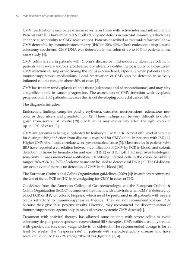
Treatment with antiviral therapy has allowed some patients with severe colitis to avoidcolectomy despite poor response to conventional IBD therapies. CMV colitis is usually treatedwith ganciclovir, foscarnet, valganciclovir, or cidofovir. The recommended dosage is for atleast 3-6 weeks. The “response rate” in patients with steroid-refractory disease who havereactivation of CMV is 72% (range 50%-100%) (figure 3) [3, 4].
Manifestations of Cytomegalovirus Infection52

Book Title
14
CMV IgG+ or IgM+
Tissue IHC or Blood CMV PCR
CMV disease active
IV Ganciclovir or oral Valganciclovir Considerar stopping immunosuppressant
if severe CMV infection
Severa/Steroid-Refractory Colitis
Figure 3. Algorithm for management of suspected CMV colitis in patients with IBD [4]
In patients having AIDS, relapse of CMV gastrointestinal disease in AIDS patients can occurbetween 9 week and 1 year after initial antiviral [3].
4.2.2. CMV hepatitis
Is defined by findings such as fever, vomiting, with hepatomegaly with hepatalgia, andatypical lymphocytosis may be approximately 50%, elevated bilirubin and/or enzyme levels,and detection of CMV by histopathologic analysis within the liver tissue is needed [13,42].
4.2.3. CMV pancreatitis
Requires the detection of CMV infection by immunohistochemical analysis together with theidentification in a pancreatic biopsy. Detection of CMV by PCR alone is insufficient fordiagnosis of CMV pancreatitis because it can imply the presence of transient viremia [42].
4.3. CMV retinitis
Retinitis can appear more than 6 months after solid organ transplantation, mainly hearttransplant recipients. The patients can be asymptomatic, or they may experience blurring ofvision, scotomata, or decreased visual acuity. Fundoscopy often reveals the diagnosis [2]. InHIV-infected patients, retinitis is the single most common manifestation of CMV disease,accounting for 85% of all cases. In developing countries, CMV retinitis is still the most frequentcause of visual loss in HIV-infected patients. Accordingly, the incidence of CMV retinitis,
Management of CMV-Associated Diseases in Immunocompromised Patientshttp://dx.doi.org/10.5772/56141
53

which is the most common CMV disease among HIV patients, decreased from 17.1 ⁄ 100 patientyears to 5.6 ⁄ 100 patient-years [2, 43].
Figure 4. Reference [43]
CMV retinitis in a patient with AIDS appears as an arcuate zone of retinitis with extensivehaemorrhages and optic disk swelling (figure 4) [43].
4.4. CMV neurological diseases
4.4.1. CMV Guillain–Barré Syndrome (GBS)
GBS has become the most frequent cause of acute flaccid paralysis in Western countries,following the near-elimination of poliomyelitis. The current annual incidence is estimated tobe 0.75–2 cases ⁄ 100 000 population. Infectious agents have been suggested as possible triggersof GBS, as some form of respiratory or gastrointestinal infection precedes nearly two-thirds ofGBS cases. Infection with CMV is the most common antecedent virus infection, as identifiedby the presence of IgM antibodies in 10–15% of patients at the onset of GBS. However, antiviraltherapy is currently not recommended in cases of GBS, since the disease is considered to bepost infectious. Recently, the presence of CMV DNA has been demonstrated in almost one-third of serum and cerebrospinal fluid samples from GBS patients who were positive for CMV-specific antibodies at the onset of the neurological disease [43]
Manifestations of Cytomegalovirus Infection54

Running Title
17
Figure 5. Reference [43]
4.4.2. CMV ventriculoencephalitis
This occurs rarely. It presents with changes in mental function. Multiple small, peri-ventricularlesions of the brain are detected following brain magnetic resonance imaging (arrow) (figure5) [2,43].
4.5. CMV genitourinary diseases
4.5.1. CMV Nephritis
It can be defined by the detection of CMV infection by immunohistochemical analysis togetherwith the identification of histological features of CMV infection in a kidney biopsy. Detectionof CMV by PCR alone is insufficient for this diagnosis [42].
4.5.2. CMV Cystitis
This CMV disease is defined by detection of CMV by immunohistochemical together withidentification of conventional histological features of CMV in a bladder biopsy obtained frompatient with symptoms of cystitis [42].
4.6. CMV myocarditis
This occurs most frequently in heart transplant recipients. This infection is defined by thedetection of CMV infection by immunohistochemical analysis together with the identificationof conventional histological features of CMV infection in a heart biopsy specimen and CMV
Management of CMV-Associated Diseases in Immunocompromised Patientshttp://dx.doi.org/10.5772/56141
55

PCR. Detection of CMV by PCR alone is insufficient for the diagnosis of CMV myocarditis [42].There is an association between CMV and left ventricular dysfunction [2].
4.7. CMV vasculopathy
The CMV vasculopathy is considered to be an inflammatory disease. CMV and othersorganisms such as Chlamydia pneumoniae, Epstein Barr virus, herpes simplex virus-1, Myco‐plasma pneumoniae and Helicobacter pylori are implicated, but evidence is strongest with CMVand Chlamydia pneumoniae. There is a correlation between CMV seropositivity and the presenceof atherosclerosis, restenosis following and coronary angioplasty and transplant vascularsclerosis. CMV antigens and nucleic acids have been detected in atherosclerotic lesions in thedifferent layers of the human aorta. Patients suffering from acute myocardial infarction havebeen found to develop CMV antigenaemia, reflecting either a primary infection or reactivationof a latent infection [44].
CMV infects cells in vessels on endotelial cells, smooth muscle cells and macrophagescontribute to the slow progression and aggravation of atherosclerosis. The virus may alsocontribute to coronary thrombosis [44].
4.8. CMV associated Hemophagocytic Syndrome (HPS)
CMV hemophagocytic syndrome, also referred as macrophage activation syndrome (MAS) orhaemophagocytic lymphohistiocytosis (HLH), is a reactive disorder, characterized by gener‐alized histiocytic proliferation, with marked hemophagocytosis. This syndrome was firstdescribed by Risdull et al in 1979 in transplant patients [45]. There are two forms of HPS,familial erytrocytic lymphohistiocytosis and the secondary or reactive HPS [34,46,47].
Reactive or secondary HPS may develop during systemic infections, immunodeficiencies ormalignancies. Infection-associated hemophagocytic syndrome (IAHS) is observed with viralinfections (CMV, Epstein Barr virus, human herpes virus 8, human herpes virus 6, ParvovirusB19 or BK polyoma virus), bacterial infections (Escherichia coli and Mycobacterium), fungalinfection (Histoplasma, Pneumocystis, and Penicillum marneffei), parasitic infections (toxoplas‐mosis, leishmaniasis or babesiosis). HPS may also develop as a complication of malignanciessuch as T-cell lymphomas and metastatic carcinomas. Secundary HPS to inflammatory/autoimmune disorders, including systemic lupus erythematosus, rheumatoid arthritis andStill´s disease, or due anticonvulsants such as phenytoin and carbamazepine [34,45,46].
Cytomegalovirus has been associated with haemophagocytic syndrome in healthy patients,patients with inflammatory bowel diseases, rheumatological diseases, and transplant recipi‐ents [45,47].
The pathophysiology of HPS is not completely understood there is an activation of lympho‐histiocytic tissue secondary to hypercytokinemia derived from the activation of T lymphocytesand activated macrophages, causing fever, shock, and organ dysfunction [48,4]).
The HLH (Henter, 2004), (Emmenegger, 2005) diagnostic criteria are shown in Table 1,
Manifestations of Cytomegalovirus Infection56
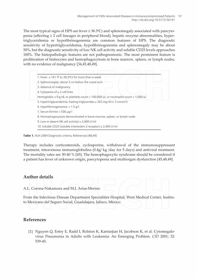
The most typical signs of HPS are fever ≥ 38.3ºC) and splenomegaly associated with pancyto‐penia (affecting ≥ 2 cell lineages in peripheral blood), hepatic enzyme abnormalities, hyper‐triglyceridemia or hypofibrinogenemia are common features of HPS. The diagnosticsensitivity of hypertriglyceridemia, hypofibrinogenemia and splenomegaly may be about50%, but the diagnostic sensitivity of low NK cell activity and soluble CD25 levels approaches100%. The histopathologic features are not pathognomonic. The most prominent feature isproliferation of histiocytes and hemophagocytosis in bone marrow, spleen, or lymph nodes,with no evidence of malignancy [34,45,48,49].
1. Fever: ≥ 101 ºF (≥ 38.3ºC) for more than a week
2. Splenomegaly: about 3 cm below the costal arch
3. Absence of malignancy
4. Cytopenia of ≥ 2 cell lines
Hemoglobin ≤ 9 g/dl, or platelets count < 100,000/μl, or neutrophil count < 1,000/μl
5. Hypertriglyceridemia: Fasting triglycerides ≥ 265 mg/dl (> 3 mmol/l)
6. Hypofibrinogenemia: < 1.5 g/l
7. Serum ferritin > 500 μg/l
8. Hemophagocytosis demonstrated in bone marrow, spleen, or lymph node
9. Low or absent NK cell activity) ≥ 2,400 U/ml
10. Soluble CD25 (soluble interleukin-2 receptor) ≥ 2,400 U/ml
Table 1. HLH-2004 Diagnostic criteria, References [48,49]
Therapy includes corticosteroids, cyclosporine, withdrawal of the immunosuppressanttreatment, intravenous immunoglobulins (0.4g/ kg /day for 5 days) and antiviral treatment.The mortality rates are 30-40 % [45]. The hemophagocytic syndrome should be considered ifa patient has fever of unknown origin, pancytopenia and multiorgan dysfunction [45,48,49].
Author details
A.L. Corona-Nakamura and M.J. Arias-Merino
From the Infectious Disease Department Specialities Hospital, West Medical Center, Institu‐to Mexicano del Seguro Social, Guadalajara, Jalisco, México
References
[1] Nguyen Q, Estey E, Radd I, Rolston K, Kantarjian H, Jacobson K, et al. Cytomegalo‐virus Pneumonia in Adults with Leukemia: An Emerging Problem. CID 2001; 32:539-45.
Management of CMV-Associated Diseases in Immunocompromised Patientshttp://dx.doi.org/10.5772/56141
57

[2] Paya CV, Razonable RR. Cytomegalovirus Infection after Solid Organ Transplanta‐tion. In:Bowden RA, Ljungman P, Paya CV, eds. Transplant Infections. 2nd ed. Phila‐delphia: Lippincott-Raven Publishers; 2003.p. 298-325.
[3] Kandiel A, Lashner B. Cytomegalovirus Colitis Complicating Inflammatory BowelDisease. Am J Gastroenterol 2006; 101: 2857-2865.
[4] Lawlor G, and Moss AC. Clinical Review. Cytomegalovirus in Inflammatory BowelDisease: Pathogen or Innocent Bystander? Inflamm Bowel Dis 2010; 16: 1620-1627.
[5] Kuo CP, Wu CL, Ho HT, Chen CG, Liu SI and Lu YT. Detection of cytomegalovirusreactivation in cáncer patients receiving chemotherapy. Clin Microbiol Infect 2008;14:221-227.
[6] Cascio A, Laria C, Ruggeri P, Fries W. Cytomegalovirus pneumonia in patients withinflammatory bowel disease: a systematic review. Int J Infect Dis 16 (2012) e474-e479.
[7] Cytomegalovirus Prophylaxis following Solid Organ Transplants Guideline Team,Cincinnati Children´s Hospital Medical Center: Evidence-based care guideline forCMV Prophylaxis following Solid Organ Transplant. Guideline 17, pages 1-16, July 6,2007. www.cincinnatichildrens.org/svc/alpha/health-policy/ev-based/CMV-Trans‐plant.htm.
[8] Boeckh MJ, Ljungman P. Cytomegalovirus Infection after Hemopoietic Stem CellTransplantation. In: Bowden RA, Ljungman P, Paya CV, eds. Transplant Infections.2nd ed. Philadelphia: Lippincott-Raven Publishers; 2003.p. 277-397.
[9] O´Brien SM, Keating MJ, Mocarski ES. Updated Guidelines on the Management ofCytomegalovirus Reactivation in Patients with Chronic Lymphocytic LeukemiaTreated with Alemtuzumab. Clin Lymphoma Myelom 2006; 7 (2): 125-130.
[10] Domenech E, Vega R, Ojanguren I, et al. Cytomegalovirus in Inflammatory BowelDisease: Time for another look? Gastroenterology 2009; 137:1163-1175.
[11] Tomblyn M., Chiller T, Einsele H, Gress R, Sepkowitz K, Storek J, et al. Guidelines forPreventing Infectious Complications among Hematopoietic Cell Transplantation Re‐cipients: A Global Perspective. Biol Blood Marrow Tr 2009; 15:1143-1148.
[12] Boeckh M. Complications, Diagnosis, Management, and Prevention of CMV Infec‐tions: Current and Future. Hematology 2011; 305-309.
[13] Crumpacker C. S, Zhang JL. Citomegalovirus. In: Mandell GL, Bennett JE, Dolin R,eds. Enfermedades Infecciosas. Principios y Práctica. 7ma. Ed. Barcelona, España: Elsevi‐er; 2012.p.1983-2000.
[14] Kim EA, Lee KS, Primack SL, Yoon HK, Byun HS, Kim TS, et al. Viral Pneumonias inAdults: Radiologic and Pathologic Findings. Radiographics 2002; 22: S137-S149.
Manifestations of Cytomegalovirus Infection58

[15] Corona Nakamura AL, Monteón Ramos FJ, Troyo Sanroman R, Arias Merino MJ,Anaya Prado R. Incidence and predictive factors for cytomegalovirus infection in re‐nal transplant recipients. Transplant Proc 2009; 41:2412–5.
[16] Kotton CN, and Fishman JA. Disease of the month. Viral Infection in the RenalTransplant Recipient. J Am Soc Nephrol 2005; 16, 1758-1774.
[17] Sun H-Y, Wagener MM and Singh N. Prevention of Posttransplant CytomegalovirusDisease and Related Outcomes with Valganciclovir: A Systematic Review. Am JTransplant 2008, 8: 2111-2118.
[18] Asberg A, Humar A, Jardine AG, Rollag H, Pescovitz MD, Mouas H, et al. Long-Term Outcomes of CMV Disease Treatment with Valganciclovir Versus IV Ganciclo‐vir in Solid Organ Transplant Recipients. Am J Transplant 2009; 9: 1205-1213.
[19] Tamm M. The Lung in the Immunocompromised Patient. Thematic Review Series.Respiration 1999; 66: 199-207.
[20] Takizawa Y, Inokuma S, Tanaka Y, Saito K, Atsumi T, Hirakata M, et al. Clinical char‐acteristics of cytomegalovirus infection in rheumatic diseases: multicentre survey ina large patient population. Rheumatology 2008; 47:1373-1378.
[21] Aries PM, Ullrich S, Gross WL. A case of destructive Wegener´s granulomatosis com‐plicated by cytomegalovirtus infection. Nature Clinical Practice Rheumatology 2006; 2:511-515.
[22] Wade JC. Viral Infection in Patients with Hematological Malignancies. Am Soc Hema‐tol 2006; 368-373.
[23] Chemaly RF, Torres HA, Hachem RY, Nogueras GM, Aguilera EA, Younes A, et al.Cytomegalovirus Pneumonia in Patients with Lymphoma. Cancer 2005; 104 (6):1213-1220.
[24] Desai S, PharmD, and Pinilla-Ibarz J. Front-Line Therapy for Chronic LymphocyticLeukemia. Cancer Control 2012; 19 (1): 26-36.
[25] Thursky KA, Worth LJ, Seymour JF, Prince HM, and Slavin MA. Spectrum of infec‐tion, risk and recmmendations for prophylaxis and screening among patients withlymphoproliferative disorders treated with alemtuzumab. Brit J Haematol 2005; 132:3-12.
[26] Badoux XC, Keating MJ, Wang X, O´Brien SM, Ferrajoli A, Fadert S, et al. Cyclophos‐phamide, fludarabine, alemtuzuman, and rituximab as salvage therapy for heavilypretreated patients with chronic lymphocytic leukemia. Blood 2011; 118 (8):2085-2092.
[27] Elter T, Hallek M, and Montillo M. Alemtuzumab: What Is the Secret to Safe Thera‐py?. Clin Advan Hematol Oncol 2011; 9 (5): 364-372.
Management of CMV-Associated Diseases in Immunocompromised Patientshttp://dx.doi.org/10.5772/56141
59

[28] Österborg A, Foá R, Bezares RF, Dearden C. Dyer MJS, Geisler C, Lin TS, et al. Re‐view. Management guidelines for the use of alemtuzumab in chronic lymphocyticleukemia. Leukemia 2009; 23:1980-1988.
[29] Cortelezzi A, Gritti G, Laurenti L, Cuneo A., Ciolli S, Di Renzo N, et al. An Italianretrospective study on the routine clinical use of low-dose alemtuzumab in relapsed/refractory chronic lymphocytic leukemia patients. Brit J Haematol 2011; 156:481-489.
[30] Petersen CC, Nederby L, Roug AS, Skovbo A, Peterslund NA, Hokland P, et al. In‐creased Expression of CD69 on T Cells as an Early Immune Marker for Human Cyto‐megalovirus Reactivation in Chronic Lymphocytic Leukemia Patients. Viral Immunol2011; 24 (2):165-169.
[31] Cavallo R. The Laboratory of clinical virology in monitoring patients undergoingmonoclonal antibody therapy. Clin Microbiol Infect 2011; 17: 1781-1785.
[32] O´Brien S, Ravandi F, Riehl T, Wierda W, Huang X, Tarrand J, et al. Valganciclovirprevents cytomegalovirus reactivation in patients receiving alemtuzumab-basedtherapy. Blood 2008; 111(4): 1816-1819.
[33] Kaufman M, Rai KR. Review. Alemtuzumab in the up-front setting. Theraputics andClinical Risk Management 2008; 4 (2):459-464.
[34] Fisman DN. Synopsis. Hemophagocytic Syndromes and Infection. Emerg Infect Dis2000; 6 (6): 601-608.
[35] Montillo M, Tedeschi A, Petrizzi VB, Ricci F, Crugnola M, Spriano M, et al. An open –label, pilot study of fludarabine, cyclophosphamide, and alemtuzumab in relapsed/refractory patients with B-cell chronic lymphocytic leukemia. Blood 2011; 118(15):4079-4085.
[36] Elter T, Gercheva-Kyuchukova L, Pylypenko H, Robak T, Jaksic B, Rekhtman G, et al.Fludarabine plus alemtuzumab versus fludarabine alone in patients with previouslytreated chronic lymphocytic leukemia: a randomised phase 3 trial. www.thelan‐cet.com/oncology 2011; 12:1204-1213.
[37] Salomon N, Gomez T, Perlman DC, Laya L, Eber C, and Mildvan D. Clinical featuresand outcome of HIV-related cytomegalovirus Pneumonia. AIDS 1997, 11:319-324.
[38] Ayyappan AP, Thomas R, Kurian S, Christopher DJ, and Cherian R. Multiple cavitat‐ing masses in an immunocompromised host with rheumatoid arthritis-related inter‐stitial lung disease: an unusual expression of cytomegalovirus pneumonitis. Brit JRadiol 2006, 79: e174-e-176.
[39] Moon JH, Kim EA, Lee KS, Kim TS, Jung KJ, and Song JH. Cytomegalovirus Pneu‐monia High-Resolution CT Findings in Ten Non-AIDS Immunocompromised Pa‐tients. Korean J Radiol 2000; 1: 73-78.
[40] Vogel MN, Brodoefel H, Hierl T, Beck R, Bethge WA, Claussen CD et al. Differencesand similarities of cytomegalovirus and pneumocystis Pneumonia in HIV-negative
Manifestations of Cytomegalovirus Infection60

immunocompromised patients- thin section CT morphology in the early phase of thedisease. Brit J Radiol 2007; 80:516-523.
[41] Ukarapol N, Chartapisak W, Lertprasertsuk N, Wongsawasdi L, Kattipattanapong V,Singhavejsakul J et al. Cytomegalovirus-Associated Manifestations Involving the Di‐gestive Tract in Children With Human Immunodeficiency Virus Infection. J PediatrGastroenterol Nutr 2002; 35: 669-673.
[42] Ljungman P, Griffiths P, Paya C. Definitions of Cytomegalovirus Infection and Dis‐ease in Transplant Recipients. CID 2002; 34 (15:1094-1097.
[43] Steininger C. Clinical relevance of cytomegalovirus infection in patients with disor‐ders of the immune system. Clin Microbiol Infec 2007; 13: 953–963.
[44] Soderberg-Naucler C. Does cytomegalovirus play a causative role in the develop‐ment of various inflammatory diseases and cancer? J Int Med 2006; 219-246.
[45] Oloomi Z, Moayeri H. Cytomegalovirus Infection-Associated Hemophagocytic Syn‐drome. Arch Iranian Med 2006; 9 (3): 284-287.
[46] Palazzi DL, McClain KL, and Kaplan SL. Major Article. Hemophagocytic Syndromein Children: An Important Diagnostic Consideration in Fever of Unknown Origin.CID 2003; 36: 306-12.
[47] Ponticelli C, and Della Casa Alberighi O. Editorial Reviews. Haemophagocytic syn‐drome- a life-threatening complication of renal transplantation. Nephrol Dial Transpl2009; 24: 2623-2627.
[48] Raschke RA, and Garcia-Orr R. Original Research. Critical Care. HemophagocyticLymphohistiocytosis. A Potentially Underrecognized Association With Systemic In‐flammatory Response Syndrome, Severe Sepsis, and Septic Shock in Adults. Chest2011; 140 (4): 933-938.
[49] Núñez Bacarreza JJ, Montiel López L, and Núñez del Prado Alcoreza JR. ElsevierDoyma. Medicina Intensiva. Síndrome hemofagocítico asociado a infección viral porcitomegalovirus. Med Intensiva 2011; 35 (3): 189-192.
Management of CMV-Associated Diseases in Immunocompromised Patientshttp://dx.doi.org/10.5772/56141
61