Clonality of Streptococcus pneumoniae in relation to antimicrobial ...
123
119 RESEARCH Clonality of Streptococcus pneumoniae in relation to antimicrobial resistance in Finland Lotta Siira
Transcript of Clonality of Streptococcus pneumoniae in relation to antimicrobial ...

Clonality of Streptococcus pneumoniae in relation to antim
icrobial resistance in Finland
1191192014
RESE
ARCH
Clonality ofStreptococcus pneumoniaein relation to antimicrobialresistance in Finland
Lotta Siira
National Institute for Health and WelfareP.O. Box 30 (Mannerheimintie 166)FI-00271 Helsinki, FinlandTelephone: 358 29 524 6000www.thl.fi
RESE
ARCH
Lotta Siira
Clonality of Streptococcus pneumoniae in relation to antimicrobial resistance in Finland
Streptococcus pneumoniae, the pneumococcus, is a commensal bacterium, which also causes respiratory tract infections and serious infections, such as bacteraemia and meningitis. Globally, the rise in pneumococcal antimicrobial resistance is a worrying trend. Over 90 pneumococcal capsules are recognised by serotyping and the most important are included in the available vaccines. The 10-valent conjugate vaccine is part of the Finnish national vaccination programme since September 2010.
In the study, the serotype and genotype clonality of the pneumococcal population in Finland was studied in relation to antimicrobial resistance in 2002-2011. Serotype 14 was predominant. The proportion of isolates non-susceptible to penicillin and erythromycin increased during the study. Non-susceptibility was particularly high among isolates from children and in serotype 14. International resistant clones dominated, but new genotypes were also found, illustrating the recombination of resistant pneumococci. In Finland a multidrug-resistant serotype 19A clone appeared prior to large-scale vaccination. The serotyping scheme set up is useful in surveillance and for monitoring of the vaccination programme.
ISBN 978-952-302-076-4119
Lotta Siira

RESEARCH 119 • 2014
Lotta Siira
Clonality of
Streptococcus pneumoniae
in relation to antimicrobial
resistance in Finland
ACADEMIC DISSERTATION
To be presented with the permission of the Faculty of Medicine,
University of Helsinki, for public examination in the Small Hall, University
Main Building, Fabianinkatu 33 on 14th February, 2014, at 12 noon.
Bacteriology Unit, Department of Infectious Disease Surveillance and
Control, National Institute for Health and Welfare
Research Program Unit, Immunobiology Research Program, University of
Helsinki
Department of Bacteriology and Immunology, Haartman Institute, University
of Helsinki
Helsinki 2014

© Lotta Siira and National Institute for Health and Welfare
Cover photo: A positive Quellung reaction photographed by Lotta Siira
ISBN 978-952-302-076-4 (printed)
ISSN 1798-0054 (printed)
ISBN 978-952-302-077-1 (online publication)
ISSN 1798-0062 (online publication)
http://urn.fi/URN:ISBN:978-952-302-077-1
Juvenes Print – Finnish University Print Ltd
Tampere, Finland 2014

Supervisor Docent Anni Virolainen-Julkunen, MD, PhD
Department for Promotion of Welfare and Health
Ministry of Social Affairs and Health
Helsinki, Finland
Reviewers Professor Ville Peltola, MD, PhD
Department of Pediatrics
University of Turku
Turku, Finland
Docent Mirja Puolakkainen, MD, PhD
Haartman Institute, Department of Virology
University of Helsinki
Helsinki, Finland
Opponent Professor Birgitta Henriques-Normark, MD, PhD
Department of Microbiology, Tumor and Cell Biology
Karolinska Institutet
Stockholm, Sweden


“As he explained the apparatus and adjusted the lens, it seemed to him that by venturing beyond the visible world he had embarked on a voyage more perilous than he had known.” – In the Reign of Harard IV by Steven Millhauser


THL — Research 119/2014 7 Clonality of Steptococcus pneumoniae in relation to antimicrobial resistance in Finland
Abstract
Lotta Siira, Clonality of Streptococcus pneumoniae in relation to antimicrobial resistance in Finland. National Institute for Health and Welfare. Research 119. 157 pages. Helsinki, Finland 2014. ISBN 978-952-302-076-4 (printed); ISBN 978-952-302-077-1 (online publication)
Streptococcus pneumoniae, or the pneumococcus, is a bacterium of the human
normal flora that also causes non-invasive respiratory tract infections, and serious
infections, such as pneumonia, septicaemia, and meningitis. Globally, the rise in
pneumococcal antimicrobial resistance is a worrying trend. Over 90 pneumococcal
capsules are recognised by serotyping; the most important serotypes are included in
the available vaccines. The 10-valent conjugate vaccine has been part of the Finnish
national vaccination programme since September 2010.
In this study, the serotype and genotype clonality of the invasive pneumococcal
population in Finland was studied in relation to antimicrobial resistance. All
invasive pneumococci isolated in Finland during 2002-2011 and a subset of non-
invasive multidrug-resistant isolates from 2008 were serotyped and studied for
antimicrobial susceptibility. The penicillin-resistant isolates were genotyped and
pilus-encoding virulence genes were detected. A sequential multiplex PCR assay for
serotyping was set up, tailored to the serotype distribution in Finland.
Serotype 14 was the predominant serotype, representing 17.5% of all invasive
isolates. The proportion of isolates non-susceptible to penicillin and erythromycin
was high, and it increased over the study period, reaching 22% and 26% for
penicillin and erythromycin, respectively. The proportion of non-susceptible isolates
was particularly high among isolates from children and in serotype 14. Among the
genotyped isolates, international resistant clones dominated, but novel genotypes
were also found, illustrating the continuous recombination of resistant pneumococci.
The results of this study showed that in Finland a globally described multidrug-
resistant serotype 19A clone appeared prior to large-scale vaccination. Conversely,
in many countries, this clone has emerged following vaccination. Pilus-encoding
gene carriage was frequent among the penicillin- and multidrug-resistant isolates. In
the future, a vaccine targeting the pilus proteins would most likely be successful in
controlling these clones. The new serotyping scheme is useful in surveillance, as
knowledge of the serotype distribution of the invasive pneumococci is essential for
vaccine development and monitoring of the vaccination programme.
Keywords: Streptococcus pneumoniae, clonality, serotyping, genotyping,
antimicrobial susceptibility

THL — Research 119/2014 8 Clonality of Steptococcus pneumoniae in
relation to antimicrobial resistance in Finland
Tiivistelmä
Lotta Siira, Clonality of Streptococcus pneumoniae in relation to antimicrobial resistance in Finland. [Streptococcus pneumoniae -bakteerin klonaalisuus suhteessa lääkeherkkyyteen Suomessa]. Terveyden ja hyvinvoinnin laitos. Tutkimus 119. 157 sivua. Helsinki, Finland 2014. ISBN 978-952-302-076-4 (painettu); ISBN 978-952-302-077-1 (verkkojulkaisu)
Streptococcus pneumoniae, pneumokokki, on normaaliflooran bakteeri, joka voi
aiheuttaa hengitystieinfektioita sekä vakavia tauteja, kuten keuhkokuumetta,
verenmyrkytyksiä ja aivokalvontulehdusta. Pneumokokkien mikrobilääkeresistenssi
on maailmanlaajuisesti ollut huolestuttavassa kasvussa. Pneumokokilla tunnetaan yli
90 eri polysakkaridikapselia, jotka määritetään serotyypittämällä. Käytössä olevat
rokotteet kattavat tärkeimmät serotyypit. 10-valenttinen pneumokokki-
konjugaattirokote on syyskuusta 2010 lähtien ollut osa kansallista rokotusohjelmaa.
Tässä tutkimuksessa tarkasteltiin invasiivisten pneumokokkien klonaalisuutta sekä
serotyyppi- että genotyyppitasolla suhteessa mikrobilääkeherkkyyteen. Serotyyppi ja
mikrobilääkeherkkyys määritettiin kaikille Suomessa vuosina 2002-2011 eristetyille
invasiivisille kannoille sekä osalle vuonna 2008 eristetyille ei-invasiivisille moni-
resistenteille pneumokokeille. Penisilliiniresistenteille kannoille määritettiin lisäksi
genotyyppi ja pilusgeenien läsnäolo. Lisäksi pystytettiin Suomen pneumokokki-
populaatioon räätälöity multiplex-PCR -pohjainen serotyypitysmenetelmä.
Serotyyppi 14 oli tärkein, se kattoi 17,5 % kaikista tutkimuksen invasiivisista
pneumokokeista. Penisilliinille ja erytromysiinille herkkyydeltään alentuneiden
kantojen osuudet olivat korkeat ja kasvoivat tutkimusjaksona kattamaan yli
viidenneksen kannoista. Herkkyydeltään alentuneiden kantojen osuus oli erityisen
korkea lapsilta eristettyjen kantojen keskuudessa sekä serotyypillä 14.
Genotyypitetyt kannat olivat sukua maailmanlaajuisille resistenteille pneumokokki-
klooneille. Myös uusia genotyyppejä havaittiin, mikä kuvastaa resistenttien kloonien
jatkuvaa kehitystä. Havaittiin, että serotyypin 19A moniresistentti klooni on
esiintynyt Suomessa jo ennen laajamittaisia rokotuksia, vaikka se on useassa maassa
yleistynyt vasta rokotusten myötä. Valtaosa tutkituista resistenteistä kannoista kantoi
pilusgeenejä. On todennäköistä, että mahdollinen pilusproteiineja sisältävä rokote
kykenisi tulevaisuudessa torjumaan näitä kantoja. Pystytetyllä serotyypitys-
menetelmällä voidaan selvittää invasiivisen pneumokokkipopulaation serotyyppi-
jakauma. Sen tunteminen on ensiarvoisen tärkeää rokoteseurannassa ja rokote-
kehittämistyössä.
Avainsanat: Streptococcus pneumoniae, klonaalisuus, serotyypitys, genotyypitys,
mikrobilääkeherkkyys

THL — Research 119/2014 9 Clonality of Steptococcus pneumoniae in relation to antimicrobial resistance in Finland
Sammandrag
Lotta Siira, Clonality of Streptococcus pneumoniae in relation to antimicrobial resistance in Finland. Institutet för hälsa och välfärd. [Streptococcus pneumoniae-bakteriens klonalitet i relation till antibiotikaresistens i Finland]. Forskning 119. 157 sidor. Helsingfors, Finland 2014. ISBN 978-952-302-076-4 (tryckt); ISBN 978-952-302-077-1 (nätpublikation) Streptococcus pneumoniae, eller pneumokocken, är en bakterie som finns i människans normalflora men också orsakar allt från milda luftvägsinfektioner till svåra invasiva sjukdomar som lunginflammationer, sepsis och hjärnhinne-inflammationer. Globalt sett har antibiotikaresistensen bland pneumokocker ökat oroväckande under de senaste decennierna. Fler än 90 olika polysackaridkapslar har beskrivits; dessa bestäms genom serotypning. De viktigaste serotyperna finns med i de vaccin som utvecklats mot pneumokocksjukdomar. I september 2010 blev det 10-valenta konjugatvaccinet en del av det nationella vaccinationsprogrammet i Finland. I den här undersökningen granskades de invasiva pneumokockernas klonalitet både på sero- och genotypnivå i förhållande till antibiotikaresistensen i Finland. Serotyper och antibiotikakänslighet bestämdes för alla invasiva pneumokocker som isolerades i Finland under åren 2002-2011 och för ett sampel av multiresistenta icke-invasiva pneumokocker från år 2008. De penicillinresistenta stammarna genotypades och deras gener för piluskodande virulensfaktorer utreddes. Ett nytt serotypnings-protokoll baserat på multiplex-PCR sattes också upp. Bland serotyperna var serotyp 14 den viktigaste, den utgjorde 17,5% av alla invasiva stammar. Andelen isolat med nedsatt känslighet mot penicillin eller erytromycin var hög och ökade under forskningsperioden till 22 %, respektive 26 %, av stammarna. Andelen stammar med nedsatt antibiotikakänslighet var speciellt hög bland isolat från barn och inom serotyp 14. Internationella resistenta kloner dominerade bland de genotypade stammarna. Nya genotyper hittades också vilket beskriver de resistenta klonernas fortsatta utveckling. Resultaten visar också att den multiresistenta serotyp 19A-klon som på många håll i världen ökat markant efter vaccinering, hade fått fotfäste i Finland redan före storskalig vaccinering inletts. Majoriteten av de penicillin- och multiresistenta pneumokockerna bar på piluskodande gener, vilket tyder på att ett vaccin som innehåller pilusprotein i framtiden kunde begränsa dessa viktiga kloners framfart. Det nya serotypningsprotokollet möjliggör också i fortsättningen granskningen av pneumokockernas serotypfördelning för vaccin-uppföljning och vaccinutvecklingsbehov. Nyckelord: Streptococcus pneumoniae, klonalitet, serotypning, genotypning, antibiotikaresistens

THL — Research 119/2014 11 Clonality of Steptococcus pneumoniae
in relation to antimicrobial resistance in Finland
Contents
Abstract ...................................................................................................................... 7
Tiivistelmä.................................................................................................................. 8
Sammandrag ............................................................................................................... 9 List of original publications ..................................................................................... 13 Abbreviations ........................................................................................................... 15 1 Introduction ........................................................................................................... 17
2 Review of the literature ......................................................................................... 18
2.1 The “sugar-coated microbe” and breakthroughs in the life sciences ............ 18
2.2 Pneumococcal disease and carriage .............................................................. 20
2.3 Pneumococcal virulence factors.................................................................... 23
2.4 The pneumococcal capsule ........................................................................... 24
2.4.1 Capsular genes, structure, and production ............................................ 26
2.4.2 Serotype nomenclature ......................................................................... 29
2.5 Antimicrobial resistance ............................................................................... 29
2.5.1 Modes of antimicrobial action and resistance mechanisms .................. 31
2.5.2 Selection pressure and fitness cost of resistance .................................. 33
2.6 Resistance and clonality ................................................................................ 34
2.7 Studying the pneumococcal population by serotyping ................................. 37
2.7.1 Phenotypic serotyping methods ............................................................ 37
2.7.2 Deducing the serotype by detecting the cps.......................................... 37
2.8 Studying the pneumococcal population by genotyping ................................ 39
2.8.1 Multi locus sequence typing ................................................................. 41
2.9 Natural genetic transformation...................................................................... 42
2.10 From whole-cell vaccines to polysaccharide and conjugate vaccines ........ 44
2.11 Intervention and the pneumococcal population .......................................... 45
3 Aims ...................................................................................................................... 48
4 Materials and methods .......................................................................................... 49
4.1 Bacterial isolates ........................................................................................... 49
4.2 Species identification .................................................................................... 49
4.3 Isolation of DNA .......................................................................................... 49
4.4 Serotyping ..................................................................................................... 52
4.4.1 Latex agglutination ............................................................................... 52
4.4.2 Counterimmunoelectrophoresis ............................................................ 52
4.4.3 The Quellung reaction .......................................................................... 53
4.4.4 Immunologic monoclonal antibody based serotyping assay ................ 53
4.4.5 Serotype deduction by multiplex PCR ................................................. 53
4.5 Antimicrobial susceptibility testing .............................................................. 55
4.6 Detection of macrolide resistance genes ....................................................... 56
4.7 Genotyping .................................................................................................... 56

THL — Research 119/2014 12 Clonality of Steptococcus pneumoniae
in relation to antimicrobial resistance in Finland
4.8 Detection of pilus-encoding genes ................................................................ 57
4.9 Sequencing of serotype 19F-like serotype 19A wzy ..................................... 57
4.10 Statistical methods ...................................................................................... 58
4.11 Cost analysis ............................................................................................... 58
5 Results ................................................................................................................... 59
5.1 Serotypes among invasive pneumococci, 2002-2011 (I, IV) ........................ 59
5.2 Antibiotic resistance among invasive pneumococci, 2002-2011 (I, IV) ....... 62
5.2.1 Penicillin ............................................................................................... 62
5.2.2 Other antimicrobial agents .................................................................... 65
5.3 Macrolide resistance determinants among the invasive pneumococci, 2002-2006 (I) ............................................................................................................... 67
5.4 Genotype clonality of penicillin-resistant and -non-susceptible invasive pneumococci, 2002-2011 (I, IV) ......................................................................... 67
5.5 Pilus islets among the penicillin-resistant invasive pneumococci, 2002-2011 (I, IV) .................................................................................................................. 72
5.6 Characteristics of non-invasive multidrug-resistant pneumococci (II) ......... 72
5.7 Validation of the mPCR based serotyping scheme (III) ............................... 73
5.8 The genotypes and wzy sequences of two serotype 19F-like 19A isolates ... 76
6 Discussion ............................................................................................................. 77
6.1 Serotypes among the invasive pneumococci................................................. 77
6.2 Recently discovered and aberrant serotypes ................................................. 78
6.2.1 Serogroup 6 .......................................................................................... 78
6.2.2 Serogroup 19 ........................................................................................ 80
6.3 Validation of the mPCR based serotyping scheme ....................................... 82
6.4 Antimicrobial resistance and macrolide-resistance determinants ................. 83
6.5 Genotype clonality and pilus-encoding islets ............................................... 87
6.5.1 Clonal complex 156 .............................................................................. 87
6.5.2 Clonal complex 320 .............................................................................. 88
6.5.3 Novel sequence types and other clonal complexes ............................... 88
6.6 Pilus-encoding islets ..................................................................................... 90
7 Conclusions and future considerations .................................................................. 92
8 Acknowledgements ............................................................................................... 94
9 References ............................................................................................................. 96

THL — Research 119/2014 13 Clonality of Steptococcus pneumoniae in relation to antimicrobial resistance in Finland
List of original publications
This thesis is based on the following original publications, which are referred to
throughout the text by the Roman numerals given below (I-IV)
I Temporal trends of antimicrobial resistance and clonality of invasive
Streptococcus pneumoniae isolates in Finland, 2002 to 2006. Siira L,
Rantala M, Jalava J, Hakanen AJ, Huovinen P, Kaijalainen T,
Lyytikäinen O, Virolainen A. Antimicrob Agents Chemother. 2009
May;53(5):2066-73.
II Clonality behind the increase of multidrug-resistance among non-
invasive pneumococci in Southern Finland. Siira L, Jalava J, Tissari
P, Vaara M, Kaijalainen T, Virolainen A. Eur J Clin Microbiol Infect
Dis. 2012 May;31(5):867-71.
III From Quellung to multiplex PCR, and back when needed, in
pneumococcal serotyping. Siira L, Kaijalainen T, Lambertsen L,
Nahm MH, Toropainen M, Virolainen A. J Clin Microbiol. 2012
Aug;50(8):2727-31.
IV Antimicrobial resistance in relation to sero- and genotypes among
invasive Streptococcus pneumoniae in Finland, 2007-2011. Siira L,
Jalava J, Kaijalainen T, Ollgren J, Lyytikäinen O, Virolainen A.
Microbial Drug Resistance. In press.
The original articles are reproduced with the kind permission of the copyright
holders. In addition, some unpublished results are included.

THL — Research 119/2014 14 Clonality of Steptococcus pneumoniae in relation to antimicrobial resistance in Finland
THL — Research 119/2014 15 Clonality of Steptococcus pneumoniae in relation to antimicrobial resistance in Finland
Abbreviations
aroE shikimate dehydrogenase gene bp base pair
CC clonal complex
CDC Centers for Disease Control and Prevention
CI confidence interval
CIEP counterimmunoelectrophoresis
CLSI Clinical and Laboratory Standards Institute
cps capsule polysaccharide synthesis locus ddl D-alanine-D-alanine ligase gene
DNA deoxyribonucleic acid
eBURST based upon related sequences ECDC the European Centre for Disease Prevention and Control
EQA external quality assurance
erm erythromycin ribosomal methylation gene
EUCAST European Committee on Antimicrobial Susceptibility Testing gdh glucose-6-phosphate dehydrogenase gene gki glucose kinase gene
HUS Hospital District of Helsinki and Uusimaa
I intermediate
kb kilo bases
LytA autolysin, N-acetylmuromyl-L-alanine amidase
mAb monoclonal antibody Mb mega bases
MDR multidrug-resistance
mef macrolide efflux gene
MIC minimum inhibitory concentration MLKSB lincosamide-ketolide-streptogramin B resistance phenotype MLST multi locus sequence typing MLVA multi locus variable number tandem repeat analysis mPCR multiplex PCR NIDR National Infectious Disease Register PBP penicillin-binding protein PCR polymerase chain reaction PEN penicillin PFGE pulsed-field gel electrophoresis PI-1 pilus islet 1 PI-2 pilus islet 2 PCV pneumococcal conjugate vaccine PCV7 7-valent pneumococcal conjugate vaccine

THL — Research 119/2014 16 Clonality of Steptococcus pneumoniae in relation to antimicrobial resistance in Finland
PCV10 10-valent pneumococcal conjugate vaccine PCV13 13-valent pneumococcal conjugate vaccine PMEN Pneumococcal Molecular Epidemiology Network
PspA pneumococcal surface protein A
PspC pneumococcal surface protein C
R resistant
RR risk ratio recP transketolase gene
S susceptible
SLV single locus variant spi signal peptidase I gene
SSI Statens Serum Institute ST sequence type THL National Institute for Health and Welfare (Terveyden ja
hyvinvoinnin laitos) UAB University of Alabama WHO World Health Organization xpt xanthine phosphoribosyl transferase gene
wzg capsular regulatory gene, formerly named cpsA wzy capsular polymerase gene
wzy pathway biosynthesis pathway for capsular polysaccharides

THL — Research 119/2014 17 Clonality of Steptococcus pneumoniae in relation to antimicrobial resistance in Finland
1 Introduction
Streptococcus pneumoniae, or the pneumococcus, is a commensal bacterium, which
also causes infections of the upper respiratory tract and serious infections such as
meningitis, septicaemia, and pneumonia. According to the Finnish National
Infectious Disease Register, more than 700 invasive pneumococcal infections are
diagnosed annually. The pneumococcus is asymptomatically carried in the
nasopharynx especially by young children, with carriage rates decreasing with
increasing age. Carriage is essential for disease to develop, and the strain causing
disease tends to originate from the nasopharynx of the patient. The pneumococcus
engages in both inter- and intraspecies competition in its natural habitat.
The pneumococcus is a diplococcus that is alpha-haemolytic when cultivated on
blood agar. A capsule made of polysaccharides covers the bacterial cell and enables
the bacterium to evade the immune system and is an important virulence factor. To
date, more than 90 different capsular types, or serotypes, have been described. These
differ in both immunogenicity and virulence and often, but not always, represent
diverse genetic backgrounds. The most frequently occurring serotypes causing
invasive disease are included in the available vaccines. The 10-valent pneumococcal
conjugate vaccine is included in the Finnish national vaccination programme as of
September 2010. The large-scale use of vaccines will assert serotype selection
pressure that is likely to bring about changes both on the serotype and genotype
level within the pneumococcal population. Over the last few decades, pneumococcal
resistance to commonly used antimicrobial drugs has emerged. This is a worrying
trend posing new treatment challenges.
The aim of this study was to examine the clonality of the invasive pneumococcal
population, both on the serotype and the genotype level, and to set up a serotyping
scheme tailored to study the invasive pneumococcal isolates in Finland. The study
also examined a subset of multidrug-resistant non-invasive isolates that have
increasingly been encountered. From a surveillance standpoint, these isolates are
important, because changes in the non-invasive population are usually reflected in
the invasive bacterial population in time. By combining virulence factor and clonal
analysis the results of this study may be useful when future prevention strategies are
considered and developed.

THL — Research 119/2014 18 Clonality of Steptococcus pneumoniae in
relation to antimicrobial resistance in Finland
2 Review of the literature
2.1 The “sugar-coated microbe” and breakthroughs in the life sciences
Streptococcus pneumoniae, or the pneumococcus, is a Gram-positive, facultatively
anaerobic catalase-negative round or lancet shaped diplococcus. It is fairly
demanding to cultivate in the laboratory and generally thrives best in an atmosphere
enriched with carbon dioxide. When cultivated on blood agar, it produces greenish
alpha-haemolysis, as the hydrogen peroxide of the bacteria oxidises haemoglobin.
The colonies are round and often dented in the middle, but the appearance depends
on the capsular type, as some serotypes have a mucoid appearance. The bacterial cell
is covered by a polysaccharide capsule, the structure of which determines the
serotype of the bacterium. The capsule is an important virulence factor [3, 160].
The history of pneumococcal research mirrors the history of key findings and
milestones in bacteriology and the life sciences. After the development of a light
microscope with sufficiently high resolution to reveal bacteria that the naked eye
could not detect, coccoid bacteria in pairs found in pulmonary tissues were reported
in the literature in 1875 [111]. In 1881, the pneumococcus was described as a
pathogen after it had been isolated independently by two researchers, George M.
Sternberg and Louis Pasteur [11, 325]. Both found diplococcoid bacteria in the
saliva of human carriers and both went on to inject the saliva into rabbits, thereby
causing disease, and were able to recover the bacteria from the rabbit blood [325].
Since its discovery, the pneumococcus has been renamed several times. Its initial
names Microbe septicemique du salive given by Pasteur and Micrococcus pasteuri by Sternberg, gave way to Pneumococcus a few years later, when its predisposition
to cause respiratory tract disease became clear. In 1920, it went on to officially carry
the name Diplococcus pneumoniae, given in an effort to describe both the shape and
clinical manifestation of the bacterium. In 1974, the current name, Streptococcus pneumoniae, was adopted to indicate that in liquid media the bacteria grow in chains
like other members of the Streptococcus genus. The pneumococcus was one of the
first bacteria to be Gram-stained, a procedure developed by Hans Christian Gram in
the 1880s and still relevant today in identifying clinically significant bacteria [325].
In 1923, the discovery that the pneumococcal capsule was comprised of
polysaccharides, i.e. sugar, caused a stir. Until then, it had been widely accepted that
only proteins were capable of acting as antigens and causing an immune response
[296, 325]. Physician Oswald Avery, who was active in pneumococcal research for

Review of the literature
THL — Research 119/2014 19 Clonality of Steptococcus pneumoniae in relation to antimicrobial resistance in Finland
several decades, affectionately called the pneumococcus the “sugar-coated microbe” [17].
Research into the pneumococcal capsule established it as a major virulence factor when
it was discovered that it protects the bacterium from opsonisation and phagocytosis by
the immune system. This research, in turn, developed into the demonstration of the
microscopically visible Quellung reaction, in which the capsule swells upon addition of
specific antiserum to pneumococci in liquid media (Figure 1) [12, 325]. This reaction is
still commonly known by the German word for swelling, Quellung, or as the capsular
reaction test, and is widely used for serotyping pneumococci. The number of known
pneumococcal serotypes increased from two in 1910 to 85 some fifty years later [325].
Today, more than 90 different serotypes are known [23].
Figure 1. A positive Quellung reaction as seen in a phase contrast microscope.
Chemotherapy against pneumococcal infections, one of the first uses of specific
antimicrobial agents as therapy for bacterial infections, took place as early as 1911.
The agent in question was the quinine derivative ethylhydrocuperine, known as
optochin, which specifically inhibits growth of pneumococci. Its therapeutic use was
abandoned because of toxicity and rapidly developing resistance, but in the
laboratory optochin remains a reliable tool for distinguishing pneumococci from
other closely related species [33, 111]. The first successful use of penicillin in a
clinical setting was against a pneumococcal conjunctivitis infection, establishing the
clinical and therapeutic usefulness of the drug [325]. When penicillin was launched
in the 1940s, it became the drug of choice and its use dramatically reduced mortality
of serious pneumococcal infections [111].
The significance of breakthroughs made in pneumococcal research extends far
beyond bacteriology. The best example of this is the discovery of deoxyribonucleic
acid (DNA) as the hereditary molecule [14, 296]. This discovery made in 1944 built

Review of the literature
THL — Research 119/2014 20 Clonality of Steptococcus pneumoniae in relation to antimicrobial resistance in Finland
upon the work from previous decades, when researchers had discovered that rather
than being stable, an avirulent non-capsulated pneumococcus could become virulent
when injected into a mouse simultaneously with heat-killed bacteria of a virulent
capsulated strain [113]. This transformation of material that changed the phenotype
of the strains in a so-called capsular switching event was shown to occur in liquid
media as well as in laboratory animals. The chemical properties of the transforming
molecule were consistent with those of DNA, although until then, it had been
generally believed that protein was the genetic material. The finding was so ground-
breaking that it took nearly a decade before it was widely accepted in the scientific
community that DNA was the hereditary molecule and contained the genes of the
organisms [201, 296].
2.2 Pneumococcal disease and carriage In 2005, the World Health Organization (WHO) estimated that 1.6 million deaths
annually were caused by the pneumococcus. Children under the age of 5 years
account for 0.7–1 million of these deaths, and developing countries are most
severely affected [333]. In 2008, it was estimated that 476,000 deaths in young
children were caused by pneumococcal infections [332]. In developed countries, the
pneumococcus is a common cause of community-acquired pneumonia in adults,
sometimes accompanied by bacteraemia [29, 176]. In Europe, the age-standardised
incidence of invasive pneumococcal disease was 5.12 per 100,000 population in
2010, according to the European Centre for Disease Prevention and Control (ECDC)
[92]. However, the incidences show great variation between countries; in the years
2002 to 2005, they were 0.4 to 25.8 per 100,000 population [270]. In the Nordic
countries, the age-standardised incidence per 100,000 population in the year 2010
was 14.82 in Sweden, 15.08 in Finland, 16.18 in Norway, and 17.26 in Denmark,
respectively [92, 246]. In Finland, the incidences of both laboratory confirmed
bloodstream infections as a whole, and invasive pneumococcal infections have
increased over the past few decades [287, 300]. In 2012, 752 cases of invasive
pneumococcal infections were registered in the National Infectious Disease Register
(NIDR) [300]. In Finland, the incidence of pneumococcal bloodstream infections is
slightly higher among males than females and the case fatality rate within a month is
10%. Nearly half of the deaths occur within two days of a positive blood culture
sample [286]. The invasive pneumococcal disease incidence displays seasonal
fluctuation, increasing in the winter months and correlating with the findings of
influenza and other respiratory viruses [105, 298]. In Finland, a temporal association
between invasive pneumococcal disease in young children and peaks in rhinovirus
circulation during spring and autumn has been established [247]. Regional and
ethnic differences have also been described in the incidence of invasive
pneumococcal disease [189].

Review of the literature
THL — Research 119/2014 21 Clonality of Steptococcus pneumoniae in relation to antimicrobial resistance in Finland
Figure 2. Pneumococcal colonisation and disease. Drawing by Hanna Siira.
The distribution of the clinical spectrum of pneumococcal colonisation and disease
can be likened to an iceberg (Figure 2). Under the surface, the widest part is made
up of colonisation; this is the most common situation, where the pneumococcus acts
as a commensal. The visible part of the iceberg represents the clinical cases. The
majority of these are non-invasive, relatively mild conditions, such as otitis media
common in children, and other respiratory infections. Above are pneumonia and
other serious conditions, and at the very top of the iceberg are bloodstream-
infections, and finally, meningitis [160, 189].
Most pneumococcal disease cases are sporadic and transmission takes place from
person to person though droplets or aerosols [134, 160]. Although rare and usually
limited in size, invasive and non-invasive outbreaks have been described in confined
settings such as nursing homes, hospital wards, day care centres, military camps, and
homeless shelters [27, 104, 114, 143, 264, 309]. In the so-called meningitis belt in
Africa, invasive outbreaks associated with serotype 1 have occurred [178, 225], and
serotype 15F strains has been found to cause outbreaks in rural communities in
Alaska [347].
Host risk factors for pneumococcal carriage and disease include alcohol abuse,
smoking or exposure to tobacco smoke, asthma, and acute upper respiratory
infections [189]. Day care or school attendance and more than four co-habitants are
risk factors for invasive pneumococcal disease in children [52, 53]. Human
immunodeficiency virus infections and the acquired immunodeficiency syndrome
are predisposing conditions to pneumococcal disease, as is any other

Review of the literature
THL — Research 119/2014 22 Clonality of Steptococcus pneumoniae in relation to antimicrobial resistance in Finland
immunocompromising condition and old age [189, 322]. Young children are also
susceptible to pneumococcal disease mainly because their immune systems have yet
to fully develop and therefore are not able to quickly eradicate polysaccharide
covered pneumococci [332]. The factors of and interplay between both the
bacterium and the human host determine whether the pneumococcus is able to cause
disease, as well as the severity and clinical manifestation of the disease. Disease is a
relatively rare event compared to carriage. Upon acquisition, a strain may be carried
for weeks or even months in the nasopharynx, where both innate and adaptive
immune responses are involved in limiting the pneumococci [135, 160]. In carriage,
the pneumococcus adheres to the resting epithelial lining of the nasopharynx. For
disease to develop, the bacterium must spread from the nasopharynx, either locally
when causing sinusitis or otitis media, by aspiration into the alveoli when causing
pneumonia, or by invading the bloodstream when causing septicaemia. In meningitis,
the brain-blood barrier is breached and the pneumococci reach the cerebrospinal
fluid [31].
The carriage rate is high during the first two years of life and declines thereafter. In
healthy 18-month-old children in a study conducted in the Netherlands it was 12%
[30], while it was as high as 55.5% in children below 24 months in a study from the
UK [245], 49.3% in 4 to 12-month-old children in Bangladesh [109], and 9-43%,
increasing with age, in 2 to 24-month-old healthy children in a Finnish study [297].
Age-related decline in pneumococcal carriage is caused by the maturation of the
immune system and occurs parallel with simultaneous increase in Staphylococcus aureus carriage [32]. The nasopharynx of children can be considered the natural
habitat of the bacterium and the reservoir from where it may be transmitted. Adults
in families with young children are often more likely to carry the bacterium than
their peers living in families without children. The adult carriage rate varies in
different populations and in different settings but is often below 10% [134].
The pneumococcus has sporadically been encountered in pets, as well as zoo and
laboratory animals [314], but humans are its main host. Nasopharyngeal colonisation
is a dynamic process, in which carried species and serotypes are in flux and vary by
age, season, geographical area, genetic background, and is further influenced by
socioeconomic factors. Interventions, such as the use of antimicrobial agents and
vaccines, also have an impact on the dynamic [30-32]. The pneumococci found in
carriage and non-invasive samples are more diverse than the strains most commonly
isolated from invasive samples [121, 175]. The pneumococcus has a complex
relationship with the estimated 700 other bacterial species that share this niche [160].
The resident bacterial flora, which includes alpha-haemolytic species, inhibits the
colonisation of invading species, such as pneumococci, Haemophilus influenzae, and
Moraxella catarrhalis. Furthermore, several of the pathogens have competitive
relationships with the other species [31]. The pneumococcus can interfere with the

Review of the literature
THL — Research 119/2014 23 Clonality of Steptococcus pneumoniae in relation to antimicrobial resistance in Finland
growth of S. aureus, M. catarrhalis, H. influenzae, and the meningococcus Neisseria meningitidis [31, 32, 248]. On the other hand, meningococcal presence in vitro
increases the growth of pneumococci [31].
2.3 Pneumococcal virulence factors The pneumococcus is able to adapt its gene expression in a site specific manner and
its different virulence factors play varyingly important roles depending on the strain,
as well as on the type and stage of disease, as [160, 239]. The most important
virulence factor of the pneumococcus is the polysaccharide capsule [192], which is
discussed in detail in the next section. However, the bacterium also has other
virulence factors that facilitate colonisation and survival in the host; some of the
most central are discussed below.
The exotoxin pneumolysin is expressed by nearly all invasive pneumococcal isolates,
and several different variants of the molecule are known [160]. Pneumolysin is
cytolytic at high concentrations, when the soluble proteins oligomerise in the
cholesterol-containing membranes of the target cells to form large round pores
consisting of more than 40 subunits [305]. At lower concentrations, pneumolysin is
cytotoxic and interferes with the immune defence by influencing ciliary beating,
complement activation, and induction of intracellular oxygen radicals [160, 198].
The role of pneumolysin in pneumococcal virulence in pneumonia is well
established. It also seems to be important in the survival and spread of bacterial from
the lungs to the bloodstream and for the clinical manifestation of bacteraemic
infections [161, 238]. Its role in meningitis remains controversial [160].
Several protein structures that influence virulence are located on the surface of the
pneumococcal cell. The most recently discovered are pili, hair-like adhesive
structures that protrude from the bacterial surface [73, 230]. The pneumococcal pili
are encoded by two pilus islets, PI-1 and PI-2, on the bacterial chromosome. These
were revealed by whole-genome sequencing. Clinical and carriage isolates may
carry none, one, or both of the islets [1]. The expression of PI-1 has been shown to
mediate adhesion to host cells and provide a competitive advantage in an animal
model of respiratory tract colonisation [18, 230]. Initially, this pathogenicity islet
was named rlrA islet, after its positive regulator gene. Pneumococcal PI-1 carriage is
associated with antimicrobial non-susceptibility [2]. The presence of PI-1 is a clonal
property, with a stronger association with the genotype than the serotype [2, 223].
Expression of PI-2 also mediates adherence to host cells [16]. In contrast to PI-1, PI-
2 is associated with antimicrobial susceptibility, although dual carriage of PI-1 and
PI-2 is associated with antimicrobial resistance [1, 343].

Review of the literature
THL — Research 119/2014 24 Clonality of Steptococcus pneumoniae in
relation to antimicrobial resistance in Finland
On its surface, the pneumococcus also carries several choline-binding proteins that
interact with host structures and influence virulence. The proteins are anchored by
their homologous C-terminal parts to the pneumococcal cell wall phosporylcholine
and vary in their protruding parts [118]. One of the choline-binding proteins is the
pneumococcal surface protein A (PspA), a variable molecule that prevents
complement mediated killing of the bacteria [141, 160]. Another is the
pneumococcal surface protein C (PspC), also known as choline-binding protein A,
which helps the bacteria adhere to epithelial cells and promotes nasopharyngeal
colonisation [160].
After reaching the stationary phase in the growth curve, the pneumococcus
undergoes characteristic autolysis by degrading its cell wall and thereby inducing its
own death. This trait seems to add to virulence and protect intact bacteria from
clearing by the immune system [196, 198, 336]. The major autolysis inducing
enzyme autolysin (LytA), or N-acetylmuromyl-L-alanine amidase, severs bonds in
the peptidoglycan cell wall [160]. Several theories of why LytA influences virulence
have been suggested: its induction releases other virulence factors or toxins for
instance pneumolysin and the cell wall components, and its induction may hinder
phagocyte activities [160, 198]. Together with pneumolysin, LytA is essential for
the survival of the pneumococcus in the bloodstream [238].
Pneumococcal surface adhesin (PsaA) is a lipoprotein located at the bacterial cell
wall that appears to be involved in providing resistance to oxidative stress [160].
Teichoic acid and peptidoglycan, both major cell wall components, induce
inflammation [307], while other pneumococcal virulence factors include LPXTG-
anchored proteins such as neuraminidases and pneumococcal histidine triad proteins
[118].
2.4 The pneumococcal capsule The capsule is the most important virulence factor for invasive pneumococcal
disease [160, 192]. The capsule inhibits complement and protects the bacterial cell
from neutrophil-mediated killing, while the protection increases with the degree of
encapsulation [160, 329]. Strains with a thick capsule are more virulent and prone to
cause invasive disease, while strains with a thinner capsule are more often found in
asymptomatic carriage [340].
More than 90 different pneumococcal serotypes have been described to date, but a
smaller number is responsible for most invasive disease. Globally, more than 80% of
invasive disease is caused by around 20 serotypes [158, 333]. The serotype
distribution of the invasive pneumococci varies depending on time, place, and age-

Review of the literature
THL — Research 119/2014 25 Clonality of Steptococcus pneumoniae in relation to antimicrobial resistance in Finland
group [126, 127, 284]. Fluctuations in the serotype frequencies or proportions may
take place over time even without selection pressure asserted by interventions [99,
122]. In children under 5 years of age, serotype 14 is the most common cause of
invasive pneumococcal disease in all regions. Often it is an important cause of
invasive disease in other age-groups as well [126, 158]. The serotypes differ in
genetic, immunological, biochemical, and epidemiological properties [23, 277].
Indeed, they show such variation in their properties, epidemiology, and invasive
disease outcomes, that from an epidemiologic point of view, it has been suggested
that each of them should be considered a separate pathogen [127, 328]. The risk
factors, disease focus, and clinical presentation are partly associated with serotype
[53, 284, 315, 347], and carriage efficiency also depends on the capsule [125].
Certain serotypes or serogroups display high invasive disease potential, i.e. they
exhibit a high propensity for causing invasive disease relative to the exposure
through carriage, while others show low invasive disease potential, i.e. they are
common in carriage but proportionately rarer in invasive disease episodes. These
serotypes or serogroups are summarised in Table 1. As illustrated, the invasive
disease potential of some serotypes, such as serotype 3, exhibit differences between
studies. Interestingly, high invasive disease potential has not been linked to high
mortality [285], but serotypes 3, 6A, 6B, 9N, 11A, 19F, and 31 are associated with
increased risk of death [285, 315, 328]. Underlying conditions allow serotypes with
otherwise low invasive disease potential to act as opportunistic pathogens and cause
invasive disease [285]. Just as the capsule affects the pathogenesis of a
pneumococcal strain, non-encapsulated isolates also display particular
characteristics and appear to have a propensity to cause conjunctivitis [119].
Table 1. Serotypes or serogroups with high or low invasive disease potential, respectively, as
identified in three studies.
Serotypes/groups with high invasive
disease potential
Serotypes/groups with low invasive
disease potential
Reference
1, 5, and 7 3, 6A, and 15 [38]
6B, 14, 18C, and 19A 6A and 11A [121]
3, 7F, 18C, 19A, 22F, and 33F 6C, 11A, 15A, 15B/C, 19F, 23A, 35B,
and 35F
[339]
To date, 97 different pneumococcal serotypes or capsular types belonging to 46
serogroups have been published [23, 34, 42, 43, 132, 164, 237, 244, 345]. In
addition to these serotypes, the DNA sequence of a novel serogroup 33 subtype,
proposed to be named 33E, is available in the nucleotide sequence database
(accession numbers EU071709 and EU071709 [302]). Currently, not all of the most
recently described serotypes are distinguishable by conventional antisera [42, 164,

Review of the literature
THL — Research 119/2014 26 Clonality of Steptococcus pneumoniae in relation to antimicrobial resistance in Finland
275]. As both genetic and immunologic methods for studying pneumococcal
serotypes evolve, it is likely that new serotypes or further structural subtypes of
previously described serotypes are discovered [85, 203, 275].
2.4.1 Capsular genes, structure, and production The capsule is the outermost layer of the pneumococcal cell. Its thickness is 200 to
400 nm and varies considerably depending on the serotype [288]. The structure of
the capsular polysaccharides varies depending on the serotype and may be linear or
branched. Branching is determined by the enzymes catalysing the polymerisation of
the polysaccharide [340]. The structure of the capsular polysaccharides also affects
the prevalence of the serotype in carriage [329].
All but two of the pneumococcal capsular polysaccharides are synthesised and
transferred to the cell surface through the so-called wzy pathway. This pathway is
also used in some Gram-negative bacteria and in nearly all other Gram-positive
bacteria for capsular synthesis [340]. The proteins required for the wzy pathway are
encoded by genes located in the capsule polysaccharide synthesis locus cps, which
can be found between dexB and aliA on the pneumococcal chromosome (Figure 3).
The locus varies in size from 10,337 bp for serotype 3, to 30,298 bp for serotype 38,
depending on the specific genes required for the synthesis of each serotype [23].
Within the locus, the genes were originally named cps to which a letter was added
for each individual gene in the sequence, i.e. cpsA, cpsB, cpsC, cpsD etc. More
recently, the cps genes have been re-named by function and orthology. This allows
homologous genes in different serotypes and across different species to carry the
same name regardless of their relative location within cps. The aforementioned four
genes are named wzg, wzh, wzd, and wze in most serotypes (Figure 3) [23, 156, 340].
At the 5’ region, the cps locus contains the four conserved genes, which are involved
in the modulation of the capsular synthesis [156, 340]. In one study, expression of
the regulatory gene wzg (cpsA) correlated inversely with the thickness of the capsule
[125]. Located downstream from the conserved genes are the serotype specific genes
that encode enzymes for carrying out the polymer-specific tasks. In most serotypes,
these enzymes consist of glycosyltransferases, polymerases, flippases, transferases,
nucleotide dephospho-sugar synthases, and modification enzymes, such as O-
acetylases [340].
Sugars needed for polysaccharide assembly are synthesised in the cytoplasm of the
bacterial cell by housekeeping genes or cps-encoded genes, depending on the
serotype [340]. The hypothetical biosynthesis of the capsule has been described as
follows. The first transferase, WchA, links the initial sugar to a membrane-
associated lipid carrier on the inside of the cytoplasmic membrane. Further glycosyl
transferases sequentially link sugars to form a repeat unit, which upon completion is

Review of the literature
THL — Research 119/2014 27 Clonality of Steptococcus pneumoniae in relation to antimicrobial resistance in Finland
transported across the cytoplasmic membrane by the Wzx flippase. The Wzy
polymerase attaches individual repeat units to form lipid-linked capsular
polysaccharides. The Wzd/Wze complex located in the cytoplasmic membrane and
the inner wall zone translocates the mature polysaccharides and may also be
responsible for their attachment to the peptidoglycan surface (Figure 3) [23].
The only two capsular types, serotypes 3 and 37, which are not synthesised by the
wzy pathway, are produced using the synthase dependent pathway [186, 340]. This
pathway involves fewer genes than the wzy pathway and the capsular
polysaccharides are simpler. The cps of the serotype 3 capsule is located between
dexB and aliA, just as the wzy pathway genes, although the conserved genes are non-
functional [10]. In serotype 37, this locus is occupied by a defect 33F-like sequence
and the single tts gene required for capsular synthesis is located elsewhere on the
chromosome [186, 340].
Pneumococci carrying and expressing more than the genes for one capsule following
transformation experiments have been described. However, not only are these kinds
of isolates extremely rare, but they are also unstable [13, 26]. The cps locus displays
heterogeneity and in some cases the difference between serotypes can arise from a
difference in a single nucleotide position. For example, in serotypes 6A and 6B a
single nucleotide polymorphism in the rhamnosyl-transferase gene wciP changes the
amino acid in position 195 from serine in 6A to asparagine in 6B. This accounts for
the difference between the two capsules [200]. That changes in the cps locus affect
the capsular production is also illustrated in the two types of spontaneous phase
variation exhibited by the pneumococcus, both of which at least partly involve the
capsule. Firstly, the colony morphology can switch from a thickly encapsulated
opaque to a thinly encapsulated transparent form. This may have implications for
colonisation, as the transparent form exhibits higher colonisation rates in animal
models, while a thicker capsule adds to virulence. The exact mechanism of
switching from the transparent to opaque form is not fully known [321]. Secondly,
the other type of phase variation, which involves switching on and off the capsular
production in a single strain, has been observed in the laboratory for serotypes 3, 8,
and 37 [323, 324]. Furthermore, serotypes 15B and 15C can interchange from one to
the other at a frequency of up to 1 in 250 by the mechanism of slipped-strand
mispairing in a tandem repeat sequence within one of the cps genes [316].

Review of the literature
THL — Research 119/2014 28 Clonality of Steptococcus pneumoniae in relation to antimicrobial resistance in Finland
F
igu
re 3
. T
he u
pper
pan
el s
how
s th
e pr
opos
ed p
osit
ions
of
som
e of
the
key
enz
ymes
in
the
Wzy
pat
hway
med
iate
d pn
eum
ococ
cal
caps
ular
syn
thes
is,
adap
ted
from
Ben
tley
et
al.
200
6 an
d Y
othe
r, 2
011
[23,
340
]. B
elow
the
gen
es o
f th
e se
roty
pe 1
9F c
ps
locu
s ar
e de
pict
ed,
adap
ted
from
Ben
tley
et
al.
200
6 an
d M
oron
a et
al.
199
9 [2
3, 2
20].
The
key
to
the
colo
urs
of t
he g
enes
and
pro
tein
s is
sho
wn
at th
e bo
ttom
of
the
figu
re.
wzg
wzh
wzd
wze
wch
Aw
ch
Ow
ch
Pw
chQ
wzy
wzx
mn
aA
rmlA
rmlC
rmlB
rmlD
dexB
aliA
1 k
b
Pep
tid
og
lycan
Cyto
pla
sm
icm
em
bra
ne
Wzx
Wzy
WchA
Wzd
/W
ze
Cap
su
lar
poly
sa
cch
ari
de
In th
e c
yto
pla
sm
:-
su
gar
bio
synth
esis
-a
dd
itio
nalt
ransfe
rases
Ke
yto
the
ge
nes a
nd p
rote
ins:
Con
se
rve
dge
ne
s/p
rote
ins
Poly
me
rase
Initia
l tr
an
sfe
rase
Flip
pa
se
Sero
typ
esp
ecific
ge
ne
s

Review of the literature
THL — Research 119/2014 29 Clonality of Steptococcus pneumoniae in relation to antimicrobial resistance in Finland
Regulation of capsule expression probably takes place both at the transcription and
post-transcription level. The cps affects the growth curve of the strain in the
laboratory, as the capsules differ in metabolic burden. The growth pattern is
transferable from one strain to another through capsule switch mutations. It further
correlates with carriage prevalence, so that the serotypes with a more pronounced
lag phase due to the burden of capsule production are less common in carriage and
require more nutrient-rich growth media [125]. It has been proposed that high
serotype prevalence in carriage is associated with metabolically less costly capsules
that contain few carbons per repeat unit. These serotypes include 3, 6A, 6B, 14, 19F
and 19A. As these serotypes are able to be more heavily capsulated without being
metabolically burdensome for the bacterium, they are well protected from the
immune system also when causing disease [329].
Given that the capsule is an important virulence factor, non-encapsulated isolates are
only rarely encountered in clinical specimens [160, 278]. Studies of non-typeable
isolates have revealed that several of the examined invasive isolates actually carried
cps genes, although capsular detection by phenotypic means had been unsuccessful.
It is possible that capsule production was downregulated in vitro [242, 278]. Non-
encapsulated pneumococci can also be grouped into clades, some of which are as
successful at colonising mice as capsulated isolates [242]. Many of the restudied
non-typeable carriage and non-invasive isolates did not carry any cps genes, or only
carried disrupted genes that would not allow for capsular production [197, 278].
2.4.2 Serotype nomenclature In 1974, the Danish serotyping nomenclature was adopted internationally [122, 131].
Serotypes are known either solely by a number, e.g. serotype 14, or if serologically
similar serotypes are known, these are gathered together into serogroups named by
numbers, e.g. serogroup 7. For each serotype within a serogroup, a letter suffix is added,
e.g. serotypes 7F, 7A, 7B, and 7C. In the majority of the serogroups, the first serotype to
be described was assigned the letter F and any subsequent serotypes were named
alphabetically starting with A [131]. The serotype nomenclature is based on the
immunological properties of the capsule. However, the genetic study of cps revealed
that sequence similarity is not necessarily greater within a serogroup than outside it. For
example, serotypes 7B and 7C share a greater sequence similarity with serotype 40 than
with serotypes 7F and 7A within the same serogroup [23].
2.5 Antimicrobial resistance The advent of the era of antibiotics reduced the mortality of serious pneumococcal
infections and penicillin continues to be the primary antimicrobial drug for treating
pneumococcal infections. [111]. For a long time, the pneumococcus was considered

Review of the literature
THL — Research 119/2014 30 Clonality of Steptococcus pneumoniae in relation to antimicrobial resistance in Finland
to be universally susceptible to penicillin [117], although already in 1943,
researchers had demonstrated that resistance to penicillin could be induced in the
laboratory. Some 25 years after penicillin had become available and used in therapy,
the first clinical pneumococcal strain that was non-susceptible to the drug was
described in 1967 in Australia and soon after elsewhere [5, 60, 183]. Initially, the
elevated minimum inhibitory concentrations (MICs) of the non-susceptible isolates
were fairly low at 0.6 mg/L. The issue did not receive much attention until a decade
later, when outbreaks involving invasive cases caused by pneumococci with higher
level MICs (2-8 mg/L) were described in South Africa. The epidemic strain also
exhibited resistance to chloramphenicol [5]. Following this observation, resistance
to several other classes of antimicrobials such as macrolides, clindamycin, and
tetracycline was described [5]. Since the 1980s and 1990s, non-susceptible or
resistant pneumococci have been increasingly encountered in both invasive and non-
invasive samples all over the world [117]. Non-susceptibility to antimicrobials is
most frequently observed in pneumococcal clones common among children and
in carriage [137, 190, 291]. The reason is that carriage in children tends to be longer
than in adults and their antimicrobial consumption can be frequent [291]. The clinical importance of pneumococcal non-susceptibility is a topic that is still
much debated, but there seems to be agreement that at least resistance with high
MIC is likely to have clinical significance [190]. Critics say factors independent of
antimicrobial susceptibility may cloud the issue. Clinical failures may reflect
circumstances other than the antimicrobial susceptibility of the pneumococcus.
These include underlying disease, comorbidities, or old age of the patient, while
other factors may be properties of the infecting strain, such as the serotype [190].
Some studies suggest penicillin-susceptibility has no major impact on the fatality
rate of invasive pneumococcal disease episodes, but it is also important to note that
certain serotypes with high invasive disease potential, such as serotype 1 and 7F, are
only rarely observed to be non-susceptible to penicillin [285]. In a setting with low
prevalence of resistance, antimicrobial drugs lower the risk of pneumococcal
carriage [245]. However, a Canadian cohort study concluded that to predict the
appropriate antimicrobial therapy for invasive pneumococcal disease, the physician
should be aware of any antimicrobial use in the previous three months, as this is the
most important risk factor for acquisition of a resistant strain [317]. Selection
pressure brought on by antimicrobials is a risk factor for the acquisition of a non-
susceptible stain [6, 190]. Hand in hand with the clinical implications of resistance goes the interpretation of
the susceptibility test results in the clinical and reference laboratories. The Clinical
and Laboratory Standards Institute (CLSI), USA, issues susceptibility breakpoints
for various pathogens and classes of antimicrobial agents. The breakpoints for
bacteria issued by the European Committee on Antimicrobial Susceptibility Testing
Review of the literature
THL — Research 119/2014 31 Clonality of Steptococcus pneumoniae in relation to antimicrobial resistance in Finland
(EUCAST) have been available since 2010, and differ in some parts from the CLSI
breakpoints [93, 194]. Microbes are categorised by the use of breakpoints into
susceptible (S), intermediate (I), and resistant (R) isolates. The intermediately and
resistant isolates are collectively referred to as non-susceptible. In 2008, the CLSI
breakpoints for pneumococcal penicillin-susceptibility were revised, taking into
consideration clinical, pharmacokinetic, and microbiological aspects. Previously,
one set of breakpoints had covered all types of cases, while the new breakpoints
differentiate between meningitis and other cases [330]. By applying the new
breakpoints, a larger proportion of the non-meningitis isolates are shifted to the
penicillin-susceptible category, while a larger proportion of the meningitis isolates
are moved to the resistant category [48].
2.5.1 Modes of antimicrobial action and resistance mechanisms Just as the mode of antimicrobial action varies, the pneumococcal resistance
mechanism also varies depending on the antimicrobial agent.
Penicillin, cephalosporins, and other β-lactam antibiotics disrupt the integrity of
the bacterial cell by interfering with a group of enzymes known as penicillin-binding
proteins (PBPs) involved in the cell wall synthesis, eventually leading to bacterial
lysis [344]. Cephalosporins have been improved upon in stages, so that each new
generation of drugs has broader spectrum of antimicrobial activity than the previous
one. Resistance to β-lactams in pneumococci is conferred by PBPs with lower
affinity for penicillin. These PBPs develop through mosaic changes in the PBP-
encoding genes. While penicillin mainly reacts with PBP2b, the most important
target for cephalosporins is PBP2x [95]. The changes affecting these proteins are
acquired through homologous recombination of genes from other streptococci,
whether other pneumococcal strains or other related species [60]. Four of the six
pneumococcal PBPs are strongly implicated in resistance. Certain amino acid
changes in PBP2b and PBP2x render the bacterium resistant, and additional changes
in PBP1a are essential for high resistance [112, 289, 344]. Changes in PBP2a may
also increase resistance further [60], and some of the other PBPs may occasionally
be involved in resistance [117]. High cephalosporin-resistance, especially, cannot be
explained solely by alterations in the PBPs [95]. It appears that mosaic changes in
murMN operon, which encodes enzymes for branching peptidoglycan muropeptides,
is vital for β-lactam resistance and that other mutations may also play important
parts [60, 95, 96]. Pneumococcal penicillin-resistance is a complicated multifactorial
process, and the variation of mosaic pbp alleles among resistant isolates is very high
[117]. Macrolides were first discovered in the 1950s. Globally, pneumococcal macrolide-
resistance appeared in tandem with penicillin-resistance. Erythromycin and other

Review of the literature
THL — Research 119/2014 32 Clonality of Steptococcus pneumoniae in relation to antimicrobial resistance in Finland
macrolides impede bacterial growth by binding to the 23S ribosomal RNA and
inhibiting protein synthesis. Pneumococcal macrolide-resistance is usually mediated
by either target site modification or active drug efflux, or both [60]. Target site
modification is most commonly carried out by post-transcriptional methylation of
the adenine in position 2058 of the pneumococcal 50S ribosomal subunit, which
reduces attachment of the drug to its target site [84]. Methylation is performed by an
enzyme usually encoded by the gene erm(B) or in some cases by erm(A) [60].
Expression of the transposon carried erm(B) results in resistance also against
lincosamides, ketolides, and streptogramin B that all share a similar molecular target.
This phenotype is named the “MLKSB phenotype” after the collective abbreviation
of the antimicrobial agents in question [84, 318]. Active efflux of the drug from the
bacterial cell is mediated by pumps encoded by the genes mef(A) or mef(E). This
results in the so called “M phenotype” which provides resistance only against
macrolides and displaying lower MICs than the MLKSB phenotype [84]. Dual
phenotype isolates, i.e. isolates that carry both the erm and mef resistance
determinants, exhibit a very high level of macrolide-resistance and are found
especially among multidrug-resistant isolates [60]. In addition to target site
modification and active efflux, pneumococci may carry ribosomal mutations that
provide resistance by preventing the drug from binding to its target site. Such
changes are similar to erm-mediated target site modification in that they often
provide resistance to more than one group of antimicrobial agents [84].
Tetracycline antimicrobials are bacteriostatic drugs. Their main mode of action is
blocking the bacterial protein synthesis by binding to the 70S ribosome. Recent
archaeological finds suggests that tetracycline exposure may have been prolonged
among the members of a Nubian tribe during Late Antiquity more than 1,600 years
ago, but in the modern era, tetracycline was first taken into clinical use in the late
1940s. For pneumococcal infections it is generally prescribed if other antimicrobials,
such as penicillin, are contraindicated [231]. Tetracycline resistance is common
among the bacteria found in the oral cavity, and can spread between oral and non-oral
bacteria [226]. Pneumococcal tetracycline resistance is mediated by ribosome
protection proteins encoded by tetM or occasionally by tetO [253, 318]. The tetM
resistance gene may be located on the same transposon as erm(B), in which case the
acquisition of macrolide and tetracycline resistance often occurs simultaneously [318].
Fluoroquinolones were originally formulated to treat Gram-negative bacterial infections.
Their development against Gram-positive bacteria was prompted by the growing
resistance of pneumococci against β-lactam antibiotics. Fluoroquinolones, such as
levofloxacin, disrupt cell division by attaching to gyrase and topoisomerase enzymes
that are essential for the supercoiling of DNA and for chromosome segregation
[190]. Fluoroquinolone resistance is mediated by single-nucleotide polymorphisms,
mutations in the genes encoding these enzymes leading to alterations in the

Review of the literature
THL — Research 119/2014 33 Clonality of Steptococcus pneumoniae in relation to antimicrobial resistance in Finland
fluoroquinolone binding sites [67, 190]. In contrast to resistance against β-lactams,
fluoroquinolone resistance is often the result of gradual accumulation of sporadic
mutations in the quinolone resistance determinant region [60].
2.5.2 Selection pressure and fitness cost of resistance Resistance to antimicrobial agents carries a fitness cost to the bacterium, and is more
common in carriage isolates especially from children than in invasive isolates [291].
To offset the fitness cost of resistance, pneumococci may employ a number of
strategies [60]. In one strategy, compensatory mutations, i.e. additional mutations in
genes not directly involved in resistance, allow the maintenance of the resistance
mechanism also in environments void of antimicrobial selection pressure by fully
compensating for their fitness cost. Heteroresistance, on the other hand, is the
presence of bacterial subpopulations that are capable of growth in higher
antimicrobial concentrations than the bacterial population as a whole. It has been
suggested that heteroresistance may allow pneumococci to test out resistance
without the full fitness cost of a resistance mechanism [60, 219].
The development of resistance has been linked to antimicrobial drug consumption
[254]. Subinhibitory concentrations of antimicrobials, which may occur in the body
during or following treatment, induce an increase in pneumococcal mutation
frequency, the so called hypermutation phenotype, which may give rise to resistance
[60]. In studies conducted in Finland, high macrolide, trimethoprim-sulfametoxazole,
and azithromycin consumption has been shown to increase pneumococcal resistance
against these antimicrobials. The studies also linked β-lactam and cephalosporin
consumption to low-level penicillin-non-susceptibility [25, 254]. A large European
study also demonstrated this correlation both for the consumption levels of
individual antimicrobial classes and for total antimicrobial consumption [268].
Non-invasive isolates usually display non-susceptibility to more antimicrobial
agents than invasive isolates [137], possibly because the non-invasive isolates are
exposed more often and to a greater number of antimicrobial agents than invasive
isolates. However, as the same isolates may be involved in both invasive and non-
invasive disease, it has been suggested that optimal surveillance of antimicrobial
resistance should take into account also a subset of non-invasive isolates and the
drugs used for treatment of infections [175].
2.6 Resistance and clonality Pneumococcal resistance is clonal when certain genetic lineages or serotypes
account for a large proportion of the non-susceptible isolates. Pneumococcal non-
susceptibility and resistance to penicillin and macrolides is mainly clonal, while the

Review of the literature
THL — Research 119/2014 34 Clonality of Steptococcus pneumoniae in relation to antimicrobial resistance in Finland
clonality of resistance to fluoroquinolones is controversial, but unlikely to be clonal
[60, 190]. In response to the observation of clonal antimicrobial resistance, the
Pneumococcal Molecular Epidemiology Network (PMEN) was founded in 1997.
The aim of the network was to standardise clone nomenclature and to curate a
collection of well-characterised and globally disseminated clones [204, 261, 261].
To date, the collection includes forty-three clones with a wide geographic spread.
The clones have been characterised by serotyping, antimicrobial susceptibility
testing, multi locus sequence typing (MLST), and sometimes additional methods,
such as pbp gene pattern analysis. Upon acceptance into the collection, PMEN
assigns numbers to the clones and names them after their original country of
isolation. In recent publications, the clones are sometimes referred to as PMEN and
their clone number, e.g. PMEN1, rather than Spain23F-1 or Spain23FST81. In this text,
the clones will be referred to by a combination of the two, i.e. PMEN1 Spain23FST81.
This includes the PMEN clone number (PMEN1), original country of isolation
(Spain), serotype (23F), and sequence type (ST81) as defined by MLST. More
recently, the PMEN collection was expanded to include pneumococcal clones that
are susceptible to antimicrobial agents, but clinically important and globally
disseminated [261]. While all the PMEN clones have spread to at least two continents, their importance
may vary between countries. By the late 1990s, 40% of the penicillin-non-
susceptible isolates studied in the USA were related to PMEN1 Spain23FST81 [60,
61], while PMEN2 Spain6BST90 was responsible for the increase in penicillin-non-
susceptibility in Iceland in the early 1990s [172], and nearly half of the penicillin-
non-susceptible isolates in Poland in 2003-2005 and in Sweden in 2003 were related
to PMEN3 Spain9VST156 [273, 284]. Serotype variants of the PMEN clones have been widely described. PMEN1
Spain23FST81 has been found to express at least eight different capsules in addition
to the original serotype 23F [67, 139, 227]. PMEN3 Spain9VST156, originally
described as serotype 9V, was later widely found also displaying serotypes 14, 19F,
19A, 11A, or 15C [57, 227]. The genetic lineage around the PMEN1 Spain23FST81
clone looks fairly conserved when compared to other genetic pneumococcal lineages
(Figure 4), displaying characteristics more often associated with young clones [293].
However, there are significant differences between isolates within the clone and
even with the same serotype, regarding systemic virulence and moribundity in test
animals [139, 147]. The ancestral strain of PMEN1 Spain23FST81 is likely to be one
of the first penicillin-non-susceptible pneumococci isolated in 1967 in Australia
[337], but mutations providing resistance to non-β-lactam antimicrobials have been
acquired multiple times within the lineage either through horizontal gene transfer or
spontaneous mutations [67]. The DNA of PMEN1 Spain23FST81 is also highly
dispersed within the pneumococcal population and the clone has acted as a donor

Review of the literature
THL — Research 119/2014 35 Clonality of Steptococcus pneumoniae in relation to antimicrobial resistance in Finland
especially for antimicrobial resistance determinants. For example, the PMEN1 and
PMEN3 reference strains share identical pbp2x, pbp1a and pbp2b alleles that
provide penicillin-resistance [337]. Genotypic variants differing from the PMEN
clones in one or a few analysed genomic loci are also common. This illustrates the
diversification of successful clones and the plasticity of the genome, although the
number of variants and subgroup founders described vary markedly between genetic
lineages (Figure 4) [227]. Non-encapsulated pneumococci are often non-susceptible to penicillin and can play
a role in the development of resistance of capsulated isolates and clones. The rate of
transformation of non-encapsulated isolates is higher compared to capsulated
isolates, and by harbouring resistance genes, they are able to act as a vector for
resistance [197]. The development from low to higher penicillin MIC by the transfer
of resistance genes from non-encapsulated isolates has been demonstrated in the
PMEN21 Portugal19FST177 clone in Switzerland [128].

Review of the literature
THL — Research 119/2014 36 Clonality of Steptococcus pneumoniae in relation to antimicrobial resistance in Finland
F
igu
re 4
. T
he g
enet
ic l
inea
ges
CC
81 (
uppe
r le
ft)
and
CC
156
as d
efin
ed b
y th
e eB
UR
ST
alg
orit
hm s
how
the
pen
icil
lin-
resi
stan
t P
neum
ococ
cal
Mol
ecul
ar
Epi
dem
iolo
gy N
etw
ork
(PM
EN
) cl
ones
PM
EN
1 S
pain
23FS
T81
and
PM
EN
3 S
pain
9VS
T15
6. C
C15
6 ha
s ex
pand
ed t
o in
clud
e th
e P
ME
N2
Spa
in6B
ST
90 c
lone
. T
he p
redi
cted
pri
mar
y fo
unde
rs o
f th
e cl
onal
com
plex
es (
CC
s) a
re d
ispl
ayed
in
blue
, sub
grou
p fo
unde
rs i
n ye
llow
, and
eac
h no
de i
n th
e di
agra
m r
epre
sent
s a
sing
le l
ocus
var
iant
of
any
geno
type
to
whi
ch i
t is
con
nect
ed. T
he s
ize
of a
nod
e is
rel
ativ
e to
the
num
ber
of i
sola
tes
of t
he g
enot
ype
in t
he d
atab
ase
[75,
227
].

Review of the literature
THL — Research 119/2014 37 Clonality of Steptococcus pneumoniae in relation to antimicrobial resistance in Finland
2.7 Studying the pneumococcal population by serotyping Before the era of antimicrobial resistance and serotype-specific vaccines, serotyping
was not considered to be of high importance, as all serotypes were susceptible to the
available antimicrobial agents [188]. As antimicrobial resistance developed and
displayed serotype dependent associations, there was an increasing interest in
characterising pneumococci further. All current vaccines offer protection against
disease caused by a selection of serotypes, making serotype-based surveillance
essential today. Serotyping also provides information about strain relatedness in
outbreak situations and the serotype can be viewed as a clonal marker [124, 136].
Serotyping traditionally refer to grouping pneumococci by the immunological
properties of their capsule, but lately, serotype deduction based on the genotype of
the isolate has become widely used.
2.7.1 Phenotypic serotyping methods Quellung, or the capsular reaction test, is the classic reference test for serotyping
pneumococci. It is still performed largely in the same way as it was first described in
1902, by mixing together broth culture of the strain to be studied and rabbit antisera
on a microscope slide. The resulting reaction is observed in a phase contrast
microscope. A positive reaction is marked by swelling of the capsule and frequently
also agglutination of the bacterial cells (Figure 1) [12, 131]. Quellung is still much
in use, but it requires a large panel of antisera, plenty of experience, and the
individual handling of isolates.
Other phenotypic serotyping techniques include latex or co-agglutination [224],
counterimmunoelectrophoresis (CIEP) [133], dot blot [102], and Fourier-transform
infrared spectroscopy with chemometrics [319]. Highly multiplexed flow cytometry-
based techniques using monoclonal and polyclonal antibodies have also been
developed [182, 341, 342]. While the newer phenotypic methods all have their
distinct benefits and often surpass Quellung in terms of rapidity and cost, some of
the methods require sophisticated and expensive instruments. On the other side of
the spectrum, the agglutination methods often rely on an array of self-prepared
reagents [224], while in CIEP, the neutral serogroups 7, 14, 33, and 37 fail to
produce precipitation lines [133].
2.7.2 Deducing the serotype by detecting the cps In recent years, several DNA-based techniques have been developed to facilitate
serotype deduction from the genetic properties of the isolate. The PCR-based
techniques are multiplexed amplification schemes targeting the genes that encode
the enzymes required for the production of each serotype. The amplification and
identification of the products is performed by multiplex PCR (mPCR) and gel

Review of the literature
THL — Research 119/2014 38 Clonality of Steptococcus pneumoniae in relation to antimicrobial resistance in Finland
electrophoresis [240], by real-time mPCR [257], by reverse line-blot hybridization
[167], or by mPCR with labelled oligonucleotide primers through fluorescent
fragment size analysis [280] and capillary electrophoresis [177]. Because of
sequence similarity between serotypes, certain serotypes or serogroups are co-
detected in PCR. In these cases, the results should be verified and subtyped by
Quellung or other phenotypic serotyping methods.
Serotyping based on sequencing, sometimes called molecular capsular typing,
sequetyping, or capsular sequence typing, rely on partially sequencing the integral
membrane regulatory protein gene wzg and the capsule biosynthesis protein-tyrosine
phosphatase gene wzh, or partially or completely sequencing only the latter. In most
serotypes, these two genes were previously denominated cpsA and cpsB,
respectively. The obtained sequence is used to interrogate either a public genetic
sequence database, such as GenBank [302], or a database set up for the purpose of
serotyping pneumococci. An identical sequence in the database or the closest match
gives the serotype of the isolate [87, 169, 180]. Phenotypic methods should be
employed for the verification of all novel DNA sequences that do not yield an exact
match.
All nucleotide sequence-based serotyping schemes are by definition indirect, as they
detect genes that encode for the proteins that synthesise the capsule, rather than the
capsule itself. Researchers that discovered serotypeable isolates among strains that
did not carry the expected cps genes caution against deducing the serotype of
isolates without screening all cps genes [275]. An advantage of cps-based serotype
deduction is that it enables typing of clinical samples rather than only cultured stains
[124]. As nucleotide based methods have been increasingly employed, novel
subtypes and features of the cps locus have been described and the diversity of the
locus has been revealed. Upon closer study, it appears that the reference strain for
serotype 22F published in 2006 contains the glycosyl- and acetyl-transferase genes
wcwA and wcwC that are not present in all serological 22F strains [275]. Similarly,
serogroups 6, 19, and 20 show sequence diversity. In the case of serogroup 20, this
has led to the characterisation of a novel subtype, while serogroup 6 now comprises
seven different proposed serotypes [42, 85, 164, 237]. Variations in the cps locus
also include the proposed serotype 33E and genes present in a variety of non-
typeable presumably non-encapsulated isolates [275]. By verifying the species prior
to serotyping or cps-sequencing, closely related species are excluded. In rare cases,
other alpha-haemolytic streptococci may carry cps-like genes [45].

Review of the literature
THL — Research 119/2014 39 Clonality of Steptococcus pneumoniae in relation to antimicrobial resistance in Finland
2.8 Studying the pneumococcal population by genotyping The three first complete pneumococcal genome sequences were published in 2001
and were obtained from virulent serotype 4 and 19F isolates and the commonly used
avirulent non-encapsulated laboratory strain R6 [81, 142, 301]. Since then, several
hundred pneumococcal whole-genome sequences have been completed or pursued
to the draft level. These cover further non-encapsulated isolates and at least 30
different serotypes including clinically important serotypes 3, 5, 6B, 9V, 14, 19A,
19F, and 23F [65, 80, 139, 302].
The single pneumococcal genome consists of a circular chromosome, which is 2.0 to
2.3 Mb in size (Figure 5). It has an average of just over 2,200 open reading frames
and a GC content of 39.7%. On average, 74% of the pneumococcal genome is
shared by all strains [80]. A core genome, consisting of just under 1,200 genes that
are present in a single copy in all studied pneumococcal genomes, has been
identified [65]. According to the so-called distributed-genome hypothesis, bacteria
possess a supragenome, or pan-genome, which exceeds the genome size of any
individual bacterium, but allow the species access to dispensable genes outside the
core genome. These genes are utilised by the pneumococcus through genetic
recombination as a means of diversity [140]. The pneumococcus is able to
incorporate genes from other species as well, and Streptococcus mitis appears to be
the main reservoir for pneumococcal genetic diversity [80].
While studies have shown that the capsule and serotype are the most important
factors in pneumococcal virulence, the issue is far from simple. In some capsular
switching experiments, the disease outcome has not been associated with the
serotype, instead possibly indicating the genetic background of the stain as an
important factor [205, 213, 285, 328, 329]. As with pneumococcal serotypes, certain
genotypes are also more associated with invasive disease and others with carriage
[71, 121]. To further complicate matters, there is also intraclonal variation between
different strains of the same genotype [36]. In some studies, there is a strong
association between serotype and genotype [39], but others found that for most
serotypes, frequent recombination events disrupt the associations between genetic
lineages and serotypes [80].
Complete genome sequencing is the most thorough method of genetic analysis of a
bacterial strain. However, several less costly and less labour-intensive standardised
genotyping methods are generally used to provide a picture of the genetic
background of the strain and its relationship to other known strains. These may be
used for outbreak investigations, surveillance purposes, as well as for the
determination of possible capsular switching events. Pulsed-field gel electrophoresis
(PFGE) is a method in which the bacterial genome is digested with a rarely cutting

Review of the literature
THL — Research 119/2014 40 Clonality of Steptococcus pneumoniae in relation to antimicrobial resistance in Finland
restriction enzyme and the resulting large DNA fragments are separated in an
agarose gel with a pulsing field rather than a continuous field. The changing
direction of the field facilitates separation of large DNA fragments and the resulting
band profile is analysed with the help of computer software and compared to
previously typed isolates [304]. While PFGE is a type of reproducible whole
genome analysis, there is little consensus when it comes to profile naming and it is
unclear whether the same sized bands in two strains actually represent the same
piece of the genome [124].
Figure 5. A schematic drawing of the pneumococcal chromosome. The positions of the capsule polysaccharide synthesis locus (cps), multi locus sequence typing genes (aroE, gdh, gki, recP, spi, xpt, ddl), and penicillin-binding protein genes (pbp) are displayed. Adapted from Brueggemann et al, 2007 [37].
Multi locus variable number tandem repeat analysis (MLVA) makes use of repeat
elements such as the so-called BOX elements, which are highly conserved repeat
elements on the pneumococcal chromosome. Depending on the scheme, eight or
sixteen repeat elements are amplified through simplex or multiplex PCR and sized to
determine the number of repeats [86, 166].

Review of the literature
THL — Research 119/2014 41 Clonality of Steptococcus pneumoniae in relation to antimicrobial resistance in Finland
2.8.1 Multi locus sequence typing Multi locus sequence typing (MLST) relies on the amplification and sequencing of
fragments of housekeeping genes located in the bacterial genome. The genes are
chosen to represent variation that arises slowly over time and is selectively neutral,
as the genes are vital for the survival of the bacteria. For the pneumococcal MLST
scheme, internal fragments of about 450 bp each of seven genes were chosen [90].
The genes encode shikimate dehydrogenase (aroE), glucose-6-phosphate
dehydrogenase (gdh), glucose kinase (gki), transketolase (recP), signal peptidase I
(spi), xanthine phosphoribosyl transferase (xpt), and D-alanine-D-alanine ligase (ddl) (Figure 5). The obtained sequences of these loci are compared to the material
deposited in the pneumococcal MLST database, which is located at Imperial College
London and can be accessed over the internet [227]. Database interrogation yields a
numeric allelic profile, e.g. 7-11-10-1-6-8-1, in which each number represents an
allele of the seven housekeeping genes. This string is collectively called the
sequence type (ST) of the strain and is identified by a unique number, e.g. ST156.
The STs can further be assembled into genetic lineages or clonal complexes (CCs)
through the based upon related sequences (eBURST) algorithm [75]. Each CC
includes STs that are descended from a predicted primary founder ST, after which
the CC is named, e.g. CC156. Submission of information about any genotyped
isolate to the database is encouraged, and all novel sequences are submitted to the
database curator for verification and inclusion in the sequence collection. In
September 2013, the pneumococcal MLST database contained 20,867 isolates and
9,061 unique STs [227].
MLST is currently the most widely used method for genotyping pneumococci. Its
benefits include uniform and unambiguous nomenclature and interpretation, as well
as the ease of comparing sequences and results between laboratories. The MLST
database is accessible to anyone with access to the internet. The database is curated,
which ensures the high quality of the deposited information [227]. Interpretation of
MLST does not vary depending on the situation or source material, while different
rules for PFGE interpretation may need to be implemented depending on the setting
[304]. In an outbreak situation, PFGE may offer some advantages, as is a more
discriminating method than MLST, which in turn, is either more or less
discriminating than MLVA, depending on the scheme used [86, 311]. Genotyping
by MLST corresponds well with whole-genome studies by microarrays and offers a
good view of the genetic content of the strain, something that serotyping does
relatively poorly [71]. MLST is also compatible with whole-genome sequencing, as
the complete chromosomal sequence includes the housekeeping gene sequences
used for MLST [124, 272]. To allow further discrimination between strains and
bridge the gap between full genome sequencing and MLST, an expanded MLST

Review of the literature
THL — Research 119/2014 42 Clonality of Steptococcus pneumoniae in relation to antimicrobial resistance in Finland
scheme has recently been developed. This is based on sequencing 96 variable loci
flanked by conserved regions identified in the pneumococcal core genome [62].
2.9 Natural genetic transformation The ability to take up free DNA from the environment and through homologous
recombination incorporating it into its own genome through transformation, i.e.
natural competence, is an important characteristic of the pneumococcus [80]. In
addition to the original observation of transformation of capsular synthesis genes,
transformation of genes involved in other biochemical pathways, such as resistance
to antimicrobial agents has been documented [65, 66, 337]. Transformation has led
to marked diversity in pneumococcal populations and the abundance of mosaic
genes in the pneumococcal genome [154]. The evolution of the pneumococcus is
mainly brought about by the horizontal genetic exchange between other
pneumococci and other species sharing the same ecological niche [80]. It has been
estimated that a pneumococcal nucleotide site is approximately fifty times more
likely to change through recombination than through mutation, and pneumococcal
alleles are changed ten times more frequently by recombination than by mutation
[100]. In a collection of PMEN1 Spain23FST81 isolates, nearly three quarters of the
genome has been found to be affected by recombination events [66]. Further, this
capacity for natural transformation has made the pneumococcus one of the most
important model organisms for studying horizontal gene transfer through natural
transformation in bacteria [129].
While natural transformation is a normal part of the physiology of the pneumococcal
species, the bacteria are not constantly in a state of competence [129]. In laboratory
cultures, competence is spontaneously achieved during the early logarithmic growth
phase and lasts for up to an hour [24]. In pneumococcal biofilm communities, which
are a form of sessile organisation found in nasopharyngeal carriage, the
pneumococci show efficient transformation capacity compared to free planktonic
growth. This may have important implications for the spread of resistance factors
and capsular genes in the pneumococcal population, especially as co-colonisation
with two different strains in the nasopharynx appears to increase the fitness and
colonising capacity of the pneumococci [195, 196]. Attached pneumococci are also
more resistant to antibiotic treatment than planktonic cells [195].
The genes required for natural transformation are dispersed throughout the genome
[129] and are co-regulated with lytic enzymes. The co-regulation has been explained
by the fact that pneumococci engage in so-called fratricide, or sibling killing, in
order to make naked DNA available. In fratricide, bacteria in the competent state
produce and emit fratricins, muralytic enzymes that lyse bacterial cells. The predator

Review of the literature
THL — Research 119/2014 43 Clonality of Steptococcus pneumoniae in relation to antimicrobial resistance in Finland
bacteria protect themselves against the bacteriolytic fratricins, while directing these
means against non-competent target pneumococci [24]. Upon the death of the target
bacteria, their DNA is released into the environment making it accessible to the
predator bacteria [24, 54]. In addition to facilitation and acquisition of DNA uptake,
the fratricins seem to provide a way of waging chemical warfare to eradicate
competing bacteria and using the free DNA from lysed bacteria to promote genetic
variation. The DNA also provides structural support in biofilm formation, which in
turn helps protect the pneumococci from phagocytosis and complement mediated
clearance [24, 79, 326].
Capsular switching is one of the consequences of pneumococcal natural competence
[14, 113]. Capsular switching refers to a pneumococcus of one genetic background
taking the capsular type of another strain, by the transfer of cps genes and has been
described in several genotypes and serotypes [154]. The other properties of the
strain such as the antibiotic resistance profile and the genotype remain unaffected,
unless the genes involved are included in the transferred DNA sequence [232].
Capsular switching may result in more effective evasion of the immune system and
in increased virulence [66, 232]. When isolates of different serotypes co-exist for
example in the nasopharynx, capsular switching and other forms of recombination
may occur [196]. Capsular switching has been occurring regularly over the past
seven decades, predating strong selection pressure asserted by the use of either
antimicrobial agents or vaccines [336]. Capsular switching events may include
recombination of cps flanking regions, in which case the PBP encoding genes pbp1A
and pbp2X can be altered; however, the mean length of the recombination sequence
is 2.3 kb [66, 336]. Antimicrobial induced pressure also appears to encourage
pneumococcal transformation. The genes required for natural competence are
activated by the presence of fluoroquinolones and aminoglycosidases and this may
result in a resistant genotype [263].
Several of the PMEN clones have serotype variants, where the genetic profile and
ST is the same as the original strain, but the serotype is different, indicating that the
clone has gone through capsular switching [67, 154, 227]. Within the PMEN3
Spain9VST156 clone, switching from serotype 9V to serotype 14 has occurred at
least twice and has been demonstrated through thorough experiments [57]. In the
long term, the cps locus has undergone horizontal transformation between clones,
but in the shorter term clonal expansion is likely to be more common than capsular
switching, if the frequency of a serotype increases. This is illustrated by the
association of the serotype and the genotype [38, 39, 90]. Reversely, in the MLST
database, identical housekeeping gene alleles can be identified in isolates from
differing CCs [75, 227], suggesting that the alleles may have relocated from one
strain to another during natural transformation events. The ddl locus is usually

Review of the literature
THL — Research 119/2014 44 Clonality of Steptococcus pneumoniae in relation to antimicrobial resistance in Finland
omitted from phylogenetic analysis because of its propensity to be involved in
horizontal gene transfer events [89, 124, 227].
2.10 From whole-cell vaccines to polysaccharide and conjugate vaccines
In the early 1900s, crude whole killed pneumococcal cells were used experimentally
for immunisation and some efficacy against pneumonia could be seen [49]. When
the capsular polysaccharide could be extracted in 1926, the ease of inoculation
improved and led to successful vaccination studies in the 1930s and 1940s [193,
325]. A hexavalent vaccine was licensed for human use following World War II, but
in the advent of antimicrobial drugs the vaccines were neglected and eventually
withdrawn [325].
The idea of vaccines as a means of preventing pneumococcal disease came back into
favour in the 1960s after the appearance of antimicrobial resistance and the research
into vaccine research was revived [193, 325]. In 1983, a 23-valent polysaccharide
vaccine containing capsule polysaccharides of the most prevalent serotypes was
introduced and it is still available today and useful for preventing disease among the
elderly and in defined clinical risk groups [193, 215, 299].
Already in the late 1920s, researchers discovered that linking the pneumococcal
capsular polysaccharides to foreign carrier proteins increases the obtained antibody
response. Later on, this technology was used successfully to create a vaccine against
another capsulated bacterial pathogen, H. influenzae type b, and in the past decades
for the development of pneumococcal conjugate vaccines (PCVs) [193].The
conjugate vaccines elicit a high immune response also in young children, the elderly,
and the immunocompromised, unlike the polysaccharide vaccines [332]. The carrier
protein in the PCV enables the activation of T-cells in eliciting an immune response
and induces an immunological memory, something the polysaccharide vaccine fails
to do [260].
The first pneumococcal conjugate vaccine was launched in the year 2000 in the
United States and in 2001 in Europe. This heptavalent conjugate vaccine (PCV7)
contains polysaccharides of serotypes 4, 6B, 9V, 14, 18C, 19F, and 23F [327]. Since
then, two other PCVs have been developed. In 2009, the 10-valent conjugate
vaccine (PCV10), covering serotypes 1, 5, and 7F in addition to the PCV7 serotypes,
was launched [241, 291]. A year later in 2010, an expanded version of PCV7 was
introduced. This 13-valent conjugate vaccine (PCV13) contains antigens of
serotypes 3, 6A, and 19A, in addition to the PCV10 serotypes [327]. The use of
PCVs has successfully decreased the invasive disease incidence in areas with high

Review of the literature
THL — Research 119/2014 45 Clonality of Steptococcus pneumoniae in relation to antimicrobial resistance in Finland
vaccine coverage. Among young children, the decrease has often been rapid and
dramatic [9, 74, 82, 145, 150, 152, 212, 255, 271, 283, 331]. As of December 2012,
86 (44%) of the WHO member states have introduced a PCV into their national
vaccination programmes [47]. Low- and middle-income countries have not
introduced PCVs in the same degree as high-income countries, although this is
where most of the deaths from pneumococcal disease take place [158]. As a result,
69% of the children born in the WHO member states are still ineligible for
vaccination [47]. In Finland, the 10-valent conjugate vaccine has been part of the
national vaccination programme since 1st September, 2010. It has since been offered
free of charge to all infants born as of 1st June, 2010, at a 2+1 schedule, with
immunisation at 3, 5, and 12 months of age [299]. Prior to universal childhood
vaccination, a cluster-randomised PCV10 trial was conducted in Finland in the years
2009 to 2010 [241]. Already in the first few years following implementation of
PCV10 vaccination in the national vaccination programme, the number of invasive
pneumococcal disease cases among young children has decreased [269].
2.11 Intervention and the pneumococcal population Both antimicrobial treatment and large-scale vaccination campaigns are
interventions that affect the pneumococcal population as a whole. As resistance to
commonly used antimicrobials such as penicillin and erythromycin is clonal, the
consumption of antimicrobials favour the resistant clones, which may be allowed to
acquire even further resistance determinants [60, 291, 312] Some studies have
indicated that a decrease in the consumption of antimicrobials leads to a decline in
resistance, but this is not always the case [6, 7]. Clonal spread of resistant lineages
may reduce the impact on resistance levels of decreased consumption of antibiotics
[7, 291]. Furthermore, large-scale vaccination and changes in the overall
consumption of antimicrobials may have synergistic or conflicting effects on the
resistance rates in a society [291].
The success of the PCVs, at least in the USA, is a combination of herd immunity
and individual protection conferred by the vaccine and both carriage and invasive
disease are affected by the vaccine [153, 327]. The use of a serotype-dependent
vaccine will inevitably assert selection pressure on the pneumococcal population and
it will lead to a decrease in vaccine serotype coverage, as the serotypes covered by
the vaccine will be eliminated or decrease in the bacterial population. Another effect
of the vaccines includes the so-called serotype replacement, in which non-vaccine
serotypes gain from the decrease of vaccine serotypes by increasing in prevalence
[212, 294, 327]. In the USA, the reduction in invasive disease was sustained in the
seven years following PCV7 introduction [255], however, the positive effect of the
vaccine may in time be partially eroded because of serotype replacement. As with

Review of the literature
THL — Research 119/2014 46 Clonality of Steptococcus pneumoniae in relation to antimicrobial resistance in Finland
pneumococcal pathogenesis, serotype replacement in disease begins with
colonisation. The incidence of non-vaccine serotypes have increased in carriage
following vaccination [151, 327]. It has been demonstrated that PCV7 reduces the
rates of colonisation with vaccines serotypes, but simultaneously increases the risk
of non-vaccine serotype isolate acquisition [235] and the colonisation prevalence as
a whole may not decrease following vaccination [148, 149]. There is indication of
serotype replacement also in invasive disease [138, 146, 212, 228, 295]. For
example, in France, the overall pneumococcal meningitis incidence in children
returned to pre-vaccination levels after five years of PCV7 use, because of
replacement primarily by serotypes 19A and 7F [181]. Capsular switching events
allow the established clones circulating in the population to adapt to the post-vaccine
environment by changing their serotype to a non-vaccine type, such as a switch from
serotype 4 to serotype 19A in the PCV7 era [4, 37].
Initially, antimicrobial resistance rates were expected to decrease following PCV use.
Some studies report significant reductions in resistance, however, the results from
different studies are contradictory and the long-term impact is still not clear [70, 206,
228, 282]. Among carriage isolates, penicillin-resistance increased in one study due
to non-PCV7 serotypes [120] and according to another study, invasive pneumococci
are more likely to be non-susceptible to penicillin in the PCV7 era compared to the
pre-PCV7 era [162]. The potential decrease in antimicrobial resistance has been
further complicated by the emergence of multidrug-resistance as a feature of
serotype replacement, mainly through the expansion of established multidrug-
resistant clones [21]. Serotype 19A is a frequently observed replacement serotype
[212], and is often resistant to several antimicrobial agents. Among the PCVs,
serotype 19A is covered only by PCV13 [70]. In the USA, the incidence of invasive
19A disease increased following vaccination, while the resistance within this
serotype increased markedly due to expansion of multidrug-resistant clones [21]. In
Norway, where the antimicrobial selection pressure is low, the increase in invasive
serotype 19A disease is caused mainly by penicillin-susceptible isolates [320].
In addition to changes in the serotype distribution, the vaccine may also indirectly
affect the epidemiology, because the serotypes vary in invasive potential and the
diseases caused. While the incidence of parapneumonic empyema has been
increasing worldwide even before PCV use, the emergence of non-vaccine serotypes
has also had an impact on this complication [40, 174]. Following PVC-7 vaccination,
the incidence of paediatric pneumococcal parapneumonic empyema increased in
Utah, USA, probably due to replacement by non-vaccine serotypes such as serotype
1 [41]. A similar situation among adults was described in Spain, where a virulent
serotype 1 strain caused pneumonia with empyema after PCV7 implementation
[110]. Non-PCV7 serotypes 1, 3, and, 19A are especially prone to causing empyema

Review of the literature
THL — Research 119/2014 47 Clonality of Steptococcus pneumoniae in relation to antimicrobial resistance in Finland
[40]. Other non-vaccine serotypes, such as 22F and 33F, have been found to have a
particular propensity to cause meningitis [315].
It has been suggested that the pneumococcal vaccine strategy is at risk of Red Queen
dynamics [153], a situation named after the character in Through the Looking-Glass
by Lewis Carroll, who said “It takes all the running you can do, to keep in the same
place” [44]. In evolutionary biology, Red Queen dynamics illustrate an arms race, in
which a pathogen and its host organism co-evolve, leading to a situation in which
their relative position to one another remains unchanged [191]. In the case of the
pneumococcus and its human host, the vaccine development and improvement
represents the evolution of the host, while serotype switching and serotype
replacement represents the pneumococcal response. Both vaccine development and
changes in serotype distribution respond and co-respond to the co-evolving party
[153]. There are a few vaccine development strategies for overcoming the Red
Queen dynamics. Firstly, a PCV covering all known serotypes would solve the
stalemate. Because of cost and technical constraints regarding the limit of carrier
protein and polysaccharides in a vaccine, this approach would more likely entail a
set of PCVs, each covering a set of serotypes and together cumulatively covering all
known serotypes. The second approach would be the development of a
pneumococcal vaccine, which would be effective against all pneumococci and
independent of serotype [153, 193, 216]. Several of the protein structures protruding
from the pneumococcal surface have been proposed as multivalent vaccine
candidates. Among these are the pneumococcal pili encoded by PI-1 and PI-2.
Immunisation with PI-1 subunits has been shown to protect mice from lethal
challenge from piliated pneumococci [16, 108, 223].
THL — Research 119/2014 48 Clonality of Steptococcus pneumoniae in relation to antimicrobial resistance in Finland
3 Aims
The general aim of this study was to examine the clonality of the invasive
pneumococcal population both on the serotype and the genotype level, focusing
especially on pneumococcal clonality in relation to antimicrobial susceptibility. For
a more comprehensive view of drug-resistant pneumococcal population, the
clonality of a subset of multidrug-resistant non-invasive isolates was also examined.
By combining clonal and virulence factor analysis, this study may be useful when
future prevention strategies are considered and developed.
The specific aims of this study were:
• to study the clonality by serotyping and genotyping, as well as the
antimicrobial susceptibility trends among the invasive pneumococcal
population in Finland in the years 2002 to 2011 (Publications I and IV)
• to study the clonality of a subset of multidrug-resistant non-invasive isolates
(Publication II)
• to set up and validate a serotyping scheme suitable for serotyping the
invasive pneumococci in Finland (Publication III)

THL — Research 119/2014 49 Clonality of Steptococcus pneumoniae in relation to antimicrobial resistance in Finland
4 Materials and methods
4.1 Bacterial isolates Table 2 presents the number of isolates included in the individual publications, the
years of isolation, type of specimen and the collection or source of the isolates, as
well as the number of isolates studied by the main methods. All isolates were stored
in skim milk vials at below -65°C until needed and cultivated on 5% sheep blood
agar at +37°C in an atmosphere enriched with 5% CO2.
The publications I and IV include the pneumococcal isolates recovered from blood or
CSF in Finland in 2002 to 2011 and collected in the Culture Collection of the NIDR
housed at THL were studied. The isolates for which unpublished genotyping results are
shown were intermediately penicillin-resistant pneumococci isolated in the year 2005.
Based on previous results, positive control strains for species verification, the
serotyping methods, and the PCR methods detecting macrolide resistance
determinants and pilus-encoding genes were selected from the culture collections of
THL, unless otherwise mentioned. For antimicrobial susceptibility testing,
Streptococcus pneumoniae (ATCC® 49619™) was used for quality control.
4.2 Species identification Verification of the bacterial species was performed primarily by colony morphology
and by optochin sensitivity testing using optochin disks (Rosco Diagnostica,
Taastrup, Denmark) [33]. When needed, the bile solubility test was performed by
mixing bacterial culture in 10% sodium deoxycholate and noting lysis of the cells. If
needed, the lytA gene was detected by PCR to confirm the species using a Taq
polymerase (AmpliTaq Gold, Applied Biosystems, Foster City, CA, USA, or
Maxima Hot Start, ThermoScientific, Waltham, MA, USA) and an annealing
temperature of 60°C (Table 3). The Primer Basic Local Alignment Search Tool
(Primer-BLAST) was used for primer design [303].
4.3 Isolation of DNA The genomic DNA of the bacterial strains was isolated either with the help of a
column kit DNeasy Tissue kit (QIAGEN GmbH, Hilden, Germany) (Publication I)
or by a heat lysis method in TE buffer containing Triton X-100, 1 % and Tween 20,
0,5% (Publications I-IV) [214].

Materials and methods
THL — Research 119/2014 50 Clonality of Steptococcus pneumoniae in relation to antimicrobial resistance in Finland
Tab
le 2
. The
num
ber
of i
sola
tes
incl
uded
in
the
indi
vidu
al p
ubli
cati
ons,
the
yea
rs o
f is
olat
ion,
spe
cim
en t
ype,
the
col
lect
ion
or s
ourc
e of
the
iso
late
s, a
nd t
he
num
ber
of i
sola
tes
stud
ied
by e
ach
of t
he m
ain
met
hods
.
Pu
bli
cati
on
Isola
tes
(n)
Yea
r(s)
of
isola
tio
n
Sp
ecim
en
typ
e C
oll
ecti
on
an
d f
urt
her
info
rm
ati
on
S
eroty
pin
g
(n)
An
tim
icro
bia
l
susc
epti
bil
ity
test
ing
(n
)
ML
ST
(n)
Rati
on
ale
for
ML
ST
su
bse
t
I
3,57
1 20
02-2
006
inva
sive
N
IDR
Cul
ture
Col
lect
ion
3,57
1 3,
571
88
PE
N
MIC≥
2 m
g/L
II
12
20
08
non-
inva
sive
H
US
12
12
12
m
ulti
drug
-re
sist
ance
III
cons
ecut
ive
NID
R C
ultu
re
Col
lect
ion
isol
ates
Vali
dati
on
set
1
70
20
09
inva
sive
70
-
- -
2
70
un
know
n un
know
n E
QA
iso
late
s fr
om S
SI
[170
] 70
-
- -
3
30
20
09
inva
sive
se
rogr
oup
6 N
IDR
Cul
ture
C
olle
ctio
n is
olat
es
30
- -
-
Rou
tin
e u
se
1,
304
2010
-201
1 in
vasi
ve
NID
R C
ultu
re C
olle
ctio
n 1,
304a
- 2
disc
repa
nt
sero
typi
ng r
esul
ts
IV
4,
194
2007
-201
1 in
vasi
ve
NID
R C
ultu
re C
olle
ctio
n 4,
194
4,19
4 12
P
EN
M
IC ≥
4 m
g/L
Un
pu
bli
shed
,
Sii
ra e
t a
l.
50
20
05
inva
sive
N
IDR
Cul
ture
Col
lect
ion
50b
50b
50
PE
N M
IC
0.12
-1 m
g/L
ML
ST
, mul
ti l
ocus
seq
uenc
e ty
ping
; N
IDR
, Nat
iona
l Inf
ecti
ous
Dis
ease
Reg
iste
r; H
US
, Hos
pita
l D
istr
ict
of H
elsi
nki
and
Uus
imaa
; S
SI,
Sta
tens
Ser
um
Inst
itut
e; E
QA
, ext
erna
l qu
alit
y as
sura
nce;
PE
N, p
enic
illi
n; M
IC, m
inim
um i
nhib
itor
y co
ncen
trat
ion;
a als
o in
clud
ed in
Pub
lica
tion
IV
; b al
so in
clud
ed in
Pub
lica
tion
I.

Materials and methods
THL — Research 119/2014 51 Clonality of Steptococcus pneumoniae in relation to antimicrobial resistance in Finland
Table 3. Oligonucleotide primers used in the study.
Method Target
gene(s) Primer sequence (5’-3’)
Publicati
on Reference
lytA TGGGGGCGGTTGGAATGCTG III This study Species
verification lytA CCAGCCTGTTCCGTCCGCTG III This study
aroE CGTTTAGCTGCAGTTGTT GC I-IV [88]
aroE CCCACACTGGTGGCATTAAC I-IV [88]
gdh ATGGACAAACCAGCNAGYTT I-IV [90]
gdh GCTTGAGGTCCCATRCTNCC I-IV [90]
gdh CTACAACTTCTTTCGCTCCT IV [27]
gdh TGCCAAGTCCATTTGGCACC IV [27]
gki GGCATTGGAATGGGATCACC I-IV [90]
gki TCTCCCGCAGCTGACAC I-IV [90]
recP GAATGTGTGATTCAATAATCACC I-IV [88]
recP TTCGATAGCAGCATGGATGG I-IV [88]
spi CGCTTAGAAAGGTAAGTTATG I-IV [259]
spi AGGCTGAGATTGGTGATTCTC I-IV [259]
MLST
xpt CCACTACAACGGGAAATATTTGA I-IV [88]
xpt AGGATAGATCCTGAGTACATG I-IV [88]
ddl TTGCCATGGATAAAATCACGAC I-IV [250]
ddl CGCGCTTGTCAAAACTTTCC I-IV [27]
ddl AGCGTGTTCTGGAATCTGC IV [28]
ddl AGGTCAACCAAACGCTCG IV [28]
erm(B) GAAAARGTACTCAACCAAATA I, II [103]
erm(B) AGTAAYGGTACTTAAATTGTTTAC I, II [103]
erm(A) CTTGTGGAAATGAGTCAACGG I, II [253]
erm(A) TTGTTCATTGGATAATTTATC I, II [253]
mef(A/E) GGCAAGCAGTATCATTAATCAC I, II [103]
mef(A/E) GACTGCAAAGACTGACTATAG I, II [103]
mef(A) TGGTTCGGTGCTTACTATTGT I [72]
mef(A) CCCCTATCAACATTCCAGA I [72]
mef(E) GGGAGATGAAAAGAAGGAGT I [72]
macrolide
resistance
determinant
detection
mef(E) GCTATAAAATGGCACCGAAAG I [265]
rlrA TCTGATAGATGAGACGCTGTTG I, II, IV [2] PI-1
detection rlrA CTCCGCTTCTTTCTACTACAA I, II, IV [2]
rrgC GACTTCGTTTGAGATGACCCTTC I, II, IV [284]
rrgC GTCGTAACAGCATAGCCTGC I, II, IV [284]
pitA-sipA CGTGGGTATCAGGTGTCCTATGATAA II, IV [16] PI-2
detection pitA-sipA GCCTCGTCTTCTAATGACTGTTAC II, IV [16]

Materials and methods
THL — Research 119/2014 52 Clonality of Steptococcus pneumoniae in relation to antimicrobial resistance in Finland
Method Target
gene(s)
Primer sequence (5’-3’) Public
ation
Reference
wzy CTTGTTTTCAAAYAAATGCAACTTT III This study wzy AGATTTGAAGAAATRACATAAGARAAA III This study
19F-like 19A wzy sequencing wzy GACGTATCTGCTTATATTTTTGGATATA III This study wzy CCTCTATCCGTGTTCTTATCCGTGT III This study wzy AAGAGTAGTAAAACGCTGGAATACCA III This study
( Table 3. continues )
MLST, multi locus sequence typing; PI-1, pilus islet 1; PI-2, pilus islet 2
4.4 Serotyping The serotyping of all studied pneumococci isolated before 2010 was performed
using phenotypic antisera-based methods, i.e. latex agglutination, CIEP, the
Quellung reaction test at a THL laboratory in Oulu (Publications I-IV). The
serotyping of pneumococci isolated since 2010 was performed using an mPCR-
based scheme supplemented when needed by the Quellung reaction at a THL
laboratory in Helsinki (Publication III-IV).
The serotyping for the verification of the validation results (Publication III) was
performed by Quellung at Statens Serum Institute (SSI), Denmark, according to
instructions from the antisera manufacturer SSI Diagnostica, and by a monoclonal
antibody based assay at the University of Alabama (UAB), Birmingham, USA [341].
The distinction of serotype 6C from serotype 6A has been possible since late 2009,
but was performed retrospectively for the years 2007-2009; however, for the ten
year study period these two serotypes were pooled together for analysis. The
serotypes 10A and 10F/C were distinguished as of the year 2010, these serotypes
were analysed at the serogroup level.
4.4.1 Latex agglutination The neutral serogroups were identified by latex agglutination using antisera (SSI
Diagnostica, Copenhagen, Denmark) and latex beads (Difco Laboratories Inc,
Detroit, MI, USA). The latex antisera reagent was mixed together with the
pneumococcus to be studied and observed for an agglutination reaction [179].
4.4.2 Counterimmunoelectrophoresis Apart from the neutral serogroups, serotypes isolated prior to 2010 were identified
primarily by CIEP using omni, pool, group or type, and factor sera (SSI
Diagnostica). The pneumococci to be studied were harvested from overnight plate
cultures, suspended in physiological NaCl solution, and heated in a water bath. After
Materials and methods
THL — Research 119/2014 53 Clonality of Steptococcus pneumoniae in
relation to antimicrobial resistance in Finland
cooling, droplets of suspension and antisera were placed in separate wells on a
counterimmunoelectrophoresis gel and run in a counterimmunoelectrophoresis
chamber [179]. Each gel was examined twice for precipitation lines between antisera
and pneumococcal suspension. The 23-valent Pneumovax vaccine (Merck, Haarlem,
Netherlands) was used as a positive control.
4.4.3 The Quellung reaction
The Quellung reaction was applied for serotyping, subtyping and confirmation,
when needed. Fixed broth culture was with antisera (SSI Diagnostica) mixed on a
microscope slide and the reaction observed in a phase contrast microscope.
Physiological NaCl solution instead of antisera was used as a negative control. This
method was used at the THL laboratories in Oulu (Publications I-IV) and in Helsinki
(Publications III-IV), as well as at SSI for the verification of discrepant serotyping in
the validation of mPCR-based serotyping (Publication III).
4.4.4 Immunologic monoclonal antibody based serotyping assay
Pneumococcal capsular polysaccharides (American Type Culture Collection,
Manassas, VA, USA) were conjugated in separate reactions to distinct Luminex
MicroPlex beads (Luminex Corporation, San Antonio, TX, USA). The bead mix
was incubated with a pneumococcal lysate of the strain to be serotyped and pooled
monoclonal antibodies (mAbs) on a filter bottom plate. After washing the beads,
bound mAbs were detected with the help of phycoerythrin conjugated goat anti-
mouse immunoglobulin (BD Pharmingen, Franklin Lakes, NJ, USA) in a Bio-Plex
200 analyser (Bio-Rad, Hercules, CA, USA) that detected the fluorescence signals.
The obtained data was normalised to remove noise and determine the serotype of the
isolate [341].
4.4.5 Serotype deduction by multiplex PCR
Setting up the serotyping scheme A serotyping scheme based on six consecutive mPCRs was compiled to cover 99%
or the serotypes found among the Finnish invasive pneumococci by analysing the
serotype distribution of 5,337 invasive isolates collected in 2002 to 2008 in Finland.
Each of the six assembled sequential mPCRs detected six serotype or serogroup loci,
as well as the capsular regulation gene wzg (cpsA) as an internal positive control.
The most commonly occurring serotypes were included in the first two mPCRs. A
separate mPCR (6C/6D mPCR) for detecting the presence of wzg (cpsA), the
serogroup 6 specific wciP, and the 6C/6D capsule gene wciNβ was also set up
according to a previously described mPCR [46]. The serotyping scheme was set up
so that mPCRs would be performed until a serotype-specific amplicon was produced
or if all six mPCRs had been performed without producing a serotype-specific
amplicon the serotyping would be performed by Quellung, as described above.
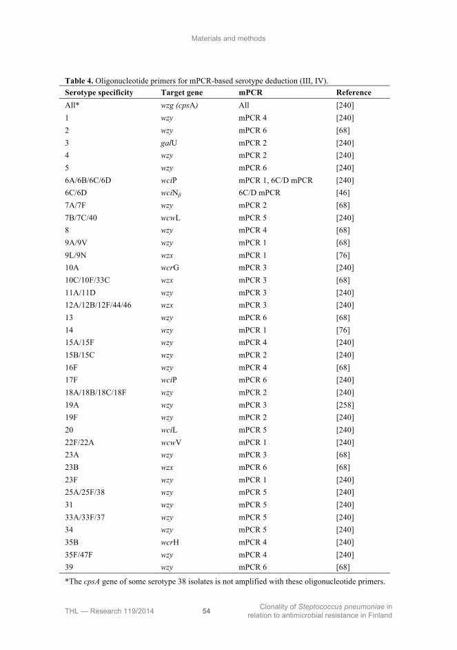
Materials and methods
THL — Research 119/2014 54 Clonality of Steptococcus pneumoniae in relation to antimicrobial resistance in Finland
Table 4. Oligonucleotide primers for mPCR-based serotype deduction (III, IV).
Serotype specificity Target gene mPCR Reference
All* wzg (cpsA) All [240]
1 wzy mPCR 4 [240]
2 wzy mPCR 6 [68]
3 galU mPCR 2 [240]
4 wzy mPCR 2 [240]
5 wzy mPCR 6 [240]
6A/6B/6C/6D wciP mPCR 1, 6C/D mPCR [240]
6C/6D wciNβ 6C/D mPCR [46]
7A/7F wzy mPCR 2 [68]
7B/7C/40 wcwL mPCR 5 [240]
8 wzy mPCR 4 [68]
9A/9V wzy mPCR 1 [68]
9L/9N wzx mPCR 1 [76]
10A wcrG mPCR 3 [240]
10C/10F/33C wzx mPCR 3 [68]
11A/11D wzy mPCR 3 [240]
12A/12B/12F/44/46 wzx mPCR 3 [240]
13 wzy mPCR 6 [68]
14 wzy mPCR 1 [76]
15A/15F wzy mPCR 4 [240]
15B/15C wzy mPCR 2 [240]
16F wzy mPCR 4 [68]
17F wciP mPCR 6 [240]
18A/18B/18C/18F wzy mPCR 2 [240]
19A wzy mPCR 3 [258]
19F wzy mPCR 2 [240]
20 wciL mPCR 5 [240]
22F/22A wcwV mPCR 1 [240]
23A wzy mPCR 3 [68]
23B wzx mPCR 6 [68]
23F wzy mPCR 1 [240]
25A/25F/38 wzy mPCR 5 [240]
31 wzy mPCR 5 [240]
33A/33F/37 wzy mPCR 5 [240]
34 wzy mPCR 5 [240]
35B wcrH mPCR 4 [240]
35F/47F wzy mPCR 4 [240]
39 wzy mPCR 6 [68]
*The cpsA gene of some serotype 38 isolates is not amplified with these oligonucleotide primers.

Materials and methods
THL — Research 119/2014 55 Clonality of Steptococcus pneumoniae in relation to antimicrobial resistance in Finland
Serotype deduction based on mPCR The amplification was performed in a 25 μl reaction volume with 35 thermal cycles
and an annealing temperature of 54°C using a Taq polymerase (AmpliTaq Gold,
Applied Biosystems, Foster City, CA, USA; or Maxima Hot Start, ThermoScientific,
Waltham, MA, USA). All oligonucleotide primer names, targets, and sources are
listed in Table 4. The genomic DNA of one positive control isolate for each
amplified locus in each mPCR was used for the PCR-based serotyping scheme. The
PCR products were visualized by gel electrophoresis using 2% NuSieve 3:1 agarose
gels (Lonza, Rockland, ME, USA) in 1×TAE buffer run for 120 to 180 minutes. As
a size marker the GeneRuler 50bp Marker (ThermoScientific) was used. Because of
primer cross-reactivity and cps sequence similarity, the Quellung reaction described
above was applied to distinguish co-detected serotypes, for subtyping when needed,
and for serotyping if no serotype- or serogroup-specific product was obtained in any
of the sequential mPCRs. During the protocol validation process, validation set 3
was studied first by the 6C/6D mPCR, after which the Quellung reaction was
applied for subtyping (Publication III).
4.5 Antimicrobial susceptibility testing The applied breakpoints for the most important antimicrobial agents studied, the
definition of multidrug-resistance, and the references for all breakpoints are listed in
Table 5. When analysing the susceptibility for the whole ten year study period, the
EUCAST 3.0 breakpoints were applied [93]. Antimicrobial susceptibility to
penicillin, erythromycin, tetracycline, and clindamycin of all available isolates was
performed by the plate dilution method at a THL laboratory in Turku (Publication I,
II and IV) [252]. Susceptibility to levofloxacin was tested from August 2004 and
susceptibility to ceftriaxone was tested from 2005 onwards. Additionally, the
susceptibility to trimethoprim/sulfamethoxazole of the non-invasive isolates was
tested (Publication II). The plates were cultivated in an atmosphere enriched with
5% CO2.
The susceptibility to penicillin, erythromycin, clindamycin, trimethoprim/
sulfamethoxazole, and doxycycline of the non-invasive isolates was initially
determined by disk diffusion test and E-test at the clinical microbiology laboratory
of the Hospital District of Helsinki and Uusimaa (Publication II).

Materials and methods
THL — Research 119/2014 56 Clonality of Steptococcus pneumoniae in relation to antimicrobial resistance in Finland
Table 5. References for the breakpoints applied in antimicrobial susceptibility testing and multidrug-resistance definitions applied in the publications.
Susceptibility breakpoints applied, minimum
inhibitory concentration (MIC) Publication
Penicillin Other antimicrobials
Multidrug-resistance
definition applied
I* CLSI pre-2008 [56] S ≤0.06 mg/L R ≥2 mg/L
CLSI pre-2008 [56],
intermediate ERY MIC ≥0.5 mg/L TET MIC ≥4 mg/L
Non-susceptibility to
PEN, ERY, and TET
II CLSI pre-2008 [56] MIC ≥0.12 mg/L
CLSI pre-2008 [56],
intermediate ERY MIC ≥0.5 mg/L TET MIC ≥4 mg/L
Non-susceptibility to
PEN, ERY, TET, CLI,
SXT, DOX
IV CLSI pre-2008 [56] S ≤0.06 mg/L R ≥2 mg/L
CLSI 2008 [55] S ≤2 mg/L R ≥8 mg/L
EUCAST 3.0 [93] S ≤0.06 mg/L R ≥4 mg/L
EUCAST 3.0 [93],
intermediate ERY MIC ≥0.5 mg/L TET MIC ≥2 mg/L
Non-susceptibility to
PEN (MIC≥0.12 mg/L),
ERY, and TET
*Includes the isolates for which unpublished genotyping results are shown; S, susceptible; R, resistant; ERY, erythromycin; TET, tetracycline; CRO, ceftriaxone; CLI, clindamycin; SXT, trimethoprim/sulfamethoxazole; DOX, doxycycline
4.6 Detection of macrolide resistance genes The macrolide resistance determinants of 223 randomly selected erythromycin-non-
susceptible isolates (MIC ≥0.5 mg/L) (Publication I) and of 12 multidrug-resistant
non-invasive isolates (Publication II) were studied at a THL laboratory in Turku.
Detection of the macrolide resistance determinants mef, erm(B), and erm(A) was
performed by mPCR (Publications I and II). Separate PCR reactions were performed
to differentiate between the efflux gene subclasses mef(A) and mef(E) in the mef
positive isolates (Publication I). The oligonucleotide primers used are presented in
Table 3, and the amplification was performed using the AmpliTaq polymerase
(Applied Biosystems).
4.7 Genotyping Selected isolates were genotyped by MLST (Table 2) [90]. The PCR reactions were
performed using a Taq polymerase, either AmpliTaq Gold (Applied Biosystems,
Foster City, CA, USA) or Maxima Hot Start (ThermoScientific, Waltham, MA,

Materials and methods
THL — Research 119/2014 57 Clonality of Steptococcus pneumoniae in relation to antimicrobial resistance in Finland
USA). The oligonucleotide primers in Table 3 were used for both PCR and
sequencing reactions. PCR product clean-up was performed by QiaQuick PCR
Purification kit (QIAGEN GmbH, Hilden, Germany), the GeneClean Turbo kit (Q-
BioGene MB Biomedicals, OH, USA), the Edge Bio QuickStep 2 PCR Purification
kit (EdgeBio Systems, Gaithersburg, MD, USA) or enzymatically using FastAP
Thermosensitive Alkaline Phosphatase and Exonuclease I (ThermoScientific).
Sequencing was performed using BigDye 1.3 chemistry (Applied Biosystems) either
in ABI Prism Genetic Analyser (Applied Biosystems) or by full sequence service at
the Institute for Molecular Medicine Finland (FIMM). The traces were analysed
using either the Vector NTI Advance 10 or 11 software suite (Invitrogen
Corporation, Carlsbad, CA, USA) or the Geneious Pro 5 or 6 software (Biomatters
Ltd, Auckland, New Zealand) and compared with the material in the MLST database,
according to which the STs were assigned [227]. Novel allele sequence traces and
STs were submitted to the MLST database. Each ST was assigned to a CC by
eBURST analysis using default stringent parameters [75]. The CCs were named
after the predicted primary founder, i.e. the ST with the highest number of single
locus variants (SLVs) at the time of writing each manuscript. For clarity and
uniformity, all observed CCs were renamed after the predicted primary founder in
September 2013.
4.8 Detection of pilus-encoding genes The presence of pilus-1 encoding PI-1 genes (rlrA, rrgC) and pilus-2 encoding PI-2
genes (pitA-sipA) were identified by PCR in selected isolates [2, 16, 284]. The
isolates were selected as follows: in Publication I, the first penicillin resistant (MIC
≥2 mg/L) isolate (n=27) of each serotype and ST as revealed by MLST, 2002-2006;
in Publication II, the multidrug-resistant non-invasive pneumococci (n=12); in
Publication IV, all penicillin-resistant (MIC ≥4 mg/L) isolates (n=12), 2007-2011. In
Publication I, the isolates were studied only for the presence of PI-1. The annealing
temperature was 59°C and the oligonucleotide primers are listed in Table 3. A Taq
polymerase was used (AmpliTaq Gold, Applied Biosystems; or Maxima Hot Start,
ThermoScientific).
4.9 Sequencing of serotype 19F-like serotype 19A wzy The wzy gene of two serotype 19F-like serotype 19A isolates, which gave discrepant
results by mPCR and Quellung based serotyping (Publication III), was sequenced using
a primer walking strategy with the oligonucleotide primes in Table 3. The obtained
sequences were edited and merged into contigs in Geneious Pro 5 software (Biomatters)
and compared to the material deposited in the GenBank through the Basic Local
Alignment Search Tool [303]. Finally, the sequences were submitted to GenBank.

Materials and methods
THL — Research 119/2014 58 Clonality of Steptococcus pneumoniae in relation to antimicrobial resistance in Finland
4.10 Statistical methods Poisson regression was used for testing the statistical significance of antimicrobial
resistance trends for the years 2002 to 2006 (Publication I). For testing the statistical
significance of the log-linear antimicrobial resistance and serotype distribution
trends, binomial regression with log-link was used in the SPSS Statistics 21 software
(IBM, Armonk, NY, USA) for the years 2007 to 2011 (Publication IV). The latter
method was used for analysis of overall serotype proportion trends and non-
susceptibility to erythromycin, tetracycline, and multidrug-resistance over the ten
year period from 2002 to 2011. The penicillin-non-susceptibility and resistance
according to EUCAST and pre-2008 CLSI breakpoints was analysed for the entire
ten year period, as was non-susceptibility by serotype and age-group. The following
age-groups were used: 0-2, 3-15, 16-64 and ≥65-year-olds. In the first five years of
the study period, the two youngest age-groups were combined for statistical testing
(Publication I). Penicillin- and erythromycin-non-susceptibility of the invasive
isolates was studied by tertiary care district (Helsinki, Turku, Tampere, Kuopio, and
Oulu) in five year periods (Publication I and IV). Risk ratio (RR) estimates per year
and 95% confidence intervals (CI) were calculated along with P values.
For detecting a possible association of PI-1 or PI-2 with serotype, serogroup, or ST,
statistical analysis was performed by Fisher’s exact test using the Internet-based
GraphPad Software (Publication II). P values <0.05 were considered statistically
significant.
4.11 Cost analysis Based on material usage per isolate at the time of the validation of the protocol, the
cost of reagents and materials of serotyping was estimated for serotyping by
Quellung and for the mPCR-based serotyping scheme including Quellung, when
needed. The mean material cost of serotyping validation set 1 was considered for
cost comparisons as this set best reflects the serotypes present in the Finnish
invasive pneumococcal population (Publication III).

THL — Research 119/2014 59 Clonality of Steptococcus pneumoniae in relation to antimicrobial resistance in Finland
5 Results
5.1 Serotypes among invasive pneumococci, 2002-2011 (I, IV) The proportions of the most important serotypes discovered are displayed in Figure
6A and Figure 6B. The proportion of certain serotypes increased over the ten-year
period. These were serotype 14, which increased from 13.5% (82/607) in 2002 to
18.0% (143/794) in 2011 (RR, 1.03; 95%CI, 1.01-1.06; P=0.010); serotype 22F,
from 3.5% to 8.8% (RR, 1.10; 95%CI 1.04-1.17; P=0.003); serotype 3 from 7.4% to
9.7% (RR, 1.04; 95%CI, 1.00-1.08; P=0.032). The proportion of serotype 4
decreased significantly from 13.6% in 2002 to 6.7% in 2011 (RR, 0.93; 95%CI,
0.90-0.96; P<0.001). The proportion of serotype 9V also decreased from 8.9% to
4.3% (RR, 0.90; 95%CI, 0.86-0.94, P<0.001).
Taken separately, the two five-year study periods showed other significant serotype
changes. Between 2002 and 2006, the proportion of serotype 6B increased from
5.8% to 9.4% (RR, 1.10; 95%CI, 1.00-1.21, P=0.05), while between 2007 and 2011
it decreased from 9.2% to 5.8% (RR, 0.91; 95%CI, 0.84-0.99; P=0.036). Reversely,
the proportion of serotype 22F decreased in the initial five years from 3.5% to 2.7%
(RR, 0.89; 95%CI, 0.84-0.94; P<0.001), while it increased in the latter five from
4.3% to 8.8% (RR, 1.20; 95%CI, 1.06-1.37; P=0.005). The proportion of PCV10
serotypes collectively remained stable in the first five years, but decreased between
2007 and 2011 from 67.0% to 55.7% (RR, 0.97; 95%CI, 0.94-0.99; P=0.008).
Serotype 6A decreased in the latter five years of the study (RR, 0.92; 95%CI, 0.86-
1.00; P=0.042), while the proportion of serotype 6C did not change in a statistically
significant manner.
There were significant changes in the serotype proportions in two age-groups from
the year 2002 to 2011. Among the adults (16-64-year-olds), the proportion of
serotype 14 increased (RR, 1.04, 95%CI, 1.01-1.07; P=0.004) reaching 20.0% in
2011; and serotype 9V decreased (RR, 0.89; 95%CI, 0.85-0.94, P<0.001) covering
5.2% of the isolates from this age-group in 2011. Among the elderly (≥65-year-olds),
the proportion of serotype 22F increased (RR, 1.09, 95%CI, 1.03-1.14; P=0.002)
reaching 9.4% in 2011, while serotype 9V decreased (RR, 0.91; 95%CI, 0.84-0.98,
P=0.017). Among the elderly, serotype 9V covered 3.6% in the year 2011.

Results
THL — Research 119/2014 60 Clonality of Steptococcus pneumoniae in relation to antimicrobial resistance in Finland
F
igu
re 6
A. T
he p
ropo
rtio
n of
the
mos
t fr
eque
ntly
enc
ount
ered
ser
otyp
es, s
erot
ypes
14,
4, 2
3F, 3
, 6B
, 7F
, and
9V
, am
ong
the
Fin
nish
inva
sive
pne
umoc
occi
20
02 to
201
1.
05
10
15
20
14
423
F3
6B
7F
9V
Proportion of isolates (%)
20
02
20
03
20
04
20
05
20
06
20
07
20
08
20
09
20
10
20
11

Results
THL — Research 119/2014 61 Clonality of Steptococcus pneumoniae in relation to antimicrobial resistance in Finland
F
igu
re 6
B. T
he p
ropo
rtio
n of
ser
otyp
es 1
9F, 2
2F, 1
8C, 1
9A, a
nd 6
A/C
am
ong
the
Fin
nish
inva
sive
pne
umoc
occi
200
2 to
201
1. T
he c
ombi
ned
prop
orti
on o
f se
roty
pes
6A a
nd 6
C a
re s
how
n fo
r th
e w
hole
ten
-yea
r pe
riod
, as
wel
l as
sep
arat
ely
for
the
latt
er f
ive
year
s.
05
10
15
20
19
F2
2F
18
C19
A6A
/6C
6A
6C
Proportion of isolates (%)
20
02
20
03
20
04
20
05
20
06
20
07
20
08
20
09
20
10
20
11

Results
THL — Research 119/2014 62 Clonality of Steptococcus pneumoniae in relation to antimicrobial resistance in Finland
In the first five years for the study period, the hypothetical PCV7 serotype coverage
was highest at 72% among the isolates derived from 0-2-year-olds and lowest at
54% among those derived from 16-64-year-olds. The overall hypothetical serotype
coverage for all age-groups and isolates was 58%. The hypothetical PCV10
coverage was 81% of the isolates obtained from 0-2 year olds in 2007, and 72% in
2011. The hypothetical PCV13 coverage in this age group was 84% in 2007, and
88% in 2011. The hypothetical PCV13 coverage overall was 68% in 2007, and 73%
in 2011.
5.2 Antibiotic resistance among invasive pneumococci, 2002-2011 (I, IV)
5.2.1 Penicillin Over the ten year study period, the proportion of penicillin-non-susceptible (MIC
≥0.12 mg/L) isolates among the invasive pneumococci increased from 8.0% (48/547)
to 21.8% (173/794) (RR, 1.12; 95%CI, 1.08-1.15; P<0.001), range 8.0% to 23.3%
(Figure 7).
Figure 7. The proportion of penicillin-non-susceptible and -resistant isolates among invasive pneumococci isolated in 2002 to 2011.

Results
THL — Research 119/2014 63 Clonality of Steptococcus pneumoniae in relation to antimicrobial resistance in Finland
The proportion of penicillin-resistant isolates according to the pre-2008 breakpoint
issued by CLSI went from 0.8% (5/595) in 2002 to 3.5% (28/794) in 2011, range
0.8% to 3.8%, but the increase was not statistically significant. The penicillin-
resistance proportion as defined by EUCAST also remained stable at 0.2% overall,
range 0% to 0.6% (Figure 7). Only two isolates were found that were resistant to
penicillin as defined by the 2008 CLSI breakpoint; these were isolated in 2010 and
2011, respectively.
When the five year study periods were examined separately, the proportion of
penicillin-non-susceptible isolates (MIC ≥0.12 mg/L) doubled from 8.0% to 16.4%
(RR, 1.15; 95%CI, 1.07-1.23; P<0.0001) and the proportion of penicillin-resistant
isolates, as defined by the pre-2008 CLSI breakpoint, increased from 0.8% to 3.7%
(RR, 1.18; 95%CI, 1.02-1.36; P=0.03). In the latter five years, the proportion of
penicillin-non-susceptible isolates increased from 14.4% to 21.8% (RR, 1.11;
95%CI, 1.05-1.17; P<0.001, but the penicillin-resistance did not change
significantly.
The proportion of penicillin-non-susceptible isolates was high within some
serotypes (Figure 8), and in four serotypes it increased significantly over the ten-
year period. The proportion of penicillin-non-susceptible isolates among serotype 14
increased from 25.0% in 2002 to 70.6% in 2011 (RR, 1.11; 95%CI, 1.08-1.15;
P<0.001); among serotype 19A from 12.5% to 37.5% (RR, 1.13; 95%CI, 1.02-1.26;
P=0.024); among serotype 4 from 1.2% to 26.4% (RR, 1.41; 95%CI, 1.28-1.54;
P<0.001); among serotypes 6A and 6C combined from none to 11.4% (RR, 1.93;
95%CI, 1.42-2.61; P<0.001).
Over the ten year period, the proportion of penicillin-non-susceptible isolates
increased in all age-groups. Among the 0-2-year-olds it increased from 15.7%
(11/70) in 2002 to 29.7% (19/64) in 2011 (RR, 1.15; 95%CI, 1.08-1.21; P<0.001),
among the 3-15-year-olds from 6.3% (2/32) to 31.0% (9/29) (RR, 1.18; 95%CI,
1.10-1.26; P<0.001), among the 16-64-year-olds from 8.7% (28/324) to 20.8%
(84/403) (RR, 1.12; 95%CI, 1.08-1.16; P<0.001), and among the ≥65-year-olds
from 5.7% (10/179) to 20.4% (61/299) (RR, 1.07; 95%CI, 1.01-1.13; P=0.034).
When studied separately in five year periods, the proportion of penicillin-non-
susceptible isolates increased only among the 16-64 year-olds (RR, 1.22; P<0.001)
and among the ≥65-year-olds (RR, 1.15; P=0.02) in 2002 to 2006. In 2007 to 2011,
the increase in the proportion of penicillin-non-susceptible isolates was not
significant in the youngest age-group, however, there was significant increase in all
other age-groups: among the 3-15-year-olds (RR, 1.23; 95%CI, 1.13-1.34; P<0.001),

Results
THL — Research 119/2014 64 Clonality of Steptococcus pneumoniae in relation to antimicrobial resistance in Finland
the 16-64-year-olds (RR 1.11; 95%CI, 1.05-1.19; P=0.001), among the ≥65-year-
olds (RR 1.09; 95%CI, 1.03-1.14; P=0.002).
Figure 8. Penicillin non-susceptibility in selected serotypes among the invasive pneumococci isolated in 2011, top panel. Erythromycin non-susceptibility in selected serotypes among the invasive pneumococci isolated in 2011, bottom panel.
The proportion of penicillin-non-susceptible isolates (MIC ≥0.12 mg/L) in the year
2011 varied between 18.2% and 25.6% in the five tertiary care districts. In the first

Results
THL — Research 119/2014 65 Clonality of Steptococcus pneumoniae in relation to antimicrobial resistance in Finland
five years of the study, the proportion of penicillin-non-susceptible isolates
increased significantly in the tertiary care districts of Tampere, from 5% in 2002 to
15% in 2006 (RR, 1.25; 1.07-1.45; P=0.004), and Kuopio, from 5% to 20% (RR,
1.30; 1.04-1.61, P=0.02). The latter five years, the increase was significant in the
districts of Helsinki from 18% to 26% (RR, 1.10; 95% CI, 1.01-1.20; P=0.033),
Kuopio from 8% to 23% (RR, 1.30; 95% CI, 1.27-1.33; P<0.001), and Turku from
11% to 19% (RR, 1.10; 95% CI, 1.00-1.22; P=0.044).
In the first five years of the study, the hypothetical PCV7 coverage was 87% of the
penicillin-non-susceptible isolates; in the last five years, the hypothetical PCV10
coverage was 90% of the penicillin-non-susceptible isolates.
5.2.2 Other antimicrobial agents The proportion of erythromycin-non-susceptible isolates increased over the ten-year
period from 16.3% to 26.4% (RR, 1.05; 95%CI, 1.02-1.07; P<0.001), and was
24.3% overall, range 16.3% to 28.9% (Figure 9). In the first five years, the increase
was significant from 16.3% to 28.4% (RR, 1.12; 95%CI, 1.06-1.18; P<0.001),
however in the latter five years the proportion did not increase significantly and was
26.6% overall, range 23.2% to 28.9%.
Figure 9. The proportion of invasive isolates non-susceptible to erythromycin and tetracycline, respectively, and the proportion of multidrug-resistant (MDR) isolates non-susceptible to PEN, ERY, and TET among the invasive pneumococci in Finland in 2002 to 2011.

Results
THL — Research 119/2014 66 Clonality of Steptococcus pneumoniae in relation to antimicrobial resistance in Finland
The proportion of erythromycin-non-susceptible isolates was consistently highest in
the youngest age-group. In the year 2011, the proportion of non-susceptible isolates
was 46.9% among isolates from 0-2-year-olds, 27.6% among isolates from 3-15-
year-olds, 25.6% among isolates from 16-64-year-olds, and 23.1% among isolates
from ≥65-year-olds. The proportion of non-susceptible isolates increased among the
isolates from all tested age-groups during the first five-year period, 2002 to 2006:
among the 0-15-year-olds (RR, 1.12; P=0.01); among the 16-64-year-olds (RR, 1.11;
P=0.003); and among the ≥65-year-olds (RR, 1.16; P<0.001). In the latter five years,
there was a significant increase in non-susceptibility over the study period only
among the 16-64-year-olds (RR, 1.06; P=0.049). In the year 2011, the proportion of erythromycin-non-susceptible isolates varied
between 18.7% and 34.3% among the five tertiary care districts. In 2002 to 2006, a
significant increase in the proportion of erythromycin-non-susceptible isolates was
observed in the tertiary care district of Tampere from 7% to 29% (RR, 1.35; 95%CI
1.22-1.51; P<0.001) and Oulu from 20% to 28% (RR, 1.12; 95% CI 1.00-1.25;
P=0.04). In 2007 to 2011, the non-susceptibility increased significantly only in the
district of Kuopio from 22% to 29% (RR, 1.07; 95% CI, 1.04-1.10; P<0.001).
In the first five years of the study, the hypothetical PCV7 coverage of the
erythromycin-non-susceptible isolates was 80%, and in the last five years, the
hypothetical PCV10 coverage was 86% of the erythromycin-non-susceptible isolates.
Serotype 14 especially, but also certain other serotypes, contained a high proportion
of erythromycin-non-susceptible isolates in the year 2011 (Figure 8).
The proportion of tetracycline-non-susceptible isolates decreased slightly over the
ten years from 9.8% to 6.0% (RR, 0.94; 95%CI, 0.91-0.98; P=0.001) and was 8.9%
overall, range 6.0% to 13.0% (Figure 9). When studied separately, there was no
significant change during either of the two five year periods.
Ceftriaxone susceptibility was studied from 2005 onwards and 3.7%, range by year,
2.5% to 4.2%, of all tested isolates were non-susceptible to this antimicrobial agent.
Twelve isolates or 0.2% had ceftriaxone MICs ≥2 mg/L. The non-susceptibility to
levofloxacin was similarly rare at 0.2% overall, with 16 non-susceptible isolates
discovered since testing started in August, 2004.
The proportion of multidrug-resistant isolates, defined as non-susceptibility to
penicillin, erythromycin, and tetracycline, was 4.2% overall, range 3.3% to 5.6%,
and did not change significantly over the ten years or in the two five year periods
(Figure 9). Of the erythromycin-non-susceptible isolates, 38% (2002-2006) and 55%
(2007-2011), respectively, were also non-susceptible to penicillin (MIC ≥0.12 mg/L).

Results
THL — Research 119/2014 67 Clonality of Steptococcus pneumoniae in relation to antimicrobial resistance in Finland
Overall, 74% (2002-2006) and 76% (2007-2011), respectively, of the penicillin-non-
susceptible isolates were also non-susceptible to erythromycin.
5.3 Macrolide resistance determinants among the invasive pneumococci, 2002-2006 (I)
The mef gene was the most common macrolide resistance determinant. It was
carried by 56% of the 223 randomly selected erythromycin-non-susceptible isolates.
Of the subtyped mef positive isolates, 72% carried mef(E) and 28% carried mef(A).
All mef(A) positive isolates were serotype 14 and susceptible to penicillin, which
suggests they may be clonally related. The erm(B) gene was present in 30% of the
screened isolates. Double carriage of macrolide resistance determinants, mef(E) and
erm(B), was displayed by only two of the studied isolates (0.9%). These isolates
were both serotype 19F and part of CC320. The macrolide resistance mechanism
remained unknown in 13% of the studied isolates.
5.4 Genotype clonality of penicillin-resistant and -non-susceptible invasive pneumococci, 2002-2011 (I, IV)
Among the penicillin-resistant (MIC ≥2 mg/L) invasive isolates from 2002 to 2006,
25 STs belonging to nine CCs and one singleton was discovered. The CCs and their
STs were CC156 with ST156, ST2918, ST2306, ST3247, ST143, ST2916, ST671,
ST90, ST3246, and ST138; CC320 with ST236, ST271, ST2694, ST2917, and
ST3245; CC81 with ST81, ST961, and ST3250; CC15 with ST13; CC63 with
ST2687; CC496 with ST496; CC205 with ST205; CC460 with ST461; and CC2599
with ST3248. The singleton was ST3249.
The penicillin-resistant (MIC ≥4 mg/L) isolates from 2007 to 2011 displayed eight
STs belonging to two CCs, CC156 and CC320. These CCs were present also among
the highly penicillin-resistant invasive isolates in the previous five-year period
(Table 6).
The penicillin-non-susceptible isolates (MIC 0.12-1.0 mg/L) from the year 2005
displayed 34 different STs belonging to 14 CCs and one singleton (Table 7).
Among all the invasive penicillin-non-susceptible isolates genotyped by MLST, 14
novel STs were discovered. Among these, four novel ddl alleles, and one novel
allele at the aroE, gki, recP, and xpt loci each were discovered. CC320 and lineage
termed i within CC156, as defined by the eBURST algorithm are shown in Figure
10 and Figure 11, respectively.

Results
THL — Research 119/2014 68 Clonality of Steptococcus pneumoniae in relation to antimicrobial resistance in Finland
Tabl
e 6.
Clo
nal c
ompl
exes
, seq
uenc
e ty
pes,
sero
type
s, an
d an
timic
robi
al su
scep
tibili
ty o
f the
pen
icill
in-r
esis
tant
(MIC
>4
mg/
L) p
neum
ococ
ci is
olat
ed in
the
year
s 200
2 to
201
1. O
ne is
olat
e pe
r cas
e is
incl
uded
(n=2
0).
CC
ST
A
llelic
pro
file
Sero
type
A
ntim
icro
bial
su
scep
tibili
ty (m
g/L)
R
elat
ed P
MEN
cl
one
Pilu
s isle
ts*
Yea
r of
isol
atio
n (n
o. o
f iso
late
s)
PEN
ER
Y
TET
320
236
15-1
6-19
-15-
6-20
-26
19F
4 8
64
PMEN
14
Taiw
an19
F ST23
6 PI
-1
2006
(1)
29
17
4-16
-19-
15-6
-20-
266
19F
4 >1
28
32
PI-1
, PI-
2 20
05 (1
), 20
09 (2
)
32
0 4-
16-1
9-15
-6-2
0-1
19F
4 >1
28
0.25
PI-1
, PI-
2 20
10 (1
)
32
0 4-
16-1
9-15
-6-2
0-1
19A
8
>128
64
PI-1
, PI-
2 20
11 (1
)
27
1 4-
16-1
9-15
-6-2
0-26
19
F 4-
8 >1
28
16-3
2
PI-1
, PI-
2 20
09 (2
), 20
10 (1
)
32
45
4-16
-19-
15-6
-20-
286
19F
4 >1
28
32
PI
-1
2002
(1)
65
1 15
-5-1
9-15
-6-2
0-26
19
F 4
4 0.
25
PI
-1, P
I-2
2009
(1)
87
94
15-1
6-19
-234
-6-2
0-50
19
F 4
>128
32
PI-1
, PI-
2 20
10 (1
)
156
156
7-11
-10-
1-6-
8-1
14
4 0.
125
0.25
PM
EN3
Spai
n9VST
156
PI-1
20
05 (1
)
14
3 7-
5-10
-18-
6-8-
1 14
4
>128
0.
25
PI-1
20
08 (1
)
29
16
7-5-
10-1
8-6-
5-1
14
4 >1
28
64
PI
-1
2008
(1)
29
18
7-11
-10-
1-6-
8-26
7 14
4
>128
0.
5
PI-1
20
10 (1
)
13
8 7-
5-8-
5-10
-6-1
4 6B
4
16
0.25
PI-1
20
06 (1
)
81
81
4-4-
2-4-
4-1-
1 23
F 4
0.12
5 1
PMEN
1 Sp
ain23
F ST
81
Non
e 20
03 (1
)
15
13
1-5-
4-5-
5-27
-8
14
4 32
0.
25
PMEN
9 En
glan
d14ST
9 N
one
2003
(1)
63
2678
2-
60-3
6-12
-17-
21-1
4 14
4
>128
64
PM
EN25
Sw
eden
15AST
63
Non
e 20
05 (1
)
CC
, clo
nal c
ompl
ex; S
T, s
eque
nce
type
; PEN
, pen
icill
in, E
RY
, ery
thro
myc
in; T
ET, t
etra
cycl
ine;
PM
EN, P
neum
ococ
cal M
olec
ular
Epi
dem
iolo
gy N
etw
ork;
PI
-1, p
ilus
isle
t 1; P
I-2, p
ilus
isle
t 2. *
PI-1
was
det
ecte
d in
the
isola
tes
from
200
2-20
06, w
hile
bot
h PI
-1 a
nd P
I-2 w
ere
dete
cted
in th
e is
olat
es fr
om 2
007-
2011
.
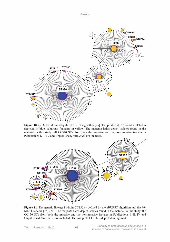
Results
THL — Research 119/2014 69 Clonality of Steptococcus pneumoniae in relation to antimicrobial resistance in Finland
Figure 10. CC320 as defined by the eBURST algorithm [75]. The predicted CC founder ST320 is depicted in blue, subgroup founders in yellow. The magenta halos depict isolates found in the material in this study, all CC320 STs from both the invasive and the non-invasive isolates in Publications I, II, IV and Unpublished, Siira et al. are included.
Figure 11. The genetic lineage i within CC156 as defined by the eBURST algorithm and the 96-MLST scheme [75, 221]. The magenta halos depict isolates found in the material in this study, the CC156 STs from both the invasive and the non-invasive isolates in Publications I, II, IV and Unpublished, Siira et al. are included. The complete CC156 is depicted in Figure 4.

Results
THL — Research 119/2014 70 Clonality of Steptococcus pneumoniae in relation to antimicrobial resistance in Finland
T
ab
le 7
. Clo
nal
com
plex
es, s
eque
nce
type
s, s
erot
ypes
, and
ant
imic
robi
al s
usce
ptib
ilit
y of
the
pen
icil
lin-
non-
susc
epti
ble
pneu
moc
occi
iso
late
d in
the
year
20
05. O
ne i
sola
te p
er c
ase
is i
nclu
ded
(n=
66).
CC
S
T
All
elic
pro
file
S
ero-
typ
e
An
tim
icro
bia
l su
scep
tib
ilit
y (
mg
/L)
No.
of
isola
tes
Rel
ate
d P
ME
N c
lon
e
PE
N
ER
Y
TE
T
156
156
7-11
-10-
1-6-
8-1
9V
2 0.
125
0.25
1
PM
EN
3 S
pain
9VS
T15
6
15
6 7-
11-1
0-1-
6-8-
1 14
1-
4 0.
063-
16
0.25
-0.5
5
15
6 7-
11-1
0-1-
6-8-
1 9N
1
0.06
3-0.
125
0.25
2
14
3 7-
5-10
-18-
6-8-
1 14
2
>12
8 32
1
67
1 7-
11-1
0-1-
5-76
-98
14
0.12
5 32
-64
0.12
5-0.
5 9
29
16
7-5-
10-1
8-6-
5-1
14
1-2
>12
8 32
2
23
06
7-11
-10-
1-6-
8-11
9 14
2
>12
8 32
1
15
69
8-11
-10-
1-6-
8-1
9V
1 >
128
64
1
33
8 7-
13-8
-6-1
-6-8
23
F
0.12
5 0.
125
0.25
1
PM
EN
26 C
olom
bia23
FS
T33
8
90
5-
6-1-
2-6-
3-4
6B
1-2
>12
8 0.
5-64
3
PM
EN
2 S
pain
6BS
T90
90
69
7-5-
8-5-
10-6
-570
6B
0.
125
0.12
5 0.
25
1
63
63
2-5-
36-1
2-17
-21-
14
14
0.25
0.
125
0.25
1
PM
EN
25 S
wed
en15
AS
T63
78
2 2-
5-36
-12-
17-2
1-5
14
0.12
5-0.
25
>12
8 32
-64
6
26
78
2-60
-36-
12-1
7-21
-14
14
1-4
>12
8 32
-64
3
315
315
20-2
8-1-
1-15
-14-
14
6B
0.12
5 >
128
32-6
4 2
PM
EN
20 P
olan
d6BS
T31
5
38
6 32
-28-
1-1-
15-5
2-14
6B
0.
125
>12
8 32
4
90
68
20-2
8-1-
1-15
-526
-14
6B
0.12
5 64
0.
5 1
230
319
12-1
9-2-
17-6
-22-
9 19
F
0.5
>12
8 32
3
PM
EN
32 D
enm
ark14
ST
230
27
6 2-
19-2
-17-
6-22
-14
19A
1
>12
8 0.
5 1
25
62
11-1
9-2-
17-6
-215
-75
19F
0.
5 >
128
32
1
91
08
12-1
9-70
-17-
6-22
-14
19A
0.
25
0.12
5 32
1
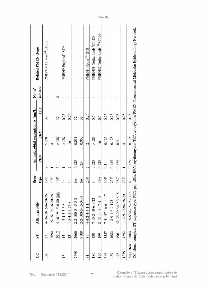
Results
THL — Research 119/2014 71 Clonality of Steptococcus pneumoniae in relation to antimicrobial resistance in Finland
CC
S
T
All
elic
pro
file
S
ero-
typ
e
An
tim
icro
bia
l su
scep
tib
ilit
y (
mg
/L)
No.
of
isola
tes
Rel
ate
d P
ME
N c
lon
e
PE
N
ER
Y
TE
T
320
271
4-16
-19-
15-6
-20-
26
19F
2
>12
8 32
1
PM
EN
14 T
aiw
an19
FS
T23
6
26
94
15-1
6-19
-1-6
-20-
26
19F
1
4 1
1
29
17
4-16
-19-
15-6
-20-
266
19F
2-
4 >
128
32
2
15
15
1-5-
4-5-
5-3-
8 14
1
>12
8 0.
25
1 P
ME
N9
Eng
land
14S
T9
13
1-
5-4-
5-5-
27-8
14
2
16
0.25
1
2660
26
60
2-2-
100-
5-15
-3-8
6A
0.
125
0.03
1 32
1
91
09
2-2-
100-
5-15
-3-1
4 6A
0.
25
0.06
3 32
1
81
81
4-4-
2-4-
4-1-
1 23
F
2 2
0.25
1
PM
EN
1 S
pain
23F S
T81
180
180
7-15
-2-1
0-6-
1-22
3
0.12
5 >
128
0.5
1 P
ME
N31
Net
herl
ands
3 ST
180
199
199
8-13
-14-
4-17
-4-1
4 19
A
0.12
5 16
0.
5 1
PM
EN
37 N
ethe
rlan
ds15
BS
T19
9
346
3557
7-
41-4
7-16
-6-1
4-17
19
F
0.5
0.12
5 0.
25
1
439
1682
1-
8-9-
1-6-
4-17
5 23
F
0.12
5 0.
125
0.25
1
496
496
42-3
5-29
-36-
9-39
-18
18C
0.
125
0.5
0.25
1
1379
12
92
1-13
-9-1
2-94
-28-
20
23F
1
4 0.
25
1
Sin
glet
on
6864
11
8-60
-4-1
5-25
-14-
9 4
0.12
5 0.
125
0.25
1
C
C, c
lona
l co
mpl
ex;
ST
, seq
uenc
e ty
pe;
PE
N, p
enic
illi
n, E
RY
, ery
thro
myc
in;
TE
T, t
etra
cycl
ine;
PM
EN
, Pne
umoc
occa
l M
olec
ular
Epi
dem
iolo
gy N
etw
ork.
Results
THL — Research 119/2014 72 Clonality of Steptococcus pneumoniae in relation to antimicrobial resistance in Finland
5.5 Pilus islets among the penicillin-resistant invasive pneumococci, 2002-2011 (I, IV)
Pilus islet carriage was limited to STs of certain CCs. Among the penicillin-resistant
pneumococci (MIC ≥2 mg/L) isolated from 2002 to 2006, PI-1 was carried by the
isolates in CC156, CC320, CC205 and the singleton ST3249. CC81, CC460,
CC2599, CC15, CC63 and CC496 were negative for PI-1. The isolates isolated
before 2007 were not studied for the presence of PI-2. Of the penicillin-resistant
(MIC ≥2 mg/L) isolates from the years 2002 to 2006, 79.5% carried PI-1 if
extrapolated from the results of the studied ST representatives. In 2007 to 2011, all
penicillin-resistant isolates (MIC ≥4 mg/L) carried PI-1, and in addition, the
serogroup 19 CC320 isolates (75%) also carried PI-2 (Table 6).
5.6 Characteristics of non-invasive multidrug-resistant pneumococci (II)
Five serotypes were found among the 12 isolates: 19F (n=5), 19A (n=3), 6B (n=2),
23F (n=1), and 14 (n=1). Six STs from three genetic CCs were represented: CC320
with ST271 (n=4), ST1451 (n=3), ST283 (n=1); CC156 with ST90 (n=2), ST2918
(n=1); and CC81 with ST81 (n=1). CC320 claimed all serogroup 19 isolates. CC320
and the lineage termed i within CC156, as defined by the eBURST algorithm, are
depicted in Figure 10 and Figure 11, respectively. Of the 12 isolates, 11 (92%)
carried PI-1 and 8 (67%) carried PI-2. All CC320 isolates were positive for both
pilus islets, while the CC156 isolates were positive only for PI-1. The CC81 isolate
was negative for both pilus islets. PI-2 was associated with serogroup 19 and with
CC320 (P=0.002). All CC320 isolates displayed the double macrolide resistance
determinant phenotype, carrying both erm(B) and mef(A/E), while the CC156 and
CC81 isolates carried only the erm(B) gene.

Results
THL — Research 119/2014 73 Clonality of Steptococcus pneumoniae in relation to antimicrobial resistance in Finland
5.7 Validation of the mPCR based serotyping scheme (III) Overall, the three sets consisted of 42 different serotypes and three non-encapsulated
pneumococci and one non-pneumococcal isolate (Figure 12). The initial results
obtained at the two THL laboratories by mPCR-based serotyping supplemented with
Quellung (Figure 13) in Helsinki and by traditional immunological methods in Oulu,
were compatible for 95.3% (162/170) of the isolates overall. For the first validation
set, the results from the THL laboratories in Helsinki and Oulu were congruent for
97% (68/70) of the isolates; for the second set, for 96% (67/70) of the isolates; and
for the third set, for 90% (27/30) of the isolates. For the serotypes where the
protocols used differed in resolution power, results such as serotype 10F/C vs.
serogroup 10 were not considered contradictory.
Figure 12. Serotypes included in the three validation sets (serotype; number of isolates) sorted by serogroup. Serotypes are named to the level that is in use for the mPCR based serotyping scheme for invasive pneumococci in Finland. PNC, pneumococcus; NC, non-encapsulated.

Results
THL — Research 119/2014 74 Clonality of Steptococcus pneumoniae in relation to antimicrobial resistance in Finland
External verification of discrepant serotyping results was performed at one or both
of the reference laboratories at SSI and UAB. Following external verification, the
results obtained by the mPCR-based serotyping scheme were compatible with the
verified results for 98.6% (69/70) of the first validation set, 98.6% (69/70) for the
second set, and 100% (30/30) for the third set. The combined compatibility between
the results obtained by the mPCR based serotyping scheme and the external
reference laboratory results was 98.8% (168/170). For isolate IH163205-2, serotype
6D, the serotyping performed at UAB was considered decisive, as this was where
serotype 6D was first described and studied in detail [34, 35]. The results for this
isolate have been published separately [229]. For isolate IH163216-4, the result of
non-pneumococcus from SSI was considered decisive, as species verification tests
had been performed there.
Together, the first two mPCRs were anticipated to give a result for over 80% of the
invasive isolates when setting up the new serotyping scheme (Figure 13). This
expectation proved accurate, as 86% of the invasive pneumococci isolated in the
initial 18 months of application produced either a serotype or serogroup specific
amplicon in one of the first two mPCRs. Routine application of the serotyping
scheme was successful and interpretation of the electrophoresis gel amplicon
patterns was straightforward. It also confirmed that the six main mPCRs combined
produced a serotype- or serogroup-specific mPCR amplicon for 99.4% of the
Finnish invasive isolates.
For serotyping validation set 1, the approximate mean material cost by Quellung
was 17 € per isolate, range 14-20 €. The approximate mean material cost for
serotyping by the mPCR based serotyping scheme including Quellung when needed,
as depicted in Figure 13, was 6 € per isolate, range 3-13 €.
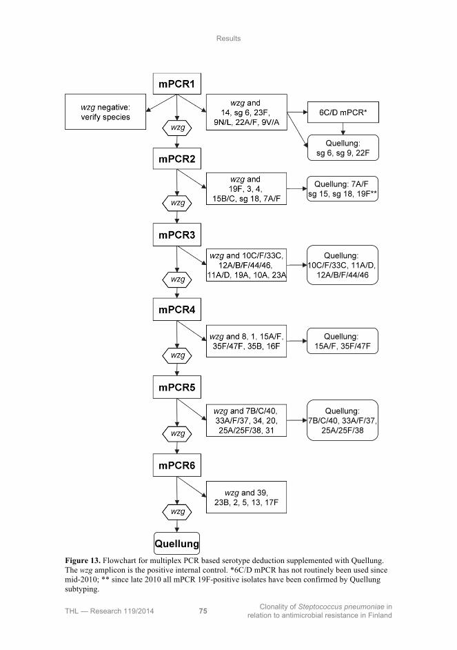
Results
THL — Research 119/2014 75 Clonality of Steptococcus pneumoniae in relation to antimicrobial resistance in Finland
Figure 13. Flowchart for multiplex PCR based serotype deduction supplemented with Quellung. The wzg amplicon is the positive internal control. *6C/D mPCR has not routinely been used since mid-2010; ** since late 2010 all mPCR 19F-positive isolates have been confirmed by Quellung subtyping.

Results
THL — Research 119/2014 76 Clonality of Steptococcus pneumoniae in
relation to antimicrobial resistance in Finland
5.8 The genotypes and wzy sequences of two serotype 19F-like 19A isolates
Two isolates that produced the 19F specific amplicon in mPCR 2, but were serotype
19A when examined by Quellung, were studied by MLST. These isolates had novel
STs. Isolate IH163643-0 had ST8025 and had the allelic profile 1-5-41-5-9-1-18,
while isolate IH166473-6 had ST8026 and had the allelic profile 13-8-4-5-9-1-18.
The sequenced wzy genes were identical in both isolates and showed 93-92%
identity with the serotype 19F wzy genes and 80% identity with the serotype 19A
wzy genes from other isolates found in GenBank. The wzy gene sequences of isolates
IH163643-0 and IH166473-6 were deposited in GenBank under the accession
numbers JX112900 and JX112901, respectively.

THL — Research 119/2014 77 Clonality of Steptococcus pneumoniae in relation to antimicrobial resistance in Finland
6 Discussion
6.1 Serotypes among the invasive pneumococci The proportions of individual serotypes may fluctuate over time in a pneumococcal
population even prior to extensive use of vaccines or antimicrobials. The use of
antimicrobials is likely to have affected the serotype distribution before systematic
surveillance of resistance was implemented [99, 122]. In this study, the three most
frequently identified serotypes in the years 2002 and 2007 were all included in
PCV10. The serotypes were 4, 14, and 9V in 2002; and 14, 6B, and 4 in 2007. In
contrast, in 2011, the three most frequent serotypes were 14, 3, and 22F, and the
proportion of all these serotypes increased over the study period. Serotype 14
covered 17.5% of the isolates from the whole period, while it only covered 12.2% of
invasive pneumococcal disease cases in 1995 to 2002 [163]. The increase of
serotype 22F, especially, was quite dramatic in the last year of the study reaching
8.8% in 2011. In 1995 to 2002 serotype 22F covered only 3.5% of the isolates [163].
Of the three most common serotypes isolated in 2011, only serotype 14 is a vaccine
serotype, while serotypes 3 and 22F may turn out to be important in replacement
following PCV10 vaccination. Serotype replacement by serotype 3 and 22F has been
described elsewhere following PCV7 use [212, 249]. In this study, the proportions
of the PCV10 serotypes 4 and 9V declined over the study period of ten years, 2002-
2011. In the latter half of the study period, the PCV10 serotypes combined also
decreased, although they had remained stable in the previous five years. These
changes may not be a direct consequence of vaccine use, but their timing is
interesting in relation to the PCV10 implementation in Finland in September 2010,
and they warrant careful monitoring in the coming years. It has been proposed that
replacement serotypes will include serotypes which are metabolically less costly for
the bacteria to produce and therefore may be more heavily capsulated [329]. These
low carbon containing serotypes include serotype 3, while other potential
replacement serotypes proposed by this theory are 19A and 6A.
The hypothetical serotype coverage calculated based on the serotypes discovered in
this material may not give the complete picture. For PCV13, the immunological
response to serotype 3 has been low in some studies, for example [233]. This is an
important consideration to bear in mind when comparing the serotype coverage
provided by different PCVs, especially as the proportion of serotype 3 has been
increasing in Finland, possibly partly in response to the use of PCV10. The
distribution of invasive serotypes may also differ markedly for example from the
distribution of serotypes in non-bacteraemic pneumococcal pneumonia. This was

Discussion
THL — Research 119/2014 78 Clonality of Steptococcus pneumoniae in relation to antimicrobial resistance in Finland
illustrated by a recent Danish study, in which the PCV10 and PCV13 serotypes
covered only a sixth and a third of non-invasive cases, respectively, while the
proportions were much higher for the bacteraemic cases [22]. In a study from the
UK, the loss of quality of life, clinical presentation, and mortality was calculated and
found to differ markedly between serotypes, giving different vaccines different
disease coverage profiles [315].
The proportions of serotype 14 and serotype 9V changed radically and
simultaneously from one year to the next during the study. These changes took place
from 2005 to 2006, when serotype 14 increased, while serotype 9V decreased. It is
tempting to speculate that this may have been caused by the rise of a particularly
successful serotype 14 clone at the expense of a declining serotype 9V clone.
Interestingly, serotype 3, has in some studies been found to display low invasiveness
[38], but was frequently encountered in invasive samples in Finland during the
whole ten year period. Other studies have found that this serotype has a high
invasive disease potential especially among adults and the elderly, and also high
mortality [249, 285, 339]. On the other hand, serotype 1, which has high invasive
disease potential [38], is very rare among the invasive isolates in Finland. This may
perhaps be explained by the low number of strains of this serotype circulating in
human society. Similarly low serotype 1 proportions have been described in the
USA, Canada, and Australia, while in much of Europe, Asia, Latin America and
Africa higher proportions of up to 30% are found [127].
6.2 Recently discovered and aberrant serotypes
6.2.1 Serogroup 6 In the years covered by this study, several novel serotypes have been discovered.
This is most notable within serogroup 6, where the two previously known serotypes,
6A and 6B, have been joined by serotypes 6C and 6D, and even more recently, by
the proposed serotypes 6E, 6F, and 6G [34, 164, 237, 244]. Serotypes 6A and 6B,
the two oldest members of this serogroup, were described in the late 1920s [130].
Genetically, they differ from each other only by a single nucleotide polymorphism at
the 584 bp position in the wciP gene, which changes the amino acid residue at
position 195 [200, 292]. This is the genetic basis for the different rhamnose ribitol
linkages found in the serotype 6A and 6B capsular polysaccharides: a 1,3 linkage in
serotype 6A and a 1,4 linkage in serotype 6B [200].
Serotype 6A subtypes were suspected already in 2006, when “dodgy 6As” isolates
were found. These bound only to certain types of monoclonal antibodies developed

Discussion
THL — Research 119/2014 79 Clonality of Steptococcus pneumoniae in relation to antimicrobial resistance in Finland
against serotype 6A and displayed a weak reaction with the Pool B antiserum in the
Quellung reaction [123, 182]. A year later, the genetic basis for serotype 6C was
described. This serotype is similar to serotype 6A, but carries the wciNβ gene, a
roughly 200 bp shorter version of the serotype 6A wciNα gene. The gene product is a
α-1,3-galactosyltransferase involved in capsular production. Pneumococci with this
serotype have been isolated from both invasive and non-invasive samples from
around the world [243].
A few years after the discovery of serotype 6C, the same research group generated
an analogous variant of serotype 6B, i.e. an isolate that harboured the serotype 6B
wciP gene, but also carrying the wciNβ gene. This serotype was named 6D [35].
Isolates carrying this capsular type were later discovered among natural clinical
isolates [34], and a study from South Korea has established that serotype 6D has
been circulating there at least since 1996 [51]. In Finland, three serotype 6D isolates
have been discovered among the invasive pneumococci since the initial publication
of the serotype. The isolates are included in this study, and one of them has been
described separately [229]. In addition to this, there are only a handful of reports of
invasive serotype 6D isolates worldwide to date [51, 173, 199, 262, 346]. According
to reports from several countries, it appears to be more commonly encountered
among non-invasive isolates [151, 157, 165, 208, 236, 276].
In this study, the serotype 6A isolates from the years 2007 to 2011 were studied
either by 6C/6D mPCR or the Quellung reaction to differentiate the serotype 6C
isolates. This showed that serotype 6C had been present among the invasive isolates
at least since the year 2007. As not all previous serotyping results have been
confirmed using the larger selection of serogroup 6 specific antisera or the 6C/6D
specific mPCR, it is likely that these newer serotypes may be present also among
older isolates. Serotype 6C has been in existence at least since 1979 [243] and it
appears that the serotype 6C cps has appeared through several independent capsular
switching events within several different genetic lineages [187]. Because of its rarity
among the invasive pneumococci isolated since 2010, there has at this time been no
attempt to retrospectively distinguish serotype 6D from 6B in Finland. It is still
unclear whether the other proposed serotypes of this serogroup, serotypes 6E, 6F
and G, are more likely to be encountered in invasive or non-invasive sites and how
common they are.
This study and others show that among the serogroup 6 serotypes, penicillin-non-
susceptibility is most frequent in serotype 6B. Serotype 6A isolates display non-
susceptibility (MIC ≥0.12 mg/L) in some cases, though rarely full resistance [202,
313]. Penicillin-non-susceptibility among serotype 6C is low, but has increased at
least in the USA, exceeding 30% among invasive isolates in the year 2007 [46]. In
Portugal, multidrug-resistance among serotype 6C has been on the increase [234]. In

Discussion
THL — Research 119/2014 80 Clonality of Steptococcus pneumoniae in relation to antimicrobial resistance in Finland
the Finnish material, only one penicillin-non-susceptible serotype 6C isolate was
discovered between 2007 and 2011, therefore one can conclude that the increase in
non-susceptibility in serotype 6A and 6C combined is largely due to serotype 6A
non-susceptibility. All three Finnish serotype 6D isolates in this study were
susceptible to the tested antimicrobials, and many other studies similarly report
penicillin-susceptible serotype 6D isolates [202, 313, 346]. However, in a large
collection containing both invasive and non-invasive serotype 6D isolates from
Israel, more than 90% displayed non-susceptibility to at least one antimicrobial
agent and multidrug-resistance exceeded 70% [262]. This finding underlines the
importance future monitoring, especially as non-invasive pneumococcal clones
appear to have a tendency to develop resistance [137].
From a PCV standpoint, the discovery of any new serogroup 6 serotypes is of
interest. Serotype 6B is included in all three available PCVs, and additionally,
serotype 6A is included in PCV13. The discovery of serotype 6C raised concerns of
post-vaccination replacement. Indeed, in the PCV7 era serotype 6C makes up the
majority of invasive serogroup 6 isolates in the USA and has increased in Australia
[46, 211, 346]. A study from the UK reported a four-fold increase in serotype 6C
carriage in the PCV7 era, due to one genetic lineage, CC395 [187]. By contrast, in
Germany, the prevalence of serotype 6C has remained low throughout the period of
PCV7, PCV10, and PCV13 use [313]. Preliminary results concerning PCV13
indicate that it may provide cross-protection against serotypes 6C and 6D, and that
PCV7 may offer cross-protection against serotype 6D [58, 59, 202]. Similarly
interesting from a prevention standpoint is that the epidemiology of newly defined
serotypes may differ from the previously known ones, as suggested by a South
African study, which showed that serotype 6C seems more prone to cause
meningitis than 6A [83].
Other newly described serotypes from other serogroups may also be present among
the invasive pneumococci in Finland. Preliminary testing of a monoclonal antibody
based serotyping scheme [341] indicated the presence of an 11E isolate
(Unpublished, Siira et al.). This serotype is not discernible by either mPCR or the
Quellung reaction and would currently be serotyped as 11A. Taken together, all
these findings indicate that the newly described serotypes have been present in the
pneumococcal population for a long time, but their clinical significance may become
apparent only when the use of serotype-specific interventions, e.g. serotype-
dependent vaccines, exercise selection pressure on the pneumococcal population.
6.2.2 Serogroup 19 Novel and aberrant serotypes, for which the molecular and phenotypic serotyping
results are not in agreement, have been described in the advent of new serotyping

Discussion
THL — Research 119/2014 81 Clonality of Steptococcus pneumoniae in relation to antimicrobial resistance in Finland
methods. In 2009, a serotype 19F isolate produced a serotype 19A specific PCR
amplicon. This discovery led to development of the improved 19A specific
oligonucleotide primers designed at the CDC [258] that were used in the current
study. Following the implementation of the mPCR-based serotyping protocol into
continuous use at THL, Helsinki, a discrepancy between the results obtained by
mPCR and Quellung was discovered. The isolate in question was serotype 19A by
Quellung but had produced a serotype 19F amplicon in mPCR. The isolate did not
produce any amplicon with the 19A specific oligonucleotide primers. As a
precaution, all isolates that produced 19F-specific mPCR results were
retrospectively and subsequently subtyped by Quellung (Figure 13), and one further
discrepancy was discovered. The two isolates producing discrepant serotyping
results by mPCR and Quellung each had novel genotypes, ST8025 and ST8026,
with four shared alleles. The closest matches for these isolates in the MLST database
were serotype 19A, 19F, and 10A isolates [227].
The capsular polysaccharide synthesis loci of serotype 19A and serotype 19F are
very similar. It has been proposed that differences in one gene, wzy (formerly cpsI), give rise to the two different serotypes [220]. This gene encodes the Wzy
polymerase that links oligosaccharide repeat units to form lipid-linked capsular
polysaccharides. Wzy determines the serotype by facilitating different types of
bonds between the otherwise identical oligosaccharide repeat units in the capsules of
serotypes 19A and 19F [23, 220]. The nucleotide identity between wzy of serotype
19A and serotype 19F is 78.5%. The amino acid identity between the two Wzy
proteins is 80.7% [220, 240, 258]. The wzy gene in the two Finnish isolates that gave
discrepant results by mPCR and Quellung showed 92-93% nucleotide identity with
the serotype 19F wzy sequences in the GenBank database and 80% identity with the
serotype 19A wzy sequences, which explains the amplification with the 19F-specific
wzy primers.
As with, serogroup 6, serogroup 19 is interesting from a PCV perspective, since 19F
is included in all available vaccines, but 19A is covered only by PCV13. As
mentioned previously, serotype 19A is a frequently observed replacement serotype
following PCV7 use [212]. Several genotypes connected with serogroup 19 have
undergone capsular switching, which illustrates the plasticity of these strains [154,
227]. At least one serotype 19A vaccine escape recombinant has been identified [37].
It has been suggested that PCV7 may have a lower effectiveness against serotype
19F than against the other vaccine serotypes [184] and this serogroup warrants
careful surveillance, regardless of the vaccine in use.

Discussion
THL — Research 119/2014 82 Clonality of Steptococcus pneumoniae in relation to antimicrobial resistance in Finland
6.3 Validation of the mPCR based serotyping scheme Serotype deduction based on the pneumococcal genotype has recently gained ground.
Although indirect, the methods appear to be reliable and precise, especially if
combined with phenotypic methods when needed. In recent years, a variety of
molecular methods that rely on the cps locus for the deduction of pneumococcal
serotypes have been described [20, 87, 167, 168, 240, 306]. The mPCR based
serotyping scheme originally developed at the CDC has been modified in different
ways for use, not only to better suit the invasive pneumococci in Finland, but also in
several other countries or areas, such as Alaska [210], Belgium [159], Brazil [76],
Italy [15], and South-Saharan Africa [218]. The flexibility to combine the
oligonucleotide primers used for amplification in several ways is important, as it
allows the modification of a scheme according to the serotype distribution of the
material to be serotyped. The serotype distribution may vary because samples
originate from different geographical regions of the world, or from colonising or
invasive isolates. PCR-based serotyping is usually less expensive than traditional
serotyping, because the reagents used for mPCR are generally available in
laboratories and are inexpensive compared to antisera. The most useful and cost-
effective mPCR-based serotyping scheme is set up according to the regional
serotype distribution, when it is known. When setting up an mPCR-based serotyping
scheme, the size of the amplicons should be taken into account, as they must be of
sufficiently different sizes to facilitate visual interpretation. Careful handling of
samples is essential in mPCR-based serotyping schemes, as well as the use of
appropriate negative controls at every stage of the serotyping process. Facilities
suitable for PCR work are also crucial and pure cultures must be used for DNA
extraction for the results to be reliable. The senisitivity of DNA-based methods is
illustrated by a false positive serotype 14 amplicon that was discovered during the
validation process. It is likely that this was caused by is a mix up of isolates or
contamination at the DNA extraction level. Appropriate negative control practices
help to prevent problems associated with contamination. The oligonucleotide
primers are not completely serotype specific; some amplify the same size products
in all serotypes of a serogroup, such as the primers for serogroup 18. Other primers
recognise distinct serotypes that share sequence similarity, such as the primers that
amplify serotypes 7B, 7C, and 40 [240]. For accurate serotype deduction,
complementary phenotypic methods are needed for the subtyping and confirmation
of the serotypes that are not distinguishable due to this kind of primer cross-
reactivity, or those serotypes that are not included in any mPCR. Phenotypic
serotyping remains the most reliable way to discover possible problems with DNA-
based serotyping schemes, as described above in the case of serogroup 19.
One of the benefits of mPCR-based serotype deduction is that isolates can be
handled in bulk when isolating DNA and performing mPCRs. This reduces the

Discussion
THL — Research 119/2014 83 Clonality of Steptococcus pneumoniae in relation to antimicrobial resistance in Finland
required hands-on time per sample. More than 85% of the invasive pneumococci
isolated in Finland can be identified either fully or to the serogroup level in the first
two mPCRs meaning that the hands-on time per isolate is reasonable. The single
disadvantage when handling samples in bulk is the delay in obtaining serotyping
results for isolates with rare serotypes identified in mPCRs 5 and 6. The delay is
particularly noticeable if only full mPCR runs are performed. Schemes that include
detection of cpsA, such as the mPCR-based serotyping protocol set up in this study,
have the added benefit of confirming the presence of the cps genes. This allows for
circumvention of downregulated capsule production, which can sometimes be
problematic when employing phenotypic methods [278]. Possible untypical cps loci
will not be detected, however, as the oligonucleotide primers may not be able to
attach to the DNA. Because of the indirect nature of the mPCR-based serotyping
schemes, it may be beneficial to verify a subset of the results by phenotypic methods,
such as Quellung, to make sure the results are in harmony. The reliable
interpretation of serotyping reactions by the Quellung reaction requires experience
and the regular application of these skills.
6.4 Antimicrobial resistance and macrolide-resistance determinants
The proportion of isolates non-susceptible to penicillin or erythromycin increased
significantly over the ten year period. The proportion of penicillin-non-susceptible
isolates reached nearly 22% in 2011, i.e. the increase was four-fold compared to the
years 1999 to 2000 [251]. The proportion of penicillin-non-susceptible isolates
(MIC ≥0.12 mg/L) is higher in Finland than in several other countries. After PCV7
implementation in Italy, non-susceptibility was 14.4% in 2006-2010 [107]. Similarly,
the penicillin-non-susceptibility in Finland is very high compared to Sweden, where
it was only 3.1% in 2007-2011 [91]. In the whole ten year period, especially the
proportion of intermediately penicillin-resistant isolates was higher in Finland than
in the other Nordic countries [91]. In Italy in 2006-2010, the proportion of
penicillin-resistant isolates (MIC ≥4 mg/L) was 1.3% [107], which is higher than
among the Finnish isolates during the same time. However, in stark contrast, the
proportion of isolates with penicillin MICs ≥2 mg/L is higher in Finland than in the
neighbouring country Sweden, where it was only 1.6% in 2007-2011 [91]. In the first five years of the ten year study period, the proportion of isolates non-
susceptibile to erythromycin increased from 16% to 28% among the invasive
isolates. Compared to the years 1999 to 2000, the increase was four-fold [251]. Over
the last five years of the ten year period, the non-susceptibility to erythromycin
remained high but stable at 26.6%, suggesting that the increase observed in the first

Discussion
THL — Research 119/2014 84 Clonality of Steptococcus pneumoniae in relation to antimicrobial resistance in Finland
five years of the study period levelled off, even though high levels of non-
susceptibility were sustained.
Globally, the highest erythromycin-resistance prevalence is found in the Far East,
where it was as high as 80% in 2001 to 2004 in a study that included both invasive
and non-invasive isolates [101]. While the non-susceptibility of invasive isolates is
much lower in Finland, it is again worryingly high compared to the other Nordic
countries Denmark, Iceland, Norway, and Sweden. For example, in Sweden, the
erythromycin-non-susceptibility was 4.8% in 2007-2011 [91]. The situation in
Finland is more comparable to that in Southern Europe [91]. Definitions of high
erythromycin MICs vary from ≥32 to 120 mg/L. More than half of the
erythromycin-non-susceptible invasive isolates from 2007 to 2011 have an
erythromycin MIC ≥64 mg/L; this is likely to be clinically important and may lead
to treatment failures [190, 291]. Even if the clinical significance of non-
susceptibility is still debated, it seems clear that non-susceptibility allows isolates to
proliferate under antimicrobial selection pressure and may give them the opportunity
to develop full resistance through recombination. Similar to non-susceptibility levels
to penicillin and erythromycin, antimicrobial consumption is higher in Finland than
in other Nordic countries, according to a European study [91, 268]. The Nordic
countries may have been able to keep their resistance and non-susceptibility levels
low at least partly thanks to conservative antimicrobial prescription policies, which
help lower the antimicrobial selection pressure in the community [268, 320].
Resistance prevalence and high antimicrobial use may lead to a vicious circle, where
each circumstance feeds the increase of the other. Th proportion of ceftriaxone-non-susceptible pneumococci remained fairly low
among the invasive isolates in Finland during the whole study period. The main
reason for this is most likely that the proportion of fully penicillin-resistant
pneumococci is still relatively low. Ceftriaxone-resistance is more common where
highly penicillin-resistant isolates are abundant [94]. In countries with high
penicillin-resistance prevalence, such as in Taiwan and Spain, pneumococcal non-
susceptibility to ceftriaxone is reported to be 7-9% [50, 310], while in countries with
a lower penicillin-resistance prevalence, such as in Italy, Hungary, and Portugal, the
respective proportion is around or below 1% [78, 116, 217]. The relationship
between penicillin- and ceftriaxone-non-susceptibility is not always a simple one. In
Spain, the ceftriaxone-non-susceptibility decreased, although the penicillin-non-
susceptibility increased from 1994 to 2004 [78].
The proportion of multidrug-resistant isolates remained stable among the invasive
pneumococci over the ten year study period, although both penicillin- and
erythromycin-non-susceptibility increased. Multidrug-resistance was defined as non-
susceptibility to penicillin, erythromycin, and tetracycline, and the slight decrease in

Discussion
THL — Research 119/2014 85 Clonality of Steptococcus pneumoniae in relation to antimicrobial resistance in Finland
tetracycline-non-susceptibility seen during 2002-2011 may have tempered the
multidrug-resistance proportion. The stability of the multidrug-resistance prevalence
is a more hopeful situation than for example in the USA, where the rate of growth of
multidrug-resistance in respiratory tract infections in 1992 to 2001 was higher than
that of single antibiotic resistance [207]. In the PCV7 era the multidrug-resistance
has decreased in the USA [291]. In general, the higher the resistance to
erythromycin or penicillin among pneumococci, the higher the prevalence of
multidrug-resistance. For example in the USA, multidrug-resistance proportions
range from 9% to 25% by region and seems to correspond with the proportion of
penicillin- and erythromycin-non-susceptibility [63, 155]. It is worth noting,
however, that the specimen types in the aforementioned US studies also included
non-invasive pneumococci.
Direct comparisons of antimicrobial resistance percentages between studies are not
necessarily straightforward, as the proportions may be affected by the time-period,
the use of PCVs, and the breakpoints used, as well as the isolate inclusion criteria.
Further, the lack of a uniform definition of pneumococcal multidrug-resistance also
makes comparison difficult.
It has been put forth that a subset of non-invasive isolates should be studied for
comprehensive surveillance of resistance, as the same pneumococcal isolates are
able to cause both invasive and non-invasive disease [175]. The Finnish Study
Group for Antimicrobial Resistance (FiRe) monitors the susceptibility of
pneumococci regardless of the specimen type and they report increasing non-
susceptibility to all examined antimicrobials in 1997-2010 [115]. The non-invasive
isolates are not regularly serotyped or genotyped, and Publication II of this study,
focusing on a subset of multidrug-resistant non-invasive isolates, is an attempt to
broaden the view of the clonality of drug-resistant pneumococci in Finland. The
study of multidrug-resistant non-invasive isolates showed that highly resistant
serotype 19A isolates were established in the pneumococcal population in Finland
already prior to large-scale vaccination. Indeed, three years after the non-invasive
isolates had been isolated and roughly one year after PCV10 had been included in
the Finnish national vaccination programme, the first highly resistant invasive
serotype 19A isolate part of the same genetic lineage was discovered (Publication
IV). Isolates and characteristics that first appear among non-invasive isolates are
likely to appear also among the invasive isolates before long.
The hypothetical PCV7 serotype coverage exceeded 80% among both the penicillin-
and erythromycin-non-susceptible invasive isolates in the years 2002 to 2006, and
the PCV10 coverage of non-susceptible isolates was even higher in the years 2007
to 2011. These results indicate that large-scale vaccination that begun in September
Discussion
THL — Research 119/2014 86 Clonality of Steptococcus pneumoniae in relation to antimicrobial resistance in Finland
2010, with the inclusion of PCV10 in the national vaccination programme, probably
will reduce non-susceptibility rates in the coming years. Indeed, some of the more
recent antimicrobial susceptibility results presented in this study, as well as by
others [171], give reason for cautious hope. Over the whole ten year period in the
current study, the non-susceptibility proportions to penicillin and erythromycin were
high in the youngest age-groups. However, in 2011, the last year of the study, the
proportions of isolates non-susceptibile to erythromycin and penicillin were lower
compared to the previous year among isolates from children aged 2 years or younger.
Many in this age-group are eligible for vaccination and the slight decline in the
proportion of resistant isolates may be a consequence of PCV10 use. Future
surveillance will be able to determine if this decrease continues and to which extent
it will be reflected in the other age-groups. It is also worth noting that the proportion
of non-susceptible isolates may vary slightly from one year to the next, as they have
done before. This emphasises that it is too early to draw definite conclusions about
the effect of the vaccine on resistance in Finland. Studies from several other
countries report declining non-susceptibility proportions following PCV use,
especially when it comes to penicillin [60, 70, 291]. However, other reports suggest
that the proportion of non-susceptibility to antimicrobial agents has remained stable
or increased in the PCV era, sometimes even in the age-group eligible for
vaccination [77, 82, 106, 107, 146, 185, 206, 209]. In the long-term, the effect of
PCVs on resistance may be a short-lived decrease followed by a rebound [206, 291].
The prevalence of antimicrobial resistance may rapidly increase in non-vaccine
serotype pneumococci [98, 149]. The current study shows that non-susceptible non-
vaccine serotype isolates are already circulating in the pneumococcal population in
Finland. Although their numbers are low, they may increase in the PCV10 era.
The mef gene was the most frequently discovered macrolide-resistance determinant
among the studied invasive isolates from 2002 to 2006, which is in agreement with
the previous studies conducted in Finland [251, 266]. Interestingly, the distribution
of macrolide-resistance determinants in Finland is more comparable, or even nearly
identical, to that of North America and Scotland, while differing from much of
continental Europe [97, 334, 335]. The serotype and susceptibility pattern to
antimicrobial agents of many of the Finnish mef(A) positive isolates suggest that
they may be part of one clone. A previous study has shown that a tenth of the
erythromycin-non-susceptible invasive pneumococci in Finland carry a mutation in
the ribosome or ribosomal protein that provides macrolide resistance [251]. The
presence of ribosomal mutations was not investigated among the isolates in this
study, but 13% of macrolide-non-susceptible pneumococci selected for resistance
determinant study were not positive for either the known efflux or methylase genes
and may therefore carry ribosomal mutations.

Discussion
THL — Research 119/2014 87 Clonality of Steptococcus pneumoniae in relation to antimicrobial resistance in Finland
6.5 Genotype clonality and pilus-encoding islets It is clear that the most important CCs among the penicillin-resistant invasive
pneumococcal population in Finland are CC156 and CC320, regardless of whether
the pre-2008 CLSI breakpoint or the EUCAST breakpoint for penicillin-resistance is
used. Members of these two CCs are consistently found among the studied isolates
over the whole ten year period and also feature strongly among the non-invasive
multidrug-resistant isolates studied.
6.5.1 Clonal complex 156 The largest single CC in the MLST database, CC156, has grown to include several
previous independent CCs in recent years [227]. This is illustrated by the fact that
CC156 now also covers CC90 and CC138, which were identified among the
genotyped isolates of Publication I, and were independent CCs at the time of writing
the original manuscript. Whether or not the eBURST algorithm is successful in its
goal to group together only STs descended from a common ancestor stain has been
questioned, especially when it comes to CC156 [221]. The pneumococcus is
unrestrained in donating and incorporating DNA [67, 337], and intraclonal variants
have also been shown to have differing invasive disease potential [36]. CC156
contains isolates that are so dissimilar that they may not share any alleles in the
seven sequenced housekeeping gene loci [75, 227]. More thorough genotyping
methods that include up to 96 different loci are able to distinguish ten different
lineages (a-j) within CC156. The discovery of the fairly rare genotype, ST4945, has
been pinpointed as the culprit for merging previous CCs, yet analysis clearly
indicated that this ST belongs to lineage h within CC156 [221]. The genotypes
identified in the Finnish penicillin-resistant material are mainly concentrated within
the lineage termed i. This is the lineage which includes the predicted primary CC
founder ST156. This CC and its predicted primary founder PMEN3 Spain9VST156
are important in several other countries as well. Close to half of the penicillin-non-
susceptible isolates in Poland in the years 2003 to 2005 and in Sweden in 2003 were
related to PMEN3 Spain9VST156 [273, 284]. A double locus variant of the PMEN
clone, ST143, is important both in Finland and in Poland [274]. CC156 is also
strongly represented among the intermediately but not fully penicillin-resistant
isolates studied. ST671 was the most frequent ST among the penicillin-intermediate
isolates. This underlines the different ST distribution compared to the penicillin-
resistant isolates, where the most frequent genotype was ST156. In addition, ST671
among the intermediate, ST143 among the fully resistant isolates, and ST2916 found
in both groups, as well as several other CC156 isolates, display non-susceptibility to
a greater number of antimicrobial agents than the primary founder strain PMEN3
Spain9VST156.

Discussion
THL — Research 119/2014 88 Clonality of Steptococcus pneumoniae in relation to antimicrobial resistance in Finland
6.5.2 Clonal complex 320 Among the genetic lineages and serotypes discovered in the non-invasive isolate
collection (Publication II), CC320 and serogroup 19 dominates, covering two thirds
of all studied isolates. This CC also has a strong presence among the invasive
penicillin-resistant isolates (Publications I and IV). The CC320 isolates are related to
previously known drug-resistant serogroup 19 isolates, such as the global PMEN14
Taiwan19FST236 clone [204]. CC320 isolates often are multidrug-resistant and
display high penicillin MICs [291], and this may explain their near absence among
the intermediate but not fully resistant isolates. The isolates carrying double
macrolide-resistance determinants in this study were all part of CC320 (Publications
I and II). The members of this genetic lineage also tend to carry both pilus-encoding
islets, which may give them a competitive advantage in colonisation, and through
that in opportunities to cause disease [18, 230]. CC320 is implicated in the rise of
multidrug-resistance following large-scale use of PCV7 in the USA, Spain, and
other countries [8, 21, 256]. After some years of PCV7 use in the USA, the serotype
19A prevalence among invasive cases stabilised at a higher level than in the pre-
PCV7 era. However, CC320 continued to increase in proportion among the serotype
19A isolates, and along with the success of this clone, the multidrug-resistance
within the serotype increased [21]. In contrast, in Norway, where the antimicrobial
resistance levels are low, the invasive serotype 19A disease following PCV
implementation was found to be caused by the success of a penicillin-susceptible
clone [320]. In some countries, including Finland, as shown in this study, as well as
Israel, Korea, and Taiwan, members of the CC320 serotype 19A clone were present
already before or during low PCV use [69, 144, 281]. This finding implies that
selection pressure from antibiotics is an important factor in the success of the clone.
It is likely that unless PCV10 provides cross-protection against serotype 19A, this
clone will be sustained in the PCV10 era in Finland and may impact the multidrug-
resistance situation. Some members of CC320 or serotype 19A display capsular
switching within serogroup 19 [227] and even outside the serogroup [4, 338]. The
serotype 19A ST320 clone that has been widely encountered globally appears to
have evolved from its ancestral clone PMEN14 Taiwan19FST236 through serotype
switching. Other changes have also taken place in the genome that allow ST320 to
outcompete the parent strain in colonisation experiments [144]. Compared to
serotype 19F ST320, the serotype 19A variant carries similar pbp, erm(B), mef(A/E),
and tetM genes, but has additional mutations in several other genes that are
connected with antimicrobial resistance or virulence [256].
6.5.3 Novel sequence types and other clonal complexes As many as four novel ddl genes were found among the subset of invasive isolates
studied by genotyping. The ddl gene is situated only 783 bp downstream from the
pbp2b gene and known to be especially prone to variations during the development

Discussion
THL — Research 119/2014 89 Clonality of Steptococcus pneumoniae in relation to antimicrobial resistance in Finland
of penicillin-resistance. Interspecies recombination at the pbp2b locus often occurs
under selection pressure brought on by the use of penicillin, and the changes in the
genome often extend through the ddl gene. This so-called gene hitchhiking has been
described extensively and is the reason for eliminating the locus from some tools for
the analysis of pneumococcal clonality, as it may distort the relationship between
strains [89].
Of the penicillin-resistant isolates genotyped in 2002 to 2006, 16% had STs that
have to date not been described elsewhere, and at the time of writing the manuscript
this proportion covered as many as a third of the isolates. An even higher proportion
was described in a study from Norway, where 42% of the genotyped penicillin-non-
susceptible isolates had a previously unknown genotype [290]. Interestingly, with
one exception (ST2918), the novel genotypes described among these Finnish
isolates are multidrug-resistant and many of them are also related to successful
international clones. The time and place of origin of the novel STs detected in this
study is unknown. The MLST database relies on the voluntary submission of
genotyped isolates and is thus unlikely to include all analysed isolates. Furthermore,
it is probable that a bias towards studying drug-resistant and invasive pneumococci
is reflected as oversampling in the database material. Genotyping may also not be
performed by the same frequency everywhere. These considerations combined with
the limitations of the eBURST algorithm may also affect the assignment of both
subgroup founders and the predicted primary founder of a CC [75]. For instance, it
is likely that the true founder of CC156, and of lineage i within it, is the penicillin-
susceptible genotype ST162. However, the algorithm assigns ST156 as the predicted
primary founder of the CC [293]. ST162 has nearly as many SLVs as ST156, and
more double and triple locus variants, which places this genotype at the centre of
CC156 [75].
While several of the same CCs were present among the intermediately penicillin-
resistant isolates from the year 2005 as among the penicillin-resistant isolates, there
are marked differences in the distribution of STs within the populations. It is
interesting to note that several of the PMEN strains, such as PMEN31
Netherlands3ST180 or PMEN9 England14ST9, identified or related to isolates among
the intermediately resistant isolates are susceptible to penicillin. Serotypes, such as 3,
6A, 9N, and 18C, which rarely exhibit penicillin-non-susceptibility, are represented
by single or a couple of isolates. These observations indicate recombination of non-
susceptible isolates, in addition to the presence of clonal non-susceptibility. These
kinds of isolates may become significant especially in the PCV10 era as several of
them are not covered by the vaccine. Antimicrobial resistance determinants such as
the altered pbp genes required for penicillin-resistance have been introduced through
horizontal gene transfer after the CCs have been established. This is illustrated by
the fact that the CCs also contain susceptible strains [337]. Taken together, this

Discussion
THL — Research 119/2014 90 Clonality of Steptococcus pneumoniae in relation to antimicrobial resistance in Finland
indicates that established and successful clones may acquire resistance determinants
and survive in a setting with antimicrobial selection pressure.
Although the serotype is not a completely reliable clonal marker, the increasing
levels of penicillin- and erythromycin-non-susceptibility among serotype 14 isolates
in this study, suggests that clonal expansion may be a driving factor in intermediate
resistance. Intermediately penicillin-resistant and erythromycin-non-susceptible
serotype 14 isolates were represented in the material by CC156, CC63, and CC15.
These are important widely disseminated CCs and CC15 has a high invasive disease
potential [36, 227], therefore it is likely that they are present among the invasive
pneumococci in Finland also in the later years.
6.6 Pilus-encoding islets Two pneumococcal pili encoded by PI-1 and PI-2 have been described on the
surface of pneumococci and have been suggested as potential vaccine candidates for
a multivalent protein vaccine against pneumococcal disease [16, 223]. In a mouse
model, this pilus provided a competitive advantage over non-piliated pneumococci
in the nasopharynx, and it has been suggested that this may contribute to the spread
of the piliated clones [284]. The presence of PI-1 has previously been shown to
correlate with antimicrobial resistance [2], but no consistent similar association has
been confirmed for PI-2. However, in a study conducted in Atlanta, Georgia, USA,
in the PCV7 era, all the PI-2 positive serogroup 19 isolates in CC320 were resistant
to penicillin, erythromycin, trimethoprim/sulfamethoxazole, tetracycline, and
chloramphenicol [343]. This resembles the non-susceptibility pattern of the CC320
isolates from Southern Finland and of the majority of the invasive CC320 isolates
for the antimicrobial agents tested. Members of CC320 generally carry both PI-1
and PI-2.
Among the invasive pneumococci, the PI-1 positive genotypes were found in five
CCs, including CC156 and CC320, discussed above, and in one singleton. Carriage
of PI-1 has previously been found to associate both with the serotype and the
genotype, and PI-1 gene prevalence is high among drug-resistant isolates [2, 19,
223]. In isolates from Portugal, PMEN3 Spain9VST156 is associated with the
presence of PI-1, regardless of serotype and 61% of the penicillin-non-susceptible
isolates carried PI-1, while the corresponding figure for susceptible isolates was
16% [2].
In a Finnish study into non-invasive isolates from the early 1990s, 20% of the
pneumococci isolated from children with acute otitis media carried the PI-1 genes,
but the PI-2 genes were not found [308]. In contrast, the large proportion of PI-2

Discussion
THL — Research 119/2014 91 Clonality of Steptococcus pneumoniae in relation to antimicrobial resistance in Finland
positive non-invasive and invasive isolates in this study, which focuses on clonality
with reference to antimicrobial resistance, may suggest that PI-2 could have spread
the pneumococcal population in tandem with resistant clones.
The frequency of PI-1 has reportedly initially decreased since the introduction of
PCV7, probably because a majority of the PI-1 associated serotypes described so far
are covered by the available vaccines [19]. Recently it has been reported that PI-1
re-emerged after a few years of PCV7 use and in some areas it has eventually
exceeded the pre-vaccination rate [267]. The prevalence of PI-1 reported to be
approximately 15% to 30% depending on the area and the vaccination programme
status in the community [1, 222, 267, 279]. On the other hand, reports indicate that
PI-2 carrying invasive pneumococci have increased following wide use of PCV7,
partly because of the emergence of piliated serotype 19A isolates [1, 343]. In a large
study of paediatric isolates from Alabama, USA, the proportion of PI-1, pspA, and
pspC gene carrying isolates was unaffected by seven years of PCV7 use [64].
Overall, 92% of the non-invasive isolates, and 74% (2002-2006) or 100% (2007-
2011) of the invasive isolates genotyped in this study carried the genes for one or
both pili. Clearly, pilus gene carriage among drug resistant pneumococci in Finland
is high, which indicates that a potential future vaccine containing pilus proteins
would be helpful in controlling the spread of these clones.

THL — Research 119/2014 92 Clonality of Steptococcus pneumoniae in relation to antimicrobial resistance in Finland
7 Conclusions and future
considerations This study illustrates the importance of surveillance of both clonality and
antimicrobial resistance. Large-scale interventions, such as the use of PCV10, which
since September 2010 is part of the Finnish national vaccination programme,
underlines the importance of ongoing surveillance. The serotyping scheme set up
and validated in this study will aid in the continued serotype-specific surveillance of
the invasive pneumococcal population. Studying the pneumococcal population by
genotyping provides more information about the circulating strains than serotyping
alone can provide, and will help to determine if the established clones proliferate or
are replaced by others.
This study shows that several globally disseminated resistant clones, such as
PMEN3 Spain9VST156, have established themselves in Finland. These clones also
include the multidrug-resistant serotype 19A clone within CC320 that globally has
emerged primarily following extensive PCV use. The impact of these clones on the
antimicrobial resistance following large-scale vaccination in Finland is still unclear.
However, it is likely, that as they are already well-established, these successful
resistant clones will be sustained and possibly expand. As seen in this study, clones
found among non-invasive isolates, are likely to turn up also in the invasive
population before long. Comprehensive surveillance of clonality in relation to
antimicrobial resistance should therefore also in the future aim to examine a subset
of non-invasive isolates. The novel genotypes in this material attest to the
continuous recombination and diversification of existing resistant clones.
The antimicrobial non-susceptibility and resistance levels in Finland are higher than
in its neighbouring countries, and increased over the ten year study period 2002-
2011. This is worrying, especially as high non-susceptibility levels may lead to a
vicious circle, where more antimicrobials are used to treat infections, which in turn
often leads to higher resistance. In Finland, the proportion of isolates non-
susceptible to penicillin or erythromycin is especially high, covering a fifth and a
fourth of the invasive isolates from the year 2011, respectively. PCV10 covers the
majority of the non-susceptible isolates among the invasive isolates, which gives
hope that the resistance prevalence will be restrained by vaccine use in the coming
years. However, some non-vaccine serotypes are also present among the non-

Conclusions and future considerations
THL — Research 119/2014 93 Clonality of Steptococcus pneumoniae in relation to antimicrobial resistance in Finland
susceptible isolates and these may quickly develop full resistance under selection
pressure from antimicrobials.
New typing methods and the increasing use of PCVs are likely to bring about the
discovery of novel serotypes. This study shows that some newly identified serotypes,
such as 6C and 6D, have been present in the Finnish invasive pneumococcal
population for several years. This has implications for potential serotype
replacement following PCV use, as these and previously rare serotypes may
proliferate when previously successful clones are controlled by vaccines.
To conclude, this study deepens our understanding of the clonality of pneumococci
in relation to antimicrobial resistance in Finland and illustrates the dynamic nature
of the pneumococcal population. Due to the years included in this study, the main
focus is on the pneumococci isolated before extensive PCV use, but future
monitoring will be able to determine how the population evolves in the PCV era. It
is essential that the surveillance of both serotype clonality and antimicrobial
resistance continues in the PCV era.

THL — Research 119/2014 94 Clonality of Steptococcus pneumoniae in relation to antimicrobial resistance in Finland
8 Acknowledgements
This study was carried out at the Bacteriology Unit at the National Institute for
Health and Welfare, THL, Helsinki. THL General Director Pekka Puska, the present
Head of Department Mika Salminen, the former Heads of Department Petri Ruutu
and Pentti Huovinen, the present Head of Unit Anja Siitonen, and the former Head
of Unit Hanna Soini are thanked for providing excellent working facilities. The
University of Helsinki and the Ministry of Social Affair and Health are gratefully
acknowledged for providing financial support for this study.
This thesis was supervised by Docent Anni Virolainen-Julkunen. I want to thank
Anni for encouraging me to do this work and allowing me discover the fascinating
world of the pneumococcus, for sharing her knowledge and her genuine enthusiasm
for research.
I am very grateful to the reviewers of this thesis, Professor Ville Peltola and Docent
Mirja Puolakkainen, for their advice, time, and effort. I am convinced that their
constructive comments helped improve the thesis immensely.
This project has allowed me to collaborate with and learn from many talented
researchers. I am very grateful to Docent Jari Jalava, on whom I have relied for
invaluable advice and a critical point of view in all matters involving antimicrobial
susceptibility. My warmest thanks also go to Dr Tarja Kaijalainen, who has always
been generous with both her expertise and her encouragement. I thank Professor
Outi Lyytikäinen for swiftly and efficiently answering all my questions. I am
grateful to Dr Merja Rantala, from whom I learnt a lot during our close and
enjoyable collaboration on the first paper of this thesis work. My sincere thanks go
to all my other co-authors Docent Antti Hakanen, Professor Pentti Huovinen, Dr
Lotte Lambertsen, Professor Moon Nahm, MSc Jukka Ollgren, Dr Päivi Tissari, Dr
Maija Toropainen, and Professor Martti Vaara for their collaboration and the work
they did on our papers.
Much of the work would have been not only infinitely more laborious but also much
less fun, had it not been for the excellent laboratory personnel at THL. I want to
thank Aila Soininen, as well as Anne Bryk, Arja Kanervo-Nordstöm, Riitta
Pulkkinen, and Anne Rinta-Opas at THL, Helsinki; ladies, it has been an absolute
honour. I am also very grateful to Aili Hökkä and Eeva-Liisa Korhonen for
serotyping work performed at THL, Oulu, as well as Minna Lamppu and Katri Kylä-

Acknowledgements
THL — Research 119/2014 95 Clonality of Steptococcus pneumoniae in relation to antimicrobial resistance in Finland
Mattila for antimicrobial susceptibility testing at THL, Turku. Toni Huovinen is
thanked for quickly dispatching any pneumococcal isolates I could think to ask for.
I thank Jukka Lindeman for helping me tame the quirks of the bibliography software,
Sanna Koivumäki and Anita Pesola for patiently guiding me through the layout and
printing process, and Pia Korkeamäki and Antti Hirvonen for IT help. I thank Saara
Salmenlinna and Silja Mentula for their straightforward approach, which helped
resolve some issues that cropped up.
I want to thank all past and present members of staff at the Bacteriology Unit and
neighbouring units for being so pleasant and proficient to work with and for always
making our break room a jolly place to be. I have been fortunate to make many good
friends through work and university; my heartfelt but insufficient thanks go to them.
I am especially indebted to Anni Vainio for all manner of advice, including helpful
comments on this manuscript, enjoyable discussion both on and off topic, and
crucial support particularly in the final stages of this project. I thank Sinikka Latvala
for all the laughs and cheering chats over cups of hot chocolate. My warmest thanks
also go to Minna Kardén-Lilja, Susanna Vähäkuopus, Tuula Siljander, Taru
Lienemann, and Milla Pietiäinen for their excellent company and wry sense of
humour. I am grateful to Salha Ibrahem for her steady and gentle encouragement
and to Mari Hyvönen for her kindness.
I am very grateful to all my dear friends outside microbiology for the joy and
comfort they bring to my life. Finally, I thank my family. I can always count on my
sister Hanna Siira for her unfailing support and friendship. Hanna also kindly put her
skills as a graphic designer at my disposal. I am grateful to my brother-in-law
Miikka Miestamo and my nephew Paul for always welcoming me as part of their
family. I thank my mother Maria Brommels for her solid support and trust.
Helsinki, November 2013
Lotta Siira

THL — Research 119/2014 96 Clonality of Steptococcus pneumoniae in relation to antimicrobial resistance in Finland
9 References
1. Aguiar SI, Melo-Cristino J, Ramirez M. Use of the 13-valent conjugate vaccine has the potential to eliminate pilus carrying isolates as causes of invasive pneumococcal disease. Vaccine 2012;30(37):5487-5490.
2. Aguiar SI, Serrano I, Pinto FR, Melo-Cristino J, Ramirez M. The presence of the pilus locus is a clonal property among pneumococcal invasive isolates. BMC Microbiol 2008;8(1):41.
3. AlonsoDeVelasco E, Verheul AF, Verhoef J, Snippe H. Streptococcus pneumoniae: virulence factors, pathogenesis, and vaccines. Microbiol Rev 1995;59(4):591-603.
4. Ansaldi F, Canepa P, de Florentiis D, Bandettini R, Durando P, Icardi G. Increasing incidence of Streptococcus pneumoniae serotype 19A and emergence of two vaccine escape recombinant ST695 strains in Liguria, Italy, 7 years after implementation of the 7-valent conjugated vaccine. Clin Vaccine Immunol 2011;18(2):343-5.
5. Appelbaum PC. Antimicrobial resistance in Streptococcus pneumoniae: an overview. Clin Infect Dis 1992;15(1):77-83.
6. Arason VA, Sigurdsson JA, Erlendsdottir H, Gudmundsson S, Kristinsson KG. The role of antimicrobial use in the epidemiology of resistant pneumococci: A 10-year follow up. Microb Drug Resist 2006;12(3):169-76.
7. Arason VA, Gunnlaugsson A, Sigurdsson JA, Erlendsdottir H, Gudmundsson S, Kristinsson KG. Clonal spread of resistant pneumococci despite diminished antimicrobial use. Microb Drug Resist 2002;8(3):187-192.
8. Ardanuy C, Rolo D, Fenoll A, Tarrago D, Calatayud L, Linares J. Emergence of a multidrug-resistant clone (ST320) among invasive serotype 19A pneumococci in Spain. J. Antimicrob. Chemother. 2009;64(3):507-510.
9. Aristegui J, Bernaola E, Pocheville I, Garcia C, Arranz L, Duran G, Perez L, Bastida M, Canduela C, Herranz Aguirre M, Garrote E, Fletcher MA, Perez C. Reduction in pediatric invasive pneumococcal disease in the Basque Country and Navarre, Spain, after introduction of the heptavalent pneumococcal conjugate vaccine. Eur J Clin Microbiol Infect Dis 2007;26(5):303-310.
10. Arrecubieta C, Garcia E, Lopez R. Sequence and transcriptional analysis of a DNA region involved in the production of capsular polysaccharide in Streptococcus pneumoniae type 3. Gene 1995;167(1-2):1-7.
11. Austrian R. Pneumococcus: the first one hundred years. Rev Infect Dis 1981;3(2):183-189.
12. Austrian R. The quellung reaction, a neglected microbiologic technique. Mt Sinai J Med 1976;43(6):699-709.
13. Austrian R, Bernheimer HP, Smith EE, Mills GT. Simultaneous production of two capsular polysaccharides by pneumococcus. II. The genetic and biochemical bases of binary capsulation. J Exp Med 1959;110:585-602.

References
THL — Research 119/2014 97 Clonality of Steptococcus pneumoniae in relation to antimicrobial resistance in Finland
14. Avery OT, MacLeod CM, McCarty M. Studies on the chemical nature of the substance inducing transformation of pneumococcal types: Induction of transformation by a desoxyribonucleic acid fraction isolated from pneumococcus type III. J Exp Med 1944;79(2):137-158.
15. Azzari C, Moriondo M, Indolfi G, Massai C, Becciolini L, de Martino M, Resti M. Molecular detection methods and serotyping performed directly on clinical samples improve diagnostic sensitivity and reveal increased incidence of invasive disease by Streptococcus pneumoniae in Italian children. J Med Microbiol 2008;57(Pt 10):1205-1212.
16. Bagnoli F, Moschioni M, Donati C, Dimitrovska V, Ferlenghi I, Facciotti C, Muzzi A, Giusti F, Emolo C, Sinisi A, Hilleringmann M, Pansegrau W, Censini S, Rappuoli R, Covacci A, Masignani V, Barocchi MA. A second pilus type in Streptococcus pneumoniae is prevalent in emerging serotypes and mediates adhesion to host cells. J Bacteriol 2008;190(15):5480-92.
17. Bardossi F. Oswald Avery and the sugar-coated microbe. The Rockefeller University Research Profiles 1988;Spring 1988(31).
18. Barocchi MA, Ries J, Zogaj X, Hemsley C, Albiger B, Kanth A, Dahlberg S, Fernebro J, Moschioni M, Masignani V, Hultenby K, Taddei AR, Beiter K, Wartha F, von Euler A, Covacci A, Holden DW, Normark S, Rappuoli R, Henriques-Normark B. A pneumococcal pilus influences virulence and host inflammatory responses. Proc Natl Acad Sci U S A 2006;103:2857-2862.
19. Basset A, Trzcinski K, Hermos C, O'Brien KL, Reid R, Santosham M, McAdam AJ, Lipsitch M, Malley R. Association of the pneumococcal pilus with certain capsular serotypes but not with increased virulence. J Clin Microbiol 2007;45(6):1684-9.
20. Batt SL, Charalambous BM, McHugh TD, Martin S, Gillespie SH. Novel PCR-restriction fragment length polymorphism method for determining serotypes or serogroups of Streptococcus pneumoniae isolates. J Clin Microbiol 2005;43(6):2656-61.
21. Beall BW, Gertz RE, Hulkower RL, Whitney CG, Moore MR, Brueggemann AB. Shifting genetic structure of invasive serotype 19A pneumococci in the United States. J Infect Dis 2011;203(10):1360-1368.
22. Benfield T, Skovgaard M, Schonheyder HC, Knudsen JD, Bangsborg J, Ostergaard C, Slotved HC, Konradsen HB, Thomsen RW, Lambertsen L. Serotype distribution in non-bacteremic pneumococcal pneumonia: association with disease severity and implications for pneumococcal conjugate vaccines. PLoS One 2013;8(8):e72743.
23. Bentley SD, Aanensen DM, Mavroidi A, Saunders D, Rabbinowitsch E, Collins M, Donohoe K, Harris D, Murphy L, Quail MA, Samuel G, Skovsted IC, Kaltoft MS, Barrell B, Reeves PR, Parkhill J, Spratt BG. Genetic analysis of the capsular biosynthetic locus from all 90 pneumococcal serotypes. PLoS Genet 2006;2(3):e31.
24. Berg KH, Biornstad TJ, Johnsborg O, Håvarstein LS. Properties and biological role of streptococcal fratricins. Appl Environ Microbiol 2012;78(10):3515-3522.

References
THL — Research 119/2014 98 Clonality of Steptococcus pneumoniae in relation to antimicrobial resistance in Finland
25. Bergman M, Huikko S, Huovinen P, Paakkari P, Seppälä H. Macrolide and azithromycin use are linked to increased macrolide resistance in Streptococcus pneumoniae. Antimicrob Agents Chemother 2006;50(11):3646-50.
26. Bernheimer HP, Wermundsen IE. Unstable binary capsulated transformants in pneumococcus. J Bacteriol 1969;98(3):1073-1079.
27. Birtles A, McCarthy N, Sheppard CL, Rutter H, Guiver M, Haworth E, George RC. Multilocus sequence typing directly on DNA from clinical samples and a cultured isolate to investigate linked fatal pneumococcal disease in residents of a shelter for homeless men. J Clin Microbiol 2005;43(4):2004-8.
28. Bishop CJ. Pneumococcal MLST oligonucleotide primer sequence, personal communication by email 30th January, 2007.
29. Blasi F, Mantero M, Santus P, Tarsia P. Understanding the burden of pneumococcal disease in adults. Clin Microbiol Infect 2012;18 Suppl 5:7-14.
30. Bogaert D, Keijser B, Huse S, Rossen J, Veenhoven R, van Gils E, Bruin J, Montijn R, Bonten M, Sanders E. Variability and diversity of nasopharyngeal microbiota in children: a metagenomic analysis. PLoS One 2011;6(2):e17035.
31. Bogaert D, De Groot R, Hermans PW. Streptococcus pneumoniae colonisation: the key to pneumococcal disease. Lancet Infect Dis 2004;4(3):144-154.
32. Bogaert D, van Belkum A, Sluijter M, Luijendijk A, de Groot R, Rumke HC, Verbrugh HA, Hermans PW. Colonisation by Streptococcus pneumoniae and Staphylococcus aureus in healthy children. Lancet 2004;363(9424):1871-1872.
33. Bowers EF, Jeffries LR. Optochin in the identification of Str. pneumoniae. J. clin. Path. 1955;8:58-60.
34. Bratcher PE, Kim KH, Kang JH, Hong JY, Nahm MH. Identification of natural pneumococcal isolates expressing serotype 6D by genetic, biochemical and serological characterization. Microbiology 2010;156(Pt 2):555-60.
35. Bratcher PE, Park IH, Hollingshead SK, Nahm MH. Production of a unique pneumococcal capsule serotype belonging to serogroup 6. Microbiology 2009;155(Pt 2):576-583.
36. Browall S, Norman M, Tangrot J, Galanis I, Sjöström K, Dagerhamn J, Hellberg C, Pathak A, Spadafina T, Sandgren A, Battig P, Franzen O, Andersson B, Örtqvist A, Normark S, Henriques-Normark B. Intraclonal variations among Streptococcus pneumoniae isolates influence the likelihood of invasive disease in children. J Infect Dis 2013;[Epub ahead of print] 10.1093/infdis/jit481.
37. Brueggemann AB, Pai R, Crook DW, Beall B. Vaccine escape recombinants emerge after pneumococcal vaccination in the United States. PLoS Pathog 2007;3(11):e168.
38. Brueggemann AB, Peto TE, Crook DW, Butler JC, Kristinsson KG, Spratt BG. Temporal and geographic stability of the serogroup-specific invasive disease potential of Streptococcus pneumoniae in children. J Infect Dis 2004;190(7):1203-1211.
39. Brueggemann AB, Griffiths DT, Meats E, Peto T, Crook DW, Spratt BG. Clonal relationships between invasive and carriage Streptococcus pneumoniae and serotype- and clone-specific differences in invasive disease potential. J Infect Dis 2003;187(9):1424-1432.

References
THL — Research 119/2014 99 Clonality of Steptococcus pneumoniae in relation to antimicrobial resistance in Finland
40. Burgos J, Falco V, Pahissa A. The increasing incidence of empyema. Curr Opin Pulm Med 2013;19(4):350-356.
41. Byington CL, Korgenski K, Daly J, Ampofo K, Pavia A, Mason EO. Impact of the pneumococcal conjugate vaccine on pneumococcal parapneumonic empyema. Pediatr Infect Dis J 2006;25(3):250-254.
42. Calix JJ, Porambo RJ, Brady AM, Larson TR, Yother J, Abeygunwardana C, Nahm MH. Biochemical, genetic, and serological characterization of two capsule subtypes among Streptococcus pneumoniae serotype 20 strains: discovery of a new pneumococcal serotype. J Biol Chem 2012;287(33):27885-27894.
43. Calix J, Nahm M. A new pneumococcal serotype, 11E, has a variably inactivated wcjE gene. J Infect Dis 2010;202(1):29-38.
44. Carroll L. Alice's Adventures in Wonderland and Through the Looking Glass and What Alice Found There. London, UK: Penguin Classics; 2009.
45. Carvalho Mda G, Bigogo GM, Junghae M, Pimenta FC, Moura I, Roundtree A, Li Z, Conklin L, Feikin DR, Breiman RF, Whitney CG, Beall B. Potential nonpneumococcal confounding of PCR-based determination of serotype in carriage. J Clin Microbiol 2012;50(9):3146-3147.
46. Carvalho Mda G, Pimenta FC, E. GR,Jr, Joshi HH, Trujillo AA, Keys LE, Findley J, Moura IS, Park IH, Hollingshead SK, Pilishvili T, Whitney CG, Nahm MH, Beall BW. PCR-based quantitation and clonal diversity of the current prevalent invasive serogroup 6 pneumococcal serotype, 6C, in the United States in 1999 and 2006 to 2007. J Clin Microbiol 2009;47(3):554-9.
47. Centers for Disease Control and Prevention (CDC). Progress in introduction of pneumococcal conjugate vaccine - worldwide, 2000-2012. MMWR Morb Mortal Wkly Rep 2013;62(16):308-311.
48. Centers for Disease Control and Prevention (CDC). Effects of new penicillin susceptibility breakpoints for Streptococcus pneumoniae--United States, 2006-2007. MMWR Morb Mortal Wkly Rep 2008;57(50):1353-1355.
49. Chien YW, Klugman KP, Morens DM. Efficacy of whole-cell killed bacterial vaccines in preventing pneumonia and death during the 1918 influenza pandemic. J Infect Dis 2010;202(11):1639-1648.
50. Chiu CH, Su LH, Huang YC, Lai JC, Chen HL, Wu TL, Lin TY. Increasing ceftriaxone resistance and multiple alterations of penicillin-binding proteins among penicillin-resistant Streptococcus pneumoniae isolates in Taiwan. Antimicrob Agents Chemother 2007;51(9):3404-6.
51. Choi EH, Lee HJ, Cho EY, Oh CE, Eun BW, Lee J, Kim MJ. Prevalence and genetic structures of Streptococcus pneumoniae serotype 6D, South Korea. Emerg Infect Dis 2010;16(11):1751-1753.
52. Ciruela P, Soldevila N, Hernandez S, Selva L, de Sevilla MF, Garcia-Garcia JJ, Moraga F, Planes AM, Munoz-Almagro C, Dominguez A. Risk factors for invasive pneumococcal disease in a community with a high proportion of non vaccine serotypes. Vaccine 2013;31(6):960-966.
53. Ciruela P, Soldevila N, Selva L, Hernandez S, Garcia-Garcia JJ, Moraga F, de Sevilla MF, Codina G, Planes AM, Esteva C, Coll F, Cardenosa N, Jordan I, Batalla J, Salleras L, Munoz-Almagro C, Dominguez A. Are risk factors

References
THL — Research 119/2014 100 Clonality of Steptococcus pneumoniae in relation to antimicrobial resistance in Finland
associated with invasive pneumococcal disease according to different serotypes? Hum Vaccin Immunother 2013;9(3).
54. Claverys JP, Martin B, Håvarstein LS. Competence-induced fratricide in streptococci. Mol Microbiol 2007;64(6):1423-1433.
55. Clinical and Laboratory Standards Institute. Performance standards for antimicrobial susceptibility testing: 18th informational supplement. CLSI document M100-S18. Wayne, PA, USA: Clinical and Laboratory Standards Institute; 2008.
56. Clinical and Laboratory Standards Institute. Performance standards for antimicrobial susceptibility testing: 15th informational supplement. Pennsylvania, USA: Clinical and Laboratory Standards Institute; 2005.
57. Coffey TJ, Daniels M, Enright MC, Spratt BG. Serotype 14 variants of the Spanish penicillin-resistant serotype 9V clone of Streptococcus pneumoniae arose by large recombinational replacements of the cpsA-pbp1a region. Microbiology 1999;145 ( Pt 8)(Pt 8):2023-2031.
58. Cohen R, Levy C, Bingen E, Koskas M, Nave I, Varon E. Impact of 13-valent pneumococcal conjugate vaccine on pneumococcal nasopharyngeal carriage in children with acute otitis media. Pediatr Infect Dis J 2012;31(3):297-301.
59. Cooper D, Yu X, Sidhu M, Nahm MH, Fernsten P, Jansen KU. The 13-valent pneumococcal conjugate vaccine (PCV13) elicits cross-functional opsonophagocytic killing responses in humans to Streptococcus pneumoniae serotypes 6C and 7A. Vaccine 2011;29(41):7207-7211.
60. Cornick JE, Bentley SD. Streptococcus pneumoniae: the evolution of antimicrobial resistance to beta-lactams, fluoroquinolones and macrolides. Microbes Infect 2012;14(7-8):573-583.
61. Corso A, Severina EP, Petruk VF, Mauriz YR, Tomasz A. Molecular characterization of penicillin-resistant Streptococcus pneumoniae isolates causing respiratory disease in the United States. Microb Drug Resist 1998;4(4):325-337.
62. Crisafulli G, Guidotti S, Muzzi A, Torricelli G, Moschioni M, Masignani V, Censini S, Donati C. An extended multi-locus molecular typing schema for Streptococcus pneumoniae demonstrates that a limited number of capsular switch events is responsible for serotype heterogeneity of closely related strains from different countries. Infect Genet Evol 2013;13:151-161.
63. Critchley IA, Brown SD, Traczewski MM, Tillotson GS, Janjic N. National and regional assessment of antimicrobial resistance among community-acquired respiratory tract pathogens identified in a 2005-2006 U.S. Faropenem surveillance study. Antimicrob Agents Chemother 2007;51(12):4382-9.
64. Croney CM, Nahm MH, Juhn SK, Briles DE, Crain MJ. Invasive and noninvasive Streptococcus pneumoniae capsule and surface protein diversity following use of conjugate vaccine. Clin Vaccine Immunol 2013.
65. Croucher NJ, Finkelstein JA, Pelton SI, Mitchell PK, Lee GM, Parkhill J, Bentley SD, Hanage WP, Lipsitch M. Population genomics of post-vaccine changes in pneumococcal epidemiology. Nat Genet 2013.

References
THL — Research 119/2014 101 Clonality of Steptococcus pneumoniae in relation to antimicrobial resistance in Finland
66. Croucher NJ, Harris SR, Barquist L, Parkhill J, Bentley SD. A high-resolution view of genome-wide pneumococcal transformation. PLoS Pathog 2012;8(6):e1002745.
67. Croucher NJ, Harris SR, Fraser C, Quail MA, Burton J, van der Linden M, McGee L, von Gottberg A, Song JH, Ko KS, Pichon B, Baker S, Parry CM, Lambertsen LM, Shahinas D, Pillai DR, Mitchell TJ, Dougan G, Tomasz A, Klugman KP, Parkhill J, Hanage WP, Bentley SD. Rapid pneumococcal evolution in response to clinical interventions. Science 2011;331(6016):430-434.
68. da Gloria Carvalho M, Pimenta FC, Jackson D, Roundtree A, Ahmad Y, Millar EV, O'Brien KL, Whitney CG, Cohen AL, Beall BW. Revisiting pneumococcal carriage by use of broth enrichment and PCR techniques for enhanced detection of carriage and serotypes. J Clin Microbiol 2010;48(5):1611-1618.
69. Dagan R, Givon-Lavi N, Leibovitz E, Greenberg D, Porat N. Introduction and proliferation of multidrug-resistant Streptococcus pneumoniae serotype 19A clones that cause acute otitis media in an unvaccinated population. J Infect Dis 2009;199(6):776-85.
70. Dagan R, Klugman KP. Impact of conjugate pneumococcal vaccines on antibiotic resistance. The Lancet Infectious Diseases 2008;8(12):785-795.
71. Dagerhamn J, Blomberg C, Browall S, Sjöström K, Morfeldt E, Henriques-Normark B. Determination of accessory gene patterns predicts the same relatedness among strains of Streptococcus pneumoniae as sequencing of housekeeping genes does and represents a novel approach in molecular epidemiology. J Clin Microbiol 2008;46(3):863-868.
72. Daly MM, Doktor S, Flamm R, Shortridge D. Characterization and prevalence of MefA, MefE, and the associated msr(D) gene in Streptococcus pneumoniae clinical isolates. J Clin Microbiol 2004;42(8):3570-4.
73. Danne C, Dramsi S. Pili of gram-positive bacteria: roles in host colonization. Res Microbiol 2012;163(9-10):645-658.
74. De Wals P, Lefebvre B, Defay F, Deceuninck G, Boulianne N. Invasive pneumococcal diseases in birth cohorts vaccinated with PCV-7 and/or PHiD-CV in the province of Quebec, Canada. Vaccine 2012;30(45):6416-6420.
75. eBURSTv3. Department of Infectious Disease Epidemiology at Imperial College London. Available at: http://eburst.mlst.net/. Accessed 15th September, 2013.
76. Dias CA, Teixeira LM, Carvalho Mda G, Beall B. Sequential multiplex PCR for determining capsular serotypes of pneumococci recovered from Brazilian children. J Med Microbiol 2007;56(Pt 9):1185-8.
77. Dias R, Canica M. Trends in resistance to penicillin and erythromycin of invasive pneumococci in Portugal. Epidemiol Infect 2008;136(7):928-39.
78. Dias R, Louro D, Canica M. Antimicrobial susceptibility of invasive Streptococcus pneumoniae isolates in Portugal over an 11-year period. Antimicrob Agents Chemother 2006;50(6):2098-105.
79. Domenech M, Ramos-Sevillano E, Garcia E, Moscoso M, Yuste J. Biofilm formation avoids complement immunity and phagocytosis of Streptococcus pneumoniae. Infect Immun 2013.

References
THL — Research 119/2014 102 Clonality of Steptococcus pneumoniae in relation to antimicrobial resistance in Finland
80. Donati C, Hiller NL, Tettelin H, Muzzi A, Croucher NJ, Angiuoli SV, Oggioni M, Dunning Hotopp JC, Hu FZ, Riley DR, Covacci A, Mitchell TJ, Bentley SD, Kilian M, Ehrlich GD, Rappuoli R, Moxon ER, Masignani V. Structure and dynamics of the pan-genome of Streptococcus pneumoniae and closely related species. Genome Biol 2010;11(10):R107-2010-11-10-r107. Epub 2010 Oct 29.
81. Dopazo J, Mendoza A, Herrero J, Caldara F, Humbert Y, Friedli L, Guerrier M, Grand-Schenk E, Gandin C, de Francesco M, Polissi A, Buell G, Feger G, Garcia E, Peitsch M, Garcia-Bustos JF. Annotated draft genomic sequence from a Streptococcus pneumoniae type 19F clinical isolate. Microb Drug Resist 2001;7(2):99-125.
82. Dos Santos SR, Passadore LF, Takagi EH, Fujii CM, Yoshioka CR, Gilio AE, Martinez MB. Serotype distribution of Streptococcus pneumoniae isolated from patients with invasive pneumococcal disease in Brazil before and after ten-pneumococcal conjugate vaccine implementation. Vaccine 2013.
83. du Plessis M, von Gotteberg A, Madhi SA, Hattingh O, de Gouveia L, Klugman KP. Serotype 6C is associated with penicillin-susceptible meningeal infections in human immunodeficiency virus (HIV)-infected adults among invasive pneumococcal isolates previously identified as serotype 6A in South Africa. Int J Antimicrob Agents 2008;32(Suppl 1):S66-S70.
84. Edelstein PH. Pneumococcal resistance to macrolides, lincosamides, ketolides, and streptogramin B agents: molecular mechanisms and resistance phenotypes. Clin Infect Dis 2004;38 Suppl 4:S322-7.
85. Elberse K, Witteveen S, van der Heide H, van de Pol I, Schot C, van der Ende A, Berbers G, Schouls L. Sequence diversity within the capsular genes of Streptococcus pneumoniae serogroup 6 and 19. PLoS One 2011;6(9):e25018.
86. Elberse KE, Nunes S, Sa-Leao R, van der Heide HG, Schouls LM. Multiple-locus variable number tandem repeat analysis for Streptococcus pneumoniae: comparison with PFGE and MLST. PLoS One 2011;6(5):e19668.
87. Elberse KE, van de Pol I, Witteveen S, van der Heide HG, Schot CS, van Dijk A, van der Ende A, Schouls LM. Population structure of invasive Streptococcus pneumoniae in The Netherlands in the pre-vaccination era assessed by MLVA and capsular sequence typing. PLoS One 2011;6(5):e20390.
88. Enright MC, Knox K, Griffiths D, Crook DW, Spratt BG. Molecular typing of bacteria directly from cerebrospinal fluid. Eur J Clin Microbiol Infect Dis 2000;19(8):627-30.
89. Enright MC, Spratt BG. Extensive variation in the ddl gene of penicillin-resistant Streptococcus pneumoniae results from a hitchhiking effect driven by the penicillin-binding protein 2b gene. Mol Biol Evol 1999;16(12):1687-95.
90. Enright MC, Spratt BG. A multilocus sequence typing scheme for Streptococcus pneumoniae: identification of clones associated with serious invasive disease. Microbiology 1998;144 ( Pt 11):3049-60.
91. European Antimicrobial Resistance Surveillance Network (EARS-Net), European Centre for Disease Prevention and Control. Available at:

References
THL — Research 119/2014 103 Clonality of Steptococcus pneumoniae in relation to antimicrobial resistance in Finland
http://www.ecdc.europa.eu/en/activities/surveillance/EARS-Net. Accessed 6th September, 2013.
92. European Centre for Disease Prevention and Control. Annual Epidemiological Report 2012. Surveillance reports 2013.
93. European Committee on Antimicrobial Susceptibility Testing. Breakpoint tables for interpretation of MICs and zone diameters. Version 3.0. 2013;www.eucast.org.
94. Faella F, Pagliano P, Fusco U, Attanasio V, Conte M. Combined treatment with ceftriaxone and linezolid of pneumococcal meningitis: a case series including penicillin-resistant strains. Clin Microbiol Infect 2006;12(4):391-394.
95. Fani F, Brotherton MC, Leprohon P, Ouellette M. Genomic analysis and reconstruction of cefotaxime resistance in Streptococcus pneumoniae. J Antimicrob Chemother 2013.
96. Fani F, Leprohon P, Legare D, Ouellette M. Whole genome sequencing of penicillin-resistant Streptococcus pneumoniae reveals mutations in penicillin-binding proteins and in a putative iron permease. Genome Biol 2011;12(11):R115-2011-12-11-r115.
97. Farrell DJ, Couturier C, Hryniewicz W. Distribution and antibacterial susceptibility of macrolide resistance genotypes in Streptococcus pneumoniae: PROTEKT Year 5 (2003-2004). Int J Antimicrob Agents 2008;31(3):245-9.
98. Farrell DJ, Klugman KP, Pichichero M. Increased antimicrobial resistance among nonvaccine serotypes of Streptococcus pneumoniae in the pediatric population after the introduction of 7-valent pneumococcal vaccine in the United States. Pediatr Infect Dis J 2007;26(2):123-8.
99. Feikin DR, Klugman KP. Historical changes in pneumococcal serogroup distribution: implications for the era of pneumococcal conjugate vaccines. Clin Infect Dis 2002;35(5):547-555.
100. Feil EJ, Smith JM, Enright MC, Spratt BG. Estimating recombinational parameters in Streptococcus pneumoniae from multilocus sequence typing data. Genetics 2000;154(4):1439-1450.
101. Felmingham D, Canton R, Jenkins SG. Regional trends in beta-lactam, macrolide, fluoroquinolone and telithromycin resistance among Streptococcus pneumoniae isolates 2001-2004. J Infect 2007;55(2):111-8.
102. Fenoll A, Jado I, Vicioso D, Casal J. Dot blot assay for the serotyping of pneumococci. J Clin Microbiol 1997;35(3):764-766.
103. Figueira-Coelho J, Ramirez M, Salgado MJ, Melo-Cristino J. Streptococcus agalactiae in a Large Portuguese Teaching Hospital: Antimicrobial Susceptibility, Serotype Distribution, and Clonal Analysis of Macrolide-Resistant Isolates. Microb Drug Resist 2004;10(1):31-36.
104. Fleming-Dutra K, Mbaeyi C, Link-Gelles R, Alexander N, Guh A, Forbes E, Beall B, Winchell JM, Carvalho Mda G, Pimenta F, Kodani M, Vanner C, Stevens H, Brady D, Caulcrick-Grimes M, Bandy U, Moore MR. Streptococcus pneumoniae serotype 15A in psychiatric unit, Rhode Island, USA, 2010-2011. Emerg Infect Dis 2012;18(11):1889-1893.
105. Fleming-Dutra KE, Taylor T, Link-Gelles R, Garg S, Jhung MA, Finelli L, Jain S, Shay D, Chaves SS, Baumbach J, Hancock EB, Beall B, Bennett N, Zansky
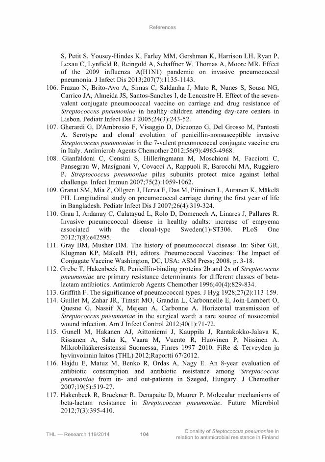
References
THL — Research 119/2014 104 Clonality of Steptococcus pneumoniae in relation to antimicrobial resistance in Finland
S, Petit S, Yousey-Hindes K, Farley MM, Gershman K, Harrison LH, Ryan P, Lexau C, Lynfield R, Reingold A, Schaffner W, Thomas A, Moore MR. Effect of the 2009 influenza A(H1N1) pandemic on invasive pneumococcal pneumonia. J Infect Dis 2013;207(7):1135-1143.
106. Frazao N, Brito-Avo A, Simas C, Saldanha J, Mato R, Nunes S, Sousa NG, Carrico JA, Almeida JS, Santos-Sanches I, de Lencastre H. Effect of the seven-valent conjugate pneumococcal vaccine on carriage and drug resistance of Streptococcus pneumoniae in healthy children attending day-care centers in Lisbon. Pediatr Infect Dis J 2005;24(3):243-52.
107. Gherardi G, D'Ambrosio F, Visaggio D, Dicuonzo G, Del Grosso M, Pantosti A. Serotype and clonal evolution of penicillin-nonsusceptible invasive Streptococcus pneumoniae in the 7-valent pneumococcal conjugate vaccine era in Italy. Antimicrob Agents Chemother 2012;56(9):4965-4968.
108. Gianfaldoni C, Censini S, Hilleringmann M, Moschioni M, Facciotti C, Pansegrau W, Masignani V, Covacci A, Rappuoli R, Barocchi MA, Ruggiero P. Streptococcus pneumoniae pilus subunits protect mice against lethal challenge. Infect Immun 2007;75(2):1059-1062.
109. Granat SM, Mia Z, Ollgren J, Herva E, Das M, Piirainen L, Auranen K, Mäkelä PH. Longitudinal study on pneumococcal carriage during the first year of life in Bangladesh. Pediatr Infect Dis J 2007;26(4):319-324.
110. Grau I, Ardanuy C, Calatayud L, Rolo D, Domenech A, Linares J, Pallares R. Invasive pneumococcal disease in healthy adults: increase of empyema associated with the clonal-type Sweden(1)-ST306. PLoS One 2012;7(8):e42595.
111. Gray BM, Musher DM. The history of pneumococcal disease. In: Siber GR, Klugman KP, Mäkelä PH, editors. Pneumococcal Vaccines: The Impact of Conjugate Vaccine Washington, DC, USA: ASM Press; 2008. p. 3-18.
112. Grebe T, Hakenbeck R. Penicillin-binding proteins 2b and 2x of Streptococcus pneumoniae are primary resistance determinants for different classes of beta-lactam antibiotics. Antimicrob Agents Chemother 1996;40(4):829-834.
113. Griffith F. The significance of pneumococcal types. J Hyg 1928;27(2):113-159. 114. Guillet M, Zahar JR, Timsit MO, Grandin L, Carbonnelle E, Join-Lambert O,
Quesne G, Nassif X, Mejean A, Carbonne A. Horizontal transmission of Streptococcus pneumoniae in the surgical ward: a rare source of nosocomial wound infection. Am J Infect Control 2012;40(1):71-72.
115. Gunell M, Hakanen AJ, Aittoniemi J, Kauppila J, Rantakokko-Jalava K, Rissanen A, Saha K, Vaara M, Vuento R, Huovinen P, Nissinen A. Mikrobilääkeresistenssi Suomessa, Finres 1997–2010. FiRe & Terveyden ja hyvinvoinnin laitos (THL) 2012;Raportti 67/2012.
116. Hajdu E, Matuz M, Benko R, Ordas A, Nagy E. An 8-year evaluation of antibiotic consumption and antibiotic resistance among Streptococcus pneumoniae from in- and out-patients in Szeged, Hungary. J Chemother 2007;19(5):519-27.
117. Hakenbeck R, Bruckner R, Denapaite D, Maurer P. Molecular mechanisms of beta-lactam resistance in Streptococcus pneumoniae. Future Microbiol 2012;7(3):395-410.

References
THL — Research 119/2014 105 Clonality of Steptococcus pneumoniae in relation to antimicrobial resistance in Finland
118. Hammerschmidt S. Pneumococcal virulence factors and adhesion proteins targeting the host. In: Hakenbeck R, Chhatwal S, editors. Molecular Biology of Streptococci Wymondham, UK: Horizon Biocience; 2007. p. 141-203.
119. Hanage WP. Fuzzy species revisited. BMC Biol 2013;11:41-7007-11-41. 120. Hanage WP, Huang SS, Lipsitch M, Bishop CJ, Godoy D, Pelton SI, Goldstein
R, Huot H, Finkelstein JA. Diversity and antibiotic resistance among nonvaccine serotypes of Streptococcus pneumoniae carriage isolates in the post-heptavalent conjugate vaccine era. J Infect Dis 2007;195(3):347-352.
121. Hanage WP, Kaijalainen TH, Syrjanen RK, Auranen K, Leinonen M, Makela PH, Spratt BG. Invasiveness of serotypes and clones of Streptococcus pneumoniae among children in Finland. Infect Immun 2005;73(1):431-435.
122. Harboe ZB, Benfield TL, Valentiner-Branth P, Hjuler T, Lambertsen L, Kaltoft M, Krogfelt K, Slotved HC, Christensen JJ, Konradsen HB. Temporal trends in invasive pneumococcal disease and pneumococcal serotypes over 7 decades. Clin Infect Dis 2010;50(3):329-337.
123. Hare KM, Smith-Vaughan H, Binks M, Park IH, Nahm MH, Leach AJ. "Dodgy 6As": differentiating pneumococcal serotype 6C from 6A by use of the Quellung reaction. J Clin Microbiol 2009;47(6):1981-1982.
124. Harrison OB, Brueggemann AB, Caugant DA, van der Ende A, Frosch M, Gray S, Heuberger S, Krizova P, Olcen P, Slack M, Taha MK, Maiden MC. Molecular typing methods for outbreak detection and surveillance of invasive disease caused by Neisseria meningitidis, Haemophilus influenzae and Streptococcus pneumoniae, a review. Microbiology 2011;157(Pt 8):2181-2195.
125. Hathaway LJ, Brugger SD, Morand B, Bangert M, Rotzetter JU, Hauser C, Graber WA, Gore S, Kadioglu A, Muhlemann K. Capsule type of Streptococcus pneumoniae determines growth phenotype. PLoS Pathog 2012;8(3):e1002574.
126. Hausdorff WP, Bryant J, Kloek C, Paradiso PR, Siber GR. The contribution of specific pneumococcal serogroups to different disease manifestations: implications for conjugate vaccine formulation and use, part II. Clin Infect Dis 2000;30(1):122-140.
127. Hausdorff WP, Bryant J, Paradiso PR, Siber GR. Which pneumococcal serogroups cause the most invasive disease: implications for conjugate vaccine formulation and use, part I. Clin Infect Dis 2000;30(1):100-121.
128. Hauser C, Aebi S, Muhlemann K. An internationally spread clone of Streptococcus pneumoniae evolves from low-level to higher-level penicillin resistance by uptake of penicillin-binding protein gene fragments from nonencapsulated pneumococci. Antimicrob Agents Chemother 2004;48(9):3563-3566.
129. Håvarstein LS. Increasing competence in the genus Streptococcus. Mol Microbiol 2010;78(3):541-544.
130. Heidelberger M, Rebers PA. Immunochemistry of the pneumococcal types II. V. and VI. I. The relation of Type VI to Type II and other correlations between chemical constitution and precipitation in antisera to type VI. J Bacteriol 1960;80:145-153.

References
THL — Research 119/2014 106 Clonality of Steptococcus pneumoniae in relation to antimicrobial resistance in Finland
131. Henrichsen J. Typing of Streptococcus pneumoniae: past, present, and future. Am J Med 1999;107(1A):50S-54S.
132. Henrichsen J. Six newly recognized types of Streptococcus pneumoniae. J. Clin. Microbiol. 1995;33(10):2759-2762.
133. Henrichsen J, Berntsson E, Kaijser B. Comparison of counterimmunoelectrophoresis and the capsular reaction test for typing of pneumococci. J Clin Microbiol 1980;11(6):589-92.
134. Henriques-Normark B, Tuomanen EI. The pneumococcus: epidemiology, microbiology, and pathogenesis. Cold Spring Harb Perspect Med 2013;3(7):10.1101/cshperspect.a010215.
135. Henriques-Normark B, Normark S. Commensal pathogens, with a focus on Streptococcus pneumoniae, and interactions with the human host. Exp Cell Res 2010;316(8):1408-1414.
136. Henriques-Normark B, Blomberg C, Dagerhamn J, Battig P, Normark S. The rise and fall of bacterial clones: Streptococcus pneumoniae. Nat Rev Microbiol 2008;6(11):827-837.
137. Henriques-Normark B. Molecular epidemiology and mechanisms for antibiotic resistance in Streptococcus pneumoniae. In: Hakenbeck R, Chhatwal S, editors. Molecular Biology of Streptococci Wymondham, UK: Horizon Biocience; 2007. p. 269-290.
138. Hicks LA, Harrison LH, Flannery B, Hadler JL, Schaffner W, Craig AS, Jackson D, Thomas A, Beall B, Lynfield R, Reingold A, Farley MM, Whitney CG. Incidence of pneumococcal disease due to non-pneumococcal conjugate vaccine (PCV7) serotypes in the United States during the era of widespread PCV7 vaccination, 1998-2004. J Infect Dis 2007;196(9):1346-54.
139. Hiller NL, Eutsey RA, Powell E, Earl JP, Janto B, Martin DP, Dawid S, Ahmed A, Longwell MJ, Dahlgren ME, Ezzo S, Tettelin H, Daugherty SC, Mitchell TJ, Hillman TA, Buchinsky FJ, Tomasz A, de Lencastre H, Sa-Leao R, Post JC, Hu FZ, Ehrlich GD. Differences in genotype and virulence among four multidrug-resistant Streptococcus pneumoniae isolates belonging to the PMEN1 clone. PLoS One 2011;6(12):e28850.
140. Hiller NL, Janto B, Hogg JS, Boissy R, Yu S, Powell E, Keefe R, Ehrlich NE, Shen K, Hayes J, Barbadora K, Klimke W, Dernovoy D, Tatusova T, Parkhill J, Bentley SD, Post JC, Ehrlich GD, Hu FZ. Comparative genomic analyses of seventeen Streptococcus pneumoniae strains: insights into the pneumococcal supragenome. J Bacteriol 2007;189(22):8186-8195.
141. Hollingshead SK, Becker R, Briles DE. Diversity of PspA: mosaic genes and evidence for past recombination in Streptococcus pneumoniae. Infect Immun 2000;68(10):5889-5900.
142. Hoskins J, Alborn WE,Jr, Arnold J, Blaszczak LC, Burgett S, DeHoff BS, Estrem ST, Fritz L, Fu DJ, Fuller W, Geringer C, Gilmour R, Glass JS, Khoja H, Kraft AR, Lagace RE, LeBlanc DJ, Lee LN, Lefkowitz EJ, Lu J, Matsushima P, McAhren SM, McHenney M, McLeaster K, Mundy CW, Nicas TI, Norris FH, O'Gara M, Peery RB, Robertson GT, Rockey P, Sun PM, Winkler ME, Yang Y, Young-Bellido M, Zhao G, Zook CA, Baltz RH,

References
THL — Research 119/2014 107 Clonality of Steptococcus pneumoniae in relation to antimicrobial resistance in Finland
Jaskunas SR, Rosteck PR,Jr, Skatrud PL, Glass JI. Genome of the bacterium Streptococcus pneumoniae strain R6. J Bacteriol 2001;183(19):5709-5717.
143. Hoti F, Erästö P, Leino T, Auranen K. Outbreaks of Streptococcus pneumoniae carriage in day care cohorts in Finland - implications for elimination of transmission. BMC Infect Dis 2009;9:102-2334-9-102.
144. Hsieh YC, Lin TL, Chang KY, Huang YC, Chen CJ, Lin TY, Wang JT. Expansion and evolution of Streptococcus pneumoniae serotype 19A ST320 clone as compared to its ancestral clone, Taiwan19F-14 (ST236). J Infect Dis 2013;208(2):203-210.
145. Hsu K, Pelton S, Karumuri S, Heisey-Grove D, Klein J, Massachusetts Department of Public Health Epidemiologists. Population-based surveillance for childhood invasive pneumococcal disease in the era of conjugate vaccine. Pediatr Infect Dis J 2005;24(1):17-23.
146. Hsu KK, Shea KM, Stevenson AE, Pelton SI, Massachusetts Department of Public Health. Changing serotypes causing childhood invasive pneumococcal disease: Massachusetts, 2001-2007. Pediatr Infect Dis J 2010;29(4):289-293.
147. Hu FZ, Eutsey R, Ahmed A, Frazao N, Powell E, Hiller NL, Hillman T, Buchinsky FJ, Boissy R, Janto B, Kress-Bennett J, Longwell M, Ezzo S, Post JC, Nesin M, Tomasz A, Ehrlich GD. In vivo capsular switch in Streptococcus pneumoniae -- analysis by whole genome sequencing. PLoS One 2012;7(11):e47983.
148. Huang SS, Hinrichsen VL, Stevenson AE, Rifas-Shiman SL, Kleinman K, Pelton SI, Lipsitch M, Hanage WP, Lee GM, Finkelstein JA. Continued impact of pneumococcal conjugate vaccine on carriage in young children. Pediatrics 2009;124(1):e1-11.
149. Huang SS, Platt R, Rifas-Shiman SL, Pelton SI, Goldmann D, Finkelstein JA. Post-PCV7 changes in colonizing pneumococcal serotypes in 16 Massachusetts communities, 2001 and 2004. Pediatrics 2005;116(3):e408-13.
150. Ingels H, Rasmussen J, Andersen PH, Harboe ZB, Glismann S, Konradsen H, Hoffmann S, Valentiner-Branth P, Lambertsen L, Danish Pneumococcal Surveillance Collaboration Group 2009-2010. Impact of pneumococcal vaccination in Denmark during the first 3 years after PCV introduction in the childhood immunization programme. Vaccine 2012;30(26):3944-3950.
151. Ip M, Chau SS, Lai LS, Ma H, Chan PK, Nelson EA. Increased nasopharyngeal carriage of serotypes 6A, 6C, and 6D Streptococcus pneumoniae after introduction of childhood pneumococcal vaccination in Hong Kong. Diagn Microbiol Infect Dis 2013;76(2):153-157.
152. Isaacman DJ, McIntosh ED, Reinert RR. Burden of invasive pneumococcal disease and serotype distribution among Streptococcus pneumoniae isolates in young children in Europe: impact of the 7-valent pneumococcal conjugate vaccine and considerations for future conjugate vaccines. Int J Infect Dis 2010;14(3):e197-209.
153. Jefferies JM, Clarke SC, Webb JS, Kraaijeveld AR. Risk of Red Queen dynamics in pneumococcal vaccine strategy. Trends Microbiol 2011;19(8):377-81.

References
THL — Research 119/2014 108 Clonality of Steptococcus pneumoniae in relation to antimicrobial resistance in Finland
154. Jefferies JM, Smith A, Clarke SC, Dowson C, Mitchell TJ. Genetic analysis of diverse disease-causing pneumococci indicates high levels of diversity within serotypes and capsule switching. J Clin Microbiol 2004;42(12):5681-5688.
155. Jenkins SG, Brown SD, Farrell DJ. Trends in antibacterial resistance among Streptococcus pneumoniae isolated in the USA: update from PROTEKT US Years 1-4. Ann Clin Microbiol Antimicrob 2008;7:1.
156. Jiang SM, Wang L, Reeves PR. Molecular characterization of Streptococcus pneumoniae type 4, 6B, 8, and 18C capsular polysaccharide gene clusters. Infect Immun 2001;69(3):1244-1255.
157. Jin P, Kong F, Xiao M, Oftadeh S, Zhou F, Liu C, Russell F, Gilbert G. First report of putative Streptococcus pneumoniae serotype 6D among nasopharyngeal isolates from Fijian children. J Infect Dis 2009;200(9):1375-1380.
158. Johnson HL, Deloria-Knoll M, Levine OS, Stoszek SK, Freimanis Hance L, Reithinger R, Muenz LR, O'Brien KL. Systematic evaluation of serotypes causing invasive pneumococcal disease among children under five: the pneumococcal global serotype project. PLoS Med 2010;7(10):10.1371/journal.pmed.1000348.
159. Jourdain S, Dreze PA, Vandeven J, Verhaegen J, Van Melderen L, Smeesters PR. Sequential multiplex PCR assay for determining capsular serotypes of colonizing S. pneumoniae. BMC Infect Dis 2011;11:100.
160. Kadioglu A, Weiser JN, Paton JC, Andrew PW. The role of Streptococcus pneumoniae virulence factors in host respiratory colonization and disease. Nat Rev Microbiol 2008;6(4):288-301.
161. Kadioglu A, Taylor S, Iannelli F, Pozzi G, Mitchell TJ, Andrew PW. Upper and lower respiratory tract infection by Streptococcus pneumoniae is affected by pneumolysin deficiency and differences in capsule type. Infect Immun 2002;70(6):2886-2890.
162. Karnezis TT, Smith A, Whittier S, Haddad J, Saiman L. Antimicrobial resistance among isolates causing invasive pneumococcal disease before and after licensure of heptavalent conjugate pneumococcal vaccine. PLoS One 2009;4(6):e5965.
163. Klemets P, Lyytikäinen O, Ruutu P, Kaijalainen T, Leinonen M, Ollgren J, Nuorti J. Trends and geographical variation in invasive pneumococcal infections in Finland. Scand J Infect Dis 2008;40(8):621-8.
164. Ko KS, Baek JY, Song JH. Capsular gene sequences and genotypes of 'serotype 6E' Streptococcus pneumoniae isolates. J Clin Microbiol 2013.
165. Ko KS, Baek JY, Song JH. Multidrug-resistant Streptococcus pneumoniae serotype 6D clones in South Korea. J Clin Microbiol 2012;50(3):818-822.
166. Koeck JL, Njanpop-Lafourcade BM, Cade S, Varon E, Sangare L, Valjevac S, Vergnaud G, Pourcel C. Evaluation and selection of tandem repeat loci for Streptococcus pneumoniae MLVA strain typing. BMC Microbiol 2005;5:66.
167. Kong F, Brown M, Sabananthan A, Zeng X, Gilbert GL. Multiplex PCR-based reverse line blot hybridization assay to identify 23 Streptococcus pneumoniae polysaccharide vaccine serotypes. J Clin Microbiol 2006;44(5):1887-91.

References
THL — Research 119/2014 109 Clonality of Steptococcus pneumoniae in relation to antimicrobial resistance in Finland
168. Kong F, Wang W, Tao J, Wang L, Wang Q, Sabananthan A, Gilbert GL. A molecular-capsular-type prediction system for 90 Streptococcus pneumoniae serotypes using partial cpsA-cpsB sequencing and wzy- or wzx-specific PCR. J Med Microbiol 2005;54(Pt 4):351-6.
169. Kong F, Gilbert GL. Using cpsA-cpsB sequence polymorphisms and serotype-/group-specific PCR to predict 51 Streptococcus pneumoniae capsular serotypes. J Med Microbiol 2003;52(Pt 12):1047-1058.
170. Konradsen HB. Validation of serotyping of Streptococcus pneumoniae in Europe. Vaccine 2005;23(11):1368-73.
171. Korppi M, Karppa H, Vuento R. Universal pneumococcal vaccination of <2-year-olds decreased pneumococcal bacteremia and antibiotic resistance in <5-year-olds. Acta Paediatr 2013;102(6):e239-40.
172. Kristinsson KG. Epidemiology of penicillin resistant pneumococci in Iceland. Microb Drug Resist 1995;1(2):121-125.
173. Kuch A, Sadowy E, Skoczynska A, Hryniewicz W. First report of Streptococcus pneumoniae serotype 6D isolates from invasive infections. Vaccine 2010;28(39):6406-6407.
174. Lahti E, Peltola V, Virkki R, Alanen M, Ruuskanen O. Development of parapneumonic empyema in children. Acta Paediatr 2007;96(11):1686-1692.
175. Lambertsen LM, Harboe ZB, Konradsen HB, Christensen JJ, Hammerum AM. Non-invasive erythromycin-resistant pneumococcal isolates are more often non-susceptible to more antimicrobial agents than invasive isolates. Int J Antimicrob Agents 2010;35(1):72-75.
176. Laupland KB, Gregson DB, Flemons WW, Hawkins D, Ross T, Church DL. Burden of community-onset bloodstream infection: a population-based assessment. Epidemiol Infect 2007;135(6):1037-1042.
177. Lawrence ER, Griffiths DB, Martin SA, George RC, Hall LM. Evaluation of semiautomated multiplex PCR assay for determination of Streptococcus pneumoniae serotypes and serogroups. J Clin Microbiol 2003;41(2):601-607.
178. Leimkugel J, Adams Forgor A, Gagneux S, Pfluger V, Flierl C, Awine E, Naegeli M, Dangy JP, Smith T, Hodgson A, Pluschke G. An outbreak of serotype 1 Streptococcus pneumoniae meningitis in northern Ghana with features that are characteristic of Neisseria meningitidis meningitis epidemics. J Infect Dis 2005;192(2):192-199.
179. Leinonen MK. Detection of pneumococcal capsular polysaccharide antigens by latex agglutination, counterimmunoelectrophoresis, and radioimmunoassay in middle ear exudates in acute otitis media. J Clin Microbiol 1980;11(2):135-40.
180. Leung MH, Bryson K, Freystatter K, Pichon B, Edwards G, Charalambous BM, Gillespie SH. Sequetyping: serotyping Streptococcus pneumoniae by a single PCR sequencing strategy. J Clin Microbiol 2012;50(7):2419-2427.
181. Levy C, Varon E, Bingen E, Lecuyer A, Boucherat M, Cohen R, Bacterial Meningitis Study Group. Pneumococcal meningitis in french children before and after the introduction of pneumococcal conjugate vaccine. Pediatr Infect Dis J 2011;30(2):168-170.
182. Lin J, Kaltoft MS, Brandao AP, Echaniz-Aviles G, Brandileone MC, Hollingshead SK, Benjamin WH, Nahm MH. Validation of a multiplex

References
THL — Research 119/2014 110 Clonality of Steptococcus pneumoniae in relation to antimicrobial resistance in Finland
pneumococcal serotyping assay with clinical samples. J Clin Microbiol 2006;44(2):383-388.
183. Linares J, Ardanuy C, Pallares R, Fenoll A. Changes in antimicrobial resistance, serotypes and genotypes in Streptococcus pneumoniae over a 30-year period. Clin Microbiol Infect 2010;16(5):402-410.
184. Lipsitch M, O'Neill K, Cordy D, Bugalter B, Trzcinski K, Thompson CM, Goldstein R, Pelton S, Huot H, Bouchet V, Reid R, Santosham M, O'Brien KL. Strain characteristics of Streptococcus pneumoniae carriage and invasive disease isolates during a cluster-randomized clinical trial of the 7-valent pneumococcal conjugate vaccine. J Infect Dis 2007;196(8):1221-7.
185. Liu Z, Nachamkin I, Edelstein PH, Lautenbach E, Metlay JP. Serotype emergence and genotype distribution among macrolide-resistant invasive Streptococcus pneumoniae isolates in the postconjugate vaccine (PCV-7) era. Antimicrob Agents Chemother 2012;56(2):743-750.
186. Llull D, Munoz R, Lopez R, Garcia E. A single gene (tts) located outside the cap locus directs the formation of Streptococcus pneumoniae type 37 capsular polysaccharide. Type 37 pneumococci are natural, genetically binary strains. J Exp Med 1999;190(2):241-251.
187. Loman NJ, Gladstone RA, Constantinidou C, Tocheva AS, Jefferies JM, Faust SN, O'Connor L, Chan J, Pallen MJ, Clarke SC. Clonal expansion within pneumococcal serotype 6C after use of seven-valent vaccine. PLoS One 2013;8(5):e64731.
188. Lund E. Laboratory diagnosis of Pneumococcus infections. Bull World Health Organ 1960;23:5-13.
189. Lynch JP,3rd, Zhanel GG. Streptococcus pneumoniae: epidemiology and risk factors, evolution of antimicrobial resistance, and impact of vaccines. Curr Opin Pulm Med 2010;16(3):217-225.
190. Lynch JP,3rd, Zhanel GG. Streptococcus pneumoniae: does antimicrobial resistance matter? Semin Respir Crit Care Med 2009;30(2):210-238.
191. Lythgoe KA, Read AF. Catching the Red Queen? The advice of the Rose. Trends Ecol Evol 1998;13(12):473-474.
192. MacLeod CM, Kraus MR. Relation of virulence of pneumococcal strains for mice to the quantity of capsular polysaccharide formed in vitro. J Exp Med 1950;92(1):1-9.
193. Malley R, Anderson PW. Serotype-independent pneumococcal experimental vaccines that induce cellular as well as humoral immunity. Proc Natl Acad Sci U S A 2012;109(10):3623-3627.
194. Marchese A, Esposito S, Barbieri R, Bassetti M, Debbia E. Does the adoption of EUCAST susceptibility breakpoints affect the selection of antimicrobials to treat acute community-acquired respiratory tract infections? BMC Infect Dis 2012;12:181.
195. Marks LR, Parameswaran GI, Hakansson AP. Pneumococcal interactions with epithelial cells are crucial for optimal biofilm formation and colonization in vitro and in vivo. Infect Immun 2012;80(8):2744-2760.

References
THL — Research 119/2014 111 Clonality of Steptococcus pneumoniae in relation to antimicrobial resistance in Finland
196. Marks LR, Reddinger RM, Hakansson AP. High levels of genetic recombination during nasopharyngeal carriage and biofilm formation in Streptococcus pneumoniae. MBio 2012;3(5):e00200-12.
197. Marsh R, Smith-Vaughan H, Hare KM, Binks M, Kong F, Warning J, Gilbert GL, Morris P, Leach AJ. The nonserotypeable pneumococcus: phenotypic dynamics in the era of anticapsular vaccines. J Clin Microbiol 2010;48(3):831-835.
198. Martner A, Skovbjerg S, Paton JC, Wold AE. Streptococcus pneumoniae autolysis prevents phagocytosis and production of phagocyte-activating cytokines. Infect Immun 2009;77(9):3826-3837.
199. Massire C, Gertz RE,Jr, Svoboda P, Levert K, Reed MS, Pohl J, Kreft R, Li F, White N, Ranken R, Blyn LB, Ecker DJ, Sampath R, Beall B. Concurrent serotyping and genotyping of pneumococci by use of PCR and electrospray ionization mass spectrometry. J Clin Microbiol 2012;50(6):2018-2025.
200. Mavroidi A, Godoy D, Aanensen DM, Robinson DA, Hollingshead SK, Spratt BG. Evolutionary genetics of the capsular locus of serogroup 6 pneumococci 1. J Bacteriol 2004;186:8181-8192.
201. McCarty M. Discovering genes are made of DNA. Nature 2003;421(6921):406. 202. McEllistrem MC, Nahm MH. Novel pneumococcal serotypes 6C and 6D:
anomaly or harbinger. Clin Infect Dis 2012;55(10):1379-1386. 203. McEllistrem MC. Genetic diversity of the pneumococcal capsule: implications
for molecular-based serotyping. Future Microbiol 2009;4(7):857-865. 204. McGee L, McDougal L, Zhou J, Spratt BG, Tenover FC, George R, Hakenbeck
R, Hryniewicz W, Lefevre JC, Tomasz A, Klugman KP. Nomenclature of major antimicrobial-resistant clones of Streptococcus pneumoniae defined by the Pneumococcal Molecular Epidemiology Network. J. Clin. Microbiol. 2001;39(7):2565-2571.
205. Melin M, Trzcinski K, Meri S, Käyhty H, Väkeväinen M. The capsular serotype of Streptococcus pneumoniae is more important than the genetic background for resistance to complement. Infect Immun 2010;78(12):5262-5270.
206. Mera RM, Miller LA, Amrine-Madsen H, Sahm DF. The impact of the pneumococcal conjugate vaccine on antimicrobial resistance in the United States since 1996: evidence for a significant rebound by 2007 in many classes of antibiotics. Microb Drug Resist 2009;15(4):261-268.
207. Mera RM, Miller LA, Daniels JJ, Weil JG, White AR. Increasing prevalence of multidrug-resistant Streptococcus pneumoniae in the United States over a 10-year period: Alexander Project. Diagn Microbiol Infect Dis 2005;51(3):195-200.
208. Mercado E, Srinivasan V, Hawkins P, Chochua S, Ochoa T, Beall B, McGee L. First report of Streptococcus pneumoniae serotype 6D in South America. J Clin Microbiol 2011;49(5):2080-2081.
209. Messina AF, Katz-Gaynor K, Barton T, Ahmad N, Ghaffar F, Rasko D, H. MG,Jr. Impact of the pneumococcal conjugate vaccine on serotype distribution and antimicrobial resistance of invasive Streptococcus pneumoniae isolates in

References
THL — Research 119/2014 112 Clonality of Steptococcus pneumoniae in relation to antimicrobial resistance in Finland
Dallas, TX, children from 1999 through 2005. Pediatr Infect Dis J 2007;26(6):461-7.
210. Miernyk K, Debyle C, Harker-Jones M, Hummel KB, Hennessy T, Wenger J, Rudolph K. Serotyping of Streptococcus pneumoniae isolates from nasopharyngeal samples: use of an algorithm combining microbiologic, serologic, and sequential multiplex PCR techniques. J Clin Microbiol 2011;49(9):3209-14.
211. Millar EV, Pimenta FC, Roundtree A, Jackson D, Carvalho Mda G, Perilla MJ, Reid R, Santosham M, Whitney CG, Beall BW, O'Brien KL. Pre- and post-conjugate vaccine epidemiology of pneumococcal serotype 6C invasive disease and carriage within Navajo and White Mountain Apache communities. Clin Infect Dis 2010;51(11):1258-1265.
212. Miller E, Andrews NJ, Waight PA, Slack MP, George RC. Herd immunity and serotype replacement 4 years after seven-valent pneumococcal conjugate vaccination in England and Wales: an observational cohort study. Lancet Infect Dis 2011;11(10):760-768.
213. Mizrachi Nebenzahl Y, Porat N, Lifshitz S, Novick S, Levi A, Ling E, Liron O, Mordechai S, Sahu RK, Dagan R. Virulence of Streptococcus pneumoniae may be determined independently of capsular polysaccharide. FEMS Microbiol Lett 2004;233(1):147-152.
214. MLVA Streptococcus pneumoniae. MLVA.eu. Available at: http://www.mlva.eu/pdf/FicheTechnique_v3.pdf. Accessed 15th September, 2013.
215. Moberley S, Holden J, Tatham DP, Andrews RM. Vaccines for preventing pneumococcal infection in adults. Cochrane Database Syst Rev 2013;1:CD000422.
216. Moffitt KL, Malley R. Next generation pneumococcal vaccines. Curr Opin Immunol 2011;23(3):407-413.
217. Montagnani F, Stolzuoli L, Zanchi A, Cresti S, Cellesi C. Antimicrobial susceptibility of Streptococcus pyogenes and Streptococcus pneumoniae: surveillance from 1993 to 2004 in Central Italy. J Chemother 2006;18(4):389-93.
218. Morais L, Carvalho MdG, Roca A, Flannery B, Mandomando I, Soriano-Gabarro M, Sigauque B, Alonso P, Beall B. Sequential multiplex PCR for identifying pneumococcal capsular serotypes from south-Saharan African clinical isolates. J Med Microbiol 2007;56(9):1181-1184.
219. Morand B, Muhlemann K. Heteroresistance to penicillin in Streptococcus pneumoniae. Proc Natl Acad Sci U S A 2007;104(35):14098-14103.
220. Morona JK, Morona R, Paton JC. Comparative genetics of capsular polysaccharide biosynthesis in Streptococcus pneumoniae types belonging to serogroup 19. J Bacteriol 1999;181(17):5355-64.
221. Moschioni M, Lo Sapio M, Crisafulli G, Torricelli G, Guidotti S, Muzzi A, Barocchi MA, Donati C. Sequence analysis of 96 genomic regions identifies distinct evolutionary lineages within CC156, the largest Streptococcus pneumoniae clonal complex in the MLST database. PLoS One 2013;8(4):e61003.
References
THL — Research 119/2014 113 Clonality of Steptococcus pneumoniae in relation to antimicrobial resistance in Finland
222. Moschioni M, De Angelis G, Melchiorre S, Masignani V, Leibovitz E, Barocchi MA, Dagan R. Prevalence of pilus-encoding islets among acute otitis media Streptococcus pneumoniae isolates from Israel. Clin Microbiol Infect 2010;16(9):1501-1504.
223. Moschioni M, Donati C, Muzzi A, Masignani V, Censini S, Hanage WP, Bishop CJ, Reis JN, Normark S, Henriques-Normark B, Covacci A, Rappuoli R, Barocchi MA. Streptococcus pneumoniae contains 3 rlrA pilus variants that are clonally related. JID 2008;197.
224. Mudany MA, Kikuchi K, Totsuka K, Uchiyama T. Evaluation of a new serotyping kit for Streptococcus pneumoniae. J Med Microbiol 2003;52(Pt 11):975-980.
225. Mueller JE, Yaro S, Ouedraogo MS, Levina N, Njanpop-Lafourcade BM, Tall H, Idohou RS, Sanou O, Kroman SS, Drabo A, Nacro B, Millogo A, van der Linden M, Gessner BD. Pneumococci in the African meningitis belt: meningitis incidence and carriage prevalence in children and adults. PLoS One 2012;7(12):e52464.
226. Mullany P, Allan E, Warburton PJ. Tetracycline resistance genes and mobile genetic elements from the oral metagenome. Clin Microbiol Infect 2012;18 Suppl 4:58-61.
227. Multi locus sequence typing (MLST) of Streptococcus pneumoniae. 2013; Available at: http://spneumoniae.mlst.net/. Accessed 15th September, 2013.
228. Munoz-Almagro C, Jordan I, Gene A, Latorre C, Garcia-Garcia JJ, Pallares R. Emergence of invasive pneumococcal disease caused by nonvaccine serotypes in the era of 7-valent conjugate vaccine. Clin Infect Dis 2008;46(2):174-82.
229. Nahm MH, Oliver MB, Siira L, Kaijalainen T, Lambertsen LM, Virolainen A. A report of Streptococcus pneumoniae serotype 6D in Europe. J Med Microbiol 2011;60(1):46-48.
230. Nelson AL, Ries J, Bagnoli F, Dahlberg S, Falker S, Rounioja S, Tschop J, Morfeldt E, Ferlenghi I, Hilleringmann M, Holden DW, Rappuoli R, Normark S, Barocchi MA, Henriques-Normark B. RrgA is a pilus-associated adhesin in Streptococcus pneumoniae. Mol Microbiol 2007;66(2):329-340.
231. Nelson ML, Levy SB. The history of the tetracyclines. Ann N Y Acad Sci 2011;1241:17-32.
232. Nesin M, Ramirez M, Tomasz A. Capsular transformation of a multidrug-resistant Streptococcus pneumoniae in vivo. J Infect Dis 1998;177(3):707-713.
233. Nunes MC, Madhi SA. Review on the immunogenicity and safety of PCV-13 in infants and toddlers. Expert Rev Vaccines 2011;10(7):951-980.
234. Nunes S, Valente C, Sa-Leao R, de Lencastre H. Temporal trends and molecular epidemiology of recently described serotype 6C of Streptococcus pneumoniae. J Clin Microbiol 2009;47:472-474.
235. O'Brien KL, Moisi J, Moulton LH, Madore D, Eick A, Reid R, Weatherholtz R, Millar E, Hu D, Hackell J, Kohberger R, Siber G, Santosham M. Predictors of pneumococcal conjugate vaccine immunogenicity among infants and toddlers in an American Indian PnCRM7 efficacy trial. J Infect Dis 2007;196(1):104-114.

References
THL — Research 119/2014 114 Clonality of Steptococcus pneumoniae in relation to antimicrobial resistance in Finland
236. Oftadeh S, Satzke C, Gilbert GL. Identification of newly described Streptococcus pneumoniae serotype 6D by use of the Quellung reaction and PCR. J. Clin. Microbiol. 2010;48(9):3378-3379.
237. Oliver MB, van der Linden MP, Kuntzel SA, Saad JS, Nahm MH. Discovery of Streptococcus pneumoniae serotype 6 variants with glycosyltransferases synthesizing two differing repeating units. J Biol Chem 2013;288(36):25976-25985.
238. Orihuela CJ, Gao G, Francis KP, Yu J, Tuomanen EI. Tissue-specific contributions of pneumococcal virulence factors to pathogenesis. J Infect Dis 2004;190(9):1661-1669.
239. Orihuela CJ, Radin JN, Sublett JE, Gao G, Kaushal D, Tuomanen EI. Microarray analysis of pneumococcal gene expression during invasive disease. Infect Immun 2004;72(10):5582-5596.
240. Pai R, Gertz RE, Beall B. Sequential multiplex PCR approach for determining capsular serotypes of Streptococcus pneumoniae isolates. J. Clin. Microbiol. 2006;44(1):124-131.
241. Palmu AA, Jokinen J, Borys D, Nieminen H, Ruokokoski E, Siira L, Puumalainen T, Lommel P, Hezareh M, Moreira M, Schuerman L, Kilpi TM. Effectiveness of the ten-valent pneumococcal Haemophilus influenzae protein D conjugate vaccine (PHiD-CV10) against invasive pneumococcal disease: a cluster randomised trial. Lancet 2013;381(9862):214-222.
242. Park IH, Kim KH, Andrade AL, Briles DE, McDaniel LS, Nahm MH. Nontypeable pneumococci can be divided into multiple cps types, including one type expressing the novel gene pspK. MBio 2012;3(3):10.1128/mBio.00035.
243. Park IH, Park S, Hollingshead SK, Nahm MH. Genetic basis for the new pneumococcal serotype, 6C. Infect Immun 2007;75:4482-4489.
244. Park IH, Pritchard DG, Cartee R, Brandao A, Brandileone MC, Nahm MH. Discovery of a new capsular serotype (6C) within serogroup 6 of Streptococcus pneumoniae. J Clin Microbiol 2007;45:1225-1233.
245. Pebody RG, Morgan O, Choi Y, George R, Hussain M, Andrews N. Use of antibiotics and risk factors for carriage of Streptococcus pneumoniae: a longitudinal household study in the United Kingdom. Epidemiol Infect 2009;137(4):555-561.
246. Pebody RG, Hellenbrand W, D'Ancona F, Ruutu P, European Union funded Pnc-EURO contributing group. Pneumococcal disease surveillance in Europe. Euro Surveill 2006;11(9):171-178.
247. Peltola V, Heikkinen T, Ruuskanen O, Jartti T, Hovi T, Kilpi T, Vainionpaa R. Temporal association between rhinovirus circulation in the community and invasive pneumococcal disease in children. Pediatr Infect Dis J 2011;30(6):456-461.
248. Pericone CD, Overweg K, Hermans PW, Weiser JN. Inhibitory and bactericidal effects of hydrogen peroxide production by Streptococcus pneumoniae on other inhabitants of the upper respiratory tract. Infect Immun 2000;68(7):3990-3997.

References
THL — Research 119/2014 115 Clonality of Steptococcus pneumoniae in relation to antimicrobial resistance in Finland
249. Pichon B, Ladhani SN, Slack MP, Segonds-Pichon A, Andrews NJ, Waight PA, Miller E, George R. Changes in molecular epidemiology of Streptococcus pneumoniae causing meningitis following introduction of pneumococcal conjugate vaccination in England and Wales. J Clin Microbiol 2013;51(3):820-827.
250. Pichon B. Pneumococcal MLST oligonucleotide primer sequence, personal communication by email 24th April, 2006.
251. Pihlajamäki M, Jalava J, Huovinen P, Kotilainen P. Antimicrobial resistance of invasive pneumococci in Finland in 1999-2000. Antimicrob Agents Chemother 2003;47(6):1832-5.
252. Pihlajamäki M, Kaijalainen T, Huovinen P, Jalava J. Rapid increase in macrolide resistance among penicillin non-susceptible pneumococci in Finland, 1996-2000. J Antimicrob Chemother 2002;49(5):785-92.
253. Pihlajamäki M, Kataja J, Seppala H, Elliot J, Leinonen M, Huovinen P, Jalava J. Ribosomal mutations in Streptococcus pneumoniae clinical isolates. Antimicrob Agents Chemother 2002;46(3):654-8.
254. Pihlajamäki M, Kotilainen P, Kaurila T, Klaukka T, Palva E, Huovinen P, Finnish Study Group for Antimicrobial Resistance (FiRe-Network). Macrolide-resistant Streptococcus pneumoniae and use of antimicrobial agents. Clin Infect Dis 2001;33(4):483-488.
255. Pilishvili T, Lexau C, Farley M, Hadler J, Harrison L, Bennett N, Reingold A, Thomas A, Schaffner W, Craig A, Smith P, Beall B, Whitney C, Moore M. Sustained reductions in invasive pneumococcal disease in the era of conjugate vaccine. J Infect Dis 2010;201(1):32-41.
256. Pillai D, Shahinas D, Buzina A, Pollock R, Lau R, Khairnar K, Wong A, Farrell D, Green K, McGeer A, Low D. Genome-wide dissection of globally emergent multi-drug resistant serotype 19A Streptococcus pneumoniae. BMC Genomics 2009;10(1):642.
257. Pimenta FC, Roundtree A, Soysal A, Bakir M, du Plessis M, Wolter N, von Gottberg A, McGee L, Carvalho Mda G, Beall B. Sequential triplex real-time PCR assay for detecting 21 pneumococcal capsular serotypes that account for a high global disease burden. J Clin Microbiol 2013;51(2):647-652.
258. Pimenta FC, E. GR,Jr, Roundtree A, Yu J, Nahm MH, McDonald RR, Carvalho Mda G, Beall BW. Rarely occurring 19A-like cps locus from a serotype 19F pneumococcal isolate indicates continued need of serology-based quality control for PCR-based serotype determinations. J Clin Microbiol 2009;47(7):2353-4.
259. Platt S, Pichon B, George R, Green J. A bioinformatics pipeline for high-throughput microbial multilocus sequence typing (MLST) analyses. Clin Microbiol Infect 2006;12(11):1144-6.
260. Pletz MW, Maus U, Krug N, Welte T, Lode H. Pneumococcal vaccines: mechanism of action, impact on epidemiology and adaption of the species. Int J Antimicrob Agents 2008;32(3):199-206.
261. Pneumococcal Molecular Epidemiology Network (PMEN). Available at: http://www.sph.emory.edu/PMEN/. Accessed 11th September, 2013.

References
THL — Research 119/2014 116 Clonality of Steptococcus pneumoniae in relation to antimicrobial resistance in Finland
262. Porat N, Benisty R, Trefler R, Ozalvo D, Givon-Lavi N, Dagan R. Baseline epidemiology and genetic structure of Streptococcus pneumoniae serotype 6D in southern Israel prior to introduction of pneumococcal conjugate vaccines. J Clin Microbiol 2013;51(5):1580-1582.
263. Prudhomme M, Attaiech L, Sanchez G, Martin B, Claverys JP. Antibiotic stress induces genetic transformability in the human pathogen Streptococcus pneumoniae. Science 2006;313(5783):89-92.
264. Rakov AV, Ubukata K, Robinson DA. Population structure of hyperinvasive serotype 12F, clonal complex 218 Streptococcus pneumoniae revealed by multilocus boxB sequence typing. Infect Genet Evol 2011;11(8):1929-1939.
265. Rantala M. Antimicrobial resistance in Streptococcus pneumoniae in Finland with special reference to macrolides and telithromycin. PhD Thesis, University of Helsinki. 2009.
266. Rantala M, Huikko S, Huovinen P, Jalava J. Prevalence and molecular genetics of macrolide resistance among Streptococcus pneumoniae isolates collected in Finland in 2002. Antimicrob Agents Chemother 2005;49(10):4180-4.
267. Regev-Yochay G, Hanage WP, Trzcinski K, Rifas-Shiman SL, Lee G, Bessolo A, Huang SS, Pelton SI, McAdam AJ, Finkelstein JA, Lipsitch M, Malley R. Re-emergence of the type 1 pilus among Streptococcus pneumoniae isolates in Massachusetts, USA. Vaccine 2010;28(30):4842-4846.
268. Riedel S, Beekmann SE, Heilmann KP, Richter SS, Garcia-de-Lomas J, Ferech M, Goosens H, Doern GV. Antimicrobial use in Europe and antimicrobial resistance in Streptococcus pneumoniae. Eur J Clin Microbiol Infect Dis 2007;26(7):485-90.
269. Rinta-Kokko H, Jokinen J, Siira L, Palmu AA, Virtanen MJ, Nohynek H, Virolainen A, Nuorti JP. Impact of 10-valent pneumococcal conjugate vaccine (PCV10) against invasive pneumococcal disease (IPD) among vaccine-eligible children in Finland. 31st Meeting of the European Society for Paediatric Infectious Diseases (ESPID), Milan, Italy, 28 May - 1 June, 2013 2013.
270. Roberts J, Chandra M, Pebody R, Stuart J. Variation in incidence of pneumococcal and meningococcal disease across Europe. Euro Surveill 2007;12(11):E071115.5.
271. Ruckinger S, van der Linden M, Reinert RR, von Kries R, Burckhardt F, Siedler A. Reduction in the incidence of invasive pneumococcal disease after general vaccination with 7-valent pneumococcal conjugate vaccine in Germany. Vaccine 2009;27(31):4136-4141.
272. Sabat AJ, Budimir A, Nashev D, Sa-Leao R, van Dijl J, Laurent F, Grundmann H, Friedrich AW, ESCMID Study Group of Epidemiological Markers (ESGEM). Overview of molecular typing methods for outbreak detection and epidemiological surveillance. Euro Surveill 2013;18(4):20380.
273. Sadowy E, Kuch A, Gniadkowski M, Hryniewicz W. Expansion and evolution of the Streptococcus pneumoniae Spain9V-ST156 clonal complex in Poland. Antimicrob. Agents Chemother. 2010;54(5):1720-1727.
274. Sadowy E, Izdebski R, Skoczynska A, Grzesiowski P, Gniadkowski M, Hryniewicz W. Phenotypic and molecular analysis of penicillin-nonsusceptible
References
THL — Research 119/2014 117 Clonality of Steptococcus pneumoniae in relation to antimicrobial resistance in Finland
Streptococcus pneumoniae isolates in Poland. Antimicrob. Agents Chemother. 2007;51(1):40-47.
275. Salter SJ, Hinds J, Gould KA, Lambertsen L, Hanage WP, Antonio M, Turner P, Hermans PW, Bootsma HJ, O'Brien KL, Bentley SD. Variation at the capsule locus, cps, of mistyped and non-typable Streptococcus pneumoniae isolates. Microbiology 2012;158(Pt 6):1560-1569.
276. Satzke C, Ortika BD, Oftadeh S, Russell FM, Robins-Browne RM, Mulholland EK, Gilbert GL. Molecular epidemiology of Streptococcus pneumoniae serogroup 6 isolates from Fijian children, including newly identified serotypes 6C and 6D. J Clin Microbiol 2010;48(11):4298-300.
277. Scott JA, Hall AJ, Dagan R, Dixon JM, Eykyn SJ, Fenoll A, Hortal M, Jette LP, Jorgensen JH, Lamothe F, Latorre C, Macfarlane JT, Shlaes DM, Smart LE, Taunay A. Serogroup-specific epidemiology of Streptococcus pneumoniae: associations with age, sex, and geography in 7,000 episodes of invasive disease. Clin Infect Dis 1996;22(6):973-981.
278. Scott JR, Hinds J, Gould KA, Millar EV, Reid R, Santosham M, O'Brien KL, Hanage WP. Nontypeable pneumococcal isolates among navajo and white mountain apache communities: are these really a cause of invasive disease? J Infect Dis 2012;206(1):73-80.
279. Selva L, Ciruela P, Blanchette K, del Amo E, Pallares R, Orihuela CJ, Munoz-Almagro C. Prevalence and clonal distribution of pcpA, psrP and Pilus-1 among pediatric isolates of Streptococcus pneumoniae. PLoS One 2012;7(7):e41587.
280. Selva L, del Amo E, Brotons P, Munoz-Almagro C. Rapid and easy identification of capsular serotypes of Streptococcus pneumoniae by use of fragment analysis by automated fluorescence-based capillary electrophoresis. J Clin Microbiol 2012;50(11):3451-3457.
281. Shin J, Baek JY, Kim SH, Song JH, Ko KS. Predominance of ST320 among Streptococcus pneumoniae serotype 19A isolates from 10 Asian countries. J Antimicrob Chemother 2011;66(5):1001-1004.
282. Simoes AS, Pereira L, Nunes S, Brito-Avo A, de Lencastre H, Sa-Leao R. Clonal evolution leading to maintenance of antibiotic resistance rates among colonizing pneumococci in the PCV7 era in Portugal. J Clin Microbiol 2011;49(8):2810-2817.
283. Singleton R, Wenger J, Klejka JA, Bulkow LR, Thompson A, Sarkozy D, Emini EA, Gruber WC, Scott DA. The 13-valent pneumococcal conjugate vaccine for invasive pneumococcal disease in Alaska native children: results of a clinical trial. Pediatr Infect Dis J 2013;32(3):257-263.
284. Sjöström K, Blomberg C, Fernebro J, Dagerhamn J, Morfeldt E, Barocchi MA, Browall S, Moschioni M, Andersson M, Henriques F, Albiger B, Rappuoli R, Normark S, Henriques-Normark B. Clonal success of piliated penicillin nonsusceptible pneumococci. Proc Natl Acad Sci U S A 2007;104:12907-12912.
285. Sjöström K, Spindler C, Ortqvist A, Kalin M, Sandgren A, Kühlmann-Berenzon S, Henriques-Normark B. Clonal and capsular types decide whether

References
THL — Research 119/2014 118 Clonality of Steptococcus pneumoniae in relation to antimicrobial resistance in Finland
pneumococci will act as a primary or opportunistic pathogen. Clinical Infectious Diseases 2006;42(4):451-459.
286. Skogberg K, Lyytikäinen O, Ollgren J, Nuorti JP, Ruutu P. Population-based burden of bloodstream infections in Finland. Clin Microbiol Infect 2012;18(6):E170-6.
287. Skogberg K, Lyytikäinen O, Ruutu P, Ollgren J, Nuorti JP. Increase in bloodstream infections in Finland, 1995-2002. Epidemiol Infect 2008;136(1):108-114.
288. Skov Sorensen UB, Blom J, Birch-Andersen A, Henrichsen J. Ultrastructural localization of capsules, cell wall polysaccharide, cell wall proteins, and F antigen in pneumococci. Infect Immun 1988;56(8):1890-1896.
289. Smith AM, Klugman KP. Alterations in PBP 1A essential-for high-level penicillin resistance in Streptococcus pneumoniae. Antimicrob Agents Chemother 1998;42(6):1329-1333.
290. Sogstad MKR, Hoiby EA, Caugant DA. Molecular characterization of non-penicillin-susceptible Streptococcus pneumoniae in Norway. J. Clin. Microbiol. 2006;44(9):3225-3230.
291. Song JH, Dagan R, Klugman KP, Fritzell B. The relationship between pneumococcal serotypes and antibiotic resistance. Vaccine 2012;30(17):2728-2737.
292. Song JH, Baek JY, Ko KS. Comparison of capsular genes of Streptococcus pneumoniae serotype 6A, 6B, 6C, and 6D isolates. J Clin Microbiol 2011;49(5):1758-1764.
293. Spratt BG, Hanage WP, Li B, Aanensen DM, Feil EJ. Displaying the relatedness among isolates of bacterial species -- the eBURST approach. FEMS Microbiol Lett 2004;241:129-134.
294. Spratt BG, Greenwood BM. Prevention of pneumococcal disease by vaccination: does serotype replacement matter? Lancet 2000;356(9237):1210-1211.
295. Steenhoff AP, Shah SS, Ratner AJ, Patil SM, McGowan KL. Emergence of vaccine-related pneumococcal serotypes as a cause of bacteremia. Clin Infect Dis 2006;42(7):907-14.
296. Steinman RM, Moberg CL. A triple tribute to the experiment that transformed biology. J Exp Med 1994;179(2):379-384.
297. Syrjänen RK, Kilpi TM, Kaijalainen TH, Herva EE, Takala AK. Nasopharyngeal carriage of Streptococcus pneumoniae in Finnish children younger than 2 years old. J Infect Dis 2001;184(4):451-459.
298. Talbot TR, Poehling KA, Hartert TV, Arbogast PG, Halasa NB, Mitchel E, Schaffner W, Craig AS, Edwards KM, Griffin MR. Reduction in high rates of antibiotic-nonsusceptible invasive pneumococcal disease in Tennessee after introduction of the pneumococcal conjugate vaccine. Clin Infect Dis 2004;39(5):641-8.
299. Rokottajan käsikirja, penumokokkirokotukset. Terveyden ja hyvinvoinnin laitos. Available at: http://www.thl.fi/fi_FI/web/rokottajankasikirja-fi/pneumokokkirokotukset. Accessed 12th September, 2013.

References
THL — Research 119/2014 119 Clonality of Steptococcus pneumoniae in relation to antimicrobial resistance in Finland
300. THL Statistical Database. Terveyden ja hyvinvoinnin laitos. Available at: http://www3.thl.fi/stat/. Accessed 21st September, 2013.
301. Tettelin H, Nelson KE, Paulsen IT, Eisen JA, Read TD, Peterson S, Heidelberg J, DeBoy RT, Haft DH, Dodson RJ, Durkin AS, Gwinn M, Kolonay JF, Nelson WC, Peterson JD, Umayam LA, White O, Salzberg SL, Lewis MR, Radune D, Holtzapple E, Khouri H, Wolf AM, Utterback TR, Hansen CL, McDonald LA, Feldblyum TV, Angiuoli S, Dickinson T, Hickey EK, Holt IE, Loftus BJ, Yang F, Smith HO, Venter JC, Dougherty BA, Morrison DA, Hollingshead SK, Fraser CM. Complete genome sequence of a virulent isolate of Streptococcus pneumoniae. Science 2001;293(5529):498-506.
302. GenBank. The National Center for Biotechnology Information (NCBI). Available at: http://www.ncbi.nlm.nih.gov/genbank. Accessed 14th September, 2013.
303. Basic Local Alignment Search Tool (BLAST). The National Center for Biotechnology Information (NCBI). Available at: http://blast.ncbi.nlm.nih.gov/Blast.cgi. Accessed 17th August, 2012.
304. Thomas JC, Pettigrew MM. Multilocus Sequence Typing and Pulsed Field Gel Electrophoresis of Otitis Media Causing Pathogens. In: Sokolowski B, editor. Auditory and Vestibular Research: Methods and Protocols; 2008.
305. Tilley SJ, Orlova EV, Gilbert RJ, Andrew PW, Saibil HR. Structural basis of pore formation by the bacterial toxin pneumolysin. Cell 2005;121(2):247-256.
306. Tomita Y, Okamoto A, Yamada K, Yagi T, Hasegawa Y, Ohta M. A new microarray system to detect Streptococcus pneumoniae serotypes. J Biomed Biotechnol 2011;2011:352736.
307. Tuomanen E, Liu H, Hengstler B, Zak O, Tomasz A. The induction of meningeal inflammation by components of the pneumococcal cell wall. J Infect Dis 1985;151(5):859-868.
308. Vainio A, Kaijalainen T, Hakanen AJ, Virolainen A. Prevalence of pilus-encoding islets and clonality of pneumococcal isolates from children with acute otitis media. Eur J Clin Microbiol Infect Dis 2011;30(4):515-519.
309. Vainio A, Lyytikäinen O, Sihvonen R, Kaijalainen T, Teirilä L, Rantala M, Lehtinen P, Ruuska P, Virolainen A. An outbreak of pneumonia associated with S. pneumoniae at a military training facility in Finland in 2006. APMIS 2009;117(7):488-91.
310. Valles X, Marcos A, Pinart M, Piner R, Marco F, Mensa JM, Torres A. Hospitalized community-acquired pneumonia due to Streptococcus pneumoniae: Has resistance to antibiotics decreased? Chest 2006;130(3):800-806.
311. van Cuyck H, Pichon B, Leroy P, Granger-Farbos A, Underwood A, Soullie B, Koeck JL. Multiple-locus variable-number tandem-repeat analysis of Streptococcus pneumoniae and comparison with multiple loci sequence typing. BMC Microbiol 2012;12:241-2180-12-241.
312. van de Sande-Bruinsma N, Grundmann H, Verloo D, Tiemersma E, Monen J, Goossens H, Ferech M, European Antimicrobial Resistance Surveillance System Group, European Surveillance of Antimicrobial Consumption Project

References
THL — Research 119/2014 120 Clonality of Steptococcus pneumoniae in relation to antimicrobial resistance in Finland
Group. Antimicrobial drug use and resistance in Europe. Emerg Infect Dis 2008;14(11):1722-1730.
313. van der Linden M, Winkel N, Kuntzel S, Farkas A, Perniciaro SR, Reinert RR, Imohl M. Epidemiology of Streptococcus pneumoniae serogroup 6 isolates from IPD in children and adults in Germany. PLoS One 2013;8(4):e60848.
314. van der Linden M, Al-Lahham A, Nicklas W, Reinert RR. Molecular characterization of pneumococcal isolates from pets and laboratory animals. PLoS One 2009;4(12):e8286.
315. van Hoek AJ, Andrews N, Waight PA, George R, Miller E. Effect of serotype on focus and mortality of invasive pneumococcal disease: coverage of different vaccines and insight into non-vaccine serotypes. PLoS One 2012;7(7):e39150.
316. van Selm S, van Cann LM, Kolkman MA, van der Zeijst BA, van Putten JP. Genetic basis for the structural difference between Streptococcus pneumoniae serotype 15B and 15C capsular polysaccharides. Infect Immun 2003;71(11):6192-6198.
317. Vanderkooi OG, Low DE, Green K, Powis JE, McGeer A, Toronto Invasive Bacterial Disease Network. Predicting antimicrobial resistance in invasive pneumococcal infections. Clin Infect Dis 2005;40(9):1288-1297.
318. Varaldo PE, Montanari MP, Giovanetti E. Genetic elements responsible for erythromycin resistance in streptococci. Antimicrob Agents Chemother 2009;53(2):343-353.
319. Vaz M, Meirinhos-Soares L, Sousa CC, Ramirez M, Melo-Cristino J, Lopes JA. Serotype discrimination of encapsulated Streptococcus pneumoniae strains by Fourier-transform infrared spectroscopy and chemometrics. J Microbiol Methods 2013;93(2):102-107.
320. Vestrheim DF, Steinbakk M, Aaberge IS, Caugant DA. Postvaccination increase in serotype 19A pneumococcal disease in Norway is driven by expansion of penicillin-susceptible strains of the ST199 complex. Clin Vaccine Immunol 2012;19(3):443-445.
321. Vollmer W. Structure and biosynthesis of the pneumococcal cell wall. In: Hakenbeck R, Chhatwal S, editors. Molecular Biology of Streptococci Wymondham, UK: Horizon Biocience; 2007. p. 83-117.
322. von Gottberg A, Cohen C, de Gouveia L, Meiring S, Quan V, Whitelaw A, Crowther-Gibson P, Madhi SA, Whitney CG, Klugman KP. Epidemiology of invasive pneumococcal disease in the pre-conjugate vaccine era: South Africa, 2003-2008. Vaccine 2013;31(38):4200-4208.
323. Waite RD, Penfold DW, Struthers JK, Dowson CG. Spontaneous sequence duplications within capsule genes cap8E and tts control phase variation in Streptococcus pneumoniae serotypes 8 and 37. Microbiology 2003;149(Pt 2):497-504.
324. Waite RD, Struthers JK, Dowson CG. Spontaneous sequence duplication within an open reading frame of the pneumococcal type 3 capsule locus causes high-frequency phase variation. Mol Microbiol 2001;42(5):1223-1232.
325. Watson DA, Musher DM, Jacobson JW, Verhoef J. A brief history of the pneumococcus in biomedical research: a panoply of scientific discovery. Clin Infect Dis 1993;17(5):913-924.

References
THL — Research 119/2014 121 Clonality of Steptococcus pneumoniae in relation to antimicrobial resistance in Finland
326. Wei H, Håvarstein LS. Fratricide is essential for efficient gene transfer between pneumococci in biofilms. Appl Environ Microbiol 2012;78(16):5897-5905.
327. Weinberger DM, Malley R, Lipsitch M. Serotype replacement in disease after pneumococcal vaccination. Lancet 2011;378(9807):1962-1973.
328. Weinberger DM, Harboe ZB, Sanders EA, Ndiritu M, Klugman KP, Ruckinger S, Dagan R, Adegbola R, Cutts F, Johnson HL, O'Brien KL, Scott JA, Lipsitch M. Association of serotype with risk of death due to pneumococcal pneumonia: a meta-analysis. Clin Infect Dis 2010;51(6):692-699.
329. Weinberger DM, Trzcinski K, Lu YJ, Bogaert D, Brandes A, Galagan J, Anderson PW, Malley R, Lipsitch M. Pneumococcal capsular polysaccharide structure predicts serotype prevalence. PLoS Pathog 2009;5(6):e1000476.
330. Weinstein MP, Klugman KP, Jones RN. Rationale for revised penicillin susceptibility breakpoints versus Streptococcus pneumoniae: coping with antimicrobial susceptibility in an era of resistance. Clin Infect Dis 2009;48(11):1596-1600.
331. Whitney CG, Farley MM, Hadler J, Harrison LH, Bennett NM, Lynfield R, Reingold A, Cieslak PR, Pilishvili T, Jackson D, Facklam RR, Jorgensen JH, Schuchat A, Active Bacterial Core Surveillance of the Emerging Infections Program Network. Decline in invasive pneumococcal disease after the introduction of protein-polysaccharide conjugate vaccine. N Engl J Med 2003;348(18):1737-1746.
332. WHO Publication. Pneumococcal vaccines WHO position paper - 2012 - recommendations. Vaccine 2012;30(32):4717-4718.
333. WHO Publication. Pneumococcal conjugate vaccine for childhood immunization--WHO position paper. Wkly Epidemiol Rec 2007;82(12):93-104.
334. Wierzbowski AK, Karlowsky JA, Adam HJ, Nichol KA, Hoban DJ, Zhanel GG, on behalf of the Canadian Antimicrobial Resistance Alliance (CARA). Evolution and molecular characterization of macrolide-resistant Streptococcus pneumoniae in Canada between 1998 and 2008. J Antimicrob Chemother 2013.
335. Wierzbowski AK, Nichol K, Laing N, Hisanaga T, Nikulin A, Karlowsky JA, Hoban DJ, Zhanel GG. Macrolide resistance mechanisms among Streptococcus pneumoniae isolated over 6 years of Canadian Respiratory Organism Susceptibility Study (CROSS) (1998 2004). J Antimicrob Chemother 2007;60(4):733-40.
336. Wyres KL, Lambertsen LM, Croucher NJ, McGee L, von Gottberg A, Linares J, Jacobs MR, Kristinsson KG, Beall BW, Klugman KP, Parkhill J, Hakenbeck R, Bentley SD, Brueggemann AB. Pneumococcal capsular switching: a historical perspective. J Infect Dis 2013;207(3):439-449.
337. Wyres KL, Lambertsen LM, Croucher NJ, McGee L, von Gottberg A, Linares J, Jacobs MR, Kristinsson KG, Beall BW, Klugman KP, Parkhill J, Hakenbeck R, Bentley SD, Brueggemann AB. The multidrug-resistant PMEN1 pneumococcus is a paradigm for genetic success. Genome Biol 2012;13(11):R103.

References
THL — Research 119/2014 122 Clonality of Steptococcus pneumoniae in relation to antimicrobial resistance in Finland
338. Xu X, Cai L, Xiao M, Kong F, Oftadeh S, Zhou F, Gilbert GL. Distribution of serotypes, genotypes, and resistance determinants among macrolide-resistant Streptococcus pneumoniae isolates. Antimicrob. Agents Chemother. 2010;54(3):1152-1159.
339. Yildirim I, Hanage WP, Lipsitch M, Shea KM, Stevenson A, Finkelstein J, Huang SS, Lee GM, Kleinman K, Pelton SI. Serotype specific invasive capacity and persistent reduction in invasive pneumococcal disease. Vaccine 2010;29(2):283-288.
340. Yother J. Capsules of Streptococcus pneumoniae and other bacteria: paradigms for polysaccharide biosynthesis and regulation. Annu Rev Microbiol 2011;65:563-581.
341. Yu J, Lin J, Kim KH, H. BW,Jr, Nahm MH. Development of an automated and multiplexed serotyping assay for Streptococcus pneumoniae. Clin Vaccine Immunol 2011;18(11):1900-7.
342. Yu J, Lin J, Benjamin WH,Jr, Waites KB, Lee CH, Nahm MH. Rapid multiplex assay for serotyping pneumococci with monoclonal and polyclonal antibodies. J Clin Microbiol 2005;43(1):156-162.
343. Zähner D, Gudlavalleti A, Stephens DS. Increase in pilus islet 2-encoded pili among Streptococcus pneumoniae isolates, Atlanta, Georgia, USA. Emerg Infect Dis 2010;16(6):955-62.
344. Zapun A, Contreras-Martel C, Vernet T. Penicillin-binding proteins and beta-lactam resistance. FEMS Microbiol Rev 2008;32(2):361-385.
345. Zartler ER, Porambo RJ, Anderson CL, Chen LH, Yu J, Nahm MH. Structure of the capsular polysaccharide of pneumococcal serotype 11A reveals a novel acetylglycerol that is the structural basis for 11A subtypes. J Biol Chem 2009;284(11):7318-7329.
346. Zhuo F, Xiao M, Kong F, Oftadeh S, Zhou F, Zhang J, Gilbert GL. Prevalence and genetic diversity of pneumococcal serogroup 6 in Australia. Clin Microbiol Infect 2011;17(8):1246-1253.
347. Zulz T, Wenger JD, Rudolph K, Robinson DA, Rakov AV, Bruden D, Singleton RJ, Bruce MG, Hennessy TW. Molecular characterization of Streptococcus pneumoniae serotype 12F isolates associated with rural community outbreaks in Alaska. J Clin Microbiol 2013;51(5):1402-1407.