Clip-off Chemistry: Synthesis by Programmed Disassembly of ...
64
doi.org/10.26434/chemrxiv.14518632.v1 Clip-off Chemistry: Synthesis by Programmed Disassembly of Reticular Materials Yunhui Yang, Anna Broto-Ribas, Borja Ortín-Rubio, Inhar Imaz, Felipe Gándara, arnau carne-sanchez, Vincent Guillerm, Sergio Jurado, Félix Busqué, Judith Juanhuix, Daniel Maspoch Submitted date: 30/04/2021 • Posted date: 03/05/2021 Licence: CC BY-NC-ND 4.0 Citation information: Yang, Yunhui; Broto-Ribas, Anna; Ortín-Rubio, Borja; Imaz, Inhar; Gándara, Felipe; carne-sanchez, arnau; et al. (2021): Clip-off Chemistry: Synthesis by Programmed Disassembly of Reticular Materials. ChemRxiv. Preprint. https://doi.org/10.26434/chemrxiv.14518632.v1 Bond-breaking is an essential process in natural and synthetic chemical transformations. Accordingly, the ability for researchers to strategically dictate which bonds in a given system are broken translates to greater synthetic control, as historically evidenced in fields such as organic synthesis. Here, we report extending the concept of selective bond-breaking to reticular materials, in a new synthetic approach that we call Clip-off Chemistry. We show that bond-breaking in these structures can be controlled at the molecular level; is periodic, quantitative and selective; is effective in reactions performed in either solid or liquid phases; and can occur in a single-crystal-to-single crystal fashion involving the entire bulk precursor sample. Clip-off Chemistry opens the door to programmed disassembly of reticular materials and thus, to the design and synthesis of new molecules and materials. File list (2) download file view on ChemRxiv Manuscript.pdf (1.38 MiB) download file view on ChemRxiv Supporting Information.pdf (2.70 MiB)
Transcript of Clip-off Chemistry: Synthesis by Programmed Disassembly of ...

doi.org/10.26434/chemrxiv.14518632.v1
Clip-off Chemistry: Synthesis by Programmed Disassembly of ReticularMaterialsYunhui Yang, Anna Broto-Ribas, Borja Ortín-Rubio, Inhar Imaz, Felipe Gandara, arnau carne-sanchez,Vincent Guillerm, Sergio Jurado, Félix Busqué, Judith Juanhuix, Daniel Maspoch
Submitted date: 30/04/2021 • Posted date: 03/05/2021Licence: CC BY-NC-ND 4.0Citation information: Yang, Yunhui; Broto-Ribas, Anna; Ortín-Rubio, Borja; Imaz, Inhar; Gandara, Felipe;carne-sanchez, arnau; et al. (2021): Clip-off Chemistry: Synthesis by Programmed Disassembly of ReticularMaterials. ChemRxiv. Preprint. https://doi.org/10.26434/chemrxiv.14518632.v1
Bond-breaking is an essential process in natural and synthetic chemical transformations. Accordingly, theability for researchers to strategically dictate which bonds in a given system are broken translates to greatersynthetic control, as historically evidenced in fields such as organic synthesis. Here, we report extending theconcept of selective bond-breaking to reticular materials, in a new synthetic approach that we call Clip-offChemistry. We show that bond-breaking in these structures can be controlled at the molecular level; isperiodic, quantitative and selective; is effective in reactions performed in either solid or liquid phases; and canoccur in a single-crystal-to-single crystal fashion involving the entire bulk precursor sample. Clip-off Chemistryopens the door to programmed disassembly of reticular materials and thus, to the design and synthesis of newmolecules and materials.
File list (2)
download fileview on ChemRxivManuscript.pdf (1.38 MiB)
download fileview on ChemRxivSupporting Information.pdf (2.70 MiB)
Clip-off Chemistry: Synthesis by Programmed Disassembly of Reticular
Materials
Yunhui Yang1,†, Anna Broto-Ribas1,†, Borja Ortín-Rubio1,†, Inhar Imaz1,*, Felipe Gándara2,
Arnau Carné-Sánchez1, Vincent Guillerm1, Sergio Jurado3, Félix Busqué3, Judith Juanhuix4,
Daniel Maspoch1,5,*
Affiliations:
1 Catalan Institute of Nanoscience and Nanotechnology (ICN2), CSIC and The Barcelona
Institute of Science and Technology, Campus UAB, Bellaterra, 08193 Barcelona, Spain.
2 Department of New Architectures in Materials Chemistry, Materials Science Institute of
Madrid – CSIC, Sor Juana Inés de la Cruz 3, Madrid 28049, Spain.
3 Departament de Química, Universitat Autònoma de Barcelona (UAB), Cerdanyola del
Vallès, 08193, Spain.
4 ALBA Synchrotron, 08290 Cerdanyola del Vallès, Barcelona, Spain.
5 ICREA, Pg. Lluís Companys 23, 08010 Barcelona, Spain.
*Corresponding authors. Email: [email protected] and [email protected]
†These authors contributed equally to this work.
Abstract: Bond-breaking is an essential process in natural and synthetic chemical
transformations. Accordingly, the ability for researchers to strategically dictate which bonds in
a given system are broken translates to greater synthetic control, as historically evidenced in
fields such as organic synthesis. Here, we report extending the concept of selective bond-
breaking to reticular materials, in a new synthetic approach that we call Clip-off Chemistry.
We show that bond-breaking in these structures can be controlled at the molecular level; is
periodic, quantitative and selective; is effective in reactions performed in either solid or liquid
phases; and can occur in a single-crystal-to-single crystal fashion involving the entire bulk
precursor sample. Clip-off Chemistry opens the door to programmed disassembly of reticular
materials and thus, to the design and synthesis of new molecules and materials.
One Sentence Summary: Bond-breaking in reticular materials enables their programmed
disassembly for synthesis of new molecules and materials.

Main Text:
Throughout history, innovations in chemical synthesis have yielded previously
inaccessible new molecules and materials that have enabled vast improvements in human life,
ranging from fine chemicals to complex functional materials. Every new reaction and
methodology not only help to expand accessible chemical space, but also inspire researchers
to further innovate in the iterative design and preparation of new chemical targets of social,
economic or industrial value. To date, most state-of-the-art synthetic approaches use bottom-
up strategies that, at the latter stage, mainly entail controlling the formation of new bonds. A
relatively recent example of this is reticular chemistry (1-7), in which judiciously designed,
rigid, molecular building blocks (MBBs) are linked by strong bonds to create crystalline open-
framework materials (8), such as metal-organic frameworks (MOFs) (1-4), covalent-organic
frameworks (COFs) (9) and metal-organic polyhedra (MOPs) (10-12).
Reticular materials are a fascinating source of metal-organic and purely organic
structures built up from an endless variety of fragments and MBBs (13) (e.g. metal clusters,
cages, cycles, metal layers, metal chains, etc.) that often do not exist in their isolated form. We
envisaged that by selectively breaking certain bonds in such reticular materials, we could
transform them into new frameworks or to break them into new molecular fragments or isolated
MBBs, as a synthetic strategy to new materials and molecules. We hypothesized that we could
use a chemical reaction for programmed bond-breaking, so that the de-reticulation process
would occur at the molecular level. We reasoned that this would require the presence of
cleavable groups at specific positions within the structures of the corresponding reticular
materials. Reported reticular materials that feature linkers containing alkene bonds would be
ideal starting materials for this strategy. However, we reasoned that, in the likely event that the
targeted structure-precursor does not contain any alkene groups, then we could generalize such
an approach to numerous reticular materials by inserting such groups into the pre-selected
linkers without modifying their size or geometry, via reticular chemistry. Indeed, reticular
chemistry dictates that for a given framework, the constituents can be chemically
functionalized pre- and/or post-synthetically (14,15), without any loss to framework
connectivity. This idea translates to the ability to encode the organic linker of an existing MOF
by inserting cleavable alkene groups, without modifying the linker size or topology, to enable
assembly of the corresponding isoreticular MOF structures containing the desired cleavable
group (16).
Here we report a new synthetic approach, called Clip-off Chemistry, which is based on
the programmed disassembly of reticular materials by controlling the breaking of bonds. We
validated Clip-Off Chemistry by synthesizing two topologically distinct, three-dimensional (3-
D) MOFs from two reported 3-D MOFs in single-crystal-to-single-crystal transformations.
Through these examples, we demonstrated that reticular materials could be modified via
cleavage, rather than formation, of bonds in their frameworks, enabling alterations to the
connectivity of their constituent MBBs and therefore, to their topology. Next, we anticipated
that Clip-off Chemistry could be generalized by applying it to 0-D molecular systems. To this
end, we synthesized a novel metal-organic macrocycle from a MOP precursor. In all the
examples we report here, we used ozonolysis as the chemical reaction to cut off constituent
organic MBBs or linkers via direct cleavage of their alkene bonds.
Reaction design in clip-off chemistry: synthesis of a first 3-D MOF
Clip-off chemistry is based on using, as starting materials, structures that contain cleavable
groups (in this study, alkenes) at specific positions, such that cleavage of said groups generates

new molecules or materials. Following this principle, we first targeted the clip-off synthesis of
a new 3-D MOF from a 3-D MOF precursor. When selecting precursors in clip-off chemistry,
reticular analysis of their underlying nets (17-19) is important as bond-breaking in the structure
relates to disassembly of certain circuits of connections (20). In reticular materials, this
disassembly can be performed mainly by erasing some of the edges or nodes. Translating this
analysis to the chemical field, the clip-off synthesis of a structure (in this example, a 3-D MOF)
from the disassembly of another structure (in this example, another 3-D MOF) can be achieved
by selecting either of two potential precursors i) a structure built from one type
of polytopic linker that contains different circuits of connections between the clusters,
containing in at least one of the circuits a cleavable bond; or ii) a mixed-linker structure, where
linkers are localized at different crystallographic positions and one of them contains a cleavable
bond. Cleavage of the polytopic linker will provoke disassembly of the circuits of connections
in which the cleavable bond is involved, thereby forming a structure built up exclusively from
those circuits that do not contain the cleavable groups, with a distinct underlying topology.
Similarly, in the mixed-linker structure, cleavage of the linker containing the cleavable bond
will provoke disassembly of the circuits of connections in which it is involved, thereby forming
a structure built up exclusively from those circuits that involve the metal clusters and the other
linkers that do not contain the cleavable groups.
In our choice of precursor for the first 3-D MOF synthesis, we followed the polytopic
linker approach described above (Fig. 1). Thus, we selected the Zr-scu-MOF (21), in which the
assembly of eight-connected (8-c), quadrangular prismatic Zr6O4(OH)4 clusters to 4-c
rectangular linkers 5-[(3,5-dicarboxyphenyl)diazenyl]benzene-1,3-dicarboxylate (L1) forms a
3-D framework with a 4,8-c scu/3,3,8T132 underlying topology (22) and 1-D channels (size:
~7 Å) along one direction. However, as this reported structure does not contain cleavable
alkene groups, we had to insert them chemically, using the well-known isoreticular principle
(16). This step of introducing cleavable groups into precursors can be performed before the
cleavage step as needed, by replacing at least one of the original linkers with one of similar
size and geometry that contains the desired cleavable groups at strategic positions. Once
inserted into the isoreticular structure, such linkers generate different circuits of connections,
in which at least one circuit contains the cleavable bonds. In our example, we replaced the
linker L1 with 5-[2-(3,5-dicarboxyphenyl)ethenyl]benzene-1,3-dicarboxylate (L2), which
contains olefinic bonds for cleavage. We thus synthesized an isoreticular Zr-scu-MOF
framework built up from different circuits of connections, wherein only some of them contain
the cleavable olefinic bonds of L2. We anticipated that, upon treatment of this framework with
ozone (23-27), each 4-c L2 linker would be split into two 2-c linkers (doubly deprotonated
trimesic acid and/or 5-formylisophthalate). Consequently, only the circuits of connections
between the Zr6O4(OH)4 clusters and these 2-c linkers would remain, forming a Zr-pcu-MOF.
In contrast to the isoreticular Zr-scu-MOF precursor, which is based on quadrangular prismatic
8-c Zr-clusters, each Zr6O4(OH)4 cluster is still coordinated by 8 linkers in this new Zr-pcu-
MOF, however its topological connectivity decreases from 8-c to a 6-c because two pairs of
the eight 2-c linkers connect the same two clusters (fig. S2) (28).

Fig 1. Clip-off chemistry approach. Shown is the synthetic steps that define the clip-off
chemistry, starting with the insertion of cleavable (alkene) groups in a MOF structure through
the isoreticular approach (16) and followed then by the quantitative break of these cleavable
(alkene) groups through ozonolysis to synthesize a new structure. Top and bottom parts show
these steps for the clip-off synthesis of Zr-pcu-MOF and Sc-pcu-MOF from Zr-scu-MOF and
Sc-soc-MOF, respectively. Middle part shows the chemistry involved in this approach. The
formation of the aldehyde/carboxylic acid groups after ozonolysis are not shown in the lower
magnification nets for better illustration of the breaking process. The formation of these groups
is shown in the magnified structures, wherein the red groups represent aldehyde or carboxylic
acid groups.

We expected that this first clip-off reaction would afford the first-ever example of a 3-
D MOF made of archetypical Zr-oxo-hydroxo-clusters linked by isophthalate-like linkers in a
pcu underlying net. Interestingly, the closest literature example of such a structure is that of a
2-D MOF made of Zr-oxo-hydroxo-clusters linked by isophthalate but in an hcb topology (29).
Our target 3-D Zr-pcu MOF could also be seen as exemplifying a connection of Zr-oxo-
hydroxo-clusters through trimesate linkers, which are coordinated only through two of their
three carboxylate groups. Accordingly, it could also be considered as the first example of a Zr-
structure with a free carboxylic acid using trimesate, as researchers have previously reported
combination of Zr-oxo-hydroxo-clusters with this linker in a spn topology of 3,6-connectivity
(30).
Clip-off synthesis: from Zr-scu-MOF to Zr-pcu-MOF
We synthesized colorless cubic crystals of isoreticular Zr-scu-MOF by solvothermal reaction
(120 ºC) of a mixture of ZrOCl2·8H2O and L2 in N,N-dimethylformamide (DMF) and formic
acid for 5 days. The crystal structure revealed the expected isoreticular scu framework, wherein
the L1 had been replaced by L2 (Fig. 2A and section S2). Next, we confirmed the phase purity
of the bulk sample by X-ray powder diffraction (XRPD) and scanning electron microscopy
(SEM) (Fig. 2B and S3,12).
We began the clip-off synthesis of Zr-pcu-MOF by packing 20 mg of synthesized Zr-
scu-MOF crystals into a plastic tube, connected on one side to the ozonator (through a CaCl2
humidity trap), and on the other side, to a vacuum pump (through a KI trap), to ensure a
continuous flow of ozone through the column (23,24). The reaction was run at room
temperature for 30 min. Afterwards, the sample was connected to vacuum for another 30 min.
This ozonation/vacuum cycle was repeated nine times for the complete cleavage of all the
alkene bonds in Zr-scu-MOF, as evidenced by the gradual disappearance of the characteristic
olefinic peak at 7.69 ppm in the 1H NMR spectra of the digested Zr-scu-MOF crystals (vide
infra; fig. S8). After nine cycles, the resulting solid was directly collected from the tube and
stored in tetrahydrofurane.
We then characterized the ozonated Zr-scu-MOF crystals by single crystal X-ray
diffraction (SCXRD), which confirmed that they had retained the single-crystal character (Fig.
2C and S1,2). For these crystals, synchrotron diffraction data were collected at a maximum
resolution of 1.2 Å. Data analyses revealed a variation in the lattice parameters, as compared
to the pristine structure. Both pre- and post-ozonolysis crystal structures were solved in the
monoclinic C2/m space group. However, the cell volume of the ozonated crystal was found to
be 16076.00(10) Å3, whereas that of the pristine Zr-scu-MOF was 16957.46(10) Å3. Analysis
of the diffraction data demonstrated the integrity of the inorganic Zr-oxo-hydroxo-clusters after
post-synthetic reaction, with no changes in their coordination. Thus, the position of all the
atoms in the inorganic secondary building units (SBUs, metal and carboxylate atoms) were
assigned and anisotropically refined. As for the rest of the organic linker, carbon atoms
belonging to the phenyl ring were located in the difference electron density maps. However,
their high thermal parameter values indicated a possible positional disorder. Moreover, the
olefinic carbon atoms could no longer be located, supporting the completeness of the
ozonolysis reaction. This is in stark contrast to the pristine MOF, where the positions of these
atoms were clearly visible and refined. The position of the resulting aldehydes or carboxylic
groups, newly inserted in the MOF upon cleavage of the carbon-carbon double bond, could not
be determined from the analysis of the difference electron density maps. Instead, a large
residual electron density was observed in the vicinity of the positions that were initially
occupied by the olefinic group. Additional differences could be observed in the disposition of

other framework atoms when comparing both structures. Most notably, the relative orientation
of opposite phenyl rings that were part of a same linker in Zr-scu-MOF is different after
ozonolysis (Fig. 2A,C), indicating that the olefin bond is no longer present to fix the angle
between these rings.
As pristine Zr-scu-MOF and ozonated Zr-scu-MOF (or Zr-pcu-MOF) showed marked
differences in their crystal structures, we also studied them by XRPD. Remarkably, the XRPD
patterns for each sample perfectly matched those calculated from the corresponding structures
determined by SCXRD and full pattern profile refinements further demonstrated phase purity
of the samples (Fig. 2B and S3-5). Altogether, these results corroborate two crucial phenomena
for validation of Clip-off Chemistry: firstly, that it is indeed possible to cleave all the periodic
olefinic bonds in a 3D structure without destroying it or its single-crystal character; and
secondly, that said cleavage and the resultant formation of a new structure (in this case, Zr-
pcu-MOF (19)) are homogeneous throughout the bulk crystalline sample.
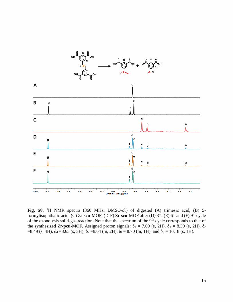
Next, we gained further evidence of the formation of Zr-pcu-MOF by measuring the 1H NMR spectra of the digested ozonized samples (5% HF/DMSO-d6) after each cycle, and
then comparing each spectrum to that of the starting Zr-scu-MOF (Fig. 2D and S6-8). Under
these digestion conditions, the MOF structures are destroyed and the linkers released, which
makes possible using 1H NMR to unveil the composition of the initial MOF structures. The
spectrum of the digested Zr-scu-MOF showed the characteristic peak of equivalent olefinic
protons of L2 at δ = 7.69 ppm. In contrast, the spectra of ozonated crystals after each cycle
confirmed a gradual fading of this olefinic signal, which, by the ninth cycle, had fully
disappeared. Indeed, 1H NMR of digested ozonated Zr-scu-MOF after the ninth cycle
confirmed the full conversion of L2 into doubly deprotonated trimesic acid and 5-
formylisophthalate. Beyond the disappearance of the olefinic protons at δ = 7.69 ppm, there
was also fading of the phenyl (δ = 8.49 ppm and 8.39 ppm) and the carboxylic acid (δ = 13.32
ppm) protons of L2, while the characteristic signals for the trimesic acid (δ = 13.57 ppm and
8.65 ppm) and 5-formylisophthalic acid (δ = 10.18 ppm, 8.70 ppm and 8.64 ppm) were clearly
identifiable. Cleavage of the olefinic bonds was corroborated by negative-mode mass
spectrometry and Fourier transform infrared (FT-IR) spectroscopy of both pristine and
ozonated Zr-MOFs (figs. S10,11). In the mass spectrum, the molecular ions [M-H]-
corresponding to trimesic acid (m/z = 209.01) and 5-formylisophthalic acid (m/z = 193.01)
were found, in strong contrast to the absence of any molecular ion [M-H]- corresponding to L2
(m/z = 355.05). Similarly, in comparison to the spectrum of pristine Zr-scu-MOF, the FT-IR
spectrum of ozonated Zr-pcu-MOF revealed the appearance of a typical carbonyl (C=O)
stretching band at 1700 cm-1 and the complete disappearance of an alkene (C=C) stretching
band at 1622 cm-1.
Having demonstrated the single-crystal-to-single-crystal synthesis of Zr-pcu-MOF by
selectively cleaving the olefinic bonds in Zr-scu-MOF into a mixture of aldehyde and
carboxylic acid groups, we next sought to perform analogous chemistry to synthesize Zr-pcu-
MOF functionalized exclusively with carboxylic acids; thus, showing improved control on the
final output of the clip-off chemistry. To this end, ozonated crystals were soaked in 1 mL of
hydrogen peroxide (30 wt. % in H2O) solution for three weeks. During this period, the sample
was refreshed with fresh hydrogen peroxide every 24 h. Next, the sample was centrifuged and
washed twice with water. The transformation to carboxylic acid groups was quantitative, as
confirmed by 1H NMR analysis (Fig. 2D and S9). Moreover, the Zr-pcu-MOF framework had
been preserved, as confirmed by XRPD (Fig. 2B and S3).

Fig 2. Structural and molecular characterization of Zr-pcu-MOF and Sc-pcu-MOF.
(A,C) Crystal structures of isoreticular Zr-scu-MOF (A) and Zr-pcu-MOF (C). Magnified
views highlight the changes observed in the distances and angles of both phenyl rings (from a1
= 3.77-3.79 Å and α1 = 14.95-19.71º to b1 = 3.39-3.65 Å and β1 = 0º) initially composing L2.
(B) Magnified view of XRPD of calculated Zr-scu-MOF (black), synthesized Zr-scu-MOF
(blue), calculated Zr-pcu-MOF (grey), synthesized Zr-pcu-MOF (red) and oxidized Zr-pcu-
MOF (green). Main structural changes with signal shifts are highlighted. (D) 1H NMR spectra
(360 MHz, DMSO-d6) of digested L2 (black), Zr-scu-MOF (orange), Zr-pcu-MOF (red), 5-
formylisophthalic acid (dark grey), trimesic acid (light grey) and oxidized Zr-pcu-MOF (light
red). Assigned proton signals: δ1 = 7.69 (s, 2H), δ2 = 8.39 (s, 2H), δ3 = 8.49 (s, 4H), δ4 = 8.64
(m, 2H), δ5 = 8.70 (m, 1H), δ6 = 10.18 (s, 1H) and δ7 = 8.65 (s, 3H). (E,G) Crystal structures

of isoreticular Sc-soc-MOF (E) and Sc-pcu-MOF (G). Magnified views highlight the changes
observed in the distances and angles of both phenyl rings (from a2 = 3.82-3.84 Å and α2 =
15.63-17.34º to a2 = 3.62-3.63 Å and β2 = 0º) initially composing L2. (F) Magnified view of
XRPD of calculated Sc-soc-MOF (black), synthesized Sc-soc-MOF (blue), calculated Sc-pcu-
MOF (grey) and synthesized Zr-pcu-MOF (red). (H) 1H NMR spectra (360 MHz, DMSO-d6)
of digested L2 (black), Sc-soc-MOF (orange), Sc-pcu-MOF (red), 5-formylisophthalic acid
(dark grey) and trimesic acid (light grey). Assigned proton signals: δ1 = 7.68 (s, 2H), δ2 = 8.38
(s, 2H), δ3 = 8.47 (s, 4H), δ4 = 8.64 (m, 2H), δ5 = 8.71 (m, 1H), δ6 = 10.17 (s, 1H) and δ7 = 8.65
(s, 3H).
Clip-off synthesis: from Sc-soc-MOF to Sc-pcu-MOF
To demonstrate that Clip-off Chemistry could be generalized to other 3D MOF
structures, we next attempted to synthesize a 3-D Sc-pcu-MOF from an Sc-soc-MOF precursor
(31) (Fig. 1 and section S3), a 3-D structure built up from linking 6-c trigonal, prismatic Sc3O
clusters to 4-c L1 linkers in a 4,6-c soc/edq topology (32). This structure exhibits a mixture of
cubic cavities (size: 11 Å) and 1-D channels (size: 8 Å) along the three axes. As in the previous
case, we designed the precursor by replacing L1 with L2, thus forming an isoreticular Sc-soc-
MOF that contains different circuits of connections, some of which contains the cleavable
olefins. We envisioned that, upon treatment with ozone, this circuit would be disrupted to form
a 3-D Sc-pcu-MOF whose Sc3O clusters would be connected through trimesate/5-
formylisophthalate linkers.
We began the clip-off synthesis of Sc-pcu-MOF via the aforementioned route, by first
preparing the isoreticular Sc-soc-MOF precursor. Colorless cubic crystals of Sc-soc-MOF were
prepared by heating a solution of Sc(NO3)3·xH2O and L2 in DMF, ethanol and formic acid at
120 ºC for 48 h (Fig. 2E). However, all attempts at complete ozonolysis were unsuccessful,
whether using the same synthetic protocol as for Zr-scu-MOF or trying slight modifications
(e.g. extending the reaction time up to 8 h and/or the number of ozonolysis cycles up to 12;
figs. S13-16). Consequently, cleavage of the olefinic bonds was incomplete, reaching a
maximum value of 60% to 70% under certain conditions (e.g. exposing the Sc-soc-MOF
crystals to ozone gas for 2 h). Consequently, a distinct ozonolysis protocol was employed: Sc-
soc MOF crystals were immersed in water, and then ozone was bubbled through the suspension
under stirring at room temperature for 12 h. These conditions afforded greater cleavage of the
olefinic bonds than in the previous strategy, leading to a mixture of trimesic acid, 5-
formylisophthalic acid and the intermediate 1,2,4-trioxolane ring (fig. S17). Importantly,
presence of this intermediate indicated that the olefinic bonds had not all been completely
cleaved to aldehyde/carboxylic acid groups. Moreover, increasing the reaction time under these
solid/liquid conditions did not provide any major improvement in cleavage, although it did lead
to a slight decrease in the crystallinity of the ozonated Sc-pcu-MOF.
To address the aforementioned drawbacks, we decided to combine the two ozonolysis
strategies, starting with a solid-gas reaction for 2 h and then, cleaving the remaining alkene
bonds via suspension/gas reaction for an additional 6 h. Under these conditions, SCXRD of the
resulting crystals confirmed quantitative single-crystal-to-single-crystal cleavage of the
olefinic bonds of Sc-soc-MOF to synthesize Sc-pcu-MOF (19) (Fig. 2G). However, in this
case, no changes were observed in the volume of the R-3 rhombohedral unit cell relative to that
of the precursor, as also confirmed by XRPD (Fig. 2F and S20). Nevertheless, SCXRD analysis
showed a very similar case to that of ozonated Zr-scu-MOF. Thus, the connectivity of the atoms
belonging to the inorganic trimeric ScO3 clusters remained identical to that of the pristine Sc-

soc-MOF. Refinement of the carbon atoms from the organic linkers evidenced a positional
disorder for some of them, and, unlike Sc-soc-MOF, the Sc-pcu-MOF product did not contain
any olefinic atoms. Instead, areas of electron density were observed only between the opposite
phenyl rings, with a maximum value at the middle point between the two rings, but at a distance
longer than that expected for a C=C bond. This area of higher electron-density points
perpendicularly above and below the plane of the phenyl rings. Although the refinement
indicates the presence of atoms in this area, these atoms could not be unambiguously assigned
to aldehyde or carboxylate groups (fig. S18). These findings indicate that the functional groups
are not arranged in an orderly fashion within the crystals after ozonolysis, but are rather
randomly oriented in the pores. This is not surprising, considering that the resulting aldehydes
and/or carboxylic groups are random distributed and most likely dangle in different orientations
into the pores, rather than having a periodically fixed position; accordingly, they would not
contribute to the periodic diffraction of the crystal. Consistent with these SCXRD data,
cleavage of all olefinic bonds was confirmed by 1H NMR (Fig. 2H and S21-23) and negative-
mode mass spectrometry of the digested ozonated samples (5% HF/DMSO-d6) (fig. S24), and
FT-IR of ozonated Sc-pcu-MOF (fig. S25), from which the olefinic protons (δ = 7.68 ppm),
molecular ion [M-H]- of L2 and the typical alkene (C=C) stretching band, respectively, were
not observed. Contrariwise, the 1H NMR signals, the molecular ions [M-H]- and the carbonyl
(C=O) stretching band corresponding to doubly deprotonated trimesic acid and 5-
formylisophthalate were all clearly identifiable, thus further corroborating the clip-off
synthesis of Sc-pcu-MOF.
Extending clip-off chemistry to a 0-D system: from a Rh-MOP to a Rh-macrocycle
Finally, we endeavored to use Clip-off Chemistry to synthesize a metal-organic
macrocycle (33-36) from a 0-D MOP system. To this end, we followed the second
aforementioned approach, designing a mixed-linker Rh-MOP that we could transform into a
triangular metal-organic macrocycle (Fig. 3A and section S4). We selected a Rh-MOP (37,38)
built from six 4-c Rh paddle-wheel clusters linked by six 5-tert-butylbenzene-1,3-
dicarboxylate (L3) linkers and by six 3-[(3-carboxyphenyl)diazenyl]benzoate (L4) linkers,
forming a cage with a diameter of 12 Å. In this cage, three paddle-wheel clusters are linked by
three L3 linkers to form a triangular macrocycle, and two of these units are linked by six L4
linkers. Following a similar strategy as those explained in the previous examples, we
functionalized this MOP with alkene groups by replacing L4 with 3-[2-(3-
carboxyphenyl)ethenyl]benzoate (L5). Accordingly, the isoreticular Rh-MOP would contain a
non-cleavable L3 linker and a cleavable L5 linker, which, upon cleavage via ozonolysis, would
split the Rh-MOP into two equal fragments. We hypothesize that this splitting would release
the metal-organic macrocycle built up from the three 4-c Rh(II) paddle-wheel units linked to
each other by the three L3 linkers and with six pendant partly deprotonated isophthalic linkers.
We synthesized isoreticular Rh-MOP by heating a N,N-dimethylacetamide (DMA)
suspension of L3, L5, Na2CO3 and Rh2(OAc)4 at 100 ºC. After 4 days, the reaction yielded a
dark precipitate that was centrifuged to separate out the Na2CO3. The resultant solution was
precipitated in MeOH to yield a green powder, which was washed several times with MeOH.
Finally, diffusion of diethyl ether into a DMF solution of this green powder afforded green
parallelogram-shaped crystals of Rh-MOP. SCXRD confirmed the formation of the expected
isoreticular MOP, built up from six L3 linkers and six cleavable L5 linkers (Fig. 3A). Next, the
six linkers were cleaved to synthesize the metal-organic macrocycle by bubbling ozone through
a suspension of Rh-MOP in DMA and hydrogen peroxide (50 wt. % in H2O) at room
temperature for 30 min. Note that oxidative conditions were used in this clip-off reaction to
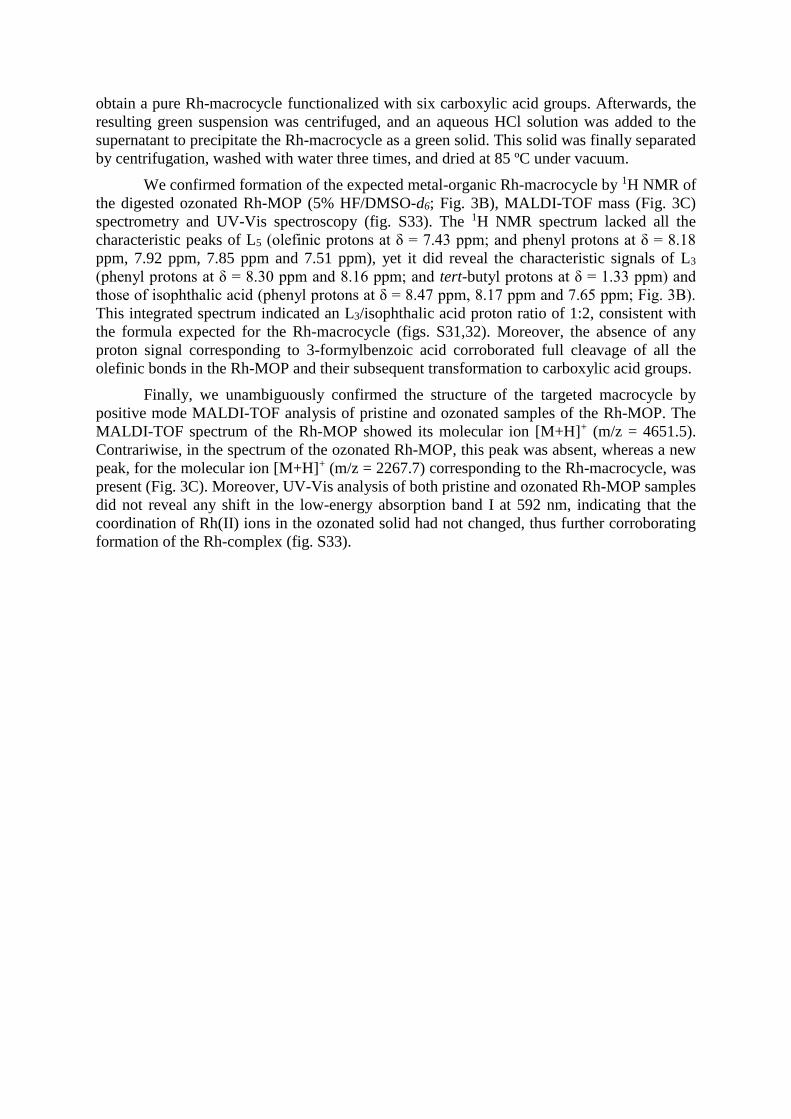
obtain a pure Rh-macrocycle functionalized with six carboxylic acid groups. Afterwards, the
resulting green suspension was centrifuged, and an aqueous HCl solution was added to the
supernatant to precipitate the Rh-macrocycle as a green solid. This solid was finally separated
by centrifugation, washed with water three times, and dried at 85 ºC under vacuum.
We confirmed formation of the expected metal-organic Rh-macrocycle by 1H NMR of
the digested ozonated Rh-MOP (5% HF/DMSO-d6; Fig. 3B), MALDI-TOF mass (Fig. 3C)
spectrometry and UV-Vis spectroscopy (fig. S33). The 1H NMR spectrum lacked all the
characteristic peaks of L5 (olefinic protons at δ = 7.43 ppm; and phenyl protons at δ = 8.18
ppm, 7.92 ppm, 7.85 ppm and 7.51 ppm), yet it did reveal the characteristic signals of L3
(phenyl protons at δ = 8.30 ppm and 8.16 ppm; and tert-butyl protons at δ = 1.33 ppm) and
those of isophthalic acid (phenyl protons at δ = 8.47 ppm, 8.17 ppm and 7.65 ppm; Fig. 3B).
This integrated spectrum indicated an L3/isophthalic acid proton ratio of 1:2, consistent with
the formula expected for the Rh-macrocycle (figs. S31,32). Moreover, the absence of any
proton signal corresponding to 3-formylbenzoic acid corroborated full cleavage of all the
olefinic bonds in the Rh-MOP and their subsequent transformation to carboxylic acid groups.
Finally, we unambiguously confirmed the structure of the targeted macrocycle by
positive mode MALDI-TOF analysis of pristine and ozonated samples of the Rh-MOP. The
MALDI-TOF spectrum of the Rh-MOP showed its molecular ion [M+H]+ (m/z = 4651.5).
Contrariwise, in the spectrum of the ozonated Rh-MOP, this peak was absent, whereas a new
peak, for the molecular ion [M+H]+ (m/z = 2267.7) corresponding to the Rh-macrocycle, was
present (Fig. 3C). Moreover, UV-Vis analysis of both pristine and ozonated Rh-MOP samples
did not reveal any shift in the low-energy absorption band I at 592 nm, indicating that the
coordination of Rh(II) ions in the ozonated solid had not changed, thus further corroborating
formation of the Rh-complex (fig. S33).

Fig 3. Clip-off synthesis of a triangular Rh-macrocycle. (A) Schematic representation of the
synthesis of a Rh-macrocycle from a discrete mixed-linker Rh-MOP, in which a linker
containing an olefinic bond had been incorporated by reticular chemistry previous to
ozonolysis. (B) 1H NMR spectra (360 MHz, DMSO-d6) of digested L3 (black), L5 (dark grey),
Rh-MOP (orange), Rh-macrocycle (red), and isophthalic acid (light grey). Assigned proton
signals for digested Rh-MOP (orange): δ1 = 8.30 (s, 1H), δ2 = 8.16 (s, 2H), δ3,5 = 7.92 (d, J =
7.7 Hz, 2H), δ3,5 = 7.85 (d, J = 7.7 Hz, 2H), δ4 = 7.51 (t, J = 7.7 Hz, 2H), δ6 = 8.18 (s, 2H), and
δ7 = 7.43 (s, 2H). Assigned proton signals for digested Rh macrocycle (red): δ1 = 8.30 (s, 1H),
δ2, 8, 10 = 8.17 - 8.15 (m, 6H), δ9 = 7.65 (t, J = 7.8 Hz, 2H), and δ11 = 8.47 (s, 2H). (C) MALDI-
TOF spectrometry of Rh-MOP (blue) and Rh-macrocycle fully functionalized with COOH
(red). The mass corresponding to the formula [Rh12(L3)6(L5)6(DMA)(H2O)2(MeOH)4 + H]+
(expected = 4403.8; found = 4405.3) and [Rh12(L3)6(L5)6(H2O)2(DMA)2(CH3CN)7 + H]+
(expected = 4650.0; found = 4651.5) have been highlighted for the Rh-MOP sample. In the
case of the Rh-macrocycle, the mass corresponding to the formula [Rh6(L3)3(C8H5O4)6 + H]+
has been highlighted (expected = 2268.8; found = 2267.7).
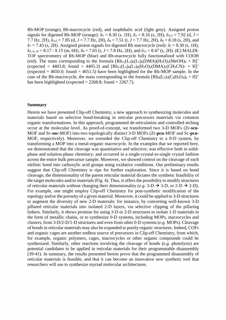
Summary
Herein we have presented Clip-off Chemistry, a new approach to synthesizing molecules and
materials based on selective bond-breaking in reticular precursors materials via common
organic transformations. In this approach, programmed de-reticulation and controlled etching
occur at the molecular level. As proof-of-concept, we transformed two 3-D MOFs (Zr-scu-
MOF and Sc-soc-MOF) into two topologically distinct 3-D MOFs (Zr-pcu-MOF and Sc-pcu-
MOF, respectively). Moreover, we extended the Clip-off Chemistry to a 0-D system, by
transforming a MOP into a metal-organic macrocycle. In the examples that we reported here,
we demonstrated that the cleavage was quantitative and selective; was effective both in solid-
phase and solution-phase chemistry; and occurred in a single-crystal-to-single crystal fashion
across the entire bulk precursor sample. Moreover, we showed control on the cleavage of each
olefinic bond into carboxylic acid groups using oxidative conditions. Our preliminary results
suggest that Clip-off Chemistry is ripe for further exploration. Since it is based on bond
cleavage, the dimensionality of the parent reticular material dictates the synthetic feasibility of
the target molecules and/or materials (Fig. 4). Thus, it offers the possibility to modify structures
of reticular materials without changing their dimensionality (e.g. 3-D 3-D, or 2-D 2-D).
For example, one might employ Clip-off Chemistry for post-synthetic modification of the
topology and/or the porosity of a given material. Moreover, it could be applied to 3-D structures
to augment the diversity of new 2-D materials: for instance, by converting well-known 3-D
pillared reticular materials into isolated 2-D layers, via selective clipping of the pillaring
linkers. Similarly, it shows promise for using 3-D or 2-D structures to isolate 1-D materials in
the form of metallic chains, or to synthesize 0-D systems, including MOPs, macrocycles and
clusters, from 3-D/2-D/1-D structures and even from other 0-D systems (e.g. MOPs). Cleavage
of bonds in reticular materials may also be expanded to purely organic structures. Indeed, COFs
and organic cages are another endless source of precursors in Clip-off Chemistry, from which,
for example, organic polymers, cages, macrocycles or other organic compounds could be
synthesized. Similarly, other reactions involving the cleavage of bonds (e.g. photolysis) are
potential candidates to be applied in reticular materials for their programmable disassembly
(39-41). In summary, the results presented herein prove that the programmed disassembly of
reticular materials is feasible, and that it can become an innovative new synthetic tool that
researchers will use to synthesize myriad molecular architectures.

Fig 4. Clip-off Chemistry for the synthesis of molecular architectures of different
dimensionality. Schematic illustrating the potential outcomes of clip-off chemistry, in which
the dimensionality of the parent reticular material dictates the dimensionality of the target
molecules and structures.
REFERENCES AND NOTES:
1. H. Li, M. Eddaoudi, T. L. Groy, O. M. Yaghi, Establishing microporosity in open metal-
organic frameworks: gas sorption isotherms for Zn(BDC) (BDC = 1,4-
benzenedicarboxylate). J. Am. Chem. Soc. 120, 8571-8572 (1998).
2. O. M. Yaghi, M. O’Keeffe, N. W. Ockwig, H. K. Chae, M. Eddaoudi, J. Kim, Reticular
synthesis and the design of new materials. Nature 423, 705-714 (2003).
3. H. Li, M. Eddaoudi, M. O’Keeffe, O. M. Yaghi, Design and synthesis of an exceptionally
stable and highly porous metal-organic framework. Nature 402, 276-279 (1999).
4. O. M. Yaghi, G. Li, H. Li, Selective binding and removal of guests in a microporous metal-
organic framework. Nature 378, 703-706 (1995).
5. S. Kitagawa, R. Kitaura, S. Noro, Functional porous coordination polymers. Angew. Chem.
Int. Ed. 43, 2334-2375 (2004).
6. B. F. Abrahams, B. F. Hoskins, D. M. Michail, R. Robson, Assembly of porphyrin building
blocks into network structures with large channels. Nature 369, 727-729 (1994).
7. B. F. Hoskins, R. Robson, Infinite polymeric frameworks consisting of three dimensionally
linked rod-like segments. J. Am. Chem. Soc. 111, 5962-5964 (1989).
8. O. M. Yaghi, M. J. Kalmutzki, C. S. Diercks, Introduction to Reticular Chemistry: Metal-
Organic Frameworks and Covalent Organic Frameworks (Wiley, 2019).
9. X. Feng, X. Ding, D. Jiang, Covalent organic frameworks. Chem. Soc. Rev. 41, 6010-6022
(2012).
10. D. J. Tranchemontagne, Z. Ni, M. O’Keeffe, O. M. Yaghi, Reticular chemistry of metal-
organic polyhedra. Angew. Chem. Int. Ed. 47, 5136-5147 (2008).

11. J. J. Perry IV, J. A. Perman, M. J. Zaworotko, Design and synthesis of metal-organic
frameworks using metal-organic polyhedra as supermolecular building blocks. Chem. Soc.
Rev. 38, 1400-1417 (2009).
12. S. Lee, H. Jeong, D. Nam, M. S. Lah, W. Choe, The rise of metal-organic polyhedra. Chem.
Soc. Rev. 50, 528-555 (2021).
13. M. Eddaoudi, D. B. Moler, H. Li., B. Chen, T. M. Reineke, M. O’Keeffe, O. M. Yaghi,
Modular chemistry: secondary building units as a basis for the design of highly porous and
robust metal-organic carboxylate frameworks. Acc. Chem. Res. 34, 319-330 (2001).
14. M. Eddaoudi, J. Kim, N. Rosi, D. Vodak, J. Wachter, M. O’Keeffe, O. M. Yaghi,
Systematic design of pore size and functionality in isoreticular MOFs and their application
in methane storage. Science 295, 469-472 (2002).
15. S. M. Cohen, Postsynthetic methods for the functionalization of metal-organic frameworks.
Chem. Rev. 112, 970-1000 (2012).
16. O. M. Yaghi, Reticular chemistry: molecular precision in infinite 2D and 3D. Mol. Front.
J. 3, 66-83 (2019).
17. M. O’Keeffe, O. M. Yaghi, Deconstructing the crystal structures of metal-organic
frameworks and related materials into their underlying nets. Chem. Rev. 112, 675-702
(2012).
18. M. O’Keeffe, M. A. Peskov, S. J. Ramsden, O. M. Yaghi, The reticular chemistry structure
resource (RCSR) database of, and symbols for, crystal nets. Acc. Chem. Res. 41, 1782-1789
(2008).
19. V. A. Blatov, A. P. Shevchenko, D. M. Proserpio, Applied topological analysis of crystal
structures with the program package Topospro. Cryst. Growth Des. 14, 3576-3586 (2014).
20. A. F. Wells, Three-Dimensional Nets and Polyhedral (Wiley, 1977).
21. H. Wang, X. Dong, J. Lin, S. J. Teat, S. Jensen, J. Cure, E. V. Alexandrov, Q. Xia, K. Tan,
Q. Wang, D. H. Olson, D. M. Proserpio, Y. J. Chabal, T. Thonhauser, J. Sun, Y. Han, J. Li,
Topologically guided tuning of Zr-MOF pore structures for highly selective separation of
C6 alkane isomers. Nat. Commun. 9, 1745 (2018).
22. M. Li, D. Li, M. O’Keeffe, O. M. Yaghi, Topological analysis of metal-organic frameworks
with polytopic linkers and/or multiple building units and the minimal transitivity principle.
Chem. Rev. 114, 1343-1370 (2014).
23. J. Albalad, H. Xu, F. Gándara, M. Haouas, C. Martineau-Corcos, R. Mas-Ballesté, S. A.
Barnett, J. Juanhuix, I. Imaz, D. Maspoch, Single-crystal-to-single-crystal postsynthetic
modification of a metal-organic framework via ozonolysis. J. Am. Chem. Soc. 140, 2028-
2031 (2018).
24. V. Guillerm, H. Xu, J. Albalad, I. Imaz, D. Maspoch, Postsynthetic selective ligand
cleavage by solid-gas phase ozonolysis fuses micropores into mesopores in metal-organic
frameworks. J. Am. Chem. Soc. 140, 15022-15030 (2018).
25. R. Criegee, Mechanism of ozonolysis. Angew. Chem. Int. Ed. 14, 745-752 (1975).
26. C. E. Schiaffo, P. H. Dussault, Ozonolysis in solvent/water mixtures: direct conversion of
alkenes to aldehydes and ketones. J. Org. Chem. 73, 4688-4690 (2008).
27. L. B. Harding, A. W. Goddard, Mechanisms of gas-phase and liquid-phase ozonolysis. J.
Am. Chem. Soc. 100, 7180-7188 (1978).
28. L. H. Xie, X. M. Liu, T. He, J. R. Li, Metal-organic frameworks for the capture of trace
aromatic volatile organic compounds. Chem 4, 1911-1927 (2018).
29. S. Wang, L. Chen, M. Wahiduzzaman, A. Tissot, L. Zhou, I. A. Ibarra, A. Gutiérrez-
Alejandre, J. S. Lee, J. S. Chang, Z. Liu, J. Marrot, W. Shepard, G. Maurin, Q. Xu, C. Serre,
A mesoporous zirconium-isophthalate multifunctional platform. Matter 4, 182-194 (2021).

30. H. Furukawa, F. Gándara, Y. B. Zhang, J. Jiang, W. L. Queen, M. R. Hudson, O. M. Yaghi,
Water adsorption in porous metal-organic frameworks and related materials. J. Am. Chem.
Soc. 136, 4369-4381 (2014).
31. J. W. Zhang, P. Qu, M. C. Hu, S. N. Li, Y. C. Jiang, Q. G. Zhai, Topology-guided design
for Sc-soc-MOFs and their enhanced storage and separation for CO2 and C2-hydrocarbons.
Inorg. Chem. 58, 16792-16799 (2019).
32. Y. Liu, J. F. Eubank, A. J. Cairns, J. Eckert, V. C. Kravtsov, R. Luebke, M. Eddaoudi,
Assembly of metal-organic frameworks (MOFs) based on indium-trimer building blocks:
a porous MOF with soc topology and high hydrogen storage. Angew. Chem. Int. Ed. 46,
3278-3283 (2007).
33. P. J. Stang, B. Olenyuk, Self-assembly, symmetry and molecular architecture: coordination
as the motif in the rational design of supramolecular metallacyclic polygons and polyhedra.
Acc. Chem. Res. 30, 502-518 (1997).
34. B. Olenyuk, A. Fechtenkötter, P. J. Stang, Molecular architecture of cyclic nanostructures:
use of co-ordination chemistry in the building of supermolecules with predefined geometric
shapes. J. Chem. Soc., Dalton Trans. 1998, 1707-1728 (1998).
35. M. Fujita, O. Sasaki, T. Mitsuhashi, T. Fujita, J. Yazaki, K. Yamaguchi, K. Ogura, On the
structure of transition-metal-linked molecular squares. Chem. Commun. 1996, 1535-1536
(1996).
36. F. A. Cotton, L. M. Daniels, C. Lin, C. A. Murillo, Square and triangular arrays based on
Mo24+ and Rh2
4+ units. J. Am. Chem. Soc. 121, 4538-4539 (1999).
37. Unpublished results.
38. J. R. Li, H. C. Zhou, Bridging-ligand-substitution strategy for the preparation of metal-
organic polyhedra. Nat. Chem. 2, 893-898 (2010).
39. L. Feng, K. Y. Wang, G. S. Day, M. R. Ryder, H. C. Zhou, Destruction of metal-organic
frameworks: positive and negative aspects of stability and lability. Chem. Rev. 120, 13087-
13133 (2020).
40. K. Y. Wang, L. Feng, T. H. Yan, S. Wu, E. A. Joseph, H. C. Zhou, Rapid generation of
hierarchically porous metal-organic frameworks through laser photolysis. Angew. Chem.
Int. Ed. 59, 11349-11354 (2020).
41. P. Shieh, Hill, M. R. Hill, W. Zhang, S. L. Kristufek, J. A. Johnson, Clip chemistry: diverse
(bio)(macro) molecular and material function through breaking covalent bonds. Chem. Rev.
2021, 0c01282 (2021).
ACKNOWLEDGMENTS
We thank Thais Grancha, Jordi Martínez-Esaín and Teodor Parella for technical assistance.
Funding: This work was supported by the Spanish MINECO (projects RTI2018-095622-B-
I00 and PID2019-106403RB-I00) and the Catalan AGAUR (project 2017 SGR 238). It was
also funded by the CERCA Programme /Generalitat de Catalunya. ICN2 is supported by the
Severo Ochoa programme from the Spanish MINECO (Grant No. SEV-2017-0706). F.G.
acknowledges funding from the Spanish Research Agency (AEI, CTQ2017-87262-R,
EUR2019-103824). The project that generated these results received support from a fellowship
(LCF/BQ/PR20/11770011) of the “la Caixa” Foundation (ID 100010434). Y.Y. acknowledges
the China Scholarship Council for scholarship support. Author contributions: Y.Y., B.O.R.
and V.G. performed the design and synthesis of the 3-D MOFs via clip-off chemistry. A.B.R.
and A.C.S. performed the design and synthesis of the Rh-macrocycle via clip-off chemistry.
F.G., J.J. and I.I. performed SCXRD experiments and resolution. S.J. and F.B. contributed on
the synthesis of non-commercial linkers and advised on the project. D.M. wrote the manuscript
with substantial input from all authors. I.I. and D.M. participated in conceptual development.

Competing interests: the authors declare no competing interests. Data and materials
availability: All data are available in the main text or in the supplementary materials.

download fileview on ChemRxivManuscript.pdf (1.38 MiB)

1
Supplementary Materials for
Clip-off Chemistry: Synthesis by Programmed Disassembly of
Reticular Materials
Yunhui Yang†, Anna Broto-Ribas†, Borja Ortín-Rubio†, Inhar Imaz*, Felipe Gándara, Arnau
Carné-Sánchez, Vincent Guillerm, Sergio Jurado, Félix Busqué, Judith Juanhuix, Daniel
Maspoch*
Correspondence to: [email protected] and [email protected]
†These authors contributed equally to this work.

2
Section S1. Materials and Methods
Zirconium oxychloride octahydrate (ZrOCl2·8H2O), formic acid, potassium iodide (KI),
scandium(III) nitrate hydrate, trimesic acid (H3BTC), anhydrous granular CaCl2 (2-6 mm),
trimethylamine, sodium carbonate (Na2CO3) and hydrogen peroxide 30 wt. % in H2O and 50 wt.
% in H2O were purchased from Sigma-Aldrich Co. N,N-dimethylformamide (DMF), absolute
ethanol (EtOH), tetrahydrofuran (THF), N,N-dimethylacetamide (DMA), dichloromethane,
methanol (MeOH), diethyl ether, acetone, 3-vinylbenzoic acid and hydrochloric acid (HCl) were
obtained from Fisher Chemical. 5-[(E)-2-(3,5-dicarboxyphenyl)ethenyl]benzene-1,3-dicarboxylic
acid (linker L2) was obtained from Chemextention Co. 3-iodobenzoic acid, palladium(II) acetate
and 5-tert-butylbenzene-1,3-dicarboxylic acid (linker L3) were obtained from TCI. Rhodium(II)
acetate dimer was obtained from Acros Organics. All the reagents and solvents were used without
further purification unless otherwise specified. Deionized water was obtained with a Milli-Q®
system (18.2 MΩ·cm). The ozone generator (model N1668A, 10.4 mmol/h O3 at room
temperature) was purchased from Ozonotec.
Section S1.1. Clip-off synthesis: from Zr-scu-MOF to Zr-pcu-MOF
Synthesis of Zr-scu-MOF
A solution of ZrOCl2·8H2O (16 mg, 0.05 mmol) and L2 (7 mg, 0.02 mmol) in DMF (8 mL) and
formic acid (8 mL) was prepared in a 23 mL scintillation vial. Then, the sealed vial was placed
into a preheated oven at 120 ºC for 5 days. After this period, colorless cubic crystals suitable for
single-crystal X-ray diffraction (SCXRD) were collected by filtration and washed three times by
incubating them with 20 mL of fresh DMF for 12 h. Afterwards, solvent exchange with THF was
performed by incubating the crystals three times with 20 mL of THF for 12 h, and the resulting
Zr-scu-MOF crystals were dried at room temperature (15 mg).
Clip-off synthesis of Zr-pcu-MOF via ozonolysis
The synthesis of Zr-pcu-MOF was achieved by a solid-gas ozonolysis reaction. This reaction
started by packing 20 mg of Zr-scu-MOF crystals into a plastic tube, connected on one side to the
ozonator (through a CaCl2 humidity trap), and on the other side, to a vacuum pump (through a KI
trap), to ensure a continuous flow of ozone through the column. The reaction was run at room
temperature for 30 min. Afterwards, the sample was held in vacuum for another 30 min. This
ozonation/vacuum cycle was repeated nine times. Finally, the resulting solid (20 mg) was directly
collected from the plastic tube and stored in THF.
Oxidation of Zr-pcu-MOF to full –COOH pending groups
Dried ozonated crystals were soaked in 1 ml of aqueous H2O2 solution (30 wt. % in H2O) for 3
weeks. During this period, the sample was exchanged with a fresh hydrogen peroxide solution
every 24 h. After that, the sample was centrifuged and washed twice with water.

3
Section S1.2. Clip-off synthesis: from Sc-soc-MOF to Sc-pcu-MOF
Synthesis of Sc-soc-MOF
A solution of Sc(NO3)3·xH2O (48 mg, 0.21 mmol) and L2 (16 mg, 0.04 mmol) in DMF (8 mL),
EtOH (3.2 mL) and formic acid (1.84 mL) was prepared in a 23 mL scintillation vial. Then, the
sealed vial was placed into a preheated oven at 120 ºC for 48 h. Colorless cubic crystals suitable
for SCXRD were collected by filtration and washed three times by incubating them with 20 mL
of fresh DMF for 12 h. Afterwards, solvent exchange with acetone was performed by incubating
the crystals three times with 20 mL of acetone for 12 h, and the resulting crystals were dried at
room temperature (20 mg).
Clip-off synthesis of Sc-pcu-MOF via ozonolysis
The clip-off synthesis of Sc-pcu-MOF was done in two steps: a first step consisting of a solid-gas
ozonolysis reaction; and a second step based on a suspension-gas ozonolysis reaction. In the first
step, 20 mg of synthesized Sc-soc-MOF crystals were packed into the plastic tube, connected on
one side to the ozonator (through a CaCl2 humidity trap), and on the other side, to a vacuum pump
(through a KI trap). The reaction was run at room temperature for 2 hours. Afterwards, the crystals
were collected from the plastic tube and suspended in 4 ml of water. The ozone generator was
directly connected to the vial through a syringe. Ozone was bubbled in the suspension for 6 h while
magnetically stirring at 880 rpm. The resulting crystals (20 mg) were collected by centrifugation
and stored in acetone.
Section S1.3. Clip-off synthesis: from Rh-MOP to a Rh-macrocycle
Synthesis of 3-[2-(3-carboxyphenyl)ethenyl]benzoic acid (linker L5)
Scheme S1. Synthesis of L5
3-vinylbenzoic acid (2 g, 13.50 mmol), 3-iodobenzoic acid (3.34 g, 13.47 mmol),
triphenylphosphine (118 mg, 0.45 mmol), triethylamine (20 mL) and palladium(II) acetate (80 mg,
0.36 mmol) were allowed to react in THF (40 ml) at 80 ºC for 24 hours. The obtained suspension
was filtered, and the obtained solution was evaporated under reduced pressure. The obtained solid

4
was dissolved in 100 mL of dichloromethane and poured into 600 mL of water under vigorous
stirring. Some drops of concentrated HCl (12 M) were added to the resulting suspension to induce
the precipitation of a brown solid. The solid was recovered by filtration and washed with water
until pH = 7. Finally, the solid was purified by dissolving it in dichloromethane and precipitating
it with 500 mL of MeOH. The precipitate was recovered by filtration and dried at room temperature
(2 g, yield: 56%). 1H NMR (250 MHz, DMSO-d6): δ 13.05 (s, 1H), 8.21 (s, 1H), 7.91 (d, J = 8.0
Hz, 1H), 7.86 (d, J = 7.9 Hz, 1H), 7.52 (t, J = 7.7 Hz, 1H), 7.45 (s, 1H). 13C NMR (250 MHz,
DMSO-d6): δ 168.05, 138.10, 132.17, 131.54, 129.87, 129.47, 129.41, 128.33. MS [C15H11O4]- =
267.0651 g/mol / Err<2.5 mDa.
Rh-MOP synthesis
L3 (250 mg, 5 eq, 1.13 mmol), L5 (422.24 mg, 7 eq, 1.57 mmol), Na2CO3 (119.65 mg, 5 eq, 1.13
mmol), rhodium(II) acetate dimer (100 mg, 1 eq, 0.226 mmol) and 1 mL of DMA were sonicated
for 5 minutes and placed into an oven at 100 ºC for 72 h. After cooling down to room temperature,
the resulting mixture was centrifuged to take out the Na2CO3. Then, 25 mL of cold MeOH was
added into the obtained solution to precipitate the Rh-MOP, which was washed three times with
MeOH. The obtained green solid was dried at room temperature (20 mg). Single-crystals suitable
for SCXRD were obtained by slow ether vapor diffusion into a DMF solution of the synthesized
Rh-MOP.
Clip-off synthesis of Rh-based metal-organic macrocycle via ozonolysis
2.4 mL of aqueous H2O2 solution (50 wt. % in H2O) were added into a solution of 10 mg of Rh-
MOP in 4 mL of DMA, leading to the formation of a green suspension. Afterwards, the ozone
generator was directly connected to the vial through a syringe. Ozone was bubbled in the
suspension for 30 min. After ozonolysis, the obtained green suspension was centrifuged, and the
resulting supernatant was treated with 5.5 mL of HCl 3M solution to precipitate the Rh-based
macrocycle as a green solid. This solid was finally separated by centrifugation, washed three times
with water and dried at 85 ºC under vacuum (3.5 mg).
Section S1.4. Characterization
Single-Crystal X-Ray Diffraction (SCXRD) data of Zr-scu-MOF, Zr-pcu-MOF, Sc-soc-MOF,
Sc-pcu-MOF were collected at 100 K at BL13-XALOC beamline at the ALBA synchrotron (λ =
0.82656 Å) (1). Data for Rh-MOP was collected with a Bruker four-circle kappa diffractometer
equipped with a Cu INCOATED microsource, operated at 50-W power (50 kV, 1.00 mA) to
generate Cu Kα radiation (λ = 1.54178 Å), and a Bruker VANTEC-500 area detector (microgap
technology). Data frames were indexed, integrated and scaled using the XDS program, and the
APEX3 software (2). The structures were solved by direct methods and subsequently refined by
corrections of F2 against all reflections, using SHELXST2013 (3) and SHELXL2013 (4) within
the Olex2 package (5). Non-hydrogen atoms were refined with anisotropic thermal parameters by
full-matrix least-squares calculations on F2 using the program SHELXL2013. Hydrogens atoms
were inserted at calculated positions and constrained with isotropic thermal parameters.

5
X-Ray Powder Diffraction (XRPD) diagrams were collected on a Panalytical X’pert mpd
diffractometer with monochromatic Cu-K radiation (Cu = 1.5406 Å).
Proton Nuclear Magnetic Resonance (1H NMR) spectra were acquired in Bruker Avance DPX
of 250 MHz, 360 MHz NMR spectrometer at “Servei de Ressonància Magnètica Nuclear” from
Autonomous University of Barcelona (UAB).
Carbon Nuclear Magnetic Resonance (13C NMR) spectra were acquired in Bruker Avance DPX
of 250 MHz NMR spectrometer at “Servei de Ressonància Magnètica Nuclear” from Autonomous
University of Barcelona (UAB).
Field-emission Scanning Electron Microscopy (FESEM) images were performed in a SEM
Quanta 650 scanning electron microscopy at an acceleration voltage of 2 KV, using aluminium as
a support.
Fourier Transform Infrared (FT-IR) spectra were recorded on a Bruker Tensor 27FT-IR
spectrometer equipped with a Golden Gate diamond attenuated total reflection (ATR) cell, in
transmittance mode at room temperature.
Ultraviolet-visible (UV−vis) spectra were measured in an Agilent Cary 4000 at room temperature
(ca. 25 °C).
Electrospray Ionization Mass Spectrometry (ESI-MS) spectra were obtained in an Agilent 6210
G1969A LC/MSD TOF mass spectrometer. All the digested MOFs were measured in the negative-
ionization mode. The samples for MS analysis were obtained from the 1H NMR digestion samples.
Mass Spectrometry (MALDI-TOF) spectra were run in an Applied Biosystems 4700 Proteomics
Analyzer. The Rh-MOP and Rh-macrocycle after cleavage were measured in positive-ionization
mode.

6
Section S2. Clip-off chemistry: from Zr-scu-MOF to Zr-pcu-MOF
Section S2.1 Single-Crystal X-Ray Diffraction
Diffraction data for Zr-scu-MOF and Zr-pcu-MOF was collected at the BL13-XALOC beamline
at the ALBA synchrotron with λ = 0.82653 Å. All tested specimens were found to be grown as
twinned crystals, and they could not be separated as single components. Best results were obtained
by indexing and reducing the data with the cell parameters corresponding to a single component,
although with unavoidable presence of a number of overlying reflections, which eventually
resulted in high refinement residual values, and negative anisotropic displacement parameters for
some atoms. Nonetheless, the structure could be solved, and the location of the framework atoms
unambiguously determined, confirming the formation of the isoreticular Zr-scu-MOF.
Table S1. Crystal data and structure refinement for Zr-scu-MOF.
Formula C72O64Zr12
Formula weight (g.mol-1) 2983.36
Temperature (K) 100(2)
Wavelength (Å) 0.82653
Crystal system Monoclinic
Space group C2/m
Unit cell dimensions a = 25.637(5)Å α = 90º
b = 36.223(7) Å β = 122.54(3)º
c = 21.661(4) Å γ = 90º
Volume (Å3) 16958(8)
Z 4
Density calculated (g/cm-3) 1.169
Absorption coefficient (mm-1) 1.157
F(000) 5696.0
Crystal size (mm) 0.33 × 0.30 × 0.30 mm
2Theta range for data collection (º) 2.594 to 58.182
Index ranges -30 ≤ h ≤ 25, 0 ≤ k ≤ 42, 0 ≤ l ≤ 25
Reflection collected 44506
Independent reflections 14607 [Rint = 0.0968, Rsigma = 0.1369]
Refinement method Full-matrix least-squares on F2
Data / restraints / parameters 14607/114/556
Goodness-of-fit on F2 1.942
Final R indices [I>2sigma(I)] R1 = 0.1983, wR2 = 0.4825
R indices (all data) R1 = 0.2125, wR2 = 0.5069
Largest diff. peak and hole 8.61, -6.78e.Å-3

7
Table S2. Crystal data and structure refinement for Zr-pcu-MOF.
Formula C71.25O67.5Zr12
Formula weight (g.mol-1) 3030.35
Temperature (K) 100(2)
Wavelength (Å) 0.82653
Crystal system Monoclinic
Space group C2/m
Unit cell dimensions a = 35.703(7)Å α = 90º
b = 25.210(5) Å β = 134.89(3)º
c = 25.210(5) Å γ = 90º
Volume (Å3) 16076(8)
Z 4
Density calculated (g/cm-3) 1.252
Absorption coefficient (mm-1) 0.812
F(000) 5790.0
Crystal size (mm) 0.38 × 0.32 × 0.32 mm
2Theta range for data collection (º) 2.28 to 34.45
Index ranges -29 ≤ h ≤ 29, -20 ≤ k ≤ 20, -20 ≤ l ≤ 20
Reflection collected 28412
Independent reflections 4867 [Rint = 0.1337, Rsigma = 0.0877]
Refinement method Full-matrix least-squares on F2
Data / restraints / parameters 4867/0/513
Goodness-of-fit on F2 1.336
Final R indices [I>2sigma(I)] R1 = 0.1277, wR2 = 0.3291
R indices (all data) R1 = 0.1736, wR2 = 0.3849
Largest diff. peak and hole 1.13, -0.76 e.Å-3

8
Fig. S1. Representation of the single-crystal structures of (A) Zr-scu-MOF and (B) Zr-pcu-MOF,
showing the differences in the orientation of the phenyl rings of the linkers before (A) and after
(B) the cleavage of the olefinic bonds.

9
Fig. S2. Comparison of the cluster environment in (A) Zr-scu-MOF and (B) Zr-pcu-MOF. In Zr-
scu-MOF, each Zr-oxo-hydroxo-cluster is connected to 14 neighboring Zr-oxo-hydroxo-clusters
by eight 4-c linkers L2. After clip-off treatment, each Zr-oxo-hydroxo-cluster in Zr-pcu-MOF is
surrounded by 6 neighboring Zr-oxo-hydroxo-clusters, which are interconnected by eight 2-c
linkers. In Zr-scu-MOF, the connectivity of the Zr-oxo-hydroxo-cluster is in a cubic fashion,
matching with the shape of the hexamer cluster. In Zr-pcu-MOF, the connectivity of the Zr-oxo-
hydroxo-cluster decreases to an octahedral fashion because two pairs of linkers connect to the
same clusters; despite the cubic shape of the hexamer cluster.

10
Section S2.2 XRPD Analysis
Fig. S3. XRPD of calculated Zr-scu-MOF (black), pristine Zr-scu-MOF (blue), calculated Zr-pcu-
MOF (green), synthesized Zr-pcu-MOF (red) and oxidized Zr-pcu-MOF (purple).

11
Fig. S4. XRPD pattern and Pawley refinement of Zr-scu-MOF. The experimental pattern (red),
the refined Pawley fitting (black), the difference plot (orange), simulated pattern (blue) and the
Bragg positions (grey) are provided.

12
Fig. S5. XRPD pattern and Pawley refinement of Zr-pcu-MOF. The experimental pattern (red),
the refined Pawley fitting (black), the difference plot (orange), simulated pattern (blue) and the
Bragg positions (grey) are provided.

13
Section S2.3 1H NMR Analysis
Fig. S6. 1H NMR spectrum (360 MHz, DMSO-d6) of the digested Zr-scu-MOF. Zr-scu-MOF
crystals were digested in 5% HF aqueous solutions at 120 ºC overnight.
*DMSO
*H2O

14
Fig. S7. 1H NMR spectrum (360 MHz, DMSO-d6) of the digested Zr-pcu-MOF after ozonolysis
solid reaction. Zr-pcu-MOF crystals were digested in 5% HF aqueous solutions at 120 ºC
overnight.
*DMSO
*H2O
15
Fig. S8. 1H NMR spectra (360 MHz, DMSO-d6) of digested (A) trimesic acid, (B) 5-
formylisophthalic acid, (C) Zr-scu-MOF, (D-F) Zr-scu-MOF after (D) 3rd, (E) 6th and (F) 9th cycle
of the ozonolysis solid-gas reaction. Note that the spectrum of the 9th cycle corresponds to that of
the synthesized Zr-pcu-MOF. Assigned proton signals: δa = 7.69 (s, 2H), δb = 8.39 (s, 2H), δc
=8.49 (s, 4H), δd =8.65 (s, 3H), δe =8.64 (m, 2H), δf = 8.70 (m, 1H), and δg = 10.18 (s, 1H).
a
a
b
b
c
c
d
d
d
d
f
f
f
e
e
e
e
g
g
g
g
f
a
b
c d f
e
g
+
A
B
C
D
E
F
ab
c

16
Fig. S9. 1H NMR spectrum (360 MHz, DMSO-d6) of the digested oxidized Zr-pcu-MOF. Zr-pcu-
MOF crystals were digested in 5% HF aqueous solutions at 120 ºC overnight.

17
Section S2.4 ESI-MS Analysis
Fig. S10. ESI-MS spectrometry of digested Zr-scu-MOF (black) and Zr-pcu-MOF (red). The peak
at m/z 355.05 is assigned to [L2 - H]- in Zr-scu-MOF, corresponding to the formula [C18H12O8 -
H]-: expected = 355.05, found = 355.05. The peak at m/z 193.01 in both samples is assigned to 5-
formylisophthalic acid, corresponding to the formula [C6H3(COOH)2(CHO) - H]-: expected =
193.02, found = 193.01. The peak at m/z 209.01 in both samples is assigned to trimesic acid,
corresponding to the formula [C6H3(COOH)3 - H]-: expected = 209.02, found = 209.01. The
assignment of the most relevant species in the spectra are presented in Table S3.
Table S3.The most relevant species in ESI-MS of Zr-scu-MOF and Zr-pcu-MOF.

18
Section S2.5 FT-IR Analysis
Fig. S11. (A) Full FT-IR spectra of Zr-scu-MOF (black), Zr-pcu-MOF (red) and (B) magnified
area. In FT-IR spectra, Zr-pcu-MOF displayed a broad band in the region of 3300-2500 cm-1,
which was not found in Zr-scu-MOF. This could be ascribed to the O-H stretching of the free
carboxyl groups. The enhanced vibration signal of the stretching carbonyl (C=O) groups at 1700
cm-1 was seen in Zr-pcu-MOF. The olefinic bond, which has the characteristic absorbance at 1622
cm-1, disappeared in Zr-pcu-MOF, demonstrating the total cleavage of linker L2 from Zr-scu-
MOF.

19
Section S2.6 FESEM Analysis
Fig. S12. FESEM images of (A) Zr-scu-MOF and (B) Zr-pcu-MOF. Scale bars: 25 μm (A, B) and
5 μm (insets). In these images, it can be observed that the surface of Zr-pcu-MOF crystals shows
some degree of cracking. Nevertheless, this cracking does not prevent keeping their single crystal
nature.
A B

20
Section S3. Clip-off chemistry: from Sc-soc-MOF to Sc-pcu-MOF
Section S3.1 Optimization of the Synthesis
Fig. S13. 1H NMR spectrum (360 MHz, DMSO-d6) of the digested Sc-soc-MOF. Sc-soc-MOF
crystals were digested in 5% HF aqueous solutions at 120 ºC overnight.
*DMSO
21
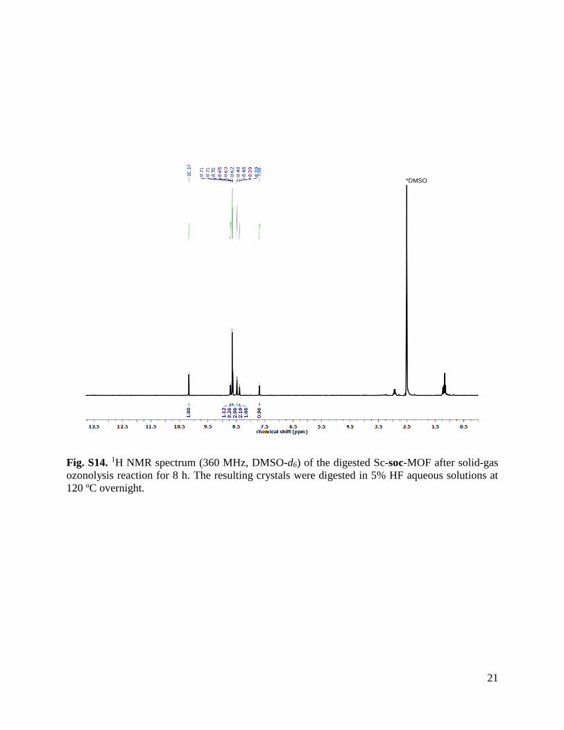
Fig. S14. 1H NMR spectrum (360 MHz, DMSO-d6) of the digested Sc-soc-MOF after solid-gas
ozonolysis reaction for 8 h. The resulting crystals were digested in 5% HF aqueous solutions at
120 ºC overnight.
*DMSO

22
Fig. S15. 1H NMR spectrum (360 MHz, DMSO-d6) of the digested Sc-soc-MOF after 12 cycles
of solid-gas ozonolysis reaction. The resulting crystals were digested in 5% HF aqueous solutions
at 120 ºC overnight.
*DMSO

23
Fig. S16. 1H NMR spectra (360 MHz, DMSO-d6) of digested (A) trimesic acid, (B) 5-
formylisophthalic acid, (C) Sc-soc-MOF, (D) Sc-soc-MOF after solid-gas reaction for 8 h, and (E)
Sc-soc-MOF after solid-gas reaction for 12 cycles. Assigned proton signals: δa = 7.68 (s, 2H), δb
= 8.38 (s, 2H), δc =8.47 (s, 4H), δd =8.65 (s, 3H), δe =8.64 (m, 2H), δf = 8.71 (m, 1H), δg = 10.17
(s, 1H). Note that the characteristic protons from the olefinic bond signal at 7.68 ppm of Sc-soc-
MOF could still be observed after both solid-gas ozonolysis reactions, confirming that the cleavage
of olefinic bonds under these synthetic conditions was not quantitative.
d
d
d
f
e
ab
c
abc
fe
ab
c
fe
g
g
g
B
C
D
E
A
a
b
c d f
e
g
+

24
Fig. S17. Characterization of Sc-soc-MOF after ozonolysis only in water suspension for 12 h. (a) 1H
NMR spectrum (360 MHz, DMSO-d6) of digested Sc-soc-MOF after only in water suspension
ozonolysis reaction for 12 h. This sample was digested in 5% HF aqueous solutions at 120 ºC overnight.
Note that the peak intensity of protons from the olefinic bond at 7.70 ppm (orange dot) was very weak
but it could still be observed, indicating a high degree of cleavage but not quantitative. Moreover, the
signal of the proton at 6.64 ppm (red star) was identified as the intermediate trioxolane-metathesis
product, which is in accordance with the previous paper published by our group (6). (b) ESI-MS
spectrometry of digested Sc-soc-MOF after in water suspension ozonolysis reaction for 12 h. The peak
at m/z 355.05 is assigned to [L2 - H]- in Sc-soc-MOF, corresponding to the formula [C18H12O8 - H]-:
excepted = 355.05, found = 355.05. The peak at m/z 193.01 in both samples is assigned to 5-
formylisophthalic acid, corresponding to the formula [C6H3(COOH)2(CHO) - H]-: expected = 193.02,
found = 193.01. The peak at m/z 209.01 is assigned to H3BTC, corresponding to the formula
[C6H3(COOH)3 - H]-: expected = 209.02, found= 209.01. The peak at m/z 403.03 is assigned to the
trioxolane intermediate, [C18H12O11 - H]-: expected = 403.04, found = 403.03. Altogether, these results
confirm that, under these suspension-gas conditions, ozonolysis could not fully cleave the olefinic
bonds. Moreover, the trioxolane intermediate was also detected.

25
Section S3.2 Single-Crystal X-Ray Diffraction
Table S4. Crystal data and structure refinement for Sc-soc-MOF.
Formula C108O64Sc12
Formula weight (g.mol-1) 2860.60
Temperature (K) 100
Wavelength (Å) 0.82653
Crystal system Trigonal
Space group R -3
Unit cell dimensions a = 31.750(5) Å α = 90º
b = 31.750(5) Å β = 90º
c = 38.800 (5) Å γ = 120º
Volume (Å3) 33872.7
Z 6
Density calculated (g/cm-3) 0.842
Absorption coefficient (mm-1) 0.594
F(000) 8480
Crystal size (mm) 0.27 × 0.23 × 0.21 mm
2Theta range for data collection (º) 2.584 to 24.409
Index ranges -31<=h<=31, -31<=k<=31, -38<=l<=38
Reflection collected 78341
Independent reflections 7872 [Rint = 0.0448, Rsigma = 0.0741]
Refinement method Full-matrix least-squares on F2
Data / restraints / parameters 7872 / 0 / 548
Goodness-of-fit on F2 1.161
Final R indices [I>2sigma(I)] R1 = 0.0998, wR2 = 0.2812
R indices (all data) R1 = 0.1047, wR2 = 0.2870
Largest diff. peak and hole 0.949, -1.316 e.Å-3

26
Table S5. Crystal data and structure refinement for Sc-pcu-MOF.
Formula C31.75O21.83Sc4
Formula weight (g.mol-1) 910.49
Temperature (K) 293(2)
Wavelength (Å) 0.82653
Crystal system Trigonal
Space group R -3
Unit cell dimensions a = 31.650(5) Å α = 90º
b = 31.650(5) Å β = 90º
c = 38.763(8) Å γ = 120º
Volume (Å3) 33628 (12)
Z 18
Density calculated (g/cm-3) 0.809
Absorption coefficient (mm-1) 0.392
F(000) 8085
Crystal size (mm) 0.21 × 0.18 × 0.17 mm
2Theta range for data collection (º) 0.910 to 20.797
Index ranges -31<=h<=31, -31<=k<=31, -38<=l<=38
Reflection collected 74052
Independent reflections 7822 [Rint=0.0494, Rsigma = 0.0241]
Refinement method Full-matrix least-squares on F2
Data / restraints / parameters 7822 / 0 / 539
Goodness-of-fit on F2 2.036
Final R indices [I>2sigma(I)] R1 = 0.1497, wR2 = 0.4331
R indices (all data) R1 = 0.1610, wR2 = 0.4579
Largest diff. peak and hole 0.559, -0.530 e.Å-3

27
Fig. S18. 2Fo-Fc maps calculated for both (A) Sc-soc-MOF and (B) Sc-pcu-MOF, showing only
presence of electron density above and below the plane of the phenyl rings, with no presence of
the olefinic carbon atoms.

28
Fig. S19. Comparison of the cluster environment in (A) Sc-soc-MOF and (B) Sc-pcu-MOF. In Sc-
soc-MOF, each Sc3O cluster is connected to 12 neighboring clusters by six 4-c linkers L2. After
clip-off treatment, each Sc3O in Sc-pcu-MOF is surrounded by only 6 neighboring clusters, which
are interconnected by six 2-c linkers. In both cases, the connectivity of Sc3O clusters is in an
octahedral fashion.

29
Section S3.3 XRPD Analysis
Fig. S20. XRPD of calculated Sc-soc-MOF (black), pristine Sc-soc-MOF (blue), calculated Sc-
pcu-MOF (green) and synthesized Sc-pcu-MOF (red).
30
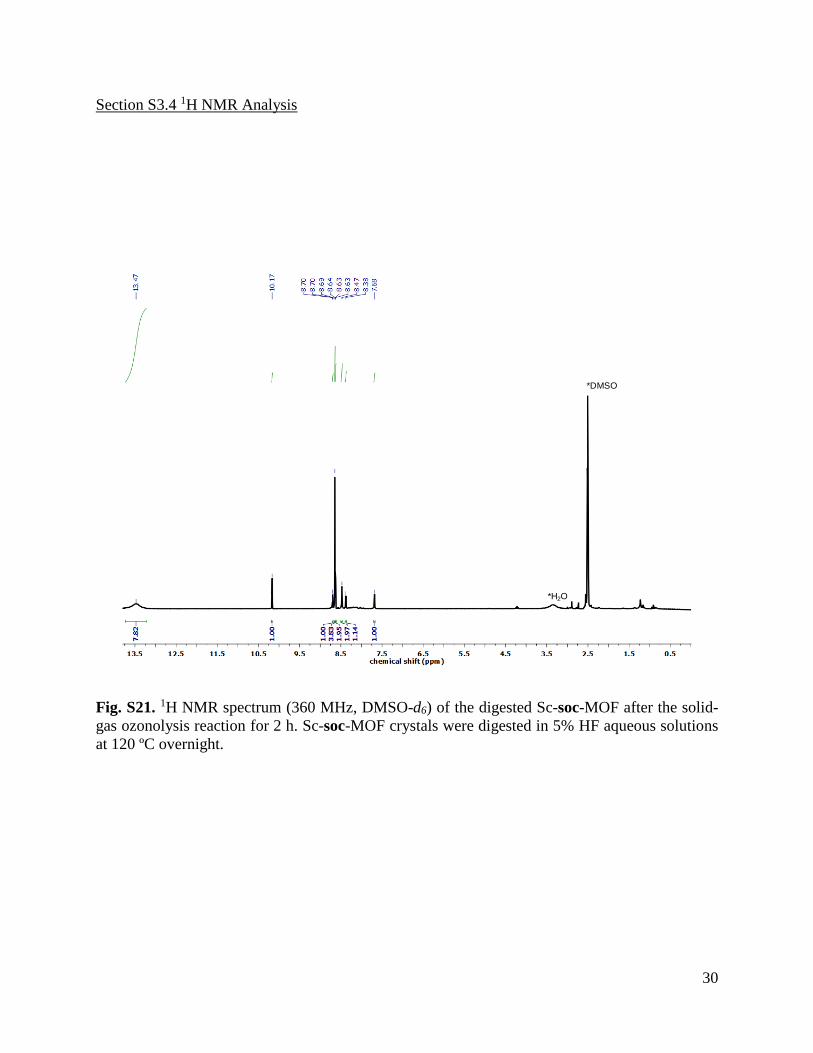
Section S3.4 1H NMR Analysis
Fig. S21. 1H NMR spectrum (360 MHz, DMSO-d6) of the digested Sc-soc-MOF after the solid-
gas ozonolysis reaction for 2 h. Sc-soc-MOF crystals were digested in 5% HF aqueous solutions
at 120 ºC overnight.
*DMSO
*H2O

31
Fig. S22. 1H NMR spectrum (360 MHz, DMSO-d6) of the digested Sc-pcu-MOF synthesized after
the two-step ozonolysis reaction (solid-gas ozonolysis for 2 h and suspension-gas ozonolysis for 6
h). Sc-pcu-MOF crystals were digested in 5% HF aqueous solutions at 120 ºC overnight.
*DMSO*DMSO

32
Fig. S23. 1H NMR spectra (360 MHz, DMSO-d6) of digested (A) trimesic acid, (B) 5-
formylisophthalic acid, (C) Sc-soc-MOF, (D) Sc-soc-MOF after solid-gas reaction (2 h), and (E)
Sc-soc-MOF after solid-gas ozonolysis for 2 h and suspension-gas ozonolysis for 6 h. Note that
this latter spectrum corresponds to that of the synthesized Sc-pcu-MOF. Assigned proton signals:
δa = 7.68 (s, 2H), δb = 8.38 (s, 2H), δc =8.47 (s, 4H), δd =8.65 (s, 3H), δe =8.64 (m, 2H), δf = 8.71
(s, 1H), and δg = 10.17 (s, 1H).
a
b
d f
e
g
+
c
a
ab
b
c
c
d
d
d
e
e
e
f
f
g
g
g
f
A
B
C
D
E

33
Section S3.5 ESI-MS Analysis
Fig. S24. ESI-MS spectrometry of digested Sc-soc-MOF (black) and Sc-pcu-MOF (red). The peak
at m/z 355.05 is assigned to [L2 - H]- in Sc-soc-MOF, corresponding to the formula [C18H12O8 -
H]-: expected = 355.05, found = 355.05. The peak at m/z 193.01 in both samples is assigned to 5-
formylisophthalic acid, corresponding to the formula [C6H3(COOH)2(CHO) - H]-: expected =
193.02, found = 193.01. The peak at m/z 209.01 in both samples is assigned to trimesic acid,
corresponding to the formula [C6H3(COOH)3 - H]-: expected = 209.02, found = 209.01. The
assignment of the most relevant species in the spectra are presented in Table S6.
Table S6. The most relevant species in ESI-MS of the Sc-soc-MOF and Sc-pcu-MOF.
34
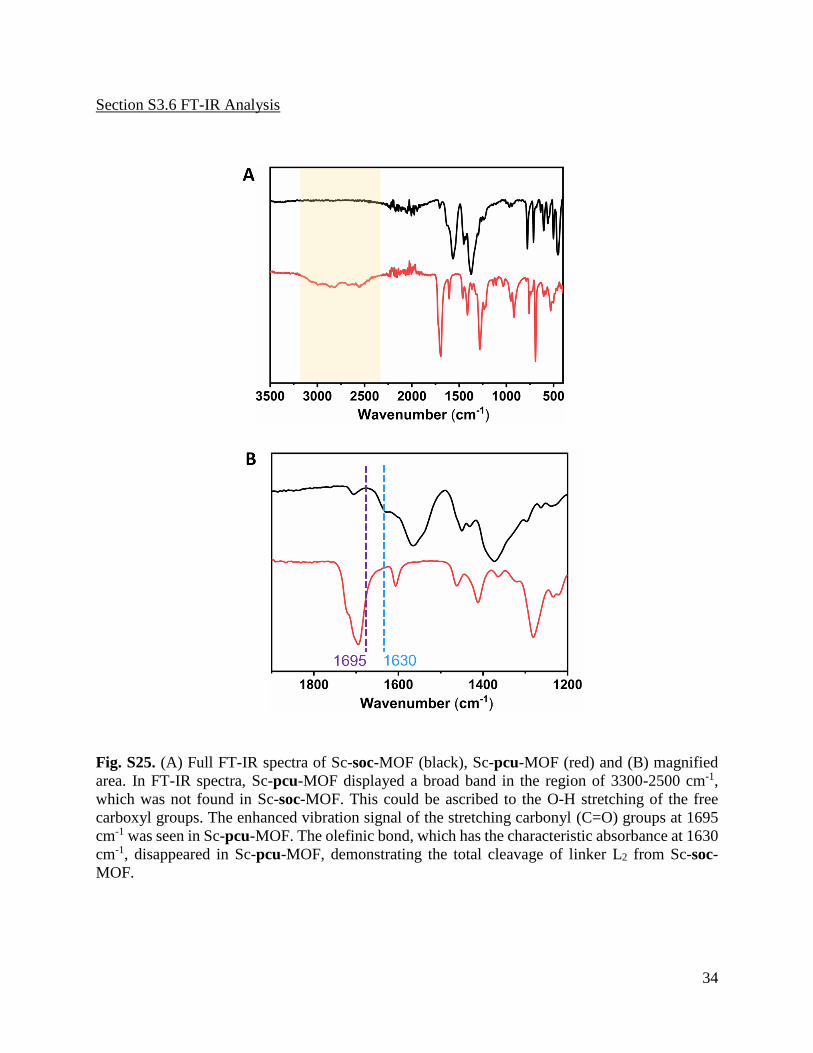
Section S3.6 FT-IR Analysis
Fig. S25. (A) Full FT-IR spectra of Sc-soc-MOF (black), Sc-pcu-MOF (red) and (B) magnified
area. In FT-IR spectra, Sc-pcu-MOF displayed a broad band in the region of 3300-2500 cm-1,
which was not found in Sc-soc-MOF. This could be ascribed to the O-H stretching of the free
carboxyl groups. The enhanced vibration signal of the stretching carbonyl (C=O) groups at 1695
cm-1 was seen in Sc-pcu-MOF. The olefinic bond, which has the characteristic absorbance at 1630
cm-1, disappeared in Sc-pcu-MOF, demonstrating the total cleavage of linker L2 from Sc-soc-
MOF.
35
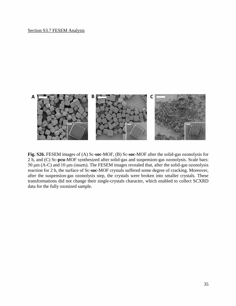
Section S3.7 FESEM Analysis
Fig. S26. FESEM images of (A) Sc-soc-MOF, (B) Sc-soc-MOF after the solid-gas ozonolysis for
2 h, and (C) Sc-pcu-MOF synthesized after solid-gas and suspension-gas ozonolysis. Scale bars:
50 μm (A-C) and 10 μm (insets). The FESEM images revealed that, after the solid-gas ozonolysis
reaction for 2 h, the surface of Sc-soc-MOF crystals suffered some degree of cracking. Moreover,
after the suspension-gas ozonolysis step, the crystals were broken into smaller crystals. These
transformations did not change their single-crystals character, which enabled to collect SCXRD
data for the fully ozonized sample.
A B C

36
Section S4. Clip-off chemistry: from a Rh-MOP to a Rh-macrocycle
Section S4.1 Characterization of Linker 3-[2-(3-carboxyphenyl)ethenyl]benzoic acid (L5)
Fig. S27. 1H NMR spectrum (250 MHz, DMSO-d6) of L5. Assigned proton signals: δf = 13.05 (s,
1H), δd = 8.21 (s, 1H), δa or c = 7.91 (d, J = 8.0 Hz, 1H), δa or c = 7.86 (d, J = 7.9 Hz, 1H), δb = 7.52
(t, J = 7.7 Hz, 1H), and δe = 7.45 (s, 1H).

37
Fig. S28. 13C NMR spectrum (250 MHz, DMSO-d6) of L5. Assigned carbon signals: δ = 168.10,
138.10, 132.17, 131.54, 129.87, 129.47, 129.41, and 128.33.

38
Fig. S29. High-resolution ESI-MS spectrometry of L5. The molecular weight corresponding to [L5
- H]- and its isotopic distribution has been highlighted. The peak at m/z = 267.0651 is assigned to
[C16H12O4 - H]-: expected = 267.0658, found = 267.0651.
267.0651
268.0685
-1
269.0714
-1
-1
C16H12O4
M/Z I I%
1 267.0651 2492 100.0 2 268.0685 459 18.4 3 269.0714 88 3.5

39
Section S4.2 Single-Crystal X-Ray Diffraction
Table S7. Crystal data and structure refinement for Rh-MOP.
Formula C182H66N6O62Rh12
Formula weight (g.mol-1) 4563.32
Temperature (K) 296(2)
Wavelength (Å) 1.54178
Crystal system Triclinic
Space group P -1
Unit cell dimensions a = 18.634(2) Å α = 70.289(6)º
b = 19.5019(18)Å β = 70.220(6)º
c = 25.014(3) Å γ = 70.390(6)º
Volume (Å3) 7796.7(15)
Z 1
Density calculated (g/cm-3) 0.972
Absorption coefficient (mm-1) 5.437
F(000) 2236.0
Crystal size (mm) 0.06 × 0.06 × 0.02 mm
2Theta range for data collection (º) 3.88 to 76.152
Index ranges -14 ≤ h ≤ 13, -15 ≤ k ≤ 15, -20 ≤ l ≤ 20
Reflection collected 25520
Independent reflections 8124 [Rint = 0.1690, Rsigma = 0.1616]
Refinement method Full-matrix least-squares on F2
Data / restraints / parameters 8124/342/1009
Goodness-of-fit on F2 0.931
Final R indices [I>2sigma(I)] R1 = 0.0795, wR2 = 0.2013
R indices (all data) R1 = 0.1304, wR2 = 0.2336
Largest diff. peak and hole 0.58, -0.87 e.Å-3

40
Fig. S30. Lateral (A) and top (B) views of the crystal structure of Rh-MOP.

41
Section S4.3 1H NMR Analysis
Fig. S31. 1H NMR spectrum (360 MHz, DMSO-d6) of digested Rh-MOP. 1 mg of Rh-MOP was
digested in a mixture of DMSO (450 µL) and DCl (20 L, 35 wt. % in D2O) at 100 ºC overnight.
Note that the ratio between both linkers L3 and L5 is 1:1, which agrees with the formula of Rh-
MOP (expected integrations in green and experimental integrations in red).

42
Fig. S32. 1H NMR spectrum (360 MHz, DMSO-d6) of digested Rh-macrocycle. 1 mg of Rh-
macrocycle was digested in a mixture of DMSO (450 µL) and DCl (20 L, 35 wt. % in D2O) at
100 ºC overnight. Note that the ratio between both linkers L3 and isophthalic acid is 1:2, which
agrees with the ratio expected in formula of the Rh-macrocycle (expected integrations in green
and experimental integrations in red).

43
Section S4.4 UV-Vis Analysis
Fig. S33. UV-vis spectra of Rh-MOP (black) and Rh-macrocycle (red) in THF solution. The
maximum of adsorption band I (λmax) of Rh-MOP is centered at 595 nm. The maximum of
adsorption band I (λmax) of Rh-macrocycle is centered at 592 nm.

44
Section S4.6 FT-IR analysis
Fig. S34. (A) Full and (B) magnified area of the FT-IR spectra of Rh-MOP (black) and Rh-based
metal-organic macrocycle (red). The main differences between both spectra are the increase of the
band at 1709 cm-1 in the ozonized sample, which corresponds to the stretching of uncoordinated
carbonyl (C=O) groups. Also, the disappearance of the peak at 1634 cm-1, which corresponds to
the characteristic absorbance of the olefin bonds.
A
45
References
1. J. Juanhuix, F. Gil-Ortiz, G. Cuní, C. Colldelram, J. Nicolás, J. Lidón, E. Boter, C. Ruget, S.
Ferrer, J. Benach, Developments in optics and performance at BL13-XALOC, the
macromolecular crystallography beamline at the Alba Synchrotron. J. Synchrotron Radiat. 21,
679-689 (2014). 2. W. Kabsch, Integration, scaling, space-group assignment and post-refinement. Acta Cryst. D
66, 125-132 (2010). 3. Sheldrick, G. M. SHELXT-Integrated space-group and crystal-structure determination. Acta
Cryst. A, 2015, 71 (1), 3-8.
4. G. M. Sheldrick, Crystal structure refinement with SHELXL. Acta Cryst. C 71, 3-8 (2015). 5. O. V. Dolomanov, L. J. Bourhis, R. J. Gildea, J. A. K. Howard, H. Puschmann, OLEX2: a
complete structure solution, refinement and analysis program. J. Appl. Cryst. 42, 339-341
(2009). 6. J. Albalad, H. Xu, F. Gándara, M. Haouas, C. Martineau-Corcos, R. Mas-Ballesté, S. Barnett,
J. Juanhuix, I. Imaz, D. Maspoch, Single-crystal-to-single-crystal postsynthetic modification
of a metal-organic framework via ozonolysis. J. Am. Chem. Soc. 140, 2028−2031 (2018).

download fileview on ChemRxivSupporting Information.pdf (2.70 MiB)