Clinicobiological, Immunophenotypic, and...
11
Stem Cells, Tissue Engineering and Hematopoietic Elements Clinicobiological, Immunophenotypic, and Molecular Characteristics of Monoclonal CD56 /dim Chronic Natural Killer Cell Large Granular Lymphocytosis Margarida Lima,* Julia Almeida, †‡ Andre ´s Garcı ´a Montero, †‡ Maria dos Anjos Teixeira,* Maria Luı ´s Queiro ´ s,* Ana Helena Santos,* Ana Balanzategui, ‡§ Alexandra Estevinho,* Maria del Ca ´ rmen Alguero ´, ‡ Paloma Barcena, ‡ So ´ nia Fonseca,* Maria Luı ´s Amorim, Jose ´ Manuel Cabeda,* Luciana Pinho,* Marcos Gonzalez, ‡§ Jesus San Miguel, § Benvindo Justic ¸a,* and Alberto Orfa ˜o †‡ From the Servic ¸os de Hematologia * and Microbiologia, Hospital Geral de Santo António, Porto, Portugal; the Servicios de Citometria, † and Hematologia, § Hospital Universitario de Salamanca, Spain; and the Centro de Investigacion del Cancer, ‡ Salamanca, Spain Indolent natural killer (NK) cell lymphoproliferative disorders include a heterogeneous group of patients in whom persistent expansions of mature, typically CD56 , NK cells in the absence of any clonal marker are present in the peripheral blood. In the present study we report on the clinical , hematological , immu- nophenotypic , serological , and molecular features of a series of 26 patients with chronic large granular NK cell lymphocytosis, whose NK cells were either CD56 or expressed very low levels of CD56 (CD56 /dim NK cells), in the context of an aberrant activation-related mature phenotype and proved to be monoclonal using the human androgen receptor gene polymerase chain reaction-based assay. As normal CD56 NK cells, CD56 /dim NK cells were granzyme B , CD3 , TCR/ , CD5 , CD28 , CD11a bright , CD45RA bright , CD122 , and CD25 and they showed variable and het- erogeneous expression of both CD8 and CD57. Never- theless, they displayed several unusual immunopheno- typic features. Accordingly, besides being CD56 /dim , they were CD11b /dim (heterogeneous), CD7 /dim (heterogeneous), CD2 (homogeneous), CD11c bright (homogeneous), and CD38 /dim (heterogeneous). Moreover, CD56 /dim NK cells heterogeneously ex- pressed HLA-DR. In that concerning the expression of killer receptors, CD56 /dim NK cells showed bright and homogeneous CD94 expression, and dim and het- erogeneous reactivity for CD161, whereas CD158a and NKB1 expression was variable. From the functional point of view, CD56 /dim showed a typical Th1 pattern of cytokine production (interferon- , tumor necro- sis factor- ). From the clinical point of view, these patients usually had an indolent clinical course, pro- gression into a massive lymphocytosis with lung in- filtration leading to death being observed in only one case. Despite this, they frequently had associated cy- topenias as well as neoplastic diseases and/or viral infections. In summary, we describe a unique and homogeneous group of monoclonal chronic large granular NK cell lymphocytosis with an aberrant ac- tivation-related CD56 /dim /CD11b /dim phenotype and an indolent clinical course, whose main clinical features are related to concomitant diseases. (Am J Pathol 2004, 165:1117–1127) Indolent natural killer (NK) cell lymphoproliferative disor- ders (LPDs) are usually designated as chronic NK cell lymphocytosis (CNKCL) or chronic NK-large granular lymphocytes (LGL) LPD. 1–6 CNKCLs are heterogeneous and they most probably include reactive chronic NK cell lymphocytosis, true chronic NK cell large granular lym- phocyte (LGL) leukemia, and even some aggressive NK cell lymphoma/leukemia cases, 7–9 because of difficulties in establishing the clonal nature of the NK cell proliferation. Previous studies have shown that in most CNKCL cases the expanded population of NK cells co-expresses CD56, CD16, and CD94 and displays NK activity. Such phenotypic and functional features are similar to those Partially supported by the Ministerio de Sanidad y Consumo, Madrid, Spain (grant FIS 02/1244); the Consejerı ´a de Educacio ´n y Cultura, Junta de Castilla y Leo ´ n, Valladolid, Spain (grant SA103/03); the University of Salamanca (Reg. N. 430 to P.B.); and the Instituto de Salud Carlos III, Spain (grant CP03/00035 to A.C.G.-M.). Accepted for publication June 10, 2004. Address reprint requests to Margarida Lima, Servic ¸ o de Hematologia, Unidade de Citometria, Hospital Geral de Santo Anto ´ nio, Rua D Manuel II, s/n, 4099-001 Porto, Portugal. E-mail: [email protected]. American Journal of Pathology, Vol. 165, No. 4, October 2004 Copyright © American Society for Investigative Pathology 1117
Transcript of Clinicobiological, Immunophenotypic, and...

Stem Cells, Tissue Engineering and Hematopoietic Elements
Clinicobiological, Immunophenotypic, and MolecularCharacteristics of Monoclonal CD56�/�dim ChronicNatural Killer Cell Large Granular Lymphocytosis
Margarida Lima,* Julia Almeida,†‡
Andres Garcıa Montero,†‡ Maria dos Anjos Teixeira,*Maria Luıs Queiros,* Ana Helena Santos,*Ana Balanzategui,‡§ Alexandra Estevinho,*Maria del Carmen Alguero,‡ Paloma Barcena,‡
Sonia Fonseca,* Maria Luıs Amorim,�
Jose Manuel Cabeda,* Luciana Pinho,*Marcos Gonzalez,‡§ Jesus San Miguel,§
Benvindo Justica,* and Alberto Orfao†‡
From the Servicos de Hematologia * and Microbiologia,� Hospital
Geral de Santo António, Porto, Portugal; the Servicios de
Citometria,† and Hematologia,§ Hospital Universitario de
Salamanca, Spain; and the Centro de Investigacion del Cancer,‡
Salamanca, Spain
Indolent natural killer (NK) cell lymphoproliferativedisorders include a heterogeneous group of patientsin whom persistent expansions of mature, typicallyCD56�, NK cells in the absence of any clonal markerare present in the peripheral blood. In the presentstudy we report on the clinical, hematological, immu-nophenotypic, serological, and molecular features ofa series of 26 patients with chronic large granular NKcell lymphocytosis, whose NK cells were either CD56�
or expressed very low levels of CD56 (CD56�/�dim NKcells), in the context of an aberrant activation-relatedmature phenotype and proved to be monoclonal usingthe human androgen receptor gene polymerase chainreaction-based assay. As normal CD56� NK cells,CD56�/�dim NK cells were granzyme B�, CD3�, TCR��/���, CD5�, CD28�, CD11a�bright, CD45RA�bright,CD122�, and CD25� and they showed variable and het-erogeneous expression of both CD8 and CD57. Never-theless, they displayed several unusual immunopheno-typic features. Accordingly, besides being CD56�/�dim,they were CD11b�/�dim (heterogeneous), CD7�/�dim
(heterogeneous), CD2� (homogeneous), CD11c�bright
(homogeneous), and CD38�/�dim (heterogeneous).Moreover, CD56�/�dim NK cells heterogeneously ex-pressed HLA-DR. In that concerning the expression ofkiller receptors, CD56�/�dim NK cells showed bright
and homogeneous CD94 expression, and dim and het-erogeneous reactivity for CD161, whereas CD158a andNKB1 expression was variable. From the functionalpoint of view, CD56�/�dim showed a typical Th1 patternof cytokine production (interferon-��, tumor necro-sis factor-��). From the clinical point of view, thesepatients usually had an indolent clinical course, pro-gression into a massive lymphocytosis with lung in-filtration leading to death being observed in only onecase. Despite this, they frequently had associated cy-topenias as well as neoplastic diseases and/or viralinfections. In summary, we describe a unique andhomogeneous group of monoclonal chronic largegranular NK cell lymphocytosis with an aberrant ac-tivation-related CD56�/�dim/CD11b�/�dim phenotypeand an indolent clinical course, whose main clinicalfeatures are related to concomitant diseases. (Am JPathol 2004, 165:1117–1127)
Indolent natural killer (NK) cell lymphoproliferative disor-ders (LPDs) are usually designated as chronic NK celllymphocytosis (CNKCL) or chronic NK-large granularlymphocytes (LGL) LPD.1–6 CNKCLs are heterogeneousand they most probably include reactive chronic NK celllymphocytosis, true chronic NK cell large granular lym-phocyte (LGL) leukemia, and even some aggressive NKcell lymphoma/leukemia cases,7–9 because of difficulties inestablishing the clonal nature of the NK cell proliferation.
Previous studies have shown that in most CNKCLcases the expanded population of NK cells co-expressesCD56, CD16, and CD94 and displays NK activity. Suchphenotypic and functional features are similar to those
Partially supported by the Ministerio de Sanidad y Consumo, Madrid,Spain (grant FIS 02/1244); the Consejerıa de Educacion y Cultura, Juntade Castilla y Leon, Valladolid, Spain (grant SA103/03); the University ofSalamanca (Reg. N. 430 to P.B.); and the Instituto de Salud Carlos III,Spain (grant CP03/00035 to A.C.G.-M.).
Accepted for publication June 10, 2004.
Address reprint requests to Margarida Lima, Servico de Hematologia,Unidade de Citometria, Hospital Geral de Santo Antonio, Rua D Manuel II,s/n, 4099-001 Porto, Portugal. E-mail: [email protected].
American Journal of Pathology, Vol. 165, No. 4, October 2004
Copyright © American Society for Investigative Pathology
1117

observed in neoplastic NK cells from aggressive NK cellleukemia/lymphoma, nasal, and nasal-type lympho-mas.10 Despite this, few cases of CNKCL and LGL leu-kemias of CD56�/�dim NK cells have been sporadicallydescribed10–13 or used to establish NK cell lines.14 NKcells showing decreased or no expression of CD56 havenot been reported either in healthy individuals or in dis-ease conditions associated to either a transient or per-sistent increase in the number of NK cells in the periph-eral blood (PB);15 in such cases, the expanded cellpopulation constantly shows co-expression of CD16 andCD56 frequently in association with acute or chronicNK cell activation features. These findings may sug-gest that a CD56�/�dim NK cell phenotype could beaberrant and reflect underlying clonal genetic abnor-malities. However, to the best of our knowledge nostudy has been reported so far in which this uniquegroup of CNKCL is studied in detail.
In contrast to both CD8�/TCR��� and CD4�/TCR���
T-cell LGL leukemias, in which the most characteristicclinical manifestations of the disease—such as its asso-ciation with cytopenias and arthritis or second neopla-sias—are well known,16–18 the clinical features associ-ated with CNKCL including NK cell LGL leukemia, are faraway from being well established. Previous studies havesuggested a possible association of CNKCL with cytope-nias, vasculitis, neutropenic or nonneutropenic fever, BMgranulomas,1–3,19,20 and even Epstein-Barr virus (EBV)infection in endemic areas.21,22
In the present study we report on the clinical, hemato-logical, immunophenotypical, functional, serological, andmolecular features of 26 patients with CNKCL in whomthe expanded PB NK cells were either CD56� or ex-pressed very low levels of surface CD56 (CD56�/�dim
CNKCL) in the context of an aberrant immunophenotypeand a mature LGL morphological appearance.
Materials and Methods
Patients
A total of 26 consecutive patients with CNKCL whoseLGL appearing circulating NK cells were CD56�/�dim,were studied at diagnosis in two different centers. Thesecenters received cases referred for the diagnosis of T/NKchronic LPD from other hospitals in Portugal (CytometryUnit of the Hematology Service at the Hospital Geral deSanto Antonio, Porto, Portugal) and in Spain (CytometryService, University and University Hospital of Salamanca,Salamanca, Spain). Altogether, these CD56�/dim patientsrepresented 20% of the total group of 130 CNKCL casesand 1% of a total of 2368 mature chronic LPDs diagnosedin both institutions between September 1997 and Decem-ber 2002, from which 64% were B-cell chronic lympho-cytic leukemias. Results obtained were compared tothose observed on PB CD56� NK cells from 12 age- andsex-matched healthy individuals, 15 patients with acuteviral infection, and 15 patients with CD56� chronic NKcell lymphocytosis associated with tumors or chronic in-
fections whose immunophenotypic features have beenpreviously published in detail.15,23
Immunophenotypic Studies
In all cases cell surface immunophenotypic studies wereperformed as previously described in detail.10 Stainingswere performed using an ethylenediaminetetraacetic ac-id-anti-coagulated whole blood stain-lyse-and-then-washmethod and a four-color panel of monoclonal antibodiesconjugated with fluorescein isothiocyanate, phyco-erythrin, phycoerythrin-cyanine 5, or peridin chlorophyllprotein and allophycocyanine directed against the follow-ing antigens: TCR��, TCR��, CD8, CD4, CD3, CD28,CD2, CD7, CD56, CD5, CD57, CD11c, CD38, CD11b,CD45RA, CD45RO, CD122, CD25, CD11a, HLA-DR,CD158a, CD161, CD16, NKB1, and CD94.23 In addition,the expression of cytoplasmic granzyme B was evaluatedusing the Fix and Perm reagent kit (Caltag Laboratories,Burlingame, CA), according to the manufacturer’s in-structions.
Functional analysis of cytokine production by the ex-panded NK cells was also performed as previously de-scribed.18 Briefly, 500 �l of heparinized blood wereplaced into a tube containing 500 �l of RPMI 1640 culturemedium (BioWhittaker, Walkersville, MD) supplementedwith 2 �mol/L of L-glutamine. Cells were cultured for 4hours at 37°C in a 5% CO2 and 95% humidity sterileenvironment in the presence of 25 ng/ml of phorbol-12myristate 13-acetate, 1 �g/ml of ionomycin, and 10 �g/mlof brefeldin A (stimulated samples) or only brefeldin A(nonstimulated samples). Once this incubation periodwas completed, each sample was sequentially stainedfor the CD3 and CD19 surface antigens—and intracellu-lar cytokines—interleukin (IL)-2, IL-4, IL-10, IL-13, inter-feron (IFN)-�, tumor necrosis factor (TNF)-�, and TNF-�.18 In these studies NK cells were identified as CD19�,CD3� lymphocytes.
Data acquisition was performed in each of the twocenters in FACSCalibur flow cytometers [Becton/Dickin-son Biosciences (BDB), San Jose, CA], equipped withtwo lasers using the Cell QUEST software program(BDB). Information on a minimum of 2 � 105 events wasacquired and stored as list mode data for each staining.For data analysis the Paint-A-Gate PRO software pro-gram (BDB) was used. Identification and enumeration ofNK cells present in the sample was performed as follows:the percentage of NK cells from all PB nucleated cellsand lymphocytes was calculated by selecting CD3� lym-phoid cells that expressed CD2 and/or CD7; then, thenormal and abnormal NK cell populations were identifiedaccording to the unique pattern of expression of theCD56, CD2, and CD7 antigens observed in normal NKcells15,23 and the proportion of abnormal CD56�/�dim,normal CD56�, and normal CD56�� NK cells within totalNK cells was determined. Afterward, the abnormal CD56�/
�dim NK cell population was characterized for the expres-sion of the other cell surface antigens analyzed. For eachantigen studied, the following characteristics were record-ed: 1) percentage of positive cells; 2) intensity of expres-
1118 Lima et alAJP October 2004, Vol. 165, No. 4
sion— absent (�), low (�/�), moderate (�), high (��),very high (���)—evaluated as their mean fluorescenceintensity (MFI) (arbitrary relative linear units scaled from 0to 10,000 fluorescence channels) and; 3) pattern of anti-gen expression (homogeneous versus heterogeneous),evaluated by the coefficient of variation (CV).
Flow Cytometry DNA Cell Content Studies
DNA studies were performed in a subset of patients (n �6). Briefly, PB cells were double stained for either surfaceCD16 or CD94 and nuclear DNA (DNA-Prep; Beckman-Coulter, Hialeah, FL) and acquired in an EPICS-XL-MCLflow cytometer using the XL2 software (Beckman-Coulter).Both the DNA ploidy status and the cell cycle distributionof PB NK cells were evaluated using the MultiCycle soft-ware (Phoenix Flow Systems, San Diego, CA).
Human Androgen Receptor GeneAssay (HUMARA)
To confirm the clonal origin of the aberrant NK cell pop-ulations, the pattern of inactivation of chromosome X wasstudied in 4 of 15 female patients by assessing the meth-ylation of human androgen receptor gene using the HU-MARA polymerase chain reaction (PCR)-based assay.24
For that purpose, purified CD56�/dim NK cells (mean,97 � 2.6%; range, 93.6 to 99.6%) and the correspondingNK cell-depleted leukocyte fractions from the four femalepatients were studied in parallel. NK cells were purifiedby a single immunomagnetic depletion step using theHuman NK Cell Isolation Kit II in the automated magneticcell separator autoMACS (Miltenyi Biotec, Bergisch Glad-bach, Germany), strictly following the manufacturer’s rec-ommendations. Digestion of genomic DNA with the meth-ylation-sensitive restriction endonuclease HapII andsubsequent PCR amplification of the methylated (inacti-vated) alleles was performed, as previously described.24
Molecular Analysis of TCR GeneRearrangements
Molecular analyses of TCR gene rearrangements wereperformed in most cases using either conventional South-ern blot-based TCR-� gene (n � 2) or PCR-based TCR-�gene (n � 12) techniques or both (n � 6). Briefly, DNAwas extracted from mononuclear cells obtained by den-sity gradient centrifugation, using the chloroform method.For Southern blot, DNA was digested with the EcoRI andHindIII restriction enzymes and DNA fragments were sep-arated by a 0.8% agarose gel electrophoresis and trans-ferred to nitrocellulose membranes by vacuum blotting,UV fixed, and hybridized with 32P-labeled probes for theTCR-� gene region (C�, TCRBC, and TCRBJ2; DAKOA/S, Glostrup, Denmark). PCR amplification of the TCR-�gene was done using the BIOMED-2 primer sequencesand protocols,25 with a limit of detection varying from 1 to10%, depending on the polyclonal TCR repertoire com-plexity, and the type of TCR-� gene rearrangements.26
Serological and Molecular Studies for ViralInfection
Sera from nine patients were tested for the presence ofIgM and IgG antibodies against cytomegalovirus (CMV),EBV viral capside (EBV-VCA), and nuclear (EBNA) anti-gens, parvovirus, herpes simplex (HSV) type 1 and 2,varicella zoster (VZV), rubella, and hepatitis A, B, and Cviruses. In addition, 13 patients were tested for the pres-ence of antibodies against human immunodeficiency(HIV) and human T-cell leukemia/lymphoma (HTLV) typeI and type II viruses.
The presence of HTLV-1, CMV, EBV, HSV, VZV, andhuman herpesvirus type 6 (HHV-6) DNA sequences wasalso evaluated in nine patients by real-time PCR assaysusing DNA extracted from PB mononuclear cells with theMagNA Pure machine and the MagNA Pure LC totalnucleic acid isolation kit (Roche Diagnostics, Mannheim,Germany). Amplification of CMV, HSV, and HHV-6 DNAwas done using an in-house developed real-time PCRassay with synthetic genetic primers and probes (Epoch,USA) in the SmartCycler system (Cepheid, USA) whereasEBV and VZV DNA were detected using commerciallyavailable kits (Artus, Hamburg, Germany) and the Light-Cycler System (Roche Diagnostics). HTLV-1 DNA wasdetected using an in-house developed real-time PCRassay with previously described primers and probes27
and the LightCycler FastStart DNA Master HybridizationProbes (Roche Diagnostics).
Other Laboratory Parameters
Other laboratory parameters included a full hematologi-cal blood cell count, a morphological evaluation of bloodsmears, and a routine biochemical survey including se-rum levels of liver enzymes, creatinine, lactate dehydro-genase, �2-microglobulin, immunoglobulins (Igs), anti-nuclear antibodies, and rheumatoid factor.
Statistical Analyses
For all variables under study, median, mean, SD, andrange values were calculated. To explore for the statisti-cal significance of the differences observed betweengroups the Mann-Whitney U-test was used (SPSS software,version 11; SPSS, Chicago, IL). P values �0.05 were con-sidered to be associated with statistical significance.
Results
Clinical and Laboratory Findings
Table 1 summarizes the clinical and hematological find-ings at diagnosis for the 26 patients studied. As may beseen in Table 1, all patients were adults more than 35years of age and there was a slight predominance infemales (male/female ratio of 0.73). In the great majorityof the cases (18 of 26) lymphocytosis was an occasionalfinding in either a routine blood analysis (n � 9) or in
CD56�/�dim Chronic NK Cell Lymphocytosis 1119AJP October 2004, Vol. 165, No. 4

blood analysis performed during investigation or monitor-ing of another pathological condition (n � 9), consistingof diabetes mellitus (n � 1), arthritis (n � 1), adenomega-lies (n � 1), idiopathic thrombocytopenic purpura (n �3), autoimmune hemolytic anemia (n � 1), and B-celllymphoma (n � 2). In the remaining eight cases, diagno-sis was obtained in blood analyses performed becauseof co-existing active pathological conditions: HIV infec-tion (n � 3), prostatic carcinoma (n � 1), colon carci-noma, refractory anemia with excess of blasts, autoim-mune hemolytic anemia (n � 1), hepatitis C virus infection(n � 1), idiopathic thrombocytopenic purpura (n � 1),and breast carcinoma (n � 1).
At the moment of diagnosis 96% of the patients hadlymphocytosis (�5.0 � 109/L). Hemoglobin, neutrophil,and platelet counts were decreased in 46%, 44%, and28% of the cases, respectively. Symptomatic anemia(�75 g/L) requiring red blood cell transfusion was ob-served in only three patients who suffered from severeCoomb’s-negative (n � 2) or Coomb’s-positive (n � 1)hemolytic anemia; in addition, one patient had a pasthistory of autoimmune hemolytic anemia that respondedcompletely to corticosteroids plus splenectomy and an-other patient had a past episode of hemolytic anemia ofunknown origin that solved spontaneously. Neutropeniawas severe (�500 � 106/L) in six cases, although onlyone patient had a past history of repeated or severeinfections. Clinically relevant thrombocytopenia (�50 �109/L) was observed at diagnosis of CNKCL in only twocases, one of them being diagnosed has idiopathicthrombocytopenic purpura; in addition, three other pa-tients had a past history of idiopathic thrombocytopenicpurpura. One of these cases had a partial response to
corticosteroids and a complete response to splenec-tomy; the other two cases had been successfully treatedwith corticosteroids. Four patients underwent splenec-tomy 2 to 12 years before the diagnosis of CNKCL, eitherfor an associated condition (n � 3) or because of trauma(n � 1).
At diagnosis, all except three patients were asymptom-atic. In these three cases symptoms were related tocytopenias and/or an associated-disease. Physical ex-amination (Table 1) showed the presence of splenomeg-aly, adenomegalies, and hepatomegaly in only a minorproportion of cases (15%). Only one patient suffered fromarthritis, which was diagnosed as rheumatoid arthritis. Skinlesions were not found neither were fever or vasculitis.
Increased lactate dehydrogenase (mean � SD, 484 �185 U/L; range, 208 to 856 U/L) and �2-microglobulin(mean � SD, 3.24 � 1.61; range, 1.30 to 7.10) serumlevels were observed in 50% and 80% of the cases,respectively. Ig serum levels were abnormal in 15 of 21patients and consisted of increased IgM (n � 3), IgG (n �7), IgA (n � 5), and/or IgE (n � 2); in addition, 5 patientshad moderate IgA (n � 4) or IgG (n � 1) hypogammaglobu-linemia. Serum rheumatoid factor was positive and serumlevels of anti-nuclear antibodies were increased in 4 of 9and 3 of 11 patients tested, respectively.
Studies aimed at detecting underlying viral infectionsconfirmed the presence of HIV infection in three patients,being associated with HTLV1 infection in one of them.Hepatitis C virus infection was detected in three addi-tional patients. Moreover, serological studies suggestedpast infection with CMV, EBV, and HSV-1 each in nine ofnine cases tested, HSV-2 in three of seven cases, VZV inseven of eight patients studied, parvovirus in five of nine,and hepatitis B virus in two of nine cases tested. Sero-logical data from a control group of 5853 outcare adultpatients showed a high incidence of previous infectionwith the viruses analyzed here (CMV, 80%; EBV/anti-VCA, 88%; EBV/anti-EBNA, 80%; HSV, 88%; VZV, 85%;and parvovirus, 77%).
Immunophenotypic Features of CD56�/�dim
NK Cells
NK cells accounted for 68 � 18% of all PB lymphocytes(range, 19 to 95%) increased PB NK cell counts beingobserved in all cases. The vast majority (97.6 � 4.1%) ofall PB NK cells had an abnormal immunophenotype asillustrated in Figure 1 and the main phenotypic differ-ences and similarities between CD56�/�dim NK cells,normal, and activated CD56� NK cells are detailed inFigure 2 and illustrated in Figure 3. As shown there, themost relevant immunophenotypic feature consisted on anabnormally low (P � 0.0001) CD56 expression on themajority of PB NK cells (CD56�/�dim NK cells); most ofthe CD56�/�dim NK cells (78 � 21%) were CD56� and aminority of them (22 � 21%) showed dim and heteroge-neous CD56 expression, as compared to normal NKcells. Residual CD56� and CD56�� NK cells were de-tected in all but one patient, accounting for only 2.3 �4.1% and 0.3 � 0.3% of total PB NK cells, respectively.
Table 1. Clinical and Laboratory Characteristics of Individualswith Chronic CD56�/�dim NK Cell LGLLymphocytosis
Age 68 � 17 (37–94)Sex (male/female) 11/15 (42%/58%)Reason for consulting
Routine blood analysis (65%)Adenomegalies (4%)Viral infection (15%)Concomitant malignant neoplasias (12%)Other (ITP) (4%)
Physical examinationAdenophaties (13%)Splenomegaly (15%)*Hepatomegaly (4%)Skin lesion (0%)†
Arthritis (4%)Peripheral blood cell counts
Hemoglobin (g/L) 123 � 27 (66–163)Platelets (�109/L) 186 � 83 (26–417)WBC (�109/L) 128 � 96 (2–43)Neutrophils (�109/L) 2.3 � 2.4 (0.1–11.4)Lymphocytes (�109/L) 9.0 � 8.9 (1.2–40.8)NK cells (�109/L) 6.7 � 8.4 (0.5–38.7)CD56�/�dim NK-cells (�109/L) 6.6 � 8.3 (0.5–38.7)
* Four cases had splenectomy before diagnosis of CNKCL for anassociated condition corresponding to an idiopathic thrombocytopeniapurpura (n � 1), autoimmune hemolytic anemia (n � 1), trauma (n �1), or splenic B-cell lymphoma (n � 1).
† Two of the patients had past history of herpetic skin lesions, infectionwith HZV being confirmed with serological analysis in one of them.
1120 Lima et alAJP October 2004, Vol. 165, No. 4

As normal CD56� cells, CD56�/�dim NK cells weregranzyme B�, CD3/TCR���, and CD5� and displayed avariable and heterogeneous reactivity for CD8 except inthree patients who had abnormally high and homoge-neous CD8 expression. Besides showing defective CD56expression, the expanded NK cells also had abnormallylow expression of CD7 (P � 0.0001), CD38 (P � 0.0002),and other surface molecules involved in NK cell adhesionand cytotoxic activity, such as CD11b (P � 0.0001, CD11b(P � 0.0001), CD57 (P � 0.008), and CD16 (P � 0.05)(Figure 2). In contrast, abnormal CD56�/�dim NK cells hada stronger and more homogeneous (ho) CD2 (P � 0.0001)and CD11c expression (P � 0.0005) (Figure 2).
In that concerning the expression of killer receptors,most CD56�/�dim NK cells were CD94� with a higher(P � 0.03) and more homogeneous (P � 0.02) expres-sion than that observed in normal CD56� NK cells.CD161 and CD158a were dimly and heterogeneouslyexpressed in a variable fraction of CD56�/�dim NK cellswhereas NKB1 expression was found in a variable pro-portion (49 � 34%) of CD56�/�dim NK cells in only 29% ofthe patients.
As normal PB NK cells, CD56�/�dim NK cells wereCD25�, CD5�, CD11a�bright, ho CD45RA�bright, ho, andCD122�. Nevertheless, CD56�/�dim NK cells expressed
much higher levels of HLA-DR (P � 0.0001) than normalCD56� NK cells, the majority of CD56�/�dim NK cellsbeing HLA-DR� (mean of 62 � 23%, versus 16 � 14%observed on normal CD56� NK cells; P � 0.0001). Inaddition, CD122 and CD45RA expression were weaker(P � 0.0001 and P � 0.0009, respectively) and moreheterogeneous (P � 0.01 and P � 0.03, respectively)whereas reactivity for CD45RO was slightly higher (P �0.03) in the aberrant CD56�/�dim NK cells as comparedto normal PB NK cells.
Some of the immunophenotypic differences found be-tween normal and CD56�/�dim NK cells—ie, overexpres-sion of CD11c and HLA-DR and down-regulation ofCD45RA accompanied in some cases by co-expressionof CD45RO in a variable fraction of NK cells—do in factreproduce the immunophenotypic changes that typicallyoccur on recently activated CD56� NK cells, whereasother—ie, increased CD2 expression and decreased andheterogeneous expression of CD7, CD11b, and CD38—were similar to those typically observed in conditions ofchronic NK cell stimulation.15 Despite these similaritiesbetween CD56�/�dim NK cells and either recently orchronically activated CD56� NK cells, major differenceswere observed in the pattern of expression of the above-mentioned markers. Accordingly, CD56�/�dim NK cells
Figure 1. Illustrative bivariate histograms of the most characteristic immunophenotypic features of abnormally expanded CD56�/�dim NK cells (red dots) in apatient with CNKCL. Black and gray dots correspond to the T and B cells present in the same blood sample, respectively.
CD56�/�dim Chronic NK Cell Lymphocytosis 1121AJP October 2004, Vol. 165, No. 4
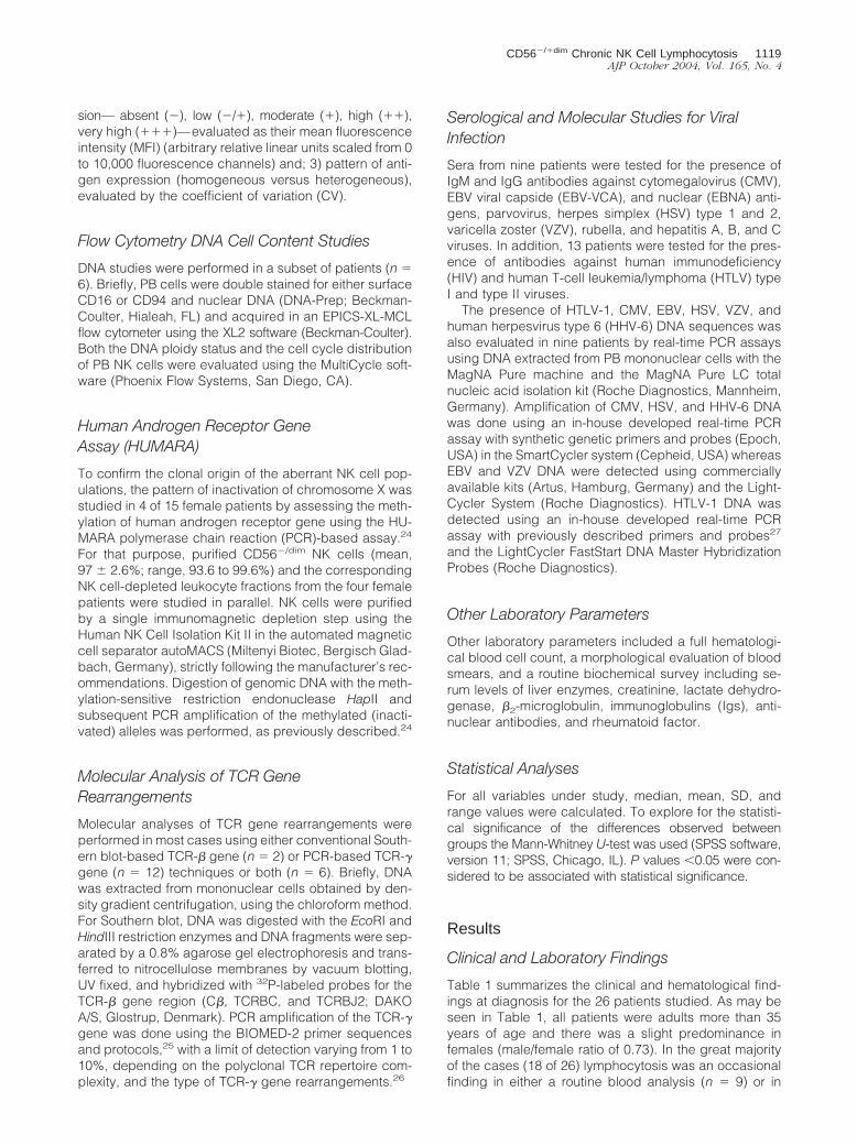
had higher and more homogenous (P � 0.002) expres-sion of CD11c, greater percentages of CD57� cells (P �0.007), and lower levels of HLA-DR (P � 0.0006) ascompared to recently activated NK cells. Additionally,CD2 expression was more homogeneous (P � 0.004),down-regulation of CD7 and CD11b were more pro-
nounced (P � 0.0001), and CD7 expression was evenmore heterogeneous (P � 0.02) on CD56�/�dim NK cellsthan on chronically activated CD56� NK cells. Moreover,disagreement between down-regulation of CD11b andCD38 expression, consisting of a CD11b�/CD38� phe-notype, was frequently found in CD56�/�dim NK cells (10
1122 Lima et alAJP October 2004, Vol. 165, No. 4

of 26 cases) but not on chronically activated CD56� NKcells.
Cytokine Production by CD56�/�dim NK Cells
As normal PB NK cells, CD56�/�dim NK cells showed atypical Th1 pattern of cytokine production after stimula-tion with phorbol-12 myristate 13-acetate plus ionomycin(Figure 4). Accordingly, these cells produced IFN-� andTNF-�, at the same time they failed to secrete IL-2, IL-4,
IL-10, IL-13, and TNF-� under these stimulatory condi-tions. A more detail comparison with normal PB NK cellsshowed that the proportion of CD56�/�dim NK cells pro-ducing both IFN-� and TNF-� and the amount of thesecytokines produced per cell was similar to that of normalCD56� PB NK cells (75 � 31% versus 75 � 21%, P � 0.7,and 52% � 36% versus 36 � 12%, P � 0.5, respectively).
Flow Cytometry DNA Cell Contents ofCD56�/�dim NK Cells
In all cases analyzed, CD56�/�dim NK cells had a diploidDNA content and a very low fraction of cells in the S(0.4 � 0.5%) and G2/M (0.3 � 0.4%) cell cycle phases.
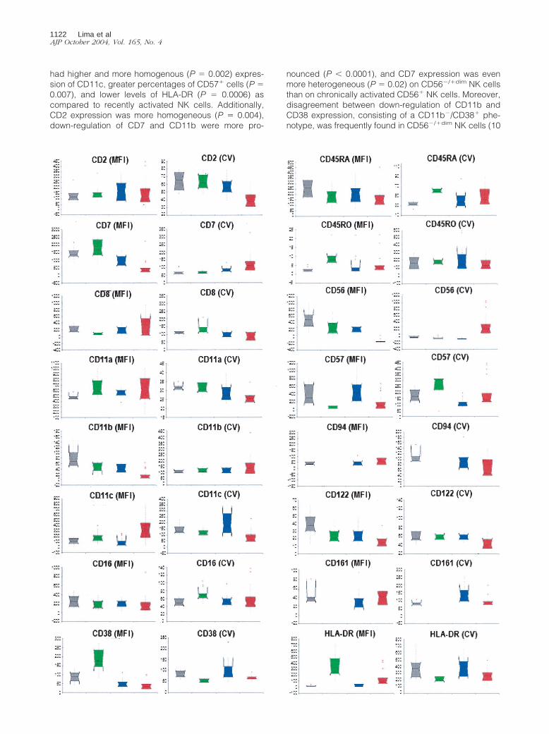
Figure 2. Immunophenotypic characteristics of PB NK cells from patients with CD56�/�dim CNKCL (red boxes, n � 26) as compared to healthy individuals (grayboxes, n � 12), patients with acute viral infection (green boxes, n � 15), and patients with CD56� CNKCL associated with chronic viral infection or neoplasias(blue boxes, n � 15). The notched box and whiskers show nonparametric statistics, including the median, the lower and upper quartiles, and the 95%confidence interval around the median (notched box); the dotted lines connect the nearest observations within 1.5 interquartile ranges of the lower and upperquartiles; red crosses or circles indicate outliers. Differences between the groups were explored using the Mann-Whitney U-test. Statistically significant P values(�0.05): 1) CD56�/�dim CNKCL versus healthy individuals: CD2(CV): �0.0001; CD7(MFI): �0.0001; CD7(CV): �0.0001; CD8(MFI): 0.04; CD11a(CV): 0.006;CD11b (MFI): � 0.0001; CD11c (MFI): 0.002; CD11c(CV): 0.0005; CD16 (MFI): 0.04; CD38 (MFI): 0.0002; CD38(CV): 0.003; CD45RA (MFI): 0.0009; CD45RA(CV):0.03; CD45RO (MFI): 0.03; CD56 (MFI): �0.0001; CD56(CV): �0.0001; CD57 (MFI): 0.008; CD94(CV): 0.02; CD122(MFI): �0.0001; CD122(CV): 0.01; HLA-DR(MFI): 0.0001; HLA-DR(CV): 0.05.2) CD56�/�dim CNKCL versus acute viral infection: CD2(CV): � 0.0001; CD7(MFI): �0.0001; CD7(CV); �0.0001; CD11a(CV):0.002; CD11b (MFI): 0.0005; CD11c(CV): 0.002; CD16(CV): 0.005; CD38 (MFI): �0.0001; CD38(CV): 0.0002; CD45RA(CV): 0.02; CD45RO (MFI): 0.0007; CD56(MFI): � 0.0001; CD56(CV): �0.0001; CD57(CV): 0.02; CD122(MFI): 0.02; CD122(CV): 0.009; HLA-DR (MFI): 0.0006.3) CD56�/�dim CNKCL versus CD56� CNKCLassociated to neoplasias or chronic viral infections: CD2(CV): 0.004; CD7(MFI): �0.0001; CD7(CV); 0.02; CD11b (MFI): �0.0001; CD11c (MFI): 0.0003;CD11c(CV): � 0.0001; CD38(CV): 0.002; CD45RA (MFI): 0.03; CD56 (MFI): � 0.0001; CD56(CV): �0.0001; CD57 (MFI): 0.0003; CD57(CV): 0.003; CD94(CV): 0.01;CD161(CV): 0.0002; HLA-DR (MFI): � 0.0001.
Figure 3. Representative bivariate dot plots illustrating the most relevantphenotypic differences between PB NK cells (red dots) from a healthyindividual (column A), and patients with acute (column B) and chronic(column C) viral infections as well as a case of CD56�/�dim CNKCL (col-umn D).
Figure 4. In vitro TNF-� and IFN-� production by phorbol-12 myristate13-acetate plus ionomycin-stimulated NK cells from CD56�/�dim CNKCLpatients (filled boxes) and healthy individuals (open boxes). Results areexpressed as percentage of cytokine� NK cells (A) and mean fluorescenceintensity levels of expression of cytoplasmic cytokines in NK cells (B). Boxesextend from the 25th to the 75th percentiles; the line in the middle and thevertical lines represent median values and 95% confidence intervals, re-spectively. No significant differences (P � 0.05) were observed between themedian values of the patient and the control groups using the Mann-WhitneyU-test.
CD56�/�dim Chronic NK Cell Lymphocytosis 1123AJP October 2004, Vol. 165, No. 4

Clonality of CD56�/�dim NK Cells
Phenotipically aberrant CD56�/dim NK cell from four femalepatients studied showed a monoclonal pattern of chromo-some X inactivation with a single HUMARA gene allele; incontrast, the NK-depleted leukocyte fractions from thesame female patients, used as polyclonal controls, showedan heterozygous genotype for the HUMARA gene, with arandom pattern of chromosome X inactivation.
TCR Gene Rearrangement Molecular Studies
Presence of (mono)clonal TCR-� and/or gamma generearrangements was found in three cases (none of themcorresponding to patients whose CD56�/�dim NK cellswere proven to be monoclonal by HUMARA PCR-basedassays). A detailed analysis of PB T cells, including thestudy of the repertoire of the TCR-� chain variable re-gions (TCR-V�),28 revealed T-cell abnormalities compat-ible with a clonal T-cell proliferation in one of these threeCD56�/�dim CNKCL cases. This patient had a phenotyp-ically abnormal CD8�/CD56� TCR-V�-restricted T-cellpopulation, representing 36% of CD8�/TCR��� PB Tcells (2% of lymphocytes). No phenotypically aberrantT-cell populations showing a TCR-V� pattern suggestiveof T-cell (mono)clonality were detected in the other twocases.
Follow-Up and Clinical Outcome
At the moment of closing the study, median follow-up was12 months (range, 1 to 63 months) and all patients re-mained alive except four patients who died because ofdisease progression (n � 1) or an associated malignantneoplasia (n � 3).
Lymphocyte counts remained stable during follow-upin all but three cases: one patient showed a progressiveincrease in PB lymphocyte counts from 4.1 � 109/L up to80.0 � 109/L throughout a 58-month follow-up period,associated with lung infiltration and death; in anotherpatient lymphocyte counts decreased from 5.4 � 109/Ltoward normal values after an episode of septicemia,although the abnormal NK cell population still persisted inblood, increasing again afterward up to 20.0 � 109/L;and a third patient experienced spontaneous completeremission. At the moment of closing this study none butone of the patients required specific cytotoxic treatmentbecause of CNKCL.
During follow-up, recurrent/severe infections occurredin six cases, four of whom corresponded to patients withsevere neutropenia. Overall, seven patients had a past orpresent history of hematological (n � 3) or nonhematologi-cal (n � 3) tumors, or developed nonhematological tumorsduring follow-up (n � 2). Hematological malignancies cor-responded to B-cell lymphomas (n � 2) and to a myelodys-plastic syndrome (n � 1). Nonhematological tumors corre-sponded to prostatic, bladder, colon, breast, and skincarcinomas (one case each). Three of these seven casesdied because of the evolution of the neoplastic disease.
Discussion
LGL leukemia has been recognized as a distinct entity inthe Revised European American29 and World Health Or-ganization30 classifications and a number of NK cell-derived cases have been included in LGL-leukemia se-ries.16 Nevertheless, assessment of NK cell clonality stillremains a challenge and no definitive consensus existson the criteria to establish the diagnosis of NK cell leu-kemia. Indeed, although in T-cell disorders clonality canbe detected through molecular analyses of TCR genes,molecular markers for the assessment of the clonal na-ture of NK cells such as X-linked DNA analysis and clonalintegration of viral DNA in the genome of the infectedcell31,32 are frequently not applicable or available in mostlaboratories. Moreover, cytogenetic studies are of limitedutility because recurrent chromosomal abnormalitieshave not been described in patients with CNKCL.
Recent studies show that neoplastic cells from almostevery chronic and acute leukemia display phenotypicaberrations that can be used for the reliable identificationof neoplastic cells.33 Although no similar phenotypicstudy has been performed in CNKCL and NK cell leuke-mia/lymphoma, we have recently established the basesto define aberrant immunophenotypes in CNKCL througha detailed analysis of the immunophenotypes of CD56�
and CD56�� PB NK cells in normal individuals23 and thephenotypic changes that occur in vivo in early and latephases of NK cell activation.15 This strategy allowed us toidentify a particular type of CNKCL, whose most strikingabnormal immunophenotypic feature consisted on lack ordim CD56 expression. Our results indicate that CD56�/�dim
CNKCL accounts for an important proportion of all CNKCLcases; despite this, cases of CD56�/�dim CNKCL havebeen only sporadically described in the literature.10–14
Besides displaying abnormally low or even no CD56expression, CD56�/�dim NK cells showed other immuno-phenotypic differences with respect to normal bloodCD56� NK cells; they had a more homogeneous CD2expression, an increased and more homogenous expres-sion of CD11c and CD94, and an increased percentageof HLA-DR� and CD45RA�/CD45RO� cells, together with alower reactivity for CD7, CD11b, CD38, and CD45RA.23
Interestingly, once individually considered, most of thesefeatures are also observed after NK cell activation However,once the whole phenotypic pattern of CD56�/�dim NK cell istaken into account (CD2�ho, CD7�/�dim,ht, CD11c�bright,ho,CD57�/�dim, CD11b�/�dim,ht, CD94�bright,ho, HLA-DR�,ht) itcould never be detected in normal or activated/reactive NKcells. Overall, these findings would indicate that the ex-panded NK cell population is under the effect of some kindof activation stimulus in vivo but it does not maturate nor-mally. Such aberrant activation-related immunophenotypewas, in fact, the very first argument supporting the (mono-)clonal nature of these neoplastic NK cells, which was sub-sequently confirmed in a subset of female patients throughthe HUMARA assay.
Although it is currently accepted that NK cells do notrearrange the TCR genes, some cases of CNKCL withclonal rearrangement of TCR-� and/or TCR-� genes havebeen sporadically reported12,34 as also found here. The
1124 Lima et alAJP October 2004, Vol. 165, No. 4

fact that concomitant clonal CD8� T-LGL and CNKCLhave also been described in the literature35 and that adetailed molecular and phenotypic analysis of blood Tcells revealed T-cell abnormalities compatible with aclonal T-cell proliferation in one of the three CD56�/�dim
CNKCL cases here described, indicates that the possi-bility of co-existence of a monoclonal T-cell LGL prolifer-ation and a CNKCL in a single patient should always beconsidered. Except for reasons related to a commonorigin of T and NK cells, no other reasonable explanationcan be found for the other two cases who revealed mono-clonal TCR-� and/or TCR-� gene rearrangements; in fact,the monoclonal rearrangement pattern was confirmed indistinct assays although none of these patients showedimmunophenotypic evidence of a concomitant clonal T-cell expansion.
The identification of those stimuli potentially responsi-ble for the activation of CD56�/�dim NK cells remains achallenge. Although we found evidence for a past infec-tion with herpes viruses in most patients included in thisstudy, a high prevalence of past herpes virus infectionswas also observed in controls. In agreement with previ-ous studies,36,37 we did not find serological or molecularevidence for EBV infection. The possibility of infectionwith HHV-8, has also been ruled out in patients with T-celland NK cell LGL LPD.38
The fact that transgenic mice for the HTLV-I tax genedevelop NK cell LGL leukemia39,40 and that sera frompatients with both T-cell and NK cell LGL leukemia fre-quently react with HTLV-I/II p21 envelope proteins41 hadlead to the hypothesis of a direct relationship betweenCNKCL and infection by retroviruses. As in this series,cases of HTLV42,43 or HIV-infected individuals44 associ-ated with CNKCL have been previously described. How-ever, efforts to document association of CNKCL and in-fection with these45,46 or other retroviruses47 have beenalso unsuccessful.
An alternative possibility to explain the failure in asso-ciating CNKCL with any particular type of virus is thatinfection by different viruses may play a role in the patho-genesis of the disease by inducing a persistent NK cellstimulation but that it may no longer be necessary insustaining the LGL proliferation. In line with this hypoth-esis, and the results reported by Zambello and col-leagues48 our findings provided serological/molecularevidence for a viral infection in the large majority of pa-tients with CD56�/�dim CNKCL.
Data presented here clearly shows that CD56�/�dim
CNKCL has an indolent clinical course and that most ofthe clinical problems in patients with CD56�/�dim CNKCLare related to the associated cytopenias, infections, andneoplasias that are typically found in a great proportion ofcases. In contrast, organomegalies were rarely ob-served, as did symptoms directly attributed to the LPDitself. Despite this and the low tumor mass found in mostcases increased lactate dehydrogenase and �2-micro-globulin levels were observed in most patients.
As in CD8� T-LGL, the exact mechanism by whichcytopenias associate to CD56�/�dim CNKCL is notknown. The possibility that a direct effect of BM infiltrationby leukemia cells is not likely, as cytopenias were usually
selective and their severity did not correlate with themagnitude of the BM infiltration, which was mild in mostcases and only detected when careful cytological andimmunophenotypic studies were performed (data notshown). The fact that Coomb’s direct-positive anemia hasbeen previously described in association with CNKCL49,50
and that increased levels of neutrophil and platelet-associ-ated Igs were detected in some of our cases presentingwith neutropenia and thrombocytopenia, (data not shown)may suggest an antibody-mediated immune mechanism.Nevertheless, the true significance of these findings is notknown because only patients with cytopenias were testedfor the presence of these antibodies and they may be onlya consequence of the polyclonal hypergammaglobulinemiaassociated with CNKCL;51 this was also probably the caseof the increased levels of other autoantibodies, observed insome patients, without any apparent clinical significance. Infact, clinical features that were previously reported in asso-ciation to CD8� T-cell LGL leukemia, ie, arthritis, andCNKCL, ie, fever of unknown origin and vasculitis, were onlysporadically found in patients with CD56�/�dim CNKCL.
The possibility of a direct cytotoxic effect of the ex-panded CD56�/�dim NK cell population against normalblood cells50 or a Fas/Fas-ligand mediated disturbanceof normal blood cells’ apoptosis52,53 should be furtherinvestigated. In line with this hypothesis, one patient withCNKCL and hemolytic anemia, whose NK cells displayedcytotoxic activity against autologous erythrocytes hasbeen previously reported51 and anemia associated withCNKCL usually responds to cyclosporine A.54,55 Irre-spectively of the mechanism involved, it seems obviousthat cytopenias occur as a consequence of peripheraldestruction because they usually responded to cortico-steroids and splenectomy and laboratorial evidence ofhemolysis was a frequent finding.
The mechanism that explains the high incidence ofneoplasias associated to CD56�/�dim NK cell is alsounclear. It could be speculated that the expandedCD56�/�dim NK cell population are functionally abnormaland unable to perform an adequate immune surveillanceagainst tumors. The marked deficiency found in the ex-panded NK cells from our patients on the expression ofCD56 and CD11b, both of which are involved in adhesionof NK cells to their targets, and target cell killing56–60
supports this hypothesis. Also in agreement with thishypothesis is the fact that low levels of CD16 expressionwere also observed in some cases. Nevertheless, theoverall amounts of TNF-� and IFN-� produced by NKcells from normal PB and by CD56�/�dim NK cells inpatients with CNKCL were similar, although the latterdisplayed a more heterogeneous response.
A major clinical concern in patients with CNKCL relatesto its potential transformation into a more aggressiveLPD. As previously described for CD8� and CD4� T-cellLGL-leukemia, transformation of CNKCL into aggressiveNK cell malignancies may occur suggesting that bothT-cell and NK cell LGL-leukemia could be premalignantconditions.61–65 In such cases, new chromosomal abnor-malities are frequently detected suggesting that the ma-lignant clinical behavior of the disease could probablydepend on additional oncogenic events.61,62 In line with
CD56�/�dim Chronic NK Cell Lymphocytosis 1125AJP October 2004, Vol. 165, No. 4

these findings, one of our patients with longer follow-up hadevidence for disease progression with lung infiltration.
In summary our results would support the neoplasticnature of expanded CD56�/�dim NK cells. Despite show-ing an indolent clinical course CD56�/�dim CNKCL isfrequently associated with infections, cytopenias, andneoplasias. Although the former may be involved in thepathogenesis of the disease, the latter may translate theexistence of an altered immunosurveillance.
Acknowledgments
This study was made possible by the kind support of ourcolleagues who sent us patients, and to whom we are mostgrateful: C. Goncalves and M. Guerra (Hospital Santo An-tonio, Porto, Portugal), M. Cunha (Hospital de Vila Real, VilaReal, Portugal), A. Silva Rodrigues (Hospital dos Capuchos,Lisboa, Portugal), M.J. Arroz (Hospital Egas Moniz, Lisboa,Portugal), E. Correia Junior (Hospital Santa Cruz, Lisboa,Portugal), E. Pedroso (Hospital de Setubal, Setubal, Portu-gal), J. Candeias e F. Prıncipe (Hospital de Sao Joao, Porto,Portugal), C. Menezes (Centro Hospitalar de Coimbra, Co-imbra, Portugal), M. Barbon (Complejo Hospitalario deLeon, Leon, Spain), I. Abuin (Hospital Clinico Universitariode Santiago, Santiago de Compostela, Spain) J.M. De Pab-los and R. Cabello (Hospital Universitario Virgen de lasNieves, Granada, Spain), P. Mayano (Hospital MiguelServet, Zaragoza, Spain), P. Fisac (Hospital General deSegovia, Segovia, Spain), A. Bermejo (Hospital Virgen deAltagracia, Ciudad Real, Spain).
References
1. Tefferi A, Li CY, Witzig TE, Dhodapkar MV, Okuno SH, Phyliky RL:Chronic natural killer cell lymphocytosis: a descriptive clinical study.Blood 1994, 84:2721–2725
2. Tefferi A, Morris CN, Adlakha A: Fever of unknown origin associatedwith chronic natural killer cell lymphocytosis. Am J Hematol 1996,53:56
3. Tefferi A: Chronic natural killer cell lymphocytosis. Leuk Lymphoma1996, 20:245–248
4. Jaffe ES: Classification of natural killer (NK) cell and NK-like T cellmalignancies. Blood 1996, 87:1207–1210
5. Lamy T, Loughran TP: Large granular lymphocyte leukemia. CancerControl 1998, 5:25–33
6. Morice WG, Leibson PJ, Tefferi A: Natural killer cells and the syn-drome of chronic natural killer cell lymphocytosis. Leuk Lymphoma2001, 41:277–284
7. Gelb AB, van de Rijn M, Regula Jr DP, Cornbleet JP, Kamel OW,Horoupian DS, Cleary ML, Warnke RA: Epstein-Barr virus-associatednatural killer-large granular lymphocyte leukemia. Hum Pathol 1994,25:953–960
8. Chan WC, Gu LB, Masih A, Nicholson J, Vogler WR, Yu G, Nasr S:Large granular lymphocyte proliferation with the natural killer-cellphenotype. Am J Clin Pathol 1992, 97:353–358
9. Nichols GE, Normansell DE, Williams ME: Lymphoproliferative disor-der of granular lymphocytes: nine cases including one with featuresof CD56 (NKH1)-positive aggressive natural killer cell lymphoma.Mod Pathol 1994, 7:819–824
10. Mori KL, Egashira M, Oshimi K: Differentiation stages of natural killercell lineage lymphoproliferative disorders based on phenotypic anal-ysis. Br J Haematol 2001, 155:225–228
11. Orange JS, Chehimi J, Ghavimi D, Campbell D, Sullivan KE: De-creased natural killer (NK) cell function in chronic NK cell lymphocy-
tosis associated with decreased surface expression of CD11b. ClinImmunol 2001, 99:53–64
12. Manteiga R, Florensa L, Sole F, Prat M, Besses C, Woessner S: CD3�large granular lymphocytic leukemia with clonal rearrangement ofthe � and � genes of the T-cell receptor. Haematologica 2000,85:879–880
13. Adachi A, Horikawa T, Kunisada M, Hayashi K, Ohshima K, MatsuokaH, Tokura Y: Hypersensitivity to mosquito bites in association withchronic Epstein-Barr virus infection and natural killer (NK) leukaemia/lymphoma with expansion of NK cells expressing a low level of CD56.Br J Dermatol 2002, 147:1036–1037
14. Scott-Algara D, Vuillier F, Dauguet C, Dighiero G: Establishment oflong term cell lines from 2 patients with large granular lymphocytelymphocytosis displaying an unusual phenotype. Nouv Rev Fr Hema-tol 1991, 33:237–243
15. Lima M, Almeida J, Teixeira MA, Queiros ML, Justica B, Orfao A: The“ex vivo” patterns of CD2/CD7, CD57/CD11c, CD38/CD11b,CD45RA/CD45RO, and CD11a/HLA-DR expression identify acute/early and chronic/late NK-cell activation states. Blood Cells Mol Dis2001, 28:181–190
16. Loughran Jr TP: Clonal diseases of large granular lymphocytes.Blood 1993, 82:1–14
17. Lamy T: Current concepts: large granular lymphocyte leukemia.Blood Rev 1999, 13:230–240
18. Lima M, Almeida A, Teixeira MA, Alguero MC, Santos AH, Balanza-tegui A, Queiros ML, Barcena P, Izarra A, Fonseca S, Bueno C,Justica B, Gonzalez M, San Miguel JF, Orfao A: TCR���/CD4� largegranular lymphocytosis: a new clonal T-cell lymphoproliferative dis-order. Am J Pathol 2003, 163:763–771
19. Sivakumaran M, Richards SJ, Scott CS: Clinical and laboratory char-acteristics of chronic natural killer cell lymphocytosis. Blood 1996,87:1659–1660
20. Rabbani GR, Phyliky RL, Tefferi A: A long-term study of patients withchronic natural killer cell lymphocytosis. Br J Haematol 1999, 106:960–966
21. Tokura Y, Tamura Y, Takigawa M, Koide M, Satoh T, Sakamoto T,Horiguchi D, Yamada M: Severe hypersensitivity to mosquito bitesassociated with natural killer cell lymphocytosis. Arch Dermatol 1990,126:362–368
22. Satoh M, Oyama N, Akiba H, Ohtsuka M, Iwatsuki K, Kaneko F:Hypersensitivity to mosquito bites with natural-killer celllymphocytosis: the possible implication of Epstein-Barr virus reacti-vation. Eur J Dermatol 2002, 12:381–384
23. Lima M, Teixeira MA, Queiros ML, Leite M, Santos AH, Justica B,Orfao A: Immunophenotypic characterization of normal bloodCD56�lo versus CD56�hi NK cell subsets and its impact on theunderstanding of their tissue distribution and functional properties.Blood Cells Mol Dis 2001, 27:731–743
24. Wu Y, Basir Z, Kajdacsy-Balla A, Strawn E, Macias V, Montgomery K,Guo SW: Resolution of clonal origins for endometriotic lesions usinglaser capture microdissection and the human androgen receptor(HUMARA) assay. Fertil Steril 2003, 79:710–717
25. van Dongen JJM, Langerak AW, Bruggemann M, Evans PAS, Hum-mel M, Lavender FL, Delabesse E, Davi F, Schuuring E, Garcıa-SanzR, van Krieken JHJM, Droese J, Gonzalez D, Bastard C, White HE,Spaargaren M, Gonzalez M, Parreira A, Smith JL, Morgan GJ, KnebaM, Macintyre EA: Design and standardization of PCR primers andprotocols for detection of clonal immunoglobulin and T-cell receptorgene recombinations in suspect lymphoproliferations: report of theBIOMED-2 Concerted Action BMH4-CT98-3936. Leukemia 2003, 17:2257–2317
26. Delabesse E, Burtin ML, Millien C, Madonik A, Arnulf B, Beldjord K,Valensi F, Macintyre EA: Rapid, multifluorescent TCRG Vgamma andJgamma typing: application to T cell acute lymphoblastic leukemiaand to the detection of minor clonal populations. Leukemia 2000,14:1143–1152
27. Dehee A, Cesaire R, Desire N, Lezin A, Bourdonne O, Bera O,Plumelle Y, Smadja D, Nicolas J-C: Quantification of HTLV-1 proviralload by TaqMan real-time PCR assay. J Virol Methods 2002, 102:37–51
28. Lima M, Almeida J, Santos AH, dos Anjos Teixeira M, Alguero MC,Queiros ML, Balanzategui A, Justica B, Gonzalez M, San Miguel JF,Orfao A: Immunophenotypic analysis of the TCR-Vbeta repertoire in98 persistent expansions of CD3(�)/TCR-alpha-beta(�) large gran-
1126 Lima et alAJP October 2004, Vol. 165, No. 4

ular lymphocytes: utility in assessing clonality and insights into thepathogenesis of the disease. Am J Pathol 2001, 159:1861–1868
29. Harris NL, Jaffe ES, Stein H, Banks PM, Chan JK, Cleary ML, DelsolG, De Wolf-Peeters C, Falini B, Gatter KC: A revised European-American classification of lymphoid neoplasms: a proposal from theInternational Lymphoma Study Group. Blood 1994, 84:1361–1392
30. Harris NL, Jaffe ES, Diebold J, Flandrin G, Muller-Hermelink HK,Vardiman J, Lister TA, Bloomfield CD: The World Health Organizationclassification of neoplasms of the hematopoietic and lymphoidtissues: report of the Clinical Advisory Committee meeting–AirlieHouse, Virginia, November, 1997. Hematol J 2000, 1:53–66
31. Nash R, McSweeney P, Zambello R, Semenzato G, Loughran Jr TP:Clonal studies of CD3-lymphoproliferative disease of granular lym-phocytes. Blood 1993, 81:2363–2368
32. Kelly A, Richards SJ, Sivakumaran M, Shiach C, Stewart AD, RobertsBE, Scott CS: Clonality of CD3 negative large granular lymphocyteproliferations determined by PCR based X-inactivation studies. J ClinPathol 1994, 47:399–404
33. Orfao A, Schmitz G, Brando B, Ruiz-Arguelles A, Basso G, BraylandR, Rothe G, Lacombe F, Lanza F, Papa S, Lucio P, San Miguel JF:Clinically useful information provided by the flow cytometric immuno-phenotyping of hematological malignancies: current status and futuredirections. Clin Chem 1999, 45:1708–1717
34. Hara J, Yumura-Yagi K, Tagawa S, Ishihara S, Tawa A, Ninomiya T,Wakiguchi H, Shimazaki C, Kitani T, Kawa-Ha K: Molecular analysis ofT cell receptor and CD3 genes in CD3� large granular lymphocytes(LGLs): evidence for the existence of CD3� LGLs committed to the Tcell lineage. Leukemia 1990, 4:580–583
35. Kondo H, Watanabe J, Iwasaki H: T large granular lymphocyte leu-kemia accompanied by an increase of natural killer cells (CD3�) andassociated with ulcerative colitis and autoimmune hepatitis. LeukLymphoma 2001, 41:207–212
36. Loughran Jr TP, Zambello R, Ashley R, Guderian J, Pellenz M, Se-menzato G, Starkebaum G: Failure to detect Epstein-Barr virus DNAin peripheral blood mononuclear cells of most patients with largegranular lymphocyte leukemia. Blood 1993, 81:2723–2727
37. Pellenz M, Zambello R, Semenzato G, Loughran Jr TP: Detection ofEpstein-Barr virus by PCR analyses in lymphoproliferative disease ofgranular lymphocytes. Leuk Lymphoma 1996, 23:371–374
38. Loughran Jr TP, Abbott L, Gentile TC, Love J, Cunningham C, Fried-man-Kien A, Huang YQ, Poiesz BJ: Absence of human herpes virus8 DNA sequences in large granular lymphocyte (LGL) leukemia. LeukLymphoma 1997, 26:177–180
39. Portis T, Grossman WJ, Harding JC, Hess JL, Ratner L: Analysis ofp53 inactivation in a human T-cell leukemia virus type 1 Tax trans-genic mouse model. J Virol 2001, 75:2185–2193
40. Grossman WJ, Ratner L: Cytokine expression and tumorigenicity oflarge granular lymphocytic leukemia cells from mice transgenic forthe tax gene of human T-cell leukemia virus type I. Blood 1997,90:783–794
41. Loughran Jr TP, Hadlock KG, Yang Q, Perzova R, Zambello R,Semenzato G, Foung SK, Poiesz BJ: Seroreactivity to an envelopeprotein of human T-cell leukemia/lymphoma virus in patients withCD3� (natural killer) lymphoproliferative disease of granular lympho-cytes. Blood 1997, 90:1977–1981
42. Loughran Jr TP, Coyle T, Sherman MP, Starkebaum G, Ehrlich GD,Ruscetti FW, Poiesz BJ: Detection of human T-cell leukemia/lym-phoma virus, type II, in a patient with large granular lymphocyteleukemia. Blood 1992, 80:1116–1119
43. Martin MP, Biggar RJ, Hamlin-Green G, Staal S, Mann D: Largegranular lymphocytosis in a patient infected with HTLV-II. AIDS ResHum Retroviruses 1993, 9:715–719
44. Ghali V, Castella A, Louis-Charles A, Agranovsky E, Croxson ST:Expansion of large granular lymphocytes (natural killer cells) withlimited antigen expression (CD2�, CD3�, CD4�, CD8�, CD16�,NKH-1�) in a human immunodeficiency virus-positive homosexualman. Cancer 1990, 65:2243–2247
45. Loughran Jr TP, Sherman MP, Ruscetti FW, Frey S, Coyle T, Mon-tagna RA, Jones B, Starkebaum G, Poiesz BJ: Prototypical HTLV-I/IIinfection is rare in LGL leukemia. Leuk Res 1994, 18:423–429
46. Pawson R, Schulz TF, Matutes E, Catovsky D: The human T-celllymphotropic viruses types I/II are not involved in T prolymphocyticleukemia and large granular lymphocytic leukemia. Leukemia 1997,11:1305–1311
47. Perzova RN, Loughran TP, Dube S, Ferrer J, Esteban E, Poiesz BJ: Lackof BLV and PTLV DNA sequences in the majority of patients with largegranular lymphocyte leukaemia. Br J Haematol 2000, 109:64–70
48. Zambello R, Loughran Jr TP, Trentin L, Pontisso P, Battistella L,Raimondi R, Facco M, Sancetta R, Agostini C, Pizzolo G, SementanoG: Serologic and molecular evidence for a possible pathogenetic roleof viral infection in CD3-negative natural killer-type lymphoprolifera-tive disease of granular lymphocytes. Leukemia 1995, 9:1207–1211
49. Dunphy CH, Velasquez WS, Morris RW, Smith J: Natural killer celllymphoproliferative disorder of granular lymphocytes presenting ashemolytic anemia: case report and review of the literature. Clin Im-munol Immunopathol 1995, 76:37–43
50. Gilsanz F, De La Serna J, Molto L, Alvarez-Mon M: Hemolytic anemiain chronic large granular lymphocytic leukemia of natural killer cells:cytotoxicity of natural killer cells against autologous red cells is as-sociated with hemolysis. Transfusion 1996, 36:463–466
51. Bassan R, Pronesti M, Buzzetti M, Allavena P, Rambaldi A, MantovaniA, Barbui T: Autoimmunity and B-cell dysfunction in chronic prolifer-ative disorders of large granular lymphocytes/natural killer cells. Can-cer 1989, 63:90–95
52. Oshimi Y, Oda S, Honda Y, Nagata S, Miyazaki S: Involvement of Fasligand and Fas-mediated pathway in the cytotoxicity of human naturalkiller cells. J Immunol 1996, 157:2909–2915
53. Liu JH, Wei S, Lamy T, Epling-Burnette PK, Starkebaum G, Djeu JY,Loughran TP: Chronic neutropenia mediated by fas ligand. Blood2000, 95:3219–3222
54. Bible KC, Tefferi A: Cyclosporine A alleviates severe anaemiaassociated with refractory large granular lymphocytic leukaemiaand chronic natural killer cell lymphocytosis. Br J Haematol 1996,93:406–408
55. Ambach A, Bonnekoh B, Gollnick H: Perforin granule release fromcytotoxic lymphocytes ex vivo is inhibited by ciclosporin but not bymethotrexate. Skin Pharmacol Appl Skin Physiol 2001, 14:249–260
56. Lanier LL, Chang C, Azuma M, Ruitenberg JJ, Hemperly JJ, PhillipsJH: Molecular and functional analysis of human natural killer cell-associated neural cell adhesion molecule (N-CAM/CD56). J Immunol1995, 146:4421–4426
57. Takasaki S, Hayashida K, Morita C, Ishibashi H, Niho Y: CD56 directlyinteracts in the process of NCAM-positive target cell killing by NKcells. Cell Biol Int 2000, 24:101–108
58. Sanchez-Madrid F, Nagy JA, Robbins E, Simon P, Springer TA: Ahuman leukocyte differentiation antigen family with distinct alpha-subunits and a common beta-subunit: the lymphocyte function-asso-ciated antigen (LFA-1), the C3bi complement receptor (OKM1/Mac-1), and the p150,95 molecule. J Exp Med 1983, 158:1785–1803
59. Xia Y, Vetvicka V, Yan J, Hanikyrova M, Mayadas T, Ross GD: Thebeta-glucan-binding lectin site of mouse CR3 (CD11b/CD18) and itsfunction in generating a primed state of the receptor that mediatescytotoxic activation in response to iC3b-opsonized target cells. J Im-munol 1999, 162:2281–2290
60. Ross GD: Regulation of the adhesion versus cytotoxic functions of theMac-1/CR3/alphaMbeta2-integrin glycoprotein. Crit Rev Immunol2000, 20:197–222
61. Ohno Y, Amakawa R, Fukuhara S, Huang CR, Kamesaki H, Amano H,Imanaka T, Takahashi Y, Arita Y, Uchiyama T, Kita K, Miwa H: Acutetransformation of chronic large granular lymphocyte leukemia asso-ciated with additional chromosome abnormality. Cancer 1989, 64:63–67
62. Tagawa S, Mizuki M, Onoi U, Nakamura Y, Nozima J, Yoshida H,Kondo K, Mukai T, Yamanishi K, Kitani T: Transformation of largegranular lymphocytic leukemia during the course of a reactivatedhuman herpesvirus-6 infection. Leukemia 1992, 6:465–469
63. Matsubara A, Matsumoto M, Takada K, Hato T, Hasegawa H, TamaiT, Yasukawa M, Fujita S: Acute transformation of chronic large gran-ular lymphocyte leukemia into an aggressive form associated withpreferential organ involvement. Acta Haematol 1994, 91:206–210
64. De Lord C, Mercieca J, Ashton-Key M, Singh L, Ryley S, Isaacson P,Catovsky D: Aggressive NK cell lymphoma preceded by a ten-yearhistory of neutropenia associated with large granular lymphocytelymphocytosis. Leuk Lymphoma 1998, 31:417–421
65. Matutes E, Wotherspoon AC, Parker NE, Osuji N, Isaacson PG, Ca-tovsky D: Transformation of T-cell large granular lymphocyte leukae-mia into a high-grade large T-cell lymphoma. Br J Haematol 2001,115:801–806
CD56�/�dim Chronic NK Cell Lymphocytosis 1127AJP October 2004, Vol. 165, No. 4