Clinical management of infertile men with Male Infertility nonobstructive...
12
Asian Journal of Andrology (2015) 17, 1–12 © 2015 AJA, SIMM & SJTU. All rights reserved 1008-682X www.asiaandro.com; www.ajandrology.com personal perspective on the clinical management of this male infertility condition, as illustrated in Figure 1. DIFFERENTIAL DIAGNOSIS IN AZOOSPERMIA Semen analysis Ejaculates of men with nonobstructive azoospermia (NOA) usually present with normal volume (>1.5 ml) and pH (>7.2), which indicates both functional seminal vesicles and patent ejaculatory ducts. But as azoospermia is defined based on the absence of spermatozoa in a given ejaculate, proper laboratory technique is crucial to reduce analytical error and enhance precision when analyzing semen specimens. 2,10 e assessment of an initially normal volume azoospermic ejaculate should be followed by the examination of the pelleted semen to exclude cryptozoospermia, which is defined by the presence of very small number of live sperm. 2 Centrifugation should be carried out using high forces as the accuracy of any protocol of <1000 ×g in pelleting all the spermatozoa in an ejaculate is uncertain. 1,11,12 e finding of live sperm may allow ICSI to be performed with ejaculated sperm, thus obviating the need of surgical SR. We perform centrifugation at high speed (3000 ×g) for 15 min, which is followed by a meticulous microscopic examination of the pellet. Up to 10% of our patients with an initially azoospermic semen specimen will have sperm usable for ICSI aſter an extended analysis of the centrifuged specimen. 13 The confirmation of azoospermia should be based on the examination of multiple semen specimens as a transient azoospermia secondary to toxic, environmental, infectious or iatrogenic conditions may occur. 14,15 Assessment of ejaculates on multiple occasions is also INTRODUCTION Men in reproductive age deliver, on average, 96 million sperm at each ejaculation. 1 Azoospermia, defined by a complete absence of spermatozoa in the ejaculate without implying an underlying etiology, affects approximately 1% of the male population and 10%–15% of the males with infertility. 2,3 In about 2/3 of these men, azoospermia is associated with a spectrum of untreatable testicular disorders that results in spermatogenic failure (SF), which has been recognized as the most severe presentation of male infertility. 4 Despite being invariably infertile, azoospermic men with SF do not necessarily have an unattainable potential to initiate a pregnancy. Direct evaluation of testis biopsy specimens reveals focal areas of spermatogenesis in 30%–60% of these men despite the severe spermatogenic dysfunction. 5,6 Such extremely low production precludes sperm appearance in the ejaculate, but sperm can be retrieved from the testis and used for intracytoplasmic sperm injection (ICSI). 5–9 Testicular sperm are capable of inducing normal fertilization and embryo development, as well as result in the production of healthy offspring with ICSI. 7–9 Notwithstanding, the challenges faced by health professionals providing care to infertile men with SF are manifold. Counseling about the chances of a successful sperm retrieval (SR), definition of the best method of sperm acquisition, decision on the usefulness of any intervention to improve sperm production, as well as the uncertainty concerning the reproductive potential of retrieved gametes and the risk of birth defects in pregnancies achieved through assisted reproductive technology (ART) are some of them. In this review, I present a Clinical management of infertile men with nonobstructive azoospermia Sandro C Esteves The clinical management of men with nonobstructive azoospermia (NOA) seeking fertility has been a challenge for andrologists, urologists, and reproductive medicine specialists alike. This review presents a personal perspective on the clinical management of NOA, including the lessons learned over 15 years dealing with this male infertility condition. A five-consecutive-step algorithm is proposed to manage such patients. First, a differential diagnosis of azoospermia is made to confirm/establish that NOA is due to spermatogenic failure. Second, genetic testing is carried out not only to detect the males in whom NOA is caused by microdeletions of the long arm of the Y chromosome, but also to counsel the affected patients about their chances of having success in sperm retrieval. Third, it is determined whether any intervention prior to a surgical retrieval attempt may be used to increase sperm production. Fourth, the most effective and efficient retrieval method is selected to search for testicular sperm. Lastly, state-of-art laboratory techniques are applied in the handling of retrieved gametes and cultivating the embryos resulting from sperm injections. A coordinated multidisciplinary effort is key to offer the best possible chance of achieving a biological offspring to males with NOA. Asian Journal of Andrology (2015) 17, 1–12; doi: 10.4103/1008-682X.148719; published online: 13 February 2015 Keywords: intracytoplasmic sperm injection; male infertility; nonobstructive azoospermia; pregnancy outcome; sperm retrieval; spermatogenic failure Male Infertility Open Access INVITED REVIEW ANDROFERT, Center for Male Reproduction, Campinas 13075-460, Brazil. Correspondence: Dr. SC Esteves ([email protected]) Received: 16 November 2014; Revised: 19 December 2014; Accepted: 24 December 2014 [Downloaded free from http://www.ajandrology.com on Thursday, February 19, 2015, IP: 187.2.18.82] || Click here to download free Android application for this
Transcript of Clinical management of infertile men with Male Infertility nonobstructive...

Asian Journal of Andrology (2015) 17, 1–12 © 2015 AJA, SIMM & SJTU. All rights reserved 1008-682X
www.asiaandro.com; www.ajandrology.com
personal perspective on the clinical management of this male infertility condition, as illustrated in Figure 1.
DIFFERENTIAL DIAGNOSIS IN AZOOSPERMIASemen analysisEjaculates of men with nonobstructive azoospermia (NOA) usually present with normal volume (>1.5 ml) and pH (>7.2), which indicates both functional seminal vesicles and patent ejaculatory ducts. But as azoospermia is defined based on the absence of spermatozoa in a given ejaculate, proper laboratory technique is crucial to reduce analytical error and enhance precision when analyzing semen specimens.2,10
The assessment of an initially normal volume azoospermic ejaculate should be followed by the examination of the pelleted semen to exclude cryptozoospermia, which is defined by the presence of very small number of live sperm.2 Centrifugation should be carried out using high forces as the accuracy of any protocol of <1000 ×g in pelleting all the spermatozoa in an ejaculate is uncertain.1,11,12 The finding of live sperm may allow ICSI to be performed with ejaculated sperm, thus obviating the need of surgical SR. We perform centrifugation at high speed (3000 ×g) for 15 min, which is followed by a meticulous microscopic examination of the pellet. Up to 10% of our patients with an initially azoospermic semen specimen will have sperm usable for ICSI after an extended analysis of the centrifuged specimen.13
The confirmation of azoospermia should be based on the examination of multiple semen specimens as a transient azoospermia secondary to toxic, environmental, infectious or iatrogenic conditions may occur.14,15 Assessment of ejaculates on multiple occasions is also
INTRODUCTIONMen in reproductive age deliver, on average, 96 million sperm at each ejaculation.1 Azoospermia, defined by a complete absence of spermatozoa in the ejaculate without implying an underlying etiology, affects approximately 1% of the male population and 10%–15% of the males with infertility.2,3 In about 2/3 of these men, azoospermia is associated with a spectrum of untreatable testicular disorders that results in spermatogenic failure (SF), which has been recognized as the most severe presentation of male infertility.4
Despite being invariably infertile, azoospermic men with SF do not necessarily have an unattainable potential to initiate a pregnancy. Direct evaluation of testis biopsy specimens reveals focal areas of spermatogenesis in 30%–60% of these men despite the severe spermatogenic dysfunction.5,6 Such extremely low production precludes sperm appearance in the ejaculate, but sperm can be retrieved from the testis and used for intracytoplasmic sperm injection (ICSI).5–9 Testicular sperm are capable of inducing normal fertilization and embryo development, as well as result in the production of healthy offspring with ICSI.7–9
Notwithstanding, the challenges faced by health professionals providing care to infertile men with SF are manifold. Counseling about the chances of a successful sperm retrieval (SR), definition of the best method of sperm acquisition, decision on the usefulness of any intervention to improve sperm production, as well as the uncertainty concerning the reproductive potential of retrieved gametes and the risk of birth defects in pregnancies achieved through assisted reproductive technology (ART) are some of them. In this review, I present a
Clinical management of infertile men with nonobstructive azoospermia
Sandro C Esteves
The clinical management of men with nonobstructive azoospermia (NOA) seeking fertility has been a challenge for andrologists, urologists, and reproductive medicine specialists alike. This review presents a personal perspective on the clinical management of NOA, including the lessons learned over 15 years dealing with this male infertility condition. A five-consecutive-step algorithm is proposed to manage such patients. First, a differential diagnosis of azoospermia is made to confirm/establish that NOA is due to spermatogenic failure. Second, genetic testing is carried out not only to detect the males in whom NOA is caused by microdeletions of the long arm of the Y chromosome, but also to counsel the affected patients about their chances of having success in sperm retrieval. Third, it is determined whether any intervention prior to a surgical retrieval attempt may be used to increase sperm production. Fourth, the most effective and efficient retrieval method is selected to search for testicular sperm. Lastly, state-of-art laboratory techniques are applied in the handling of retrieved gametes and cultivating the embryos resulting from sperm injections. A coordinated multidisciplinary effort is key to offer the best possible chance of achieving a biological offspring to males with NOA.Asian Journal of Andrology (2015) 17, 1–12; doi: 10.4103/1008-682X.148719; published online: 13 February 2015
Keywords: intracytoplasmic sperm injection; male infertility; nonobstructive azoospermia; pregnancy outcome; sperm retrieval; spermatogenic failure
Mal
e In
fert
ility
Open Access
INVITED REVIEW
ANDROFERT, Center for Male Reproduction, Campinas 13075-460, Brazil.Correspondence: Dr. SC Esteves ([email protected])Received: 16 November 2014; Revised: 19 December 2014; Accepted: 24 December 2014
[Downloaded free from http://www.ajandrology.com on Thursday, February 19, 2015, IP: 187.2.18.82] || Click here to download free Android application for this journal

Asian Journal of Andrology
Management of nonobstructive azoospermia SC Esteves
2
important, given the large biological variability in semen specimens from the same individuals.13–15 Determination of fructose is usually not necessary in the presence of a normal volume ejaculate with normal pH because these findings practically exclude any problem at the ejaculatory ducts or seminal vesicles levels.3
Clinical diagnosisIn the vast majority of patients, NOA may be distinguished clinically from obstructive azoospermia (OA) with a thorough analysis of diagnostic parameters, including history, physical examination, and hormonal analysis. Together, these parameters provide a >90% prediction of whether azoospermia is obstructive or nonobstructive (Table 1).16
This differentiation is important because OA is considered a favorable prognostic condition in male infertility since spermatogenesis is not disrupted, unlike NOA.9,17,18 OA is attributed to a mechanical blockage occurring anywhere along the reproductive tract, including the vas deferens, epididymis, and ejaculatory duct. While in OA both reconstructive procedures and SR are overall highly successful,19–21 NOA is associated with a spectrum of various severe and untreatable conditions associated with an intrinsic testicular impairment.4
A notable exception is hypogonadotropic hypogonadism (HH), a rare endocrine disorder characterized by failure of spermatogenesis due to
lack of appropriate stimulation by gonadotropins.22 Patients with HH are easily recognized as the levels of pituitary gonadotropins and androgens are remarkably low (follicle-stimulating hormone [FSH] and luteinizing hormone [LH] <1.2 IU ml−1; testosterone [T] levels <300 ng dl−1), and they present with signs of either absent or poor virilization.3,22 This NOA category includes both congenital and acquired forms of HH, such as those in which spermatogenesis have been suppressed by excess exogenous androgen administration. Patients with HH benefit from specific hormonal therapy and often show remarkable recovery of spermatogenic function after exogenously administered gonadotropins or gonadotropin-releasing hormone (GnRH).22 A comprehensive review about HH can be found elsewhere.22 From this point on, unless otherwise specified, NOA will be considered a synonym of SF.
Common etiology conditions associated with NOA include genetic (e.g., Y chromosome microdeletions [YCMDs] and Klinefelter syndrome [KS]) and congenital abnormalities (e.g., cryptorchidism), postinfectious (e.g., mumps orchitis), exposure to gonadotoxins (e.g., radiotherapy/chemotherapy), testicular trauma, and idiopathic. Therefore, a detailed medical history and physical examination should be obtained in all azoospermic patients to identify those with NOA (Table 1).3,7 During the physical examination, attention should be given to sexual development. Men with incomplete masculinization, such as
Table 1: Differential diagnosis in azoospermia
Nonobstructive Obstructive
Spermatogenic failure Hypogonadotropic hypogonadism
Most common etiologies
Y chromosome microdeletions; Klinefelter syndrome; cryptorchidism; postinfectious (e.g. mumps orchitis); radiotherapy; chemotherapy; testicular trauma/torsion; idiopathic
Congenital (e.g. Kallman syndrome, normosmic HH), Prader–Willi), acquired (e.g. pituitary tumor, steroid abuse)
Postsurgical (e.g. vasectomy, epididymal cysts removal, hernia repair, scrotal surgery, prostatectomy), CBAVD; postinfectious; ejaculatory duct obstruction; iatrogenic (e.g. urological endoscopic instrumentation); idiopathic
Physical examination
Either small‑sized (volume <15 ml or long axis 4.6 cm or less) or normal testes; normal epididymides and palpable vasa deferentia
Small‑sized testes (volume <15 ml or testicular long axis 4.6 cm or less); small epididymides and palpable vasa deferentia
Either normal or enlarged epididymides, and palpable or nonpalpable vasa deferentia (e.g. CBAVD); normal‑sized testes (volume ≥15 ml and long axis >4.6 cm)
Semen analysis
Normal (>1.5 ml) ejaculate volume and pH (>7.2); hypospermia (<1.5 ml ejaculate) may be found in males with hypogonadism
Low ejaculate volume (<1.5 ml) and normal pH
Either normal or low pH and ejaculate volume (e.g. CBAVD and EDO)
Endocrine profile
Either elevated (>7.6 mIU ml−1) or normal FSH levels; either elevated or normal LH levels; either low (<300 ng dl−1) or normal total testosterone levels
Low FSH and LH levels (<1.2 mIU ml−1); low (<300 ng dl−1) total testosterone levels
Normal FSH (<7.6 mIU ml−1) and LH levels; normal total testosterone levels (>300 ng dl−1)
Genetic testing
Nonmosaic (47,XXY) and mosaic (46,XY/47, XXY) Klinefelter syndrome, and AZF Yq microdeletions seen in about 15% of cases
KAL‑1, FGFR‑1, PROK‑2, PROKR‑2, CHD‑7 and FGF‑8 gene mutations can be found in congenital HH
CFTR gene mutations usually found in males with CBAVD
Testicular biopsy
Hypospermatogenesis; Maturation arrest; Sertoli‑cell only; Tubular atrophy; Mixed pattern
Not applicable Normal spermatogenesis
CBAVD: congenital bilateral absence of the vas deferens; CFTR: cystic fibrosis transmembrane conductance regulator; CHD-7: Chromodomain helicase DNA binding protein 7; EDO: ejaculatory duct obstruction; FGFR-1: Fibroblast growth factor receptor 1; FGF-8: Fibroblast growth factor 8; FSH: follicle‑stimulating hormone; LH: luteinizing hormone; HH: hypogonadotropic hypogonadism; KAL-1: Kallmann syndrome 1 sequence; PROK-2: Prokineticin 2; PROKR-2: Prokineticin receptor 2; YCMD: Y chromosome microdeletions; Yq: Y chromosome long arm; AZF: azoospermia factor
Figure 1: Step‑by‑step approach for the clinical management of infertile men with nonobstructive azoospermia.
[Downloaded free from http://www.ajandrology.com on Thursday, February 19, 2015, IP: 187.2.18.82] || Click here to download free Android application for this journal

Asian Journal of Andrology
Management of nonobstructive azoospermia SC Esteves
3
those with KS, typically present with excessively long extremities and reduced body hair distribution.23 Palpation and measurement of both testes are essential because NOA is usually associated with the presence of small-sized and soft testes. Given that approximately 85% of the testicular parenchyma is involved in spermatogenesis, the lower the testicular size, the lower the sperm production.3,16 Noteworthy, patients with maturation arrest (MA) generally have well-developed and normal-sized testes. In such cases, testicular size will not be a reliable clinical marker of NOA.24,25 Owing to the fact that approximately 30% and 80% of patients with bilaterally-treated and untreated undescended testes, respectively, present with NOA, and given the higher risk of testicular cancer in such men, any abnormality in testes consistency and/or the presence of small nodules should be further evaluated by scrotal ultrasound examination.3,23 Men with NOA normally have flat epididymides and palpable vasa deferentia. During physical examination, the presence of a palpable varicocele should also be noted.
For all patients with azoospermia, serum levels of FSH, LH, and total T are obtained at our institution.3 Prolactin levels are determined in azoospermic men with a complaint of concomitant sexual dysfunction or when there is clinical evidence of pituitary disease. Levels of FSH >7.6 mIU ml−1 are found in about 89% of men with NOA but only if the levels are greater than twice the upper normal limit do they provide a reasonably precise diagnosis of SF.16,18,26 LH levels are usually elevated or within normal upper limits in these men. Given that control feedback of FSH and LH secretions is driven by the number of spermatogonia and Leydig cells, respectively, FSH and LH values within normal range may be found in such patients.24
Hypogonadism, as indicated by low T levels (<300 ng dl−1), is seen in approximately half of the patients with NOA and generally reflect Leydig cell insufficiency.27–29 Low T levels may also result from obesity, which is associated with elevated serum levels of estradiol due to an increased peripheral aromatization of C19 androgens under the influence of aromatase.30–32 Elevated estradiol levels (>60 pg ml−1) suppress LH and FSH secretion and also directly inhibit T biosynthesis.30 Low T in obese men can also reflect an adaptation to changed sex hormone binding globulin (SHBG) levels and not a true T deficiency.33 Therefore, it is our routine to assess the serum levels of estradiol and SHBG in overweight/obese azoospermic patients as these measurements may be helpful for decision making with regard to medical therapy prior to SR (see “interventions prior to SR”). Because of diurnal variation, blood samples for measuring T are taken before 10:00 am.27,34 Based on levels of total T and SHBG, free and bioavailable T can be calculated (http://www.issam.ch/freetesto.htm).
Testis biopsyThe “gold-standard” diagnostic test in NOA is testicular biopsy. Histopathologic examination of biopsy specimens reveals either one of the following: (i) hypospermatogenesis; (ii) germ cell MA; (iii) germ cell aplasia (Sertoli-cell-only [SCO] ); (iv) tubular sclerosis; or (v) combined patterns.35 Biopsy results have been used not only to confirm the diagnosis of NOA, but also to predict the chances of retrieving testicular sperm.7,26 In a study evaluating 356 patients with NOA, we found that 19.5% of males with SCO and 40.3% of those with MA had sperm retrieved (P = 0.007). SR rates (SRRs) were significantly higher in men with hypospermatogenesis (SRR: 100.0%; P < 0.001).36
Although histopathology phenotypes have prognostic value for SR, the isolated diagnostic testicular biopsy should be rarely indicated as it will not provide a definitive proof of whether sperm will be found during SR, particularly in SCO and MA cases. Moreover, extraction of tissue with the sole purpose of histopathology evaluation may remove
focal areas of spermatogenesis that will jeopardize future retrieval attempts.6 At Androfert, diagnostic biopsies are only indicated if the differential diagnosis between OA and NOA cannot be established based on clinical and endocrine parameters. Occasionally, we perform a biopsy if a couple will not proceed to SR unless a fair chance of success is anticipated. If a uniform pattern of SCO is revealed, the couple has obtained useful information that SR will be associated with only 20% chance of success.36 Our approach is to perform diagnostic biopsies using percutaneous or open “window” methods.3,37 Testicular specimens are sent to the in vitro fertilization (IVF) laboratory for wet examination. When sperm is found, we routinely cryopreserve testicular spermatozoa using the liquid nitrogen vapor technique.37,38 A fragment is placed in Bouin’s solution and sent for histopathology examination.
DETERMINING WHO ARE ELIGIBLE FOR SPERM RETRIEVALOwed to the untreatable nature of NOA, SR and ART are generally the only options for the affected males to generate their own biological offspring. Uncertainty of sperm acquisition, however, makes prognostic factors very desirable.
Clinical and hormonal dataWe evaluated 60 men with NOA who were candidates for SR to determine the accuracy of preoperative markers to identify the patients with SR success.39 The prediction power of serum FSH and T as well as testicular volume was low, as shown by the areas under the receiver-operating characteristic curves of 0.53, 0.59 and 0.52, respectively. In another study, the diagnostic accuracy was only fair (0.74), even after combining clinical and laboratory parameters, such as testicular volume and FSH levels and histopathology findings.40
Etiology is not predictive of SR success as well. A notable exception is the presence of YCMD that will be discussed later. Evaluation of 176 men with NOA at our institution revealed that sperm were found in 63.1% of those with history of cryptorchidism, 50.0% of the men who had undergone radio- or chemo-therapy, and 52.4% in idiopathic NOA.41 Retrieval rates ranging from 25% to 70% have been also reported in men with postorchitis and KS.42–45 Though factors such as etiology, testicular volume and serum levels of pituitary gonadotropins may reflect the global spermatogenic function, they cannot accurately determine whether or not a patient should undergo SR or discriminate the ones with a higher likelihood of SR success.
Molecular genetic testingMolecular diagnosis and subtyping of YCMD have been useful preoperative markers not only to detect the males in whom NOA is caused by YCMD, but also to counsel the affected patients about their chances of SR success.46–53 A microdeletion is like a chromosomal deletion that usually spans over several genes, but is small in size and cannot be detected using conventional cytogenetic methods such as karyotyping.53,54 The long arm of the Y chromosome contains a region at Yq11 that clusters 26 genes involved in spermatogenesis regulation.49,53,55,56 This region is referred to as “AZF” because microdeletions at this interval is often associated with azoospermia. Molecular biology diagnosis has recognized three AZF subregions designated as AZFa, AZFb and AZFc, each one containing a major AZF candidate gene.56 Approximately 10% of men with NOA harbor microdeletions within the AZF region that might explain their condition.49
The most frequent deletion subtypes that have recurrently been found in men with NOA comprises the AZFc region (~80%) followed
[Downloaded free from http://www.ajandrology.com on Thursday, February 19, 2015, IP: 187.2.18.82] || Click here to download free Android application for this journal
Asian Journal of Andrology
Management of nonobstructive azoospermia SC Esteves
4
by AZFb (1%–5%), AZFa (0.5%–4%) and AZFbc (1%–3%) regions.54–56 Deletions differentially affecting these AZF subregions result in a distinct disruption of germ cell development.
Azoospermia factor deletions that remove the entire AZFa are invariably associated with the testicular histopathology phenotype of pure SCO with no residual areas of active spermatogenesis. Although partial AZFa deletions have been described and may be eventually associated with residual spermatogenesis, this event is either extremely rare or merely represent a false positive due to a single-nucleotide polymorphism.57,58 Therefore, finding a microdeletion within the AZFa region implies that the chances of SR success is virtually nonexistent.46,48–50,59
The clinical features of complete AZFb and AZFbc deletions are similar to that of AZFa since SR success rates are close to zero.46,48,50 MA is the most common testicular histopathology phenotype in AZFb and AZFbc deletions, but SCO may also be found.49 Surprisingly enough, severe oligozoospermia has been found in three patients with complete AZFb or AZbc deletions, and spermatozoa have been identified in ejaculates of rare cases involving partial AZFb and AZFbc deletions.50,60,61 At present, however, given the difficulties to explain the biological nature of these unusual phenotypes, it is sound to assume that the diagnosis of complete deletions involving AZFb or AZFbc implies that the chances of a successful testicular SR is virtually nil.
In contrast, patients with AZFc deletions usually have residual spermatogenesis as SR success in the range of 50%–70% has been reported.47,49 In these men the probability of fatherhood by ICSI seems to be unaltered by the presence of AZFc microdeletions,47,62–66 although an impaired embryo development has been noted by
some investigators.49,67 The male offspring of fathers with AZFc microdeletions will inherit the YCMD and as a result infertility. However, the exact testicular phenotype cannot be predicted as AZFc deletions may jeopardize Y chromosome integrity, thus predisposing to chromosome loss and sex reversal. There is a potential risk to the 45,X0 karyotype and to the mosaic phenotype 45,X/46,XY, which may lead to spontaneous abortion or genital ambiguity.68–70 Genetic counseling is, therefore, mandatory to provide information about the risk of conceiving a son with infertility and other genetic abnormalities.
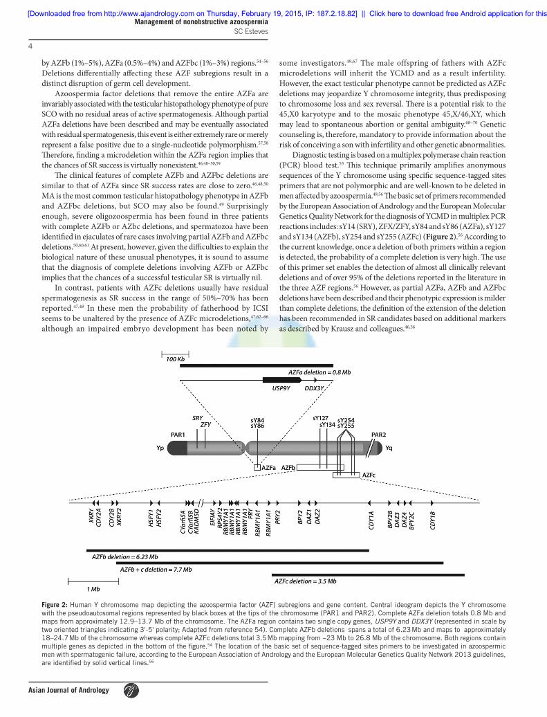
Diagnostic testing is based on a multiplex polymerase chain reaction (PCR) blood test.53 This technique primarily amplifies anonymous sequences of the Y chromosome using specific sequence-tagged sites primers that are not polymorphic and are well-known to be deleted in men affected by azoospermia.49,56 The basic set of primers recommended by the European Association of Andrology and the European Molecular Genetics Quality Network for the diagnosis of YCMD in multiplex PCR reactions includes: sY14 (SRY), ZFX/ZFY, sY84 and sY86 (AZFa), sY127 and sY134 (AZFb), sY254 and sY255 (AZFc) (Figure 2).56 According to the current knowledge, once a deletion of both primers within a region is detected, the probability of a complete deletion is very high. The use of this primer set enables the detection of almost all clinically relevant deletions and of over 95% of the deletions reported in the literature in the three AZF regions.56 However, as partial AZFa, AZFb and AZFbc deletions have been described and their phenotypic expression is milder than complete deletions, the definition of the extension of the deletion has been recommended in SR candidates based on additional markers as described by Krausz and colleagues.46,56
Figure 2: Human Y chromosome map depicting the azoospermia factor (AZF) subregions and gene content. Central ideogram depicts the Y chromosome with the pseudoautosomal regions represented by black boxes at the tips of the chromosome (PAR1 and PAR2). Complete AZFa deletion totals 0.8 Mb and maps from approximately 12.9–13.7 Mb of the chromosome. The AZFa region contains two single copy genes, USP9Y and DDX3Y (represented in scale by two oriented triangles indicating 3'‑5' polarity; Adapted from reference 54). Complete AZFb deletions spans a total of 6.23 Mb and maps to approximately 18–24.7 Mb of the chromosome whereas complete AZFc deletions total 3.5 Mb mapping from ~23 Mb to 26.8 Mb of the chromosome. Both regions contain multiple genes as depicted in the bottom of the figure.54 The location of the basic set of sequence‑tagged sites primers to be investigated in azoospermic men with spermatogenic failure, according to the European Association of Andrology and the European Molecular Genetics Quality Network 2013 guidelines, are identified by solid vertical lines.56
[Downloaded free from http://www.ajandrology.com on Thursday, February 19, 2015, IP: 187.2.18.82] || Click here to download free Android application for this journal

Asian Journal of Andrology
Management of nonobstructive azoospermia SC Esteves
5
Our approach is to offer genetic testing to all men with NOA, including karyotype and YCMD screening. Although the finding of a 47,XXY karyotype or an AZFc microdeletion is not irreconcilable with SR, some men opt to pursue other options after knowing their genetic condition. For men with complete AZFa, AZFb or AZFbc microdeletions we do not recommend proceeding with SR. However, given the limited number of cases documented in the literature to allow a categorical statement against SR, we sporadically perform SR in such cases provided the patient had given consent after exhaustive counseling. In our experience, sperm have been retrieved in 1/9 men with complete AZFb deletions, and in 0/5 cases with AZFa. ICSI was performed with freshly retrieved testicular sperm in the aforementioned AZFb patient, which resulted in a singleton delivery of a healthy male neonate. Regardless of treatment choice, our patients with genetic abnormalities found it reassuring to know the cause of their infertility.
ROLE OF INTERVENTIONS PRIOR TO SPERM RETRIEVALAfter genetic testing, the next step is to define whether or not any medical and/or surgical interventions should be used prior to SR. Any treatment that might improve sperm production would be highly recommended since nearly half of men with NOA will be halted in their attempt to conceive due to an unsuccessful SR.7,9,30
Medical therapyWhile a positive outcome is warranted following exogenous gonadotropin treatment in HH, it is generally believed that empirical medical treatment is ineffective in men with SF, particularly in the presence of high plasma levels of gonadotropins. Nevertheless, there may be a potential role for such treatments in men with NOA given the paradoxically weak stimulation of Leydig and Sertoli cells by endogenous gonadotropins. Gonadotropin secretion is determined by the frequency, amplitude and duration of its secretory pulses, but due to the high baseline levels of endogenous gonadotropins seen in most of men with NOA the relative amplitudes of both FSH and LH are low.71–74 Furthermore, approximately 50% of these men have low endogenous levels of total T (<300 ng dl−1), and therefore they may lack adequate levels of intratesticular T (ITT) that are essential for regulating the spermatogenic process in combination with adequate Sertoli cell stimulation by FSH.27–29
Drugs that have been utilized include clomiphene citrate (CC), gonadotropins and aromatase inhibitors (AIs).30,75 CC, a selective estrogen receptor modulator, binds competitively to estrogen on its receptors at the hypothalamus and pituitary gland. After CC administration, the pituitary perceives less estrogen that leads to the secretion of both FSH and LH. The latter binds to LH receptors in the Leydig cells and induces androgen production. As a result, there is a rise in T levels.30 Human chorionic gonadotropin (hCG) is a glycoprotein similar to the native LH, but with higher receptor affinity and half-life.76 hCG binds to the same LH receptor at the Leydig cell level and also stimulates the production of androgens. On the other hand, AIs block the aromatase enzyme, which is present in the adipose tissue, liver, testis and skin, and is responsible for converting T and other androgens to estradiol. An imbalance in T to estradiol (T/E) ratio , which is frequently seen in obese men, may be reversed by oral administration of AI.29,31,77
Empirical treatments have been exploited with varying degrees of success in terms of sperm production, albeit all of them were effective for increasing endogenous T levels even under a hypergonadotropic condition (Table 2).29,77–82 In one study, ITT levels were increased by 5-fold after hCG-based therapy (post: 1348.1 ± 505.4 ng ml−1; pre:
273.6 ± 134.4 ng ml−1; P < 0.0001).71 Approximately half of the men treated with hCG exhibit suppressed endogenous FSH levels through a negative feedback mechanism of elevated serum T.71,81 Such an effect may be beneficial since high plasma FSH levels lead to down-regulation of FSH receptors, which has been associated with an impaired tubular function. In fact, improvement in Sertoli cell function was achieved after reduction of FSH plasma concentration by administration of a GnRH analogue in men with NOA.83 Sertoli cells are major targets for T signaling via the activation of nuclear androgen receptors, whose expression is upregulated in men with NOA compared to those with normal spermatogenesis.84–88 Since the Sertoli cells support male germ cell development and survival, their function may be restored by increasing endogenous T, whose levels are normally 100-fold greater within the testes compared with the serum.71,83,89
The exact mechanism underlying the potential beneficial role of medical interventions remains unclear, but it has been speculated that increased ITT levels act by stimulating spermatogonia DNA synthesis and spermiogenesis in patients with residual spermatogenic activity.71,90,91 These effects may result in the formation of well-differentiated seminiferous tubules that would be detected during SR. Although current evidence indicate that the aforesaid medication enhances endogenous T production, a definitive conclusion regarding sperm production cannot yet be drawn due to the lack of well-designed clinical trials. At present, we routinely evaluate T and estradiol levels in men with NOA. Men with low T or a low T/E ratio are treated as depicted in Figure 3.
Varicocele repairVaricocele, found in approximately 5% of men with NOA, has been another target for intervention.92 While it is still debatable whether varicocele is coincidental or contributory to spermatogenesis disruption in these men its surgical treatment has been aimed at improving sperm production.92–94 Treatment goals are either to allow the appearance of small quantities of sperm in the ejaculate, thus obviating the need for SR, or increase the likelihood of SR success.
An early retrospective cohort study of 31 treated patients indicated that only 9.6% achieved adequate motile sperm in the ejaculate for ICSI to be performed without SR, thus suggesting that varicocele repair would be of limited value.95 However, in a study examining the role of microsurgical subinguinal varicocelectomy in a group of 17 men with clinical varicocele and NOA, we found that sperm returned to the ejaculate in 35.3% (6/17) of men at an average follow-up of 19 months.94 The mean motile sperm count was 0.8 million ml−1 (range 0.1–1.8) and one patient was able to initiate a natural pregnancy. Testicular biopsies obtained during the operations revealed that the histopathology phenotype was associated with the surgical outcome. Sperm were identified in the ejaculates of 72.7% (8/11) of the patients with hypospermatogenesis or MA, in contrast to none (0/6) of those with SCO. Recently, a meta-analysis of eleven cohort studies involving 233 patients with NOA and clinical varicocele corroborated our results.93 After microsurgical varicocele repair and at a mean postoperative follow-up of 13 months, motile sperm was found in the ejaculates of 39% of the males. With a mean sperm count of 1.6 million ml−1, natural and assisted conceptions were obtained in 26% of these men. Analysis of testicular biopsies taken either prior or during varicocele repair revealed that hypospermatogenesis and MA were significantly more likely to be associated with the presence of sperm in the postoperative ejaculate compared with SCO (odds ratio [OR]: 9.4; 95% confidence interval [CI]: 3.2–27.3).
While the aforementioned studies indicate that an improvement in sperm production is achieved in up to 1/3 of the men with NOA after varicocelectomy, most of the treated individuals remain azoospermic
[Downloaded free from http://www.ajandrology.com on Thursday, February 19, 2015, IP: 187.2.18.82] || Click here to download free Android application for this journal

Asian Journal of Andrology
Management of nonobstructive azoospermia SC Esteves
6
and will require SR. In one study, SRR were identical (60%) in men who had their varicoceles treated before SR as compared with those who did not.95 Nevertheless, a beneficial role of intervention was suggested by others.96,97 Inci et al.96 studying a group of 96 men observed that SR success was significantly higher in treated compared with untreated men (53 vs 30%, OR: 2.63, 95% CI: 1.05–6.60; P = 0.03). In another study involving 66 men, Haydardedeoglu et al.97 reported higher SR success in men who had varicocele repair prior to SR (61%) compared with untreated men (38%; P < 0.01).
Based on the literature and our own data, we offer microsurgical repair of varicoceles before SR, particularly to young men (<35 years) with large bilateral varicoceles (Grades 2 and 3) after proper counseling. Given the above-mentioned studies were small retrospective series, there is a need for controlled trials to evaluate the role of varicocele repair in NOA.
CHOICE OF SPERM RETRIEVAL METHODThe preferred method in NOA has been conventional testicular sperm extraction (TESE).98 Typically, multiple randomly testicular biopsies are taken and examined since it is not possible to predict before TESE whether sperm will be found or where islets of normal spermatogenesis, if existent, are located.5,7,40,98,99 A disadvantage of TESE is that the removal of large fragments of testicular tissue may jeopardize in a transient or
permanent way the already compromised androgen production, which may lead to severe hypogonadism.100 Moreover, laboratory processing of large quantities of testicular tissue is time-consuming and labor intensive, and may miss rare sperm within the sea of cells and debris.37,38
Ideally, SR in NOA should be aimed at offering the highest possible chance of obtaining an adequate number of good quality testicular sperm, which can be immediately used for ICSI or cryopreserved for future ICSI attempts. In addition, it should minimize testicular damage to maintain androgen activity and the chances of success in repeated retrieval attempts.
Microdissection testicular sperm extractionMicrodissection TESE (micro-TESE) is a microsurgical SR method originally described by Schlegel, which has been proposed as a better alternative to TESE in cases of NOA.101 The principle of micro-TESE is to identify areas of active spermatogenesis with the aid of an operating microscope. After testis delivery, a large incision is made in an avascular area of the tunica albuginea under ×6–8 magnification to widely expose the testicular parenchyma. Dissection at ×15–25 magnification enables the surgeon to search and isolate the seminiferous tubules that exhibit larger diameter in comparison with nonenlarged or collapsed counterparts (Figure 4).42 Enlarged tubules are selectively extracted based on the assumption that they are more likely to contain sperm production.101
Table 2: Summary review of empirical medical therapy for infertile men with NOA
Study Design Study group Control group Medication Main findings
Pavlovich et al.77
Prospective cohort
43 men with T/E ratio<10
N/A Testolactone, 50–100 mg twice daily for a mean of 5 months
None of the 12 men who completed 3 months of treatment had sperm in ejaculate; T/E ratios were restored to normal range (>10) in all treated men
Hussein et al.78
Prospective cohort
42 men with favorable histology (hypospermatogenesis or maturation arrest)
N/A CC, 50 mg every other day for a mean of 5 months; dose titrated by 25 mg increments until target T levels between 600 and 800 ng dl−1 were achieved
64.3% of the men had sperm in posttreatment semen analysis (mean density of 3.8×10⁶ ml−1, and motility of 20.8%); all of the men (n=15) who remained azoospermic had success at SR
Selman et al.79
Prospective cohort
49 men with normal endocrine and genetic profile in whom diagnostic testis biopsy showed maturation arrest and no sperm on wet examination
N/A rec‑hFSH, 75 IU SC on alternate days for 2 months, then dose increased to 150 IU and hCG (2000 IU SC twice weekly) added for another 4 months
None of the patients had return of sperm in ejaculate; posttreatment SRR were 21.4%
Ramasamy et al.80
Retrospective cohort
56 men with nonmosaic Klinefelter’s syndrome and T levels lower than 300 ng dl−1
N/A Testolactone (50–100 mg) or anastrozole (1 mg) were used orally, alone or combined with SC hCG (up to 2500 IU three times a week) for at least 3 months
SRR were 1.4‑fold higher (77% vs 55%; P=0.03) in men who responded to treatment with a resultant T level of 250 ng dl‑1 or higher compared with those men in whom posttreatment T was less than 250 ng dl‑1
Reifsnyder et al.29
Retrospective cohort
307 men with T levels lower than 300 ng dl−1
41 men with T levels lower than 300 ng dl−1; 388 men with T levels higher than 300 ng dl‑1
AI (50–100 mg testolactone orally twice daily or 1 mg anastrozole daily), hCG (at a dose of 1500–2000 IU SC twice or three times a week) and CC were used, alone or combined, for at least 2–3 months before surgery
None of the patients had return of sperm in ejaculate; SRR were not different in treated (51%; n=307) and untreated (61% ; n=41) men with baseline low T levels; SRR were not different between treated males with T levels lower than 300 ng dl−1 (51%; n=307) and untreated ones with T levels>300 ng dl−1 (51%; n=388)
Shiraishi et al.81
Prospective cohort
28 men with idiopathic* NOA who had negative SR
20 men with idiopathic* NOA who had negative SR
At least 6 months after the first SR attempt, patients received hCG (5000 IU SC three times a week) for 3 months. When FSH levels decreased after hCG (<3 mIU ml−1) recombinant FSH (rec‑hFSH, 150 IU SC three times a week) was added for 2 months
Sperm was obtained at the second SR attempt in 6 (21%) of the 28 treated men, but in none of the untreated men (P<0.05)
Hussein et al.82
Prospective cohort
612 unselected men 116 unselected men
CC (50 mg every other day) alone or combined with hCG (5000 IU SC twice a week) and hMG (75IU SC once weekly) were administered for an average of 5.4 months
Sperm were found in ejaculates of 10.9% of the treated males; in the patients who remained azoospermic, SRR were higher in those who received medical therapy compared with controls (57.0 vs 33.6%, P<0.001)
AI: aromatase inhibitor; CC: clomiphene citrate; FSH: follicle‑stimulating hormone; hCG: human chorionic gonadotropin; hMG: human menopausal gonadotropin; IU: International Units; NOA: nonobstructive azoospermia; rec‑hFSH: recombinant human FSH; SC: subcutaneous; SR: sperm retrieval; SRR: sperm retrieval rates; T: total testosterone; T/E: testosterone to estradiol ratio; N/A: not applicable. *Exclusion criteria were men with Klinefelter syndrome, testis volume<4 ml, T levels<200 ng dl−1, varicocele and cryptorchidism
[Downloaded free from http://www.ajandrology.com on Thursday, February 19, 2015, IP: 187.2.18.82] || Click here to download free Android application for this journal

Asian Journal of Andrology
Management of nonobstructive azoospermia SC Esteves
7
In a controlled study involving 60 men with NOA, we found that SR success was higher in the micro-TESE group compared with the conventional single-biopsy TESE group (45% vs 25%; P = 0.005). Results also favored micro-TESE after stratification according to the testicular histopathology phenotype (hypospermatogenesis: 93 vs 64%; MA: 64% vs 9%; SCO syndrome: 20% vs 6%; P < 0.001).39 Others have corroborated our findings, and added that complication rates were lower with micro-TESE.101–106 The use of optical magnification reduces the chances of vascular injury since preservation of testicular blood supply is easily achieved, thus reducing the chances of hematoma formation and testicular devascularization. After micro-TESE a transient decrease in serum T is followed by return to baseline levels in about 95% of the cases within 18 months, except in men with very small testes and severely compromised androgen activity such as those with KS in whom these effects tend to be permanent.45,106
Our SR success with micro-TESE in an updated experience involving 356 patients was 41.4% overall, and 100.0%, 40.3% and 19.5% according to the histopathology phenotypes of hypospermatogenesis, MA and SCO, respectively.9,35 A recent systematic review pooling seven comparative studies and 1062 patients confirmed that the micro-TESE was associated with a more favorable SRR ranging from 42.9% to 63% compared with 16.7%–45% in conventional TESE.107 Micro-TESE has been shown to rescue approximately one-third of the cases that had failed in the previous retrieval attempts with conventional TESE or percutaneous testicular aspiration (TESA), and is particularly helpful for men with NOA presenting the worst-case scenarios.100,107,108 A new micro-TESE after an initially successful procedure can be carried out, but should be delayed for at least 6 months due to inflammatory changes. SR success is markedly lower (25% vs 80%) if repeat micro-TESE is performed within 6 months of the first operation.100
Figure 3: Algorithm for the medical management of infertile men with nonobstructive azoospermia.
[Downloaded free from http://www.ajandrology.com on Thursday, February 19, 2015, IP: 187.2.18.82] || Click here to download free Android application for this journal

Asian Journal of Andrology
Management of nonobstructive azoospermia SC Esteves
8
In our center, micro-TESE is the method of choice for SR in men with NOA. Procedures are carried out under intravenous anesthesia in an outpatient basis.99 When coupled with ICSI, we prefer to perform micro-TESE on the day before oocyte retrieval. Importantly, we always confirm azoospermia by analyzing a centrifuged semen specimen obtained immediately before the procedure since rare sperm may occasionally spill over into the ejaculates of such patients.5 Studies focusing on quantitative spermatogenesis have shown that the threshold of 3 mature spermatids per seminiferous tubule’s cross-section must be exceeded in order for spermatozoa to spill over into the ejaculate. Men with NOA have a mean of 0–3 mature spermatids per seminiferous tubule, thus explaining why rare sperm are occasionally found in ejaculates.5,11 The technical details of how we perform micro-TESE can be found elsewhere (http://www.brazjurol.com.br/videos/may_june_2013/Esteves_440_441video.htm).42
LABORATORY HANDLING OF RETRIEVED GAMETES AND EMBRYOSAfter SR, testicular parenchyma is transferred to the IVF laboratory for tissue processing and sperm search. The laboratory management of surgically retrieved specimens requires special attention because spermatozoa collected from men with NOA are often compromised in quality and more fragile than ejaculated counterparts (Table 3). Both sperm DNA fragmentation (SDF) and aneuploidy rates are higher in testicular sperm obtained from men with NOA compared with ejaculated sperm obtained from infertile men with various etiology categories.109,110 As a result, lower fertilization, embryo development and pregnancy rates have been reported with ICSI using such gametes.9,17,111,112
Testicular tissue handlingThe extraction of a minimum volume of tissue is advantageous because processing of TESE specimens may be incredibly labor-intensive. Tissue removal is approximately 50- to 70-fold lower in micro-TESE compared with conventional TESE.37–39,101 The lower the amount of tissue extracted, the easier the tissue processing and sperm search. Micro-TESE is, therefore, helpful to increase laboratory process
efficiency in addition to minimizing testicular damage and increasing SR success.38,42,108
Testicular tissue processing techniques have been developed to increase sperm yield. While mechanical disruption of the tubules is achieved by mincing them repeatedly using needled-tuberculin syringe or by passing the suspensions through a 24-gauge angiocatheter, enzymatic digestion is carried out by incubation of testicular suspensions with collagenase.38,114–116 Our approach is to perform an initial examination of microsurgically-extracted specimens under the inverted microscope after mechanical mincing (Figure 4). The procedure is deemed successful if an adequate number of sperm for ICSI is found, otherwise the surgeon is promptly informed that more specimens are needed. Additional samples are taken until both testes are examined if no sperm are initially identified.38 All excised specimens are extensively minced mechanically and meticulously re-examined. These specimens are subjected to enzymatic digestion with collagenase Type IV (1000 IU ml−1) for approximately 2–4 h if no sperm are found after mechanical processing. These methods ensure tubular wall break down and cellular content loss.
After confirmation for sperm, specimens are processed by either simple washing or gradient centrifugation.37,117 If needed, erythrocyte lysing solution is used to eliminate excessive red blood cells.37 Aliquots of testicular suspension are then cryopreserved or loaded in microdroplets of an oil-covered sperm medium if ICSI is to be performed with fresh sperm. When only immotile spermatozoa are obtained after processing of fresh or cryo-thawed testicular specimens, different methods can be used to differentiate live immotile spermatozoa from dead ones. The hypoosmotic swelling test, the sperm tail flexibility test and motility stimulants, such as pentoxifylline, are some of these methods.38 Viable and preferentially motile sperm are chosen because ICSI outcomes are negatively affected by the use of immotile sperm for injections.118–120
Laboratory environmentGood laboratory practices, including sterile techniques, temperature and pH stability of working solutions, and air quality control, will ensure optimal safety conditions for micromanipulation.37,121 At our center, SR and all related-laboratory steps are carried out in controlled
Figure 4: Microdissection testicular sperm extraction. The illustration depicts its main technical aspects, including the use of an operating microscope and the identification of enlarged seminiferous tubules, and initial processing of extracted specimens.
[Downloaded free from http://www.ajandrology.com on Thursday, February 19, 2015, IP: 187.2.18.82] || Click here to download free Android application for this journal

Asian Journal of Andrology
Management of nonobstructive azoospermia SC Esteves
9
environments. Our facility, comprised of reproductive laboratories (IVF and andrology), an operating room where microsurgical sperm extractions and oocyte collections are carried out, and embryo transfer rooms, was constructed according to cleanroom standards for air particles and volatile organic compounds filtration as previously described.121 In an observational study evaluating 2315 sperm injection cycles mainly involving severe male factor infertility, we noted that treatment effectiveness was markedly improved after cleanroom technology implementation. Live birth rates increased (35.6% vs 25.8%; P = 0.02) and miscarriage rates decreased (28.7% vs 20.0%; P = 0.04), while an additional high quality embryo, on average, was generated per treatment cycle (3.2 vs 2.3; P = 0.01).121
Cryopreservation of testicular spermAfter a successful SR in NOA, cryopreservation of surplus testicular sperm is recommended given that success in repeated retrievals is not warranted. Moreover, such patients often require more than one ICSI attempt until a pregnancy is established. While some centers prefer to retrieve and intentionally cryopreserve testicular sperm for future use, others simultaneously coordinate SR and oocyte collection. A comprehensive review of the advantages and disadvantages of performing sperm injections with fresh or frozen-thawed testicular sperm, as well as the methods of selecting viable immotile sperm for ICSI can be found elsewhere.38 Our experience is that frozen-thawed testicular sperm from men with NOA usually will be immotile.37 Although limited data suggest that freezing these gametes inside an empty zona pellucida or using vitrification may improve survival, and that postthaw incubation with motility stimulants such as pentoxifylline can help in selecting viable sperm for ICSI, patients
should be advised that ICSI outcomes will be lowered if only immotile frozen-thawed testicular sperm were available.122–124 A meta-analysis of ten studies involving 734 treatments showed a significantly lower implantation rate when frozen–thawed testicular sperm had been used compared with fresh sperm (relative risk: 1.75; 95% CI: 1.10–2.80).120 Therefore, our strategy is to plan repeat SR as a back-up option in such cases. Given that best results in NOA will be obtained with fresh testicular sperm, SR and controlled ovarian stimulation in the same cycle has been our preferred strategy despite carrying the risk of no availability of testicular sperm.
REPRODUCTIVE POTENTIAL OF MEN WITH NONOBSTRUCTIVE AZOOSPERMIA AND HEALTH OF RESULTING OFFSPRINGIn our experience pregnancy rates in ICSI cycles using sperm obtained from men with NOA are lower when compared with both ejaculated sperm and epididymal/testicular sperm of men with OA. In one series we studied 188 couples that underwent ICSI using sperm from partners with NOA, and the results were compared with groups of 182 and 465 couples whose partners had OA and nonazoospermia male infertility, respectively. Live birth rates were significantly lower in the NOA group (21.4%) compared with OA (37.5%) and ejaculated sperm (32.3%) groups (P = 0.003). A total of 326 live births resulted in 427 babies born. Differences were not observed among the groups in gestational age, preterm birth, birth weight and low birth weight, although we noted a tendency toward poorer neonatal outcomes in the azoospermia categories.17
In an updated series involving a larger cohort of 365 men with NOA who underwent micro‐TESE for ICSI, we compared treatment
Table 3: Laboratory management of testicular specimens extracted from infertile men with nonobsructive azoospermia
Process Procedures Useful techniques Main goals
Testicular tissue handling
Extraction of minimum volume of tissue
Micro‑TESE Improvement in tissue processing and searching efficiency
Mechanical mincing Disruption of seminiferous tubules using needles or microscissors, and forced passing through small lumen angiocatheters
Tubular break down and cellular content loss
Enzymatic digestion Incubation of testicular suspensions with collagenase type IV (1000 IU ml−1) and/or DNAse (25 µg ml−1)
Tubular break down and cellular content loss
Erythrocyte lysing Incubation of testicular suspensions with erythrocyte lysing buffer solution (1:1, v/v)a
Elimination of excessive blood cells
Motility enhancement Incubation of testicular suspensions with pentoxifylline (1:1, v/v)b Selection of viable sperm for ICSI
Laboratory environment and “good laboratory practices”
Air quality control Air filtration for particles (HEPA filtration) and volatile organic compounds (potassium permanganate impregnated zeolite plus activated carbon filtration)
Assurance of optimal safety conditions for gamete handling, sperm injection and embryo culture
Maintenance of temperature and pH stability
Quality control and quality assurance of instruments, equipment and reagents
Avoidance of iatrogenic cellular damage
Centrifugation Simple washing with buffered sperm medium or gradient centrifugation using low centrifugation forces (200–300×g)
Avoidance of iatrogenic cellular damage
Sterile techniques Manipulation of gametes and embryos in laminar flow cabinets or inside controlled environments
Assurance of optimal safety conditions for gamete handling, sperm injection and embryo culture
ICSI Sperm selection Hyposmotic swelling testc
Mechanical touch techniqued
Laser‑assisted sperm selectione
Selection of viable immotile sperm for ICSI
Testicular sperm storage
Cryopreservation Sperm freezing inside an empty zona pellucida, and sperm vitrification
Enhancement of postthaw sperm recovery and survival
a155 mmol NH4Cl +10 mmol KHCO3 +2 mmol EDTA dissolved in sterile water. Adjust pH to 7.2 if needed. b1.391 mg of pentoxifylline dissolved in 1 ml of sperm medium gives a 5 mM solution. cPrepared by mixing 1 ml sperm medium to 1 ml of sterile water (approximately 139 mOsm kg−1). A single morphologically normal immotile spermatozoon is picked up from the sperm medium and aspirated head‑first into the microinjection pipette. Then, the pipette is immersed into a hyposmotic solution microdroplet and only the sperm tail is moved out of the pipette tip into the solution. The sperm tail is kept for 5–10 s into the solution, and it is observed if a tail tip swelling occurs. Sperm tail swelling is often minimal and is a marker of sperm viability in fresh specimens. dA morphologically normal immotile spermatozoon is picked up from the sperm medium using the microinjection pipette and transferred to the PVP microdroplet. Then, the sperm tail is touched with the tip of the microinjection pipette to move the tail up and down. The sperm tail is considered flexible when it moves independently of the sperm head. Sperm tail flexibility is considered a marker of sperm viability. If the tail remains rigid upon touching and both the sperm head and tail move together as a block, spermatozoon is then considered nonviable for ICSI. eThe application of a single laser shot to the far end of the sperm tail using a noncontact 1.48 µm diodide laser has been shown to cause a curling of the tail only in viable sperm, similar to the reaction observed with the hyposmotic swelling test. HEPA: high‑efficiency particulate air; ICSI: intracytoplasmic sperm injection; PVP: polyvinylpyrrolidone; EDTA: ethylenediaminetetraacetic acid; TESE: testicular sperm extraction
[Downloaded free from http://www.ajandrology.com on Thursday, February 19, 2015, IP: 187.2.18.82] || Click here to download free Android application for this journal

Asian Journal of Andrology
Management of nonobstructive azoospermia SC Esteves
10
results in cycles that testicular sperm had been successfully retrieved with those of 40 couples who used donor sperm for ICSI due to failed retrieval.9 A group of 146 men with OA who underwent percutaneous SR was also included for comparison. Not surprisingly, the SRR was lower in NOA compared with OA (41.4% vs. 100%; adjusted-OR: 0.033; 95% CI: 0.007–0.164; P < 0.001). Live birth rates after sperm injections were lower in men with NOA (19.9%) compared with donor sperm (37.5%; adjusted-OR: 0.377, 95% CI: 0.233–0.609, P < 0.001) and OA (34.2%; adjusted-OR: 0.403, 95% CI: 0.241–0.676, P = 0.001). In this aforementioned study, neither miscarriage nor the newborn parameters (gestational age, birth weight, malformation rate, perinatal mortality) of infants conceived was significantly different among the groups.
Our observations that pregnancy rates are negatively affected by NOA have been corroborated by others, and may be related to the higher tendency of spermatozoa obtained from men with NOA to carry deficiencies related to the centrioles and genetic material, which ultimately affect their capability of triggering the development of a viable embryo.110–113,124,125 In one report assessing SDF levels in testicular sperm, it has been shown that sperm of patients with NOA exhibited, on average, significantly higher DNA damage (46.9%) compared with OA counterparts (35.9%; P < 0.05). In this aforementioned study in which the authors assessed SDF using the sperm chromatin dispersion test, it was noted that embryo morphology was negatively affected by SDF (r = −0.163; P = 0.01).111 Altogether, these findings indicate that the testicular sperm obtained from men with NOA hold a lower developmental competence. Nevertheless, our data on the health of offspring is reassuring and have been endorsed by others. Despite that, a call for continuous monitoring is essential given the limited population analyzed.8,126,127 Furthermore, long-term follow-up studies are needed as data on the physical, neurological, and developmental outcomes of children conceived are lacking.
FUTURE PERSPECTIVES FOR MEN WITH NONOBSTRUCTIVE AZOOSPERMIAIn vitro fertilization with immature germ cells and in vitro culture of these cells have been proposed as an approach to overcome the cases where no mature spermatozoa is retrieved.128 ICSI with immature germ cells, including elongating and round spermatids, has yielded conflicting results and despite reported deliveries of healthy offspring, the method has very low efficiency as currently used.129 Ethical and safety concerns related to the potential transmission of genomic imprinted disorders have been raised leading to the ban of spermatid injection in countries such as the United Kingdom.129 Human spermatozoa are highly specialized cells with the purpose of not only delivering competent paternal DNA to the oocyte but also providing a robust epigenetic contribution to embryogenesis. The latter requires that chromatin contains layers of regulatory elements sufficient to drive genes toward activation or silencing upon delivery to the oocyte.130
Because assisted reproduction techniques require mature germ cells, research efforts are now focused on the differentiation of preexisting immature germ cells or the production/derivation of sperm from somatic cells. Biotechnology has been investigated as a valuable tool for rescuing fertility while maintaining biological fatherhood. Breakthrough advancement in this field has been accomplished by Asian scientists who used stem cells from mouse embryos to create primordial germ cells, which differentiated in spermatozoa after testis transplantation in mice.131 In humans, formation of human haploid-like cells has already been obtained from pluripotent stem cells of somatic origin using the novel technique of in vitro sperm derivation.128 Haploidization is another technique under investigation as an option to create gametes based on
biological cloning technology. Despite promising, these methodologies are still experimental. The production of gametes in the laboratory is a highly complex process, which is yet to be fully translated to humans.
CONCLUSIONSNonobstructive azoospermia is the most severe presentation of male infertility. Despite lacking sperm in the ejaculate, approximately 50% of men with NOA have minimal sperm production within their dysfunctional testes. Such sperm can be extracted and used for IVF techniques to produce a viable offspring. The scope of NOA-related infertility covers a wide spectrum from genetic studies to hormonal control, microsurgical and medical therapy to assisted reproduction techniques, as well as innovative stem cell research aiming at creating artificial gametes. From a medical perspective, the management of men with NOA seeking fertility involves a series of steps that includes the differential diagnosis of azoospermia, genetic testing and counseling, identification of those who could benefit from medical and surgical interventions prior to SR, application of the best method to surgically retrieve testicular spermatozoa, and the use of state-of-art IVF techniques. A coordinated multidisciplinary effort involving urologists, andrologists, geneticists, reproductive endocrinologists and embryologists is key to offer the best possible chance of achieving a biological offspring to men with NOA.
COMPETING INTERESTSThe author declares no competing interests.
ACKNOWLEDGMENTSFabiola Bento assisted with language revision.
REFERENCES1 Cooper TG, Hellenkemper B, Jonckheere J, Callewaert N, Grootenhuis AJ, et al.
Azoospermia: virtual reality or possible to quantify? J Androl 2006; 27: 483–90.2 Aziz N. The importance of semen analysis in the context of azoospermia. Clinics
(Sao Paulo) 2013; 68 Suppl 1: 35–8.3 Esteves SC, Miyaoka R, Agarwal A. An update on the clinical assessment of the
infertile male. [corrected]. Clinics (Sao Paulo) 2011; 66: 691–700.4 Esteves SC, Agarwai A. The azoospermic male: current knowledge and future
perspectives. Clinics (Sao Paulo) 2013; 68 Suppl 1: 1–4.5 Silber SJ. Microsurgical TESE and the distribution of spermatogenesis in
non‑obstructive azoospermia. Hum Reprod 2000; 15: 2278–84.6 Esteves SC, Miyaoka R, Agarwal A. Sperm retrieval techniques for assisted
reproduction. Int Braz J Urol 2011; 37: 570–83.7 Carpi A, Sabanegh E, Mechanick J. Controversies in the management of
nonobstructive azoospermia. Fertil Steril 2009; 91: 963–70.8 Belva F, De Schrijver F, Tournaye H, Liebaers I, Devroey P, et al. Neonatal outcome of 724
children born after ICSI using non‑ejaculated sperm. Hum Reprod 2011; 26: 1752–8.9 Esteves SC, Prudencio C, Seol B, Verza S, Knoedler C, et al. Comparison of sperm
retrieval and reproductive outcome in azoospermic men with testicular failure and obstructive azoospermia treated for infertility. Asian J Androl 2014; 16: 602–6.
10 Esteves SC, Hamada A, Kondray V, Pitchika A, Agarwal A. What every gynecologist should know about male infertility: an update. Arch Gynecol Obstet 2012; 286: 217–29.
11 Jaffe TM, Kim ED, Hoekstra TH, Lipshultz LI. Sperm pellet analysis: a technique to detect the presence of sperm in men considered to have azoospermia by routine semen analysis. J Urol 1998; 159: 1548–50.
12 Corea M, Campagnone J, Sigman M. The diagnosis of azoospermia depends on the force of centrifugation. Fertil Steril 2005; 83: 920–2.
13 Esteves SC. Clinical relevance of routine semen analysis and controversies surrounding the 2010 World Health Organization criteria for semen examination. Int Braz J Urol 2014; 40: 443–53.
14 Castilla JA, Alvarez C, Aguilar J, González‑Varea C, Gonzalvo MC, et al. Influence of analytical and biological variation on the clinical interpretation of seminal parameters. Hum Reprod 2006; 21: 847–51.
15 Keel BA. Within‑ and between‑subject variation in semen parameters in infertile men and normal semen donors. Fertil Steril 2006; 85: 128–34.
16 Schoor RA, Elhanbly S, Niederberger CS, Ross LS. The role of testicular biopsy in the modern management of male infertility. J Urol 2002; 167: 197–200.
17 Esteves SC, Agarwal A. Reproductive outcomes, including neonatal data, following sperm injection in men with obstructive and nonobstructive azoospermia: case series and systematic review. Clinics (Sao Paulo) 2013; 68 Suppl 1: 141–50.
[Downloaded free from http://www.ajandrology.com on Thursday, February 19, 2015, IP: 187.2.18.82] || Click here to download free Android application for this journal

Asian Journal of Andrology
Management of nonobstructive azoospermia SC Esteves
11
18 Practice Committee of American Society for Reproductive Medicine in Collaboration with Society for Male Reproduction and Urology. Evaluation of the azoospermic male. Fertil Steril 2008; 90: S74–7.
19 Baker K, Sabanegh Jr E. Obstructive azoospermia: reconstructive techniques and results. Clinics (Sao Paulo) 2013; 68 Suppl 1: 61–73.
20 Esteves SC, Lee W, Benjamin DJ, Seol B, Verza Jr A, et al. Reproductive potential including neonatal outcomes of men with obstructive azoospermia undergoing percutaneous sperm retrieval and intracytoplasmic sperm injection according to the cause of obstruction. J Urol 2013; 189: 232–7.
21 Miyaoka R, Esteves SC. Predictive factors for sperm retrieval and sperm injection outcomes in obstructive azoospermia: do etiology, retrieval techniques and gamete source play a role? Clinics (Sao Paulo) 2013; 68 Suppl 1: 111–9.
22 Fraietta R, Zylberstejn DS, Esteves SC. Hypogonadotropic hypogonadism revisited. Clinics (Sao Paulo) 2013; 68 Suppl 1: 81–8.
23 Cocuzza M, Alvarenga C, Pagani R. The epidemiology and etiology of azoospermia. Clinics (Sao Paulo) 2013; 68 Suppl 1: 15–26.
24 Hung AJ, King P, Schlegel PN. Uniform testicular maturation arrest: a unique subset of men with nonobstructive azoospermia. J Urol 2007; 178: 608–12.
25 Sokol RZ, Swerdloff RS. Endocrine evaluation. In: Lipshultz LI, Howards SS, editors. Infertility in the Male. 3rd ed. New York: Churchill Livingstone; 1997. p. 210–8.
26 Gudeloglu A, Parekattil SJ. Update in the evaluation of the azoospermic male. Clinics (Sao Paulo) 2013; 68 Suppl 1: 27–34.
27 Sussman EM, Chudnovsky A, Niederberger CS. Hormonal evaluation of the infertile male: has it evolved? Urol Clin North Am 2008; 35: 147–55, vii.
28 Bobjer J, Naumovska M, Giwercman YL, Giwercman A. High prevalence of androgen deficiency and abnormal lipid profile in infertile men with non‑obstructive azoospermia. Int J Androl 2012; 35: 688–94.
29 Reifsnyder JE, Ramasamy R, Husseini J, Schlegel PN. Role of optimizing testosterone before microdissection testicular sperm extraction in men with nonobstructive azoospermia. J Urol 2012; 188: 532–6.
30 Kumar R. Medical management of non‑obstructive azoospermia. Clinics (Sao Paulo) 2013; 68 Suppl 1: 75–9.
31 Hammoud A, Carrell DT, Meikle AW, Xin Y, Hunt SC, et al. An aromatase polymorphism modulates the relationship between weight and estradiol levels in obese men. Fertil Steril 2010; 94: 1734–8.
32 Isidori AM, Caprio M, Strollo F, Moretti C, Frajese G, et al. Leptin and androgens in male obesity: evidence for leptin contribution to reduced androgen levels. J Clin Endocrinol Metab 1999; 84: 3673–80.
33 Strain G, Zumoff B, Rosner W, Pi‑Sunyer X. The relationship between serum levels of insulin and sex hormone‑binding globulin in men: the effect of weight loss. J Clin Endocrinol Metab 1994; 79: 1173–6.
34 Vesper HW, Botelho JC, Wang Y. Challenges and improvements in testosterone and estradiol testing. Asian J Androl 2014; 16: 178–84.
35 Dohle GR, Elzanaty S, van Casteren NJ. Testicular biopsy: clinical practice and interpretation. Asian J Androl 2012; 14: 88–93.
36 Esteves SC, Agarwal A. Re: sperm retrieval rates and intracytoplasmic sperm injection outcomes for men with non‑obstructive azoospermia and the health of resulting offspring. Asian J Androl 2014; 16: 642.
37 Esteves SC, Verza S Jr. PESA/TESA/TESE sperm processing. In: Nagy ZP, Varghese AC, Agarwal A, editors. Practical Manual of In Vitro Fertilization. New York: Springer; 2012. p. 207–20.
38 Esteves SC, Varghese AC. Laboratory handling of epididymal and testicular spermatozoa: what can be done to improve sperm injections outcome. J Hum Reprod Sci 2012; 5: 233–43.
39 Verza S Jr, Esteves SC. Microsurgical versus conventional single – Biopsy testicular sperm extraction in nonobstructive azoospermia: a prospective controlled study. Fertil Steril 2011; 96 Suppl: S53.
40 Tournaye H, Verheyen G, Nagy P, Ubaldi F, Goossens A, et al. Are there any predictive factors for successful testicular sperm recovery in azoospermic patients? Hum Reprod 1997; 12: 80–6.
41 Esteves SC, Verza S, Prudencio C, Seol B. Sperm retrieval rates (SRR) in nonobstructive azoospermia (NOA) are related to testicular histopathology results but not to the etiology of azoospermia. Fertil Steril 2010; 94 Suppl: S132.
42 Esteves SC. Microdissection testicular sperm extraction (micro‑TESE) as a sperm acquisition method for men with nonobstructive azoospermia seeking fertility: operative and laboratory aspects. Int Braz J Urol 2013; 39: 440.
43 Chan PT, Palermo GD, Veeck LL, Rosenwaks Z, Schlegel PN. Testicular sperm extraction combined with intracytoplasmic sperm injection in the treatment of men with persistent azoospermia postchemotherapy. Cancer 2001; 92: 1632–7.
44 Raman JD, Schlegel PN. Testicular sperm extraction with intracytoplasmic sperm injection is successful for the treatment of nonobstructive azoospermia associated with cryptorchidism. J Urol 2003; 170: 1287–90.
45 Schiff JD, Palermo GD, Veeck LL, Goldstein M, Rosenwaks Z, et al. Success of testicular sperm extraction [corrected] and intracytoplasmic sperm injection in men with Klinefelter syndrome. J Clin Endocrinol Metab 2005; 90: 6263–7.
46 Krausz C, Quintana‑Murci L, McElreavey K. Prognostic value of Y deletion analysis: what is the clinical prognostic value of Y chromosome microdeletion analysis? Hum Reprod 2000; 15: 1431–4.
47 Peterlin B, Kunej T, Sinkovec J, Gligorievska N, Zorn B. Screening for Y chromosome microdeletions in 226 Slovenian subfertile men. Hum Reprod 2002; 17: 17–24.
48 Hopps CV, Mielnik A, Goldstein M, Palermo GD, Rosenwaks Z, et al. Detection of sperm in men with Y chromosome microdeletions of the AZFa, AZFb and AZFc regions. Hum Reprod 2003; 18: 1660–5.
49 Simoni M, Tüttelmann F, Gromoll J, Nieschlag E. Clinical consequences of microdeletions of the Y chromosome: the extended Münster experience. Reprod Biomed Online 2008; 16: 289–303.
50 Kleiman SE, Yogev L, Lehavi O, Hauser R, Botchan A, et al. The likelihood of finding mature sperm cells in men with AZFb or AZFb‑c deletions: six new cases and a review of the literature (1994‑2010). Fertil Steril 2011; 95: 2005–12.e1.
51 Esteves SC, Agarwal A. Novel concepts in male infertility. Int Braz J Urol 2011; 37: 5–15.52 Kleiman SE, Almog R, Yogev L, Hauser R, Lehavi O, et al. Screening for partial AZFa
microdeletions in the Y chromosome of infertile men: is it of clinical relevance? Fertil Steril 2012; 98: 43–7.
53 Hamada AJ, Esteves SC, Agarwal A. A comprehensive review of genetics and genetic testing in azoospermia. Clinics (Sao Paulo) 2013; 68 Suppl 1: 39–60.
54 Navarro‑Costa P, Plancha CE, Gonçalves J. Genetic dissection of the AZF regions of the human Y chromosome: thriller or filler for male (in)fertility? J Biomed Biotechnol 2010; 2010: 936569.
55 Repping S, Skaletsky H, Lange J, Silber S, Van Der Veen F, et al. Recombination between palindromes P5 and P1 on the human Y chromosome causes massive deletions and spermatogenic failure. Am J Hum Genet 2002; 71: 906–22.
56 Krausz C, Hoefsloot L, Simoni M, Tüttelmann F, European Academy of Andrology, et al. EAA/EMQN best practice guidelines for molecular diagnosis of Y‑chromosomal microdeletions: state‑of‑the‑art 2013. Andrology 2014; 2: 5–19.
57 Tyler‑Smith C, Krausz C. The will‑o’‑the‑wisp of genetics – Hunting for the azoospermia factor gene. N Engl J Med 2009; 360: 925–7.
58 Wu Q, Chen GW, Yan TF, Wang H, Liu YL, et al. Prevalent false positives of azoospermia factor a (AZFa) microdeletions caused by single – Nucleotide polymorphism rs72609647 in the sY84 screening of male infertility. Asian J Androl 2011; 13: 877–80.
59 Vogt PH, Bender U. Human Y chromosome microdeletion analysis by PCR multiplex protocols identifying only clinically relevant AZF microdeletions. Methods Mol Biol 2013; 927: 187–204.
60 Soares AR, Costa P, Silva J, Sousa M, Barros A, et al. AZFb microdeletions and oligozoospermia – Which mechanisms? Fertil Steril 2012; 97: 858–63.
61 Longepied G, Saut N, Aknin‑Seifer I, Levy R, Frances AM, et al. Complete deletion of the AZFb interval from the Y chromosome in an oligozoospermic man. Hum Reprod 2010; 25: 2655–63.
62 Kent‑First MG, Kol S, Muallem A, Ofir R, Manor D, et al. The incidence and possible relevance of Y‑linked microdeletions in babies born after intracytoplasmic sperm injection and their infertile fathers. Mol Hum Reprod 1996; 2: 943–50.
63 Mulhall JP, Reijo R, Alagappan R, Brown L, Page D, et al. Azoospermic men with deletion of the DAZ gene cluster are capable of completing spermatogenesis: fertilization, normal embryonic development and pregnancy occur when retrieved testicular spermatozoa are used for intracytoplasmic sperm injection. Hum Reprod 1997; 12: 503–8.
64 Kamischke A, Gromoll J, Simoni M, Behre HM, Nieschlag E. Transmission of a Y chromosomal deletion involving the deleted in azoospermia (DAZ) and chromodomain (CDY1) genes from father to son through intracytoplasmic sperm injection: case report. Hum Reprod 1999; 14: 2320–2.
65 Cram DS, Ma K, Bhasin S, Arias J, Pandjaitan M, et al. Y chromosome analysis of infertile men and their sons conceived through intracytoplasmic sperm injection: vertical transmission of deletions and rarity of de novo deletions. Fertil Steril 2000; 74: 909–15.
66 Oates RD, Silber S, Brown LG, Page DC. Clinical characterization of 42 oligospermic or azoospermic men with microdeletion of the AZFc region of the Y chromosome, and of 18 children conceived via ICSI. Hum Reprod 2002; 17: 2813–24.
67 van Golde RJ, Wetzels AM, de Graaf R, Tuerlings JH, Braat DD, et al. Decreased fertilization rate and embryo quality after ICSI in oligozoospermic men with microdeletions in the azoospermia factor c region of the Y chromosome. Hum Reprod 2001; 16: 289–92.
68 Siffroi JP, Le Bourhis C, Krausz C, Barbaux S, Quintana‑Murci L, et al. Sex chromosome mosaicism in males carrying Y chromosome long arm deletions. Hum Reprod 2000; 15: 2559–62.
69 Rajpert‑De Meyts E, Ottesen AM, Garn ID, Aksglaede L, Juul A. Deletions of the Y chromosome are associated with sex chromosome aneuploidy but not with Klinefelter syndrome. Acta Paediatr 2011; 100: 900–2.
70 Patsalis PC, Sismani C, Quintana‑Murci L, Taleb‑Bekkouche F, Krausz C, et al. Effects of transmission of Y chromosome AZFc deletions. Lancet 2002; 360: 1222–4.
71 Shinjo E, Shiraishi K, Matsuyama H. The effect of human chorionic gonadotropin‑based hormonal therapy on intratesticular testosterone levels and spermatogonial DNA synthesis in men with non‑obstructive azoospermia. Andrology 2013; 1: 929–35.
72 Spratt DI, Carr DB, Merriam GR, Scully RE, Rao PN, et al. The spectrum of abnormal patterns of gonadotropin – Releasing hormone secretion in men with idiopathic hypogonadotropic hypogonadism: clinical and laboratory correlations. J Clin Endocrinol Metab 1987; 64: 283–91.
73 Keenan DM, Veldhuis JD. Divergent gonadotropin‑gonadal dose – Responsive coupling in healthy young and aging men. Am J Physiol Regul Integr Comp Physiol 2004; 286: R381–9.
[Downloaded free from http://www.ajandrology.com on Thursday, February 19, 2015, IP: 187.2.18.82] || Click here to download free Android application for this journal

Asian Journal of Andrology
Management of nonobstructive azoospermia SC Esteves
12
74 Coviello AD, Bremner WJ, Matsumoto AM, Herbst KL, Amory JK, et al. Intratesticular testosterone concentrations comparable with serum levels are not sufficient to maintain normal sperm production in men receiving a hormonal contraceptive regimen. J Androl 2004; 25: 931–8.
75 Ramasamy R, Stahl PJ, Schlegel PN. Medical therapy for spermatogenic failure. Asian J Androl 2012; 14: 57–60.
76 Leão Rde B, Esteves SC. Gonadotropin therapy in assisted reproduction: an evolutionary perspective from biologics to biotech. Clinics (Sao Paulo) 2014; 69: 279–93.
77 Pavlovich CP, King P, Goldstein M, Schlegel PN. Evidence of a treatable endocrinopathy in infertile men. J Urol 2001; 165: 837–41.
78 Hussein A, Ozgok Y, Ross L, Niederberger C. Clomiphene administration for cases of nonobstructive azoospermia: a multicenter study. J Androl 2005; 26: 787–91.
79 Selman H, De Santo M, Sterzik K, Cipollone G, Aragona C, et al. Rescue of spermatogenesis arrest in azoospermic men after long‑term gonadotropin treatment. Fertil Steril 2006; 86: 466–8.
80 Ramasamy R, Ricci JA, Palermo GD, Gosden LV, Rosenwaks Z, et al. Successful fertility treatment for Klinefelter’s syndrome. J Urol 2009; 182: 1108–13.
81 Shiraishi K, Ohmi C, Shimabukuro T, Matsuyama H. Human chorionic gonadotrophin treatment prior to microdissection testicular sperm extraction in non‑obstructive azoospermia. Hum Reprod 2012; 27: 331–9.
82 Hussein A, Ozgok Y, Ross L, Rao P, Niederberger C. Optimization of spermatogenesis‑regulating hormones in patients with non‑obstructive azoospermia and its impact on sperm retrieval: a multicentre study. BJU Int 2013; 111: E110–4.
83 Foresta C, Bettella A, Spolaore D, Merico M, Rossato M, et al. Suppression of the high endogenous levels of plasma FSH in infertile men are associated with improved Sertoli cell function as reflected by elevated levels of plasma inhibin B. Hum Reprod 2004; 19: 1431–7.
84 Griswold MD. Perspective on the function of Sertoli cells. In: Skinner MK, Griswold MD, editors. Sertoli Cell Biology. San Diego: Elsevier Science; 2005. p. 15–8.
85 Boukari K, Meduri G, Brailly‑Tabard S, Guibourdenche J, Ciampi ML, et al. Lack of androgen receptor expression in Sertoli cells accounts for the absence of anti – Mullerian hormone repression during early human testis development. J Clin Endocrinol Metab 2009; 94: 1818–25.
86 Quigley CA, De Bellis A, Marschke KB, el‑Awady MK, Wilson EM, et al. Androgen receptor defects: historical, clinical, and molecular perspectives. Endocr Rev 1995; 16: 271–321.
87 McLachlan RI, O’Donnell L, Meachem SJ, Stanton PG, de Kretser DM, et al. Identification of specific sites of hormonal regulation in spermatogenesis in rats, monkeys, and man. Recent Prog Horm Res 2002; 57: 149–79.
88 Kato Y, Shiraishi K, Matsuyama H. Expression of testicular androgen receptor in non‑obstructive azoospermia and its change after hormonal therapy. Andrology 2014; 2: 734–40.
89 Jarow JP, Chen H, Rosner TW, Trentacoste S, Zirkin BR. Assessment of the androgen environment within the human testis: minimally invasive method to obtain intratesticular fluid. J Androl 2001; 22: 640–5.
90 O’Shaughnessy PJ, Verhoeven G, De Gendt K, Monteiro A, Abel MH. Direct action through the Sertoli cells is essential for androgen stimulation of spermatogenesis. Endocrinology 2010; 151: 2343–8.
91 Matthiesson KL, McLachlan RI, O’Donnell L, Frydenberg M, Robertson DM, et al. The relative roles of follicle‑stimulating hormone and luteinizing hormone in maintaining spermatogonial maturation and spermiation in normal men. J Clin Endocrinol Metab 2006; 91: 3962–9.
92 Miyaoka R, Esteves SC. A critical appraisal on the role of varicocele in male infertility. Adv Urol 2012; 2012: 597495.
93 Weedin JW, Khera M, Lipshultz LI. Varicocele repair in patients with nonobstructive azoospermia: a meta‑analysis. J Urol 2010; 183: 2309–15.
94 Esteves SC, Glina S. Recovery of spermatogenesis after microsurgical subinguinal varicocele repair in azoospermic men based on testicular histology. Int Braz J Urol 2005; 31: 541–8.
95 Schlegel PN, Kaufmann J. Role of varicocelectomy in men with nonobstructive azoospermia. Fertil Steril 2004; 81: 1585–8.
96 Inci K, Hascicek M, Kara O, Dikmen AV, Gürgan T, et al. Sperm retrieval and intracytoplasmic sperm injection in men with nonobstructive azoospermia, and treated and untreated varicocele. J Urol 2009; 182: 1500–5.
97 Haydardedeoglu B, Turunc T, Kilicdag EB, Gul U, Bagis T. The effect of prior varicocelectomy in patients with nonobstructive azoospermia on intracytoplasmic sperm injection outcomes: a retrospective pilot study. Urology 2010; 75: 83–6.
98 Donoso P, Tournaye H, Devroey P. Which is the best sperm retrieval technique for non‑obstructive azoospermia? A systematic review. Hum Reprod Update 2007; 13: 539–49.
99 Esteves SC, Miyaoka R, Orosz JE, Agarwal A. An update on sperm retrieval techniques for azoospermic males. Clinics (Sao Paulo) 2013; 68 Suppl 1: 99–110.
100 Schlegel PN, Su LM. Physiological consequences of testicular sperm extraction. Hum Reprod 1997; 12: 1688–92.
101 Schlegel PN. Testicular sperm extraction: microdissection improves sperm yield with minimal tissue excision. Hum Reprod 1999; 14: 131–5.
102 Amer M, Ateyah A, Hany R, Zohdy W. Prospective comparative study between microsurgical and conventional testicular sperm extraction in non‑obstructive azoospermia: follow‑up by serial ultrasound examinations. Hum Reprod 2000; 15: 653–6.
103 El‑Haggar S, Mostafa T, Abdel Nasser T, Hany R, Abdel Hadi A. Fine needle aspiration vs. mTESE in non‑obstructive azoospermia. Int J Androl 2008; 31: 595–601.
104 Okada H, Dobashi M, Yamazaki T, Hara I, Fujisawa M, et al. Conventional versus microdissection testicular sperm extraction for nonobstructive azoospermia. J Urol 2002; 168: 1063–7.
105 Tsujimura A. Microdissection testicular sperm extraction: prediction, outcome, and complications. Int J Urol 2007; 14: 883–9.
106 Ramasamy R, Yagan N, Schlegel PN. Structural and functional changes to the testis after conventional versus microdissection testicular sperm extraction. Urology 2005; 65: 1190–4.
107 Deruyver Y, Vanderschueren D, Van der Aa F. Outcome of microdissection TESE compared with conventional TESE in non‑obstructive azoospermia: a systematic review. Andrology 2014; 2: 20–4.
108 Ashraf MC, Singh S, Raj D, Ramakrishnan S, Esteves SC. Micro‑dissection testicular sperm extraction as an alternative for sperm acquisition in the most difficult cases of azoospermia: technique and preliminary results in India. J Hum Reprod Sci 2013; 6: 111–23.
109 Verza S Jr, Esteves SC. Sperm defect severity rather than sperm Source is associated with lower fertilization rates after intracytoplasmic sperm injection. Int Braz J Urol 2008; 34: 49–56.
110 Vozdova M, Heracek J, Sobotka V, Rubes J. Testicular sperm aneuploidy in non‑obstructive azoospermic patients. Hum Reprod 2012; 27: 2233–9.
111 Meseguer M, Santiso R, Garrido N, Gil‑Salom M, Remohí J, et al. Sperm DNA fragmentation levels in testicular sperm samples from azoospermic males as assessed by the sperm chromatin dispersion (SCD) test. Fertil Steril 2009; 92: 1638–45.
112 Ubaldi F, Nagy ZP, Rienzi L, Tesarik J, Anniballo R, et al. Reproductive capacity of spermatozoa from men with testicular failure. Hum Reprod 1999; 14: 2796–800.
113 He X, Cao Y, Zhang Z, Zhao J, Wei Z, et al. Spermatogenesis affects the outcome of ICSI for azoospermic patients rather than sperm retrieval method. Syst Biol Reprod Med 2010; 56: 457–64.
114 Baukloh V, German Society for Human Reproductive Biology. Retrospective multicentre study on mechanical and enzymatic preparation of fresh and cryopreserved testicular biopsies. Hum Reprod 2002; 17: 1788–94.
115 Aydos K, Demirel LC, Baltaci V, Unlü C. Enzymatic digestion plus mechanical searching improves testicular sperm retrieval in non‑obstructive azoospermia cases. Eur J Obstet Gynecol Reprod Biol 2005; 120: 80–6.
116 Ramasamy R, Reifsnyder JE, Bryson C, Zaninovic N, Liotta D, et al. Role of tissue digestion and extensive sperm search after microdissection testicular sperm extraction. Fertil Steril 2011; 96: 299–302.
117 Ozkavukcu S, Ibis E, Kizil S, Isbacar S, Aydos K. A laboratory modification to testicular sperm preparation technique improves spermatogenic cell yield. Asian J Androl 2014; 16: 852–7.
118 Nagy ZP, Liu J, Joris H, Verheyen G, Tournaye H, et al. The result of intracytoplasmic sperm injection is not related to any of the three basic sperm parameters. Hum Reprod 1995; 10: 1123–9.
119 Fischer R, Baukloh V, Naether OG, Schulze W, Salzbrunn A, et al. Pregnancy after intracytoplasmic sperm injection of spermatozoa extracted from frozen‑thawed testicular biopsy. Hum Reprod 1996; 11: 2197–9.
120 Dafopoulos K, Griesinger G, Schultze‑Mosgau A, Orief Y, Schöpper B, et al. Factors affecting outcome after ICSI with spermatozoa retrieved from cryopreserved testicular tissue in non‑obstructive azoospermia. Reprod Biomed Online 2005; 10: 455–60.
121 Esteves SC, Bento FC. Implementation of air quality control in reproductive laboratories in full compliance with the Brazilian Cells and Germinative Tissue Directive. Reprod Biomed Online 2013; 26: 9–21.
122 Kovacic B, Vlaisavljevic V, Reljic M. Clinical use of pentoxifylline for activation of immotile testicular sperm before ICSI in patients with azoospermia. J Androl 2006; 27: 45–52.
123 Endo Y, Fujii Y, Shintani K, Seo M, Motoyama H, et al. Simple vitrification for small numbers of human spermatozoa. Reprod Biomed Online 2012; 24: 301–7.
124 Nicopoullos JD, Gilling‑Smith C, Almeida PA, Norman‑Taylor J, Grace I, et al. Use of surgical sperm retrieval in azoospermic men: a meta‑analysis. Fertil Steril 2004; 82: 691–701.
125 Palermo GD, Schlegel PN, Hariprashad JJ, Ergün B, Mielnik A, et al. Fertilization and pregnancy outcome with intracytoplasmic sperm injection for azoospermic men. Hum Reprod 1999; 14: 741–8.
126 Vernaeve V, Bonduelle M, Tournaye H, Camus M, Van Steirteghem A, et al. Pregnancy outcome and neonatal data of children born after ICSI using testicular sperm in obstructive and non‑obstructive azoospermia. Hum Reprod 2003; 18: 2093–7.
127 Fedder J, Gabrielsen A, Humaidan P, Erb K, Ernst E, et al. Malformation rate and sex ratio in 412 children conceived with epididymal or testicular sperm. Hum Reprod 2007; 22: 1080–5.
128 Aponte PM, Schlatt S, Franca LR. Biotechnological approaches to the treatment of aspermatogenic men. Clinics (Sao Paulo) 2013; 68 Suppl 1: 157–67.
129 Vloeberghs V, Verheyen G, Tournaye H. Intracytoplasmic spermatid injection and in vitro maturation: fact or fiction? Clinics (Sao Paulo) 2013; 68 Suppl 1: 151–6.
130 Kumar M, Kumar K, Jain S, Hassan T, Dada R. Novel insights into the genetic and epigenetic paternal contribution to the human embryo. Clinics (Sao Paulo) 2013; 68 Suppl 1: 5–14.
131 Sato T, Katagiri K, Gohbara A, Inoue K, Ogonuki N, et al. In vitro production of functional sperm in cultured neonatal mouse testes. Nature 2011; 471: 504–7.
[Downloaded free from http://www.ajandrology.com on Thursday, February 19, 2015, IP: 187.2.18.82] || Click here to download free Android application for this journal