CLINICAL IMPLICATIONS OF THE VARIATIONS OF SCIATIC …
96
1 CLINICAL IMPLICATIONS OF THE VARIATIONS OF SCIATIC NERVE BIFURCATION ON THE POPLITEAL NERVE BLOCK By Chancy Rosine Mady-Goma Thesis presented in partial fulfilment of the requirements for the degree of Master of Science in Anatomy at Stellenbosch University Supervisor: Dr Venant Tchokonte-Nana, PhD Anatomy and Histology Faculty of Medicine and Health Sciences Stellenbosch University December 2018
Transcript of CLINICAL IMPLICATIONS OF THE VARIATIONS OF SCIATIC …

1
CLINICAL IMPLICATIONS OF THE VARIATIONS OF SCIATIC
NERVE BIFURCATION ON THE POPLITEAL NERVE BLOCK
By
Chancy Rosine Mady-Goma
Thesis presented in partial fulfilment of the requirements for the degree of
Master of Science in Anatomy at
Stellenbosch University
Supervisor: Dr Venant Tchokonte-Nana, PhD
Anatomy and Histology
Faculty of Medicine and Health Sciences
Stellenbosch University
December 2018

ii
DECLARATION
By submitting this thesis electronically, I declare that the entirety of the work contained therein
is my own, original work, that I am the sole author thereof (save to the extent explicitly
otherwise stated), that reproduction and publication thereof by Stellenbosch University will not
infringe any third-party rights and that I have not previously in its entirety or in part submitted
it for obtaining any qualification.
Chancy Rosine MADY-GOMA
Copyright © 2018 Stellenbosch University
All rights reserved
Stellenbosch University https://scholar.sun.ac.za

iii
ABSTRACT
The sciatic nerve (SN) is a major nerve of the lower limb, innervating the posterior thigh, the
hip and knee as well as structures below the knee through its branches. The SN division occurs
in the popliteal fossa (PF) at the level of the knee. However, various studies report great
variations in the level of division of the SN, ranging between 3.3 and 65.1%. These variations
were suggested as a possible cause in failures of the popliteal block (PB). Therefore, the aim
of this study is to describe the level of division of the SN in a South African cohort and to
evaluate the success rate of three approaches to the PB. Following the simulation of the PB in
22 lower limb specimens, the popliteal fossae of 61 cadavers were dissected and the sciatic
nerve properly exposed. The level of division was described and the location and distance
between the dye and the nerve measured. Variations represented only 11.48% of cases, similar
to textbooks’ description (12%). The bifurcation pattern of the SN in this South African cohort
was therefore comparable to the standard one. The distance between the SN and the PC varied
between 20 mm and 405 mm, with a median of 55 mm, close to 60 – 70 mm found in most
studies. The prevalence of variations was higher in females (ratio F:M=2.78) and 55.56% were
bilateral. With the SN dividing in the PF, the simulation predicted a 100% success rate with no
difference between the approaches used. Nevertheless, a higher division of the SN would
compromise the success of the block. Overall, the SN division in our study population follows
the normal pattern with a lesser degree of variations (11.48%). The division of the nerve in the
PF might ensure a successful block in 95 to 100% of cases, in contrast to cases of high
variations. Nevertheless, a preoperative imagery is strongly recommended, especially in
women for early identification of variations to avoid failures of the PB, irrespective of the
approach used.
Stellenbosch University https://scholar.sun.ac.za

iv
OPSOMMING
N. ischiadicus is 'n hoof senuwee in die onderste ledemaat, wat die posterior kompartement
van die dy, en die heup en knie voorsien, asook strukture onder die knie deur takke afkomstig
vanaf die senuwee. Verdeling van n. ischiadicus vind plaas in die popliteale fossa (PF) op die
vlak van die knie. Verskeie studies toon egter ʼn groot aantal variasies, wat wissel tussen 3.3%
en 65.1%, ten opsigte van die vlak van verdeling van die senuwee. Daar is voorgestel dat
hierdie variasies moontlik die oorsaak van mislukte toediening van ʼn popliteale blok (PB) kan
wees. Die doel van hierdie studie is dus om die vlak van verdeling van n. ischiadicus in 'n Suid-
Afrikaanse kohort te beskryf, en die voorkoms van sukses ten opsigte van drie benaderings tot
die toediening van ʼn PB te evalueer. Na simulasie toediening van ʼn PB in 22 onderste
ledemaatmonsters, is die popliteale fossa in 61 kadawers oopgedissekteer en n. ischiadicus
blootgelê. Die vlak van verdeling is beskryf, en die ligging van die kleurstof en afstand tussen
die kleurstof en die senuwee bepaal. Variasies het slegs in 11.48% van die gevalle voorgekom,
vergelykbaar met gemiddelde aanduidings in handboeke (12%). Die bifurkasie patroon van n.
ischiadicus in hierdie Suid-Afrikaanse kohort is dus vergelykbaar met die aanvaarde
standaarde. Die afstand tussen n. ischiadicus en die popliteale plooi (PC) wissel tussen 20 en
405 mm, met 'n mediaan van 55 mm, in vergelyking met 60 tot 70 mm in die meeste studies.
Die voorkoms van variasies was hoër by vroue as by mans (verhouding V : M = 2.78 : 1), en
in 55.56% was die voorkoms van variasies bilateraal. Met verdeling van n. ischiadicus in die
PF, het simulasie van ʼn PB 'n 100% voorspelde suksessyfer, met geen verskil ten opsigte van
die benadering wat gebruik word nie. 'n Hoër verdeling van n. ischiadicus kan die toediening
van ʼn PB moontlik nadeling beïnvloed. Oor die algemeen volg die verdeling van n. ischiadicus
in ons studiepopulasie die normale patroon met 'n lae voorkoms van variasies (11.48%).
Verdeling van die senuwee in die PF kan 'n suksesvolle blok in 95 tot 100% van gevalle
waarborg, in teenstelling met hoër variasies. Beelding word preoperatief aanbeveel, veral by
Stellenbosch University https://scholar.sun.ac.za

v
vroue, vir vroeër identifikasie van variasies om mislukking met toediening van ʼn PB te vermy,
ongeag die benadering wat gebruik word.
Stellenbosch University https://scholar.sun.ac.za

vi
RESEARCH OUTPUTS
Peer-reviewed manuscripts submitted for publications
Simulation of the popliteal block: anatomical correlation with the sciatic nerve division
C. Mady-Goma and V Tchokonte-Nana
Variations of the sciatic nerve division in a South African coloured population:
implications on the popliteal block
C. Mady-Goma and V Tchokonte-Nana
Conferences
Oral Presentations
Variations of the sciatic nerve division in a mixed ancestry Western cape cohort in
South Africa: implications on the popliteal block
C. Mady-Goma and V Tchokonte-Nana
45th annual conference of the Anatomical society of Southern Africa (ASSA) at
Langebaan, South Africa, April 2017.
Variations of the sciatic nerve in a mixed ancestry South African population
C. Mady-Goma and V Tchokonte-Nana
Annual Academic Day, Faculty of Medicine and Health Sciences, Stellenbosch
University, August 2016.
Poster Presentation
A cadaveric simulation of the popliteal block
C. Mady-Goma and V Tchokonte-Nana
Annual Academic Day, Faculty of Medicine and Health Sciences, Stellenbosch
University, August 2017
Stellenbosch University https://scholar.sun.ac.za

vii
ACKNOWLEDGEMENTS
I am forever grateful unto the Lord God Almighty for his favour upon my life and the grace to
finish this work. I am also grateful to my parents, sisters, South African family and husband
for their perpetual support (financial, moral, and physical).
I am thankful to the division of Anatomy and Histology for trusting me with this research and
the opportunities offered to me in the meantime. Thanks to Mr RP Williams for providing me
with all the tools needed for my work and to Mr Pretorius for allowing me to work on an extra
set of cadavers. To my colleagues of the Islet & MSK Research group, it was an honour to
know you and work with you. Thanks for the advices and encouragements.
Stellenbosch University https://scholar.sun.ac.za

viii
TABLE OF CONTENTS
DECLARATION...................................................................................................................... ii
ABSTRACT ............................................................................................................................ iii
OPSOMMING......................................................................................................................... iv
RESEARCH OUTPUTS ........................................................................................................ vi
ACKNOWLEDGEMENTS .................................................................................................. vii
TABLE OF CONTENTS ....................................................................................................... viii
LIST OF FIGURES ............................................................................................................... xii
LIST OF TABLES ................................................................................................................ xiv
ABBREVIATIONS ................................................................................................................ xv
Chapter 1 - INTRODUCTION .................................................................................................. 1
Chapter 2 - LITERATURE REVIEW ....................................................................................... 3
. Sciatic nerve ................................................................................................................... 3
Development of the sciatic nerve ............................................................................. 3
Anatomy ................................................................................................................... 4
Branches of the sciatic nerve .................................................................................... 7
The popliteal fossa .................................................................................................. 14
Structures innervated by SN ................................................................................... 15
. Popliteal block .............................................................................................................. 17
Definition ................................................................................................................ 17
Indications .............................................................................................................. 18
Technique ............................................................................................................... 18
Stellenbosch University https://scholar.sun.ac.za

ix
Advantages and disadvantages of the PB ............................................................... 22
Outcome of the PB ................................................................................................. 24
Problem statement .......................................................................................................... 25
Aim ................................................................................................................................. 26
Objectives ....................................................................................................................... 26
Chapter 3 - MATERIALS AND METHODS .......................................................................... 27
. Study population .......................................................................................................... 27
. Ethical issues ................................................................................................................ 27
. Exclusion criteria.......................................................................................................... 28
. Procedure ...................................................................................................................... 28
The dye ................................................................................................................... 28
Popliteal block ........................................................................................................ 30
Dissection of the lower limb ................................................................................... 35
. Morphometric analysis ................................................................................................. 38
. Statistical analysis ........................................................................................................ 38
Chapter 4 - RESULTS ............................................................................................................. 40
. Level of division........................................................................................................... 40
Prevalence of variations .......................................................................................... 44
Level of division per population group .................................................................. 44
Symmetry................................................................................................................ 44
Level of division by side ........................................................................................ 45
Stellenbosch University https://scholar.sun.ac.za

x
Level of division by sex .......................................................................................... 46
. Distance to the popliteal crease .................................................................................... 46
Distance by level of division .................................................................................. 47
Distance by population group ................................................................................. 48
Distance by sex and side ......................................................................................... 48
. Popliteal Block Simulation .......................................................................................... 49
Pilot study ............................................................................................................... 49
Simulation of the PB ............................................................................................... 49
Chapter 5 - DISCUSSION ....................................................................................................... 52
. Variations in the level of division of SN ...................................................................... 52
Level of division by population group ................................................................... 55
Level of division of the SN by sex ......................................................................... 56
Level of division by side and symmetry ................................................................. 56
Variations in the distance between the point of bifurcation and the popliteal crease .... 57
Distance by level of division .................................................................................. 58
Distance by sex ....................................................................................................... 59
Distance by side ...................................................................................................... 60
. Popliteal block simulation ............................................................................................ 60
Chapter 6 - CONCLUSION, STRENGHTS, LIMITATIONS & RECOMMENDATIONS .. 63
. CONCLUSION ............................................................................................................ 63
. STRENGHTS ............................................................................................................... 64
Stellenbosch University https://scholar.sun.ac.za

xi
. LIMITATIONS ............................................................................................................ 64
. RECOMMENDATIONS ............................................................................................. 64
REFERENCES ........................................................................................................................ 66
ADDENDA .............................................................................................................................. 76
Stellenbosch University https://scholar.sun.ac.za

xii
LIST OF FIGURES
Figure 2-1 Origin of the sciatic nerve and components. ....................................................... 5
Figure 2-2: Exit of the sciatic nerve below the piriformis muscle ....................................... 6
Figure 2-3: Route and termination of the SN. ....................................................................... 7
Figure 2-4: Branching pattern of the sciatic nerve ............................................................... 9
Figure 2-5: Popliteal fossa. Margins and landmarks. ......................................................... 14
Figure 2-6: Cutaneous innervation of the lower limb through the branches of the sciatic
nerve ........................................................................................................................................ 16
Figure 2-7: Popliteal block using the classic posterior approach. ..................................... 20
Figure 2-8: Popliteal block using the intertendinous approach......................................... 21
Figure 2-9: Popliteal block through the lateral approach. ................................................. 22
Figure 3-1: Mixtures of silicone for the three approaches of popliteal block ................... 30
Figure 3-2: Equipment used for the simulation .................................................................. 31
Figure 3-3: Classic posterior approach ................................................................................ 33
Figure 3-4: Intertendinous approach ................................................................................... 34
Figure 3-5: Lateral approach to the popliteal block ........................................................... 35
Figure 3-6: Skin landmarks for dissection........................................................................... 35
Figure 3-7: Sciatic nerve exposed after dissection of the posterior thigh. ........................ 36
Figure 3-8: Dye located outside the popliteal fossa just superficial to the fascia ............. 37
Figure 3-9: Sciatic nerve exposed in the popliteal fossa ..................................................... 38
Figure 4-1: Levels of division of the SN ............................................................................... 40
Figure 4-2: Division of the SN in the popliteal fossa ........................................................... 41
Figure 4-3: Division of the SN in the proximal third of the posterior thigh ..................... 42
Figure 4-4: Division of the sciatic nerve in the gluteal region............................................ 43
Figure 4-5: Scatter plot of the distances recorded .............................................................. 47
Stellenbosch University https://scholar.sun.ac.za

xiii
Figure 4-6: Diagram showing the dye in the PF with a high division of the SN. ............. 49
Figure 4-7: Case of simulation with the dye outside of the popliteal fossa. ...................... 50
Figure 4-8: Dye visible in the PF, in direct contact with the Sciatic nerve. ...................... 51
Stellenbosch University https://scholar.sun.ac.za

xiv
LIST OF TABLES
Table 2-1: Variations in the SN anatomy ............................................................................ 10
Table 3-1: Demographic characteristics of the study population ...................................... 27
Table 3-2: Tools used for the preparation of the dye ......................................................... 29
Table 3-3: Composition of a mixture of silicone ................................................................. 29
Table 3-4: Variables considered for analysis....................................................................... 39
Table 4-1: Representation of the level of division of the SN by population group .......... 44
Table 4-2: Level of division in each side .............................................................................. 45
Table 4-3: Level of division for each gender ....................................................................... 46
Table 4-4: Mean distance from popliteal crease and standard deviation per level of
division (in mm)...................................................................................................................... 47
Table 4-5: Overall statistics by sex and side ........................................................................ 48
Table 4-6: Overall statistics by side ...................................................................................... 48
Table 5-1: Variations observed in different studies ............................................................ 53
Table 5-2: Distances (in mm) reported in different studies ............................................... 57
Table 5-3: Compilation of means by sex for different studies ........................................... 59
Table 5-4: Compilation of mean by side in different studies ............................................. 60
Stellenbosch University https://scholar.sun.ac.za

xv
ABBREVIATIONS
BF Biceps femoris
BMI Body mass index
CPA Classic posterior approach
CFN Common fibular nerve
GM Gluteus maximus
IT Intertendinous approach
LA Lateral approach
LAT Lateral
MED Medial
MRI Magnetic Resonance Imagery
NS Neurostimulation/neurostimulator
PB Popliteal block
PC Popliteal crease
PCB Distance between the popliteal crease and the point of bifurcation
PF Popliteal fossa
PM Piriformis muscle
PT Posterior thigh
RA Regional anaesthesia
SM Semimembranosus
SN Sciatic nerve
ST Semitendinosus
SD Standard deviation
US Ultrasound
Stellenbosch University https://scholar.sun.ac.za
1
Chapter 1 - INTRODUCTION
The sciatic nerve (SN) is one of the major nerves of the lower limb with clinical and therapeutic
significance. It is the longest and largest nerve of the body. It runs from the pelvis to the distal
third of the posterior thigh (PT), where it normally divides after entering the popliteal fossa
(PF), giving two main branches: the tibial nerve (TN) and the common fibular nerve (CFN)
(Drake, Vogl and Mitchell, 2015). The SN can be involved in pathologic mechanisms or used
in regional anaesthesia (RA) for surgical procedures of the lower limb (Lewis et al., 2016).
Different techniques of SN block are available, the most distal approach being the popliteal
block (PB) , defined as the block of the SN in the PF, before its bifurcation (single-injection
technique) (Chelly, 2004, 2009; Hadzic and Vloka, 2004). It is the method of choice for foot
and ankle surgery. Nevertheless, variations of the SN, specifically in its level of division (high
divisions or high variations), were frequent and acknowledged to be one of the main causes of
unsuccessful SN block. As a matter of fact, the SN is generally described as dividing in the PF
in 85-89% of cases (Kiros and Woldeyes, 2015). However, different studies reported variations
in the levels of division with a high percentage of high divisions (division above or out of the
PF). The normal pattern (division in the PF) occured in only 72% in Poland (Okraszewska et
al. 2002), 72,5% in Serbia (Ugrenović et al., 2005) and 67,1% in Kenya (Ogeng’O et al.,
2011). These results differ from the normal description. On the other hand, when performing
the PB, the needle is generally inserted approximately 7cm above the popliteal crease (Hadžic
et al., 2002; Hadzic and Vloka, 2004; Nader et al., 2009), which explains the failure in case of
high division of the SN (Saleh, El-Fark and Abdel-Hamid, 2009; Prakash et al., 2010). The
neurostimulator (NS) and the ultrasound (US), are two main methods used to help localizethe
nerve (Chelly, 2004; De Andrés et al., 2005), thus improving the success rate of the PB.
Stellenbosch University https://scholar.sun.ac.za

2
Regional anaesthesia such as PB is preferred to general anaesthesia because of the many
advantages and the few complications. The PB provides sufficient anaesthesia and excellent
control of pain, which shortens the length of hospital stay; theses characteristics are essential
for foot and ankle surgery because of the pain induced. In addition, the consumption of
analgesics (opioids) is lowered as well as their side effects. Furthermore, the effects on the
cardiorespiratory function and the vagal system such as headaches, vomiting or nausea are
limited (Hansen, Eshelman and Cracchiolo 3rd, 2000).
At present, studies on SN division are not well documented in Southern African populations
and data available are limited because of the small samples used. Besides, the South African
population is heterogenous… Therefore, this study, which aims both to describe the level of
division in a South African cohort and evaluate implications of variations on the popliteal
block, will add to the knowledge of the SN anatomy and improve the success rate of the PB.
Stellenbosch University https://scholar.sun.ac.za

3
Chapter 2 - LITERATURE REVIEW
The term “popliteal block” (PB) refers to a regional anaesthetic (RA) technique that targets the
sciatic nerve close to its termination in the popliteal fossa (PF) (Hadzic and Vloka, 2004). This
anaesthetic technique encompasses many approaches which can be performed during surgical
procedures below the knee, especially for foot and ankle (Barbosa et al., 2015). The advantages
offered are numerous and of better value than other anaesthetic techniques such as general
anaesthesia or spinal anaesthesia (Benzon, 2005; Hansen and Netter, 2014). Nevertheless, the
success of the PB is related to a deep knowledge of the sciatic nerve (SN) and the identification
of the landmarks among which are the superior margins of the PF (Reinoso-Barbero et al.,
2014; Barbosa et al., 2015). The following sections describe the anatomy of the SN, the PB as
well as the anatomical landmarks used, and the clinical implications of SN variations.
. Sciatic nerve
The sciatic nerve (SN) is part of the peripheral nervous system (PNS) that establishes
connections between the centre of command in the central nervous system and the periphery.
Through the PNS, sensory information is carried to the CNS (centre of command) and
responses are sent to the muscles (motor). Peripheral nerves such as the SN therefore have both
a sensory and motor function (Schoenwolf et al., 2014).
Development of the sciatic nerve
The development of peripheral nerves is simultaneous with the limb development (Schoenwolf
et al., 2014). The lower limb buds appear at a later stage than the upper limb buds, around 28-
29 days (Cochard et al., 2012). According to Schoenwolf et al. ( 2014), around 10 weeks post-
coitum (pc), the SN is essentially made of unmyelinated fibres. Schwann cells, that will later
Stellenbosch University https://scholar.sun.ac.za

4
form the myelin sheath start proliferating from 12 to 17 weeks pc. Consequently, the structure
of the nerve changes progressively and myelination begins at 18 weeks pc.
The final structure of the SN is similar to that of other peripheral nerves (Benzon, 2005) with
three main connective tissue layers. First, the endoneurium encloses each nervous fibre, which
are later grouped in fascicles. Secondly, these fascicles are bound together by the perineurium
and held together by some connective tissue. The most external layer is the epineurium. The
whole structure is surrounded by the epineural tissue.
Anatomy
Most prominent nervous structure of the human body, the SN is also one of the major nerves
of the lower limb (Okraszewska et al., 2002; Prakash et al., 2010; Drake, Vogl and Mitchell,
2015). This nerve innervates a large area in the lower limb and mainly the posterior thigh
(Drake, Vogl and Mitchell, 2015). The SN finds its origin in the lumbosacral plexus and arises
from the fusion of the anterior rami of spinal nerves, namely the fourth and fifth lumbar nerves
as well as the first, second and third sacral nerves (Vloka et al., 1997; Adibatti, Sangeetha and
Adibatti M, 2014; Hansen and Netter, 2014; Drake, Vogl and Mitchell, 2015). The merging of
these spinal branches of the lumbosacral plexus occurs in the pelvis (Kukiriza, Ibingira and
Ochieng, 2015). From its origin, the sciatic nerve presents two distinct components: the tibial
nerve (TN) and the common fibular nerve (CFN) (Figure 2-1).
Stellenbosch University https://scholar.sun.ac.za
5
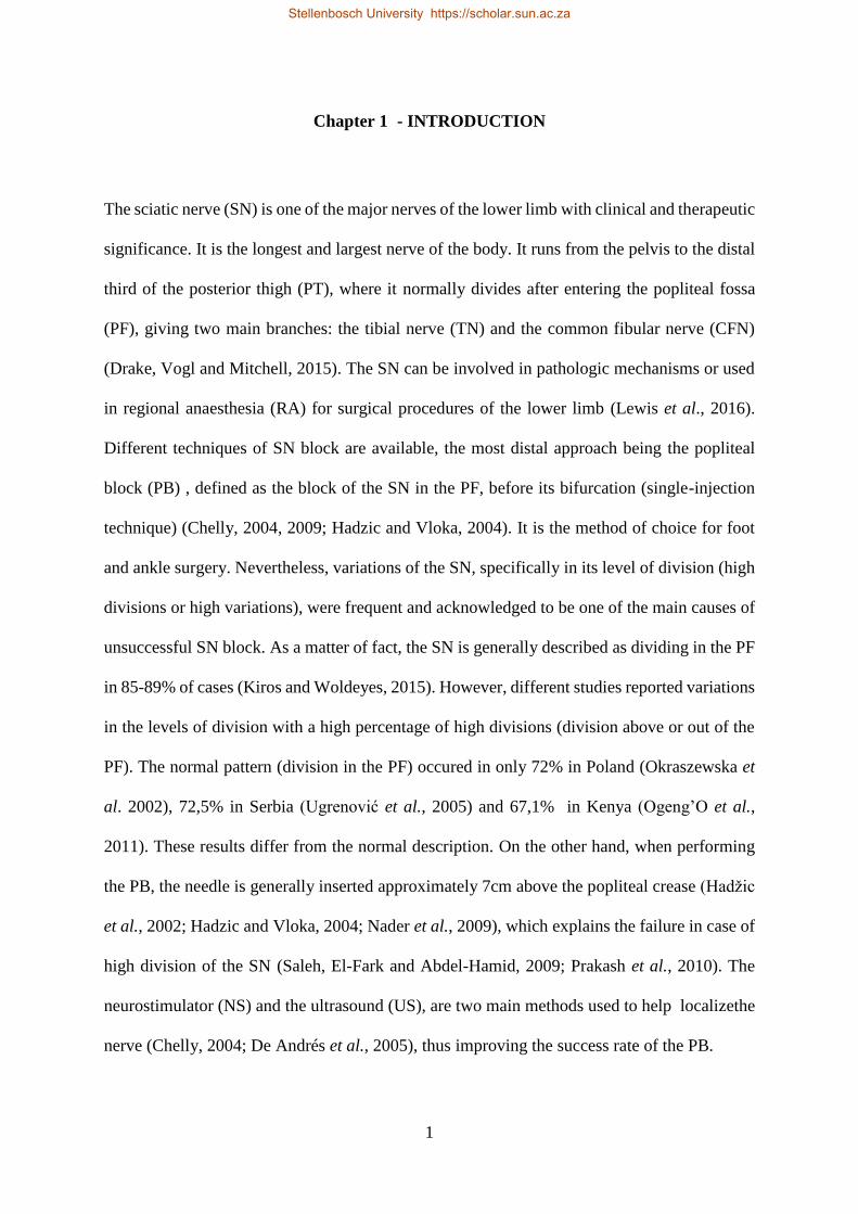
Figure 2-1 Origin of the sciatic nerve and components.
Source: Atlas of Anatomy (Netter, 2011)
Anatomically, these branches are enjoined in a common epineural sheath; however, they are
functionally distinct (Guvencer et al., 2009). This sheath plays a role in the diffusion of the
anaesthetic and contributes to the anaesthesia of both the TN and the CFN (Vloka et al., 1997).
The rami contributing to the formation of the SN gives rise to anterior divisions (L4-S3) for
the tibial component and posterior divisions (L4-S2) for the common fibular component.
Between its origin and termination, the SN successively runs through the pelvis, the gluteal
region and the posterior thigh until its distal third, in the popliteal fossa (PF).
The sciatic nerve (SN) enters the gluteal region via the greater sciatic foramen, underneath the
piriformis muscle (Saleh, El-Fark and Abdel-Hamid, 2009; Ekanem et al., 2015) (Figure 2-2).
Stellenbosch University https://scholar.sun.ac.za

6
Figure 2-2: Exit of the sciatic nerve below the piriformis muscle. This figure shows the
exit of the SN hust below the piriformis muscle as a single trunk.
Source: Gary’s Anatomy for students (Drake, Vogl and Mitchell, 2015).
The rapport between the SN and the PM shows anatomic variations which have previously
been reported in many populations (Guvencer et al., 2009). They are mostly associated with a
bifurcation of the SN in the pelvis and a classification, elaborated by Beaton and Anson in 1937
(Okraszewska et al., 2002; Guvencer et al., 2009), is used to label the different variants
(Guvencer et al., 2009). These variations can lead to clinical manifestations recognised as the
piriformis syndrome, caused by the compression of one of the branches or the common trunk
of the sciatic nerve (Rani and Kalra, 2015). However, they are not the focus of this study.
In the gluteal region, the SN is in relation with the posterior surfaces of the gemelli muscles,
the quadratus femoris and the obturator internus that are crossed posteriorly (Figure 2-2)
(Adibatti, Sangeetha and Adibatti, 2014). Close to the inferior border of the quadratus femoris,
the SN enters the posterior thigh and runs parallel to the biceps femoris muscle until its
termination in the PF (Drake, Vogl and Mitchell, 2015) (Figure 2-3).
Stellenbosch University https://scholar.sun.ac.za

7
Figure 2-3: Route and termination of the SN. The SN from its origin to the PF and along
its route gives muscular branches to the hamstring.
Source: Gary’s Anatomy for students (Drake, Vogl and Mitchell, 2015)
The SN terminates and bifurcates in the PF and at this level, the SN is superficial, localised at
a depth of 20-30 mm (James Phillips, Troutman and Lerant, 2011). The description of the
pattern of division is given below and is comparable to findings by Sohn et al. (2015) who
also found the SN at a depth of 20.6 ± 7.1 mm from the skin.
Branches of the sciatic nerve
The tibial nerve (TN) and common fibular nerve (CFN) are two subdivisions of the sciatic
nerve after it branches in the PF. The TN descends vertically, whereas the CFN descends
Stellenbosch University https://scholar.sun.ac.za

8
laterally and twists around the neck of the fibula. Their anatomical description as given in
textbooks (Hansen and Netter, 2014; Drake, Vogl and Mitchell, 2015) will be detailed here.
Tibial nerve
The TN is the branch of the SN made of the anterior divisions of L4 to S3. After the division
of the SN in the PF, the TN travels vertically downward, parallel to the midline. From there,
it penetrates the posterior compartment of the leg in the space between the two heads of the
gastrocnemius. Along its course, the TN gives a branch (medial), which combined to another
branch from the CFN (lateral), forms the sural nerve. Close to the termination, the TN gives
the medial calcaneal nerve before ending its course in the foot with the plantar nerves (medial
and lateral). The two branches of the SN are surrounded by the epineural sheath until their
separation in the PF (Vloka et al., 1996).
Common fibular nerve
The CFN exits the PF by following the Biceps Femoris’ long head. It spirals around the neck
of the fibula, crossing successively the lateral and anterior sides of the leg. The CFN divides
and gives two branches: Superficial and Deep fibular nerves, respectively for the lateral and
anterior compartments of leg.
Figure 2-4 gives a brief overview of the divisions and subdivisions of the sciatic nerve. All
these branches contribute to the muscular innervation of the foot and leg as well as a partial
cutaneous innervation of that area. Terminal branches are not represented in this diagram.
Stellenbosch University https://scholar.sun.ac.za

9
Figure 2-4: Branching pattern of the sciatic nerve (diagram based on descriptions).
Source: Gary’s Anatomy for students (Drake, Vogl and Mitchell, 2015).
The distance of the point at which the SN bifurcates from the popliteal crease is not described
in textbooks. Vloka et al. (1997) first evaluated this distance as ranging between 0 and 73 mm,
while the distance ranged between 0 and 115 mm in another of their study (Vloka et al., 2001).
However, these observations are limited, due to the small sample sizes used for the analysis
(10 specimens only for the first study and 28 in the second).
Variations in the branching pattern
The anatomy of the sciatic nerve varies greatly (Table 2-1) according to studies carried out in
different populations (Okraszewska et al., 2002; Ugrenović et al., 2005; Guvencer et al., 2009;
Ogeng’O et al., 2011; Adibatti, Sangeetha and Adibatti M, 2014; Kiros and Woldeyes, 2015).
Sciatic nerve
Tibial nerve
Medial plantar nerve
Lateral plantar nerve
Medial sural cutaneous
nerve
Common fibular nerve
Superficial fibular nerve
Deep fibular nerve
Lateral sural cutaneous
nerve
Stellenbosch University https://scholar.sun.ac.za
10
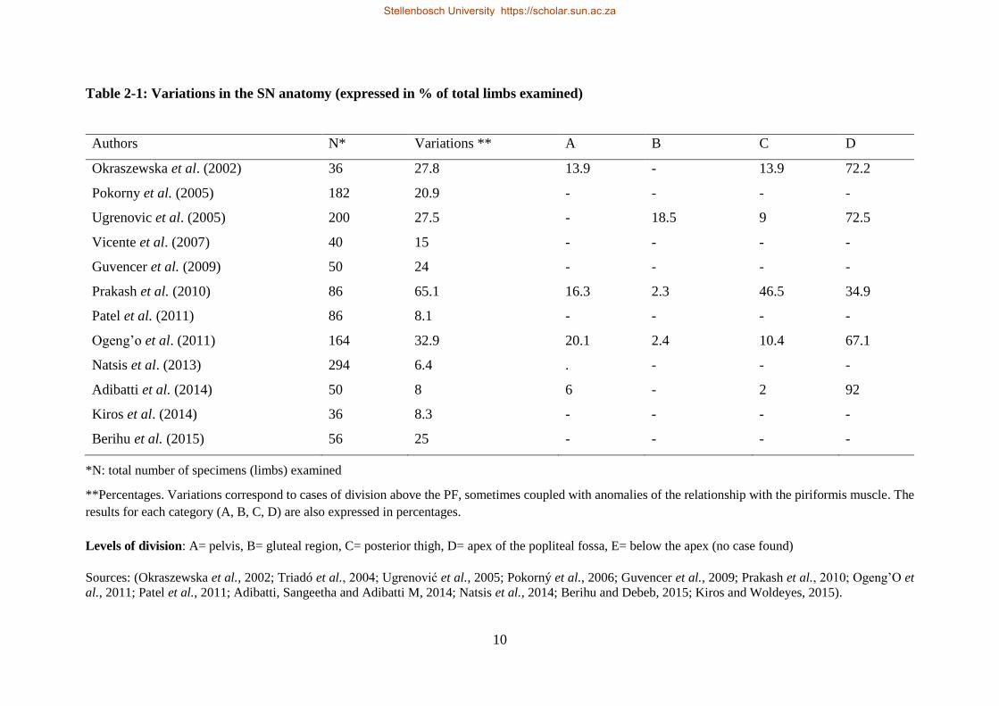
Table 2-1: Variations in the SN anatomy (expressed in % of total limbs examined)
Authors N* Variations ** A B C D
Okraszewska et al. (2002) 36 27.8 13.9 - 13.9 72.2
Pokorny et al. (2005) 182 20.9 - - - -
Ugrenovic et al. (2005) 200 27.5 - 18.5 9 72.5
Vicente et al. (2007) 40 15 - - - -
Guvencer et al. (2009) 50 24 - - - -
Prakash et al. (2010) 86 65.1 16.3 2.3 46.5 34.9
Patel et al. (2011) 86 8.1 - - - -
Ogeng’o et al. (2011) 164 32.9 20.1 2.4 10.4 67.1
Natsis et al. (2013) 294 6.4 . - - -
Adibatti et al. (2014) 50 8 6 - 2 92
Kiros et al. (2014) 36 8.3 - - - -
Berihu et al. (2015) 56 25 - - - -
*N: total number of specimens (limbs) examined
**Percentages. Variations correspond to cases of division above the PF, sometimes coupled with anomalies of the relationship with the piriformis muscle. The
results for each category (A, B, C, D) are also expressed in percentages.
Levels of division: A= pelvis, B= gluteal region, C= posterior thigh, D= apex of the popliteal fossa, E= below the apex (no case found)
Sources: (Okraszewska et al., 2002; Triadó et al., 2004; Ugrenović et al., 2005; Pokorný et al., 2006; Guvencer et al., 2009; Prakash et al., 2010; Ogeng’O et
al., 2011; Patel et al., 2011; Adibatti, Sangeetha and Adibatti M, 2014; Natsis et al., 2014; Berihu and Debeb, 2015; Kiros and Woldeyes, 2015).
Stellenbosch University https://scholar.sun.ac.za
Stellenbosch University https://scholar.sun.ac.za

11
Generally speaking, most of the variations in the SN anatomy are of two types: variations in
the relationship with the piriformis muscle (PM) (Guvencer et al., 2009; Natsis et al., 2014)
and variations in the level of division of the SN. Both types are sometimes associated,
especially in cases of SN division in the pelvis (Okraszewska et al., 2002). Besides, variations
such as trifurcations or emergence of the sural nerve directly from the sciatic nerve could be
rarely observed (Prakash et al., 2010; Adibatti, Sangeetha and Adibatti M, 2014; Berihu and
Debeb, 2015). In this study, the focus was only on variants in the level of bifurcation of the
SN. In most cases, the terms high variations and high divisions were used interchangeably to
describe a SN dividing above (outside of) the PF.
Many studies reported a bifurcation of the SN at any point along its course between the pelvis
and the PF (see Table 2-1). The symmetry (division at the same level on both sides) was
individually variable (Rani and Kalra, 2015) and only a few comparisons were made between
male and female. Because multiple surgical procedures are carried out in the gluteal region, a
precise knowledge of the SN division would be essential for surgeons (Rani and Kalra, 2015).
Also, many clinical situations such as nerve injuries (Adibatti, Sangeetha and Adibatti M, 2014;
Berihu and Debeb, 2015; Ekanem et al., 2015; Kiros and Woldeyes, 2015), muscular atrophy
(Ogeng’O et al., 2011), non-discogenic sciatica or piriformis syndrome by entrapment of the
sciatic nerve or its branches (Guvencer et al., 2009; Patel et al., 2011; Supriya, 2012; Adibatti,
Sangeetha and Adibatti M, 2014) are the most frequent clinical presentations related to
variations in the level of division of the sciatic nerve (Kiros and Woldeyes, 2015; Tomaszewski
et al., 2016). To avoid all these complications, surgeons and physicians in general must be
aware of these variations (Ogeng’O et al., 2011). In addition, any case of SN injury leading to
paralysis could cause an atrophy of the muscles supplied.
Stellenbosch University https://scholar.sun.ac.za
12
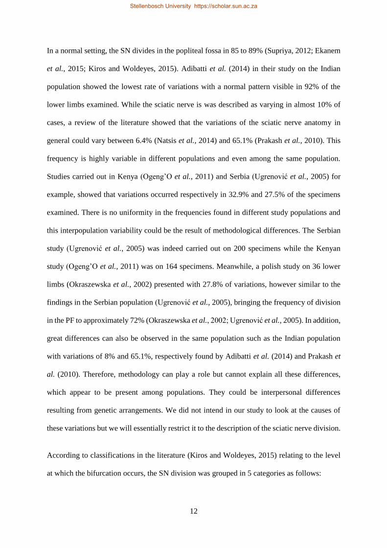
In a normal setting, the SN divides in the popliteal fossa in 85 to 89% (Supriya, 2012; Ekanem
et al., 2015; Kiros and Woldeyes, 2015). Adibatti et al. (2014) in their study on the Indian
population showed the lowest rate of variations with a normal pattern visible in 92% of the
lower limbs examined. While the sciatic nerve is was described as varying in almost 10% of
cases, a review of the literature showed that the variations of the sciatic nerve anatomy in
general could vary between 6.4% (Natsis et al., 2014) and 65.1% (Prakash et al., 2010). This
frequency is highly variable in different populations and even among the same population.
Studies carried out in Kenya (Ogeng’O et al., 2011) and Serbia (Ugrenović et al., 2005) for
example, showed that variations occurred respectively in 32.9% and 27.5% of the specimens
examined. There is no uniformity in the frequencies found in different study populations and
this interpopulation variability could be the result of methodological differences. The Serbian
study (Ugrenović et al., 2005) was indeed carried out on 200 specimens while the Kenyan
study (Ogeng’O et al., 2011) was on 164 specimens. Meanwhile, a polish study on 36 lower
limbs (Okraszewska et al., 2002) presented with 27.8% of variations, however similar to the
findings in the Serbian population (Ugrenović et al., 2005), bringing the frequency of division
in the PF to approximately 72% (Okraszewska et al., 2002; Ugrenović et al., 2005). In addition,
great differences can also be observed in the same population such as the Indian population
with variations of 8% and 65.1%, respectively found by Adibatti et al. (2014) and Prakash et
al. (2010). Therefore, methodology can play a role but cannot explain all these differences,
which appear to be present among populations. They could be interpersonal differences
resulting from genetic arrangements. We did not intend in our study to look at the causes of
these variations but we will essentially restrict it to the description of the sciatic nerve division.
According to classifications in the literature (Kiros and Woldeyes, 2015) relating to the level
at which the bifurcation occurs, the SN division was grouped in 5 categories as follows:
Stellenbosch University https://scholar.sun.ac.za

13
Category A: Division in the pelvis
Category B: Division in the gluteal region
Category C: Division in the posterior thigh
Category D: Division at the apex of the popliteal fossa
Category E: Division below the apex of the popliteal fossa
In certain classifications, category C (division in the posterior thigh) has three subtypes namely
divisions in the proximal, mid and distal third. Besides, division at the apex and below the
popliteal fossa can be grouped under popliteal fossa, as we are more interested in the high
divisions. Division below the apex of the popliteal fossa would not be inconvenient,
considering also the fact that no study was precise about what they considered as being below
the apex of the PF and it does not affect the outcome of the PB.
Concerning the distance at which the SN divides above the PC, high variations affect the
distance between the PC and the point of bifurcation of the nerve. The mean varied between
44 and 82 mm (Vloka et al., 1997, 2001; Saleh, El-Fark and Abdel-Hamid, 2009; Schiarite et
al., 2015; Sohn et al., 2015; Tomaszewski et al., 2016). When the distance was recorded, the
lowest point at which the sciatic nerve divided was at the level of the popliteal crease (Vloka
et al., 1997, 2001). The highest point was in the posterior thigh at a distance of 180 mm (Saleh,
El-Fark and Abdel-Hamid, 2009).
As discussed earlier, these variations of the sciatic nerve would affect the outcome of the PB.
The common technique consists in locating the common trunk of the SN before the bifurcation;
Stellenbosch University https://scholar.sun.ac.za

14
it therefore shows the importance of the sciatic nerve anatomy and the existing variations,
paramount to a successful PB.
The popliteal fossa
The popliteal fossa is a transition area between the thigh and leg, posterior to the knee joint. Its
shape corresponds to that of a diamond (Figure 2-5) and the boundaries are essentially muscular
(Gosling, 2008; Creech and Meyr, 2013). As described in Figure 2-5, the biceps femoris
constitutes the superior lateral border, while the semitendinosus and semimembranosus form
the superior medial border. These muscles all together are called hamstring. On the other side,
the gastrocnemius muscles and the plantaris muscle form the inferior boundaries (Creech and
Meyr, 2013).
Figure 2-5: Popliteal fossa. Margins and landmarks. The superior margins are biceps
femoris, semitendinosus, and semimembranosus while the inferior margins are both lateral
and medial head of gastrocnemius.
Source: Gary’s Anatomy for students (Drake, Vogl and Mitchell, 2015).
Stellenbosch University https://scholar.sun.ac.za

15
The popliteal fossa (PF) is considered to be the normal level for the division of the SN (Adibatti,
Sangeetha and Adibatti M, 2014). However, there are cases of high bifurcation which occur
outside of the PF, at any point along the course of the nerve (Ekanem et al., 2015). Besides the
SN and its branches, the PF contains a lot of adipose tissue and the popliteal vessels, located
medially and deeper to the SN (Rorie et al., 1980; Creech and Meyr, 2013). This detail is of
importance when injecting in that area, to avoid the risk of vascular punctures. Also, the fat
present in the PF could be favourable as it extends the duration of action of local anaesthetics
(Creech and Meyr, 2013) even though there is a risk of impairment in the distribution of the
anesthetic around the nerve (Rorie et al., 1980).
Structures innervated by SN
The sciatic nerve (SN) is described as a mixed nerve with a motor and sensory function
(Adibatti, Sangeetha and Adibatti M, 2014; Drake, Vogl and Mitchell, 2015). It innervates the
hamstring (Biceps femoris, Semitendinosus and Semimembranosus) as well as a portion of the
Adductor magnus. It also supplies the hip joint and the knee joint through articular branches
(Okraszewska et al., 2002; Drake, Vogl and Mitchell, 2015). It therefore contributes to the
flexion of the knee and the extension of the hip. After bifurcation, the tibial nerve (TN) and the
common fibular nerve (CFN) serve most muscular structures of the leg and foot. The TN
supplies muscles of the posterior aspect of the leg and the planter surface of the foot while the
CFN supplies the other muscles of the leg and of the dorsal surface of the foot. Both nerves
participate in the cutaneous innervation below the knee, save the anteromedial side of the leg
and the foot (Lewis et al., 2016).
According to the territory innervated, the TN and CFN participate to musculoskeletal
movements of the lower limb. The TN supplies the muscles that produce plantarflexion and
inversion movements of the foot as well as flexion of the toes. The CFN on the other hand,
Stellenbosch University https://scholar.sun.ac.za
16
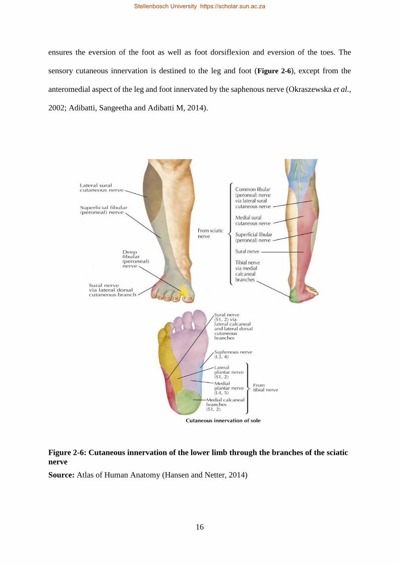
ensures the eversion of the foot as well as foot dorsiflexion and eversion of the toes. The
sensory cutaneous innervation is destined to the leg and foot (Figure 2-6), except from the
anteromedial aspect of the leg and foot innervated by the saphenous nerve (Okraszewska et al.,
2002; Adibatti, Sangeetha and Adibatti M, 2014).
Figure 2-6: Cutaneous innervation of the lower limb through the branches of the sciatic
nerve
Source: Atlas of Human Anatomy (Hansen and Netter, 2014)
Stellenbosch University https://scholar.sun.ac.za

17
. Popliteal block
The popliteal block is one the distal approaches to the blockade of the SN and is realised in the
PF. While a blockade of the nerve above that point is more difficult due to the deep location of
the nerve, the popliteal block is easier and gives more satisfaction (Herring et al., 2011; Barbosa
et al., 2015).
Definition
The popliteal block (PB) is a peripheral nerve block which entails an obstruction to the nervous
conduction of the SN from the PF and downwards. It is the most distal approach to the sciatic
nerve, first performed by Labat in 1923 (Hegewald et al., 2014). Compared to the double-
injection technique that targets each branch (TN and CFN), the single-injection targets the
common trunk of the SN before its bifurcation (March et al. , 2006); this would reduce the risk
of vascular puncture because the popliteal vessels are still located deeper (Phillips et al., 2011).
The infrequent use of the popliteal block was due to the risk of complications or failure. Yet,
it remained a technique of choice for foot surgery (Rorie et al., 1980). Hadzic et al. (2002) also
suggested the insufficient preparation of the operating room, the variable outcome and a
problem in the training of anaesthesiologists. There are different ways to perform a peripheral
nerve blockade: blind methods, PB using a neurostimulator or PB using ultrasound (US). Blind
techniques are still used even though the use of neurostimulator or ultrasound (US) is more
beneficial. The neurostimulator is a great tool in localising the nerve and therefore improving
the success rate by allowing a better localization of the nerve than blind methods (Hadzic and
Vloka, 2004). Nevertheless, lately the US brings a new dimension to PB with more precision,
the possibility to visualize the nerve and therefore less complications (Prasad et al., 2010).
Stellenbosch University https://scholar.sun.ac.za

18
Indications
The PB is the preferred anaesthetic technique used for foot and ankle surgery (Rorie et al.,
1980; Prasad et al., 2010). It can be used as a single technique or combined to other regional
anesthetic techniques for a larger action in below-the-knee anaesthesia or analgesia (Bouaziz
et al., 1999). Initially this technique of RA was not widely used. Nowadays, it can even be
used as a postoperative analgesic technique in the emergency department, as reported by
Philips et al. (2011).
Technique
The PB encompasses different approaches and techniques. The block is commonly easy to
perform (Chelly, 2003; Triadó et al., 2004). Nevertheless, with the blind method (without the
use of NS or US to locate the nerve), the PB has a high rate of failure especially in situations
of high variations of the nerve (Reinoso-Barbero et al., 2014). The PB is sometimes combined
to the saphenous block to obtain a good desensitisation of the lower limb (Mendicino, Statler
and Catanzariti, 2002).
The equipment needed to perform a popliteal block includes needles of 22 gauge (function of
the approach used), syringes, ruler, marking pen, disinfectant, drapers, sterile gloves, gauze
pack, etc. When injecting the anaesthetic, it is essential to place the needle near the nerve to
avoid impairment of the substance distribution in the popliteal fossa (Rorie et al., 1980). As
stated before, the US or the NS (neurostimulator) aids the localisation of the nerve. When using
the NS, the best predictive factor for complete blockade is foot inversion (Ter Rahe and Suresh,
2002; De Andrés et al., 2005; Arcioni et al., 2007).
The approaches to the PB described in the literature are numerous but only three will be the
focus of this study: classic posterior, intertendinous and lateral approaches. They differ in the
Stellenbosch University https://scholar.sun.ac.za

19
placement of the patient and the point at which the needle is inserted. The surface landmarks
and the points of injection will be described in the description of each technique. The posterior
approaches have more constraints when it comes to placing the patient (Muñiz et al., 2003);
the prone position is indeed difficult to adopt for certain categories of patients while the
position for the lateral approach offers more comfort with the patient lying face upwards.
Posterior approaches
The patient is commonly placed lying on the bed, face downwards (prone position). The classic
landmarks are the hamstring borders and the popliteal crease (PC); they are outlined with a
marker. Slight differences in the point of injection were described. Some authors would place
the needle at 5 to 7 cm (Rorie et al., 1980; Vloka et al., 1997; Hadzic, 2007) above the crease
while others advocate a placement of the needle at 10 cm to ensure a higher success rate
(Singelyn, Gouverneur and Gribomont, 1991). More recently, modified approaches were
experimented with the needle inserted at 12 to 14 cm above the PC (Nader et al., 2009). Many
factors should be taken into consideration when choosing the approach, including the
preference of the surgeon, the patient pathology and the position of the patient (Chelly, 2003).
Two variants of the posterior approach have been developed: the classic posterior approach
and the intertendinous approach. The needle would be introduced at a depth of approximately
3 cm (Casalia, Carradori and Moreno, 2006).
2.2.3.1.1. Classic posterior approach (CPA)
For this approach (Figure 2-7), the needle is generally inserted at 7cm above the popliteal
crease. However, variants have been described. Rorie et al. (1980) suggested a placement of
the needle at 5 cm above the crease. The anaesthesia was successful in 82.3% of cases though
the point of injection was lower than in techniques proposed by Hadzic et al. (2002) who
Stellenbosch University https://scholar.sun.ac.za

20
inserted the needle 7 cm above the PC and perpendicular to the skin. Other authors inclined the
needle 60° proximally (Chelly, 2003).
Figure 2-7: Popliteal block using the classic posterior approach. The point of injection,
represented in red is 1 cm lateral to the midline axis.
2.2.3.1.2. Intertendinous approach (IA)
The landmarks are the same as those used for the CPA. In this approach (Figure 2-8), the needle
is inserted at 7 cm above the crease, at an equal distance of the tendons of the hamstring. A
modified approach was proposed by Nader et al. (2009) in which the needle was pushed from
a point just distal to the junction of the muscular tendons (semitendinosus and biceps femoris),
approximately 12 to 14 cm from the crease. The same approach was also experimented by
Minville et al. (2007), with the knee not flexed.
Stellenbosch University https://scholar.sun.ac.za

21
Figure 2-8: Popliteal block using the intertendinous approach. The point of injection is at
equal distance of the medial and lateral margins, just on the midline axis.
Lateral approach
The benefit of this approach is the placement of the patient in supine, which contributes to
his/her comfort (Casalia, Carradori and Moreno, 2006). The lower limb is extended on the bed
and the intermuscular groove on the lateral side of the thigh identified. As described by Vloka
et al. (1996) and Hadzic et al. (1998), the needle is first introduced horizontally in that space
7cm above the lateral femoral condyle (Figure 2-9). After contact with the femur, the needle
is withdrawn and reoriented 30° posteriorly.
Stellenbosch University https://scholar.sun.ac.za

22
Figure 2-9: Popliteal block through the lateral approach. The point of injection is in the
intermuscular groove, 7 cm above the lateral femoral condyle.
Source: Illustration made by the investigator using “Paint”
To reduce the pain that can be caused by the passage of the needle through the muscles, Al-
Nasser (2002) proposed a modified approach by inserting the needle posterior to the tendon of
the biceps femoris. Nevertheless, this variant called for more trials before considering it as a
clinical approach with tangible results (Al-Nasser, 2002).
Advantages and disadvantages of the PB
Like any procedure for peripheral nerves, the PB offers both positive and negative aspects.
Among the disadvantages, the risk of toxicity, intravascular injection or injury of the nerve was
highlighted in the literature (Mendicino, Statler and Catanzariti, 2002). One of the most
reported complications was a temporary paraesthesia in the postoperative period (Herring et
al., 2011; Joshi et al., 2016). Complications were observed in up to 8.2% of cases (Joshi et al.,
2016), which contrasts with the observations of Rorie et al. (1980) who reported that
complications were rare in the PB. Another difficulty encountered is the positioning of the
patients, mainly for the posterior approaches. While in these approaches the patients tend to be
Stellenbosch University https://scholar.sun.ac.za

23
placed in prone, this approach is not suitable for patients with conditions like pregnancy,
obesity, traumatisms, those displaying an hemodynamic instability or ventilated (Arcioni et al.,
2007). However, an alternative was proposed by placing the subject in supine and elevating his
leg (James Phillips, Troutman and Lerant, 2011; Sohn et al., 2015).
On the other hand, the benefits of the PB are many (Joshi et al., 2016). Peripheral techniques
become popular because of their advantages and the good control of pain (Casalia, Carradori
and Moreno, 2006). This regional anaesthetic technique is secure, compared to other
anaesthetic techniques (general, spinal), the side effects being lowered (Sohn et al., 2015; Joshi
et al., 2016). Indeed, the side effects related to general or spinal anaesthesia include headaches,
vomiting, cardiorespiratory depression, toxicity to only mention a few (Hadžic et al., 2002).
The PB provides with an analgesia that is prolonged in the postoperative period, therefore
reducing the consumption of opioids (Hadžic et al., 2002; Ter Rahe and Suresh, 2002; Perlas
et al., 2008; Creech and Meyr, 2013; Joshi et al., 2016) for a pain that is either moderate or
severe in foot and ankle surgery. Consequently, the side effects associated with the opioids
(urinary retention, nausea, constipation, vomiting, addiction) are also reduced (Hadžic et al.,
2002; Ter Rahe and Suresh, 2002; Perlas et al., 2008; Joshi et al., 2016). With reduced
morbidity, the length of hospital stay is decreased (Mendicino, Statler and Catanzariti, 2002).
Furthermore, the PB provides an adequate desensitization, adapted to the surgical procedure to
realise.
Another advantage of this regional technique of anaesthesia is the conservation of the
hamstring; this allows an early ambulation with crushes and therefore reduces the hospital stay,
overall decreasing the medical costs (Ter Rahe and Suresh, 2002; Joshi et al., 2016).
All these characteristics contribute to the popularity of the PB in today’s setting where
outpatient surgery is growing and there is a need to make surgery costs affordable for everyone
Stellenbosch University https://scholar.sun.ac.za

24
(Greenberg, 1995; Joshi et al., 2016; Ng-Kamstra et al., 2016). Overall, this anaesthetic method
has proven to be satisfactory for patients.
Outcome of the PB
The outcome of a PB varies widely from one study to another (Muñiz et al., 2003). Despite its
reliability, many factors influence the outcome such as the level of training of the operator and
the proximity of the needle (Rorie et al., 1980). Hegewald et al. (2014) also evoked the patient
age and BMI as factors influencing the outcome of the PB; a low BMI and increasing age were
considered as improving the success rate. The use of neurostimulators or imagery also enhances
success rates. The imagery could be the ultrasound or like in certain cases the MRI (Minville
et al., 2007; Sohn et al., 2015). The ultrasound appeared in some studies as the method of
choice to have a good success rate (McCartney, Brauner and Chan, 2004; Sinha and Chan,
2004). Hence, those studies reported a successful block in 76.2%, close to the results of
Provenzano et al. reported in a study by Hegewald et al. (2014) who found 79% of successful
cases. On the other hand, Rorie et al. (1980) had slightly better results with 82.3% of
satisfactory blocks. Many other authors (Barbosa et al. 2015; Danelli et al. 2009; Hansen et
al., 2000; Mariano et al. 2009; Migues et al.2005; Phillips et al., 2011) had the best outcomes
with a block successful in 90 to 100% of cases. Monso et al. (2000) reviewed the PB in more
than 700 patients and found 86% successful blocks, closely similar to the reports from Rorie et
al. (1980) or Perlas et al. (2008) who found 89% successful blocks. Comparable to the findings
of Herring et al. (2011), Wadhwa et al. (2010) reported a better outcome with the use of
ultrasound compared to blocks aided by neurostimulation. The use of both techniques (US and
NS) indeed improves to a certain extent the outcome of the block (De Andrés et al., 2005), as
confirmed by Van Geffen et al. (2010) who asserted that US helped in visualising the nerve
and therefore with a good placement of the needle and the observation of the anaesthetic’s
Stellenbosch University https://scholar.sun.ac.za

25
spread. Besides, US also improves the patient’s comfort by reducing the number of attempts in
needle puncture and position (Danelli et al., 2009). However, a combination of both techniques
is recommended to avoid wrong interpretations of US images (Van Geffen et al., 2010; Creech
and Meyr, 2013).
Compared to previous authors, Hadzic et al. (2002) used MRI and succeeded in 100% of cases
for the intertendinous block. When the block fails, one of the main causes suggested was the
high division of the sciatic nerve, such as reported in a case study by Clendenen et al. (2008).
In this case, the block would not have been complete without the use of US. Another cause of
failure was the difficulty to clearly identify the boundaries of the PF in living patients (Hadžic
et al., 2002). A preoperative imagery is therefore advisable for a better success (Adibatti,
Sangeetha and Adibatti M, 2014).
Problem statement
The popliteal block (PB) is a widespread anaesthetic procedure nowadays (Sinha and Chan,
2004; Migues et al., 2005; Bruhn et al., 2008). It relies heavily on the knowledge of the sciatic
nerve (SN) anatomy, essential for surgeons and anaesthetists (Casalia, Carradori and Moreno,
2006). However, the variations highlighted in the literature are prejudicial to the block to the
extent that they contribute to cases of failures, even more during blind injections (Prakash et
al., 2010; Reinoso-Barbero et al., 2014).
Variations of the SN division are highly inconsistent from one population to another. In
addition, only few data are available in Africa (Ogeng’O et al., 2011) and on a homogenous
population. This study is therefore beneficial as it identifies these variations in a heterogeneous
South African population as well as determines the outcome of the block after simulation using
three different approaches.
Stellenbosch University https://scholar.sun.ac.za

26
Aim
The aim of this study is to describe the level of division of the sciatic nerve in a South African
cohort and evaluate the implications of variations on the popliteal block.
Objectives
To simulate the popliteal block by injecting coloured silicone
To examine the branching patterns of the sciatic nerve and identify high variations
To evaluate the distance between the popliteal crease and the point of bifurcation of the
nerve
To establish an anatomical correlation between the dye of silicone and the nerve
To compare the results between males and females
To analyse the results and compare with other reports in the literature.
Stellenbosch University https://scholar.sun.ac.za

27
Chapter 3 - MATERIALS AND METHODS
This cross-sectional study took place at the division of Anatomy and Histology of the Faculty
of Medicine and Health Sciences, Stellenbosch University.
. Study population
The study population was composed of 61 cadavers of mixed ancestry or Coloured (n = 45),
African descent (n = 9) and European descent (n = 7) as detailed in Table 3-1. The cadaveric
cohort was representative of the heterogeneity of the Western Cape population with a high
representativity of the Coloured population (73.77%).
Table 3-1: Demographic characteristics of the study population
Group
Sex Black Mixed White Total
Male 8 23 6 37
Female 1 22 1 24
Total 9 45 7 61
These embalmed cadavers were prepared for the anatomical and physiological training of the
students in the Faculty of Medicine and Health Sciences.
. Ethical issues
To comply with the University’s policy on the use of human material, ethics approval
(S16/03/052) was obtained from the Health Research and Ethics Committee (HREC) prior to
the commencement of the study.
Stellenbosch University https://scholar.sun.ac.za

28
. Exclusion criteria
Both left and right lower limbs of 65 cadavers (n = 130 specimens) were examined. Specimens
whose sciatic nerve was damaged during the dissection process were excluded as well as one
cadaver with results on the right limb only (SN damaged during the dissection of the left lower
limb) because it could not be used to evaluate symmetry. This resulted in a total of 61 cadavers
(n = 122 lower limb specimens).
. Procedure
The study consisted in three consecutive steps. Firstly, a pilot study was carried out to test the
preparation of the dye and ascertain the technique of the popliteal block (PB) on 7 cadavers.
The second step consisted in data collection during the medical students’ dissections of the
lower limb specifically. Lastly, the investigator performed the simulation of the PB, then
dissected the lower limbs of the cadavers prepared for physiology training.
The preparation of the dye, the PB simulation as well as the dissection procedure are developed
in this section.
The dye
Matching a work on cerebral arteries in the division of Anatomy and Histology, the dye
consisted of mixtures of silicone.
Materials and equipment
The tools and substances used for the preparation are listed in Table 3-2. Most tools were
provided by the university. Plastic tubes, wooden sticks, syringes, gloves and disposable
spoons were changed as often as necessary. Therefore, the quantities of these items are not
mentioned in the table.
Stellenbosch University https://scholar.sun.ac.za

29
Table 3-2: Tools used for the preparation of the dye
Material/Equipment Trade name Quantity
Silicone MM922 Acc silicone Europe, UK 5 kg
Catalyst Acc silicone Europe, UK
Turpentine DEKRO PAINTS, SA 1 bottle of
Powder paint Dala, SA 3 bottles of
Plastic tubes - -
Disposable coffee spoons - -
Wooden stick - -
Nitrile Gloves Lasec, Malaysia
Disposable syringes (1ml) China -
Composition of the dye
The dye consisted in a mixture of silicone. The quantities used for each preparation are detailed
in Table 3-3.
Table 3-3: Composition of a mixture of silicone
Material Quantity
Silicone 30 ml
Catalyst 1.5ml
Turpentine 3 drops
Powder paint Half spoon
During the pilot test, the 30 ml of silicone were put in a tube using a 10 ml syringe. Thereafter,
the limit was drawn on the tube with a marker and reported on the other tubes. As for the
catalyst, the quantity was measured using 1ml syringes.
Stellenbosch University https://scholar.sun.ac.za

30
Preparation of the dye
The silicone was mixed with half a teaspoon of powder paint in a tube. When the mixture
obtained became homogenous, the catalyst was slowly incorporated in a clockwise movement.
The drops of turpentine were then added last to slow down the hardening process. During PB
simulation, three different approaches were used. Hence, three separate mixtures of different
colours (Figure 3-1) were made to differentiate each technique.
Figure 3-1: Mixtures of silicone for the three approaches of popliteal block. Each colour
of dye corresponded to a specific approach; in this image, the dye was solidified 48h after
preparation.
The blue, red and green mixtures were used respectively for the lateral approach (LA), the
classic posterior approach (CPA) and the intertendinous approach (IA). The green dye was
obtained after mixing the blue and yellow powder paints.
Popliteal block
Three different approaches were used: the classic posterior approach, the intertendinous
approach and the lateral approach.
Stellenbosch University https://scholar.sun.ac.za

31
Materials and equipment
The different instruments used for the simulation of the block are illustrated in Figure 3-2. They
consisted of:
Mixtures of silicone (Figure 3-1),
Needles 14-18G a length of 48-80mm by Viggo-spectramed, Bio-On and B.Braun with
short to regular bevels,
Disposable syringes of 1ml,
A measuring tape,
A black marker,
Nitrile gloves by Lasec, Malaysia.
Figure 3-2: Equipment used for the simulation. As shown in the picture, it consisted of
mixture of silicone, needles, syringes, measuring tape, and marker.
Simulation of the popliteal block
The cadavers were successively placed in prone and supine position to perform the block. The
prone position was used for the posterior approaches (classic posterior and intertendinous)
while the supine position was for the lateral approach. Classic posterior and intertendinous
Stellenbosch University https://scholar.sun.ac.za

32
approaches are two variants of the posterior approach to the PB, which only differ in the
distance from the midline axis to the point of insertion of the needle. They were both used in
this study because not only did we study the level at which the dye was deposited but also the
proximity between the dye and the nerve.
The simulation was performed on the lower limbs of 11 cadavers and the three approaches
were used on each limb.
3.4.2.2.1. Classic posterior approach
The cadaver was placed in prone position. The steps followed were as previously described by
Hadzic et al. (2002). The superior borders of the popliteal fossa (biceps femoris,
semitendinosus and semimembranosus) were identified and outlined with a permanent marker.
The bisector of the superior angle of the PF or midline axis was traced. After identification of
the landmarks, the needle connected to a syringe containing the red dye was inserted 7 cm
above the popliteal crease and 1cm lateral to the midline axis (
Figure 3-3), perpendicular to the skin and at a depth of 4 cm approximately.
Stellenbosch University https://scholar.sun.ac.za

33
Figure 3-3: Classic posterior approach: point of injection on a right lower limb with the
popliteal fossa highlighted in green.
Source: modified from Kenhub (Jones, 2015).
3.4.2.2.2. Intertendinous approach
The intertendinous approach (IA), variant of the CPA, followed the same steps with the
difference that the needle was mounted on a syringe filled with a green dye and inserted on
the midline axis (at midpoint between the muscular tendons), still 7 cm above the PC (Figure
3-4). Similarly to Triado et al. (2004), a 48 mm needle was used. The depth of insertion of the
needle was 4 cm approximately from the insertion point on the skin.
Stellenbosch University https://scholar.sun.ac.za

34
Figure 3-4: Intertendinous approach: point of injection in the popliteal fossa highlighted
in green. MED = Medial, LAT = Lateral, PC = Popliteal Crease, MA = Midline Axis.
Source: modified from Kenhub (Jones, 2015).
3.4.2.2.3. Lateral approach
The cadaver placed in supine position, the landmarks were identified: vastus lateralis, biceps
femoris and lateral femoral condyle. These landmarks were traced with the marker and the
syringe filled with the blue dye. The needle was introduced in the groove between both
muscles, 7 cm above the lateral femoral condyle (Figure 3-5). From the skin, the needle was
directed posteriorly with an inclination of 30 degrees and inserted at a depth of approximately
4 cm, knowing that Vloka et al. (1996) suggested 33 to 54 mm.
Stellenbosch University https://scholar.sun.ac.za

35
Figure 3-5: Lateral approach to the popliteal block
Source: modified image from “google search”
Dissection of the lower limb
The lower limb was dissected following the instructions from the Grant’s Dissector (Tank,
2013) as indicated in Figure 3-6. After skin removal, the fascia was opened and posterior to
the knee the popliteal fossa cleaned and explored to retrieve the nerve.
Figure 3-6: Skin landmarks for dissection (Duke Anatomy - Lab 13: Gluteal Region &
Posterior Thigh, 2013)
A good exposure of the SN (Figure 3-7) in the PF required a thorough cleaning of the area
because of the significance of the adipose tissue; however, the cleaning was careful to avoid
any damage to the nerve or its branches.
Stellenbosch University https://scholar.sun.ac.za

36
Legend: GM = Gluteus Maximus, BF = Biceps Femoris, CFN = Common Fibular Nerve, TN = Tibial Nerve,
ST= Semitendinosus, SM = Semimembranosus
Figure 3-7: Sciatic nerve exposed after dissection of the posterior thigh. The SN divided in
the upper third of the posterior thigh
After identification, the level of division was classified based on previous classifications and
adapted to this study. It consisted of 4 categories:
Category A: division of the nerve in the pelvis. The branches of the SN are already
individualised at the exit of the pelvis below the piriformis muscle.
Category B: division of the nerve in the gluteal region. The nerve divides between the
inferior border of the piriformis muscle and the gluteal fold.
Category C: division of the nerve in the posterior thigh, between the gluteal fold and
the popliteal fossa.
Category D: division of the nerve in the popliteal fossa (Figure 2-5), from the apex and
below.
The division of the SN in the popliteal fossa (category D) corresponded to the normal pattern
described in textbooks, whereas Categories A, B and C were considered as high variations of
the nerve. After identification of the SN and categorisation of the level of division, the PF was
Stellenbosch University https://scholar.sun.ac.za

37
examined to locate the dye and observe its position to the nerve. The success of the PB was
related to the location of the dye above SN bifurcation while failures or incomplete blocks
would be considered when the dye was deposited below the point of bifurcation of the nerve.
Cases where the dye was outside of the PF (Figure 3-8) were attributed to technical glitches
and excluded from the study.
Figure 3-8: Dye located outside the popliteal fossa just superficial to the fascia
On the other hand, we also evaluated the proximity of the needle by looking at the closeness
between the dye and the nerve to see if there would be a significant difference between the
approaches used, especially the classic posterior and the intertendinous approaches.
Consequently, an approach was superior to another if the dye was closer to the nerve compared
to other approaches.
Stellenbosch University https://scholar.sun.ac.za

38
. Morphometric analysis
Measurements were taken to give a quantitative evaluation of the distance (in mm) at which
the sciatic nerve divides. In addition, they also helped in appreciating the closeness of the dye
to the nerve. Because the distribution of data was skewed, the results were expressed in median,
25th percentile, 75th percentile as well as the minimum and maximum. On the other hand, the
distance between the nerve and the dye was also recorded to determine the closeness of the tip
of the needle to the nerve.
Figure 3-9: Sciatic nerve exposed in the popliteal fossa; the blue line represents the distance
between the popliteal crease and the point of bifurcation of the nerve
. Statistical analysis
Two forms (appendix C) were used for data collection. The variables in analysis, classified as
qualitative and quantitative, are detailed in Table 3-4.
Stellenbosch University https://scholar.sun.ac.za

39
Table 3-4: Variables considered for analysis
Qualitative variables Quantitative variables
Sex
Population group
Side
Level of division
Location of the dye
Distance to the PC
Distance between the nerve and the
dye
Data collected (appendix B) were reported in Excel 2016 and the analysis made using Excel
and Stata 2015. For the level of division, the prevalence of variations was determined for the
whole sample with its confidence interval (CI). The frequency of variations would then be
determined for each gender, population group as well as each side, and comparisons made
thereof using the chi square test.
On the other hand, the distances were expressed in mm for more accuracy. In most cases,
medians were used because the distribution was skewed. Nevertheless, the mean and standard
deviation were reported for each level of division. Due to the skewness of the data set, the
Kruskal-Wallis test was used to determine if the difference was statistically significant between
gender, population groups or sides.
Finally, the success rate of the simulation of the PB was predicted in terms of location of the
dye. If the dye was located below the point of bifurcation, the PB was considered as failed
whereas a dye located around or above the point of bifurcation was equivalent to a successful
PB. The proximity of the dye to the nerve was used to compare the three approaches to the
popliteal block and see which one would help deposit the dye closer to the nerve and therefore
contribute to improving the success rate.
Stellenbosch University https://scholar.sun.ac.za

40
Chapter 4 - RESULTS
The sample consisted of 122 lower limb specimens including both left and right lower limbs
of 61 cadavers. All the specimens were examined to describe the branching pattern of the SN
and the distance at which the SN divides. Twenty-two of these specimens were used for the
experi:mentation of the popliteal block.
. Level of division
From the classification used in this study (page 36), 3 categories (B, C and D) were identified
(Figure 4-1: Levels of division of the SN and corresponded to the division in the popliteal fossa
(category D), the posterior thigh (category C) and the gluteal region (category B). No case of
division of the SN in the pelvis (category A) was recorded.
Figure 4-1: Levels of division of the SN
88,52%
8,20%
3,28%
Level of division of the SN
Popliteal fossa Posterior thigh Gluteal region
Stellenbosch University https://scholar.sun.ac.za

41
The bifurcation of the SN in the popliteal fossa (Figure 4-2) was observed in 108 specimens
(88.52%) while the division in the posterior thigh (Figure 4-3) and the gluteal region (Figure
4-4) were observed in 10 (8.20%) and 4 (3.28%) specimens respectively.
Figure 4-2: Division of the SN in the popliteal fossa. The observation was made on a left
lower limb and the division occurred at the apex of the PF. Image obtained during the present
study.
Stellenbosch University https://scholar.sun.ac.za
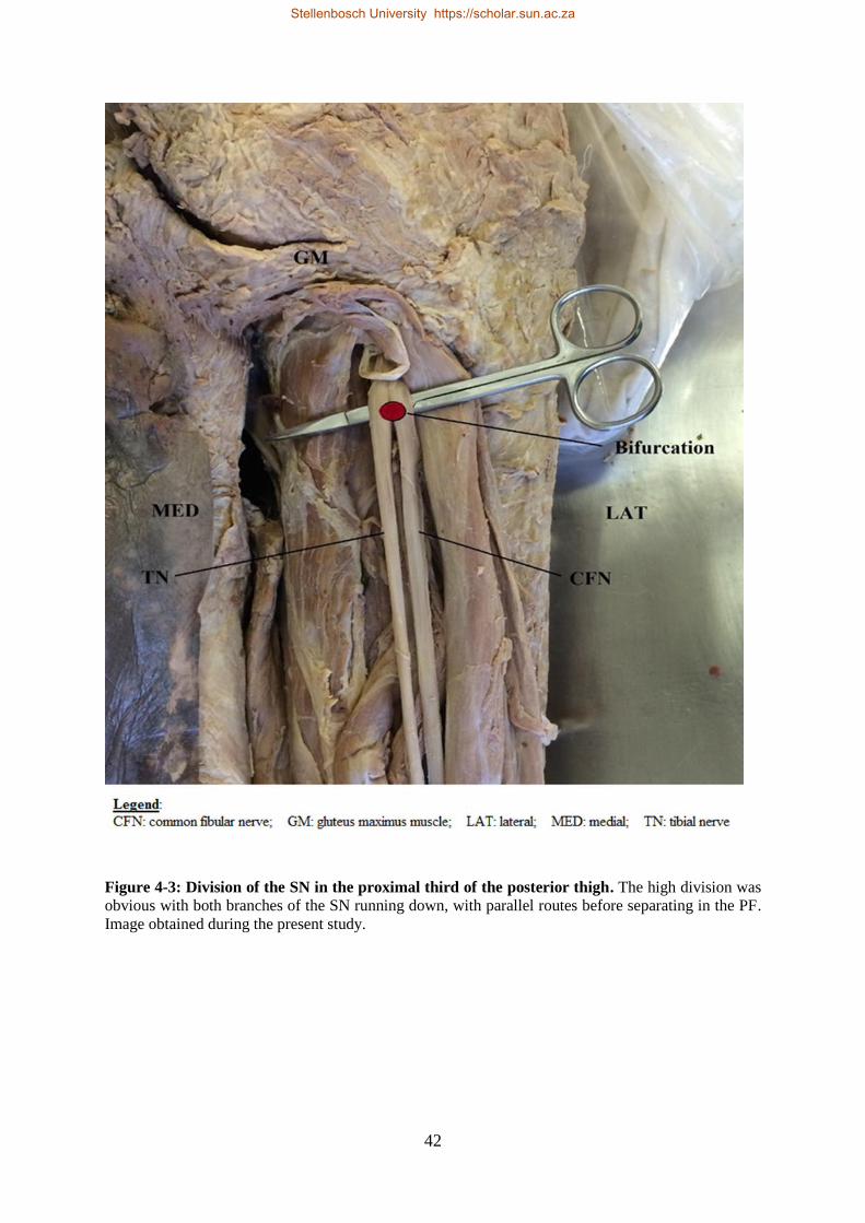
42
Figure 4-3: Division of the SN in the proximal third of the posterior thigh. The high division was
obvious with both branches of the SN running down, with parallel routes before separating in the PF.
Image obtained during the present study.
Stellenbosch University https://scholar.sun.ac.za
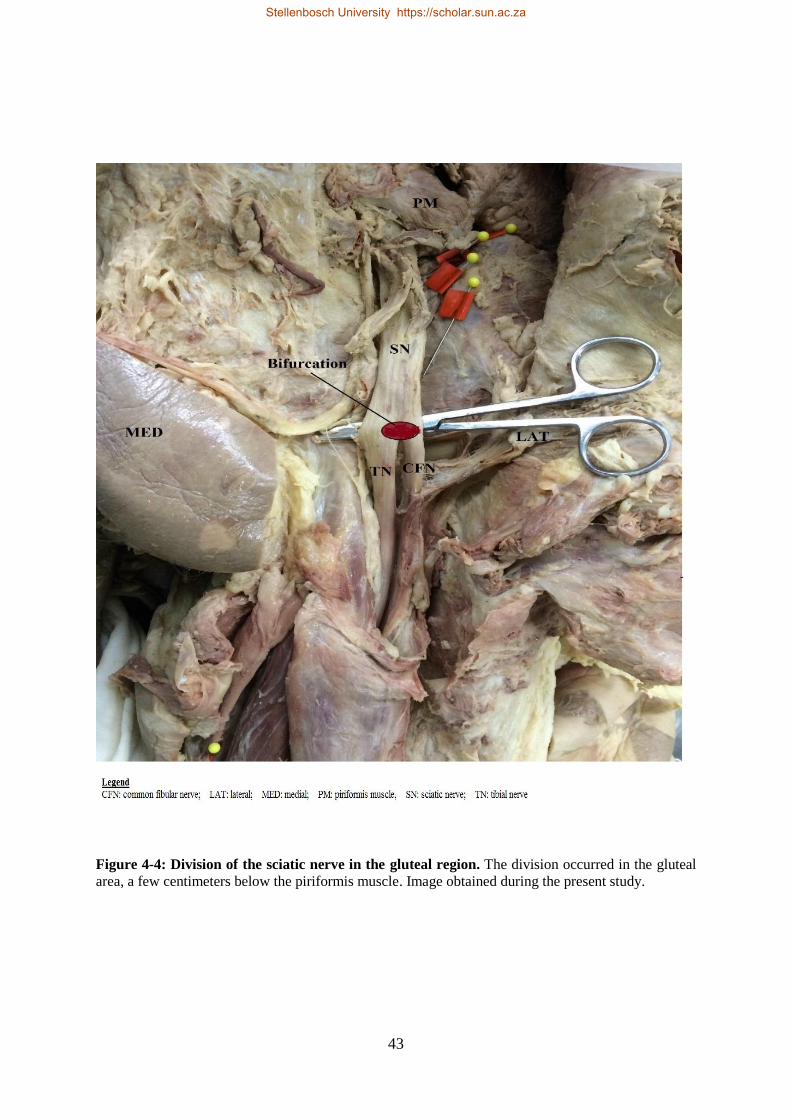
43
Figure 4-4: Division of the sciatic nerve in the gluteal region. The division occurred in the gluteal
area, a few centimeters below the piriformis muscle. Image obtained during the present study.
Stellenbosch University https://scholar.sun.ac.za

44
Prevalence of variations
Division of the SN in the posterior thigh and the gluteal region were both identified as high
variations. These variations were observed in 11.48% with a 95% confidence interval (CI) of
[5.82; 17.13], showing a risk of SN variation in 11.48 ± 5.66% of cases.
The occurrence of high variations in the posterior thigh (8.20%) was superior to that of high
variations in the gluteal region (3.28%).
Level of division per population group
Representing the heterogeneity of the South African population, three population groups were
represented: coloured, black and white. Table 4-1 depicts the level of division for each
population group.
Table 4-1: Representation of the level of division of the SN by population group
Normal division Variations Total
Group population N Percentage* N Percentage* N
Coloured 80 88.89% 10 11.11% 90
Black 16 88.89% 2 11.11% 18
White 12 85.71% 2 14.29% 14
p = 0.9
* Percentage of row total
The level of division of the SN did not differ among these population groups.
Symmetry
The symmetry explored the bilateral aspect of the level of division in each cadaver. Two main
groups were considered: normal division (popliteal fossa) and variations (posterior thigh and
gluteal region). Out of the 61 cadavers examined, the level of division appeared to be
Stellenbosch University https://scholar.sun.ac.za

45
symmetrical in both lower limbs of 57 cadavers (93.44%) with a confidence interval of [89.05
; 97.83]; this included 5 cadavers showing bilateral variations. In these cases, when the level
of division was normal (at the level of the popliteal fossa) in one limb, the same observation
applied to the other limb; or, a high variation observed in one limb specimen would be observed
in the contralateral limb.
In the four remaining cadavers out of 61 (6.56%), there was an asymmetry, meaning that in
one limb the division would be normal while a high variation would be observed in the other
limb.
Considering the variations specifically, on the 9 patients who had variations on the one side, 5
(55.56%) had variations on the other side as well. The 95% confidence interval was [23.1; 88]
and the risk of having bilateral variations was 55.56 ± 32.46%.
Level of division by side
Table 4-2: Level of division in each side
Normal Variations Total
N Percentage* N Percentage* N
Left 56 91.80% 5 8.20% 61
Right 52 85.25% 9 14.75% 61
p = 0.25
* Percentage of row total
Table 4-2: Level of division in each sideTable 4-2 depicts the level of division per side and
shows there is no significant difference between both groups of specimens, which is in favour
of the high symmetry described beforehand.
Stellenbosch University https://scholar.sun.ac.za
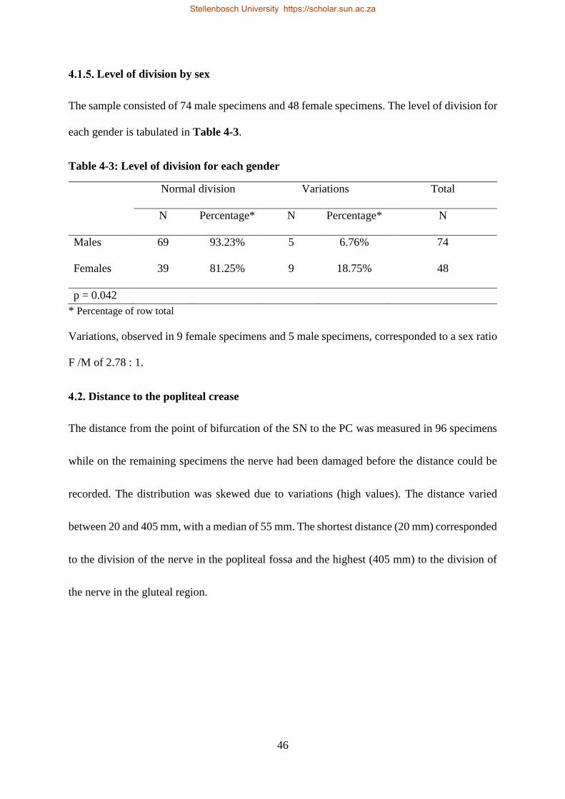
46
Level of division by sex
The sample consisted of 74 male specimens and 48 female specimens. The level of division for
each gender is tabulated in Table 4-3.
Table 4-3: Level of division for each gender
Normal division Variations Total
N Percentage* N Percentage* N
Males 69 93.23% 5 6.76% 74
Females 39 81.25% 9 18.75% 48
p = 0.042
* Percentage of row total
Variations, observed in 9 female specimens and 5 male specimens, corresponded to a sex ratio
F /M of 2.78 : 1.
. Distance to the popliteal crease
The distance from the point of bifurcation of the SN to the PC was measured in 96 specimens
while on the remaining specimens the nerve had been damaged before the distance could be
recorded. The distribution was skewed due to variations (high values). The distance varied
between 20 and 405 mm, with a median of 55 mm. The shortest distance (20 mm) corresponded
to the division of the nerve in the popliteal fossa and the highest (405 mm) to the division of
the nerve in the gluteal region.
Stellenbosch University https://scholar.sun.ac.za

47
Figure 4-5: Scatter plot of the distances recorded
Figure 4-5 illustrates distances and shows that in 85 specimens (88.54%) the distance ranged
between 0 and 100 mm. The outliers or data above 120 mm corresponded to high variations.
Distance by level of division
With regards to the level of division, the distribution was normal, therefore, means and SD are
given in Table 4-4. These data evaluate the distance at which the sciatic nerve divides for each
category of division.
Table 4-4: Mean distance from popliteal crease and standard deviation per level of
division (in mm).
Category Ranges Means SD* P25 Median P75
Popliteal fossa 20-90 51.5 19.5 40 50 65
Posterior thigh 120-300 209.4 62 150 210 250
Gluteal 370-405 394.8 16.6 385 402 404.5
* SD= standard deviation
The distance recorded was proportional to the level at which the SN divided.
0
50
100
150
200
250
300
350
400
450
0 20 40 60 80 100 120 140
Dis
tan
ce t
o P
C
Number of specimens
Distances recorded
Stellenbosch University https://scholar.sun.ac.za
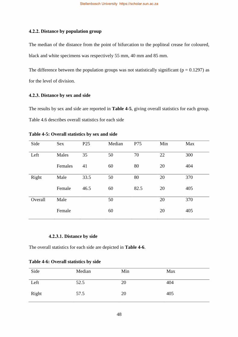
48
Distance by population group
The median of the distance from the point of bifurcation to the popliteal crease for coloured,
black and white specimens was respectively 55 mm, 40 mm and 85 mm.
The difference between the population groups was not statistically significant (p = 0.1297) as
for the level of division.
Distance by sex and side
The results by sex and side are reported in Table 4-5, giving overall statistics for each group.
Table 4.6 describes overall statistics for each side
Table 4-5: Overall statistics by sex and side
Side Sex P25 Median P75 Min Max
Left Males 35 50 70 22 300
Females 41 60 80 20 404
Right Male 33.5 50 80 20 370
Female 46.5 60 82.5 20 405
Overall Male 50 20 370
Female 60 20 405
Distance by side
The overall statistics for each side are depicted in Table 4-6.
Table 4-6: Overall statistics by side
Side Median Min Max
Left 52.5 20 404
Right 57.5 20 405
Stellenbosch University https://scholar.sun.ac.za

49
. Popliteal Block Simulation
Pilot study
In the pilot study, the popliteal block (PB) was performed on seven cadavers. After dissection,
the dye was visualised in the popliteal fossa. One cadaver out of 7 presented a bilateral variation
of the sciatic nerve division with the SN dividing in the posterior thigh on the left limb and in
the gluteal region for the right limb. The distances (PCB) were 300 mm and 370 mm
respectively. As shown in
Figure 4-6, the dye, which was found in the popliteal fossa, was far from the main trunk of the
sciatic nerve (SN). Therefore, the popliteal block in that case was considered as unsuccessful.
Legend
SN: Sciatic nerve
CFN: Common fibular nerve
TN: Tibial nerve
PC: Popliteal crease
Figure 4-6: Diagram showing the dye in the PF with a high division of the SN.
Simulation of the PB
The PB was simulated on 22 lower limbs from 11 cadavers. These cadavers were part of the
sample used to determine the level of division of the SN. Results were obtained in 69.23% of
Stellenbosch University https://scholar.sun.ac.za
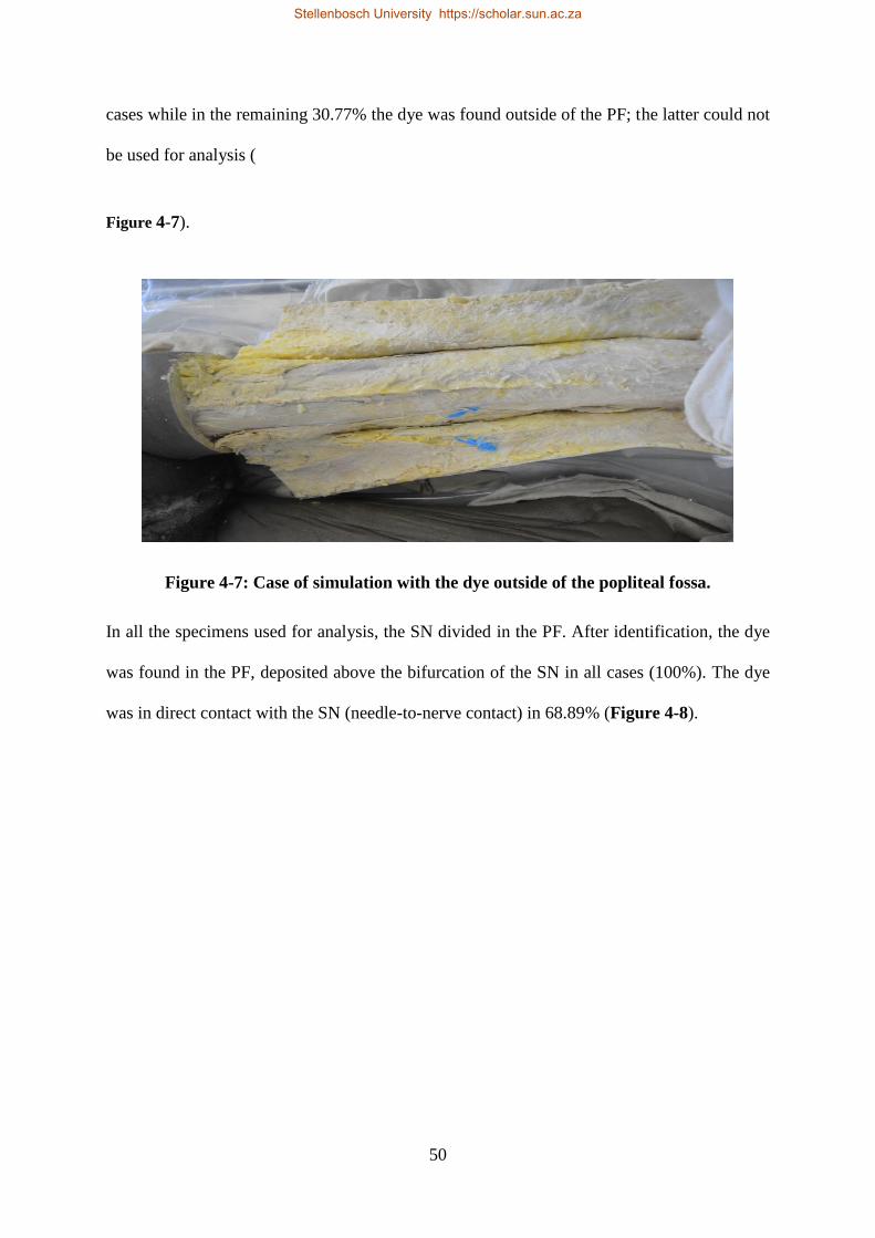
50
cases while in the remaining 30.77% the dye was found outside of the PF; the latter could not
be used for analysis (
Figure 4-7).
Figure 4-7: Case of simulation with the dye outside of the popliteal fossa.
In all the specimens used for analysis, the SN divided in the PF. After identification, the dye
was found in the PF, deposited above the bifurcation of the SN in all cases (100%). The dye
was in direct contact with the SN (needle-to-nerve contact) in 68.89% (Figure 4-8).
Stellenbosch University https://scholar.sun.ac.za
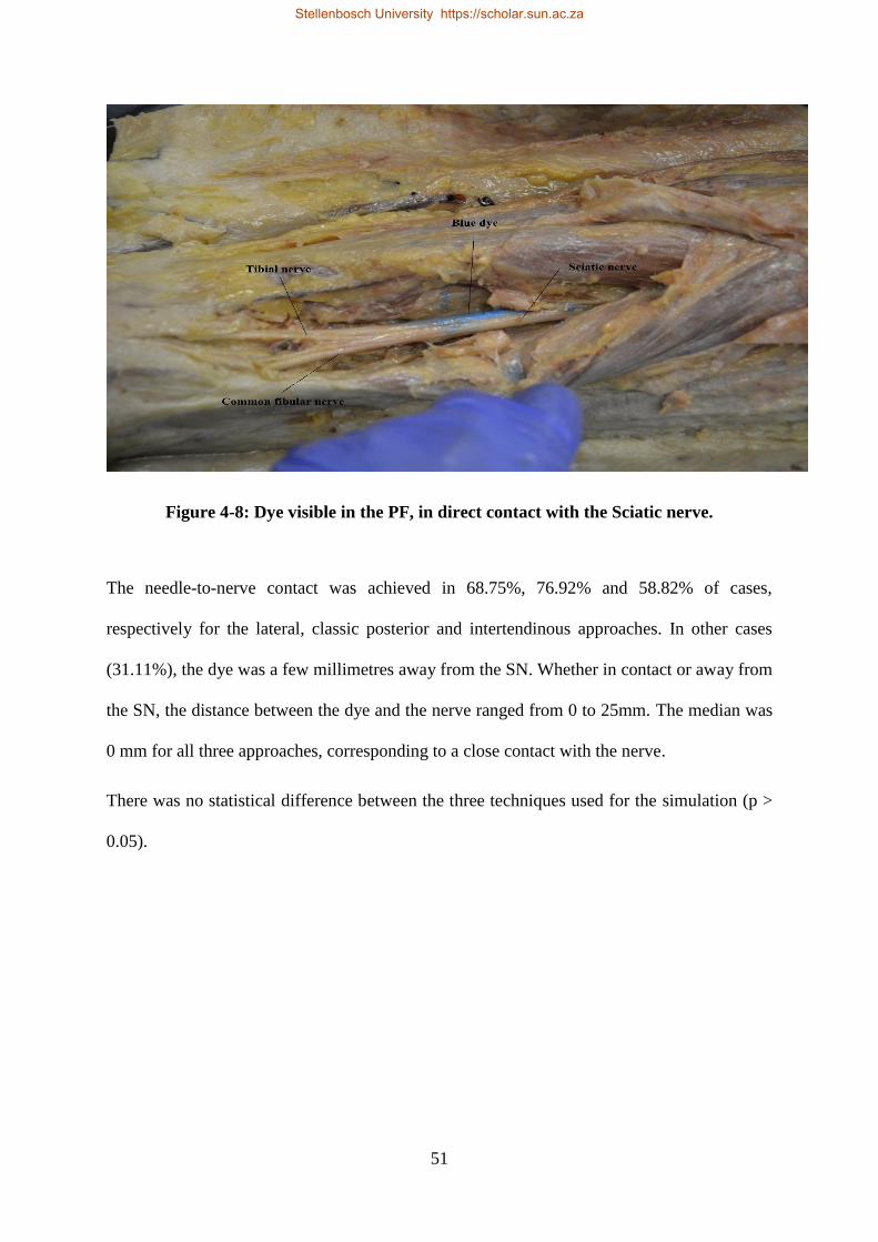
51
Figure 4-8: Dye visible in the PF, in direct contact with the Sciatic nerve.
The needle-to-nerve contact was achieved in 68.75%, 76.92% and 58.82% of cases,
respectively for the lateral, classic posterior and intertendinous approaches. In other cases
(31.11%), the dye was a few millimetres away from the SN. Whether in contact or away from
the SN, the distance between the dye and the nerve ranged from 0 to 25mm. The median was
0 mm for all three approaches, corresponding to a close contact with the nerve.
There was no statistical difference between the three techniques used for the simulation (p >
0.05).
Stellenbosch University https://scholar.sun.ac.za

52
Chapter 5 - DISCUSSION
The focus of this study was to describe the pattern of the sciatic nerve bifurcation and to identify
variations in the level of division and their frequency in a South African population. In addition,
a simulation of the popliteal block was performed with a dye made of silicone to evaluate the
success rate of the PB as well as discriminate the most successful approach.
This study described the SN division in 122 lower limbs; so far, the third largest study after
those of Ugrenovic et al. (2005) and Ogeng’O et al. (2011). While Okraszewska et al. (2002)
and Saleh et al. (2009) studied 36 and 30 specimens respectively, the smallest sample used for
the SN division was that of Vloka et al. (1997)involving 10 specimens. In most studies,
observations on the sciatic nerve bifurcation were based on the examination of 40 to 60
specimens (Triadó et al., 2004; Guvencer et al., 2009; Shewale, Karambelkar and Umarji,
2013; Adibatti, Sangeetha and Adibatti M, 2014; Barbosa et al., 2015; Kiros and Woldeyes,
2015). The results of this study therefore cover a larger population, therefore giving a better
overview of the level of division of the SN. This work is also unique in that the cadavers used
were of three population groups: black, white and coloured, a representation of the Western
Cape population of South Africa.
. Variations in the level of division of SN
The standard description of the SN division which is a bifurcation in the PF (Berihu and Debeb,
2015) was found in 88.52% of cases. This result corresponds to the normal rate of 85-89%
(Kiros and Woldeyes, 2015). Similarly, Adibatti et al. (2014) found a high frequency (92%)
of the normal pattern in their research on the Indian population. However, results in other
studies (Okraszewska et al., 2002; Ugrenović et al., 2005; Ogeng’O et al., 2011) reported a
lower prevalence of the normal division, between 65 and 75% (Table 5-1).
Stellenbosch University https://scholar.sun.ac.za

53
Table 5-1: Variations observed in different studies (ours included)
Author (Year) Country N Normal (%) Variations (%)
Okraszewska et al. (2002) Poland 36 72,2 27,8
Ugrenovic et al. (2005) Serbia 200 72,5 27,5
Prakash et al. (2010) India 86 34,9 65,1
Ogeng’o et al. (2011) Ethiopia 164 67,1 32,9
Shewale et al. (2013) India 90 77.78 32.22
Adibatti et al. (2014) India 50 92 8
Kiros et al. (2015) Ethiopia 50 64 36
Present study South Africa 122 88,5 11,5
N= number of specimens examined. Variations consisted in cases of division above the PF.
Sources: (Okraszewska et al., 2002; Ugrenović et al., 2005; Prakash et al., 2010; Ogeng’O et al., 2011; Shewale, Karambelkar and Umarji, 2013; Adibatti, Sangeetha and
Adibatti M, 2014; Kiros and Woldeyes, 2015)
Most studies display a higher prevalence of variations than the normal description.
Stellenbosch University https://scholar.sun.ac.za
Stellenbosch University https://scholar.sun.ac.za

54
High variations in the literature included cases of division in the pelvis, the gluteal region or the
posterior thigh. Most of these variants were observed in this work except from variations with
division of the SN in the pelvis, supporting previous works by Ugrenovic et al. (2005) and Shewale
et al. (2013). In studies where a division of the SN in the pelvis was observed, the prevalence
varied between 5 and 15% (Okraszewska et al., 2002; Adibatti, Sangeetha and Adibatti M, 2014;
Kiros and Woldeyes, 2015) and higher, about 16.3% for Prakash et al. (2010) and 20.1% for
Ogeng’O et al. (2011)
The low prevalence of division in the gluteal region (< 5%) found in most studies (Okraszewska
et al., 2002; Prakash et al., 2010; Ogeng’O et al., 2011; Adibatti, Sangeetha and Adibatti M, 2014;
Kiros and Woldeyes, 2015) was comparable to the findings in this South African cohort (3.28%).
Nevertheless, Ugrenovic et al. (2005) as well as Shewale et al. (2013) found more that 10% of
cases of this variant.
On the other hand, the division of the SN occurred in the posterior thigh in 8.20% of cases. The
results for this variant were comparable to those of Ugrenovic et al. (2005), Ogeng’o et al. (2011)
and Shewale et al. (2013) who found 9%, 10.4% and 11.11% respectively while Adibatti et al.
(2014) only found 2% of cases of division at that level. On the contrary, Prakash et al. (2010)
observed this variant in 46.5% of the specimens studied.
Overall, the prevalence of variations was 11.48% and this prevalence for 95% of the population to
display a variation in one or the other limb would vary between 5.82 and 17.13%. This description
is in accord with the standard description of the sciatic nerve bifurcation, but far below the findings
in most studies which are in the order of approximately 30% (Okraszewska et al., 2002; Ugrenović
et al., 2005; Ogeng’O et al., 2011; Shewale, Karambelkar and Umarji, 2013; Kiros and Woldeyes,
2015).
Stellenbosch University https://scholar.sun.ac.za
Stellenbosch University https://scholar.sun.ac.za

55
The differences in prevalence of variations from one study to another show the great changes in
the levels of the division of the SN. Both similarities and differences were observed between this
study and others. Even though a larger sample would improve the accuracy of the results, it remains
that the sciatic nerve displays a highly variable anatomy in terms of level of bifurcation. In
addition, many factors were reported by other authors as possible causes of the variations in the
level of division of the nerve. First, the branching patterns vary between population groups
(Prakash et al., 2010). For example, while in Ethiopia the frequency of variations is around 35%
in two different studies (Ogeng’O et al., 2011; Kiros and Woldeyes, 2015), the studies in India are
different in that the variations ranged between 8% and 65% of the specimens (Prakash et al., 2010;
Shewale, Karambelkar and Umarji, 2013; Adibatti, Sangeetha and Adibatti M, 2014). In South
Africa, studies on the sciatic nerve are hard to find, making it impossible for us to compare or to
have global results.
A second factor suggested by Ogengo et al. (2011), was that the division especially in the pelvis
resulted of a separate course of the two components of the SN (TN and CFN) during the embryonic
period. In neonates, high variations are also observed, hence variations could be present at any age
(Reinoso-Barbero et al., 2014).
Level of division by population group
The diversity and heterogeneity of the South African population might be symptomatic of
differences due to genetic arrangements. However, this study did not highlight any statistical
difference (p > 0.05) between the three main population groups studied, namely white, black and
coloured. Nonetheless, the sample was composed mainly of coloured specimens. Hence, a larger
Stellenbosch University https://scholar.sun.ac.za
Stellenbosch University https://scholar.sun.ac.za

56
study with a larger proportion of black and white specimens is required to confirm these findings.
Hence, the results of one group should not yet be applied to another group.
Level of division of the SN by sex
A clear numerical difference was observed between males and females in terms of variations.
Indeed, most variations were found in female specimens with a sex ratio F : M of 2.78 : 1. This
ratio showed that the risk of variation in females was two times the risk in male specimens, and
this difference was confirmed statistically (p = 0.042). Consequently, maneuvers related to the SN
would call for more precautions, such as during a popliteal block or hip surgery in females.
Level of division by side and symmetry
The comparison of both left and right side demonstrated there was no difference between both
sides, what is in favour of the high symmetry. Interestingly, symmetry was not considered as a
variable of analysis in studies on the SN, making it difficult to determine whether variations are
bilateral or not. In this work, the level of division of the SN was symmetrical in 93.44%,
comparable to the findings of Ugrenovic et al. (2005) who observed that all variations were
bilateral. On the contrary, Kiros et al. (2015) reported a high asymmetry of 32% (8 out of 25).
Even though they did not specify it, this difference could be attributed to the fact that we grouped
variations together instead of considering them as individual variants. Nevertheless, based on
previous observations on the high symmetry, a patient showing a variation in the bifurcation point
of the SN is at risk to present a variation in the other limb. For this reason, any variation identified
in one limb should call for the exploration of the other limb.
Stellenbosch University https://scholar.sun.ac.za
Stellenbosch University https://scholar.sun.ac.za

57
Variations in the distance between the point of bifurcation and the popliteal crease
In various studies, the level of division was the focus of the research while the distance was not
always recorded. Hence, only a few studies considered the distance between the PC and the
bifurcation of the sciatic nerve as a variable in their analysis (Vloka et al., 2001; Saleh, El-Fark
and Abdel-Hamid, 2009; Schiarite et al., 2015; Sohn et al., 2015). The results of these studies are
consigned in Table 5-2.
Table 5-2: Distances (in mm) reported in different studies
Authors Sample size Minimum Maximum Mean SD
Vloka et al. (1997) 10 0 73 44 20
Vloka et al. (2001) 28 0 115 60.5 27
Saleh et al. (2009) 30 50 180 81.37* 5.84
Lee et al. (2013) 50 - - 79 26
Barbero et al. (2014) 48 150 400 - -
Schiarite et al. 2015) 124 - - 61.78** 11*
Sohn et al. (2015) 40 - - 63.9 9.8
Present study 122 20 405 72.42 81.47
* Calculated from averages for male and female
**Calculated from averages for left and right limbs. SD: Standard deviation
Sources: (Vloka et al., 1997, 2001; Saleh, El-Fark and Abdel-Hamid, 2009; Lee et al., 2013; Schiarite et al., 2015;
Sohn et al., 2015).
The average distance between the popliteal crease and the point of bifurcation of the SN varied
from one study to the other. All studies combined, the distance varied between 0 and 405 mm. The
lowest distance was reported by Vloka et al. (1997, 2001), while the highest distance for the point
Stellenbosch University https://scholar.sun.ac.za
Stellenbosch University https://scholar.sun.ac.za

58
of bifurcation so far was observed in the present study, at 405 mm above the PC. The maximal
distance was related to the presence of high variations, considering that higher variation would
correspond to higher distance value. The sample size could also be a factor of difference because
a large sample improves the accuracy and gives a better overview of variations, therefore
encompassing higher distances. For example, Vloka et al. (2001) examined 28 specimens and had
the higher distance at 115 mm (in the popliteal fossa), while Barbero et al. (Reinoso-Barbero et
al., 2014) who worked on 48 specimens found a maximum at 400 mm. Much more, the large
sample used in this study increases the probability to observe variations which are not so frequent
as well as high distances.
The interpretation using the mean was made impossible due to the abnormal distribution of the
different distances recorded. According to findings reported in Table 5-2, the average was
approximately 65 mm for most studies, close to the median (55mm) in the present study.
Distance by level of division
In general, the distance is proportional to the level of division and therefore increases with it.
Casalia et al. (2006) stipulated that the division of the SN occurs in a range of 0 to 113 mm above
the popliteal crease. This corroborates this study’s results with a distance ranging between 20 and
90 mm for the normal pattern, as described in Table 4-4. This is indeed comparable to the analysis
of the scatter plot Figure 4-5 which shows that for most of the specimens, the distance was below
100 mm. These findings are close to those of Vloka et al. (2001) who found a distance ranging
between 0 and 115 mm. All these results apply for cases of division in the PF. On the other hand,
Saleh et al. (2009) reported a distance varying between 50 and 180 mm above the crease, which
according to Table 4-4 corresponds to cases of divisions in the PF and the posterior thigh.
Stellenbosch University https://scholar.sun.ac.za
Stellenbosch University https://scholar.sun.ac.za

59
Besides the normal pattern, the high variations included the cases in which either the SN branched
in the posterior thigh or the gluteal region and the ranges were 120 to 300mm and 370 to 405 mm
respectively.
Globally, the distance in the cases of normal division varies between 0 and 115 mm while the
distance for high variations varies between 120 to 405 mm. Consequently, a high distance from
the PC would increase the risk of piriformis syndrome and nerve injury during muscular injections
or even surgical procedures in the gluteal region and could reduce the success of the popliteal
block as shown in the pilot study of the PB simulation (Figure 4-6).
Distance by sex
Our results were reported as medians as opposed go other studies, due to the skewness of the
distribution. As reported in
Table 5-3, Saleh et al. (2009) reported a mean distance higher in males while the mean distance
was approximately the same in both groups for Schiarite et al. (2015). Considering the median in
this study, females displayed a greater distance (Table 4-5) though not significant. On the other
hand, Vloka et al. (2001) showed a distance greater in females than males. However, in all these
observations, the difference was not statistically significant.
Table 5-3: Compilation of means by sex for different studies
Males Females
Mean STDEV Mean STDEV
Vloka et al. (2001) 55.1 25.5 64.2 27.5
Saleh et al. (2009) 83.9 4.14 78.85 7.54
Schiarite et al. (2015) 62.56 6 61.35 5
This study 50* 60*
Stellenbosch University https://scholar.sun.ac.za
Stellenbosch University https://scholar.sun.ac.za

60
STDEV: standard deviation, *represents the median
Sources: (Vloka et al., 2001; Saleh, El-Fark and Abdel-Hamid, 2009).
Distance by side
Similarly to other studies (Vloka et al., 2001; Saleh, El-Fark and Abdel-Hamid, 2009), the
difference between the right and left lower limb in terms of distance from the PC to the bifurcation
was not statistically significant. This is in accord the bilateral characteristic of the level of division
or symmetry. Results are close for left and right lower limbs (Table 5-4). The symmetry could
also explain the absence of significant difference by side in males and females.
Table 5-4: Compilation of mean by side in different studies
Left Right
Mean STDEV Mean STDEV
Vloka et al. (2001) 57.5 25.7 66.2 23.9
Saleh et al. (2009) 83.3 3.98 79.2 7.21
Schiarite et al. (2015) 61.27 11 62.29 11
This study 52.5* 57.5*
STDEV: standard deviation
Sources: (Vloka et al., 2001; Saleh, El-Fark and Abdel-Hamid, 2009; Schiarite et al., 2015).
. Popliteal block simulation
There are no previous reports on simulation of the popliteal block in the literature. The pilot study
carried out during this study highlighted a case of high variation in one of the stimulated limbs. In
this case, the block would be unsuccessful as the dye was located 150 mm below the SN
bifurcation. Hypothetically, considering the proximal spread of the anaesthetic, the block could be
Stellenbosch University https://scholar.sun.ac.za
Stellenbosch University https://scholar.sun.ac.za
61
successful following a suggestion that for variations located 210 mm above the PC and even
higher when the needle is inserted with a proximal inclination of 40 to 60°, the blockade could
occur (Vloka et al. 1997).
On the other hand, our definitive results were comparable to the reports of Mariano et al. (Mariano
et al., 2009) with a success rate predictable in 100% of cases. In all cases, the dye was deposited
before the bifurcation of the nerve. Other authors suggested a satisfactory block in approximately
90 to 97% of cases (Rongstad et al., 1996; Hansen, Eshelman and Cracchiolo 3rd, 2000; Migues
et al., 2005; Perlas et al., 2008; Barbosa et al., 2015), and in 82.3% of cases (Rorie et al., 1980).
Nevertheless, a better outcome of the PB was obtained with the aid of the ultrasound (Wadhwa,
Gebhard and Obal, 2010) when compared to the neurostimulation. This could be as a result of a
good visualisation of the nerve with the ultrasound therefore ensuring a better placement of the
needle before injection of the anaesthetic.
The success rate predicted for the popliteal block was high because the SN showed a normal
bifurcation in the PF for all the specimens used for simulation. On the opposite, the existence of
variations would compromise the block as demonstrated in the pilot study. For this reason, the use
of a neurostimulation or better the ultrasound would improve the placement of the needle and
above all help identify such variations for a better choice of the anaesthetic method.
In most cases, the presence of an undivided nerve at 7 cm above the crease would lead to the
placement of the needle at that distance above the crease (Hadžic et al., 2002). However, taking
into account the variations in which the nerve divided at a minimum of 120 mm above the crease,
the technique suggested by Singelyn et al. (1991) would improve the quality of the block. An
approach with a placement of the needle 12 to 14 cm (Nader et al., 2009) above the crease would
Stellenbosch University https://scholar.sun.ac.za
Stellenbosch University https://scholar.sun.ac.za

62
also be beneficial. Despite the benefits of all these approaches, the technique used remains the
choice of the surgeon or the anaesthetist, adapted to the condition of the patient.
Concerning the three approaches used for the simulation, a closer needle-to-nerve contact was
achieved with the classic posterior approach, followed by the lateral approach and lastly the
intertendinous approach. This approach could therefore improve the success rate. However, this
difference was not proven statistically. Another simulation on a larger number of specimens could
be interesting for a better comparison and analysis of the proximal spread to ensure successful
blocks even in some cases of high variations.
Stellenbosch University https://scholar.sun.ac.za
Stellenbosch University https://scholar.sun.ac.za

63
Chapter 6 - CONCLUSION, STRENGHTS, LIMITATIONS & RECOMMENDATIONS
. CONCLUSION
The knowledge of the anatomy of the sciatic nerve, especially its level of division, is essential for
accurate placement of the needle during the popliteal block, therefore contributing to the success
of this anesthesia. There is no standard description of bifurcation, due to the high variability of
bifurcation of the SN. However, the South African population might display a normal pattern of
bifurcation as described in this study. The mean distance at which the nerve divides varies between
50 and 70 mm approximately above the popliteal crease, and variations of the SN bifurcation could
be observed in as low as 8% or a high as 60% of cases approximately. Therefore, because the level
of division of the SN is generally bilateral, the identification of a variation in one limb calls for the
exploration of the other limb. Moreover, more precautions would be required in females than in
males, due to the high prevalence in this subgroup. Consequently, systematic preoperative imagery
should be enforced to avoid failures of the block or injuries of the nerve.
Furthermore, as no difference was highlighted among the three approaches used, this study
demonstrated that a normal division of the sciatic nerve in the popliteal fossa ensures a 100%
success rate of the popliteal block. On the other hand, variations would compromise the block,
hence techniques such as those suggested by Singelyn et al. (Casalia, Carradori and Moreno, 2006)
or Nader et al. (2009) using a needle inserted between 10 and 14 cm above the crease would yield
better results.
In summary, high variations of the sciatic nerve lead to failures of the popliteal block, hence
surgeons and anaesthesiologists need to consider these variations when making surgical decisions.
Stellenbosch University https://scholar.sun.ac.za
Stellenbosch University https://scholar.sun.ac.za

64
. STRENGHTS
The strength of this study is based on the large sample size unlike most previous studies. It
therefore gives a large spectrum of the level of division of the sciatic nerve and the distance at
which it divided. Moreover, the heterogeneous study population contributes to a broader view of
the sciatic nerve division.
. LIMITATIONS
There could be some bias in this study arising from data being collected and analysed by a single
observer (Schiarite et al., 2015). Besides, the distance between the PC and the point of bifurcation
was recorded in only 79.5% of our sample because data collection was a two-step process. The
first step consisted in recording the levels of division on a form while measurements, part of the
second step, would be taken afterwards. For some specimens, the measurements could not be taken
because the sciatic nerve had been damaged during the dissection process after identification of
the location of the point of bifurcation. Distances were recorded in millimetres (mm) for more
precision and better comparison.
In addition, the simulation was carried out by a general practitioner with no training in block
procedures. This could explain the glitches excluded from the analysis of the popliteal block
simulation. Despite those limitations, data was collected meticulously and the pilot study was used
to understand and master the technique of the popliteal block.
. RECOMMENDATIONS
The assessment of the sciatic nerve by preoperative imagery should be a prerequisite to any
surgical procedure of the lower limb using the popliteal block as a regional anaesthesia. In addition,
Stellenbosch University https://scholar.sun.ac.za
Stellenbosch University https://scholar.sun.ac.za

65
this study should be carried further on a larger sample to confirm these findings and explore further
the differences among population groups. It would therefore improve the accuracy of the results
obtained and make them applicable to the South African population. Furthermore, multiple
observers should collect the results in order to reduce the risk of bias.
Stellenbosch University https://scholar.sun.ac.za
Stellenbosch University https://scholar.sun.ac.za

66
REFERENCES
Adibatti, M., Sangeetha, V. and Adibatti M, V. S. (2014) ‘Study on variant anatomy of sciatic
nerve’, Journal of Clinical and Diagnostic Research, 8(8), pp. 8–10. doi:
10.7860/JCDR/2014/9116.4725.
Al-Nasser, B. (2002) ‘A modified lateral approach for a single-shot sciatic nerve block in the
popliteal fossa’, Journal of Clinical Anesthesia, pp. 608–609. doi: 10.1016/S0952-8180(02)00450-
6.
De Andrés, J., Alonso-Iñigo, M., Jose, Sala-Blanch, X. and Reina, M. A. (2005) ‘Nerve stimulation
in regional anesthesia: theory and practice’, Best Practice & Research Clinical Anaesthesiology,
19(2), pp. 153–174. doi: 10.1016/j.bpa.2004.11.002.
Arcioni, R., Palmisani, S., Della Rocca, M., Romanò, S., Mercieri, M., De Blasi, R. A., Ronconi,
P. and Pinto, G. (2007) ‘Lateral popliteal sciatic nerve block: A single injection targeting the tibial
branch of the sciatic nerve is as effective as a double-injection technique’, Acta Anaesthesiologica
Scandinavica. Wiley Online Library, 51(1), pp. 115–121. doi: 10.1111/j.1399-6576.2006.01164.x.
Barbosa, F. T., Barbosa, T. R. B. W., da Cunha, R. M., Rodrigues, A. K. B., Ramos, F. W. da S.
and de Sousa-Rodrigues, C. F. (2015) ‘Anatomical basis for sciatic nerve block at the knee level’,
Brazilian journal of anesthesiology (Elsevier). Sociedade Brasileira de Anestesiologia, 65(3), pp.
177–179. doi: 10.1016/j.bjane.2014.03.010.
Benzon, H. T. (2005) Essentials of pain medicine and regional anesthesia. Philadelphia: Elsevier-
Churchill Livingstone. Available at: http://books.google.com/books?id=mthpAAAAMAAJ.
Berihu, B. A. and Debeb, Y. G. (2015) ‘Anatomical variation in bifurcation and trifurcations of
sciatic nerve and its clinical implications: in selected university in Ethiopia.’, BMC research notes.
BioMed Central, 8(1), p. 633. doi: 10.1186/s13104-015-1626-6.
Bouaziz, H., Narchi, P., Zetlaoui, P. J., Paqueron, X. and Benhamou, D. (1999) ‘Lateral approach
to the sciatic nerve at the popliteal fossa combined with saphenous nerve block’, Techniques in
Regional Anesthesia and Pain Management, pp. 19–22. doi: 10.1016/S1084-208X(99)80017-2.
Stellenbosch University https://scholar.sun.ac.za
Stellenbosch University https://scholar.sun.ac.za

67
Bruhn, J., VAN GEFFEN, G., Gielen, M. J. and Scheffer, G. J. (2008) ‘Visualization of the course
of the sciatic nerve in adult volunteers by ultrasonography’, Acta Anaesthesiologica Scandinavica.
Wiley Online Library, 52(9), pp. 1298–1302.
Casalia, A. G., Carradori, G. and Moreno, M. (2006) ‘Blockade of the sciatic nerve in the popliteal
fossa’, Techniques in Regional Anesthesia and Pain Management, 10(4), pp. 173–177. doi:
10.1053/j.trap.2006.10.006.
Chelly, J. E. (2003) ‘Sciatic nerve block’, Techniques in Regional Anesthesia and Pain
Management, pp. 18–25. doi: 10.1053/trap.2003.123521.
Chelly, J. E. (2004) Peripheral nerve blocks : a color atlas. Philadelphia: Lippincott Williams &
Wilkins.
Chelly, J. E. (2009) Peripheral nerve blocks a color atlas. Philadelphia, PA: Wolters Kluwer
Health/Lippincott Williams & Wilkins. Available at:
http://public.eblib.com/choice/publicfullrecord.aspx?p=2032163.
Clendenen, S. R., York, J. E., Wang, R. D. and Greengrass, R. A. (2008) ‘Three‐dimensional
ultrasound‐assisted popliteal catheter placement revealing aberrant anatomy: implications for
block failure’, Acta Anaesthesiologica Scandinavica. Wiley Online Library, 52(10), pp. 1429–
1431.
Cochard, L. R., Netter, F. H., Craig, john A., Machados, C. A. and Clinicalkey (2012) Netter’s
atlas of human embryology. updated ed. Edited by C. Philadelphia: Elsevier/Saunders.
Creech, C. and Meyr, A. A. J. A. A. J. (2013) ‘Techniques of popliteal nerve regional anesthesia’,
The Journal of Foot and Ankle Surgery, 52(5), pp. 681–685. doi: 10.1053/j.jfas.2013.03.021.
Danelli, G., Fanelli, A., Ghisi, D., Moschini, E., Rossi, M., Ortu, A., Baciarello, M. and Fanelli,
G. (2009) ‘Ultrasound vs nerve stimulation multiple injection technique for posterior popliteal
sciatic nerve block’, Anaesthesia, 64(6). doi: 10.1111/j.1365-2044.2009.05915.x.
Drake, R. L., Vogl, A. W. and Mitchell, A. W. (2015) Gray’s anatomy for students. Available at:
https://www.clinicalkey.com/dura/browse/bookChapter/3-s2.0-C20110061707.
Stellenbosch University https://scholar.sun.ac.za
Stellenbosch University https://scholar.sun.ac.za
68
Duke Anatomy - Lab 13: Gluteal Region & Posterior Thigh (2013). Available at:
https://web.duke.edu/anatomy/Lab13/Lab13.html (Accessed: 13 March 2018).
Ekanem, P. E., Abba, S., Mariam, K. and Assefa, H. (2015) ‘Variations of Sciatic Nerve
Bifurcation in Dissected Cadaveres from Ethiopia and their Clinical Implication: A Case Report.’
Van Geffen, G.-J., Pirotte, T., Anesthesiologist, S., Gielen, M. J., Scheffer, G. and Bruhn, J. (2010)
‘Ultrasound-guided proximal and distal sciatic nerve blocks in children’, Journal of Clinical
Anesthesia, 22, pp. 241–245. doi: 10.1016/j.jclinane.2009.07.009.
Gosling, J. A. (2008) Human anatomy color atlas and textbook. [Edinburgh?]: Mosby/Elsevier.
Available at: http://www.clinicalkey.com/dura/browse/bookChapter/3-s2.0-C20090602312.
Greenberg, C. P. (1995) ‘Practical, cost-effective regional anesthesia for ambulatory surgery’,
Journal of clinical anesthesia. Elsevier, 7(7), pp. 614–621.
Guvencer, M., Iyem, C., Akyer, P., Tetik, S. and Naderi, S. (2009) ‘Variations in the high division
of the sciatic nerve and relationship between the sciatic nerve and the piriformis.’, Turkish
Neurosurgery. Dokuz Eylul University, Anatomy Department, Izmir, Turkey.
[email protected], 19(2), pp. 139–144.
Hadzic, A. (2007) Textbook of regional anesthesia and acute pain management. McGraw-Hill,
Medical Pub. Division.
Hadzic, A. and Vloka, J. (2004) Peripheral nerve blocks : principles and practice. New York:
McGraw-Hill/Professional.
Hadžic, A., Vloka, J. D., Singson, R., Santos, A. C. and Thys, D. M. (2002) ‘A comparison of
intertendinous and classical approaches to popliteal nerve block using magnetic resonance imaging
simulation’, Anesthesia & Analgesia. LWW, 94(5), pp. 1321–1324.
Hadzić A, V. J. D. (1998) ‘A comparison of the posterior versus lateral approaches to the block of
the sciatic nerve in the popliteal fossa.’, Anesthesiology, 88(6), pp. 1480–1486.
Hansen, E., Eshelman, M. R. and Cracchiolo 3rd, A. (2000) ‘Popliteal fossa neural blockade as
Stellenbosch University https://scholar.sun.ac.za
Stellenbosch University https://scholar.sun.ac.za

69
the sole anesthetic technique for outpatient foot and ankle surgery’, Foot & ankle international.
Department of Anesthesiology, UCLA Medical Center, Los Angeles, CA 90095, USA., 21(1), pp.
38–44.
Hansen, J. T. and Netter, F. H. (2014) Netter’s clinical anatomy. Available at:
https://www.clinicalkey.com/dura/browse/bookChapter/3-s2.0-C20120065167.
Hegewald, K., McCann, K., Elizaga, A. and Hutchinson, B. L. (2014) ‘Popliteal Blocks for Foot
and Ankle Surgery: Success Rate and Contributing Factors’, Journal of Foot and Ankle Surgery.
Elsevier Ltd, 53(2), pp. 176–178. doi: 10.1053/j.jfas.2013.04.008.
Herring, A. A., Stone, M. B., Fischer, J., Frenkel, O., Chiles, K., Teismann, N. and Nagdev, A.
(2011) ‘Ultrasound-guided distal popliteal sciatic nerve block for ED anesthesia’, American
Journal of Emergency Medicine, 29(6), p. 697.e3-697.e5. doi: 10.1016/j.ajem.2010.06.015.
James Phillips, W., Troutman, G. and Lerant, A. (2011) ‘Nerve stimulator–assisted sciatic nerve
block for painful procedures in the ED’, American Journal of Emergency Medicine, 29, pp. 1130–
1135. doi: 10.1016/j.ajem.2010.08.007.
Jones, O. (2015) Popliteal Fossa, Teach Me Anatomy. Available at:
http://teachmeanatomy.info/lower-limb/areas/popliteal-fossa/ (Accessed: 15 November 2017).
Joshi, G., Gandhi, K., Shah, N., Gadsden, J. and Corman, S. L. (2016) ‘Peripheral nerve blocks in
the management of postoperative pain: challenges and opportunities’, Journal of Clinical
Anesthesia, 35, pp. 524–529. doi: 10.1016/j.jclinane.2016.08.041.
Kiros, M. D. and Woldeyes, D. H. (2015) ‘Anatomical variations in the level of bifurcation of the
sciatic nerve in Ethiopia’, Journal of Experimental and Clinical Anatomy. Medknow Publications,
14(1), p. 1. doi: 10.4103/1596-2393.158916.
Kukiriza, J., Ibingira, C. and Ochieng, J. (2015) ‘Course of the Sciatic nerve: a Review of Cases
Seen at Mulago Hospital, Kampala - Uganda’, East and Central African Journal of Surgery.
Association of Surgeons of East Africa and College of Surgeons of East Central and Southern
Africa, 20(August), pp. 62–68.
Stellenbosch University https://scholar.sun.ac.za
Stellenbosch University https://scholar.sun.ac.za

70
Lee, J. H., Lee, B. N., Lee, M. Y., An, X. and Han, S. H. (2013) ‘The significance of tibial and
common peroneal nerves in nerve blocks’, Surgical and Radiologic Anatomy, 35(3), pp. 211–215.
doi: 10.1007/s00276-012-1025-9.
Lewis, S., Jurak, J., Lee, C., Lewis, R. and Gest, T. (2016) ‘Anatomical variations of the sciatic
nerve, in relation to the piriformis muscle’, Translational Research in Anatomy. Elsevier Ltd, 5,
pp. 15–19. doi: 10.1016/j.tria.2016.11.001.
March, X., Pineda, O., Garcia, M. M., Carames, D. and Villalonga, A. (2006) ‘The posterior
approach to the sciatic nerve in the popliteal fossa: a comparison of single- versus double-injection
technique’, Anesthesia and Analgesia. Servei d’Anestesia, Reanimacio i Terapeutica del Dolor,
Hospital Universitari de Girona Dr Josep Trueta, Carretera de Franca s/n. Girona, Spain.
[email protected], 103(6), pp. 1571–1573. doi: 103/6/1571 [pii].
Mariano, E. R., Cheng, G. S., Choy, L. P., Loland, V. J., Bellars, R. H., Sandhu, N. S., Bishop, M.
L., Lee, D. K., Maldonado, R. C. and Ilfeld, B. M. (2009) ‘Electrical stimulation versus ultrasound
guidance for popliteal-sciatic perineural catheter insertion: a randomized controlled trial’, Reg
Anesth Pain Med, 34(5), pp. 480–485. doi: 10.1097/AAP.0b013e3181ada57a.
McCartney, C. J. L., Brauner, I. and Chan, V. W. S. (2004) ‘Ultrasound guidance for a lateral
approach to the sciatic nerve in the popliteal fossa’, Anaesthesia, 59(10), pp. 1023–1025. doi:
10.1111/j.1365-2044.2004.03872.x.
Mendicino, R. W., Statler, T. K. and Catanzariti, R. (2002) ‘Popliteal Sciatic Nerve Blocks after
Foot and Ankle Surgery : An Adjunct to Postoperative Analgesia’, The journal of Foot and Ankle
Surgery, 41(5), pp. 338–341. doi: 10.1016/S1067-2516(02)80055-2.
Migues, A., Slullitel, G., Vescovo, A., Droblas, F., Carrasco, M. and Turenne, H. P. (2005)
‘Peripheral foot blockade versus popliteal fossa nerve block: A prospective randomized trial in 51
patients’, Journal of Foot and Ankle Surgery, 44(5), pp. 354–357. doi: 10.1053/j.jfas.2005.07.005.
Minville, V., Zegermann, T., Hermant, N., Eychenne, B. and Otal, P. (2007) ‘A Modified Lateral
Approach to the Sciatic Nerve: Magnetic Resonance Imaging Simulation and Clinical Study’,
Regional Anesthesia and Pain Medicine, 32(2), pp. 157–161. doi: 10.1016/j.rapm.2006.12.001.
Stellenbosch University https://scholar.sun.ac.za
Stellenbosch University https://scholar.sun.ac.za
71
Monso, A., Santaliestra, J., Barbal, F., Fitó, F., Riudeubás, J. and Viladot, R. (2000) ‘Regional
anaesthesia in foot surgery: peripheral blockade of the foot versus popliteal sciatic nerve block’,
Foot and ankle surgery. Elsevier, 6(2), pp. 95–98.
Muñiz, M. T., Álvarez, J., Cortés, J., Rodrı́guez, J. and Atanassoff, P. G. (2003) ‘Lateral approach
to the sciatic nerve block in the popliteal fossa: correlation between evoked motor response and
sensory block’, Regional anesthesia and pain medicine. Elsevier, 28(5), pp. 450–455.
Nader, A., Kendall, M. C., Candido, K. D., Benzon, H. and McCarthy, R. J. (2009) ‘A randomized
comparison of a modified intertendinous and classic posterior approach to popliteal sciatic nerve
block’, Anesthesia and Analgesia. Department of Anesthesiology, Northwestern University
Feinberg School of Medicine, Chicago, Illinois, USA., 108(1), pp. 359–363. doi:
10.1213/ane.0b013e31818c9452 [doi].
Natsis, K., Totlis, T., Konstantinidis, G. A., Paraskevas, G., Piagkou, M. and Koebke, J. (2014)
‘Anatomical variations between the sciatic nerve and the piriformis muscle: a contribution to
surgical anatomy in piriformis syndrome’, Surgical and Radiologic Anatomy. Springer, 36(3), pp.
273–280.
Netter, F. H. (2011) Atlas of human anatomy. Available at:
http://www.clinicalkey.com/dura/browse/bookChapter/3-s2.0-C20090598364.
Ng-Kamstra, J. S., Greenberg, S. L. M., Abdullah, F., Amado, V., Anderson, G. A., Cossa, M.,
Costas-Chavarri, A., Davies, J., Debas, H. T. and Dyer, G. S. M. (2016) ‘Global Surgery 2030: a
roadmap for high income country actors’, BMJ Global Health. BMJ Specialist Journals, 1(1), p.
e000011.
Ogeng’O, J. A., El-Busaidy, H., Mwika, P. M., Khanbhai, M. M., Munguti, J., Ogeng’o, J. A., El-
Busaidy, H., Mwika, P. M., Khanbhai, M. M. and Munguti, J. (2011) ‘Variant anatomy of sciatic
nerve in a black Kenyan population’, Folia Morphologica, 70(3), pp. 175–179.
Okraszewska, E., Migdalski, Ł., Jędrzejewski, K. and Bolanowski, W. (2002) ‘Sciatic nerve
variations in some studies on the Polish population and its statistical significance’, FOLIA
MORPHOLOGICA-WARSZAWA-ENGLISH EDITION-. POLISH ANATOMICAL SOCIETY,
Stellenbosch University https://scholar.sun.ac.za
Stellenbosch University https://scholar.sun.ac.za

72
61(4), pp. 277–282. Available at:
http://www.fm.viamedica.pl/darmowy_pdf.phtml?indeks=22&indeks_art=310 (Accessed: 22
September 2017).
Patel, S., Shah, M., Vora, R., Zalawadia, A. and Rathod, S. P. (2011) ‘A variation in the high
division of the sciatic nerve and its relation with piriformis muscle’, National journal of medical
researh, pp. 10–13.
Perlas, A., Brull, R., Chan, V. W. S., McCartney, C. J. L. B., Nuica, A. and Abbas, S. (2008)
‘Ultrasound guidance improves the success of sciatic nerve block at the popliteal fossa’, Regional
anesthesia and pain medicine. Elsevier, 33(3), pp. 259–265. doi: 10.1016/j.rapm.2007.10.010.
Pokorný, D., Jahoda, D., Veigl, D., Pinskerová, V. and Sosna, A. (2006) ‘Topographic variations
of the relationship of the sciatic nerve and the piriformis muscle and its relevance to palsy after
total hip arthroplasty’, Surgical and Radiologic Anatomy, 28(1), pp. 88–91. doi: 10.1007/s00276-
005-0056-x.
Prakash, B. A. K., Devi, M. N., Sridevi, N. S., Rao, P. K. and Singh, G. (2010) ‘Sciatic nerve
division: a cadaver study in the Indian population and review of the literature’, Singapore Med J,
51(9), pp. 721–723. Available at:
https://pdfs.semanticscholar.org/7435/000e39174c77ebb2662926072df90ce66a87.pdf (Accessed:
13 September 2017).
Prasad, A., Perlas, A., Ramlogan, R., Brull, R. and Chan, V. W. S. (2010) ‘Ultrasound-Guided
Popliteal Block Distal to Sciatic Nerve Bifurcation Shortens Onset Time’, Regional Anesthesia
and Pain Medicine, 35(3). doi: 10.1097/AAP.0b013e3181df2527.
Ter Rahe, C. and Suresh, S. (2002) ‘Popliteal fossa block: Lateral approach to the sciatic nerve’,
Techniques in Regional Anesthesia and Pain Management, 6(3), pp. 141–143. doi:
10.1053/trap.2002.123509.
Rani, P. and Kalra, S. (2015) ‘A CADAVERIC STUDY OF NORMAL AND VARIANT LEVELS
OF DIVISION OF SCIATIC NERVE AND COUPLED ANOMALIES WITH CLINICAL
APPLICATION IN SURGICAL INTERVENTIONS’, Int J Anat Res, 3(3), pp. 1230–1236.
Stellenbosch University https://scholar.sun.ac.za
Stellenbosch University https://scholar.sun.ac.za

73
Reinoso-Barbero, F., Saavedra, B., Segura-Grau, E. and Llamas, A. (2014) ‘Anatomical
comparison of sciatic nerves between adults and newborns: Clinical implications for ultrasound
guided block’, Journal of Anatomy, 224(2), pp. 108–112. doi: 10.1111/joa.12128.
Rongstad, K., Mann, R. A., Prieskorn, D., Nichelson, S. and Horton, G. (1996) ‘Popliteal sciatic
nerve block for postoperative analgesia.’, Foot & ankle international, 17(7), pp. 378–82. doi:
10.1177/107110079601700704.
Rorie, D. K., Byer, D. E., Nelson, D. O., Sittipong, R. and Johnson, K. A. (1980) ‘Assessment of
block of the sciatic nerve in the popliteal fossa’, Anesth Analg, 59(5), pp. 371–376.
Saleh, H. A. M., El-Fark, M. M. O. and Abdel-Hamid, G. A. (2009) ‘Anatomical variation of
sciatic nerve division in the popliteal fossa and its implication in popliteal nerve blockade’, Folia
Morphologica, 68(4), pp. 256–259.
Schiarite, L., Calvo, M., Maggi, G., Abad, A. and Gilsanz, F. (2015) ‘Location of the sciatic nerve
at the popliteal fossa in the adult population: Relationship with gender and anthropometric data
studied. Implications for daily practice PALABRAS CLAVE’, Revista Española de
Anestesiología y Reanimación (English Edition). Sociedad Española de Anestesiología,
Reanimación y Terapéutica del Dolor, 62(10), pp. 565–569. doi: 10.1016/j.redare.2015.07.003.
Schoenwolf, G. C., Bleyl, S. B., Brauer, P. R. and Francis-West, P. H. (2014) ‘Larsen’s human
embryology’, in Larsen’s human embryology, pp. 307–312. doi: 10.1016/B978-0-443-06811-
9.10011-9.
Shewale, A. D., Karambelkar, R. R. and Umarji, B. N. (2013) ‘Study of variations in the divisions
, course and termination of the sciatic nerve’, Journal of Krishna Institute of Medical Sciences
University, 2(1), pp. 62–68.
Singelyn, F. J., Gouverneur, J. M. and Gribomont, B. F. (1991) ‘Popliteal sciatic nerve block aided
by a nerve stimulator: a reliable technique for foot and ankle surgery.’, Regional anesthesia, pp.
278–81. Available at: http://www.ncbi.nlm.nih.gov/pubmed/1958606.
Sinha, A. B. and Chan, V. W. S. (2004) ‘Ultrasound imaging for popliteal sciatic nerve block’,
Regional anesthesia and pain medicine. Elsevier, 29(2), pp. 130–134. doi:
Stellenbosch University https://scholar.sun.ac.za
Stellenbosch University https://scholar.sun.ac.za

74
10.1016/j.rapm.2004.01.001.
Sohn, Y., Lee, Y.-J., Lee, N. S., Han, S. Y., Tubbs, R. S. and Lee, J.-H. (2015) ‘The Effective
Injection Point of the Popliteal Region: A Cadaver Dissection Without Removing the
Perineurium’, INTERNATIONAL JOURNAL OF MORPHOLOGY. SOC CHILENA ANATOMIA
CASILLA 54-D, TEMUCO, 00000, CHILE, 33(2), pp. 701–705.
Supriya, G. (2012) ‘Anatomical Variations in the Bifurcation of the Sciatic Nerve, A Cadaveric
Study and its Clinical Implications’, Anatomy & Physiology, 02(05), pp. 2–5. doi: 10.4172/2161-
0940.1000111.
Tomaszewski, K. A., Graves, M. J., Henry, B. M., Popieluszko, P., Roy, J., Pękala, P. A., Hsieh,
W. C., Vikse, J. and Walocha, J. A. (2016) ‘Surgical anatomy of the sciatic nerve: A meta‐
analysis’, Journal of Orthopaedic Research. Wiley Online Library, 34(10), pp. 1820–1827. doi:
10.1002/jor.23186.
Triadó, V. D., Crespo, M. T., Aguilar, J. L., Atanassoff, P. G., Palanca, J. M. and Moro, B. (2004)
‘A Comparison of Lateral Popliteal Versus Lateral Midfemoral Sciatic Nerve Blockade Using
Ropivacaine 0.5%’, Regional Anesthesia and Pain Medicine, 29(1), pp. 23–27. doi:
10.1016/j.rapm.2003.09.012.
Ugrenović, S. Z., Jovanović, I. D., Krstić, V., Stojanović, V. R., Vasović, L. P., Antić, S. and
Pavlović, S. S. (2005) ‘The level of the sciatic nerve division and its relations to the piriform
muscle’, Vojnosanitetski pregled, 62(1), pp. 45–49.
Vloka, J. D., Hadžic, A., April, E. and Thys, D. M. (2001) ‘The division of the sciatic nerve in the
popliteal fossa: anatomical implications for popliteal nerve blockade’, Anesthesia & Analgesia.
LWW, 92(1), pp. 215–217.
Vloka, J. D., Hadžic, A., Kitain, E., Lesser, J. B., Kuroda, M., April, E. W. and Thys, D. M. (1996)
‘Anatomic considerations for sciatic nerve block in the popliteal fossa through the lateral
approach.’, Regional anesthesia. LWW, 21(5), pp. 414–8. Available at:
http://www.ncbi.nlm.nih.gov/pubmed/8896000.
Vloka, J. D., Hadzić, A., Koorn, R. and Thys, D. (1996) ‘Supine approach to the sciatic nerve in
Stellenbosch University https://scholar.sun.ac.za
Stellenbosch University https://scholar.sun.ac.za

75
the popliteal fossa’, Canadian journal of anaesthesia. Springer, 43(9), pp. 964–967.
Vloka, J. D., Hadzic, A., Lesser, J. B., Kitain, E., Geatz, H., April, E. W. and Thys, D. M. (1997)
‘A common epineural sheath for the nerves in the popliteal fossa and its possible implications for
sciatic nerve block’, Anesthesia and Analgesia, 84(2), pp. 387–390. doi: 10.1213/00000539-
199702000-00028.
Wadhwa, A., Gebhard, R. E. and Obal, D. (2010) ‘Single or Multiple Guidance Methods for
Peripheral Nerve Blockade in Modern-Day Practice of Regional Anesthesia’, Advances in
Anesthesia, 28, pp. 187–210. doi: 10.1016/j.aan.2010.08.002.
Stellenbosch University https://scholar.sun.ac.za
Stellenbosch University https://scholar.sun.ac.za

76
ADDENDA
ADDENDUM A: Sample forms used for data collection
FORM 1
Cadaver N° Division-left Division-right Distance apex-left Distance apex-right
00/15 popliteal fossa gluteal 25 mm 56 mm
Stellenbosch University https://scholar.sun.ac.za
Stellenbosch University https://scholar.sun.ac.za

77
FORM 2
Cadaver N° Blue Green Pink Above Below
Distance nerve-dye Location to bifurcation
Stellenbosch University https://scholar.sun.ac.za
Stellenbosch University https://scholar.sun.ac.za

78
Addendum B: Data used for analysis
Level of division and distance between the crease and the SN bifurcation
ID
cadaver
Sex Race Side Distance to apex Level of division
17/16 M C L 15 Popliteal fossa
19/16 F C L 55 Popliteal fossa
16/16 F B L 50 Popliteal fossa
11/16 F C L 50 Popliteal fossa
4/16 F C L 30 Popliteal fossa
8/16 F C L 45 Popliteal fossa
5/16 F C L 50 Popliteal fossa
7/16 M C L 20 Popliteal fossa
6/16 M W L 5 Popliteal fossa
13/16 M C L 0 Popliteal fossa
15/16 M B L 60 Popliteal fossa
127/15 M W L 50 Popliteal fossa
134/15 F C L Post thigh
118/15 M C L Popliteal fossa
60/15 F C L 15 Popliteal fossa
119/15 M C L 70 Popliteal fossa
71/15 F C L 80 Popliteal fossa
135/15 M C L 15 Popliteal fossa
100/15 M C L 15 Popliteal fossa
120/15 M C L 20 Popliteal fossa
66/15 M C L 10 Popliteal fossa
76/15 F C L 65 Popliteal fossa
133/15 M C L 50 Popliteal fossa
64/15 M C L 60 Popliteal fossa
89/15 F C L 20 Popliteal fossa
121/15 F C L 10 Popliteal fossa
126/15 M C L 70 Popliteal fossa
128/15 F C L 0 Popliteal fossa
130/15 F C L 60 Popliteal fossa
59/15 F C L Popliteal fossa
98/15 M C L 0 Popliteal fossa
57/15 M B L 30 Popliteal fossa
10/15 M C L 15 Popliteal fossa
61/15 M W L 35 Popliteal fossa
88/15 F C L 45 Popliteal fossa
101/15 F C L Gluteal
Stellenbosch University https://scholar.sun.ac.za
Stellenbosch University https://scholar.sun.ac.za

79
143/15 M B L Popliteal fossa
106/15 F C L Popliteal fossa
104/15 M B L Popliteal fossa
86/15 M C L 65 Popliteal fossa
63/15 M W L Popliteal fossa
107/15 F C L Popliteal fossa
47/15 F C L 65 Popliteal fossa
62/15 M C L Popliteal fossa
22/15 M C L 53 Popliteal fossa
42/15 M B L 60 Popliteal fossa
15/15 M B L Popliteal fossa
14/14 M C L 10 Popliteal fossa
56/15 M C L Popliteal fossa
125/15 M W L Popliteal fossa
70/15 M B L 50 Popliteal fossa
116/15 M W L 45 Popliteal fossa
140/15 F C L 25 Popliteal fossa
142/15 M B L Popliteal fossa
111/15 F C L Post thigh
146/15 M C L 48 Popliteal fossa
65/15 M C L 50 Popliteal fossa
137/15 M C L 20 Popliteal fossa
83/15 M C L 32 Popliteal fossa
75/15 F C L 34 Popliteal fossa
138/15 F W L 10 Popliteal fossa
17/16 M C R 55 Popliteal fossa
19/16 F C R 20 Popliteal fossa
16/16 F B R 50 Popliteal fossa
11/16 F C R 40 Popliteal fossa
4/16 F C R 60 Popliteal fossa
8/16 F C R 40 Popliteal fossa
5/16 F C R 70 Popliteal fossa
7/16 M C R 60 Popliteal fossa
6/16 M W R 0 Popliteal fossa
13/16 M C R 10 Popliteal fossa
15/16 M B R 70 Popliteal fossa
127/15 M W R 25 Popliteal fossa
134/15 F C R Gluteal
118/15 M C R Popliteal fossa
60/15 F C R 3 Popliteal fossa
119/15 M C R 60 Popliteal fossa
Stellenbosch University https://scholar.sun.ac.za
Stellenbosch University https://scholar.sun.ac.za

80
71/15 F C R 60 Popliteal fossa
135/15 M C R 15 Popliteal fossa
100/15 M C R 15 Popliteal fossa
120/15 M C R 0 Popliteal fossa
66/15 M C R 0 Popliteal fossa
76/15 F C R 60 Popliteal fossa
133/15 M C R 25 Popliteal fossa
64/15 M C R 65 Popliteal fossa
89/15 F C R 20 Popliteal fossa
121/15 F C R 20 Popliteal fossa
126/15 M C R 60 Popliteal fossa
128/15 F C R 0 Popliteal fossa
130/15 F C R 65 Popliteal fossa
59/15 F C R Popliteal fossa
98/15 M C R 0 Popliteal fossa
57/15 M B R 30 Popliteal fossa
10/15 M C R 0 Popliteal fossa
61/15 M W R 30 Popliteal fossa
88/15 F C R 43 Popliteal fossa
101/15 F C R Gluteal
143/15 M B R Popliteal fossa
106/15 F C R Popliteal fossa
104/15 M B R Popliteal fossa
86/15 M C R 67 Popliteal fossa
63/15 M W R Popliteal fossa
107/15 F C R Popliteal fossa
47/15 F C R 42 Popliteal fossa
62/15 M C R 62 Popliteal fossa
22/15 M C R 20 Popliteal fossa
42/15 M B R 44 Popliteal fossa
15/15 M B R 60 Popliteal fossa
14/14 M C R 20 Popliteal fossa
56/15 M C R Popliteal fossa
125/15 M W R Popliteal fossa
70/15 M B R 40 Popliteal fossa
116/15 M W R 30 Popliteal fossa
140/15 F C R 15 Popliteal fossa
142/15 M B R Post thigh
111/15 F C R Post thigh
146/15 M C R Popliteal fossa
65/15 M C R Popliteal fossa
Stellenbosch University https://scholar.sun.ac.za
Stellenbosch University https://scholar.sun.ac.za

81
137/15 M C R 46 Popliteal fossa
83/15 M C R 32 Popliteal fossa
75/15 F C R 56 Popliteal fossa
138/15 F W R 10 Popliteal fossa
Distance between the dye and the SN
Cadaver nbrSex Race Side Blue Pink Green
4/16 F C L 3 0
5/16 F C L 0 0
6/16 M W L 10
7/16 M C L 0 0
8/16 F C L 0 0 10
11/16 F C L 0 0
13/16 M C L 0
15/16 M B L 0 0
16/16 F B L 0 0 10
17/16 M C L 0
19/16 F C L 0 0 15
4/16 F C R 0 0
5/16 F C R 0 0 0
6/16 M W R 0
7/16 M C R 20 20
8/16 F C R 0
11/16 F C R 0 25 25
13/16 M C R 25 0 10
15/16 M B R 0
16/16 F B R 0 5 0
17/16 M C R 0 0
19/16 F C R 5 5
Stellenbosch University https://scholar.sun.ac.za
Stellenbosch University https://scholar.sun.ac.za