
Vascular Malformations, Rare Causes of Sciatic …campus.neurochirurgie.fr/IMG/pdf/JJM1.pdf ·...
10
Vascular Malformations, Rare Causes of Sciatic Neuropathy: A Case Series BACKGROUND: Sciatica is typically a clear-cut symptom complex commonly related to an impingement at the spinal nerve level. Etiologies of sciatic neuropathy outside the neural foramina are uncommon. OBJECTIVE: To describe 4 patients presenting with radiating leg pain due to sciatic nerve involvement, all with a vascular etiology. METHODS: Four patients presenting with neuropathic pain were retrospectively re- viewed. Preoperative 3 Tesla magnetic resonance imaging was used to identify these lesions, which most commonly showed diffuse T2 changes with nerve enhancement upon administration of contrast. RESULTS: Exploration revealed vascular lesions. All patients went on to external and limited internal neurolysis of the involved sciatic nerve segment. Intraoperative histo- logical study confirmed the presence of a venous angioma, an arteriovenous malfor- mation, a venous malformation associated with Klippel-Trenaunay syndrome, and a capillary hemangioma. Follow-up demonstrated stable neurological examinations with reduction in pain at 1 year or greater. CONCLUSION: In patients with sciatic distribution symptoms and signs, after initial negative spine imaging, high-resolution imaging of the sciatic nerve itself should be undertaken to address rarer causes such as vascular abnormalities. In these cases, ex- ploration and fascicular biopsy provided a diagnosis; external and limited internal neurolysis improved pain. KEY WORDS: Neuropathy, Peripheral nerve, Sciatic, Vascular malformation Neurosurgery 67:1133–1142, 2010 DOI: 10.1227/NEU.0b013e3181ecc84e www.neurosurgery-online.com A lthough sciatica is most often radicular in origin, new radiating pain despite nega- tive lumbar imaging studies warrants consideration of such nondiscogenic origins as plexopathy or neuropathy. Infrequent causes include benign or malignant tumors (eg, primary or secondary tumors), infections, mechanical entrapments, and vascular causes. 1,2 Of these, vascular causes, either isolated, posttraumatic, or syndromic, are the least common. 3-5 Vascular lesions include both hemangiomas and vascular malformations. About 60% of vascular lesions occur in the head and neck re- gion, followed by the trunk and extremities. 6 Their classifications are based upon the publications of Finn et al 7 and Mulliken and Glowacki. 8 Knowledge of the type of vessel af- fected, be it venous or arterial, and its catego- rization into ‘‘high or low flow,’’ permits further lesional subdivision. Capillary hemangiomas are true tumors characterized by active endothelial proliferation, whereas telangiectasias, cavernous, venous, and arteriovenous lesions are considered malformative in nature. 7,8 Hemangioma affect- ing the peripheral nerve has been only rarely reported, most commonly affecting the median nerve. 3,6,7,9-18 Less frequent is involvement of the peroneal or tibial nerves, which present with pain as the initial symptom. 3,9,11,15,17 Opposed to the low-flow hemangiomas, arteriovenous malformations (AVMs) are high-flow lesions, characterized by aggressive growth and tissue destruction. The majority of AVMs are asymp- tomatic and affect the brain, lung, and the lower extremities. Lower-extremity AVMs most Jamie J. Van Gompel, MD Department of Neurosurgery, Mayo Clinic, Rochester, Minnesota Christoph J. Griessenauer, MD Paracelsus Private Medical University, Salzburg, Austria Bernd W. Scheithauer, MD Department of Pathology, Mayo Clinic, Rochester, Minnesota Kimberly K. Amrami, MD Department of Radiology, Mayo Clinic, Rochester, Minnesota Robert J. Spinner, MD Department of Neurosurgery, Mayo Clinic, Rochester, Minnesota Reprint requests: Robert J. Spinner, MD, Mayo Clinic, Department of Neurosurgery, 200 First Street, SW, Rochester, MN 55905. E-mail: [email protected] Received, May 5, 2009. Accepted, January 11, 2010. Copyright ª 2010 by the Congress of Neurological Surgeons ABBREVIATIONS: AVM, arteriovenous malfor- mation; EMG, electromyography; KTS, Klippel- Trenaunay syndrome NEUROSURGERY VOLUME 67 | NUMBER 4 | OCTOBER 2010 | 1133 RESEARCH—CLINICAL TRIALS AND STUDIES Copyright © Congress of Neurological Surgeons. Unauthorized reproduction of this article is prohibited.
Transcript of Vascular Malformations, Rare Causes of Sciatic …campus.neurochirurgie.fr/IMG/pdf/JJM1.pdf ·...

Vascular Malformations, Rare Causes of SciaticNeuropathy: A Case Series
BACKGROUND: Sciatica is typically a clear-cut symptom complex commonly related toan impingement at the spinal nerve level. Etiologies of sciatic neuropathy outside theneural foramina are uncommon.
OBJECTIVE: To describe 4 patients presenting with radiating leg pain due to sciaticnerve involvement, all with a vascular etiology.
METHODS: Four patients presenting with neuropathic pain were retrospectively re-viewed. Preoperative 3 Tesla magnetic resonance imaging was used to identify theselesions, which most commonly showed diffuse T2 changes with nerve enhancementupon administration of contrast.
RESULTS: Exploration revealed vascular lesions. All patients went on to external andlimited internal neurolysis of the involved sciatic nerve segment. Intraoperative histo-logical study confirmed the presence of a venous angioma, an arteriovenous malfor-mation, a venous malformation associated with Klippel-Trenaunay syndrome, anda capillary hemangioma. Follow-up demonstrated stable neurological examinations withreduction in pain at 1 year or greater.
CONCLUSION: In patients with sciatic distribution symptoms and signs, after initialnegative spine imaging, high-resolution imaging of the sciatic nerve itself should beundertaken to address rarer causes such as vascular abnormalities. In these cases, ex-ploration and fascicular biopsy provided a diagnosis; external and limited internalneurolysis improved pain.
KEY WORDS: Neuropathy, Peripheral nerve, Sciatic, Vascular malformation
Neurosurgery 67:1133–1142, 2010 DOI: 10.1227/NEU.0b013e3181ecc84e www.neurosurgery-online.com
Although sciatica is most often radicular inorigin, new radiating pain despite nega-tive lumbar imaging studies warrants
consideration of such nondiscogenic origins asplexopathy or neuropathy. Infrequent causesinclude benign or malignant tumors (eg, primaryor secondary tumors), infections, mechanicalentrapments, and vascular causes.1,2 Of these,vascular causes, either isolated, posttraumatic, orsyndromic, are the least common.3-5
Vascular lesions include both hemangiomasand vascular malformations. About 60% ofvascular lesions occur in the head and neck re-gion, followed by the trunk and extremities.6
Their classifications are based upon the
publications of Finn et al7 and Mulliken andGlowacki.8 Knowledge of the type of vessel af-fected, be it venous or arterial, and its catego-rization into ‘‘high or low flow,’’ permits furtherlesional subdivision. Capillary hemangiomas aretrue tumors characterized by active endothelialproliferation, whereas telangiectasias, cavernous,venous, and arteriovenous lesions are consideredmalformative in nature.7,8 Hemangioma affect-ing the peripheral nerve has been only rarelyreported, most commonly affecting the mediannerve.3,6,7,9-18 Less frequent is involvement ofthe peroneal or tibial nerves, which present withpain as the initial symptom.3,9,11,15,17 Opposedto the low-flow hemangiomas, arteriovenousmalformations (AVMs) are high-flow lesions,characterized by aggressive growth and tissuedestruction. The majority of AVMs are asymp-tomatic and affect the brain, lung, and thelower extremities. Lower-extremity AVMs most
Jamie J. Van Gompel, MD
Department of Neurosurgery,
Mayo Clinic,
Rochester, Minnesota
Christoph J. Griessenauer, MD
Paracelsus Private Medical University,
Salzburg, Austria
Bernd W. Scheithauer, MD
Department of Pathology,
Mayo Clinic,
Rochester, Minnesota
Kimberly K. Amrami, MD
Department of Radiology,
Mayo Clinic,
Rochester, Minnesota
Robert J. Spinner, MD
Department of Neurosurgery,
Mayo Clinic,
Rochester, Minnesota
Reprint requests:
Robert J. Spinner, MD,
Mayo Clinic, Department of
Neurosurgery,
200 First Street, SW,
Rochester, MN 55905.
E-mail: [email protected]
Received, May 5, 2009.
Accepted, January 11, 2010.
Copyright ª 2010 by the
Congress of Neurological Surgeons
ABBREVIATIONS: AVM, arteriovenous malfor-
mation; EMG, electromyography; KTS, Klippel-
Trenaunay syndrome
NEUROSURGERY VOLUME 67 | NUMBER 4 | OCTOBER 2010 | 1133
RESEARCH—CLINICAL TRIALS AND STUDIES
Copyright © Congress of Neurological Surgeons. Unauthorized reproduction of this article is prohibited.

commonly manifest with dermatological signs including discol-oration and swelling; fortunately, peripheral neuropathy is rela-tively infrequent.3,9,11,15,17 To date, review of the literature by usand others reveals only 3 cases of vascular lesions causing sciat-ica.3,19-22 Moreover, although multiple-organ involvement hasbeen reported in Klippel-Trenaunay syndrome (KTS), the pe-ripheral nervous system is rarely affected. Capillary, venous, and/or lymphatic vasculature is involved, but microscopic AVMsunderlie the clinical features of KTS. To date, sciatica in KTS hasrarely been reported. Nonetheless, peripheral neuropathy isa manifestation of the disorder.8,23 Finally, although these vas-cular lesions represent a heterogeneous group of pathologies, it islikely that they share a common mechanism for their pain
generation. It is unknown whether the effects of these vascularlesions upon nerve are due to compression, diversion of bloodflow from the nerve (ischemia), or venous hypertension.The present series reports 4 cases of sciatic neuropathy of
vascular etiology collected over a 4-year period, including oneexample each of venous and capillary angioma, AVM, and he-mangiomatosis, the latter in the setting of KTS. Each patient hada preoperative neurological evaluation, high-resolution magneticresonance imaging (MRI) at 3 Tesla, and electrophysiologicaltesting, operative exploration and external and limited internalneurolysis, fascicular biopsy (frozen and permanent sectionsperformed to exclude vascular tumor), and postoperative follow-up assessment of at least 1 year.
FIGURE 1. MRI appearance of venous and cavernous angioma of the sciatic bifurcation (case 1). A, sagittal T1 weighted imagewith gadolinium of the distal thigh, arrowhead shows venous dilatation and vascular enhancement surrounding the sciaticbifurcation. Axial T1 (B), T2 (C), and T1 (with gadolinium) (D) weighted images in the distal thigh demonstrate theabnormality at the level of the distal sciatic nerve involving both the tibial and peroneal divisions (arrows). There is abnormal T2signal and gadolinium enhancement present consistent with a vascular lesion.
VAN GOMPEL ET AL
1134 | VOLUME 67 | NUMBER 4 | OCTOBER 2010 www.neurosurgery-online.com
Copyright © Congress of Neurological Surgeons. Unauthorized reproduction of this article is prohibited.

FIGURE 2. Intraoperative appearance of venous and cavernous angioma of the sciatic bifurcation (case 1). A, intraoperativeappearance of the sciatic bifurcation; top is proximal and the bottom is distal in all photos. The asterisk indicates the peronealnerve adjacent to the tibial nerve. Note the blue discoloration of sciatic nerve just prior to its bifurcation. B, intraoperative photoafter the proximal divisions of the sciatic nerve were separated in preparation for neurolysis of the blue lesion. C, despite internalneurolysis, vessels are seen to interdigitate between the nerves.
FIGURE 3. Histology of combined venous and cavernous angioma of the sciatic bifurcation (case 1). The lesion consists of a cellular venous element (A, top) and numerous slit-like cavernous spaces (A, center). These 2 elements are best seen on Masson’s trichrome stain (B). Although all vascular spaces are endothelial lined (C, Factor 8 immunostain),smooth muscle cells are most numerous in the upper element (D, hematoxylin and eosin stain; E, smooth muscle actin immunostain).
VASCULAR MALFORMATIONS AS RARE CAUSE OF SCIATIC NEUROPATHY
NEUROSURGERY VOLUME 67 | NUMBER 4 | OCTOBER 2010 | 1135
Copyright © Congress of Neurological Surgeons. Unauthorized reproduction of this article is prohibited.

CASE REPORTS
Case 1: Venous and Cavernous Angioma
A 23-year-old man presented with left hamstring and knee pain,which had had its onset 11 years before. The quality of the pain wassharp, burning, localized, waxing and waning, and was aggravated bypalpation or direct pressure, as in sitting. Furthermore, he reported left-calf pain, which had begun 1 year before and was worsened by stretchingof the hamstrings. The past medical history included an anterior cruciateligament tear in 2002, which had been arthrosopically repaired in 2005.The physical examination was unremarkable aside from nonradiatingpain upon percussion of the distal thigh. His sensory and motor ex-aminations were unremarkable. A MRI scan of the left knee was un-remarkable, but imaging of the distal sciatic nerve in the distal thighshowed an enhancing lesion resembling a nerve sheath tumor (Figure 1).High-resolution MRI examination of the left lower thigh and upper calf,both with and without gadolinium administration, demonstrated focalfusiform enlargement of the sciatic nerve over a 3.5-cm segment. Itfeatured increased T2 signal and was accompanied by mild diffuse en-hancement involving the distal segments of the sciatic nerve and mostproximal portions of the tibial and peroneal nerves (Figure 1). Elec-tromyography (EMG) showed no evidence of sciatic neuropathy orlumbosacral radiculopathy.External and limited internal neurolysis of the sciatic nerve and dis-
connection of the sciatic nerve varix were performed (Figure 2). Theintraoperative features of the lesion were impressive, consisting of a bluediscoloration between nerve fascicles. Pathological examination indicated
the presence of a mixed venous and cavernous angioma within epi-neurium (Figure 3). The patient noted worsening of his pain post-operatively for 1 month; however, at 6 month follow-up his pain hadcompletely resolved. Full sensory and motor examination demonstratedno deficit and there was no pain to palpation over the surgical site.An unsolicited letter at 2 years postoperatively reported no pain in theleft leg. MRI done at that time revealed a smaller vascular portionand decreased T2 hyperintensity in the sciatic nerve. There were nodenervation changes in the muscles.
Case 2: AVM
A 33-year-old woman presented with persistent right-sided,radiating leg pain. It began 4 years previously during pregnancy,affected mainly the right calf, and worsened during menses. In the courseof the disease, pain became more debilitating and constant. Its qualitywas that of deep throbbing associated with allodynia over the calf whenpain was intense. The pain was associated with mild swelling and dis-coloration of the distal leg. Physical examination revealed tenderness topercussion in the right lower limb, the maximal point being the poplitealfossa and distal third of the thigh, and radiating paresthesias in a sciaticdistribution from the lower buttock toward the foot. Sensorimotorfunctions were normal. An ankle jerk was present. Slight discolorationwas noted on the medial aspect of the distal leg. An MRI scan dem-onstrated enlargement and T2 hyperintensity of the distal sciatic nerveincluding both the tibial and peroneal components with prominentvessels interdigitating between the nerve fascicles (Figure 4). The ab-normality was most prominent in the distal thigh but extended
FIGURE 4. MRI appearance of AVM of the sciatic bifurcation (case 2). A, coronal T2; B, axial T2 with fat suppression; C, T1 with fat suppression with gadolinium;D, T2with fat suppression; and E, T1 weighted image (with fat suppression after intravenous gadolinium administration) at the distal thigh. The arrowheads show the sciatic nerveat both levels with diffuse fascicular infiltration by the vascular abnormality and abnormal enhancement. AVM, arteriovenous malformation.
VAN GOMPEL ET AL
1136 | VOLUME 67 | NUMBER 4 | OCTOBER 2010 www.neurosurgery-online.com
Copyright © Congress of Neurological Surgeons. Unauthorized reproduction of this article is prohibited.

proximally as well as distally toward the popliteal fossa. An EMG showedno abnormality.Surgical exploration with external neurolysis as well as limited internal
neurolysis of the sciatic nerve and its terminal branches was undertaken(Figure 5). Prominent vascularity suggestive of a vascular malformationwas apparent at the sciatic bifurcation (Figure 5). Engorged, purple veinsas well as small arterial feeders were noted, in particular, affecting theperoneal and, to a lesser degree, the tibial nerve fascicles (Figure 5). Oneprominent draining vein adjacent to peroneal fascicles was resected(Figure 5). Histological examination revealed the lesion to be an AVM(Figure 6). At 6 month follow-up and 1 year follow-up, the patient’s pain
had improved. However, her pain still occurs intermittently associatedwith her menstrual cycle. She continues to use over-the-counter non-steroidal anti-inflammatory agents for pain control on an as-needed basis.Her motor and sensory examinations were normal at 6 month follow-up.
Case 3: Venous Malformation inKlippel-Trenaunay Syndrome
A 36-year-old man presented with recurrent, short-duration, stabbingpain in the sciatic distribution, affecting the left lower extremity. Lastinga few minutes to an hour, it extended from the buttock along the thigh,popliteal fossa, and leg to the sole of his foot. At age 10, a diagnosis ofKTS was made. At age 17 and 23, he underwent surgeries to removevascular malformations of the left leg to decompress the sciatic nerve andits terminal branches. The first surgery provided symptomatic relief, butupon recurrence of symptoms, a second unsuccessful attempt was made.Over 12 years, the pain gradually worsened. On physical examination,his left lower extremity was found to be significantly shorter than theright. In addition, flexion contracture in the knee was noted as was anequinovarus deformity of the foot. Also of note were abnormal vascu-lature of plantar aspect of the fourth toe, gigantism of the fourth and fifthtoes with local overlap, diffuse atrophy of the muscles of the left leg, mildweakness of hip flexors, gluteus maximus muscle, and foot intrinsicmuscles, and moderate to severe weakness of hamstrings. Absence of thedeep tendon reflex was noted in the left ankle. Aside from decreasedperception of light touch along the lateral side of the calf and foot,sensation was unremarkable. Tenderness to percussion was worse in thepopliteal fossa and the region of the ischial tuberosity region than in thebuttock and distally. An MRI scan of the pelvis and lower extremityshowed vascular enhancement within and around the sciatic nerve, inparticular, at the level of the ischium and sciatic bifurcation (Figure 7).The nerve itself was mildly enlarged and showed T2 hyperintensity atthis level. The results of an EMGwere consistent with chronic changes ofsciatic neuropathy.The patient underwent surgical exploration and external neurolysis of
the sciatic nerve in the entire thigh and popliteal fossa. Internal neu-rolysis was performed at points of maximal tenderness noted pre-operatively. At the time of surgery, vascular involvement was extensive(Figure 8). Histological examination was consistent with a venousmalformation (Figure 9). Following surgical exploration, the patientreported significant decrease of pain, in particular, in the buttocks. Giventhe chronic nature of KTS, it is difficult to assess the effect of the mostrecent surgery. Although the patient experienced marked improvementof symptoms immediately after surgery and at 6 months follow-up,neuropathic pain persisted and required a reduced dose of gabapentinand oxycodone at 1 year. His neurological examination was unchangedcompared with preoperatively at 6 months follow-up.
Case 4: Capillary Hemangioma
An 18-year-old woman presented with paresthesias and pain in the leftlower extremity. Symptoms had been noted for 2 months before pre-sentation and were intermittent at onset but constant later in the course.She denied radiation of pain, numbness, or focal weakness. Maximalpain was noted over the medial aspect of the knee and in the thigh.The past medical history was remarkable for primary biliary atresia,
which had been treated by liver transplantation during infancy andcontinued immunosuppression throughout much of her life. At the timeof presentation, the patient was awaiting a second liver transplant.Physical examination revealed nonradiating pain upon palpation and
FIGURE 5. Intraoperative appearance of AVM of the sciatic bifurcation(case 2). A, intraoperative appearance of the sciatic bifurcation at the poplitealfossa; proximal is top and distal is bottom in all photos. The asterisk indicates theperoneal nerve adjacent to the tibial nerve. Note vascular structures runningparallel to nerve fascicles. Blue in color, they likely represent drainingveins. B, arterial structure within the perineurium and related to the lesion.C, operative field after the proximal divisions of the sciatic nerve have beenseparated. D, silk stitches indicate a large, extraneural draining vein. AVM,arteriovenous malformation.
VASCULAR MALFORMATIONS AS RARE CAUSE OF SCIATIC NEUROPATHY
NEUROSURGERY VOLUME 67 | NUMBER 4 | OCTOBER 2010 | 1137
Copyright © Congress of Neurological Surgeons. Unauthorized reproduction of this article is prohibited.

FIGURE 6. Histology of AVM of the sciatic bifurcation (case 2). Arteriovenous malformation, note the abundance of vessels within the epineurium (A) (hematoxylin andeosin). The Movat preparations show sizable arteries and smaller veins (B) as well as a large vein accompanying a nerve fascicle (C). AVM, arteriovenous malformation.
FIGURE 7. MRI appearance of venous malformation of the sciatic nerve and bifurcation in KTS (case 3). A, axial T2 with fatsuppression and (B) axial T1 with fat suppression after intravenous gadolinium administration in MR images of the lower pelvis.C, axial T2 with fat suppression and (D) axial T1 weighted images after intravenous gadolinium at the distal thigh. Thearrowheads in each instance show the sciatic nerve with diffuse fascicular infiltration by the vascular abnormality and abnormalenhancement. KTS, Klippel-Trenaunay syndrome.
VAN GOMPEL ET AL
1138 | VOLUME 67 | NUMBER 4 | OCTOBER 2010 www.neurosurgery-online.com
Copyright © Congress of Neurological Surgeons. Unauthorized reproduction of this article is prohibited.
percussion of the left thigh. Weakness of the gastrocnemius, posteriortibialis, and flexor digitorum muscles were noted, as was hypersensitivityof the plantar aspect of the foot. The sensory examination was un-remarkable, and reflexes were symmetric. MRI scan identified a lesion inher left sciatic nerve predominantly affecting the tibial portion of un-certain etiology (Figure 10) but with some features suggesting slow flowwithin a venous vascular structure, especially at the level of the sciaticbifurcation. EMG was normal without evidence for sciatic or tibialneuropathy.
Surgical exploration was undertaken. At the time of surgery, beneaththe gluteal sling and approximately 10 cm below the greater trochanter,an enlarged, pink sciatic nerve featured hypervascularity of the tibialdivision (Figure 11). Limited internal neurolysis was performed. Path-ological evaluation demonstrated a capillary hemangioma (Figure 12).At 6 months follow-up, her pain was relieved and her motor functionremained stable. Phone follow-up at 18 months revealed that the patienthad significant decrement in her pain. She still described occasional painevents which were not comparable with the preoperative pain.
DISCUSSION
Although the vast majority of sciatica is due to degenerativecauses and is radicular in nature, various other causes are to beconsidered when spine imaging is negative. Sciatica has beenreported, although rarely, to be related to vascular lesions alongthe course of the sciatic nerve from the pelvis to its bifurcation.3,21,24 Other vascular lesions discussed as causes of sciaticaare anatomic anomalies such as a sciatic artery or vein, glutealvaricoses, venous thrombosis, hemangiomatosis in KTS, andvenous or capillary hemangioma.1,3,5,25-30 Vascular causes ofperipheral neuropathy are extremely uncommon and requirea focused workup. The present series included 4 patients withpain in a sciatic nerve distribution accumulated over a period of4 years. All had presented with symptoms suggesting spinaletiologies, but investigations of the spine were unremarkable inevery instance. A thorough physical examination, in particular,the reproduction of neuropathic pain by percussion along thecourse of the sciatic nerve as well as high-resolution 3-Tesla MRIof peripheral nerve were conducted to identify the origin ofthe pain. Features suggesting lesions of the nerve on MRI scansincluded diffuse T2 and T1 changes along with abnormal en-hancement with gadolinium administration. Flow-related en-hancement was present on T2 weighted fast-spin echo images inand around the affected nerves consistent with the dilated bloodvessels of vascular malformations in all cases. Electromyographicstudies performed in all patients revealed no abnormality asidefrom chronic changes in the one patient with KTS (who hadprevious operations on the sciatic nerve).In this small series, external neurolysis and limited micro-
vascular dissection of affected nerve fascicles led to satisfactoryrelief of symptoms. Although only 1 of the 4 patients is currentlypain free, the remaining 3 are improved in their opinion com-pared with their preoperative status. This type of surgical ap-proach and the outcomes achieved are consistent with the fewcases reported in literature (Table).3,15,19,20 The most extensivedata, including treatment success and long-term follow-up, is thatregarding hemangioma of the median nerve mimicking carpaltunnel syndrome which seems to respond well to decompressionand internal neurolysis.16 Unfortunately, rare reports of AVMassociated with the sciatic nerve differ in terms of treatment,involving mainly endovascular embolization, and lack sufficientfollow-up.7,24 Nonetheless, resection of one AVM was highlysuccessful.21 Di Iorio et al13 reported on vascular lesions
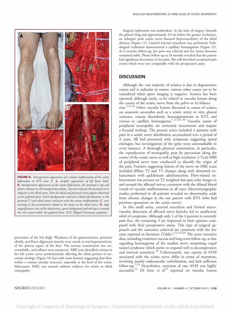
FIGURE 8. Intraoperative appearance of a venous malformation of the sciaticbifurcation in KTS (case 3). A, atrophic appearance of left lower limb.B, intraoperative appearance of the sciatic bifurcation, the proximal is top anddistal is bottom in all intraoperative photos. Asterisk indicates the peroneal nerveadjacent to the tibial nerve. Note the tibial and peroneal nerves appear abnormalat the popliteal fossa. Green backgrounds centered at bluish discoloration in theperoneal (*) and tibial nerves consistent with the venous malformation. C, veinrunning in the perineurium related to the lesion in the tibial nerve. D, highmagnification view of the tibial nerve, green background and red tape is aroundthe vein noted within the popliteal fossa. KTS, Klippel-Trenaunay syndrome.
VASCULAR MALFORMATIONS AS RARE CAUSE OF SCIATIC NEUROPATHY
NEUROSURGERY VOLUME 67 | NUMBER 4 | OCTOBER 2010 | 1139
Copyright © Congress of Neurological Surgeons. Unauthorized reproduction of this article is prohibited.

FIGURE 9. Histology of a venous malformation of the sciatic bifurcation in KTS (case 3). The biopsy, encompassed by epineurialtissue, shows numerous thin-walled but media-containing veins within the endoneurium (A and B, hematoxylin and eosin). Theveins within the endoneurium are further highlighted on Luxol-fast blue (C) and Masson trichrome stain (D). KTS, Klippel-Trenaunay syndrome.
FIGURE 10. MRI appearance of a capillary hemangioma of the sciatic nerve (case 4). A, axial T2 weighted image with fatsuppression. B, axial T1 weighted image. C, axial T1 with fat suppression after gadolinium of the distal thigh. The arrowhead ineach image shows the tibial division with fascicular infiltration by the vascular abnormality with abnormal enhancement. Notesparing of the peroneal nerve.
VAN GOMPEL ET AL
1140 | VOLUME 67 | NUMBER 4 | OCTOBER 2010 www.neurosurgery-online.com
Copyright © Congress of Neurological Surgeons. Unauthorized reproduction of this article is prohibited.

associated with peripheral nerves in the setting of KTS; however,the follow-up data in this publication are insufficient. Detailedfollow-up data are needed in all cases to determine whetherexternal and limited internal neurolysis results in long-term reliefwithout further loss of neurological function and satisfactory paincontrol.The collective experience with vascular lesions as a cause of
sciatica is limited to a small number of case reports. Collection ofthese rare cases through literature report at this time appears to bethe only viable option in understanding the pathophysiology,natural history, and treatment of these lesions.
Disclosure
The authors have no personal financial or institutional interest in any of thedrugs, materials, or devices described in this article.
REFERENCES
1. Chatillon CE, Guiot MC, Jacques L. Lipomatous, vascular, and chondromatousbenign tumors of the peripheral nerves: representative cases and review of theliterature. Neurosurg Focus. 2007;22(6):E18.
2. Sundine MJ, Wirth GA. Hemangiomas: an overview. Clin Pediatr. 2007;46(3):206-221.
3. Wood MB. Intraneural hemangioma: report of a case. Plast Reconstr Surg.1980;65(1):74-76.
FIGURE 11. Intraoperative appearance of a capillary hemangioma of the tibialdivision of the sciatic nerve (case 4). A, isolation of the sciatic nerve proximal tothe bifurcation shows no obvious abnormality. B, upon opening the epineurium,fine pink streaking corresponding to the capillary hemangioma is well seenrestricted to the tibial division (T). Limited internal neurolysis was then per-formed. Peroneal division (P).
FIGURE 12. Histology of a capillary hemangioma of the tibial nerve (case 4). A and B, capillary hemangioma, the fascicles are associated with a sizable vein (A, asterisk (*)and tufts of densely compacted capillaries (A and B, arrowheads, hematoxylin and eosin). Note their position between two nerve fascicles (B, arrowheads). A CD31 stainhighlights several of these capillary complexes (C, arrowheads)
TABLE. Literature Reported Cases of Vascular Lesions and Sciaticaa
Patient Age, y Sex Symptoms Signs Pathology Treatment Outcome
A 10 M R leg p, weakness R leg weakness, percussion tenderness
at site of the nerve lesion
Hemangioma Resection of nerve segment,
leg amputation
Pain resolved,
loss of leg
B 5 F L leg p L leg weakness Hemangioma Internal neurolysis Pain resolved
C 53 M R leg p, tenesmus
and rectal p
None AVM Internal neurolysis Pain resolved
1 23 M L leg p None Hemangioma Internal neurolysis Pain resolved
2 33 F R leg p Swelling, discoloration AVM Internal neurolysis Pain improved
3 36 M L leg p, weakness Atrophy, weakness Venous malformation Internal neurolysis Pain improved
4 18 F L leg p None Hemangioma Internal neurolysis Pain improved
aA, Stewart and Bettin24; B, Purcel and Gurdjian22; C, Vos et al12; R, right; L, left; p, pain; AVM, arteriovenous malformation.
VASCULAR MALFORMATIONS AS RARE CAUSE OF SCIATIC NEUROPATHY
NEUROSURGERY VOLUME 67 | NUMBER 4 | OCTOBER 2010 | 1141
Copyright © Congress of Neurological Surgeons. Unauthorized reproduction of this article is prohibited.

4. Bilge T, Kaya A, Alatli M, Bilge S, Alatli C. Hemangioma of the peronealnerve: case report and review of the literature. Neurosurgery. 1989;25(4):649-652.
5. Maniker A, Thurmond J, Padberg FT Jr, Blacksin M, Vingan R. Traumatic venousvarix causing sciatic neuropathy: case report. Neurosurgery. 2004;55(5):1224.
6. Van Meir N, De Smet L. Carpal tunnel syndrome in children. Acta Orthop Belg.2003;69(5):387-395.
7. Finn MC, Glowacki J, Mulliken JB. Congenital vascular lesions: clinical appli-cation of a new classification. J Pediatr Surg. 1983;18(6):894-900.
8. Mulliken JB, Glowacki J. Hemangiomas and vascular malformations in infantsand children: a classification based on endothelial characteristics. Plast ReconstrSurg. 1982;69(3):412-422.
9. Coessens B, De Mey A, Lacotte B, Vandenbroeck D. Carpal tunnel syndrome dueto an haemangioma of the median nerve in a 12-year-old child. Ann Chir MainMemb Super. 1991;10(3):255-257.
10. Ozdemir O, Calisaneller T, Altinors N. Compression of the ulnar nerve inGuyon’s canal by an arteriovenous malformation. J Hand Surg Eur Vol.2007;32(5):600-601.
11. Vigna PA, Kusior MF, Collins MB, Ross JS. Peripheral nerve hemangioma. Potentialfor clinical aggressiveness. Arch Pathol Lab Med. 1994;118(10):1038-1041.
12. Vos LD, Bom EP, Vroegindeweij D, Tielbeek AV. Congenital pelvic arterio-venous malformation: a rare cause of sciatica. Clin Neurol Neurosurg.1995;97(3):229-232.
13. Di Iorio G, Sanges G, Sannino V, et al. Peripheral nervous system involvement inKlippel-Trenaunay syndrome. Clin Neuropathol. 2005;24(1):42-47.
14. Narvaez J, Narvaez JA, Alegre-Sancho JJ, et al. Pelvic arteriovenous malformationas a rare cause of sciatica. Br J Rheumatol. 1997;36(12):1340-1341.
15. Kara M, Ozcakar L, Eken G, Ozen G, Kiraz S. Deep venous thrombosis andinferior vena cava agenesis causing double crush sciatic neuropathy in Behcet’sdisease. Joint Bone Spine. 2008;75(6):734-736.
16. Meirer R, Huemer GM, Shafighi M, Kamelger FS, Hussl H, Piza-Katzer H. Sciaticnerve enlargement in the Klippel-Trenaunay-Weber syndrome. Br J Plast Surg.2005;58(4):565-568.
17. Ricci S, Georgiev M, Jawien A, Zamboni P. Sciatic nerve varices. Eur J VascEndovasc Surg. 2005;29(1):83-87.
18. Regan PJ, Roberts JO, Bailey BN. Acute posterior interosseous nerve palsy causedby bleeding from an arteriovenous malformation. J Hand Surg Am.1991;16(2):272-273.
19. Bendszus M, Rieckmann P, Perez J, Koltzenburg M, Reiners K, Solymosi L.Painful vascular compression syndrome of the sciatic nerve caused by glutealvaricosities. Neurology. 2003;61(7):985-987.
20. Jacob AG, Driscoll DJ, Shaughnessy WJ, Stanson AW, Clay RP, Gloviczki P.Klippel-Trenaunay syndrome: spectrum and management. Mayo Clin Proc.1998;73(1):28-36.
21. Labropoulos N, Tassiopoulos AK, Gasparis AP, Phillips B, Pappas PJ. Veins alongthe course of the sciatic nerve. J Vasc Surg. 2009;49(3):690-696.
22. Purcell FH, Gurdjian ES. Hemangioma of peripheral nerves. Am J Surg.1935;30:541-544.
23. Kursumovic A, Langner C, Scharnagl E, Koch H. Digital nerve entrapment due tovascular malformation. J Reconstr Microsurg. 2003;19(5):291-294.
24. Stewart SF, Bettin ME. The motor significance of hemangioma. Surg GynecolObstet. 1924;39:307-317.
25. Ergin MT, Druckmiller WH, Cohen P. Intrinsic hemangiomas of the peripheralnerves report of a case and review of the literature. Conn Med. 1998;62(4):209-213.
26. Gasecki AP, Ebers GC, Vellet AD, Buchan A. Sciatic neuropathy associated withpersistent sciatic artery. Arch Neurol. 1992;49(9):967-968.
27. Mestdagh H, Lecomte-Houcke M, Reyford H. Intraneural haemangioma of theposterior tibial nerve. J Bone Joint Surg Br. 1990;72(2):323-324.
28. Patel CB, Tsai TM, Kleinert HE. Hemangioma of the median nerve: a report oftwo cases. J Hand Surg Am. 1986;11(1):76-79.
29. Vekris MD, Stafilas KS, Zacharis KX, Xenakis TA, Soucacos PN, Beris AE.Intrinsic haemangioma of the median nerve: report of a case and review of theliterature. Microsurgery. 2008;28(2):89-90.
30. Cherry KJ, Gloviczki P, Stanson AW. Persistent sciatic vein: diagnosis andtreatment of a rare condition. J Vasc Surg. 1996;23(3):490-497.
Acknowledgment
The authors appreciate the excellent secretarial skills of Denise Chase of MayoClinic Transcription Service.
COMMENTS
T he patient with spinal imaging negative ‘‘sciatica,’’ particularly ifneuropathic pain is present, is challenging. Most surgeons have
found that sciatic nerve imaging is usually not productive. Gompel et al,however, have demonstrated otherwise. They identified 4 cases of vas-cular malformations of the sciatic nerve. Preconceived notions in thisregard, therefore, may not always be appropriate. The character of thepain and neurological examination may provide clues regarding sucha diagnosis. Obviously, however, with such clues, magnetic resonanceimaging (MRI) must be ordered. Further sciatic nerve imaging may be inorder in selected cases. Regardless, the authors are to be congratulated fortheir observations and clinical astuteness. They have unquestionablyheightened our awareness.
Edward C. BenzelCleveland, Ohio
D r Gompel and his colleagues present 4 cases in which a patient isfound to have a vascular malformation causing radiating leg pain
from sciatic nerve involvement. Each case includes a thorough pre-operative evaluation including imaging, a complete discussion of theexternal and limited internal neurolysis performed to treat the malfor-mation, and at least 6 months of follow-up data. The surgical approachof limited internal neurolysis was shown to improve pain in venousangioma, arteriovenous malformation, Klippel-Trenaunay syndrome,and capillary hemangioma.This type of report is of great value in the literature for this rare
condition and will be an aid in determining the treatment of sciaticacaused by various vascular malformations. The authors present the casesin a complete and thoughtful manner that will be increasingly helpful asmore similar cases are identified and reported.
Daniel H. KimHouston, Texas
T his is an interesting and well-written case series in which the authorsdescribe 4 patients with sciatica caused by vascular lesions. These
cases serve as an important reminder to clinicians that thorough clinicaland imaging evaluations are needed in cases of sciatica that are not readilyexplained by lumbar MRI studies. These patients presented with someclinical features that were not typical of sciatica from the usual discogenicorigin, for example, percussion tenderness over the lesion in the thigh,progression of symptoms to constant pain even in recumbency,discoloration of the leg with swelling, exacerbation of pain during menses,etc. The latter cyclical feature in case 2 was reminiscent of endometriosis,another uncommon cause of nondiscogenic sciatica. 1 We have also treatedseveral vascular lesions presenting as nerve sheath tumors in the brachialplexus,2 and these cases have taught us to be wary of possible severehemorrhage when approaching these lesions surgically. Preoperative angi-ography and embolization may be essential in some of the high-flow lesions.
Eric L. ZagerPhiladelphia, Pennsylvania
1. Zager EL, Pfeifer SM, Brown MJ, Torosian MH, Hackney DB. Catamenialmononeuropathy and radiculopathy: a treatable neuropathic disorder. J Neurosurg.1998;88(5):827-830.
2. Ranalli NJ, Huang JH, Lee EB, Zhang PJL, Siegelman ES, Zager EL. Hemangiomasof the brachial plexus: a case series. Neurosurgery. 2009;65(4):A181-A188.
VAN GOMPEL ET AL
1142 | VOLUME 67 | NUMBER 4 | OCTOBER 2010 www.neurosurgery-online.com
Copyright © Congress of Neurological Surgeons. Unauthorized reproduction of this article is prohibited.


