EFFICACY AND SAFETY OF Pulsed radiofrequency treatment in CHRONIC pain syndromes

Clinical Efficacy of Tumor Antigen-Pulsed DC Treatmentfor High-Grade Glioma Patients: Evidence from aMeta-AnalysisJun-Xia Cao1,2*, Xiao-Yan Zhang1, Jin-Long Liu1, Duo Li1, Jun-Li Li1, Yi-Shan Liu1, Min Wang1, Bei-Lei Xu1,
Hai-Bo Wang1, Zheng-Xu Wang1*
1 Biotherapy Center, the General Hospital of Beijing Military Command, Beijing, People’s Republic of China, 2 Tsinghua-Peking Center for Life Sciences, Laboratory of
Dynamic Immunobiology, School of Medicine, School of Life Sciences, Tsinghua University, Beijing, People’s Republic of China
Abstract
Background: The effectiveness of immunotherapy for high-grade glioma (HGG) patients remains controversial. To evaluatethe therapeutic efficacy of dendritic cells (DCs) alone in the treatment of HGG, we performed a systematic review and meta-analysis in terms of patient survival with relevant published clinical studies.
Materials and methods: A total of 409 patients, including historical cohorts, nonrandomized and randomized controls withHGG, were selected for the meta-analysis.
Results: The treatment of HGG with DCs was associated with a significantly improved one-year survival (OS) (p,0.001) and1.5-, 2-, 3-, 4-, and 5-year OS (p,0.001) compared with the non-DC group. A meta-analysis of the patient outcome datarevealed that DC immunotherapy has a significant influence on progression-free survival (PFS) in HGG patients, who showedsignificantly improved 1-,1.5-, 2-, 3- and 4-year PFS (p,0.001). The analysis of Karnofsky performance status (KPS)demonstrated no favorable results for DC cell therapy arm (p = 0.23).The percentages of CD3+CD8+ and CD3+CD4+ T cellsand CD16+ lymphocyte subset were not significantly increased in the DC group compared with the baseline levels observedbefore treatment (p.0.05), whereas CD56+ lymphocyte subset were significantly increased after DC treatment (p = 0.0001).Furthermore, the levels of IFN-c in the peripheral blood of HGG patients, which reflect the immune function of the patients,were significantly increased after DC immunotherapy (p,0.001).
Conclusions: Thus, our meta-analysis showed that DC immunotherapy markedly prolongs survival rates and progression-free time, enhances immune function, and improves the efficacy of the treatment of HGG patients.
Citation: Cao J-X, Zhang X-Y, Liu J-L, Li D, Li J-L, et al. (2014) Clinical Efficacy of Tumor Antigen-Pulsed DC Treatment for High-Grade Glioma Patients: Evidencefrom a Meta-Analysis. PLoS ONE 9(9): e107173. doi:10.1371/journal.pone.0107173
Editor: Christopher Wheeler, Cedars-Sinai Medical Center, United States of America
Received May 6, 2014; Accepted August 7, 2014; Published September 12, 2014
Copyright: � 2014 Cao et al. This is an open-access article distributed under the terms of the Creative Commons Attribution License, which permits unrestricteduse, distribution, and reproduction in any medium, provided the original author and source are credited.
Data Availability: The authors confirm that all data underlying the findings are fully available without restriction. All relevant data are within the paper and itssupporting information files.
Funding: This research work was supported by the National Natural Science Foundation of China (No. 31171427 and 30971651 to Zheng-Xu Wang); BeijingMunicipal Science & Technology Project; Clinical characteristics and Application Research of Capital (No. Z121107001012136 to Zheng-Xu Wang); the NationalNatural Science Foundation of China (No. 30700974 to Jun-Xia Cao); and the Postdoctoral Foundation of China (No. 20060400775 to Jun-Xia Cao). The fundershad no role in study design, data collection and analysis, decision to publish, or preparation of the manuscript.
Competing Interests: The authors have declared that no competing interests exist.
* Email: [email protected] (ZXW); [email protected] (JXC)
Introduction
High-grade gliomas (HGGs) have an incidence currently
estimated at 14,000 new diagnoses per year, according to the
2007 World Health Organization (WHO) classification, which
includes patients with anaplastic astrocytomas (WHO grade III)
and with glioblastoma multiforme (GBM, WHO grade IV) [1].
GBM is the most common and most malignant glioma in adults
and represents approximately 75% of all newly diagnosed glioma
cases; moreover, the prognosis of these patients remains poor, with
a median survival of less than 15 months, despite the use of
trimodal therapy [2]. Indeed, there is no conventional treatment
that specifically targets tumor cells and spares normal brain
parenchyma. Immunologic approaches are theoretically able to
trace, identify, and kill dispersed tumor cells with great accuracy
and are being tested to enhance the response of these tumors to
existing therapy and/or to stimulate innate immune responses [3].
Based on previous studies, it was assumed that immune
reactions do not occur in the brain because of the blood-brain
barrier (BBB); however, we now have an in-depth understanding
that the central nervous system maintains a two-way communi-
cation network with the immune system, with each having a
profound influence on the other [4]. Several studies have clarified
that lymphocytes and antigen-presenting cells (APCs), including
macrophages and dendritic cells (DCs), are able to cross the blood-
brain barrier and migrate to a tumor within the brain parenchyma
[5]. Thus, in phase I and phase II trials, adoptive immunotherapy
including lymphokine-activated killer cells (LAK), cytotoxic T
PLOS ONE | www.plosone.org 1 September 2014 | Volume 9 | Issue 9 | e107173

lymphocytes (CTLs) and tumor-infiltrating T lymphocytes (TILs)
and active immunotherapy using autologous tumor cells (ATCs)
and DCs have demonstrated clinical efficacy, suggesting that
immunotherapy may be a useful strategy to combat HGGs [6].
DCs are the most potent APCs in the human body.
Importantly, DCs can cross the BBB and traffic into perivascular
and parenchymal spaces in the glioma [5,7]. An important
milestone has been reached with the recent approval in 2010 of
sipuleucel-T (Provenge), the first DC vaccine for hormone-
resistant metastatic prostate cancer. This vaccine is primarily an
active immunologic agent with proven activity against solid tumors
[8]. In HGG, a cohort comparison trial involving 45 children,
HGG-IMMUNO-2003, has been conducted since 2001 to
implement and improve immunotherapeutic approaches [9].
Additionally, another clinical trial of 77 newly diagnosed
glioblastoma patients was performed [10]. In addition, a Phase
I/II single-arm clinical trial, HGG-2006, was designed and
conducted using 117 patients [11]. In brief, all of the data showed
a remarkable overall survival (OS) compared with the generally
expected progression of this disease. Thus, both hope and
challenges exist for DC-based immunotherapy. These data
compelled the design of the current prospective placebo-
controlled, double-blind Phase IIb stratified randomized clinical
trial (EudraCT number 2009-018228-14) and the Phase III study
of DCVax in GBM, which has been registered at ClinicalTrials.
gov (NCT00045968).
Unfortunately, due to profound tumor-associated mechanisms
of immunosuppression and evasion, immunotherapeutic strategies
have thus far not translated into clinical success [12]. There are
several reviews that summarize more than 21 DC clinical trials
that were performed in HGG in which up to 500 patients were
involved, excluding controls. These studies always used historical
or nonrandomized cohorts due to the disease’s malignancy [13–
15]. However, evidence from the meta-analysis through logistic
regression regarding the OS, PFS, and other outcomes of the
therapy remains scarce. Here, we addressed the effect of the
autologous tumor antigen-pulsed DCs on the treatment of glioma
patients in terms of survival compared with historical cohorts or
nonrandomized and randomized control groups.
Materials and Methods
2.1 Literature search and inclusion and exclusion criteriaThe trials analyzed in this study were identified through an
electronic search of the PubMed database, the Cochrane Central
Registry of Controlled Trials, the Wanfang Database, the China
Science and Technology Periodical Database, China Journal Net,
reference lists of published trials, and relevant review articles. The
search strategy included the medical subject headings ‘‘glioma’’,
‘‘immunotherapy’’, ‘‘dendritic cells’’, and free text search. No
language limits were applied. The initial search was performed on
Nov 2013 and was updated in Jan 2014. Furthermore, manual
searches were performed in reference lists and conference
proceedings of the American Society of Clinical Oncology (ASCO)
Annual Meetings and the European Cancer Conference (ECCO).
We excluded abstracts that were never subsequently published as
full papers and studies on animals and cell lines.
2.2 Study selection and data extractionWe collected various sets of information, including the authors’
names, journal and year of publication, sample size per arm, newly
or recurrent, regimen used, median or mean age of the patients,
Karnofsky performance status (KPS), DC antigen, delivery route
and dose, and characteristics of the study design (i.e., whether the
trial reported the mode of randomization, allocation concealment,
description of withdrawals per arm, and blinding) for all of the
trials included in the study. The data were independently screened
by two reviewers.
2.3 Definition of outcome measuresOverall survival (OS) was defined as the time from the initiation
of treatment until death from any cause. The secondary endpoint
was progression-free survival (PFS), which was documented and
extracted for analysis. Quality of Life (QoL) was assessed by the
KPS. The immune response was assessed by evaluating and
comparing the data of surface phenotype of the patients’
peripheral blood lymphocytes by FACScan from the recruited
papers, including CD3+, CD4+, CD8+, CD16+ and CD56+ of each
study. Furthermore, we approximately collected the data of
CD3+CD8+ and CD3+CD4+ as the T cell subpopulation and
CD16+ and CD56+ as other cell subset. In addition we also
extracted the data of the cytokine IFN-c tested by ELISA kit from
the included papers.
2.4 Statistical analysisThe analysis was performed using Review Manager Version 5.0
(Nordic Cochran Centre, Copenhagen, Denmark). In our meta-
analysis, we compared the immunotherapy-containing arms of the
selected trials to the respective non-immunotherapy arms. The
treatment effects are reflected by the odds ratios (OR) for OS and
PFS. The OS and PFS data in each arm were extracted from each
included study, and the pooled odds ratio (OR) was calculated
through the Mantel and Haenszel method. A pooled OR,1
indicated a lower recurrence or lower survival in the immuno-
therapy arm. We used Cochran’s Q test, a chi-squared test with a
df equal to the number of studies minus one that tests the null
hypothesis and demonstrates whether the difference among the
studies based on the OR is due to chance, to evaluate whether the
studies’ results were homogeneous. Also calculated in the analysis
was the quantity I2, which describes the percentage of variation
across studies that is due to heterogeneity rather than chance.
Generally speaking, I2 values of 25% represent low heterogeneity,
and subsequently, I2 values of 50% and 75% were used as
evidence of moderate and high heterogeneity, respectively. When
no statistically significant heterogeneity existed, the OR was
calculated with a fixed-effect model; otherwise, a random-effect
model was employed. P-values of ,0.05 were considered to be
statistically significant. All reported P-values resulted from two-
sided versions of the respective tests [16].
Results
3.1 Selection of the trialsThe electronic search yielded 189 references. After a title and
abstract review, 158 publications were excluded for different
reasons (nine for being review articles, 11 for using in vitroexperiments, 26 for being animal models, 91 for being case
reports, and 21 for being DC protocol studies or comments)
(Tables S1–S5 in File S1). A total of 31 clinical trials were selected
as potentially relevant, and their full texts were retrieved for a
more detailed assessment. We then excluded 22 of these 31 studies
for not having a control arm or not providing detailed patient
clinical data and details on the therapeutic response (Table S6 in
File S1). The procedure used to select the clinical trials is shown in
Figure 1. As a result, 9 articles reporting clinical trials of DC-based
therapy were selected for the meta-analysis [17–25] (Table S7 in
File S1).
Meta-Analysis of Active Immunotherapy for High-Grade Glioma Patients
PLOS ONE | www.plosone.org 2 September 2014 | Volume 9 | Issue 9 | e107173

3.2 Characteristics of DC cell-based therapyAfter the selection process, 9 eligible trials with a total of 409
patients to date were included in the present analysis. All of the
trials were fully published: three phase I trials [20,21,23], five
phase I/II trials [17,18,19,22,24] and one phase II trials [25]. The
clinical data of the trials are shown in Table 1. The median age of
the included patients was ,50 years. The WHO grade was mainly
IV for the included HGG patients. All of the patients had
experienced surgery (ST, surgical resection), chemotherapy (CT,
chemical therapy), radiotherapy (RT, radiation therapy), and
intra-cellular hyperthermia (ICH). The included patients were
mainly recurrent containing some of new ones, and also only one
trial recruited the new patients [25], which have been listed on
Table 1. The patients’ KPS have all been reported before
immunotherapy and the value was mainly more than 60, but
after the treatment only two of them reported [17,25]. Addition-
ally, most of the included patients received the DC therapy
without any other simultaneous treatment, and the controls were
four historical cohorts [17,18,20,23], three nonrandomized
cohorts [19,21,22] and two randomized cohorts [24,25].
The method for the preparation of DCs is now well established,
and a sufficient number of DCs can be generated for injection into
patients. In Table 1, we summarized the patient information
about the DC treatment. DCs were matured using cocktails
containing GM-CSF, IL-4, TNF-a, IL-1b, or PGE2. The number
of DCs injected ranged from 16106 to 56108. The frequency of
the injections was highly variable in different trials. The sources of
antigen were also different, but most that were included in our
meta-analysis were derived from tumor cells: autologous irradiated
tumor cells (AIT), autologous tumor lysate (ATL), HLA-1-eluted
peptides (HLP), autologous acid-eluted tumor peptides (ATP), and
autologous heat-shock tumor cells (AHT). One trial reported DC
treatment with fusions of glioma cells [21]. The routes of DC
injection used were mainly intradermal (i.d.), intratumoral (i.t.),
and subcutaneous (s.c.).
3.3 Survival0.5-year overall survival. Information on the 0.5-year
survival was available for six trials [17–19,20,23,24]. These six
trials contained 320 patients in total (80 patients received DC
therapy, and 240 patients not receiving DC therapy were used as a
control). Although the 0.5-year OS rates were 96% (77/80) for
glioma patients receiving the DC treatment and 88% (211/240)
for the historical or nonrandomized and randomized control
Figure 1. Flow diagram showing record identification, screening and study inclusion process.doi:10.1371/journal.pone.0107173.g001
Meta-Analysis of Active Immunotherapy for High-Grade Glioma Patients
PLOS ONE | www.plosone.org 3 September 2014 | Volume 9 | Issue 9 | e107173

Ta
ble
1.
Clin
ical
info
rmat
ion
of
the
elig
ible
tria
lsfo
rth
em
eta
-an
alys
is.
Tri
al
refe
ren
ceT
um
or
cha
ract
eri
stic
sW
HO
gra
de
Cli
nic
al
tria
lp
ha
seP
ati
en
ts(m
ale
)a
nd
con
tro
lM
ed
ian
ag
eP
re-
Th
era
py
KP
SP
rev
iou
str
ea
tme
nt
DC
Arm
Inje
ctio
nD
Cre
gim
en
sC
ult
ure
of
DC
cell
s
Ch
en
-Ne
nC
han
g2
01
1[1
7]
Ne
wo
rR
ecu
rre
nt
III/I
VI/
II1
7(8
);6
3(U
K);
his
tori
cal
44
.7;
UK
Me
dia
n;
90
CT
/RT
DC
slo
ade
dw
ith
AIT
(s.c
)1
.0–
6.16
10
7/c
ou
rse
GM
-CSF
,IL
-4
Ryu
yaY
aman
aka
20
05
[18
]R
ecu
rre
ntI
II/IV
I/II
24
(16
);2
7(U
K);
his
tori
cal
48
.9;
55
.9M
ed
ian
;6
2.5
SR/R
T,
CT
DC
slo
ade
dw
ith
AT
L(i
.do
ri.t
)3
.9–
24
0.96
10
6/c
ou
rse
GM
-CSF
,IL
-4,
KLH
Ch
rist
op
he
rJ
Wh
ee
ler
20
04
[19
]D
en
ovo
IVI/
II2
5(1
1);
25
(13
);ra
nd
om
ize
d5
5;
50
.6
0SR
/CT
,R
TD
Cs
load
ed
wit
hA
TL
or
HLP
10
–4
06
10
6/c
ou
rse
UK
Lin
da
M.L
iau
20
05
[20
]N
ew
or
Re
curr
en
tIV
I1
2(5
);9
9(U
K);
his
tori
cal
40
.4;
,5
0$
60
CT
/IC
HD
Cs
load
ed
wit
hA
TP
1–
106
10
6/c
ou
rse
GM
-CSF
,IL
-4
Te
tsu
roK
iku
chi
20
01
[21
]R
ecu
rre
nt
UK
I8
(7)
38
Me
dia
n7
0SR
/CT
,R
TD
Cs
fuse
dw
ith
AIT
(i.d
)2
.4–
8.76
10
6/c
ou
rse
GM
-CSF
,IL
-4,
TN
F-a
RY
aman
aka
20
03
[22
]R
ecu
rre
nt
UK
I/II
10
(4)
46
Me
dia
n5
4SR
/RT
DC
slo
ade
dw
ith
AT
L(i
.d)
10
–1
37
.26
10
6/c
ou
rse
GM
-CSF
,IL
-4,
KLH
Joh
nS.
Yu
20
04
[23
]N
ew
or
Re
curr
en
tU
KI
14
(10
);2
6(U
K);
his
tori
cal
45
;5
3$
60
SR/C
TD
Cs
load
ed
wit
hA
TL
(i.d
)1
07–
10
8/c
ou
rse
GM
-CSF
,IL
-4
X.J
ie2
01
2[2
4]
Re
curr
en
tIV
I/II
13
(10
);1
2(9
);ra
nd
om
ize
d4
0.2
;4
3.1
$6
0SR
/RT
,C
TD
Cs
load
ed
wit
hA
HT
and
GM
-CSF
(s.c
)66
10
6/c
ou
rse
GM
-CSF
,IL
-4,
IL-1b
,PG
E2,T
NF-a
De
r-Y
ang
Ch
o2
01
1[2
5]
Ne
wIV
II1
8(8
);1
6(8
);ra
nd
om
ize
d5
8.6
;5
5.8
.7
0SR
/CT
,R
TD
Cs
load
ed
wit
hA
TL(
s.c)
2–
561
07/c
ou
rse
GM
-CSF
,IL
-4
Th
eta
ble
sum
mar
ize
sth
ep
atie
nts
’bas
icin
form
atio
nab
ou
tth
etu
mo
rst
age
,ne
wly
or
recu
rre
nt,
case
s,ag
e,K
PS,
op
era
tive
me
tho
db
efo
reth
eim
mu
no
the
rap
yan
dd
eta
ilso
fth
eim
mu
no
the
rap
yin
clu
din
gth
eD
C,t
um
or
anti
ge
n,
and
load
ing
rou
te.
Th
ela
stro
wis
the
cult
ure
con
dit
ion
su
sed
for
the
cells
.K
PS:
Kar
no
fsky
pe
rfo
rman
cest
atu
s;A
IT:
Au
tolo
go
us
irra
dia
ted
tum
or
cells
;A
TL:
Au
tolo
go
us
tum
or
lysa
te;
HLP
:H
LA-1
-elu
ted
pe
pti
de
s;A
TP
:A
uto
log
ou
sac
id-e
lute
dtu
mo
rp
ep
tid
es;
AH
T:
Au
tolo
go
us
he
at-s
ho
cktu
mo
rce
lls;C
T:C
he
mic
alth
era
py;
RT
:R
adia
tio
nth
era
py;
SR:
Surg
ical
rese
ctio
n;K
LH:
Ke
yho
lelim
pe
th
em
ocy
anin
;P
GE2
:P
rost
agla
nd
inE2
;T
NF-a
:Tu
mo
rn
ecr
osi
sfa
cto
r-a
;IL
-4:
Inte
rle
uki
n-4
;i.d
.:in
trad
erm
alva
ccin
atio
n;
i.t.:
intr
atu
mo
ral
vacc
inat
ion
;s.
c.:s
ub
cuta
ne
ou
sin
ject
ion
;IC
H:
intr
a-ce
llula
rh
ype
rth
erm
ia;
UK
:U
nkn
ow
n.
do
i:10
.13
71
/jo
urn
al.p
on
e.0
10
71
73
.t0
01
Meta-Analysis of Active Immunotherapy for High-Grade Glioma Patients
PLOS ONE | www.plosone.org 4 September 2014 | Volume 9 | Issue 9 | e107173
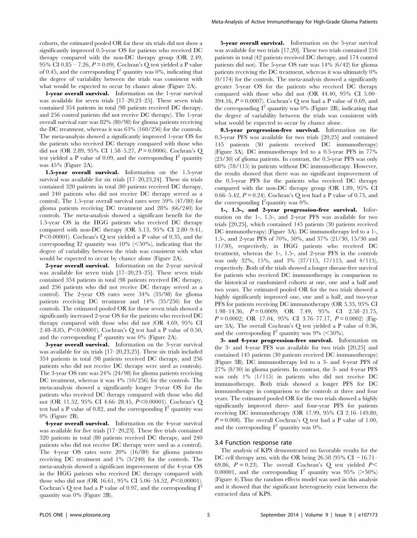
cohorts, the estimated pooled OR for these six trials did not show a
significantly improved 0.5-year OS for patients who received DC
therapy compared with the non-DC therapy group (OR 2.49,
95% CI 0.85 – 7.26, P = 0.09). Cochran’s Q test yielded a P value
of 0.45, and the corresponding I2 quantity was 0%, indicating that
the degree of variability between the trials was consistent with
what would be expected to occur by chance alone (Figure 2A).
1-year overall survival. Information on the 1-year survival
was available for seven trials [17–20,23–25]. These seven trials
contained 354 patients in total (98 patients received DC therapy,
and 256 control patients did not receive DC therapy). The 1-year
overall survival rate was 82% (80/98) for glioma patients receiving
the DC treatment, whereas it was 63% (160/256) for the controls.
The meta-analysis showed a significantly improved 1-year OS for
the patients who received DC therapy compared with those who
did not (OR 2.89, 95% CI 1.58–5.27, P = 0.0006). Cochran’s Q
test yielded a P value of 0.09, and the corresponding I2 quantity
was 45% (Figure 2A).
1.5-year overall survival. Information on the 1.5-year
survival was available for six trials [17–20,23,24]. These six trials
contained 320 patients in total (80 patients received DC therapy,
and 240 patients who did not receive DC therapy served as a
control). The 1.5-year overall survival rates were 59% (47/80) for
glioma patients receiving DC treatment and 28% (66/240) for
controls. The meta-analysis showed a significant benefit for the
1.5-year OS in the HGG patients who received DC therapy
compared with non-DC therapy (OR 5.13, 95% CI 2.80–9.41,
P,0.00001). Cochran’s Q test yielded a P value of 0.35, and the
corresponding I2 quantity was 10% (,50%), indicating that the
degree of variability between the trials was consistent with what
would be expected to occur by chance alone (Figure 2A).
2-year overall survival. Information on the 2-year survival
was available for seven trials [17–20,23–25]. These seven trials
contained 354 patients in total (98 patients received DC therapy,
and 256 patients who did not receive DC therapy served as a
control). The 2-year OS rates were 34% (33/98) for glioma
patients receiving DC treatment and 14% (35/256) for the
controls. The estimated pooled OR for these seven trials showed a
significantly increased 2-year OS for the patients who received DC
therapy compared with those who did not (OR 4.69, 95% CI
2.48–8.85, P,0.00001). Cochran’s Q test had a P value of 0.50,
and the corresponding I2 quantity was 0% (Figure 2A).
3-year overall survival. Information on the 3-year survival
was available for six trials [17–20,23,25]. These six trials included
354 patients in total (98 patients received DC therapy, and 256
patients who did not receive DC therapy were used as controls).
The 3-year OS rate was 24% (24/98) for glioma patients receiving
DC treatment, whereas it was 4% (10/256) for the controls. The
meta-analysis showed a significantly longer 3-year OS for the
patients who received DC therapy compared with those who did
not (OR 11.52, 95% CI 4.66–28.45, P,0.00001). Cochran’s Q
test had a P value of 0.82, and the corresponding I2 quantity was
0% (Figure 2B).
4-year overall survival. Information on the 4-year survival
was available for five trials [17–20,23]. These five trials contained
320 patients in total (80 patients received DC therapy, and 240
patients who did not receive DC therapy were used as a control).
The 4-year OS rates were 20% (16/80) for glioma patients
receiving DC treatment and 1% (3/240) for the controls. The
meta-analysis showed a significant improvement of the 4-year OS
in the HGG patients who received DC therapy compared with
those who did not (OR 16.61, 95% CI 5.06–54.52, P,0.00001).
Cochran’s Q test had a P value of 0.97, and the corresponding I2
quantity was 0% (Figure 2B).
5-year overall survival. Information on the 5-year survival
was available for two trials [17,20]. These two trials contained 216
patients in total (42 patients received DC therapy, and 174 control
patients did not). The 5-year OS rate was 14% (6/42) for glioma
patients receiving the DC treatment, whereas it was ultimately 0%
(0/174) for the controls. The meta-analysis showed a significantly
greater 5-year OS for the patients who received DC therapy
compared with those who did not (OR 44.40, 95% CI 5.00–
394.16, P = 0.0007). Cochran’s Q test had a P value of 0.69, and
the corresponding I2 quantity was 0% (Figure 2B), indicating that
the degree of variability between the trials was consistent with
what would be expected to occur by chance alone.
0.5-year progression-free survival. Information on the
0.5-year PFS was available for two trials [20,25] and contained
145 patients (30 patients received DC immunotherapy)
(Figure 3A). DC immunotherapy led to a 0.5-year PFS in 77%
(23/30) of glioma patients. In contrast, the 0.5-year PFS was only
68% (78/115) in patients without DC immunotherapy. However,
the results showed that there was no significant improvement of
the 0.5-year PFS for the patients who received DC therapy
compared with the non-DC therapy group (OR 1.89, 95% CI
0.66–5.42, P = 0.24). Cochran’s Q test had a P value of 0.73, and
the corresponding I2quantity was 0%.
1-, 1.5-, and 2-year progression-free survival. Infor-
mation on the 1-, 1.5-, and 2-year PFS was available for two
trials [20,25], which contained 145 patients (30 patients received
DC immunotherapy) (Figure 3A). DC immunotherapy led to a 1-,
1.5-, and 2-year PFS of 70%, 50%, and 37% (21/30, 15/30 and
11/30), respectively, in HGG patients who received DC
treatment, whereas the 1-, 1.5-, and 2-year PFS in the controls
was only 32%, 15%, and 3% (37/115, 17/115, and 4/115),
respectively. Both of the trials showed a longer disease-free survival
for patients who received DC immunotherapy in comparison to
the historical or randomized cohorts at one, one and a half and
two years. The estimated pooled OR for the two trials showed a
highly significantly improved one, one and a half, and two-year
PFS for patients receiving DC immunotherapy (OR 5.33, 95% CI
1.98–14.36, P = 0.0009; OR 7.49, 95% CI 2.58–21.75,
P = 0.0002; OR 17.04, 95% CI 3.76–77.17, P = 0.0002) (Fig-
ure 3A). The overall Cochran’s Q test yielded a P value of 0.36,
and the corresponding I2 quantity was 9% (,50%).
3- and 4-year progression-free survival. Information on
the 3- and 4-year PFS was available for two trials [20,25] and
contained 145 patients (30 patients received DC immunotherapy)
(Figure 3B). DC immunotherapy led to a 3- and 4-year PFS of
27% (8/30) in glioma patients. In contrast, the 3- and 4-year PFS
was only 1% (1/115) in patients who did not receive DC
immunotherapy. Both trials showed a longer PFS for DC
immunotherapy in comparison to the controls at three and four
years. The estimated pooled OR for the two trials showed a highly
significantly improved three- and four-year PFS for patients
receiving DC immunotherapy (OR 17.99, 95% CI 2.16–149.80,
P = 0.008). The overall Cochran’s Q test had a P value of 1.00,
and the corresponding I2 quantity was 0%.
3.4 Function response rateThe analysis of KPS demonstrated no favorable results for the
DC cell therapy arm, with the OR being 26.58 (95% CI 216.71–
69.86, P = 0.23). The overall Cochran’s Q test yielded P,
0.00001, and the corresponding I2 quantity was 95% (.50%)
(Figure 4).Thus the random effects model was used in this analysis
and it showed that the significant heterogeneity exist between the
extracted data of KPS.
Meta-Analysis of Active Immunotherapy for High-Grade Glioma Patients
PLOS ONE | www.plosone.org 5 September 2014 | Volume 9 | Issue 9 | e107173

Meta-Analysis of Active Immunotherapy for High-Grade Glioma Patients
PLOS ONE | www.plosone.org 6 September 2014 | Volume 9 | Issue 9 | e107173

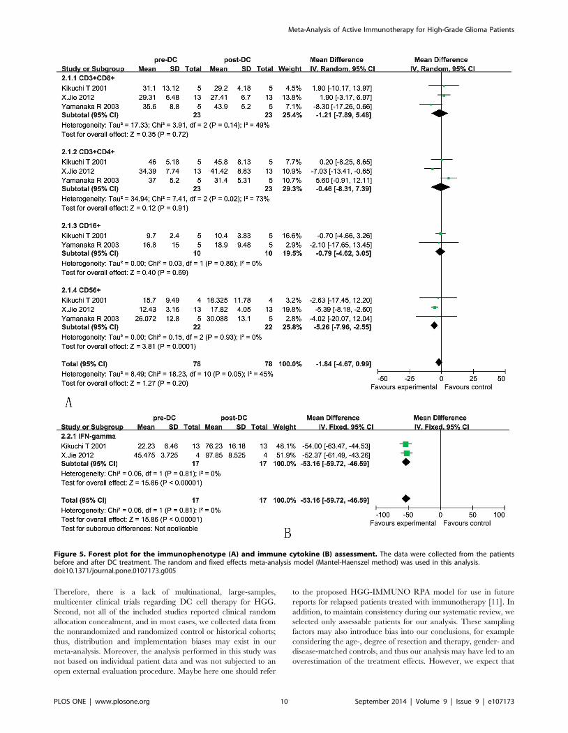
3.5 Immune responseLymphocyte/monocyte subsets in patients. The analysis
showed that the proportions of CD3+CD8+ and CD3+CD4+ cells
were not significantly increased in the DC group compared with
the baseline levels observed before treatment, as reflected by
pooled MD values of 21.21 (95% CI = 27.89–5.48, p = 0.72) and
20.46 (95% CI = 28.31–7.39, p = 0.91) [21,22,24]. Cochran’s Q
test had P values of 0.14 and 0.02, while the corresponding I2
quantities were 49% and 73%. CD16+ cells were also not
significantly increased in the DC group compared with the
baseline levels observed before treatment, as reflected by pooled
MD values of 20.79 (95% CI = 24.62–3.05, p = 0.69), cochran’s
Q test had a P value of 0.86, while the corresponding I2 quantity
was 0%. Whereas CD56+ lymphocyte subset was significantly
increased after DC treatment with pooled MD values of 25.26
(95% CI = 27.96–22.55, p = 0.0001) (Figure 5A). Cochran’s Q
test had a P value of 0.93, while the corresponding I2 quantity was
0%.Immune cytokine levels of patients. As a consequence of
stimulation by DCs, the CD4+ cells release cytokines, such as IL-2,
IL-6, IFN-c, TNF, and lymphotoxin (LT), which assist in the
expansion of the CD8+ cytotoxic T lymphocytes (CTLs). In our
analysis, the levels of IFN-c (OR = -53.16, 95% CI = 259.72–2
46.59, p,0.00001) were significantly increased after DC treatment
(Figure 5B) [21,24]. Cochran’s Q test had a P value of 0.41, while
the corresponding I2 quantity was 0%, indicating that there was no
evidence of heterogeneity among the individual studies. This
finding indicated that the degree of variability among the trials was
consistent with what would occur by chance.
Discussion
DC therapy is based on the concept that GBM cells are poor
APCs because of the down regulation of costimulatory molecules
and the release of immunoinhibitory cytokines [6]. DCs are
professional APCs that phagocytose foreign antigens and present
them in the context of MHC to activate innate and adaptive
immune cells [8]. Thus, DC immunotherapy is widely considered
as the fourth treatment modality for patients with cancer [26].
Our systematic meta-analysis yielded several major findings.
First, we demonstrated that DC immunotherapy can significantly
improve the 1-, 1.5-, 2-, 3-, 4-, and 5-year OS (p,0.001) of HGG
patients compared with the non-treatment group. Then, a meta-
analysis of the outcome of the patient data revealed that DC
immunotherapy has a significant influence on the 1-, 1.5-, 2-, 3-,
and 4-year PFS (p,0.001). But, the results of the analysis of KPS
demonstrated no favorable outcome for DC cell therapy arm
(p = 0.23). Furthermore, the percentages of CD3+CD8+ and
CD3+CD4+ T cells and CD16+ lymphocyte cells were not
significantly increased in the DC group compared with the
baseline levels observed before treatment (p.0.05), but CD56+
lymphocyte cells were significantly increased after DC treatment
(p,0.001). In addition, after DC immunotherapy, the levels of
IFN-c in the peripheral blood of HGG patients, which reflect the
immune function of the patients, were significantly increased (p,
0.001). Overall, according to our analysis, DC therapy can
prolong OS, improve the disease recurrence time, and would
involve in the immunity function. Hence, our meta-analysis
demonstrated that DC immunotherapy is a promising therapy
method for HGG patients.
To date, most recent phase I and II trials have exhibited the
median overall survival is increased by 20% after administration of
an autologous DC vaccine for patients with GBM [17,27]. Thus,
in a meta-analysis of the collected data, our comprehensive results
showed that the 1-, 1.5-, 2-, 3-, 4-, and 5-year OS rates were 82%,
59%, 34%, 24%, 20%, and 14%, respectively, which is slightly
different from the results observed with the independent trials and
the median overall survival was about 29%. Yet, the positive trend
held consistent compared with the historical or the randomized
and nonrandomized controls, which maintained 63%, 28%, 14%,
4%, 1%, and 0 OS rates, respectively and the median overall
survival was about 9%. So through logistic regression, our data
analysis also showed that DC immunotherapy can significantly
prolong the OS in HGG patients (p,0.001) by increasing median
OS 20%.
Regarding PFS, the data were described in only two studies of
145 patients with historical cohorts or randomized controls. The
summarized results showed that the 1-, 1.5-, 2-, 3-, and 4-year PFS
rates were 70%, 50%, 37%, and 27%, respectively, compared with
the controls, which maintained 32%, 15%, 3%, and 1% PFS rates.
Our meta-analysis showed that DC immunotherapy benefits the
PFS, which could be up to 50% at the 1.5-year mark. It was
previously reported that independent clinical trials of Phase II
studies with other immunotherapy methods in recurrent GBM
showed that the median PFS is 20 weeks [28], and in another
Phase II trial with newly diagnosed GBM, the time for the median
PFS is 14.2 months [29]. Although there are quite a few
differences among the trials, the positive trend of the meta-
analysis was fully confirmed, and the advantage of logistic
regression for the data analysis was obvious, revealing that DC
immunotherapy has a significant influence on the 1-, 1.5-, 2-, 3-,
and 4-year PFS (p,0.001). As a matter of fact, immunotherapy
would ameliorate some of the symptoms: patients had increased
appetite, improved sleep, gained body weight, and pain relief. But
in our meta-analysis, DC immunotherapy may not improve the
life quality of postoperative patients (p = 0.23) by comparing KPS
before and after treatment on the Figure 4.
Immunologic evidence of the response to DC therapy was
assessed by comparing the levels of immunologic cell types
(CD3+CD8+, CD4+CD8+, CD16+ and CD56+ cells) and certain
cytokines (IFN-c) before and after DC treatment. Our meta-
analysis showed that following DC therapy, there was a significant
increase in the IFN-c (p,0.001) levels compared with those in
non-DC patients, suggesting the induction of an immune response
in these patients with DC treatment. The concept of immune-
editing is to use immunotherapy as a treatment strategy in
response to a major challenge presented to the immune system
[30]. Immune-editing consists of 3 phases: elimination, equilibri-
um, and escape. Elimination refers to the antitumor function of
both the adaptive and innate immune system and is driven by the
production of IFN-c. It was demonstrated that IFN-c production
levels from post-vaccine peripheral blood mononucleated cell
(PBMC) correlated significantly with patient survival and time to
progression [31]. Here, our meta-analysis demonstrated that IFN-
c was significantly increased after DC treatment and thus would
Figure 2. Comparison of 0.5-, 1-, 1.5- and 2-year overall survival (OS) between the non-DC and DC groups (A); Forest plot for 3-, 4-,and 5-year OS between the non-DC and DC groups in HGG patients (B). The fixed-effects meta-analysis model (Mantel-Haenszel method)was used. OR, odds ratio. DC, DC-containing therapy; non-DC, non-DC-containing therapy. Each trial is represented by a square, the center of whichgives the odds ratio for that trial. The size of the square is proportional to the information in that trial. The ends of the horizontal bars denote a 95%CI. The black diamond gives the overall odds ratio for the combined results of all trials.doi:10.1371/journal.pone.0107173.g002
Meta-Analysis of Active Immunotherapy for High-Grade Glioma Patients
PLOS ONE | www.plosone.org 7 September 2014 | Volume 9 | Issue 9 | e107173

Meta-Analysis of Active Immunotherapy for High-Grade Glioma Patients
PLOS ONE | www.plosone.org 8 September 2014 | Volume 9 | Issue 9 | e107173

be helpful in immune-editing to compensate for the elimination.
But it should be noted that only two trials were included, and the
method for measure IFN-c was not used ELISPOT and/or ICS.
Furthermore, in the included studies, there were some trials in
which they detected the IFN-c with ELISPOT only reported the
individual response of the patients [18,22] or qPCR to calculate
the multiples or the percentage of the response after the
immunotherapy [19,23], but could not be analyzed in our meta-
analysis. Thus the quantification of IFN-c in sera was not
consistent in all the studies, that could induce the big heterogeneity
among these studies.
Another notable challenge is the presence of an immunosup-
pressive tumor microenvironment, which causes decreased antigen
recognition and depressed immune cell activation. Although
increased CD8+ infiltrating lymphocytes have been shown in
some studies to be associated with increased patient survival,
Hussain et al. reported that most tumor-infiltrating CD8+ cells are
not activated [2,32]. In our meta-analysis, the results showed that
the CD3+CD8+ and CD4+CD8+ levels were not significantly
changed after DC treatment. In addition, our meta-analysis that
included only three trials showed CD16+ and CD56+ lymphocyte
cells which would denote some of the NK cells, could combat the
tumor cells and play an immunomodulatory function, inducing a
Th1 immune response and improving antitumor immunoreactiv-
ity in the body [33]. Among them CD16+ was not significantly
increased after DC treatment, but CD56+ was significantly
increased after DC immunotherapy according to our meta-
analysis with the included papers.
Equilibrium is the period in which immune cells become latent
to a partially eradicated tumor. Escape occurs when the tumor
escapes from immunosurveillance and becomes resistant to
antitumor immune function, usually via genomic instability or
downregulation of key antigens [34]. Thus, to induce a tumor-
specific immune reaction via DCs, which is the basis of DC
immunotherapy, DCs are loaded with various antigens and then
activate both helper and killer T cells and B cells. So far, the
different tumor-associated antigens (TAAs) used include specific
tumor-associated peptides, tumor-derived RNA and cDNA, tumor
cell lysate, apoptotic tumor cells, and gene transfer methods using
retroviral vectors, recombinant adenoviruses or lentiviruses
encoding tumor antigens, and electroporation of tumor RNA into
DCs to increase the target accuracy and overcome the tumor
immunosuppression [35]. In our meta-analysis, the selected
clinical trials with DC only used the following tumor antigens:
autologous irradiated tumor cells, tumor lysate and acid-eluted
tumor peptides or heat-shock tumor cells that compensate for
tumor antigen exposure. Other antigens were not included in our
meta-analysis, especially EGFRvIII, IL-13Ra, EphA2, survivin,
Wilms’ tumor 1 (WT1), Sry-related high mobility group box
(SOX), and cytomegalovirus (CMV), but have been tested in some
clinical trials [36]. Furthermore, cancer stem cells (CSCs) or
cancer-initiating cells can also be a potentially useful source of
tumor antigens in DC-based immunotherapy, and some of the
preclinical data are potentially encouraging [37]. Thus, multiple
questions need to be clarified regarding the identification of
suitable antigens and improvement of the tumor cell targeting
accuracy precluding the eventually successful translation of DC
immunotherapy into clinical applications.
More recent transcriptional profiling has classified GBM
molecularly into four subtypes with distinct clinical features. Prins
et al. showed the difference of sensitivity to a whole-cell lysate DC
vaccination between two of the GBM subtypes with MGMT
promoter methylation and IDH1 mutation [38]. Interestingly, it
was also demonstrated that the mesenchymal gene expression
profile may represent a population of patients with favorable
responses to their DC vaccine, so the GBM microenvironment is
also a double-edged sword concerning DC immunotherapy [39].
Despite these challenges, DC immunotherapy still showed an
appealing benefit in terms of extending the survival of patients
with HGG. The heterogeneous methods use and the complexity of
designing and reporting on the immunotherapy trials will be
overcome in the near future, providing stronger evidence to
establish DC treatment as the standard for HGG patients.
In brief, DC immunotherapy has yielded encouraging results
with immunological and clinical benefits for HGG patients and
needs to be further tested to demonstrate significant therapeutic
efficacy in phase III clinical trials.
Limitations of the studyAlthough several early-phase clinical trials have demonstrated
promising therapeutic outcomes to date, clinical immunotherapy
trials for gliomas have not yet demonstrated objective proof of
clinical trials for lacking the randomized studies, so limitations in
our analyses should be considered in interpreting the results. Many
intrinsic and extrinsic factors might influence the systemic review’s
reliability. First, no one trial has more than 100 patients per arm.
Figure 3. Comparison of 0.5-, 1-, 1.5-, and 2-year progression-free survival (PFS) between the non-DC and DC groups (A); Forestplot for 3-, 4-, and 5-year PFS between the non-DC and DC groups in HGG patients (B). The fixed effects meta-analysis model (Mantel-Haenszel method) was used in this analysis.doi:10.1371/journal.pone.0107173.g003
Figure 4. Forest plot for KPS before and after DC treatment. The random effects model (Mantel-Haenszel method) was used in this analysis.doi:10.1371/journal.pone.0107173.g004
Meta-Analysis of Active Immunotherapy for High-Grade Glioma Patients
PLOS ONE | www.plosone.org 9 September 2014 | Volume 9 | Issue 9 | e107173
Therefore, there is a lack of multinational, large-samples,
multicenter clinical trials regarding DC cell therapy for HGG.
Second, not all of the included studies reported clinical random
allocation concealment, and in most cases, we collected data from
the nonrandomized and randomized control or historical cohorts;
thus, distribution and implementation biases may exist in our
meta-analysis. Moreover, the analysis performed in this study was
not based on individual patient data and was not subjected to an
open external evaluation procedure. Maybe here one should refer
to the proposed HGG-IMMUNO RPA model for use in future
reports for relapsed patients treated with immunotherapy [11]. In
addition, to maintain consistency during our systematic review, we
selected only assessable patients for our analysis. These sampling
factors may also introduce bias into our conclusions, for example
considering the age-, degree of resection and therapy, gender- and
disease-matched controls, and thus our analysis may have led to an
overestimation of the treatment effects. However, we expect that
Figure 5. Forest plot for the immunophenotype (A) and immune cytokine (B) assessment. The data were collected from the patientsbefore and after DC treatment. The random and fixed effects meta-analysis model (Mantel-Haenszel method) was used in this analysis.doi:10.1371/journal.pone.0107173.g005
Meta-Analysis of Active Immunotherapy for High-Grade Glioma Patients
PLOS ONE | www.plosone.org 10 September 2014 | Volume 9 | Issue 9 | e107173

our study will be valuable for the design of more comprehensive,
larger, controlled clinical trials.
Furthermore, the blood sample sources, injection modes, cell
numbers, cell purity, tumor antigen, and cell phenotype may also
affect the outcome of individual trials. All of these variables may
introduce some level of bias; for instance, there is significant
heterogeneity in the extracted data shown in Figure 4 and 5. It is
important to standardize not only the DC cell preparation but also
the criteria of the immune phenotyping system and the clinical
response assessment. Our analysis may be valuable for the
standardization of DC immune therapy as an adjuvant treatment
for patients with HGG.
Conclusions
Taken together, our data suggest that DC cells have great
potential to be a clinically efficacious therapy in the treatment of
patients suffering from advanced-stage HGG malignancies who
exhibit poor tolerance of chemotherapy or radiotherapy. These
early results from clinical trials are very promising and must be
verified more stringently before DC immunotherapy can be
applied at the bedside.
Supporting Information
Checklist S1 PRISMA Checklist.
(DOC)
File S1 Contains Tables S1–S7. Table S1. List 9 excluding
papers for reviews. Table S2. List 11 excluding papers for being
in vitro experiments. Table S3. List 26 excluding papers for being
animal models experiments. Table S4. List 91 case reports for
being no enough data. Table S5. List 21 DC protocol studies and
comments for being no clinical data. Table S6. List 22 clinical
trials for no appropriate control arm. Specify the reasons of
excluded clinical trial study’s PICOS characteristics and report
characteristics. P: participants; I: interventions; C: comparison; O:
outcomes; S: setting (study design). Table S7. List 9 including
clinical trials. Separately specify all the selected information
sources in the included 9 clinical trials for our meta-analysis with
PICOS, report characteristics, and clinical data source which
denoted every data extracted from the paper’s Title, Abstract,
Introduction, Material and methods, Results, Figure, Table,
Discussion, or Supplementary material.
(DOCX)
Author Contributions
Conceived and designed the experiments: ZXW JXC. Performed the
experiments: ZXW JXC XYZ JL-Liu YSL MW DL JL-Li BLX HBW.
Analyzed the data: JXC XYZ JL-Liu YSL DL MW JL-Li BLX HBW.
Contributed reagents/materials/analysis tools: JXC DL MW. Contributed
to the writing of the manuscript: ZXW JXC XYZ JL-Liu.
References
1. Louis DN, Ohgaki H, Wiestler OD, Cavenee WK, Burger PC, et al. (2007) The2007 WHO classification of tumours of the central nervous system. Acta
Neuropathol. 114(2):97–109. Epub 2007 Jul 6.
2. Ruzevick J, Jackson C, Phallen J, Lim M (2012) Clinical trials with
immunotherapy for high-grade glioma. Neurosurg Clin N Am. 23(3):459-470.doi: 10.1016/j.nec.2012.04.003. Epub 2012 Jun 8.
3. Badhiwala J, Decker WK, Berens ME, Bhardwaj RD (2013) Clinical trials in
cellular immunotherapy for brain/CNS tumors. Expert Rev Neurother.
13(4):405–424. doi: 10.1586/ern.13.23.
4. Wilson EH, Weninger W, Hunter CA (2010) Trafficking of immune cells in thecentral nervous system. J Clin Invest. 120(5):1368–1379. doi: 10.1172/
JCI41911. Epub 2010 May 3.
5. Sagar D, Foss C, Baz ER, Pomper MG, Khan ZK, et al. (2012) Mechanisms of
dendritic cell trafficking across the blood-brain barrier. J NeuroimmunePharmacol. 7(1):74–94. doi: 10.1007/s11481-011-9302-7. Epub 2011 Aug 6.
6. Xu X, Stockhammer F, Schmitt M (2012) Cellular-based immunotherapies forpatients with glioblastoma multiforme. Clin Dev Immunol. 2012:764213. doi:
10.1155/2012/764213. Epub 2012 Feb 28.
7. Fabry Z, Raine CS, Hart MN (1994) Nervous tissue as an immunecompartment: the dialect of the immune response in the CNS. Immunol
Today. 15(5):218–224.
8. Palucka K, Banchereau J (2012) Cancer immunotherapy via dendritic cells. Nat
Rev Cancer. 22;12(4):265–277. doi: 10.1038/nrc3258. Review.
9. Ardon H, De Vleeschouwer S, Van Calenbergh F, Claes L, Kramm CM, et al.
(2010) Adjuvant dendritic cell-based tumour vaccination for children withmalignant brain tumours. Pediatr Blood Cancer. 54(4):519–525. doi: 10.1002/
pbc.22319.
10. Ardon H, Van Gool SW, Verschuere T, Maes W, Fieuws S, et al. (2012)
Integration of autologous dendritic cell-based immunotherapy in the standard ofcare treatment for patients with newly diagnosed glioblastoma: results of the
HGG-2006 phase I/II trial. Cancer Immunol Immunother. 61(11):2033–2044.doi: 10.1007/s00262-012-1261-1. Epub 2012 Apr 22.
11. De Vleeschouwer S, Ardon H, Van Calenbergh F, Sciot R, Wilms G, et al.(2012) Stratification according to HGG-IMMUNO RPA model predicts
outcome in a large group of patients with relapsed malignant glioma treatedby adjuvant postoperative dendritic cell vaccination. Cancer Immunol
Immunother. 61(11):2105–2112. doi: 10.1007/s00262-012-1271-z. Epub 2012
May 8.
12. Van Gool S, De Vleeschouwer S (2012) Should dendritic cell-based tumorvaccination be incorporated into standard therapy for newly diagnosed
glioblastoma patients? Expert Rev Neurother. 12(10):1173–1176. doi:
10.1586/ern.12.107.
13. Bregy A, Wong TM, Shah AH, Goldberg JM, Komotar RJ (2013) Activeimmunotherapy using dendritic cells in the treatment of glioblastoma multi-
forme. Cancer Treat Rev. 39(8):891–907. doi: 10.1016/j.ctrv.2013.05.007.Epub 2013 Jun 21.
14. Van Gool S, Maes W, Ardon H, Verschuere T, Van Cauter S, et al. (2009)
Dendritic cell therapy of high-grade gliomas. Brain Pathol. 19(4):694–712. doi:
10.1111/j.1750-3639.2009.00316.x.
15. Mineharu Y, Castro MG, Lowenstein PR, Sakai N, Miyamoto S. (2013)
Dendritic Cell-Based Immunotherapy for Glioma: Multiple Regimens and
Implications in Clinical Trials. Neurol Med Chir (Tokyo). 53(11):741–754. Epub
2013 Oct 21.
16. Wang ZX, Cao JX, Liu ZP, Cui YX, Li CY, et al. (2014). Combination of
chemotherapy and immunotherapy for colon cancer in China: a meta-analysis.
World J Gastroenterol. 28;20(4):1095–1106. doi: 10.3748/wjg.v20.i4.1095.
17. Chang CN, Huang YC, Yang DM, Kikuta K, Wei KJ, et al. (2011) A phase I/II
clinical trial investigating the adverse and therapeutic effects of a postoperative
autologous dendritic cell tumor vaccine in patients with malignant glioma. J Clin
Neurosci. 18(8):1048–1054. doi: 10.1016/j.jocn.2010.11.034. Epub 2011 Jun
28.
18. Yamanaka R, Homma J, Yajima N, Tsuchiya N, Sano M, et al. (2005) Clinical
evaluation of dendritic cell vaccination for patients with recurrent glioma: results
of a clinical phase I/II trial. Clin Cancer Res. 11(11):4160–4167.
19. Wheeler CJ, Das A, Liu G, Yu JS, Black KL. (2004) Clinical responsiveness of
glioblastoma multiforme to chemotherapy after vaccination. Clin Cancer Res.
10(16):5316–5326.
20. Liau LM, Prins RM, Kiertscher SM, Odesa SK, Kremen TJ, et al. (2005)
Dendritic cell vaccination in glioblastoma patients induces systemic and
intracranial T-cell responses modulated by the local central nervous system
tumor microenvironment. Clin Cancer Res. 11(15):5515–5525.
21. Kikuchi T, Akasaki Y, Irie M, Homma S, Abe T, et al. (2001) Results of a phase
I clinical trial of vaccination of glioma patients with fusions of dendritic and
glioma cells. Cancer Immunol Immunother. 50(7):337–344.
22. Yamanaka R, Abe T, Yajima N, Tsuchiya N, Homma J, et al. (2003)
Vaccination of recurrent glioma patients with tumour lysate-pulsed dendritic
cells elicits immune responses: results of a clinical phase I/II trial. Br J Cancer.
89(7):1172–1179.
23. Yu JS, Liu G, Ying H, Yong WH, Black KL, et al. (2004) Vaccination with
tumor lysate-pulsed dendritic cells elicits antigen-specific, cytotoxic T-cells in
patients with malignant glioma. Cancer Res. 64(14):4973–4979.
24. Jie X, Hua L, Jiang W, Feng F, Feng G, et al. (2012) Clinical application of a
dendritic cell vaccine raised against heat-shocked glioblastoma. Cell Biochem
Biophys. 62(1):91–99. doi: 10.1007/s12013-011-9265-6.
25. Cho DY, Yang WK, Lee HC, Hsu DM, Lin HL, et al. (2012) Adjuvant
immunotherapy with whole-cell lysate dendritic cells vaccine for glioblastoma
multiforme: a phase II clinical trial. World Neurosurg. 77(5-6):736–744. doi:
10.1016/j.wneu.2011.08.020. Epub 2011 Nov 7.
26. Van Gool S (2013) Immunotherapy for high-grade glioma: how to go beyond
Phase I/II clinical trials. Immunotherapy. 5(10):1043–1046. doi: 10.2217/
imt.13.86.
Meta-Analysis of Active Immunotherapy for High-Grade Glioma Patients
PLOS ONE | www.plosone.org 11 September 2014 | Volume 9 | Issue 9 | e107173

27. Shah AH, Bregy A, Heros DO, Komotar RJ, Goldberg J (2013) Dendritic cell
vaccine for recurrent high-grade gliomas in pediatric and adult subjects:clinical trial protocol. Neurosurgery. 73(5):863–867. doi: 10.1227/NEU.
0000000000000107.
28. Izumoto S, Tsuboi A, Oka Y, Suzuki T, Hashiba T, et al. (2008) Phase II clinicaltrial of Wilms tumor 1 peptide vaccination for patients with recurrent
glioblastoma multiforme. J Neurosurg. 108(5):963–971. doi: 10.3171/JNS/2008/108/5/0963.
29. Sampson JH, Heimberger AB, Archer GE, Aldape KD, Friedman AH, et al.
(2010) Immunologic escape after prolonged progression-free survival withepidermal growth factor receptor variant III peptide vaccination in patients with
newly diagnosed glioblastoma. J Clin Oncol. 28(31):4722–4729. doi: 10.1200/JCO.2010.28.6963. Epub 2010 Oct 4.
30. Schreiber RD, Old LJ, Smyth MJ (2011) Cancer immunoediting: integratingimmunity’s roles in cancer suppression and promotion. Science.
331(6024):1565–1570. doi: 10.1126/science.1203486.
31. Wheeler CJ, Black KL, Liu G, Mazer M, Zhang XX, et al. (2008) Vaccinationelicits correlated immune and clinical responses in glioblastoma multiforme
patients. Cancer Res. 68(14):5955–5964. doi: 10.1158/0008-5472.CAN-07-5973.
32. Hussain SF, Heimberger AB (2005) Immunotherapy for human glioma:
innovative approaches and recent results. Expert Rev Anticancer Ther.5(5):777–790.
33. Ogbomo H, Cinatl J Jr, Mody CH, Forsyth PA (2011) Immunotherapy in
gliomas: limitations and potential of natural killer (NK) cell therapy. Trends MolMed. 17(8):433–441. doi: 10.1016/j.molmed.2011.03.004. Epub 2011 Apr 19.
34. Grivennikov SI, Greten FR, Karin M (2010) Immunity, inflammation, and
cancer. Cell. 140(6):883–899. doi: 10.1016/j.cell.2010.01.025.35. Jackson C, Ruzevick J, Brem H, Lim M. (2013) Vaccine strategies for
glioblastoma: progress and future directions. Immunotherapy. 5(2):155–167. doi:10.2217/imt.12.155.
36. Reardon DA, Wucherpfennig KW, Freeman G, Wu CJ, Chiocca EA, et al.
(2013) An update on vaccine therapy and other immunotherapeutic approachesfor glioblastoma. Expert Rev Vaccines. 12(6):597–615. doi: 10.1586/erv.13.41.
37. Li Z, Lee JW, Mukherjee D, Ji J, Jeswani SP, et al. (2012) Immunotherapytargeting glioma stem cells-insights and perspectives. Expert Opin Biol Ther.
12(2):165–178. doi: 10.1517/14712598.2012.648180. Epub 2011 Dec 26.38. Prins RM, Soto H, Konkankit V, Odesa SK, Eskin A, et al. (2011) Gene
expression profile correlates with T-cell infiltration and relative survival in
glioblastoma patients vaccinated with dendritic cell immunotherapy. ClinCancer Res. 17(6):1603–1615. doi: 10.1158/1078-0432.CCR-10-2563. Epub
2010 Dec 6.39. Jackson C, Ruzevick J, Phallen J, Belcaid Z, Lim M. (2011) Challenges in
immunotherapy presented by the glioblastoma multiforme microenvironment.
Clin Dev Immunol. 2011:732413. doi: 10.1155/2011/732413. Epub 2011 Dec10.
Meta-Analysis of Active Immunotherapy for High-Grade Glioma Patients
PLOS ONE | www.plosone.org 12 September 2014 | Volume 9 | Issue 9 | e107173