RESEARCHARTICLE Antigen … · RESEARCHARTICLE Antigen-SpecificTh17CellsArePrimedby...
23
RESEARCH ARTICLE Antigen-Specific Th17 Cells Are Primed by Distinct and Complementary Dendritic Cell Subsets in Oropharyngeal Candidiasis Kerstin Trautwein-Weidner 1☯ , André Gladiator 1☯ , Florian R. Kirchner 1,2 , Simone Becattini 1,3¤ , Thomas Rülicke 4 , Federica Sallusto 3 , Salomé LeibundGut- Landmann 1,2 * 1 Institute of Microbiology, ETH Zürich, Zürich, Switzerland, 2 Section of Immunology, Institute of Virology, University of Zürich, Zürich, Switzerland, 3 Institute for Research in Biomedicine, Università della Svizzera Italiana, Bellinzona, Switzerland, 4 Institute of Laboratory Animal Science, University of Veterinary Medicine Vienna, Vienna, Austria ☯ These authors contributed equally to this work. ¤ Present address: Sloan Kettering Institute, Infectious Diseases Service, Memorial Sloan Kettering Cancer Center, New York, New York, United States of America * [email protected] Abstract Candida spp. can cause severe and chronic mucocutaneous and systemic infections in immunocompromised individuals. Protection from mucocutaneous candidiasis depends on T helper cells, in particular those secreting IL-17. The events regulating T cell activation and dif- ferentiation toward effector fates in response to fungal invasion in different tissues are poorly understood. Here we generated a Candida-specific TCR transgenic mouse reactive to a novel endogenous antigen that is conserved in multiple distant species of Candida, including the clinically highly relevant C. albicans and C. glabrata. Using TCR transgenic T cells in com- bination with an experimental model of oropharyngeal candidiasis (OPC) we investigated antigen presentation and Th17 priming by different subsets of dendritic cells (DCs) present in the infected oral mucosa. Candida-derived endogenous antigen accesses the draining lymph nodes and is directly presented by migratory DCs. Tissue-resident Flt3L-dependent DCs and CCR2-dependent monocyte-derived DCs collaborate in antigen presentation and T cell prim- ing during OPC. In contrast, Langerhans cells, which are also present in the oral mucosa and have been shown to prime Th17 cells in the skin, are not required for induction of the Can- dida-specific T cell response upon oral challenge. This highlights the functional compartmen- talization of specific DC subsets in different tissues. These data provide important new insights to our understanding of tissue-specific antifungal immunity. Author Summary Candida spp. are present in the normal microbiota without causing damage to the host. They can become pathogenic and bear a serious health hazard for individuals with a weak- ened immune system. The continuous incidence of fungal infections and the increase in PLOS Pathogens | DOI:10.1371/journal.ppat.1005164 October 2, 2015 1 / 23 OPEN ACCESS Citation: Trautwein-Weidner K, Gladiator A, Kirchner FR, Becattini S, Rülicke T, Sallusto F, et al. (2015) Antigen-Specific Th17 Cells Are Primed by Distinct and Complementary Dendritic Cell Subsets in Oropharyngeal Candidiasis. PLoS Pathog 11(10): e1005164. doi:10.1371/journal.ppat.1005164 Editor: Mairi C Noverr, Louisiana State University Health Sciences Center, UNITED STATES Received: April 9, 2015 Accepted: August 21, 2015 Published: October 2, 2015 Copyright: © 2015 Trautwein-Weidner et al. This is an open access article distributed under the terms of the Creative Commons Attribution License, which permits unrestricted use, distribution, and reproduction in any medium, provided the original author and source are credited. Data Availability Statement: All relevant data are within the paper and its Supporting Information files. Funding: The LeibundGut-lab was supported by funding from the Swiss National Science Foundation (grant PP00P3_123342; www.snf.ch) and from ETH Zürich (grant ETH-11 09-3; www.ethz.ch). The Sallusto lab is supported by funding from the European Research Council (grant 323183 PREDICT; www.erc.eu) and the Swiss National Science Foundation (grant 149475; www.snf.ch). The Institute for Research in Biomedicine is supported by the Helmut Horten Foundation (www.helmut-horten-
-
Upload
phungkhanh -
Category
Documents
-
view
231 -
download
0
Transcript of RESEARCHARTICLE Antigen … · RESEARCHARTICLE Antigen-SpecificTh17CellsArePrimedby...
RESEARCH ARTICLE
Antigen-Specific Th17 Cells Are Primed byDistinct and Complementary Dendritic CellSubsets in Oropharyngeal CandidiasisKerstin Trautwein-Weidner1☯, André Gladiator1☯, Florian R. Kirchner1,2,Simone Becattini1,3¤, Thomas Rülicke4, Federica Sallusto3, Salomé LeibundGut-Landmann1,2*
1 Institute of Microbiology, ETH Zürich, Zürich, Switzerland, 2 Section of Immunology, Institute of Virology,University of Zürich, Zürich, Switzerland, 3 Institute for Research in Biomedicine, Università della SvizzeraItaliana, Bellinzona, Switzerland, 4 Institute of Laboratory Animal Science, University of Veterinary MedicineVienna, Vienna, Austria
☯ These authors contributed equally to this work.¤ Present address: Sloan Kettering Institute, Infectious Diseases Service, Memorial Sloan Kettering CancerCenter, New York, New York, United States of America* [email protected]
AbstractCandida spp. can cause severe and chronic mucocutaneous and systemic infections in
immunocompromised individuals. Protection frommucocutaneous candidiasis depends on T
helper cells, in particular those secreting IL-17. The events regulating T cell activation and dif-
ferentiation toward effector fates in response to fungal invasion in different tissues are poorly
understood. Here we generated aCandida-specific TCR transgenic mouse reactive to a
novel endogenous antigen that is conserved in multiple distant species ofCandida, includingthe clinically highly relevantC. albicans andC. glabrata. Using TCR transgenic T cells in com-
bination with an experimental model of oropharyngeal candidiasis (OPC) we investigated
antigen presentation and Th17 priming by different subsets of dendritic cells (DCs) present in
the infected oral mucosa. Candida-derived endogenous antigen accesses the draining lymph
nodes and is directly presented by migratory DCs. Tissue-resident Flt3L-dependent DCs and
CCR2-dependent monocyte-derived DCs collaborate in antigen presentation and T cell prim-
ing during OPC. In contrast, Langerhans cells, which are also present in the oral mucosa and
have been shown to prime Th17 cells in the skin, are not required for induction of theCan-dida-specific T cell response upon oral challenge. This highlights the functional compartmen-
talization of specific DC subsets in different tissues. These data provide important new
insights to our understanding of tissue-specific antifungal immunity.
Author Summary
Candida spp. are present in the normal microbiota without causing damage to the host.They can become pathogenic and bear a serious health hazard for individuals with a weak-ened immune system. The continuous incidence of fungal infections and the increase in
PLOS Pathogens | DOI:10.1371/journal.ppat.1005164 October 2, 2015 1 / 23
OPEN ACCESS
Citation: Trautwein-Weidner K, Gladiator A, KirchnerFR, Becattini S, Rülicke T, Sallusto F, et al. (2015)Antigen-Specific Th17 Cells Are Primed by Distinctand Complementary Dendritic Cell Subsets inOropharyngeal Candidiasis. PLoS Pathog 11(10):e1005164. doi:10.1371/journal.ppat.1005164
Editor: Mairi C Noverr, Louisiana State UniversityHealth Sciences Center, UNITED STATES
Received: April 9, 2015
Accepted: August 21, 2015
Published: October 2, 2015
Copyright: © 2015 Trautwein-Weidner et al. This isan open access article distributed under the terms ofthe Creative Commons Attribution License, whichpermits unrestricted use, distribution, andreproduction in any medium, provided the originalauthor and source are credited.
Data Availability Statement: All relevant data arewithin the paper and its Supporting Information files.
Funding: The LeibundGut-lab was supported byfunding from the Swiss National Science Foundation(grant PP00P3_123342; www.snf.ch) and from ETHZürich (grant ETH-11 09-3; www.ethz.ch). TheSallusto lab is supported by funding from theEuropean Research Council (grant 323183PREDICT; www.erc.eu) and the Swiss NationalScience Foundation (grant 149475; www.snf.ch). TheInstitute for Research in Biomedicine is supported bythe Helmut Horten Foundation (www.helmut-horten-

resistance against available antifungal drugs urge the development of novel preventive andtherapeutic strategies. Knowledge gained from understanding how immunocompetentmammals control Candida will help develop new immunotherapeutic and-prophylacticapproaches suitable to improve patient prognosis. It is well known that T helper cells, andin particular the Th17 subset, provide resistance against mucocutaneous infections withCandida. However, the mechanisms through which T cell-mediated antifungal immunityis induced in such context are not well understood. Here we developed a new experimentalsystem to study the regulation of antigen-specific T cells with high resolution. Our resultsreveal the interplay of different dendritic cell subsets associated to the oral mucosa ofinfected mice that directly present fungal antigen to Candida-specific T cells and orches-trate a protective Th17 response in a tissue specific manner. Thus, our data highlightimportant features of immune regulation in the oral mucosa, a tissue that is immunologi-cally not well characterized.
IntroductionOpportunistic fungal infections cause an increasing medical problem due to the progression inimmunosuppression worldwide [1]. Candida spp. present in the normal human microbiota cancause mucocutaneous infections when cellular immune barriers of the host are breached. Assuch, HIV+ individuals with low T cells counts are often affected by oropharyngeal candidiasis(OPC) [2], indicating that CD4+ T cells play a critical role in preventing disease symptoms. Can-dida-specific memory T helper cells are found in all healthy individuals that have been exposedto the fungus in the normal human microflora and interestingly, they belong predominantly tothe subset of Interleukin 17 (IL-17)-secreting Th17 cells [3]. The notion that IL-17 plays a keyrole in protection from fungal infections is further supported by the identification of rare familiesof patients, in which inborn errors in genes linked to the IL-17 pathway are associated withchronic and recurrent forms of mucocutaneous candidiasis [4]. Although the relevance of Th17cells in protection from Candida is well-documented, the regulation of these cells remains ill-defined. This gap in knowledge is entailed (among other things) by the limited information avail-able about Candida-derived T cell epitopes. Out of the>1015 different T cell receptors (TCRs)that are theoretically generated by gene segment rearrangement, only a minute proportion recog-nizes Candida-derived antigens. The difficulty to identify these few antigen-specific T cells withinthe entire polyclonal repertoire hampers the study of their activation and differentiation process.The use of TCR transgenic T cells proved useful to elucidate diverse aspects of adaptive immunityin experimental systems of infectious and non-infectious diseases. However, only few TCR trans-genic mouse lines specific for clinically relevant pathogens including fungi [5,6] exist to date. Tocircumvent this limitation, well-established TCR transgenic T cells specific for model antigenssuch as ovalbumin or the I-Eα chain have been used in combination with infectious agents engi-neered to express these model antigens. Although such systems are useful to interrogate the acti-vation of antigen-specific T cells in the context of an infectious setting, they have importantlimitations, such as restricted availability, processing and presentation of model antigens duringT cell priming that result in the inefficient generation of effector and memory T cells [7]. TCRtransgenic T cells which recognize endogenous antigen thus represent an important advantage tofunctionally analyze the pathogen-specific T cell response at high resolution and in a physiologi-cal context. No TCR transgenic mouse specific for Candida spp. exists to date.
Differentiation of naive T cells into effector T cells depends on antigen presentation, co-stimulation and polarizing cytokines provided in cis by antigen presenting cells (APCs) [8]. In
Antigen-Specific Th17 Priming during Oropharyngeal Candidiasis
PLOS Pathogens | DOI:10.1371/journal.ppat.1005164 October 2, 2015 2 / 23
stiftung.org). The funders had no role in study design,data collection and analysis, decision to publish, orpreparation of the manuscript.
Competing Interests: The authors have declaredthat no competing interests exist.

the context of Candida infection, Syk- and Card9-coupled C-type lectin receptors includingDectin-1 and Dectin-2 are relevant for the induction of Th17-inducing cytokines in responseto fungal recognition [9,10]. Dectin-1 and Dectin-2 are broadly expressed by diverse subsets ofmononuclear phagocytes (MNPs), many of which can potentially serve as APCs for Th17induction. MNPs comprise monocytes, macrophages and dendritic cells (DCs). Although theyare all derived from a common macrophage and DC progenitor (MDP), MNPs comprise devel-opmentally and functionally distinct cellular subsets in different tissues, which are difficult tounambiguously distinguish on the basis of their phenotype [11]. Ly6Chi monocytes differenti-ate fromMDPs and egress from the bone marrow in a CCR2-dependent manner [12]. Afterentering tissues they can give rise to monocyte-derived DCs expressing high levels of CD11cand MHC II under the influence of M-CSF in inflammatory conditions [13]. MDP can alsogive rise to common DC progenitors (CDPs), which develop in response to Flt3 signaling andgive rise to two distinct subsets of DCs: Batf3-independent and Batf3-dependent DCs, the latterof which comprises lymphoid tissue CD8α+ DCs and non-lymphoid-tissue CD103+ CD11b-
DCs [14]. In the skin, Langerhans cells (LCs) constitute a special case. Seeded before birth fromfetal liver monocytes [15] they maintain themselves under steady state conditions by self-renewing from local precursors [16]. Non-lymphoid tissue DCs migrate from the peripheryand carry antigens for presentation to T cells in the draining lymph nodes and when activatedby inflammatory or infectious stimuli promote the generation of antigen-specific effector Tcells.
The oral mucosa shares features with other mucosal tissues and the skin, but it constitutes aunique tissue with its own cellular composition and function [17]. LCs, CD103+ CD11b- DCsand CD11b+ CD103- DCs have all been identified, but their role in immune activation is notwell understood. In addition, inflammatory monocytes that give rise to monocyte-derived DCsinfiltrate the oral mucosa upon infection and inflammation. Using an experimental model ofOPC we show here how these different DC subsets orchestrate the T cell response during oralinfection. We made use of a novel TCR-transgenic mouse, whose T cells specifically recognizean endogenous Candida-derived antigen, to functionally determine the presentation capacityof individual APC subsets. We found that both monocyte-derived and Flt3L-dependent con-ventional DCs carry fungal antigen from the site of infection to the draining cervical lymphnodes where they directly present it to T cells. In a partially redundant manner they instructthe activation and differentiation of Candida-specific T cells into cytokine-producing effectorcells in vivo. This indicates that the initiation of an antifungal Th17 response depends on anintricate interplay of different APC subsets in the oral mucosa allowing the generation of arobust response.
Materials and Methods
Ethics statementAll mouse experiments described in this study were conducted in strict accordance with theguidelines of the Swiss and Austrian Animal Protection Law and were performed under proto-cols approved by the Veterinary office of the Canton Zürich, Switzerland (license number 184/2009 and 201/2012) and by the institutional ethics and animal welfare committee of the Uni-versity of Veterinary Medicine Vienna (license number 68.205/0258-II/3b/2011). All effortswere made to minimize suffering and ensure the highest ethical and humane standards.
Mice and depletion strategiesC57BL/6J mice (B6) were purchased from Janvier Elevage. Ccr7-/- [18], Batf3-/- [19], Ccr2-/-[20] and Flt3l-/- [21] were bred at the Laboratory Animal Service Center (University of Zürich,
Antigen-Specific Th17 Priming during Oropharyngeal Candidiasis
PLOS Pathogens | DOI:10.1371/journal.ppat.1005164 October 2, 2015 3 / 23
Switzerland). Langerin-DTR mice [22] were a kind gift from Björn Claussen and Dr. KordulaKautz-Neu (Mainz, Germany). All mice were on the C57BL/6 background, kept in specificpathogen-free conditions and used at 6–15 weeks of age. In some experiments, mice weretreated with diphtheria toxin via the intraperitoneal route (10 ng per gram body weight, dailystarting from 1 day prior to infection to day +2 post-infection). For blocking of CSF1R, 2 mganti-CSF1R antibody (clone AFS98, kindly provided by Melanie Greter, Zürich) was adminis-tered intraperitoneally one day prior infection, followed by a second dose of 1 mg on day 1post-infection.
Generation of the TCR transgenic mouse line HectorSplenocytes were isolated from systemically infected B6 mice (infection on day -24 and day-10) and re-activated with GM-CSF-induced bone marrow-derived DCs at a ratio 10:1 in thepresence of 2 x 10⁴ heat-killed C. albicans yeast cells. After 3 days cells were fused withBW5147 lymphoma cells (ATCC #TIB48) using polyethylene glycol 1500 (AppliChem) [23]selected in hypoxanthine, aminopterin and thymidine (HAT) medium (Invitrogen). Specificityof the CD4+ T cell hybridoma for C. albicans was assessed by co-culturing 5 x 10⁴ hybridomacells with 5 x 10⁴ DC1940 cells that were pulsed with 5 x 10⁴ heat-killed fungi. After 24h super-natants were transferred to 1x10⁴ CTLL-2 and their viability was assessed by the alamar bluecell viability test (Invitrogen) following the manufacturer’s instructions. C. albicans-specifichybridoma were subcloned by serial dilution to generate the monoclonal hybridoma cell line59.8, which was re-screened for specificity. TCR Vα2 and Vβ4 expression was determined byflow cytometry. RNA was isolated form the hybridoma using TRI reagent (Sigma) according tothe manufacturer's instructions, and cDNA was generated using M-MLV Reverse Transcrip-tase RNase, H- (Promega). cDNA was amplified with a TCRα-specific primer set [24] and aTCRβ-specific primer set [25]. Sequencing of the PCR products was done by Microsynth andthen aligned to the mouse genome using Ensemble database (http://www.ensembl.org/Mus_musculus) and analyzed with Immunogenetics Information System (www.imgt.org). The iden-tified Vα2Jα53 and Vβ4Dβ1Jβ1 gene segments were amplified from the genomic DNA usingthe following primers: Vα2 fwd, 5'-tgacccgggagcttcagtctaggaggaatg-3'; Vα2 rev, 5'-atatcggccgctcctgtaatacttacttg-3'; Vβ4 fwd, 5'-tgtctcgagagagatcctatcctgtgtgacactgctatg-3'; Vβ4rev, 5'-tgcccgcggcatcccacacccaaagaccctcaggccttaccta-3'; digested with XmaI and NotI or XhoIand SacII, respectively, and cloned into previously described TCR expression vectors [26]. Theresulting pTαVα2 and pTβVβ4 were digested with SalI respectively KpnI to excise the trans-genes from prokaryotic vector DNA. The isolated linearized fragments were co-injected inequimolar ratios into fertilized C57BL/6N oocytes according to the standard method [27]. Theresulting TCR transgenic mouse line selected for experimental use was designated according tothe standardized genetic nomenclature for mice: C57BL/6N-Tg(TcraTcrb)603Biat (Hector)[28]. It was backcrossed to express the congenic marker Thy1.1 and bred at our animal facilityRodent Center HCI.
Fungal strain and infectionsThe C. albicans laboratory strain SC5314 was used throughout unless stated otherwise. Clinicalisolates of C. albicans, C. dubliniensis, C. krusei, and C. glabrata were obtained from CristinaFragoso and Orlando Petrini (Bellinzona, Switzerland). All fungal strains were grown in YPDmedium at 30°C for 15–18 hours. Mice were infected with 2.5 x 10⁶ cfu C. albicans sublinguallyas described [29] without immunosuppression. In some experiments, mice were treated with400 μg Fluconazole (Ratiopharm) intraperitoneally on day 2 post-infection and 0.2 mg/ml Flu-conazole (Sigma-Adrich) in the drinking water from day 2 post-infection until the mice were
Antigen-Specific Th17 Priming during Oropharyngeal Candidiasis
PLOS Pathogens | DOI:10.1371/journal.ppat.1005164 October 2, 2015 4 / 23
sacrificed to prevent fungal overgrowth, which may affect the degree of the T cell response.Mice were monitored for morbidity and weight throughout the course of all experiments.Determining the body weight of infected mice represents a sensitive method for monitoringthe control of infection. While all mice sublingually infected with C. albicans strain SC5314lose 10–15% of their body weight within the first 2 days post-infection, their recovery of theoriginal weight within 5–7 days post-infection correlates with rapid fungal elimination [30].For determination of fungal burden, the tongue of euthanized animals was removed, homoge-nized in sterile 0.05% NP40 in H2O for 3 minutes at 25 Hz using a Tissue Lyzer (Qiagen) andserial dilutions were plated on YPD agar containing 100 μg/ml Ampicillin. For systemic infec-tion, mice were injected intravenously with 5 x 10⁴ cfu C. albicans.
ImmunizationsMice were immunized subcutaneously with 50 μg pADH1126-140 (EMC microcollection) inIncomplete Freund's Adjuvant (IFA, Sigma) mixed with 25 μg CpG (Microsynth).
Preparation of mannoprotein extractC. albicans strain ATCC14053 was grown YPD medium at 30°C for 16 hours, washed exten-sively and resuspended in 20mMNa citrate buffer. Samples were autoclaved for 1.5h at 121°Cand spun at max speed for 15’. The supernatant, containing highly soluble mannoproteins, washarvested and stored at -20°C. A mix of equal volumes of Fehling solution I (7% hydrate cop-per(II)sulfate in 100ml H2O) and Fehling solution II (35% potassium tartrate + 10% NaOH in100 ml H2O) was prepared and added to the thawed supernatant in a 1:1 ratio for 30’. Aftercentrifugation for 15’ at max speed a pellet was obtained that derived from precipitation ofmannoproteins. The pellet was dissolved in 3N HCl. Proteins were precipitated upon additionof 8:1 MetOH + Acetic acid, incubation for 1h on a rotating wheal at 4°C and centrifugation(step repeated twice). Finally, two steps of wash/dehydration were performed with MetOH andEther, respectively. Pellets were dried with a vacuum pump and stored at -80C. Samples wereresuspended in water and quantified using Bradford reagent (Biorad) prior to use.
Cell linesThe C. albicans-specific T cell hybridoma 59.8 was maintained in DMEMmedium supple-mented with 10% FCS, Penicillin/Streptomycin, 2mM Glutamine and 50 μM 2-Mercaptoetha-nol. The DC cell line DC1940 [31] was kept in IMDMmedium, supplemented with 10% FCS,Penicillin/Streptomycin, 2 mM Glutamine and 50 μM 2-Mercaptoethanol. CTLL-2 cells, whichare dependent on IL-2 for growth [32], were maintained in RPMI 1640 medium supplementedwith 10% FCS, Penicillin/Streptomycin and 2mM Glutamine and recombinant IL-2.
Isolation of tongue cellsMice were anaesthetized with a sublethal dose of Ketamine (100 mg/kg), Xylazin (20 mg/kg)and Acepromazin (2.9 mg/kg), and perfused by injection of PBS into the right heart ventricle.Tongues were removed, cut into fine pieces and digested with DNase I (2.4 mg/ml, Roche) andCollagenase IV (4.8 mg/ml, Invitrogen) in PBS for 45 min at 37°C. Single cell suspensions wereenriched for leukocytes using 30% Percoll and analyzed by flow cytometry.
Isolation of APC populations from cervical lymph nodesCervical lymph nodes were removed from infected mice on day 2 post-infection or from naïvecontrols and digested with DNase I (2.4 mg/ml, Roche) and Collagenase I (2.4 mg/ml,
Antigen-Specific Th17 Priming during Oropharyngeal Candidiasis
PLOS Pathogens | DOI:10.1371/journal.ppat.1005164 October 2, 2015 5 / 23
Invitrogen) in PBS for 15 min at 37°C. CD11b+ cells were enriched with the help of biotinylatedanti-CD11b antibody and Streptavidin microbeads (Miltenyi) according to the manufacturer'srecommendations.
Hybridoma activation assay5 x 104 C. albicans-specific T cell hybridoma 59.8 was co-cultured with 5 x 10⁴ DC1940 cells thatwere pulsed with 5 x 10⁴ heat-killed C. albicans, mannoprotein extract, 1 μg/ml of a pool ofoverlapping 15-mer peptides covering the entire ADH1 protein sequence or with 1 μg/ml ofindividual peptides (A&A, La Jolla, CA). Alternatively, 105 59.8 hybridoma cells were stimu-lated with 105 cervical lymph node cells from sublingually infected mice, whichwere enrichedfor CD11b+ cells, without addition of exogenous antigen. After 24h of co-culture at 37°C, IL-2production by the hybridoma cells was quantified with the CTLL-2 bioassay. For this, 1 x 10⁴
CTLL-2 cells were incubated with supernatant from the hybridoma overnight and their viabil-ity was assessed by the alamar blue cell viability test (Invitrogen) following the manufacturer’sinstructions. As a control, CTLL-2 cells were incubated with recombinant IL-2 or with mediumalone.
In vitro activation and proliferation of Hector T cellsCD4+ T cells were purified from spleen (and in some cases from spleen and lymph nodes) ofTCR transgenic Hector mice with anti-CD4 microbeads (Miltenyi) following the manufac-turer's recommendations. In some cases, they were labeled with 1 μM carboxyfluorescein succi-nimidyl ester (CFSE, Invitrogen) for 5 minutes at room temperature. 6 x 10⁴ T cells were thenco-cultured with 1 x 10⁴ CD11b+-enriched or FACS-sorted cervical lymph node cells from sub-lingually infected mice without addition of exogenous antigen. Alternatively, 6 x 10⁴ Hector Tcells were co-cultured with 1 x 10⁴ splenocytes that had been pulsed with 100ng/ml of a pool ofoverlapping 15-mer peptides covering the entire ADH1 protein sequence or with 100ng/mlpADH1126-140. Expression of CD69 as a marker of T cell activation was analyzed by flowcytometry after 24 hours of co-culture. T cell proliferation was determined by measuring dilu-tion of the CFSE signal by flow cytometry after 3 to 4 days.
Analysis of Th17 priming in vivoIn some experiments, 1 x 10⁶ CD4+ Hector T cells were prepared as described above and adop-tively transferred into recipients one day prior to infection. On day 7 post-infection, cervicallymph nodes were removed and single cell suspensions were prepared by digested with DNaseI (2.4 mg/ml, Roche) and Collagenase I (2.4 mg/ml, Invitrogen) in PBS for 15 min at 37°C. Forinducing cytokine secretion by primed T cells, 106 cervical lymph node cells were then re-stim-ulated for 6 hours with 1 x 10⁵ DC1940 cells pulsed with pADH1126-140 peptide (100 ng/ml),2.5x105/ml heat-killed C. albicans or left unpulsed. Brefeldin A (10 μg/ml, AppliChem) wasadded for the last 5 hours. IL-17 production by endogenous CD3+ CD4+ T cells and/or CD3+
CD4+ Thy1.1+ Vα2+ Hector T cells was then analyzed by flow cytometry.
Flow cytometryAll antibodies were from Biolegend, if not stated otherwise. For flow cytometry analysis ofAPCs, single cell suspensions of cervical lymph nodes and tongue were prepared as describedbefore and stained in PBS with LIVE/DEAD Fixable Near-IR Stain (Life Technologies),CD45.2 (clone 104,), CD11b (clone M1/70), Ly6C (clone AL-21, BD Biosciences), Ly6G(clone 1A8), I-A/I-E (clone M5/114.15.2), CD11c (clone N418), CCR2 (clone 475301, R&D
Antigen-Specific Th17 Priming during Oropharyngeal Candidiasis
PLOS Pathogens | DOI:10.1371/journal.ppat.1005164 October 2, 2015 6 / 23

Biosystems), Langerin (clone eBioL31, eBiosciences), CD103 (clone 2E7), CD64 (clone X54-5/7.), CD24 (clone M1/69) or SIRPα (clone P84). For flow cytometry analysis of T cells, singlecell suspensions were stained in ice-cold PBS with LIVE/DEAD Fixable Near-IR Stain, CD4(clone RM4-5), CD3ε (clone 145-2C11), and in some cases CD44 (clone IM7), CD62L (cloneMEL-14) and/or CD69 (clone H1.2F3). Thy1.1 (clone OX-7) and TCRVα2 (clone B20.1) wereadded for identification of Hector TCR transgenic T cells. For intracellular cytokine staining, Tcells were first incubated in ice-cold PBS containing LIVE/DEAD Fixable Near-IR Stain andsurface marker antibodies. After fixation and permeabilization using BD Cytofix/Cytoperm(BD Biosciences) the cells were then incubated in in Perm/Wash buffer (BD Biosciences) con-taining anti-IL-17A (clone TC11-18H10.1) and anti-IFNγ (clone XMG1.2) antibodies. Datawere acquired on a LSRII (BD Biosciences) and analyzed with FlowJo software (Tristar). For allexperiments, the data were pre-gated on live single cells.
For isolating APC subsets by FACS sorting, cervical lymph nodes single cell suspensionswere depleted of B and T cells with the help of biotinylated anti-CD19 (clone 6D5) and anti-CD3ε and Streptavidin microbeads (Miltenyi) according to the manufacturer's recommenda-tions, stained in ice-cold PBS with LIVE/DEAD Fixable Near-IR Stain, Ly6G, I-A/I-E, CD11cand CCR2, and sorted on a FACSAriaII (BD Biosciences).
StatisticsStatistical significance was determined by student's t-test using GraphPad Prism (GraphPadSoftware) with �, p< 0.05; ��, p< 0.01; ���, p< 0.001. For data plotted on a logarithmic scalethe geometric mean is indicated.
Results
Generation of a TCR transgenic mouse reactive to a novel Candida-derived T cell epitopeTo study the immune response to Candida antigen, we first developed a Candida-specific TCRtransgenic mouse. In this mouse, dubbed 'Hector', 40–60% of all peripheral CD4+ T cellsexpressed a transgenic TCR consisting of Vα2 and Vβ4 genes sequenced from the T cellhybridoma 59.8, which was generated from T cells that were isolated from a C. albicans-infected C57BL/6J (B6) mouse (S1 Fig).
The antigenic specificity of Hector T cells was determined using the T cell hybridoma 59.8and different C. albicans antigenic preparations presented by the DC1940 cell line [31]. As posi-tive control, the T cell hybridoma 59.8 exposed to C. albicans-loaded DC1940 cells produced IL-2, which was quantified using the CTLL-2 bioassay (Fig 1A). The hybridoma was found toreact against DC1940 cells pulsed with a mannoprotein-enriched fraction (Fig 1A), indicatingthat the antigenic determinant was present in the C. albicans cell wall. Mass spectrum analysisof the mannoprotein extract revealed the presence of five abundant proteins (yeast wall pro-tein1, YWP1; enolase, ENO1; glyceraldehyde-3-phosphate dehydrogenase, G3PDH; alcoholdehydrogenase, ADH1; fructose bisphosphate 1, FBA1). A peptide pool, consisting of 15-mersoverlapping by 10 amino acids covering the entire ADH1 sequence, stimulated IL-2 productionfrom the hybridoma 59.8 in a dose-dependent manner (Fig 1B), while no response wasdetected against peptide pools covering the sequences of YWP1, ENO1, G3PDH or FBA1. Byscreening the individual peptides of ADH1, we identified 3 peptides that triggered IL-2 produc-tion by the hybridoma (S2 Fig). Of these, peptide C2 induced the strongest when tested fortheir capacity to induce proliferation of Hector T cells, while peptide C3 induced a muchweaker response and peptide D1 failed to induce a response in this assay (S2 Fig). Peptide C2
Antigen-Specific Th17 Priming during Oropharyngeal Candidiasis
PLOS Pathogens | DOI:10.1371/journal.ppat.1005164 October 2, 2015 7 / 23
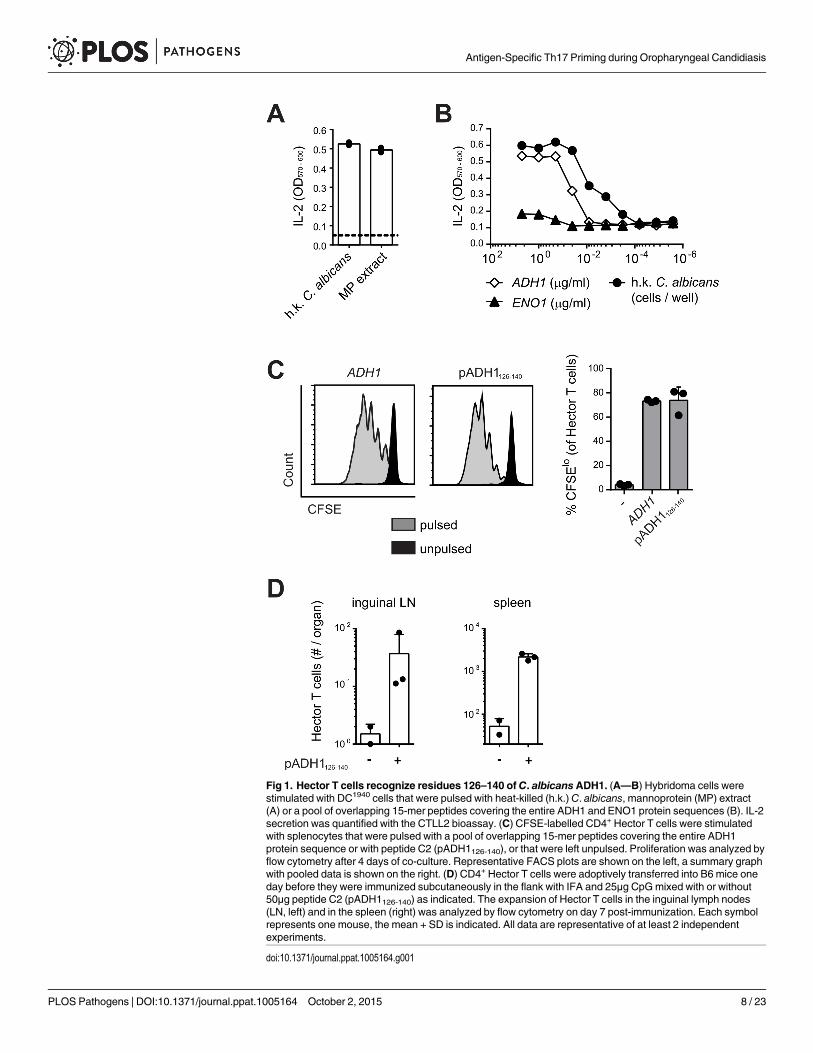
Fig 1. Hector T cells recognize residues 126–140 of C. albicansADH1. (A—B) Hybridoma cells werestimulated with DC1940 cells that were pulsed with heat-killed (h.k.) C. albicans, mannoprotein (MP) extract(A) or a pool of overlapping 15-mer peptides covering the entire ADH1 and ENO1 protein sequences (B). IL-2secretion was quantified with the CTLL2 bioassay. (C) CFSE-labelled CD4+ Hector T cells were stimulatedwith splenocytes that were pulsed with a pool of overlapping 15-mer peptides covering the entire ADH1protein sequence or with peptide C2 (pADH1126-140), or that were left unpulsed. Proliferation was analyzed byflow cytometry after 4 days of co-culture. Representative FACS plots are shown on the left, a summary graphwith pooled data is shown on the right. (D) CD4+ Hector T cells were adoptively transferred into B6 mice oneday before they were immunized subcutaneously in the flank with IFA and 25μg CpGmixed with or without50μg peptide C2 (pADH1126-140) as indicated. The expansion of Hector T cells in the inguinal lymph nodes(LN, left) and in the spleen (right) was analyzed by flow cytometry on day 7 post-immunization. Each symbolrepresents one mouse, the mean + SD is indicated. All data are representative of at least 2 independentexperiments.
doi:10.1371/journal.ppat.1005164.g001
Antigen-Specific Th17 Priming during Oropharyngeal Candidiasis
PLOS Pathogens | DOI:10.1371/journal.ppat.1005164 October 2, 2015 8 / 23
stimulated proliferation of Hector T cells to an extent that was comparable to that induced bythe peptide pool covering the entire ADH1 sequence (Fig 1C). Finally, the specificity of HectorT cells for the ADH1 peptide C2 was confirmed in vivo in mice adoptively transferred withCD4+ Hector T cells and immunized with the peptide admixed with CpG adjuvant (Fig 1D).
Peptide C2 mapped to residues 126–140 of ADH1 (pADH1126-140), corresponding tosequence GSFEQYATADAVQAA, and was predicted to have a good binding affinity to I-Ab
(IC50 = 605, SMM align method) [33]. In line with the high degree of conservation of theADH1 protein and in particular of the pADH1126-140 peptide sequence across Candida spp.,we found a comparable dose-dependent response of hybridoma 59.8 to different strains of C.albicans, C. dubliniensis, C. tropicalis, C. glabrata and C. krusei (S2 Fig). Moreover, the epitopewas also conserved in S. cerevisiae (S2 Fig), but not in other ascomycetes such as Aspergillus orthe dimorphic fungi. Thus, the Hector mouse is a source of T cells highly enriched in CD4+ Tcells specific for a novel ADH1-derived antigen and they provide a valuable tool for studying indetail and in an antigen-specific manner the adaptive immune response to C. albicans.
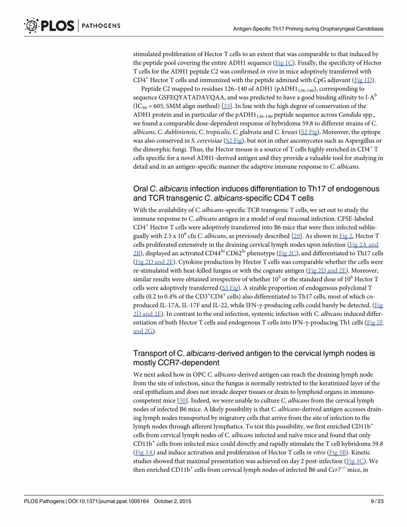
Oral C. albicans infection induces differentiation to Th17 of endogenousand TCR transgenic C. albicans-specific CD4 T cellsWith the availability of C. albicans-specific TCR transgenic T cells, we set out to study theimmune response to C. albicans antigen in a model of oral mucosal infection. CFSE-labeledCD4+ Hector T cells were adoptively transferred into B6 mice that were then infected sublin-gually with 2.5 x 10⁶ cfu C. albicans, as previously described [29]. As shown in Fig 2, Hector Tcells proliferated extensively in the draining cervical lymph nodes upon infection (Fig 2A and2B), displayed an activated CD44hi CD62lo phenotype (Fig 2C), and differentiated to Th17 cells(Fig 2D and 2E). Cytokine production by Hector T cells was comparable whether the cells werere-stimulated with heat-killed fungus or with the cognate antigen (Fig 2D and 2E). Moreover,similar results were obtained irrespective of whether 105 or the standard dose of 106 Hector Tcells were adoptively transferred (S3 Fig). A sizable proportion of endogenous polyclonal Tcells (0.2 to 0.4% of the CD3+CD4+ cells) also differentiated to Th17 cells, most of which co-produced IL-17A, IL-17F and IL-22, while IFN-γ-producing cells could barely be detected. (Fig2D and 2E). In contrast to the oral infection, systemic infection with C. albicans induced differ-entiation of both Hector T cells and endogenous T cells into IFN-γ-producing Th1 cells (Fig 2Fand 2G).
Transport of C. albicans-derived antigen to the cervical lymph nodes ismostly CCR7-dependentWe next asked how in OPC C. albicans-derived antigen can reach the draining lymph nodefrom the site of infection, since the fungus is normally restricted to the keratinized layer of theoral epithelium and does not invade deeper tissues or drain to lymphoid organs in immuno-competent mice [30]. Indeed, we were unable to culture C. albicans from the cervical lymphnodes of infected B6 mice. A likely possibility is that C. albicans-derived antigen accesses drain-ing lymph nodes transported by migratory cells that arrive from the site of infection to thelymph nodes through afferent lymphatics. To test this possibility, we first enriched CD11b+
cells from cervical lymph nodes of C. albicans infected and naïve mice and found that onlyCD11b+ cells from infected mice could directly and rapidly stimulate the T cell hybridoma 59.8(Fig 3A) and induce activation and proliferation of Hector T cells in vitro (Fig 3B). Kineticstudies showed that maximal presentation was achieved on day 2 post-infection (Fig 3C). Wethen enriched CD11b+ cells from cervical lymph nodes of infected B6 and Ccr7–/–mice, in
Antigen-Specific Th17 Priming during Oropharyngeal Candidiasis
PLOS Pathogens | DOI:10.1371/journal.ppat.1005164 October 2, 2015 9 / 23
Fig 2. Antigen-specific Th cell response to C. albicans oropharyngeal infection. (A, B) CFSE-labelled CD4+ Hector T cells were adoptively transferredone day prior to sublingual infection of B6 mice withC. albicans. Proliferation of Thy1.1+ CD4+ Vα2+ Hector cells was analyzed by flow cytometry on day 3post-infection. A representative FACS plot is shown in (A), quantification of Thy1.1+ CD4+ Vα2+ Hector cells in cervical lymph nodes is shown in (B). Eachsymbol represents an individual mouse, the mean + SD is indicated, data are pooled form 2 independent experiments. (C—E) CD4+ Hector T cells wereadoptively transferred one day prior to sublingual infection withC. albicans. In (C), Thy1.1+ CD4+ Vα2+ Hector cells in the cervical lymph nodes wereanalyzed on day 7 post-infection for expression of CD44 and CD62L. Representative FACS plots from 3 independent experiments are shown. In (D—E),cytokine production by endogenous CD3+ CD4+ Thy1.2+ T cells (left) and Thy1.1+ CD4+ Vα2+ Hector cells (right) on day 7 post-infection was analyzed byflow cytometry after re-stimulation with DC1940 cells pulsed with heat-killed (h.k.) C. albicans or pADH1126-140 as indicated. Representative FACS plots areshown in C and D, quantification of IL-17-producing cells is shown in (E). Each symbol represents an individual mouse, the mean + SD is indicated, data arepooled form 2 independent experiments. (F, G) B6 mice obtained an adoptive transfer of CD4+ Hector T cells one day prior to systemic infection with 5 x 10⁴
cfu C. albicans. Cytokine production by endogenous CD3+ CD4+ Thy1.2+ T cells (left) and Thy1.1+ CD4+ Vα2+ Hector cells (right) in the renal lymph nodes(left) and spleen (right) on day 7 post-infection was analyzed after re-stimulation with DC1940 cells pulsed with heat-killed (h.k.) C. albicans or pADH1126-140as indicated. Representative FACS plots are shown in (F), quantification of IFN-γ-producing cells is shown in (G). Each symbol represents an individualmouse, the mean + SD is indicated, data are pooled form 2 independent experiments.
doi:10.1371/journal.ppat.1005164.g002
Antigen-Specific Th17 Priming during Oropharyngeal Candidiasis
PLOS Pathogens | DOI:10.1371/journal.ppat.1005164 October 2, 2015 10 / 23
which migration of cells from the periphery to draining lymph nodes is severely impaired [34].Strikingly, activation of Hector T cells was strongly reduced (Fig 3D and 3E).
To further corroborate these data, we adoptively transferred CCR7-sufficient Hector T cellsinto Ccr7–/–or B6 mice prior to sublingual infection with C. albicans. Expression of CCR7 onHector T cells allows their normal entry into cervical lymph nodes via high endothelial venules.When lymph nodes were analyzed on day 7 post-infection, we found that Hector T cells differ-entiated into IL-17A-secreting effector cells in response to C. albicans in B6 mice, but theirexpansion and differentiation was strongly reduced in CCR7-deficient mice (Fig 3F), indicatingthat the delivery of C. albicans-derived antigen to the cervical lymph nodes was cell-associatedand dependent on CCR7-mediated cell trafficking in vivo. Attempts to visualize the cells thattake up C. albicans (either labeled with a fluorescent dye or engineered to express GFP) failedboth in the tongue or in the cervical lymph nodes of infected mice, presumably due to the factthat the majority of C. albicans hyphae, the predominant morphotype in infected oral tissue,remained extracellular and the ingested material was degraded rapidly by the phagocytosingcells.
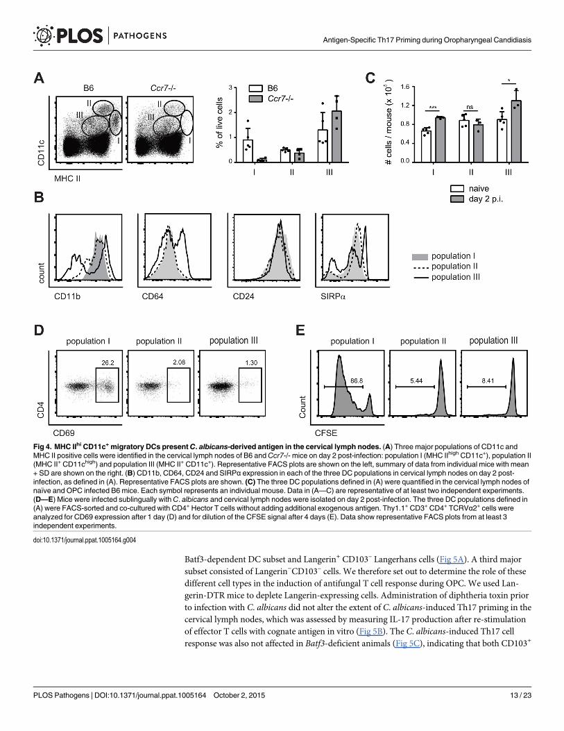
MHC IIhi CD11c+ migratory DCs present C. albicans-derived antigen inthe cervical lymph nodesThree major populations of DCs could be identified in cervical lymph nodes according to theexpression of MHC II and CD11c: Population I (MHC IIhigh CD11c+), population II(CD11chigh MCH II+), and population III (CD11cint MHC IIint) (Fig 4A). Population I and IIexpressed CD11b, CD24 and SIRP1α but not CD64, while population III was more heteroge-neous for expression of some of these markers (Fig 4B). When compared to the populationsfound in lymph nodes of Ccr7–/–mice, population II could be identified as lymph node residentDCs and population I as migratory DCs. Population III could also be identified as composedmainly of lymph node resident cells, since the number of CD11cint MHC IIint cells was notaffected in Ccr7–/–mice (Fig 4A). During the course of C. albicans infection, populations I andIII increased, while population II remained unchanged (Fig 4C).
To define the antigen-presenting capacity in OPC, we FACS-sorted to high purity the threeDC populations from cervical lymph nodes of C. albicans infected mice and directly testedtheir capacity to activate Hector T cells in vitro without addition of exogenous antigen. We alsotested Ly6G+ neutrophils isolated from the cervical lymph nodes of the same mice in the assay,since previous studies have shown that these cells can present antigen in certain conditions[35]. Hector T cells responded only very weekly, or not at all, to CD11chi MHC IIint DCs (pop-ulation II), CD11cint MHC IIint cells (population III) and Ly6G+ neutrophils. In contrast, theyrapidly upregulated CD69 and proliferated strongly when co-cultured with MHC IIhi CD11c+
migratory DCs (population I) (Fig 4D and 4E). In the assay, the T cell stimulatory capacity wasstrictly dependent on C. albicans-derived antigen, since DCs isolated from naïve animals wereunable to induce proliferation of Hector T cells. Together with the previous findings, these dataindicate that cells within the MHC IIhi CD11c+ migratory population can capture C. albicans-derived antigen from the peripheral tissue and transport them to the cervical lymph nodeswhere they are presented to CD4+ T cells. Consistent with a role of migratory DCs in the induc-tion of Th17 differentiation, cells within population I produced higher levels of IL-6 comparedto the other two DC populations in the cervical lymph nodes of OPC-infected mice (S4 Fig).
Antigen-Specific Th17 Priming during Oropharyngeal Candidiasis
PLOS Pathogens | DOI:10.1371/journal.ppat.1005164 October 2, 2015 11 / 23

Flt3L-dependent migratory DCs and monocyte-derived DCs presentC. albicans-derived antigenTo further dissect the heterogeneity of the MHC IIhi CD11c+ population in the cervicallymph nodes we performed staining with antibodies to Langerin and CD103. We detectedtwo small populations consisting of Langerin+ CD103+ cells, most likely belonging to the
Fig 3. Transport ofC. albicans-derived antigen to the cervical lymph nodes is CCR7-dependent. (A) Cervical lymph node cells that were isolated fromnaïve or from sublingually infected mice on day 2 post-infection and either enriched for CD11b+ cells or left non-enriched were co-cultured with the C.albicans-specific T cell hybridoma cells. IL-2 production as a read-out for hybridoma activation was quantified by CTLL-2 bioassay. (B) Cervical lymph nodecells were isolated from naïve mice or from sublingually infected mice on day 2 p.i., enriched for CD11b+ cells, and co-cultured with CFSE-labeled CD4+
Hector T cells. CFSE-dilution of the Thy1.1+ CD3+ CD4+ Hector cells was analyzed after 4 days. (C) CD11b+ cells were enriched from cervical lymph nodesat different time points after infection as indicated and analyzed for antigen presentation as described in (A). (D—E) cervical lymph node cells were isolatedfrom sublingually infected B6 or Ccr7-/- mice on day 2 post-infection, enriched for CD11b+ cells and co-cultured with CD4+ Hector T cells. CD69 expression(D) and CFSE dilution (E) of Thy1.1+ CD3+ CD4+ TCRVα2+ cells was analyzed on day 1 and day4 respectively. In (B), (D) and (E), representative FACS plotsare shown on the left; summary of data from individual mice with mean + SD are shown on the right. Data are from individual experiments that arerepresentative of at least two independent experiments each. (F)CD4+ Hector T cells were adoptively transferred into B6 andCcr7-/- mice one day prior tosublingual infection. Cytokine production by Thy1.1+ CD3+ CD4+ Hector cells in the cervical lymph nodes was analyzed on day 7 post-infection after re-stimulation with DC1940 cells pulsed with pADH1126-140, heat-killed (h.k.) C. albicans or left unpulsed as indicated. Symbols represent individual mice pooledfrom 2 independent experiments, the mean + SD is indicated.
doi:10.1371/journal.ppat.1005164.g003
Antigen-Specific Th17 Priming during Oropharyngeal Candidiasis
PLOS Pathogens | DOI:10.1371/journal.ppat.1005164 October 2, 2015 12 / 23
Batf3-dependent DC subset and Langerin+ CD103– Langerhans cells (Fig 5A). A third majorsubset consisted of Langerin−CD103– cells. We therefore set out to determine the role of thesedifferent cell types in the induction of antifungal T cell response during OPC. We used Lan-gerin-DTR mice to deplete Langerin-expressing cells. Administration of diphtheria toxin priorto infection with C. albicans did not alter the extent of C. albicans-induced Th17 priming in thecervical lymph nodes, which was assessed by measuring IL-17 production after re-stimulationof effector T cells with cognate antigen in vitro (Fig 5B). The C. albicans-induced Th17 cellresponse was also not affected in Batf3-deficient animals (Fig 5C), indicating that both CD103+
Fig 4. MHC IIhi CD11c+ migratory DCs presentC. albicans-derived antigen in the cervical lymph nodes. (A) Three major populations of CD11c andMHC II positive cells were identified in the cervical lymph nodes of B6 andCcr7-/- mice on day 2 post-infection: population I (MHC IIhigh CD11c+), population II(MHC II+ CD11chigh) and population III (MHC II+ CD11c+). Representative FACS plots are shown on the left, summary of data from individual mice with mean+ SD are shown on the right. (B) CD11b, CD64, CD24 and SIRPα expression in each of the three DC populations in cervical lymph nodes on day 2 post-infection, as defined in (A). Representative FACS plots are shown. (C) The three DC populations defined in (A) were quantified in the cervical lymph nodes ofnaïve and OPC infected B6 mice. Each symbol represents an individual mouse. Data in (A—C) are representative of at least two independent experiments.(D—E) Mice were infected sublingually withC. albicans and cervical lymph nodes were isolated on day 2 post-infection. The three DC populations defined in(A) were FACS-sorted and co-cultured with CD4+ Hector T cells without adding additional exogenous antigen. Thy1.1+ CD3+ CD4+ TCRVα2+ cells wereanalyzed for CD69 expression after 1 day (D) and for dilution of the CFSE signal after 4 days (E). Data show representative FACS plots from at least 3independent experiments.
doi:10.1371/journal.ppat.1005164.g004
Antigen-Specific Th17 Priming during Oropharyngeal Candidiasis
PLOS Pathogens | DOI:10.1371/journal.ppat.1005164 October 2, 2015 13 / 23

and CD103– Langerin-expressing DCs were not essential for the induction of antifungal Th17immunity in the oral mucosa.
The large Langerin−CD103– subset of migratory DCs in the cervical lymph node appearedto be phenotypically homogeneous for all markers analyzed (Fig 4B). However, the subset maystill comprise phylogenetically and functionally distinct cell types, including Flt3-dependentconventional DCs and monocyte-derived DCs that are Flt3-independent but dependent onCsf1R signaling for differentiation from inflammatory monocyte [13]. Consistent with thisnotion, we found that in Flt3l-/- mice, migratory MHC IIhi CD11c+ DCs, which include thelarge population of Langerin−CD103– cells, were strongly reduced in Flt3l-/- mice compared toB6 mice (Fig 6A). Furthermore, the CD11b+ cervical lymph node cells isolated from infectedFlt3l-/- mice were strongly impaired in their ability to induce CD69 upregulation and prolifera-tion of Hector T cells in vitro (Fig 6B and 6C). This became also clear when the APC fractionwas purified by FACS sorting: antigen presentation by Flt3L-dependent migratory DCsstrongly promoted the activation of CD4+ Hector T cells in vitro, it was however not essentialfor the response (Fig 6D), indicating that Flt3L-independent migratory DCs are also involved.Thus, Flt3L-dependent migratory DCs appear to be an important source of antigen in cervicallymph nodes for T cell activation in response to C. albicans oral infection.
To assess the role of the Flt3L-independent monocyte-derived DCs we took advantage ofthe notion that these cells express CCR2 [12]. First, we noticed that CCR2+ monocytes, whichare also Ly6Chi and CD11bhi, rapidly accumulated in the infected tongue (S5 Fig). From day 2post-infection, a proportion of CCR2+ monocytes up-regulated CD11c and MHC II anddown-regulated Ly6C, indicating that they differentiated to DCs (S5 Fig). Second, CCR2+
monocytes were also found in the cervical lymph nodes of infected mice (Fig 6E), and some ofthem differentiated to DCs, as observed in the peripheral tissue (Fig 6E and 6F). We alsoobserved that monocyte-derived DCs with the strongest expression of MHC II and CD11c hadreduced levels of CCR2 (Fig 6F). Because of these characteristic markers, these cells fell in thegate of migratory MHChi CD11chi DCs (population I) and were hardly distinguishable fromother migratory DCs such as Flt3L-dependent DCs.
To directly assess the antigen presentation capacity of the CCR2+-derived migratory DCsduring OPC, we separated MHC IIhi CD11c+ DCs from the cervical lymph nodes of infectedmice into three fractions according to their CCR2 expression and exposed them to CD4+ Hec-tor T cells in vitro. We observed T cell activation with all the subsets, irrespective of their degreeof CCR2 expression, suggesting that both monocyte-derived DCs (included mainly in the
Fig 5. Langerin+ cells are not essential for efficient Th17 priming. (A) Expression of Langerin and CD103 was analyzed in population I (MHC IIhigh
CD11c+) on day 2 p.i.. (B, C) cervical lymph node cells were isolated from Langerin-DTRmice that were treated with diphtheria toxin (DT) or not as indicated(B) or from Batf3-/- and littermate controls (C) on day 7 p.i. and re-stimulated in vitro with DC1940 pulsed with heat killed (h.k.) C. albicans or left unpulsed for5h in presence of Brefeldin A. IL-17A production by CD3+ CD4+ cells was analyzed by intracellular cytokine staining. Symbols represent individual mice, themean + SD is indicated. All data are representative of at least 2 independent experiments.
doi:10.1371/journal.ppat.1005164.g005
Antigen-Specific Th17 Priming during Oropharyngeal Candidiasis
PLOS Pathogens | DOI:10.1371/journal.ppat.1005164 October 2, 2015 14 / 23

CCR2hi and CCR2int fractions) as well as CCR2lo DCs, most likely reflecting Flt3L-dependentDCs, could directly present C. albicans-derived antigen (Fig 6G).
Fig 6. Flt3L-dependent migratory DCs andmonocyte-derived DCs both presentC. albicans derived antigen. (A) Cervical lymph node cells of B6 orFlt3l-/- mice were analyzed on day 2 post-infection. Representative FACS plots from individual mice and quantification of MHC IIhigh CD11c+ (population I),MHC II+ CD11chigh (population II) and MHC II+ CD11c+ cells (population III) from one of two independent experiments are shown. (B, C) Cervical lymph nodecells were isolated from naïve B6 mice and from infected B6 and Flt3l-/- mice on day 2 post-infection, enriched for CD11b+ cells and co-cultured with CD4+
Hector T cells. Thy1.1+ CD3+ CD4+ TCRVα2+ cells were analyzed for CD69 expression after 1 day (B) and for proliferation after 4 days, respectively (C).Representative plots are shown on the left, summary of data from individual mice with mean + SD are shown on the right. Each symbol represents onemouse. Data are representative of 2 independent experiments. (D) As in B, but MHC IIhigh CD11c+ migratory DCs were isolated by FACS-sorting from thecervical lymph nodes of infected B6 and Flt3l-/- mice. (E—F) CCR2+ CD11b+ cells in the cervical lymph nodes of naïve and infected B6 mice on day 2 post-infection were analyzed for the expression of MHC II and CD11c (D). Three distinct subsets of CCR2+ CD11b+ cells (MHC IIlo/int Ly6C+ (Q1), MHC IIint Ly6C-
(Q2) and MHC IIhigh Ly6C- (Q3)) from infected mice were further analyzed for the expression of CCR2 and CD11c (E). Representative FACS plots are shown.(G) Cervical lymph nodes were isolated from infected B6 mice on day 2 post-infection. CCR2hi, CCR2int and CCR2lo subsets within the MHC IIhigh CD11c+
population I were FACS-sorted and co-cultured with CD4+ Hector T cells for 1 day. Thy1.1+ CD3+ CD4+ TCRVα2+ cells were then analyzed for CD69expression. Representative FACS plots are shown, Data are representative of 2 independent experiments.
doi:10.1371/journal.ppat.1005164.g006
Antigen-Specific Th17 Priming during Oropharyngeal Candidiasis
PLOS Pathogens | DOI:10.1371/journal.ppat.1005164 October 2, 2015 15 / 23

Monocyte-derived and Flt3L-dependent migratory DCs complementeach other for Th17 priming during OPCTo evaluate the relative contribution of CCR2-dependent and Flt3L-dependent migratory DCsubsets in T cell priming during OPC in vivo, we examined the induction of C. albicans-specificTh17 cell in the cervical lymph nodes of OPC infected Flt3l-/- and Ccr2-/- mice on day 7 post-infection. While the response was not affected by the absence of Flt3L-dependent DCs (Fig7A), the frequency of IL-17-secreting C. albicans-specific CD4+ T was strongly reduced inCcr2-/- mice compared to B6 controls (Fig 7B). Similarly to the endogenous response, adop-tively transferred Hector T cells were also strongly impaired in their capacity to differentiateinto IL-17-producing effector cells in Ccr2-/- mice (S6 Fig). Alterations in DC and monocytepopulations may impair the innate control of the fungus; and an increased fungal burden canaugment the extent of the T cell response. Therefore, to exclude the possibility that the resultswere influenced by differences in the available amount of antigen between the experimentalgroups, we treated the mice with Fluconazole from day 2 post-infection, a time point when thefungal burden was high and similar in all groups of mice (S7 Fig). This resulted in comparableweight recovery (S7 Fig) and clearance of the fungus to undetectable levels within the periodof the experiment, which is indicative of comparable infection control in all experimentalgroups.
Fig 7. Flt3L-dependent migratory DCs andmonocyte-derived DCs complement each other for Th17 priming during OPC. (A) Cervical lymph nodeswere isolated from B6 and Flt3l-/- mice on day 7 post-infection and re-stimulated with heat-killed (h.k.) C. albicans or medium only. IL-17A production byendogenous CD3+ CD4+ cells was then analyzed by flow cytometry. (B) B6 andCcr2-/- mice were infected sublingually and IL-17 production by endogenousCD3+ CD4+ cells was analyzed on day 7 post-infection as described in (A). (C) B6 mice were treated with anti-CSF1R or left untreated prior to sublingualinfection withC. albicans. IL-17 production by endogenous CD3+ CD4+ cervical lymph node cells was analyzed on day 7 post-infection as described in (A).(D) B6 and Flt3l-/- mice were treated with anti-CSF1R or left untreated prior to sublingual infection with C. albicans. IL-17 production by endogenous CD3+
CD4+ cervical lymph node cells was analyzed on day 7 post-infection as described in (A). Each symbol represents one mouse, the mean + SD for each groupis shown.
doi:10.1371/journal.ppat.1005164.g007
Antigen-Specific Th17 Priming during Oropharyngeal Candidiasis
PLOS Pathogens | DOI:10.1371/journal.ppat.1005164 October 2, 2015 16 / 23

To further support the critical role of monocyte-derived DCs in Th17 priming during OPC,we made use of a CSF1R-specific antibody, which was shown to block the differentiation ofmonocytes into monocyte-derived DCs [13] and led to a significant reduction of CCR2+ cellswithin the migratory DC population (S8 Fig). Th17 induction in response to OPC was consid-erably lower in mice that were treated with this antibody prior to and during infection than inuntreated controls (Fig 7C). However, similarly to what we observed in the Ccr2-/- mice, thereduction in Th17 response in mice treated with anti-CSF1R antibody was only about 50%compared to the response in controls. This could be explained by inefficient/incomplete block-ade with the antibody, while in Ccr2-/- mice, monocyte trafficking may be partially compen-sated by CCR2-independent mechanisms. The impact of monocyte-derived DCs on Th17priming during OPC may thus be underestimated in our system. An explanation for the partialeffect could also be the involvement of other subsets of APCs that might even compensate to acertain extent for the absence of monocyte-derived DCs to promote Th17 priming in anti-CSF1R treated or CCR2-deficient mice. Because Flt3L-dependent DCs contribute to the pre-sentation of C. albicans-derived antigen, at least when presentation was assessed in our in vitroassay (Fig 6B and 6C), we tested the hypothesis that they may play a relevant role in Th17induction in absence of monocyte-derived DCs. For this we analyzed again the Th17 responseto C. albicans in Flt3L-deficient animals and we included a group, in which we blocked theCSF1R prior to infection. All animals were again treated with Fluconazole from day 2 post-infection to minimalize differences in fungal burden between the experimental groups thatcould affect T cell priming (S7 Fig). This resulted in a nearly complete blockade of the C. albi-cans-specific Th17 response (Fig 7D). Together, our results demonstrate a contribution of dif-ferent subsets of APCs to the Th17 response during OPC and highlight the critical role ofmonocyte-derived DCs and to a lesser extent Flt3L-dependent DCs in this process. WhileFlt3L-dependent DCs were not essential under normal conditions when monocyte-derivedDCs were present, they did critically contribute if these primary APCs were absent.
DiscussionTh17 cells have emerged as a key component of protective immunity against mucocutaneouscandidiasis. In recent years, Candida-specific T cells have been studied in detail in humans andin mice leading to the identification of Th17-inducing innate pathways [9,10,36–40], revealingCandida-derived antigenic epitopes [31,41–43], and providing detailed insights into theirclonal diversity [44]. Here we provide new insights in the mechanism that regulates fungus-specific T cell immunity. We generated a Candida-specific TCR transgenic mouse model toprovide a tool for the detection, enumeration and characterization of antigen-specific T cellsduring infection in vivo. Using this new tool we identified the cellular players that present fun-gal antigen and instruct the Th17 response against C. albicans during oropharyngeal infection.CCR2-dependent monocytes-derived DCs rapidly accumulate in the oral mucosa during infec-tion and directly present Candida-derived antigen in the draining lymph nodes. However, theyare partially redundant for Th17 induction during OPC and complemented by tissue-residentFlt3-dependent migratory DCs for antifungal T cell priming in vivo. Together, this reveals acomplex regulation of the antifungal T cell response by a multifaceted network MNPs in theoral mucosa.
T cells of the newly established Hector mouse model are responsive to a native epitope of C.albicans derived from the metabolic enzyme ADH1 that localizes to the fungal cell wall and isalso abundant in biofilms [45]. ADH1 was reported previously to have immunogenic proper-ties because it bears some structural homology to integrins and can mediate adherence to extra-cellular matrix [46], and ADH1-specific antibodies were detected in Candida-exposed
Antigen-Specific Th17 Priming during Oropharyngeal Candidiasis
PLOS Pathogens | DOI:10.1371/journal.ppat.1005164 October 2, 2015 17 / 23

individuals and mice [47,48]. Here we show that ADH1 determines T cell specificity ADH1 ishighly conserved in diverse species of the genus Candida and in other Saccharomycotina. Theshort stretch of amino acids that define the T cell epitope identified here are 100% conserved inmultiple clinically relevant species of Candida including C. albicans, C. dubliniensis and C.glabrata. The Hector mouse model thus offers new opportunities for investigating T cellresponses against distinct species of Candida. Here we explored the regulation of C. albicans-specific T cells during oropharyngeal infection.
The oral cavity is the entry port and the first site of contact with the host for a multitude ofmicrobes. Diverse DC subsets including tissue resident DCs as well as inflammatory DCs accu-mulating in the oral mucosa in response to infection and inflammation orchestrate T cell out-comes in response to these microbes. DC subsets in the oral mucosa are related to thosedescribed in other barrier tissues. However, their relative abundance and distribution as well astheir putative function partially differs. Similarly to the skin, Langerhans cells are present inthe oral mucosa, but in contrast to what was reported for an epicutaneous model of candidiasis,in which Langerhans cells were shown to be necessary and sufficient for antifungal Th17 prim-ing [49], they are not essential for Th17 induction during OPC. Moreover, while Batf3-depen-dent DCs promote Th1 and inhibit Th17 cell differentiation during epicutaneous candidiasis[49], absence of Batf3-dependent cells does not affect the T cell response during OPC. Whethermonocyte-derived DCs are involved in the T cell response against C. albicans in the skin hadnot been examined to our knowledge. The discrepancies observed between the two distinct tis-sues are likely explained by site-specific differences, including the important difference inabundance of Langerhans cells between the skin and the tongue, but may also be influenced bythe different mouse models of Langerhans cell deficiency used in the two studies. In the oralmucosa, Langerhans cells seem to be mainly tolerogenic [17]. The observed differences in regu-lation of T cell priming at different sites underline the compartmentalization of the immuneresponse against one and the same fungal organism in different tissue environments. Thiseffect is even more pronounced when antifungal immunity in barrier tissues is compared withthe response elicited during systemic candidiasis, which is dominated by type 1 immunity [50].Whether the quality of the Th17 response primed in the oral mucosa versus the skin may differ,remains to be established. In the oral mucosa, C. albicans-specific T cells form a stable long-lived population of memory Th17 cells that efficiently responds to secondary infection [51].Similarly, Th17 cells primed in the skin have recently been shown to enhance protection fromepicutaneous fungal challenge with C. albicans [50].
The DCs presenting C. albicans-derived antigen to T cells in the cervical lymph nodes ofOPC-infected mice belong to the CCR7-dependent migratory population. Distinguishingmonocyte-derived DCs from other migratory DC subsets in the cervical lymph nodes of C.albicans-infected mice on the basis of their phenotype is difficult as they gradually lose theircharacteristic expression of Ly6C and CCR2. We therefore used an approach to separate themon the basis of their phylogenetic origin. Direct assessment of antigen presentation by migra-tory subsets revealed that both monocyte-derived DCs and Flt3L-dependent DCs presented C.albicans-derived antigen in the cervical lymph nodes of infected mice. They were howeverredundant for Th17 differentiation in vivo. Interrupting the differentiation of monocyte intoDCs in absence of Flt3-dependent DCs almost abolished T cell priming, supporting the notionthat other Flt3-independent DCs such as Langerhans cells are not required and not sufficientfor Th17 induction during OPC.
Monocyte-derived DCs gain increasing attention for their role as professional APC to pro-mote T cell responses [52–54] including those elicited by fungi such as Aspergillus fumigatus orBlastomyces dermatitidis [55,56]. In addition to priming adaptive immunity, CCR2-dependentcells also contribute to innate immunity against fungi including C. albicans and A. fumigatus
Antigen-Specific Th17 Priming during Oropharyngeal Candidiasis
PLOS Pathogens | DOI:10.1371/journal.ppat.1005164 October 2, 2015 18 / 23

[57,58]. The mechanism by which these cells contribute to acute protection has not been fullyestablished. During OPC, innate lymphoid cells and innate lymphocytes provide importantsources of IL-17 during the early phase of infection [59,60]. Whether monocytes and/or mono-cyte-derived DCs impact on the regulation of innate IL-17 secretion has not yet been estab-lished. Here we show that monocyte-dependent DCs, together with Flt3L-dependent DCs,orchestrate the antigen-specific T cell response to a clinically highly relevant fungal pathogen,and may thus have implications for potential future immunotherapeutic approaches and vac-cine development against mucosal candidiasis.
Supporting InformationS1 Fig. Characterization of Hector TCR transgenic mice. (A—B) CD3+ CD4+ TCR β+ T cellsfrom the spleen, cervical lymph nodes and thymus of naïve Hector and transgene-negativecontrol mice (B6) were analyzed by flow cytometry for TCR Vα2 and Vβ4 expression. Repre-sentative FACS plots of splenic T cells are shown in A. Summary graphs with 6 individual miceare shown in B. (C) The proportion of CD4+ and CD8+ cells within the CD3+ TCRβ+ T cellpopulation was analyzed in the spleen, cervical lymph nodes and thymus of naïve Hector andtransgene-negative control mice (B6). For the thymus, the frequency of CD4+ CD8+ doublepositive (DP) T cells is also indicated. Each symbol represents an individual mouse, themean ± SD is indicated. Data are pooled from two independent experiments. Note that the dif-ferent ratio of CD4+: CD8+ T cells in spleen and lymph nodes of Hector mice is expected dueto allelic exclusion of non-transgenic TCRβ genes and preferential lineage decision towardsCD4+ T cells, as observed in other TCR transgenic mice [61,62].(EPS)
S2 Fig. Identification and characterization of the epitope recognized by Hector T cells inCandida spp. and S. cerevisiae. (A) Duplicate wells of hybridoma cells were stimulated withDC1940 cells that were pulsed with individual 15-mer peptides and IL-2 secretion by the hybrid-oma cells was quantified with the CTLL2 bioassay. The horizontal line indicates the detectionlimit of the assays (CTLL2 cells without IL-2 stimulation). (B) CFSE-labelled CD4+ Hector Tcells were stimulated with splenocytes that were pulsed with decreasing concentrations of theC2, C3 and D1 peptide, respectively, and analyzed for proliferation by flow cytometry after 4days of co-culture. (C) Hybridoma cells were cultured with DC1940 cells that were pulsed withdecreasing amounts of heat-killed yeast cells and IL-2 secretion by the hybridoma cells wasquantified by the CTLL2 bioassay. C. a., C. albicans; C. d., C. dubliniensis; C. t., C. tropicalis; C.g., C. glabrata; C. k., C. krusei; S. c., S. cerevisiae. Strain numbers refer to our internal strain col-lection. C. albicans strains SC5314 was included in all panels as a reference.(EPS)
S3 Fig. Th17 differentiation of C. albicans-specific Hector T cells during OPC. 105 or 106
CD4+ Hector T cells were adoptively transferred, as indicated, one day prior to sublingual infectionwith C. albicans. Cytokine production by Thy1.1+ CD4+ Vα2+ Hector cells on day 7 post-infectionwas analyzed by flow cytometry after re-stimulation with DC1940 cells pulsed with heat-killed (h.k.) C. albicans or pADH1126-140 as indicated. Percentage (A–B) and absolute numbers per mouse(C–D) of IL-17- (A, C) and IFN-γ-producing cells (B, D) are shown. Each symbol represents anindividual mouse, the mean is indicated, data are pooled form 2 independent experiments.(EPS)
S4 Fig. IL-6 is expressed preferentially by the MHC IIhi CD11c+ migratory DC population.Cervical lymph node cells from OPC infected mice were analyzed for IL-6 expression by intracel-lular cytokine staining and FACS analysis. Populations I, II and III were identified as indicated in
Antigen-Specific Th17 Priming during Oropharyngeal Candidiasis
PLOS Pathogens | DOI:10.1371/journal.ppat.1005164 October 2, 2015 19 / 23
Fig 4. Numbers indicate the mean ± SD of IL-6+ cells within each population. n = 5.(EPS)
S5 Fig. Monocytes and monocyte-derived DCs accumulate rapidly in the oral mucosa. (A)Accumulation of Ly6G+ CCR2- neutrophils and CCR2+ Ly6G- monocytes in the tongue was ana-lyzed at the indicated time points post-infection. Cells are pre-gated on CD11b+ cells. A represen-tative plot from day 1 post-infection is shown on the left, and the summary from three individualmice of one representative experiment is shown on the right. (B) CCR2+ Ly6G- cells in the ton-gue were analyzed for expression of MHC II and CD11c on indicated time points post-infection.A representative plot from day 1 post-infection is shown on the left, and quantification of MHCII+ CD11c-, MHC II+ CD11c+ andMHC II- CD11c- subpopulations are shown on the right. Dataare mean + SD of 3 independent mice and representative of 2 independent experiments.(EPS)
S6 Fig. The response of Hector T cells is strongly impaired in Ccr2-/- mice. (A—B) CD4+
Hector T cells were adoptively transferred into Ccr2-/- and B6 control mice one day prior tosublingually infection with C. albicans. Cervical lymph node cells were isolated on day 7 post-infection, re-stimulated with DC1940 cells pulsed with heat-killed (h.k.) C. albicans orpADH1126-140 peptide or left unpulsed and IL-17A and IFN-γ production by CD3+ CD4+ Thy1.1+ TCRVα2+ Hector T cells was analyzed by flow cytometry. Representative FACSplots are shown in (A), the summary of data from individual mice with mean + SD is shown in(B).(EPS)
S7 Fig. OPC-infected mice display high fungal burden on day 2 and normal weight recoverywithin 5 to 7 days post-infection. (A) Fungal burden in the tongue of OPC-infected B6,Ccr2-/- and Flt3l-/- mice as well as B6 and Flt3l-/- mice treated with anti-CSF1R antibody onday 2 post-infection. The dotted line indicates the detection limit. (B—E) Weight curves fromB6 and Flt3l-/- mice (B), B6 and Ccr2-/- mice (C), B6 mice treated or not with anti-CSF1R (D),and B6 and Flt3l-/- mice treated or not with anti-CSF1R (E) that were included in the experi-ments shown in Fig 7A–7D.(EPS)
S8 Fig. B6 mice were treated with anti-CSF1R or left untreated prior to sublingual infectionwith C. albicans, as described in Fig 7C. The CCR2+ subset within the MHC-IIhi migratoryDC population was analyzed on day 2 post-infection.(EPS)
AcknowledgmentsThe authors would like to thankMathias Heikenwälder, Lubor Borsig, Björn Clausen, KordulaKautz-Neu andMarkus Manz for mice; Cristina Corti-Fragoso and Orlando Petrini for Candidaisolates; Melanie Greter for providing the anti-CSF1R antibody, the staff of the Rodent Center HCIand the Laboratory Animal Service Center for animal husbandry; FranziskaWagen for technicalassistance; Annette Schütz of the ETH Zurich FACS core facility for technical assistance, NicoleTorti, Annette Oxenius and members of the LeibundGut-lab for helpful advice and discussions.
Author ContributionsConceived and designed the experiments: KTW AG SLL. Performed the experiments: KTWAG FRK TR. Analyzed the data: KTWAG FRK. Contributed reagents/materials/analysis tools:SB FS. Wrote the paper: KTW AG FS SLL.
Antigen-Specific Th17 Priming during Oropharyngeal Candidiasis
PLOS Pathogens | DOI:10.1371/journal.ppat.1005164 October 2, 2015 20 / 23
References1. Brown GD, Denning DW, Gow NA, Levitz SM, Netea MG,White TC. Hidden killers: human fungal infec-
tions. Sci Transl Med. 2012; 4: 165rv113.
2. Li X, Lei L, Tan D, Jiang L, Zeng X, Dan H, et al. Oropharyngeal Candida colonization in human immu-nodeficiency virus infected patients. APMIS. 2013; 121: 375–402. doi: 10.1111/apm.12006 PMID:23030258
3. Acosta-Rodriguez EV, Rivino L, Geginat J, Jarrossay D, Gattorno M, Lanzavecchia A, et al. Surfacephenotype and antigenic specificity of human interleukin 17-producing T helper memory cells. NatImmunol. 2007; 8: 639–646. PMID: 17486092
4. Puel A, Cypowyj S, Marodi L, Abel L, Picard C, Casanova JL. Inborn errors of human IL-17 immunityunderlie chronic mucocutaneous candidiasis. Curr Opin Allergy Clin Immunol. 2012; 12: 616–622. doi:10.1097/ACI.0b013e328358cc0b PMID: 23026768
5. Rivera A, Ro G, Van Epps HL, Simpson T, Leiner I, Sant'Angelo DB, et al. Innate immune activationand CD4+ T cell priming during respiratory fungal infection. Immunity. 2006; 25: 665–675. PMID:17027299
6. Wuthrich M, Hung CY, Gern BH, Pick-Jacobs JC, Galles KJ, Filutowicz HI, et al. A TCR transgenicmouse reactive with multiple systemic dimorphic fungi. J Immunol. 2011; 187: 1421–1431. doi: 10.4049/jimmunol.1100921 PMID: 21705621
7. Wuthrich M, Ersland K, Pick-Jacobs JC, Gern BH, Frye CA, Sullivan TD, et al. Limited model antigenexpression by transgenic fungi induces disparate fates during differentiation of adoptively transferred Tcell receptor transgenic CD4+ T cells: robust activation and proliferation with weak effector function dur-ing recall. Infect Immun. 2012; 80: 787–797. doi: 10.1128/IAI.05326-11 PMID: 22124658
8. Joffre O, Nolte MA, Sporri R, Reis e Sousa C. Inflammatory signals in dendritic cell activation and theinduction of adaptive immunity. Immunol Rev. 2009; 227: 234–247. doi: 10.1111/j.1600-065X.2008.00718.x PMID: 19120488
9. LeibundGut-Landmann S, Gross O, Robinson MJ, Osorio F, Slack EC, Tsoni SV, et al. Syk- andCARD9-dependent coupling of innate immunity to the induction of T helper cells that produce interleu-kin 17. Nat Immunol. 2007; 8: 630–638. PMID: 17450144
10. Saijo S, Ikeda S, Yamabe K, Kakuta S, Ishigame H, Akitsu A, et al. Dectin-2 recognition of alpha-man-nans and induction of Th17 cell differentiation is essential for host defense against Candida albicans.Immunity. 2010; 32: 681–691. doi: 10.1016/j.immuni.2010.05.001 PMID: 20493731
11. Chow A, Brown BD, Merad M. Studying the mononuclear phagocyte system in the molecular age. NatRev Immunol. 2011; 11: 788–798. doi: 10.1038/nri3087 PMID: 22025056
12. Serbina NV, Jia T, Hohl TM, Pamer EG. Monocyte-mediated defense against microbial pathogens.Annu Rev Immunol. 2008; 26: 421–452. doi: 10.1146/annurev.immunol.26.021607.090326 PMID:18303997
13. Greter M, Helft J, Chow A, Hashimoto D, Mortha A, Agudo-Cantero J, et al. GM-CSF controls nonlym-phoid tissue dendritic cell homeostasis but is dispensable for the differentiation of inflammatory den-dritic cells. Immunity. 2012; 36: 1031–1046. doi: 10.1016/j.immuni.2012.03.027 PMID: 22749353
14. Merad M, Sathe P, Helft J, Miller J, Mortha A. The dendritic cell lineage: ontogeny and function of den-dritic cells and their subsets in the steady state and the inflamed setting. Annu Rev Immunol. 2013; 31:563–604. doi: 10.1146/annurev-immunol-020711-074950 PMID: 23516985
15. Hoeffel G, Wang Y, Greter M, See P, Teo P, Malleret B, et al. Adult Langerhans cells derive predomi-nantly from embryonic fetal liver monocytes with a minor contribution of yolk sac-derived macrophages.J Exp Med. 2012; 209: 1167–1181. doi: 10.1084/jem.20120340 PMID: 22565823
16. Merad M, Manz MG, Karsunky H, Wagers A, Peters W, Charo I, et al. Langerhans cells renew in theskin throughout life under steady-state conditions. Nat Immunol. 2002; 3: 1135–1141. PMID:12415265
17. Hovav AH. Dendritic cells of the oral mucosa. Mucosal Immunol. 2014; 7: 27–37. doi: 10.1038/mi.2013.42 PMID: 23757304
18. Forster R, Schubel A, Breitfeld D, Kremmer E, Renner-Muller I, Wolf E, et al. CCR7 coordinates the pri-mary immune response by establishing functional microenvironments in secondary lymphoid organs.Cell. 1999; 99: 23–33. PMID: 10520991
19. Hildner K, Edelson BT, PurthaWE, Diamond M, Matsushita H, KohyamaM, et al. Batf3 deficiencyreveals a critical role for CD8alpha+ dendritic cells in cytotoxic T cell immunity. Science. 2008; 322:1097–1100. doi: 10.1126/science.1164206 PMID: 19008445
20. Kuziel WA, Morgan SJ, Dawson TC, Griffin S, Smithies O, Ley K, et al. Severe reduction in leukocyteadhesion and monocyte extravasation in mice deficient in CC chemokine receptor 2. Proc Natl AcadSci U S A. 1997; 94: 12053–12058. PMID: 9342361
Antigen-Specific Th17 Priming during Oropharyngeal Candidiasis
PLOS Pathogens | DOI:10.1371/journal.ppat.1005164 October 2, 2015 21 / 23
21. McKenna HJ, Stocking KL, Miller RE, Brasel K, De Smedt T, Maraskovsky E, et al. Mice lacking flt3ligand have deficient hematopoiesis affecting hematopoietic progenitor cells, dendritic cells, and naturalkiller cells. Blood. 2000; 95: 3489–3497. PMID: 10828034
22. Bennett CL, van Rijn E, Jung S, Inaba K, Steinman RM, Kapsenberg ML, et al. Inducible ablation ofmouse Langerhans cells diminishes but fails to abrogate contact hypersensitivity. J Cell Biol. 2005;169: 569–576. PMID: 15897263
23. Kruisbeek AM. Production of mouse T cell hybridomas. Curr Protoc Immunol. 2001; Chapter 3: Unit 314. doi: 10.1002/0471142735.im0314s24 PMID: 18432780
24. Yoshida R, Yoshioka T, Yamane S, Matsutani T, Toyosaki-Maeda T, Tsuruta Y, et al. A newmethod forquantitative analysis of the mouse T-cell receptor V region repertoires: comparison of repertoiresamong strains. Immunogenetics. 2000; 52: 35–45. PMID: 11132155
25. Pannetier C, Cochet M, Darche S, Casrouge A, Zoller M, Kourilsky P. The sizes of the CDR3 hypervari-able regions of the murine T-cell receptor beta chains vary as a function of the recombined germ-linesegments. Proc Natl Acad Sci U S A. 1993; 90: 4319–4323. PMID: 8483950
26. Kouskoff V, Signorelli K, Benoist C, Mathis D. Cassette vectors directing expression of T cell receptorgenes in transgenic mice. J Immunol Methods. 1995; 180: 273–280. PMID: 7714342
27. Rulicke T. Pronuclear microinjection of mouse zygotes. Methods Mol Biol. 2004; 254: 165–194. PMID:15041762
28. Blake JA, Bult CJ, Kadin JA, Richardson JE, Eppig JT, Mouse Genome Database G. The MouseGenome Database (MGD): premier model organism resource for mammalian genomics and genetics.Nucleic Acids Res. 2011; 39: D842–848. doi: 10.1093/nar/gkq1008 PMID: 21051359
29. Solis NV, Filler SG. Mouse model of oropharyngeal candidiasis. Nat Protoc. 2012; 7: 637–642. doi: 10.1038/nprot.2012.011 PMID: 22402633
30. Trautwein-Weidner K, Gladiator A, Nur S, Diethelm P, Leibund Gut-Landmann S. IL-17-mediated anti-fungal defense in the oral mucosa is independent of neutrophils. Mucosal Immunol. 2015; 8: 221–231.doi: 10.1038/mi.2014.57 PMID: 25005360
31. Bär E, Gladiator A, Bastidas S, Roschitzki B, Acha-Orbea H, Oxenius A, et al. A novel Th cell epitope ofCandida albicans mediates protection from fungal infection. J Immunol. 2012; 188: 5636–5643. doi:10.4049/jimmunol.1200594 PMID: 22529294
32. Gillis S, Baker PE, Ruscetti FW, Smith KA. Long-term culture of human antigen-specific cytotoxic T-celllines. J Exp Med. 1978; 148: 1093–1098. PMID: 308989
33. Nielsen M, Lundegaard C, Lund O. Prediction of MHC class II binding affinity using SMM-align, a novelstabilization matrix alignment method. BMC Bioinformatics. 2007; 8: 238. PMID: 17608956
34. von Andrian UH, Mempel TR. Homing and cellular traffic in lymph nodes. Nat Rev Immunol. 2003; 3:867–878. PMID: 14668803
35. Abi Abdallah DS, Egan CE, Butcher BA, Denkers EY. Mouse neutrophils are professional antigen-pre-senting cells programmed to instruct Th1 and Th17 T-cell differentiation. Int Immunol. 2011; 23: 317–326. doi: 10.1093/intimm/dxr007 PMID: 21422151
36. Drewniak A, GazendamRP, Tool AT, van Houdt M, Jansen MH, van Hamme JL, et al. Invasive fungalinfection and impaired neutrophil killing in human CARD9 deficiency. Blood. 2013; 121: 2385–2392.doi: 10.1182/blood-2012-08-450551 PMID: 23335372
37. Ferwerda B, Ferwerda G, Plantinga TS, Willment JA, van Spriel AB, Venselaar H, et al. Human dectin-1 deficiency and mucocutaneous fungal infections. N Engl J Med. 2009; 361: 1760–1767. doi: 10.1056/NEJMoa0901053 PMID: 19864674
38. Glocker EO, Hennigs A, Nabavi M, Schaffer AA, Woellner C, Salzer U, et al. A homozygous CARD9mutation in a family with susceptibility to fungal infections. N Engl J Med. 2009; 361: 1727–1735. doi:10.1056/NEJMoa0810719 PMID: 19864672
39. Plantinga TS, van der VeldenWJ, Ferwerda B, van Spriel AB, AdemaG, Feuth T, et al. Early stop poly-morphism in human DECTIN-1 is associated with increased candida colonization in hematopoieticstem cell transplant recipients. Clin Infect Dis. 2009; 49: 724–732. doi: 10.1086/604714 PMID:19614557
40. Robinson MJ, Osorio F, Rosas M, Freitas RP, Schweighoffer E, Gross O, et al. Dectin-2 is a Syk-cou-pled pattern recognition receptor crucial for Th17 responses to fungal infection. J Exp Med. 2009; 206:2037–2051. doi: 10.1084/jem.20082818 PMID: 19703985
41. Nisini R, Romagnoli G, Gomez MJ, La Valle R, Torosantucci A, Mariotti S, et al. Antigenic propertiesand processing requirements of 65-kilodalton mannoprotein, a major antigen target of anti-Candidahuman T-cell response, as disclosed by specific human T-cell clones. Infect Immun. 2001; 69: 3728–3736. PMID: 11349037
Antigen-Specific Th17 Priming during Oropharyngeal Candidiasis
PLOS Pathogens | DOI:10.1371/journal.ppat.1005164 October 2, 2015 22 / 23

42. Pietrella D, Lupo P, Rachini A, Sandini S, Ciervo A, Perito S, et al. A Candida albicans mannoproteindeprived of its mannan moiety is efficiently taken up and processed by human dendritic cells andinduces T-cell activation without stimulating proinflammatory cytokine production. Infect Immun. 2008;76: 4359–4367. doi: 10.1128/IAI.00669-08 PMID: 18591233
43. Stuehler C, Khanna N, Bozza S, Zelante T, Moretti S, KruhmM, et al. Cross-protective TH1 immunityagainst Aspergillus fumigatus and Candida albicans. Blood. 2011; 117: 5881–5891. doi: 10.1182/blood-2010-12-325084 PMID: 21441461
44. Becattini S, Latorre D, Mele F, Foglierini M, De Gregorio C, Cassotta A, et al. T cell immunity. Functionalheterogeneity of humanmemory CD4(+) T cell clones primed by pathogens or vaccines. Science.2015; 347: 400–406. doi: 10.1126/science.1260668 PMID: 25477212
45. Martinez-Gomariz M, Perumal P, Mekala S, Nombela C, Chaffin WL, Gil C. Proteomic analysis of cyto-plasmic and surface proteins from yeast cells, hyphae, and biofilms of Candida albicans. Proteomics.2009; 9: 2230–2252. doi: 10.1002/pmic.200700594 PMID: 19322777
46. Klotz SA, Pendrak ML, Hein RC. Antibodies to alpha5beta1 and alpha(v)beta3 integrins react with Can-dida albicans alcohol dehydrogenase. Microbiology. 2001; 147: 3159–3164. PMID: 11700367
47. Bertram G, Swoboda RK, Gooday GW, Gow NA, Brown AJ. Structure and regulation of the Candidaalbicans ADH1 gene encoding an immunogenic alcohol dehydrogenase. Yeast. 1996; 12: 115–127.PMID: 8686375
48. Pitarch A, Diez-Orejas R, Molero G, PardoM, SanchezM, Gil C, et al. Analysis of the serologic response tosystemic Candida albicans infection in a murine model. Proteomics. 2001; 1: 550–559. PMID: 11681208
49. Igyarto BZ, Haley K, Ortner D, Bobr A, Gerami-Nejad M, Edelson BT, et al. Skin-resident murine den-dritic cell subsets promote distinct and opposing antigen-specific T helper cell responses. Immunity.2011; 35: 260–272. doi: 10.1016/j.immuni.2011.06.005 PMID: 21782478
50. Kashem SW, Igyarto BZ, Gerami-Nejad M, Kumamoto Y, Mohammed J, Jarrett E, et al. Candida albi-cans Morphology and Dendritic Cell Subsets Determine T Helper Cell Differentiation. Immunity. 2015;42: 356–366. doi: 10.1016/j.immuni.2015.01.008 PMID: 25680275
51. Hernandez-Santos N, Huppler AR, Peterson AC, Khader SA, McKenna KC, Gaffen SL. Th17 cells con-fer long-term adaptive immunity to oral mucosal Candida albicans infections. Mucosal Immunol. 2012.doi: 10.1038/mi.2012.128 PMID: 23250275
52. Hammad H, Plantinga M, Deswarte K, Pouliot P, Willart MA, Kool M, et al. Inflammatory dendritic cells—not basophils—are necessary and sufficient for induction of Th2 immunity to inhaled house dust miteallergen. J Exp Med. 2010; 207: 2097–2111. doi: 10.1084/jem.20101563 PMID: 20819925
53. Leon B, Lopez-Bravo M, Ardavin C. Monocyte-derived dendritic cells formed at the infection site controlthe induction of protective T helper 1 responses against Leishmania. Immunity. 2007; 26: 519–531.PMID: 17412618
54. Segura E, Touzot M, Bohineust A, Cappuccio A, Chiocchia G, Hosmalin A, et al. Human inflammatorydendritic cells induce Th17 cell differentiation. Immunity. 2013; 38: 336–348. doi: 10.1016/j.immuni.2012.10.018 PMID: 23352235
55. Ersland K, Wuthrich M, Klein BS. Dynamic interplay among monocyte-derived, dermal, and residentlymph node dendritic cells during the generation of vaccine immunity to fungi. Cell Host Microbe. 2010;7: 474–487. doi: 10.1016/j.chom.2010.05.010 PMID: 20542251
56. Hohl TM, Rivera A, Lipuma L, Gallegos A, Shi C, Mack M, et al. Inflammatory monocytes facilitate adap-tive CD4 T cell responses during respiratory fungal infection. Cell Host Microbe. 2009; 6: 470–481. doi:10.1016/j.chom.2009.10.007 PMID: 19917501
57. Espinosa V, Jhingran A, Dutta O, Kasahara S, Donnelly R, Du P, et al. Inflammatory monocytes orches-trate innate antifungal immunity in the lung. PLoS Pathog. 2014; 10: e1003940. doi: 10.1371/journal.ppat.1003940 PMID: 24586155
58. Ngo LY, Kasahara S, Kumasaka DK, Knoblaugh SE, Jhingran A, Hohl TM. Inflammatory monocytesmediate early and organ-specific innate defense during systemic candidiasis. J Infect Dis. 2013.
59. Conti HR, Peterson AC, Brane L, Huppler AR, Hernandez-Santos N, Whibley N, et al. Oral-resident nat-ural Th17 cells and gammadelta T cells control opportunistic Candida albicans infections. J Exp Med.2014; 211: 2075–2084. doi: 10.1084/jem.20130877 PMID: 25200028
60. Gladiator A, Wangler N, Trautwein-Weidner K, Leibundgut-Landmann S. Cutting Edge: IL-17-SecretingInnate Lymphoid Cells Are Essential for Host Defense against Fungal Infection. J Immunol. 2013; 190:521–525. doi: 10.4049/jimmunol.1202924 PMID: 23255360
61. Germain RN. T-cell development and the CD4-CD8 lineage decision. Nat Rev Immunol. 2002; 2: 309–322. PMID: 12033737
62. Jackson A, Kondilis HD, Khor B, Sleckman BP, Krangel MS. Regulation of T cell receptor beta allelicexclusion at a level beyond accessibility. Nat Immunol. 2005; 6: 189–197. PMID: 15640803
Antigen-Specific Th17 Priming during Oropharyngeal Candidiasis
PLOS Pathogens | DOI:10.1371/journal.ppat.1005164 October 2, 2015 23 / 23