Clinical Diagnosis and Management of Orofacial...
33
Clinical Diagnosis and Management of Orofacial Pain Dermot Canavan Irish Pain Society 2014
Transcript of Clinical Diagnosis and Management of Orofacial...

Clinical Diagnosis and Management of Orofacial Pain
Dermot Canavan Irish Pain Society 2014
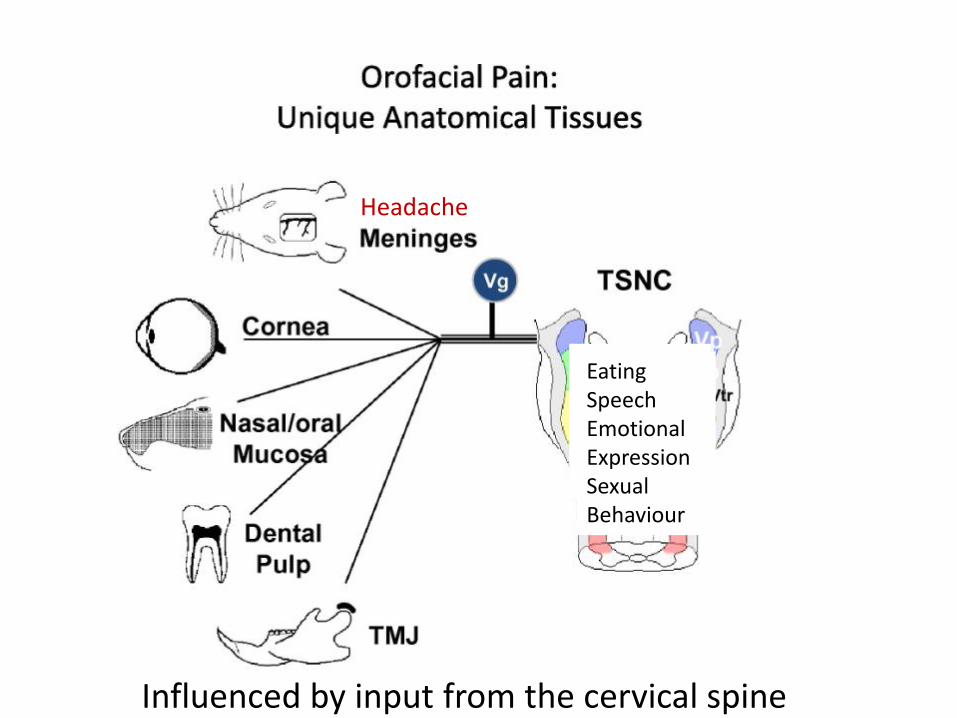
Fig. 2
Copyright © 2011 International Association for the Study of PainIN
Heachache Toothache AFP SMS Neuralgias
Eating Speech Emotional Expression Sexual Behaviour
Headache
Influenced by input from the cervical spine
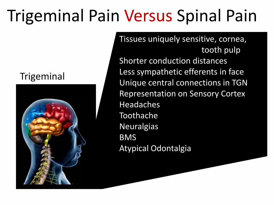
Trigeminal Pain Versus Spinal Pain
Trigeminal
Tissues uniquely sensitive, cornea, tooth pulp Shorter conduction distances Less sympathetic efferents in face Unique central connections in TGN Representation on Sensory Cortex Headaches Toothache Neuralgias BMS Atypical Odontalgia

Diagnosing Orofacial Pain Our Challenge as Clinicians
• Pulpal Pain (Toothache) is most common cause of orofacial pain
• Myofascial Pain is most common cause of extraoral pain
• 81% of patients attending a specialist orofacial center had pain sources outside the trigeminal system but failed to mention them
Turp JC et al. Pain maps from facial pain patients reveal a broad pain geography. J Dent Res 1998; 77: 1465 - 1472
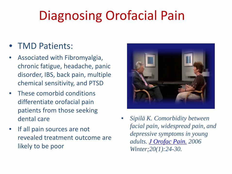
• Sipilä K. Comorbidity between facial pain, widespread pain, and depressive symptoms in young adults. J Orofac Pain. 2006 Winter;20(1):24-30.
• TMD Patients: • Associated with Fibromyalgia,
chronic fatigue, headache, panic disorder, IBS, back pain, multiple chemical sensitivity, and PTSD
• These comorbid conditions differentiate orofacial pain patients from those seeking dental care
• If all pain sources are not revealed treatment outcome are likely to be poor
Diagnosing Orofacial Pain
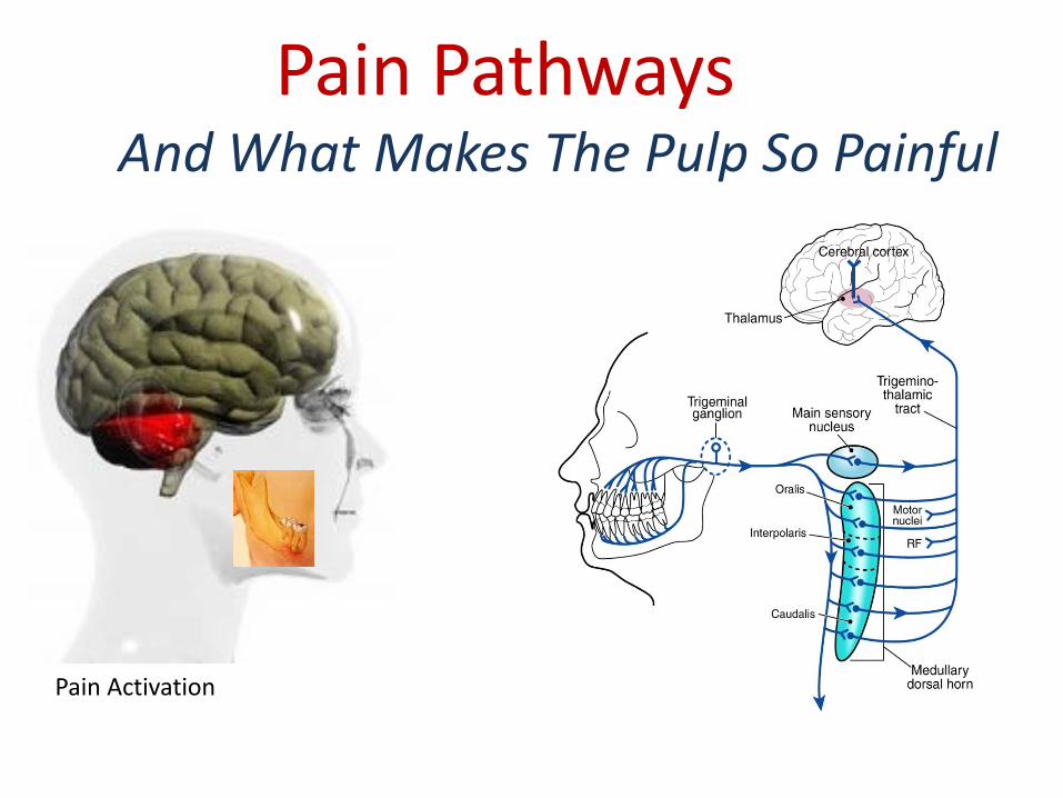
Pain Pathways And What Makes The Pulp So Painful
Pain Activation Pain Modulation
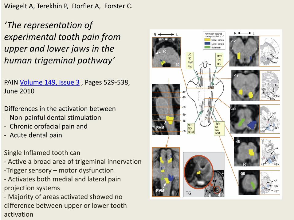
Fig. 2 Wiegelt A, Terekhin P, Dorfler A, Forster C.
‘The representation of experimental tooth pain from upper and lower jaws in the human trigeminal pathway’ PAIN Volume 149, Issue 3 , Pages 529-538, June 2010 Differences in the activation between - Non-painful dental stimulation - Chronic orofacial pain and - Acute dental pain
Single Inflamed tooth can - Active a broad area of trigeminal innervation -Trigger sensory – motor dysfunction - Activates both medial and lateral pain projection systems - Majority of areas activated showed no difference between upper or lower tooth activation
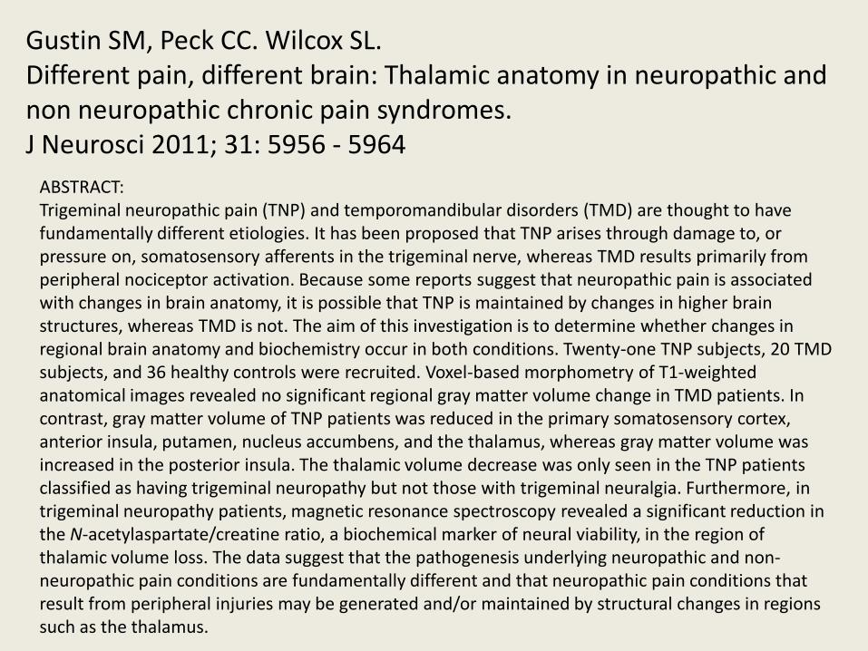
Gustin SM, Peck CC. Wilcox SL. Different pain, different brain: Thalamic anatomy in neuropathic and non neuropathic chronic pain syndromes. J Neurosci 2011; 31: 5956 - 5964
ABSTRACT: Trigeminal neuropathic pain (TNP) and temporomandibular disorders (TMD) are thought to have fundamentally different etiologies. It has been proposed that TNP arises through damage to, or pressure on, somatosensory afferents in the trigeminal nerve, whereas TMD results primarily from peripheral nociceptor activation. Because some reports suggest that neuropathic pain is associated with changes in brain anatomy, it is possible that TNP is maintained by changes in higher brain structures, whereas TMD is not. The aim of this investigation is to determine whether changes in regional brain anatomy and biochemistry occur in both conditions. Twenty-one TNP subjects, 20 TMD subjects, and 36 healthy controls were recruited. Voxel-based morphometry of T1-weighted anatomical images revealed no significant regional gray matter volume change in TMD patients. In contrast, gray matter volume of TNP patients was reduced in the primary somatosensory cortex, anterior insula, putamen, nucleus accumbens, and the thalamus, whereas gray matter volume was increased in the posterior insula. The thalamic volume decrease was only seen in the TNP patients classified as having trigeminal neuropathy but not those with trigeminal neuralgia. Furthermore, in trigeminal neuropathy patients, magnetic resonance spectroscopy revealed a significant reduction in the N-acetylaspartate/creatine ratio, a biochemical marker of neural viability, in the region of thalamic volume loss. The data suggest that the pathogenesis underlying neuropathic and non-neuropathic pain conditions are fundamentally different and that neuropathic pain conditions that result from peripheral injuries may be generated and/or maintained by structural changes in regions such as the thalamus.
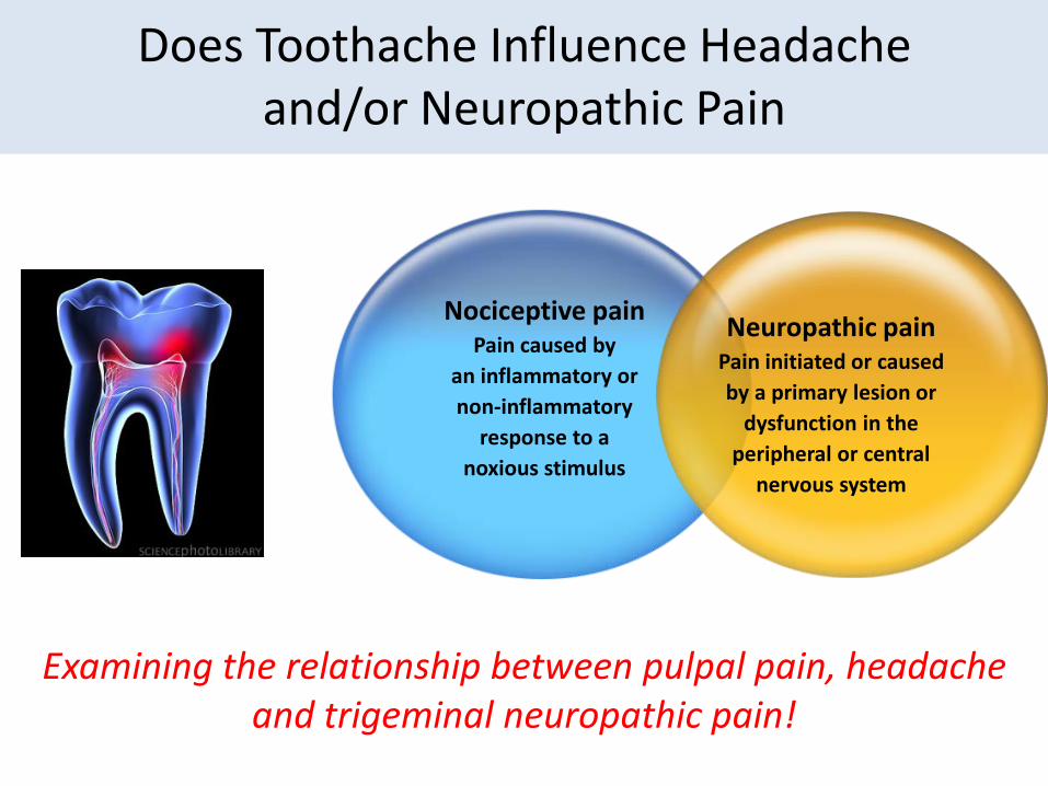
Examining the relationship between pulpal pain, headache and trigeminal neuropathic pain!
Neuropathic pain Pain initiated or caused by a primary lesion or
dysfunction in the peripheral or central
nervous system
Nociceptive pain Pain caused by
an inflammatory or non-inflammatory
response to a noxious stimulus
Does Toothache Influence Headache and/or Neuropathic Pain

Does Toothache Influence Headache and/or Neuropathic Pain
Migraine
Neuropathic Pain
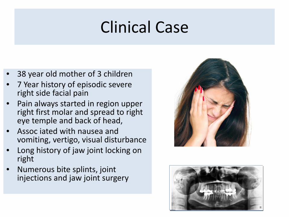
Clinical Case
• 38 year old mother of 3 children • 7 Year history of episodic severe
right side facial pain • Pain always started in region upper
right first molar and spread to right eye temple and back of head,
• Assoc iated with nausea and vomiting, vertigo, visual disturbance
• Long history of jaw joint locking on right
• Numerous bite splints, joint injections and jaw joint surgery
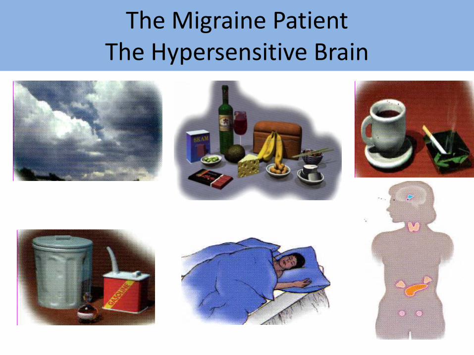
The Migraine Patient The Hypersensitive Brain
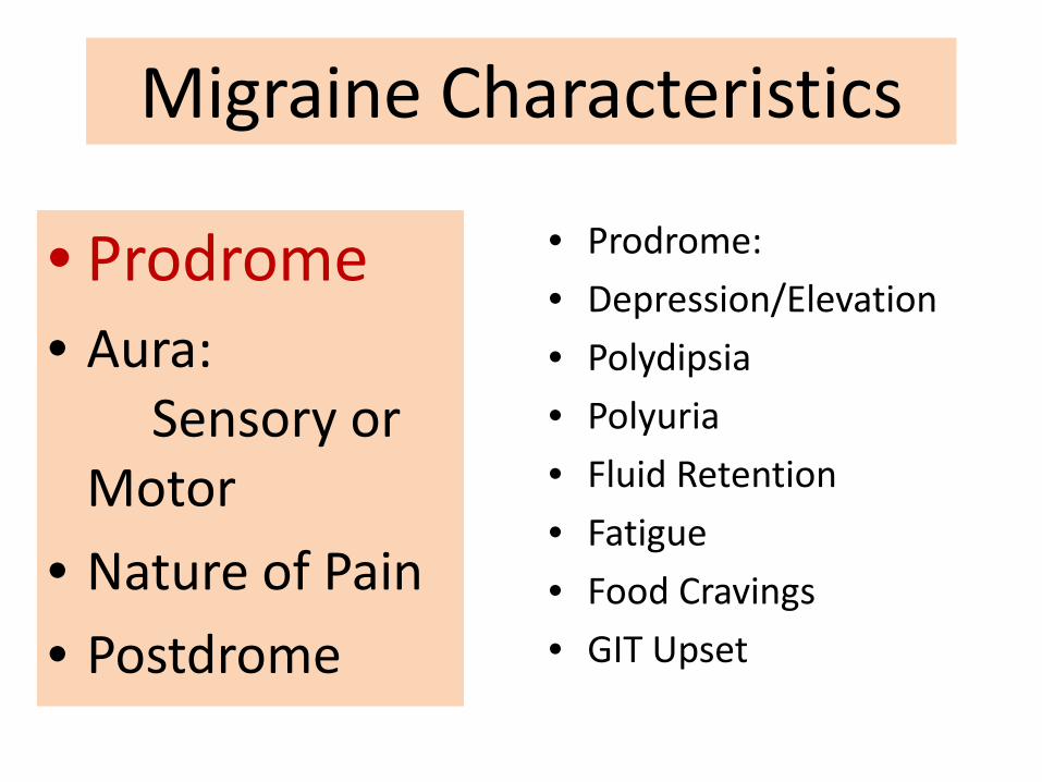
Migraine Characteristics
• Prodrome • Aura:
Sensory or Motor
• Nature of Pain
• Postdrome
• Prodrome:
• Depression/Elevation
• Polydipsia
• Polyuria
• Fluid Retention
• Fatigue
• Food Cravings
• GIT Upset

Migraine Characteristics
• Prodrome
• Aura:
- Sensory or Motor
• Nature of Pain
• Postdrome

Clinical Case
• Pain Diary
• Overuse of analgesics
• Headache prev entive meds
• Sleep hygiene, diet, exercise
• Referral for mindfulness training
• Simple exercise program for TMD symptoms
• Acute break through pain – medications or GON blocks

Cluster Headache
Typically Male Attacks 2 – 3 day Nocturnal Activation Autonomic Symptoms Possibly localised to teeth Excrutiating Intensity Duration 30 -180 mins
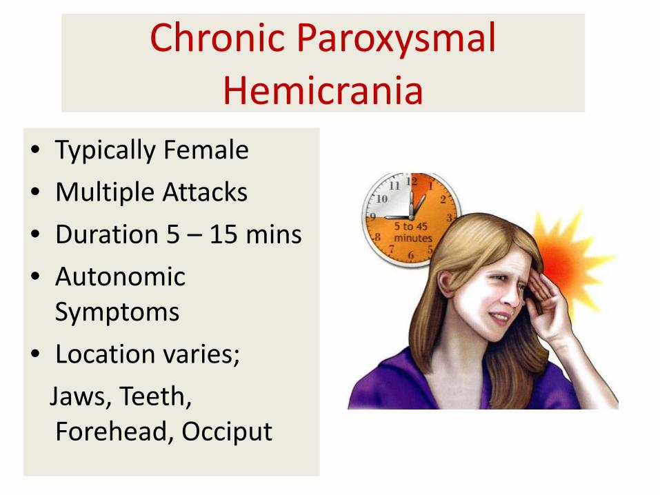
Chronic Paroxysmal Hemicrania
• Typically Female
• Multiple Attacks
• Duration 5 – 15 mins
• Autonomic Symptoms
• Location varies;
Jaws, Teeth, Forehead, Occiput
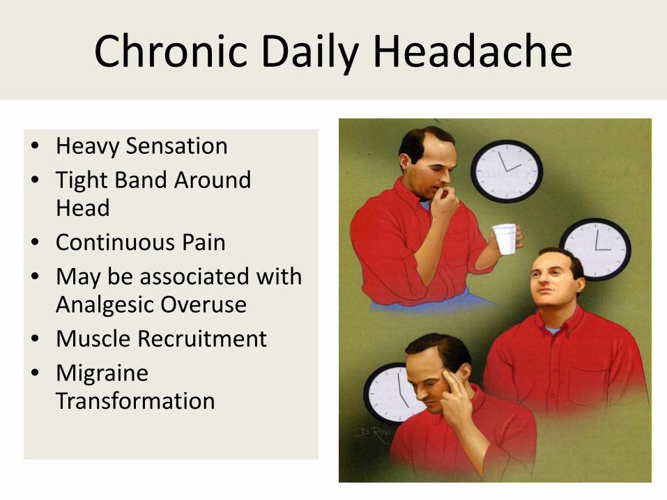
Chronic Daily Headache
• Heavy Sensation • Tight Band Around
Head • Continuous Pain • May be associated with
Analgesic Overuse • Muscle Recruitment • Migraine
Transformation

Headache Influences on The Orofacial Region
• Migraine/Headache pain frequently involves the face and oral cavity, mimicking dental and/or neuropathic trigeminal pain
• Episodic Primary Headache Disorders like migraine, cluster and CPH may appear to originate in the oral cavity
• Headache Disorders may be aggravated by dental procedures or oral surgery
• Patients with headache disorders may experience widespread dental sensitivity
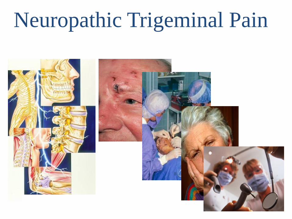
Neuropathic Trigeminal Pain

Prevalence of persistent pain after endodontic treatment and factors affecting its occurrence in
cases with complete radiographic healing N. Polycarpou, Y.-L. Ng, D. Canavan*, D. R. Moles & K. Gulabivala
Conclusions The presence and duration of preoperativepain from the tooth site, lasting at least 3 months, a positive history of previous chronic pain experience or painful treatment in the orofacial region, and female gender were important risk factors associated with persistent pain after successful endodontic treatment.
International Endodontic Journal 2005
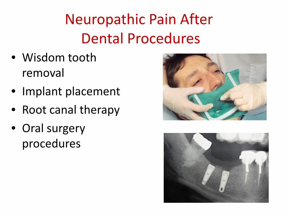
Neuropathic Pain After Dental Procedures
• Wisdom tooth removal
• Implant placement
• Root canal therapy
• Oral surgery procedures

Profiling of Patients with Postraumatic Neuropathy of the Trigeminal Nerve
Renton T, Zehra Y. J Orofacial Pain 2011 ;25:333-344s
Neuropathic pain, as well as anaesthesia, frequently occurs following iatrogenic trigeminal nerve injury similar to other posttraumatic sensory nerve injuries. This must be acknowledged by clinicians as a relatively common problem and informed consent appropriately formulated for patients at risk of trigeminal nerve injuries in relation to dentistry requires revision.
• Renton T, Dawood A et al. Post-implant neuropathy of the trigeminal nerve. A case series. Br Dent J. 2012 Jun 8;212
• RESULTS:
• Patients were aware of signing consent forms for the surgery in 11 cases and 8 of those felt they were not explicitly warned about nerve injury. Over 70% of patients were referred after six months post injury. Implant surgery planning involved intra-oral films only (30%), CBCT (10%), dental pantomograph (50%) and long cone peri-apical radiographs (48%). However, no radiographic evidence pre- or postoperatively was provided by the referring practitioner in 15% of cases. Intra-operative problems included bleeding and neurological symptoms. Proximity of the implant bed or implant to the inferior alveolar canal was evident radiographically. This showed contact with roof inferior alveolar nerve canal in 44% of cases, protrusion into the canal in 20% of cases, crossing of the canal in 20% cases and distance in one case, presumed to be due to local anaesthetic injury. All patients presented with a demonstrable neuropathy, which included neuropathic pain (50%) that interfered with speaking, kissing and socialising.
• CONCLUSIONS:
• Consent, preoperative planning and appropriate referral were inadequate in provision of mandibular implants in this patient group. Recommendations have been proposed to improve practice and possible novel strategies are suggested for the prevention and improved management of these complications
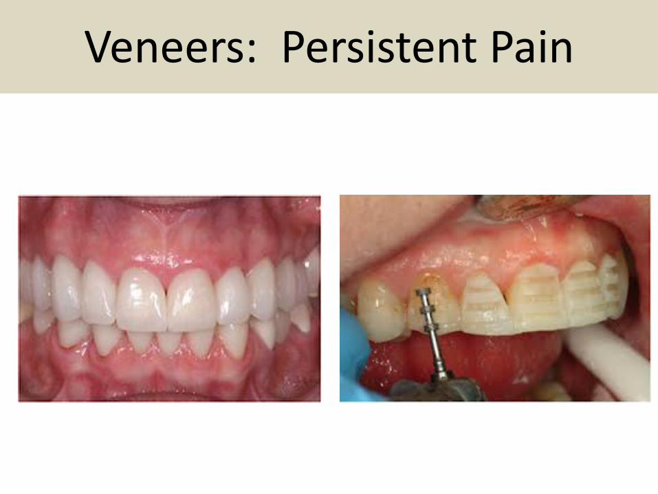
Veneers: Persistent Pain

Veneers: Persistent Pain
• Location:
• Intensity:
• Type:
• Presence:
• Agg by:
• Allev by:
• Onset:
Upper Centrals
Severe
Burning, Aching
Continuous
Nil
Nil
After tooth preps
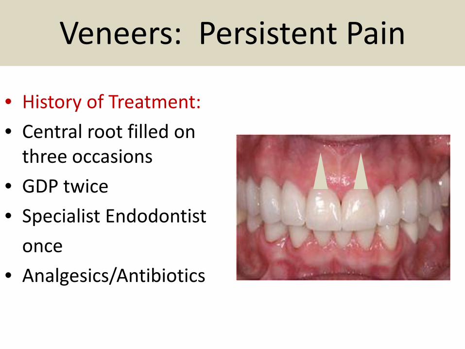
Veneers: Persistent Pain
• History of Treatment:
• Central root filled on three occasions
• GDP twice
• Specialist Endodontist
once
• Analgesics/Antibiotics

Veneers: Persistent Pain
• Relevent Findings: • Tenderness in premaxilla bilaterally • Tenderness in infraorbital regions • Centrals are extremely sensitive to
touch • Local Sensory Abnormalities:
Allodynia, Hyperalgesia, Hyperpathia, increased response to cold
• Special Tests:

Trigeminal Neuropathic Pain
Induced by External Trauma History
Lower Incisors traumatised by direct blow. RCT modified but did not eliminate the pain
Patient insisted on removal of teeth and placement of implants
Treatment Options Anticonvulsants/Tricyclics Nerve Blocks: Mandibular Division Steroid Injection Stellate Ganglion Block

Looking to the Future
PENS
Teaching Models for undergraduate and
postgraduates students
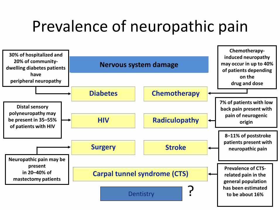
Radiculopathy HIV
Chemotherapy Diabetes
Nervous system damage
Surgery Stroke
Carpal tunnel syndrome (CTS)
7% of patients with low back pain present with
pain of neurogenic origin
30% of hospitalized and 20% of community-
dwelling diabetes patients have
peripheral neuropathy
Distal sensory polyneuropathy may be present in 35–55% of patients with HIV
Neuropathic pain may be present
in 20–40% of mastectomy patients
Chemotherapy-induced neuropathy
may occur in up to 40% of patients depending
on the drug and dose
8–11% of poststroke patients present with
neuropathic pain
Prevalence of CTS-related pain in the general population has been estimated
to be about 16%
Prevalence of neuropathic pain
Dentistry ?

Thank You Dermot Canavan Dublin Dental University Hospital and 69 Eglinton Road, Donnybrook, Dublin