

Clin. Vaccine Immunol.-2016-Schmidt-CVI.00730-15
43
1 A Tetraspecific VHH-based Neutralizing Antibody Modifies Disease Outcome in Three Animal 1 Models of Clostridium difficile Infection 2 3 Diane J. Schmidt 1 , Gillian Beamer 1 , Jacqueline M. Tremblay 1 , Jennifer A. Steele 1 , Hyeun Bum 4 Kim 2 , Yaunkai Wang 3 , Michele Debatis 1 , Xingmin Sun 1 , Elena A. Kashentseva 4 , Igor P. 5 Dmitriev 4 , David T. Curiel 4 , Charles B. Shoemaker 1 , Saul Tzipori 1# . 6 7 1 Department of Infectious Disease and Global Health, Cummings School of Veterinary 8 Medicine, Tufts University, North Grafton, MA, USA 9 2 Department of Animal Resources Science, Dankook University, Cheonan, Republic of Korea 10 3 School of Agriculture and Biology; Shanghai Jiao Tong University; Shanghai Key Laboratory 11 of Veterinary Biotechnology; Key Laboratory of Urban Agriculture (South) Ministry of 12 Agriculture; Shanghai, PR China 13 4 Department of Radiation Oncology, Washington University, St. Louis, MO, USA 14 15 Running Head: Therapeutic treatment for C. difficile Infection 16 17 # Address correspondence to Saul Tzipori, [email protected]. 18 19 20 21 22 CVI Accepted Manuscript Posted Online 13 July 2016 Clin. Vaccine Immunol. doi:10.1128/CVI.00730-15 Copyright © 2016 Schmidt et al. This is an open-access article distributed under the terms of the Creative Commons Attribution 4.0 International license. on July 26, 2016 by TUFTS UNIV http://cvi.asm.org/ Downloaded from
-
Upload
diane-schmidt -
Category
Documents
-
view
99 -
download
1
Transcript of Clin. Vaccine Immunol.-2016-Schmidt-CVI.00730-15

1
A Tetraspecific VHH-based Neutralizing Antibody Modifies Disease Outcome in Three Animal 1
Models of Clostridium difficile Infection 2
3
Diane J. Schmidt1, Gillian Beamer1, Jacqueline M. Tremblay1, Jennifer A. Steele1, Hyeun Bum 4
Kim2, Yaunkai Wang3, Michele Debatis1, Xingmin Sun1, Elena A. Kashentseva4, Igor P. 5
Dmitriev4, David T. Curiel4, Charles B. Shoemaker1, Saul Tzipori1#. 6
7
1Department of Infectious Disease and Global Health, Cummings School of Veterinary 8
Medicine, Tufts University, North Grafton, MA, USA 9
2Department of Animal Resources Science, Dankook University, Cheonan, Republic of Korea 10
3School of Agriculture and Biology; Shanghai Jiao Tong University; Shanghai Key Laboratory 11
of Veterinary Biotechnology; Key Laboratory of Urban Agriculture (South) Ministry of 12
Agriculture; Shanghai, PR China 13
4Department of Radiation Oncology, Washington University, St. Louis, MO, USA 14
15
Running Head: Therapeutic treatment for C. difficile Infection 16
17
#Address correspondence to Saul Tzipori, [email protected]. 18
19
20
21
22
CVI Accepted Manuscript Posted Online 13 July 2016Clin. Vaccine Immunol. doi:10.1128/CVI.00730-15Copyright © 2016 Schmidt et al.This is an open-access article distributed under the terms of the Creative Commons Attribution 4.0 International license.
on July 26, 2016 by TU
FT
S U
NIV
http://cvi.asm.org/
Dow
nloaded from
2
Abstract 23
Clostridium difficile infection (CDI), a leading cause of nosocomial infection, is a serious disease 24
in North America, Europe and Asia. CDI varies greatly from asymptomatic carriage to life 25
threatening diarrhea, toxic megacolon and toxemia. The incidence of community acquired 26
infection has increased due to the emergence of hypervirulent antibiotic-resistant strains. These 27
new strains contribute to the frequent occurrence of disease relapse; complicating treatment, 28
increasing hospital stays, and increasing morbidity and mortality among patients. It is critical 29
therefore to develop new therapeutic approaches that bypass the development of antimicrobial 30
resistance and avoid disruption of gut microflora. Here we describe the construction of a single 31
heteromultimeric VHH-based neutralizing agent (VNA) targeting the two primary virulence 32
factors of Clostridium difficile, toxin A (TcdA) and B (TcdB). Designated VNA2-Tcd, this agent 33
has sub-nanomolar toxin neutralization potencies for both C. difficile toxins in cell assays. When 34
given systemically by parenteral administration, VNA2-Tcd protected against CDI in gnotobiotic 35
piglets and mice, and to a lesser extent in hamsters. Protection from CDI was also observed in 36
gnotobiotic piglets treated by gene therapy with an adenovirus promoting expression of VNA2-37
Tcd. 38
39
Introduction 40
Clostridium difficile infection (CDI) is currently one of the leading causes of nosocomial 41
infection (1, 2) and is fast becoming a cause of community acquired diarrhea in previously low-42
risk populations including; children, healthy adults, and pregnant women (1-7). Manifestations 43
of CDI vary from asymptomatic colonization, mild or moderate diarrhea, a severe or fulminant 44
illness with complications including pseudomembranous colitis, toxic megacolon, small bowel 45
on July 26, 2016 by TU
FT
S U
NIV
http://cvi.asm.org/
Dow
nloaded from

3
ileus; and even systemic inflammatory response syndrome, a multisystem organ failure which 46
can be fatal (8). The emergence of antibiotic-resistant hypervirulent strains and the increase in 47
disease relapse have complicated treatment of CDI leading to increases in hospital stay, 48
morbidity, and mortality (1). 49
Clostridium difficile is a gram-positive, spore-forming anaerobic bacterium that produces 50
two toxins, designated TcdA and TcdB (9), which are the major virulence factors of CDI (10). 51
They are large exotoxins that bind to human colonocytes causing inflammation, fluid 52
accumulation, and mucosal injury manifested as pseudomembranous colitis (11). 53
C. difficile survives, persists and produces the two exotoxins in the gut after prolonged 54
treatment with broad-spectrum antibiotics reduces normal microflora (12). The extensive use of 55
antibiotics for treatment of CDI has increased the emergence of resistant strains leading to a 56
dramatic increase in the incidence of disease relapse estimated at 20-35% (13). Consequently 57
there is an urgent need to develop novel, non-antibiotic therapies that prevent persistence and 58
toxin production by C. difficile and minimally impact the normal gut microflora. Ideally, 59
approaches that specifically target toxins instead of bacterial cells and eliminate the possibility of 60
antimicrobial resistance are favored (14, 15). Several therapeutic approaches are currently under 61
development including antibiotics (8, 16, 17), probiotics (18-23), fecal-transplants (24-26), toxin 62
binding resins or polymers (27), vaccines (16, 28-30), and toxin-specific antibodies (31-38). 63
Several but not all anti-toxin antibodies (Abs) improve CDI outcomes in animal models and 64
clinical trials (32, 34, 35, 39-42), but these conventional antibodies are costly and challenging to 65
engineer. There is some evidence from the pig model (43) that antibodies against TcdB alone 66
may be sufficient for treating CDI, however there is conflicting data on the roles of the toxins in 67
disease (44-46). 68
on July 26, 2016 by TU
FT
S U
NIV
http://cvi.asm.org/
Dow
nloaded from

4
As an efficient alternative we produced and tested heavy-chain-only VH domains 69
(VHHs), generated by Camilidae species, seeking VHHs that neutralize each of the two C. 70
difficile toxins. DNAs encoding these unconventional IgGs (IgG2 and IgG3) are easily cloned 71
(47) and can be expressed at high levels in soluble form (48). The VHH protein products are 72
generally more stable than conventional antibodies and frequently bind the active sites of 73
targeted proteins (48-50). We previously showed that bispecific VHH-based neutralizing agents 74
(VNAs) are highly efficacious as antitoxins in animal models of exposures to botulinum 75
neurotoxins (51), ricin (52), Shiga toxins (53), and anthrax (54); significantly outperforming 76
their monomer VHH components. To achieve protection from CDI, a VNA was engineered and 77
expressed in bacteria containing four VHHs; two (AH3, AA6) that neutralize TcdA, and two 78
copies of the 5D VHH (5D, 5D) that neutralizes TcdB (55). This VNA, called ABA, provided 79
potent protection from CDI in a mouse model. 80
While some reports have indicated that TcdA does not play a significant role in disease 81
pathogenesis in the gnotobiotic pig model of CDI (43), other evidence has shown that both TcdA 82
and TcdB toxins contribute to fulminant disease in hamsters (56) and in some mouse models of 83
CDI (57). Since VHH agents remain functional when linked into multimers, we have chosen to 84
include VHHs neutralizing both Tcd toxins in our antitoxin agent as this should be effective in 85
all of the models of CDI. In the current study, we chose to re-engineer the ABA VNA based on 86
recent results (58) and unpublished data showing that two different toxin neutralizing VHHs 87
against the same target combined into a single linked construct is a more effective antitoxin in 88
vivo than a homodimer of only one toxin-neutralizing VHH. In our new VNA, VNA2-Tcd, we 89
replaced one of the two copies of the 5D VHH in ABA with a different TcdB-neutralizing VHH, 90
E3. Specifically VNA2-Tcd is a tetraspecific agent containing 5D and E3 VHHs targeting TcdB 91
on July 26, 2016 by TU
FT
S U
NIV
http://cvi.asm.org/
Dow
nloaded from

5
linked to the two TcdA-neutralizing VHHs, AH3 and AA6. In this report, we test the ability of 92
VNA2-Tcd to protect against CDI pathology in mouse, hamster and gnotobiotic piglet models of 93
this disease when administered both as a protein therapeutic or by adenoviral gene therapy. 94
95
Materials and Methods 96
Ethics with IACUC statement 97
Treatment and care of all animals used in experiments followed institutional animal care 98
and use committee guidelines. All animal studies performed were approved by Tufts University 99
Institutional Animal Care and Use Committee. 100
101
Plasmid Construction 102
Synthetic DNA was prepared in which coding DNA for the two most potent neutralizing 103
VHHs we reported previously for TcdA (AH3, AA6) and TcdB (5D, E3) (41) were linked 104
together, each separated by DNA encoding a flexible spacer (GGGGS)3), to encode a VHH tetra-105
specific, heteromultimeric VNA (AH3/5D/E3/AA6) called VNA2-Tcd.. This DNA was inserted 106
into expression vector pET32b in fusion with E. coli thioredoxin as described by Tremblay (53) 107
to create the Trx/VNA2-Tcd expression plasmid. 108
109
Protein Purification 110
The Trx-VNA2-Tcd-6His/pET32b plasmid was transformed into Rosetta-gami (DE3) E. 111
coli and fermentation was as follows: 20 L of LB media with 100 μg/ml ampicillin, 34 μg/ml 112
chloramphenicol, incubated at 15°C, and expression induced with 1mM IPTG at OD600=0.6 for 113
20 hrs (59). Protein was captured by Ni-affinity chromatography and eluted with 0.5M Imidazole 114
on July 26, 2016 by TU
FT
S U
NIV
http://cvi.asm.org/
Dow
nloaded from

6
gradient at pH 7.5. It was further purified by gel filtration chromatography (HiLoad Superdex 200 115
26/60, GE Life Sciences) with 20mM Hepes, pH 7.8, 200mM NaCl, 1mM DTT, 1mM EDTA 116
elution buffer. The protein eluted as a monomer from the gel filtration column. Recombinant 117
protein was visualized by SDS-PAGE/Coomassie and a Western blot using anti-E-tag antibody 118
(BETHYL) at 1:5,000 dilution. In some experiments protein was treated for endotoxin removal 119
by Triton X114 phase partitioning method. Final endotoxin concentration in endotoxin-free 120
preparation was below 0.01EU/mg as determined by PyroGene™ recombinant Factor C assay 121
(Lonza). Fermentation, purification, dialysis and endotoxin removal was performed by ARVYS 122
Proteins Inc. (www.arvysproteins.com). 123
124
Adenovirus vector construction and preparation 125
The generation of recombinant replication incompetent Ad5-based vectors has been 126
previously described (60). Briefly in a modification from (61), pShCMV-JGf7 shuttle plasmid 127
was used for subcloning the VNA2-Tcd coding sequence (54), under control of the mammalian 128
CMV promoter and followed by the bovine growth hormone polyA signal. A control vector 129
Ad/VNA1-Stx was created in a similar manner with the sequence from two VHHs against Stx 130
(62). This control vector results in secretion of a Stx reactive Ab with no binding to Tcd. Each 131
shuttle plasmid was linearized and employed for homologous recombination with pAdEasy-1 132
plasmid carrying viral genome resulting in selection of plasmid containing recombinant Ad5 133
genome incorporating either VNA2-Tcd or VNA1-Stx expression cassette in place of deleted 134
viral E1 genes. The resultant plasmids were validated by PCR, restriction analyses and 135
sequencing to confirm incorporation of VNA2-Tcd or VNA1-Stx expression cassette within the 136
corresponding recombinant viral genome. The plasmids were linearized with PacI to release the 137
on July 26, 2016 by TU
FT
S U
NIV
http://cvi.asm.org/
Dow
nloaded from

7
inverted terminal repeats of the viral genomic DNA and transfected into 293 cells to rescue 138
replication incompetent Ad/VNA2-Tcd. The rescued Ad/VNA2-Tcd and Ad/VNA1-Stx vectors 139
were upscaled, purified by centrifugation on CsCl gradients as previously described (61), 140
dialyzed against phosphate-buffered saline (PBS) (8 mM Na2HPO4, 2 mM KH2PO4 [pH 7.4], 141
137 mM NaCl, 2.7 mM KCl) containing 10% glycerol and stored at -80⁰C. The titers of physical 142
viral particles (vp) were determined by the methods of Maizel et al. (63). 143
144
Neutralization Assay 145
Vero cells (ATCC) at a concentration of 2.4x10⁴cells/100 μl of media [DMEM high 146
glucose + 1mM sodium pyruvate, 2mM L-glutamine, 50U/ml and 50 µg/ml Pen/Strep pH 7.4 147
(HyClone)] were plated in 96 well plates overnight for 90-95% confluency, prior to addition of 148
VNA2-Tcd added in serial dilutions (in media) from 100 μg/ml – 1.0 fg/ml and either 2 ng -12.5 149
ng/ml TcdA and 0.25-2 ng/ml TcdB or both TcdA and B in a 24 hr cytotoxicity/cell rounding 150
assay (41). 151
152
ELISA 153
EIA/RIA 96 well high binding plates (Corning Costar) coated with 0.5-5 µg/ml of rTcdA 154
or rTcdB or rTcdA + rTcdB overnight at 4°C were used for binding assays. Plates were washed 155
3 times with 1xPBS + 0.1% Tween followed by 3 times with 1xPBS and blocked (4-5% non-fat 156
dry milk in 1xPBS + 0.1% Tween) for 1 hr at room temperature (RT) with rocking. Serially 157
diluted VNA2-Tcd, serum or fecal samples diluted in blocking solution were incubated for 1 hr 158
at RT with rocking and washed as above. Equivalent control samples were spiked with a known 159
amount of VNA2-Tcd for use as an internal standard. Goat anti-E-tag-HRP conjugated antibody 160
on July 26, 2016 by TU
FT
S U
NIV
http://cvi.asm.org/
Dow
nloaded from

8
(Bethyl labs) diluted 1:5000 in blocking solution was incubated for 1 hr at RT with rocking, 161
washed as above before adding TMB microwell peroxidase substrate (KPL) to develop 162
(incubated for 10-40 min), stopped with 1M H2SO4 and read at 450nm on a ELx808 Ultra 163
Microplate Reader (Bio-Tek instruments) (51). VNA2-Tcd levels in unknown samples were 164
determined by comparison of their signals to that of internal standards as previously described 165
(61, 62, 64-66). 166
167
Mouse Systemic Toxin Challenge 168
6 week old C57BL/6 female mice were intraperitoneally (IP) injected with a single dose 169
of VNA2-Tcd (50 μg/mouse) 1 hr prior to IP injection of TcdA (100 ng/mouse), TcdB 170
(200ng/mouse), or TcdA + TcdB (100ng and 200ng respectively). Mice were monitored for 171
signs and symptoms of toxemia (including; lethargy, depression, anorexia, dehydration, ruffled 172
coat, and hunched posture). Moribund mice were euthanized following IACUC-approved 173
removal criteria. 174
175
Mouse CDI Challenge 176
To mimic the human condition and facilitate colonization with C. difficile, ten 6 week 177
old, C57BL/6 female mice received filter sterilized antibiotics (kanamycin, gentamycin, colistin, 178
metranidozole, and vancomycin) in drinking water for 5 days followed by 2 days of water alone. 179
After 2 days of drinking water, the mice received one 100 μl intraperitoneally (IP) injection of 180
clindamycin (2mg/ml) IP. One day later mice were orally challenged (67) with 106 spores of an 181
NAPI/027/BI C. difficile strain, designated strain UK6 (68) only (control group), or inoculated 182
on July 26, 2016 by TU
FT
S U
NIV
http://cvi.asm.org/
Dow
nloaded from

9
with spores and administered VNA2-Tcd (25-50 µg/mouse) at 4, 24 and 48hrs post-challenge 183
(treated group). Blood was collected at 72, 96 and 120 hrs post-challenge for VNA titers. 184
185
Hamster CDI Challenge 186
Again, to mimic the human condition and facilitate colonization with C. difficile, 187
seventeen, 110-135g male Golden Syrian hamsters were administered clindamycin (30mg/kg) 188
via oral gavage for 5 days prior to oral inoculation with 1000 C. difficile strain UK6 spores. 189
Infected control hamsters were administered clindamycin inoculated with UK6 spores and given 190
sterile PBS, IP, 2 times per day for the duration of the experiment. VNA2-Tcd treated hamsters 191
were administered clindamycin, inoculated with spores and given purified VNA2-Tcd (1mg/kg), 192
IP 2 times a day for the duration of the experiment. A blood sample was collected at time of 193
euthanasia for detection of VNA2-Tcd in serum. Necropsies were performed on euthanized 194
animals and tissues were collected for histopathologic examination. 195
196
Pig CDI Challenge 197
Thirty gnotobiotic piglets were derived via Caesarean section and maintained in sterile 198
isolators for the duration of the experiment (66). Five groups of piglets were orally inoculated 199
with 106 C. difficile UK6 spores (group 1-5) and group 6 was the uninfected controls, 200
summarized in Table 1. Group 1 (n=3) received VNA2-Tcd (1mg/pig) 4 hours prior to, and 201
Group 2 (n=3) 18 hours post oral inoculation with spores. After the initial dose, the treated 202
groups received 2 doses of VNA2-Tcd (1 mg/pig) per day either IP or intra muscular (IM) for the 203
duration of the experiment (Table 1). The Ad/VNA2-Tcd treated group (Group 3; n=9) was 204
given 1.0 x 10¹¹ viral particles IV, one day prior to oral inoculated with 106 C. difficile UK6 205
on July 26, 2016 by TU
FT
S U
NIV
http://cvi.asm.org/
Dow
nloaded from

10
spores and 3 days post infection (Table 1). Group 4 (n=6) received VNA-Tcd buffer as control 206
given 4 hours prior to oral inoculated with 106 C. difficile UK6 spores and at 24 hour post 207
inoculation and then every 12 hours until the termination of the experiment (Table 1). Group 5 208
was given control adenovirus expressing an unrelated VNA (n=6) 1.0 x 10¹¹ viral particles IV, 209
one day prior to oral inoculated with 106 C. difficile UK6 spores and 3 days post infection (Table 210
1). Group 6 (n=3) was uninfected (Table 1). From all piglets, fecal samples were collected for 211
bacterial culture, and blood samples were collected 1-3 times (when possible) during the 212
experiment and at time of euthanasia for VNA titers. Necropsies were performed on all animals 213
and tissues were collected for histopathologic examination. 214
215
Histology 216
Tissue samples were collected during necropsy and preserved in 10% neutral buffered 217
formalin. Formalin fixed samples were embedded in paraffin, sectioned at 5um, and stained 218
using hematoxylin and eosin using routine histochemical techniques at TCSVM Histopathology 219
Service Laboratory http://vet.tufts.edu/histology-service/. Light microscopic examination and 220
lesion evaluation was performed by a board-certified veterinary pathologist (GB) with results 221
reported as previously described for severity (minimal, mild, moderate, marked), epithelial 222
ulceration, luminal contents, and quantification (65). Briefly, a quantitative assessment of colitis 223
severity was performed by counting neutrophilic foci in colon sections from each sample. Foci 224
were observed between colonic crypts in the lamina propria in 10 random fields with ×20 225
magnification. 226
227
on July 26, 2016 by TU
FT
S U
NIV
http://cvi.asm.org/
Dow
nloaded from
11
Results 228
VNA2-Tcd construction, expression and protein purification 229
We previously identified two individual VHHs with strong neutralizing activity against 230
TcdA (AH3, AA6) and two VHHs that neutralized TcdB (5D, E3). Results with these VHHs, 231
and VHHs to other toxins (51-53), have shown that covalently linking two toxin-neutralizing 232
VHHs into bispecific heterodimer VHH-based neutralizing agents (VNAs) results in 233
substantially enhanced in vitro and in vivo antitoxin potency compared to equimolar pools of 234
unlinked VHHs. We thus engineered recombinant expression of a tetraspecific VHH 235
heteromultimer containing the four different VHHs, two each having TcdA and TcdB 236
neutralizing properties, all separated by flexible spacer peptides (Fig. 1). The VNA2-Tcd 237
heterotetramer was purified using Ni-affinity and gel filtration chromatography, treated for 238
endotoxin removal by a detergent-based method (>0.01EU/mg), and visualized following SDS-239
PAGE revealing a purity of about 71% for full-length heterotetramer and the remaining protein 240
bands almost entirely represented by trimeric and dimeric truncations of full-size VNA based on 241
Western blot (see Fig. S1 in supplemental material). For gene therapy, an adenovirus vector 242
(Ad5) was engineered to promote expression and secretion of VNA2-Tcd (Ad/VNA2-Tcd) 243
following transduction of mammalian cells. 244
245
In vitro characterization 246
The IC50 of VNA2-Tcd was determined by dilution cytotoxicity assay using Vero cells 247
and 100 pM of recombinant TcdA or 0.6 pM of recombinant TcdB and serially diluted VNA2-248
Tcd. The estimated IC50 was about 100 pM for TcdA and about 10 pM for TcdB, indicating that 249
on July 26, 2016 by TU
FT
S U
NIV
http://cvi.asm.org/
Dow
nloaded from

12
VNA2-Tcd is capable of neutralizing both toxins when present at near equimolar doses to the 250
two Tcd toxins in these assays (Fig. 2A and 2B). 251
252
Mouse systemic toxin challenge 253
We assessed the potency of VNA2-Tcd to neutralize toxins A and B in a systemic mouse 254
challenge using 6 week old C57BL/6 female mice. Six groups of mice (5 mice per group) were 255
injected IP with TcdA and/or TcdB +/- VNA, or VNA alone, and monitored for signs of toxemia. 256
Initial systemic toxin doses of 100 ng-200 ng of TcdA and 100-800 ng of TcdB were tested 257
individually to determine 24 hr LD100. The LD100 doses for each toxin were then used in the 258
systemic challenge. The control group administered 100 ng/mouse of TcdA and 200 ng/mouse of 259
TcdB (IP) were either moribund (euthanized) or died within 4 hrs post-challenge. Other groups 260
were administered: 1) no treatment; 2) VNA2-Tcd only (no toxin); 3) VNA2-Tcd + TcdA; 4) 261
VNA2-Tcd + TcdB, or; 5) VNA2-Tcd + TcdA/B. VNA2-Tcd treatments were a single IP 262
injection of VNA2-Tcd (2.5 mg/kg) at 1 hr prior to toxin challenge. No mice in these 5 groups 263
(Table S1) showed any signs or symptoms of toxemia and all remained healthy until termination 264
of the experiment at 7 days post-toxin challenge. 265
266
Mouse CDI challenge 267
Two groups (5 mice each) of 6 week old C57BL/6 female mice were treated for 5 days 268
with an antibiotic cocktail in drinking water and administered a single IP injection of 269
clindamycin prior to being orally challenge with 106 C. difficile (UK6 strain) spores. The control 270
group received spores + PBS, and the treated group received spores and VNA2-Tcd injected IP 271
at 4, 24, and 48 hrs post challenge. The VNA2-Tcd group received an initial dose of 25 272
on July 26, 2016 by TU
FT
S U
NIV
http://cvi.asm.org/
Dow
nloaded from
13
µg/mouse (1.25 mg/kg) at 4 hrs post challenge and two additional doses of VNA2-Tcd [50 273
µg/mouse (2.5 mg/kg)] at 24 and 48 hrs post-challenge. The control group (spores + PBS) 274
experienced weight loss at days 1, 2, and 3 post challenge and most mice began to gain weight 275
on day 3 or 4 post challenge, while the VNA2-Tcd treated group experienced weight loss only on 276
day 1 post challenge and began to regain weight on day 2 post challenge (Fig. 3A). The 277
difference in weight between the control and treated groups was only statistically significant on 278
day 2 post-challenge. 100% of the control animals developed diarrhea and 60% were moribund 279
or died (Fig. 3B). Only 1 mouse in the VNA2-Tcd treated group developed diarrhea, which was 280
resolved by day 2 post challenge. All mice in the VNA2-Tcd treated group survived for the 281
duration of the experiment (Fig. 3B). Blood was collected from mice 24, 48, or 72 hrs after 282
administration of the final dose of VNA2-Tcd. Serum VNA2-Tcd levels were measured by 283
ELISA and ranged from 1.9 to 6.1 µg/ml (Fig. S2A). Serum TcdA and TcdB neutralization 284
abilities tested by cytotoxicity/cell rounding assay were similar for all treated mice with a 1:10 285
dilution of serum providing about 50% protection from rounding (Fig. S2B). 286
287
Hamster CDI challenge 288
Hamsters were reported to be relatively resistant to the UK6 strain of C. difficile (69) so 289
we performed initial experiments and established that treating with five days of oral clindamycin 290
followed by 1000 UK6 spores induced CDI disease in 100% of Syrian hamsters (not shown). To 291
test VNA2-Tcd efficacy, two groups of male golden Syrian hamsters were administered 292
clindamycin (30 mg/kg) orally for 5 days prior to inoculation with 1000 UK6 spores. The control 293
group (6 hamsters) received spores + PBS, and the treated group (11 hamsters) received spores 294
and 1 mg/kg of VNA2-Tcd injected IP two times a day for the duration of the experiment. All 295
on July 26, 2016 by TU
FT
S U
NIV
http://cvi.asm.org/
Dow
nloaded from

14
hamsters in the control and treated groups except for one treated hamster, developed diarrhea. In 296
the control group hamsters developed diarrhea between day 5 through 7 and all were moribund 297
and euthanized by day 7 (Fig. 4A). As expected for the CDI hamster model, control hamsters 298
(6/6) developed mild to severe cecal dilation with or without hemorrhage (Fig. 4B and 4C). The 299
hamsters treated with purified VNA2-Tcd also developed diarrhea between day 4 and day 13 300
(10/11) but there was a trend for less severe cecal dilation and hemorrhage (Fig. 4D and 4E) and 301
a delay in onset of symptoms (not shown). By Kaplan-Meier log rank test, survival was 302
significantly increased by VNA treatment (Fig. 4A, p = 0.003). Overall, the time-frame suggests 303
the main beneficial effect was delayed morbidity, as 90% of all hamsters eventually succumbed 304
by 200 hours (8 days) post-spore administration. In the mouse model untreated mice succumbed 305
to infection by day 2 or 3 post-spore inoculation, and none of the treated mice became moribund. 306
Blood was collected from hamsters at the time of euthanasia and VNA2-Tcd serum levels 307
were measured by ELISA, ranging from 500-25000 ng/ml (Fig. S3A). Since blood was collected 308
at time of euthanasia only, which occurred at varying times throughout the experiment, and 309
hamsters received twice daily injections of VNA2-Tcd IP, the levels of VNA in serum varied 310
widely likely due to how soon after the last treatment hamsters became moribund. 311
No significant differences were detected by light microscopic examination of large 312
intestinal tissues samples, including cecum, from control and treated groups (Table S2 and Fig. 313
S3B). Together, the clinical observations, necropsy and microscopic findings suggest that 314
morbidity in the hamster CDI model includes disease mechanisms which are independent from 315
those resulting in edema and neutrophilic inflammation. Possibilities include electrolyte 316
imbalances secondary to diarrhea and dehydration, hypovolemic shock due to fluid loss, and 317
on July 26, 2016 by TU
FT
S U
NIV
http://cvi.asm.org/
Dow
nloaded from

15
poor perfusion/reduced venous return to the heart secondary to compression of the caudal vena 318
cava from massively dilated cecae. 319
320
Pig CDI challenge treated with purified VNA2-Tcd protein 321
Two groups of 5 day old gnotobiotic piglets (12 piglets) were orally challenged with 106 322
UK6 spores. The treatment group (3 piglets per treatment group and 6 untreated controls) was 323
initially administered VNA2-Tcd (1 mg/kg) either 4 hrs prior to spore challenge (via IP delivery 324
route) or 18 hrs post challenge (via IM delivery route), followed by similar doses administered 325
twice daily for the duration of the experiment. Three out of six control pigs were moribund 326
(within 5-6 days post-spore inoculation) with signs of weakness, lethargy, severe 327
(copious/continuous yellow or white mucoid or watery) diarrhea and severe (red/bloody, 328
externally visible, with thickening of the rectal wall) edematous rectal prolapse (Table 2). All 329
pigs in the control and VNA2-Tcd treated groups developed diarrhea within 48 hrs of inoculation 330
with spores. In contrast, the signs of CDI disease were much less severe in the treated groups: 331
Similar to mice and unlike the hamsters, none of the treated pigs became moribund. In addition 332
none developed rectal prolapse and diarrhea was only mild to moderate in this group (Table 2). 333
Half of the control piglets had pleural effusion and ascites (Fig. 5A and 5C). In contrast, pleural 334
effusion and ascites were absent from VNA2-Tcd treated piglets. (Figs. 5B and 5D). Control 335
piglets also had moderate to severe mesocolonic edema and dilation (Fig. 5C, 5E and 5F) as well 336
as hyperemia, mucosal ulceration, and hemorrhages (not shown). In contrast, treated piglets had 337
mild to moderate mesocolonic edema with mild dilation (Fig. 5G through 5I) and moderate 338
hyperemia (not shown). 339
on July 26, 2016 by TU
FT
S U
NIV
http://cvi.asm.org/
Dow
nloaded from
16
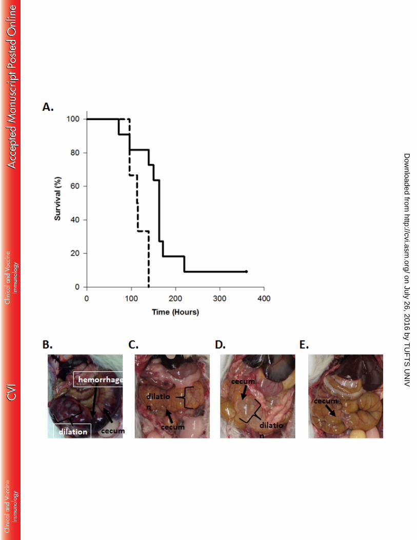
Microscopic examination of the large intestine identified submucosal edema as a cause of 340
colonic mural thickening, and mucosal neutrophilic infiltration as typical of CDI in piglets. As 341
expected based on the clinical observations and gross findings, the main difference observed by 342
light microscopy reflected lesion severity; more severe lesions were observed in control versus 343
VNA-treated piglets. There was a trend for more severe submucosal edema in control piglets 344
than VNA-treated (not shown). Similarly, there were more neutrophils in the large intestine of 345
control than VNA-treated piglets (Fig. 6A and 6B) which reached statistical significance for the 346
distal colon p=0.001) but not the spiral colon (p=0.0527). Some control pigs had epithelial 347
ulceration associated with neutrophilic colitis (Fig. 6C) while VNA-treated pigs did not develop 348
ulceration associated with neutrophilic colitis (Fig. 6D). 349
Blood collected from pigs one time during the course of the experiment and at 350
euthanasia, determined that VNA2-Tcd serum levels ranged from 2.7-4.7 µg/ml (Fig. S2C). 351
However, as observed in mice, serum neutralization abilities tested by cell cytotoxicity assay 352
were similar for all treated pigs with a 1:2 dilution of serum providing about 50% reduction in 353
cytotoxicity/cell rounding (Fig. S2D). 354
355
Pig CDI challenge following gene therapy using adenovirus expressing VNA2-Tcd 356
To further corroborate the efficacy of VNA2-Tcd in treating pigs for CDI, we employed a 357
gene therapy approach in which an adenovirus vector (Ad/VNA2-Tcd) was administered that 358
could promote the in vivo expression and secretion of VNA2-Tcd into the serum of treated pigs. 359
A total of 15 gnotobiotic pigs were used to assess the effectiveness of Ad/VNA2-Tcd. Six 360
control pigs were administered 1.0x10¹¹ vp of adenovirus (IV) expressing an unrelated VNA 361
and 9 pigs were administered 1.0x10¹¹ vp of Ad/VNA2-Tcd (IV). Each pig was treated twice 362
on July 26, 2016 by TU
FT
S U
NIV
http://cvi.asm.org/
Dow
nloaded from

17
with Ad/VNA2-Tcd; one day prior to oral inoculation with 106 UK6 C. difficile spores and again 363
three days after spore exposure. All six pigs in the control groups developed diarrhea within 48 364
hrs of inoculation with spores. Three of the six control pigs became moribund, all had moderate 365
to severe diarrhea, and two had systemic signs of disease including pleural effusion and ascites 366
(Table 2). In contrast, none of the nine Ad/VNA2-Tcd treated pigs became moribund and 367
diarrhea was predominantly mild to moderate with no other signs of disease (Table 2 and Fig. 5I 368
and 5K). Microscopic examination again supported the trend that VNA treatment reduced lesion 369
severity. More specifically, it was noted that treated pigs with high serum VNA levels had 370
predominantly mild to moderate edema with two pigs (#1 and 14) while pigs with low VNA-371
levels showed marked edema (Table3). Furthermore, Ad/VNA-treated pigs with the highest 372
levels of serum VNA (# 6 and 13) had minimal or no edema (Table 3). Two of six control pigs 373
(#7 and 15) showed minimal to marked levels of edema (Table 3). 374
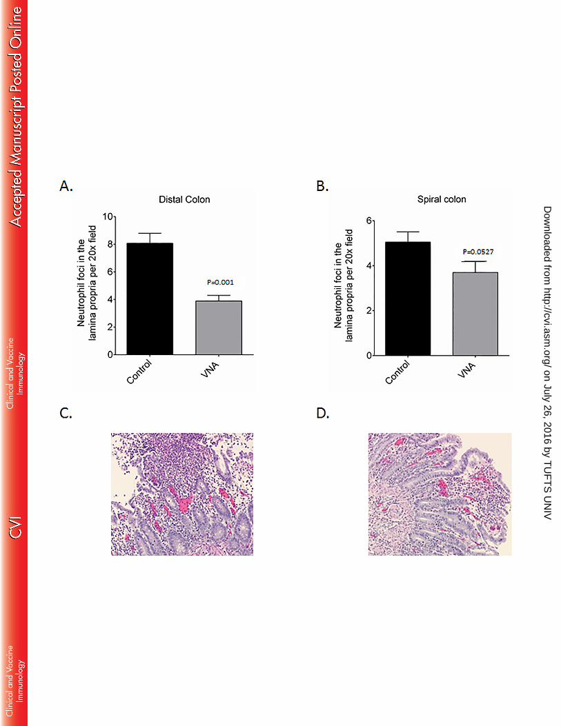
To determine whether there was a significant correlation between the severity of 375
symptoms and the serum level of VNA2-Tcd, blood was collected from pigs one to three times 376
during the course of the experiment and at euthanasia. Serum ELISAs showed that VNA2-Tcd 377
serum levels ranged from 20-1600 ng/ml at multiple time points during the experiment (Fig. 7A). 378
The association between VNA concentration in pig serum and disease severity was analyzed 379
using Spearman rank correlation. This analysis demonstrated a significant negative relationship 380
in which increased serum VNA concentration was clearly associated with lower CDI severity 381
score (rS = -0.614, p = 0.0443, Fig. 7B). 382
on July 26, 2016 by TU
FT
S U
NIV
http://cvi.asm.org/
Dow
nloaded from

18
383
Discussion 384
A single tetraspecific VNA protein comprised of four unique VHH binding agents, two 385
each targeting C. difficile toxin A or toxin B at distinct neutralizing epitopes reduced severity of 386
CDI clinical parameters and lesions in three different animal models: mice, hamsters, and 387
gnotobiotic pigs. We expressed and purified VNA2-Tcd, tested the agent for in vitro toxin 388
binding and neutralization activity, then used the VNA to treat mice that received systemically 389
delivered Tcd toxins. In the mouse model, a single dose of VNA (containing an albumin binding 390
peptide that improves VNA serum persistence in mice (61)) fully protected all groups that 391
received the VNA against any signs or symptoms of toxemia, while all the untreated mice died 392
within 4 hrs of toxin administration. The same purified VNA2-Tcd also significantly protected 393
mice, hamsters, and gnotobiotic piglets against systemic signs of disease in CDI challenge 394
models. In these models, the toxins, likely leaking from intestinal lesions, enter the peritoneal 395
space and blood stream inducing toxemia and systemic complications such as ascites and pleural 396
effusion (70, 71). 397
Since the VNA was administered systemically, we did not necessarily expect to see a 398
significant protective effect on the integrity of the mucosa or the GI tract, however previous 399
results indicated that there was a possibility that the VNA2-Tcd would be partially protective 400
against GI pathology (57, 72). In mice and pigs there was a significant protective effect against 401
diarrhea, edema, and hemorrhage in addition to protection against systemic disease. VNA2-Tcd 402
showed protection when administered IP against diarrhea in the mouse model (80% after the first 403
dose and 100% after the second dose) and against edema and severe diarrhea in the piglet model. 404
This suggests an ability of the agent to be effectively absorbed either through intestinal lesions 405
on July 26, 2016 by TU
FT
S U
NIV
http://cvi.asm.org/
Dow
nloaded from
19
(57, 72), loosening of epithelial cell tight junctions, or through normal portal absorption to some 406
degree, thereby mitigating effects of the toxin on microvilli degradation and neutrophil 407
infiltration in the lamina propria and edema in the spiral and distal colon. 408
The difference seen in protection against GI disease between the mouse and piglet 409
models of CDI may be attributed, at least in part, to the difference in the serum levels of VNA 410
achieved by the different treatments. The mice received three doses of 1.25-2.5mg/kg/day, while 411
the hamsters and piglets received a dose of 1-2 mg/kg/day. Perhaps more important, the VNA 412
contains an albumin binding peptide which was selected for mouse albumin affinity (73). This 413
peptide substantially increases the half-life of the VNA in mouse sera but has little or no 414
apparent affinity for albumins from other species (61) such as pig and therefore is unlikely to 415
extend serum half-life. Increasing the dose of VNA administered to hamster and piglets and/or 416
employing a VNA with an albumin binding peptide which binds hamster and pig albumin would 417
likely improve its efficacy in these models to become closer to the high levels achieved in the 418
mouse model. 419
Generally the hamster model of CDI is extremely sensitive and requires as few as 100 420
spores to induce diarrhea (74). However, hamsters were reported to be relatively resistant to the 421
UK6 strain of C. difficile (69). We did induce CDI disease in 100% of Syrian hamsters using 422
1000 UK6 spores and found that any sign of diarrhea in hamsters was always fatal in our model. 423
Hamsters sometimes died prior to external signs of diarrhea (wet tail) but with internal signs 424
(lack of formed feces in the GI tract). Diarrhea in mice and pigs ranged in severity, and only 425
severe diarrhea and systemic disease proved fatal in these animals. Since our treatment is 426
designed to protect against the systemic effects of CDI, with only limited access to the GI track, 427
it is not surprising that this treatment was less effective in our hamster model where hamsters did 428
on July 26, 2016 by TU
FT
S U
NIV
http://cvi.asm.org/
Dow
nloaded from

20
not develop systemic disease and instead became moribund very soon after displaying symptoms 429
of GI disease. Together, the clinical observations, necropsy and microscopic findings suggest 430
that morbidity in the hamster CDI model includes disease mechanisms which are independent 431
from those resulting in edema and neutrophilic inflammation. Possibilities include electrolyte 432
imbalances secondary to diarrhea and dehydration, hypovolemic shock due to fluid loss, and 433
poor perfusion/reduced venous return to the heart secondary to compression of the caudal vena 434
cava from massively dilated cecae. 435
Finding a method for oral administration of the bioactive VNA may permit more 436
effective protection against diarrhea and edema seen in the hamster and piglet CDI treatment 437
group. Although VHHs are generally more stable to pH and temperature extremes than 438
conventional antibodies (50), it seems likely that heteromultimeric VNAs will be susceptible to 439
gastric enzyme degradation. To overcome this problem, VNAs may be lyophilized and delivered 440
in drug capsules, or using a nanoparticle delivery system- or by some form of gene therapy that 441
promotes expression of VNA2-Tcd into the GI lumen (e.g. recombinant Lactococcus lactis) (75). 442
Alternatively, an adenovirus, that promotes secretion of VNA2-Tcd and is engineered to 443
selectively transduce intestinal epithelial cells (76, 77) may reduce intestinal edema and 444
inflammation and the severity of diarrhea. 445
VNA2-Tcd is directed against the two secreted toxins, not against the bacteria, and does 446
not involve the use of antibiotic therapy. Therefore, treatment with VNA2-Tcd is unlikely to 447
promote the occurrence of relapse associated with antibiotic treatments against CDI which is the 448
major cause of fatal disease in humans (1). Previous results using therapeutic antibodies against 449
CDI suggest this is possible (35). 450
on July 26, 2016 by TU
FT
S U
NIV
http://cvi.asm.org/
Dow
nloaded from
21
Using a single tetraspecific agent to express a polyprotein with multiple linked VHHs 451
against two different Tcd toxins, and the feasibility of using microbial hosts for production, 452
reduces manufacturing costs and reduces the complexity of clinical trials. Addition of an albumin 453
binding peptide that effectively binds human albumin would increase the serum persistence of 454
the VNA, thereby increasing steady state serum levels and reduce the frequency of doses 455
required to effectively treat CDI pathology. The fact that VNAs can be effectively delivered by 456
gene therapy (64-66) opens the possibility of developing single dose therapies that provide 457
prolonged efficacy and protect patients from relapses. 458
459
Acknowledgements 460
With thanks to our animal care technicians, Patricia Boucher, Rachel Nieminen and John Feddo 461
for providing excellent care for all of our piglets and to Yimin Zhang for statistical analysis of 462
hamster data. 463
464
Funding Information 465
This work was supported in part by the National Institutes of Health (grant R01AI088748 to ST). 466
467
References 468
1. Rupnik M, Wilcox MH, Gerding DN. 2009. Clostridium difficile infection: new 469
developments in epidemiology and pathogenesis. Nat Rev Microbiol 7:526-536. 470
on July 26, 2016 by TU
FT
S U
NIV
http://cvi.asm.org/
Dow
nloaded from
22
2. Jarvis WR, Schlosser J, Jarvis AA, Chinn RY. 2009. National point prevalence of 471
Clostridium difficile in US health care facility inpatients, 2008. Am J Infect Control 472
37:263-270. 473
3. Bartlett JG, Perl TM. 2005. The new Clostridium difficile--what does it mean? N Engl J 474
Med 353:2503-2505. 475
4. Freeman J, Bauer MP, Baines SD, Corver J, Fawley WN, Goorhuis B, Kuijper EJ, 476
Wilcox MH. 2010. The changing epidemiology of Clostridium difficile infections. Clin 477
Microbiol Rev 23:529-549. 478
5. Kim J, Smathers SA, Prasad P, Leckerman KH, Coffin S, Zaoutis T. 2008. 479
Epidemiological features of Clostridium difficile-associated disease among inpatients at 480
children's hospitals in the United States, 2001-2006. Pediatrics 122:1266-1270. 481
6. Pituch H. 2009. Clostridium difficile is no longer just a nosocomial infection or an 482
infection of adults. Int J Antimicrob Agents 33 Suppl 1:S42-45. 483
7. Sunenshine RH, McDonald LC. 2006. Clostridium difficile-associated disease: new 484
challenges from an established pathogen. Cleve Clin J Med 73:187-197. 485
8. Higa JT, Kelly CP. 2013. New Drugs and Strategies for Management of Clostridium 486
difficile Colitis. J Intensive Care Med doi:10.1177/0885066613475426. 487
9. Durai R. 2007. Epidemiology, pathogenesis, and management of Clostridium difficile 488
infection. Dig Dis Sci 52:2958-2962. 489
10. Lyerly DM, Krivan HC, Wilkins TD. 1988. Clostridium difficile: its disease and toxins. 490
Clin Microbiol Rev 1:1-18. 491
11. Bartlett JG. 2006. Narrative review: the new epidemic of Clostridium difficile- 492
associated enteric disease. Ann Intern Med 145:758-764. 493
on July 26, 2016 by TU
FT
S U
NIV
http://cvi.asm.org/
Dow
nloaded from

23
12. Voth DE, Ballard JD. 2005. Clostridium difficile toxins: mechanism of action and role 494
in disease. Clin Microbiol Rev 18:247-263. 495
13. Zar FA, Bakkanagari SR, Moorthi KM, Davis MB. 2007. A comparison of 496
vancomycin and metronidazole for the treatment of Clostridium difficile- associated 497
diarrhea, stratified by disease severity. Clin Infect Dis 45:302-307. 498
14. Barbut F, Richard A, Hamadi K, Chomette V, Burghoffer B, Petit JC. 2000. 499
Epidemiology of recurrences or reinfections of Clostridium difficile-associated diarrhea. J 500
Clin Microbiol 38:2386-2388. 501
15. Brown AT, Seifert CF. 2014. Effect of treatment variation on outcomes in patients with 502
Clostridium difficile. Am J Med 127:865-870. 503
16. Giannasca PJ, Warny M. 2004. Active and passive immunization against Clostridium 504
difficile diarrhea and colitis. Vaccine 22:848-856. 505
17. Scott LJ. 2013. Fidaxomicin: a review of its use in patients with Clostridium difficile 506
infection. Drugs 73:1733-1747. 507
18. Allen SJ, Wareham K, Wang D, Bradley C, Hutchings H, Harris W, Dhar A, Brown 508
H, Foden A, Gravenor MB, Mack D. 2013. Lactobacilli and bifidobacteria in the 509
prevention of antibiotic-associated diarrhoea and Clostridium difficile diarrhoea in older 510
inpatients (PLACIDE): a randomised, double-blind, placebo-controlled, multicentre trial. 511
Lancet 382:1249-1257. 512
19. Allen SJ, Wareham K, Wang D, Bradley C, Sewell B, Hutchings H, Harris W, Dhar 513
A, Brown H, Foden A, Gravenor MB, Mack D, Phillips CJ. 2013. A high- dose 514
preparation of lactobacilli and bifidobacteria in the prevention of antibiotic- associated 515
and Clostridium difficile diarrhoea in older people admitted to hospital: a multicentre, 516
on July 26, 2016 by TU
FT
S U
NIV
http://cvi.asm.org/
Dow
nloaded from

24
randomised, double-blind, placebo-controlled, parallel arm trial (PLACIDE). Health 517
Technol Assess 17:1-140. 518
20. Gorbach SL. 2000. Probiotics and gastrointestinal health. Am J Gastroenterol 95:S2-4. 519
21. Maziade PJ, Andriessen JA, Pereira P, Currie B, Goldstein EJ. 2013. Impact of 520
adding prophylactic probiotics to a bundle of standard preventative measures for 521
Clostridium difficile infections: enhanced and sustained decrease in the incidence and 522
severity of infection at a community hospital. Curr Med Res Opin 29:1341-1347. 523
22. Pattani R, Palda VA, Hwang SW, Shah PS. 2013. Probiotics for the prevention of 524
antibiotic-associated diarrhea and Clostridium difficile infection among hospitalized 525
patients: systematic review and meta-analysis. Open Med 7:e56-67. 526
23. Rainkie D, Kolber MR. 2013. Probiotics for the prevention of Clostridium difficile. Can 527
Fam Physician 59:957. 528
24. MacConnachie AA, Fox R, Kennedy DR, Seaton RA. 2009. Faecal transplant for 529
recurrent Clostridium difficile-associated diarrhoea: a UK case series. Qjm 102:781-784. 530
25. Rubin TA, Gessert CE, Aas J. 2009. Stool transplantation for older patients with 531
Clostridium difficile infection. J Am Geriatr Soc 57:2386. 532
26. Vindigni SM, Broussard EK, Surawicz CM. 2013. Alteration of the intestinal 533
microbiome: fecal microbiota transplant and probiotics for Clostridium difficile and 534
beyond. Expert Rev Gastroenterol Hepatol 7:615-628. 535
27. Kurtz CB, Cannon EP, Brezzani A, Pitruzzello M, Dinardo C, Rinard E, Acheson 536
DW, Fitzpatrick R, Kelly P, Shackett K, Papoulis AT, Goddard PJ, Barker RH, Jr., 537
Palace GP, Klinger JD. 2001. GT160-246, a toxin binding polymer for treatment of 538
Clostridium difficile colitis. Antimicrob Agents Chemother 45:2340-2347. 539
on July 26, 2016 by TU
FT
S U
NIV
http://cvi.asm.org/
Dow
nloaded from

25
28. Gardiner DF, Rosenberg T, Zaharatos J, Franco D, Ho DD. 2009. A DNA vaccine 540
targeting the receptor binding domain of Clostridium difficile toxin A. Vaccine 27:3598-541
3604. 542
29. Ghose C, Kalsy A, Sheikh A, Rollenhagen J, John M, Young J, Rollins SM, Qadri F, 543
Calderwood SB, Kelly CP, Ryan ET. 2007. Transcutaneous immunization with 544
Clostridium difficile toxoid A induces systemic and mucosal immune responses and toxin 545
A-neutralizing antibodies in mice. Infect Immun 75:2826-2832. 546
30. Sougioultzis S, Kyne L, Drudy D, Keates S, Maroo S, Pothoulakis C, Giannasca PJ, 547
Lee CK, Warny M, Monath TP, Kelly CP. 2005. Clostridium difficile toxoid vaccine in 548
recurrent C. difficile-associated diarrhea. Gastroenterology 128:764-770. 549
31. Abougergi MS, Kwon JH. 2011. Intravenous immunoglobulin for the treatment of 550
Clostridium difficile infection: a review. Dig Dis Sci 56:19-26. 551
32. Babcock GJ, Broering TJ, Hernandez HJ, Mandell RB, Donahue K, Boatright N, 552
Stack AM, Lowy I, Graziano R, Molrine D, Ambrosino DM, Thomas WD, Jr. 2006. 553
Human monoclonal antibodies directed against toxins A and B prevent Clostridium 554
difficile-induced mortality in hamsters. Infect Immun 74:6339-6347. 555
33. Demarest SJ, Hariharan M, Elia M, Salbato J, Jin P, Bird C, Short JM, Kimmel 556
BE, Dudley M, Woodnutt G, Hansen G. 2010. Neutralization of Clostridium difficile 557
toxin A using antibody combinations. MAbs 2:190-198. 558
34. Leav BA, Blair B, Leney M, Knauber M, Reilly C, Lowy I, Gerding DN, Kelly CP, 559
Katchar K, Baxter R, Ambrosino D, Molrine D. 2010. Serum anti-toxin B antibody 560
correlates with protection from recurrent Clostridium difficile infection (CDI). Vaccine 561
28:965-969. 562
on July 26, 2016 by TU
FT
S U
NIV
http://cvi.asm.org/
Dow
nloaded from

26
35. Lowy I, Molrine DC, Leav BA, Blair BM, Baxter R, Gerding DN, Nichol G, Thomas 563
WD, Jr., Leney M, Sloan S, Hay CA, Ambrosino DM. 2010. Treatment with 564
monoclonal antibodies against Clostridium difficile toxins. N Engl J Med 362:197-205. 565
36. Mulvey GL, Dingle TC, Fang L, Strecker J, Armstrong GD. 2011. Therapeutic 566
potential of egg yolk antibodies for treating Clostridium difficile infection. J Med 567
Microbiol 60:1181-1187. 568
37. Saito T, Kimura S, Tateda K, Mori N, Hosono N, Hayakawa K, Akasaka Y, Ishii T, 569
Sumiyama Y, Kusachi S, Nagao J, Yamaguchi K. 2011. Evidence of intravenous 570
immunoglobulin as a critical supportive therapy against Clostridium difficile toxin-571
mediated lethality in mice. J Antimicrob Chemother 66:1096-1099. 572
38. Andersen KK, Strokappe NM, Hultberg A, Truusalu K, Smidt I, Mikelsaar RH, 573
Mikelsaar M, Verrips T, Hammarstrom L, Marcotte H. 2015. Neutralization of 574
Clostridium difficile toxin B mediated by engineered lactobacilli producing single 575
domain antibodies. Infect Immun 84:395-406. 576
39. Hussack G, Arbabi-Ghahroudi M, van Faassen H, Songer JG, Ng KK, MacKenzie 577
R, Tanha J. 2011. Neutralization of Clostridium difficile toxin A with single-domain 578
antibodies targeting the cell receptor binding domain. J Biol Chem 286:8961-8976. 579
40. Taylor CP, Tummala S, Molrine D, Davidson L, Farrell RJ, Lembo A, Hibberd PL, 580
Lowy I, Kelly CP. 2008. Open-label, dose escalation phase I study in healthy volunteers 581
to evaluate the safety and pharmacokinetics of a human monoclonal antibody to 582
Clostridium difficile toxin A. Vaccine 26:3404-3409. 583
41. Yang Z, Schmidt D, Liu W, Li S, Shi L, Sheng J, Chen K, Yu H, Tremblay JM, 584
Chen X, Piepenbrink KH, Sundberg EJ, Kelly CP, Bai G, Shoemaker CB, Feng H. 585
on July 26, 2016 by TU
FT
S U
NIV
http://cvi.asm.org/
Dow
nloaded from

27
2014. A novel multivalent, single-domain antibody targeting TcdA and TcdB prevents 586
fulminant Clostridium difficile infection in mice. J Infect Dis doi:10.1093/infdis/jiu196. 587
42. Kociolek LK, Gerding DN. 2016. Breakthroughs in the treatment and prevention of 588
Clostridium difficile infection. Nat Rev Gastroenterol Hepatol 13:150-60. 589
43. Steele J, Mukherjee J, Parry N, Tzipori S. 2013. Antibody against TcdB, but not 590
TcdA, prevents development of gastrointestinal and systemic Clostridium difficile 591
disease. J Infect Dis 207:323-330. 592
44. Kuehne SA, Cartman ST, Minton NP. 2011. Both, toxin A and toxin B, are important 593
in Clostridium difficile infection. Gut Microbes 2:252-255. 594
45. Lyras D, O'Connor JR, Howarth PM, Sambol SP, Carter GP, Phumoonna T, Poon 595
R, Adams V, Vedantam G, Johnson S, Gerding DN, Rood JI. 2009. Toxin B is 596
essential for virulence of Clostridium difficile. Nature 458:1176-1179. 597
46. Young VB, Hanna PC. 2014. Overlapping roles for toxins in Clostridium difficile 598
infection. J Infect Dis 209:9-11. 599
47. Maass DR, Sepulveda J, Pernthaner A, Shoemaker CB. 2007. Alpaca (Lama pacos) 600
as a convenient source of recombinant camelid heavy chain antibodies (VHHs). J 601
Immunol Methods 324:13-25. 602
48. Arbabi Ghahroudi M, Desmyter A, Wyns L, Hamers R, Muyldermans S. 1997. 603
Selection and identification of single domain antibody fragments from camel heavy-chain 604
antibodies. FEBS Lett 414:521-526. 605
49. Dumoulin M, Conrath K, Van Meirhaeghe A, Meersman F, Heremans K, Frenken 606
LG, Muyldermans S, Wyns L, Matagne A. 2002. Single-domain antibody fragments 607
with high conformational stability. Protein Sci 11:500-515. 608
on July 26, 2016 by TU
FT
S U
NIV
http://cvi.asm.org/
Dow
nloaded from

28
50. van der Linden RH, Frenken LG, de Geus B, Harmsen MM, Ruuls RC, Stok W, de 609
Ron L, Wilson S, Davis P, Verrips CT. 1999. Comparison of physical chemical 610
properties of llama VHH antibody fragments and mouse monoclonal antibodies. Biochim 611
Biophys Acta 1431:37-46. 612
51. Mukherjee J, Tremblay JM, Leysath CE, Ofori K, Baldwin K, Feng X, Bedenice D, 613
Webb RP, Wright PM, Smith LA, Tzipori S, Shoemaker CB. 2012. A novel strategy 614
for development of recombinant antitoxin therapeutics tested in a mouse botulism model. 615
PLoS One 7:e29941. 616
52. Vance DJ, Tremblay JM, Mantis NJ, Shoemaker CB. 2013. Stepwise engineering of 617
heterodimeric single domain camelid VHH antibodies that passively protect mice from 618
ricin toxin. J Biol Chem 288:36538-36547. 619
53. Tremblay JM, Mukherjee J, Leysath CE, Debatis M, Ofori K, Baldwin K, Boucher 620
C, Peters R, Beamer G, Sheoran A, Bedenice D, Tzipori S, Shoemaker CB. 2013. A 621
single VHH-based toxin-neutralizing agent and an effector antibody protect mice against 622
challenge with Shiga toxins 1 and 2. Infect Immun 81:4592-4603. 623
54. Moayeri M, Leysath CE, Tremblay JM, Vrentas C, Crown D, Leppla SH, 624
Shoemaker CB. 2015. A heterodimer of a VHH (variable domains of camelid heavy 625
chain-only) antibody that inhibits anthrax toxin cell binding linked to a VHH antibody 626
that blocks oligomer formation is highly protective in an anthrax spore challenge model. J 627
Biol Chem 290:6584-6595. 628
55. Yang Z, Schmidt D, Liu W, Li S, Shi L, Sheng J, Chen K, Yu H, Tremblay JM, 629
Chen X, Piepenbrink KH, Sundberg EJ, Kelly CP, Bai G, Shoemaker CB, Feng H. 630
on July 26, 2016 by TU
FT
S U
NIV
http://cvi.asm.org/
Dow
nloaded from

29
2014. A novel multivalent, single-domain antibody targeting TcdA and TcdB prevents 631
fulminant Clostridium difficile infection in mice. J Infect Dis 210:964-972. 632
56. Kuehne SA, Collery MM, Kelly ML, Cartman ST, Cockayne A, Minton NP. 2014. 633
Importance of toxin A, toxin B, and CDT in virulence of an epidemic Clostridium 634
difficile strain. J Infect Dis 209:83-86. 635
57. Yang Z, Ramsey J, Hamza T, Zhang Y, Li S, Yfantis HG, Lee D, Hernandez LD, 636
Seghezzi W, Furneisen JM, Davis NM, Therien AG, Feng H. 2015. Mechanisms of 637
protection against Clostridium difficile infection by the monoclonal antitoxin antibodies 638
actoxumab and bezlotoxumab. Infect Immun 83:822-831. 639
58. Herrera C, Tremblay JM, Shoemaker CB, Mantis NJ. 2015. Mechanisms of Ricin 640
Toxin Neutralization Revealed through Engineered Homodimer Camelid Antibodies. J 641
Biol Chem 290:27880-27889. 642
59. Tremblay JM, Kuo CL, Abeijon C, Sepulveda J, Oyler G, Hu X, Jin MM, 643
Shoemaker CB. 2010. Camelid single domain antibodies (VHHs) as neuronal cell 644
intrabody binding agents and inhibitors of Clostridium botulinum neurotoxin (BoNT) 645
proteases. Toxicon 56:990-998. 646
60. Luo J, Deng ZL, Luo X, Tang N, Song WX, Chen J, Sharff KA, Luu HH, Haydon 647
RC, Kinzler KW, Vogelstein B, He TC. 2007. A protocol for rapid generation of 648
recombinant adenoviruses using the AdEasy system. Nat Protoc 2:1236-1247. 649
61. Mukherjee J, Dmitriev I, Debatis M, Tremblay JM, Beamer G, Kashentseva EA, 650
Curiel DT, Shoemaker CB. 2014. Prolonged prophylactic protection from botulism with 651
a single adenovirus treatment promoting serum expression of a VHH-based antitoxin 652
protein. PLoS One 9:e106422. 653
on July 26, 2016 by TU
FT
S U
NIV
http://cvi.asm.org/
Dow
nloaded from

30
62. Sheoran AS, Dmitriev IP, Kashentseva EA, Cohen O, Mukherjee J, Debatis M, 654
Shearer J, Tremblay JM, Beamer G, Curiel DT, Shoemaker CB, Tzipori S. 2015. 655
Adenovirus vector expressing Stx1/Stx2-neutralizing agent protects piglets infected with 656
Escherichia coli O157:H7 against fatal systemic intoxication. Infect Immun 83:286-291. 657
63. Maizel JV, Jr., White DO, Scharff MD. 1968. The polypeptides of adenovirus. I. 658
Evidence for multiple protein components in the virion and a comparison of types 2, 7A, 659
and 12. Virology 36:115-125. 660
64. Moayeri M, Tremblay JM, Debatis M, Dmietriev IP, Kashentseva EA, Yeh AJ, 661
Cheung GY, Curiel DT, Leppla S, Shoemaker CB. 2016. Adenoviral expression of a 662
bispecific VHH-based neutralizing agent targeting protective antigen provides 663
prophylactic protection from anthrax in mice. Clin Vaccine Immunol 23:213-8. 664
65. Sponseller JK, Steele JA, Schmidt DJ, Kim HB, Beamer G, Sun X, Tzipori S. 2014. 665
Hyperimmune Bovine Colostrum as a Novel Therapy to Combat Clostridium difficile 666
Infection. J Infect Dis 211:1334-41. 667
66. Tzipori S, Gunzer F, Donnenberg MS, de Montigny L, Kaper JB, Donohue-Rolfe A. 668
1995. The role of the eaeA gene in diarrhea and neurological complications in a 669
gnotobiotic piglet model of enterohemorrhagic Escherichia coli infection. Infect Immun 670
63:3621-3627. 671
67. Chen X, Katchar K, Goldsmith JD, Nanthakumar N, Cheknis A, Gerding DN, Kelly 672
CP. 2008. A mouse model of Clostridium difficile-associated disease. Gastroenterology 673
135:1984-1992. 674
68. Killgore G, Thompson A, Johnson S, Brazier J, Kuijper E, Pepin J, Frost EH, 675
Savelkoul P, Nicholson B, van den Berg RJ, Kato H, Sambol SP, Zukowski W, 676
on July 26, 2016 by TU
FT
S U
NIV
http://cvi.asm.org/
Dow
nloaded from

31
Woods C, Limbago B, Gerding DN, McDonald LC. 2008. Comparison of seven 677
techniques for typing international epidemic strains of Clostridium difficile: restriction 678
endonuclease analysis, pulsed-field gel electrophoresis, PCR- ribotyping, multilocus 679
sequence typing, multilocus variable-number tandem- repeat analysis, amplified fragment 680
length polymorphism, and surface layer protein A gene sequence typing. J Clin Microbiol 681
46:431-437. 682
69. Douce G, Goulding D. 2010. Refinement of the hamster model of Clostridiumdifficile 683
disease. Methods Mol Biol 646:215-227. 684
70. Steele J, Chen K, Sun X, Zhang Y, Wang H, Tzipori S, Feng H. 2012. Systemic 685
dissemination of Clostridium difficile toxins A and B is associated with severe, fatal 686
disease in animal models. J Infect Dis 205:384-391. 687
71. Steele J, Feng H, Parry N, Tzipori S. 2010. Piglet models of acute or chronic 688
Clostridium difficile illness. J Infect Dis 201:428-434. 689
72. Cohen OR, Steele JA, Zhang Q, Schmidt DJ, Wang Y, Hamel PE, Beamer G, Xu B, 690
Tzipori S. 2014. Systemically administered IgG anti-toxin antibodies protect the colonic 691
mucosa during infection with Clostridium difficile in the piglet model. PLoS One 692
9:e111075. doi: 10.1371/journal.pone.0111075. 693
73. Nguyen A, Reyes AE, 2nd, Zhang M, McDonald P, Wong WL, Damico LA, Dennis 694
MS. 2006. The pharmacokinetics of an albumin-binding Fab (AB.Fab) can be modulated 695
as a function of affinity for albumin. Protein Eng Des Sel 19:291-297. 696
74. Buckley AM, Spencer J, Candlish D, Irvine JJ, Douce GR. 2011. Infection of 697
hamsters with the UK Clostridium difficile ribotype 027 outbreak strain R20291. J Med 698
Microbiol 60:1174-1180. 699
on July 26, 2016 by TU
FT
S U
NIV
http://cvi.asm.org/
Dow
nloaded from

32
75. Vandenbroucke K, de Haard H, Beirnaert E, Dreier T, Lauwereys M, Huyck L, 700
Van Huysse J, Demetter P, Steidler L, Remaut E, Cuvelier C, Rottiers P. 2010. 701
Orally administered L. lactis secreting an anti-TNF Nanobody demonstrate efficacy in 702
chronic colitis. Mucosal Immunol 3:49-56. 703
76. Lu ZH, Kaliberov S, Zhang J, Muz B, Azab AK, Sohn RE, Kaliberova L, Du Y, 704
Curiel DT, Arbeit JM. 2014. The myeloid-binding peptide adenoviral vector enables 705
multi-organ vascular endothelial gene targeting. Lab Invest 94:881-892. 706
77. Tang SC, Sambanis A, Sibley E. 2005. Proteasome modulating agents induce rAAV2-707
mediated transgene expression in human intestinal epithelial cells. Biochem Biophys Res 708
Commun 331:1392-1400. 709
710
711
Table 1. Pig treatment groups 712
NA=not applicable 713 714
715
716
717
# of pigs
Control Buffer
Control Adenovirus
VNA2-Tcd Ad/VNA2-Tcd
Time (hours)
Route
Group 1 3 NA NA 1mg NA -4,24, then every 12 hours
IP
Group 2 3 NA NA 1mg NA 18, then every 12 hours
IM
Group 3 9 NA NA NA 1.0x10¹¹vp -24, 72 IV Group 4 6 1ml NA NA NA -4, 24, then
every 12 hours IM
Group 5 6 NA 1.0x10¹¹vp NA NA -24, 72 IV Group 6 3 NA NA NA NA NA NA
on July 26, 2016 by TU
FT
S U
NIV
http://cvi.asm.org/
Dow
nloaded from

33
Table 2. Pig clinical signs of disease 718
a Severity of gastrointestinal disease was determined by clinical signs of edema, hemorrhage, 719 rectal prolapse, diarrhea, and gross and histopathologic lesions, ranging from mild to severe. 720 b Systemic signs include pleural effusion and ascites. 721 c Fatal disease indicates that piglets were euthanized due to the severity of the disease. 722 723
Table 3. Clostridium difficile infection and VNA2-Tcd levels in pig serum treated with 724
Adenovirus ad/VNA2-Tcd 725
726
727
728
729
730
731
732
733
734
735
736
Treatment (# of animals) Gastrointestinal disease (%)a Systemic Disease (%) b Fatal Disease (%) c
Uninfected control (3) mild-moderate diarrhea (100), rectal prolapse (0) 0 0
UK6 spores + VNA2-Tcd (6) mild-moderate diarrhea (100), rectal prolapse (0) 0 0
UK6 spores + VNA2-Tcd - Adeno (9)
mild-severe diarrhea (100), rectal prolapse (0) 0 0
UK6 spores + buffer (6) moderate-severe diarrhea (100), rectal prolapse (83) 50 50
UK6 spores + control-Adeno (6) moderate-severe diarrhea (100), rectal prolapse (50) 33 50
Pig
#
Dise
ase
seve
rity
scor
e ng
/ml a
d/VN
A-Tc
d
Mes
ocol
on
edem
a S
ubm
ucos
a ed
ema
Lam
ina
prop
ria
edem
a Ne
utro
phili
c co
lits
Epith
eliu
m
Lum
inal
co
nten
ts
5 4 160 +/- - +/- - Intact None 9 1 300 + +/- - - Intact None 12 3 350 - - - - Intact None 13 4 1000 + - - +/- Intact None 6 0 1600 ++ - +/- - Intact None 1 3 30 NAa +++ ++ ++ Intact necrotic cell debris (+) 2 3 100 + + ++ +/- Intact None 14 10 150 + +++ ++ ++ Ulcerated necrotic cell debris (++) 7b 9 0 ++ +/- ++ ++ Intact None 15b 11 0 ++ +++ ++ - Intact None
aNA = Not Available
bControl pig treated with unrelated VNA
on July 26, 2016 by TU
FT
S U
NIV
http://cvi.asm.org/
Dow
nloaded from

34
737
Fig 1. Synthesis and purification of VNA2-Tcd. A) Schematic diagram of TcdA and TcdB 738
toxins respectively, GTD indicates the enzymatic glucosyltransferase domain, TMD indicates the 739
transmembrane domain and RBD is the receptor binding domain. AH3 and AA6 bind in the 740
GTD region and TMD region of TcdA respectively, 5D and E3 bind to different regions of the 741
GTD in TcdB. B) Diagram of the heterotetramer VNA2-Tcd, which was synthetically generated 742
containing two potent neutralizing VHHs to each toxin. The protein contains a thioredoxin 743
protein at the amino end, followed by four VHHs separated by a flexible spacer peptide with a 744
carboxyl terminal E-tag peptide and an albumin binding peptide to increase serum persistence. 745
746
Fig. 2. In vitro and in vivo neutralization of toxin. A) and B) In vitro neutralization assay 747
using Vero cells incubated with 100 pM TcdA or 0.6pM TcdB per well for 24 hrs with serial 748
dilutions of VNA2-Tcd as indicated. Percent cell rounding (cytotoxicity) was assessed after 24 749
hours. The IC50 was determined using GraphPad nonlinear fit of log transformed concentrations 750
Log IC50 for TcdA = 2.0 and the IC50 was 106.4, Log IC50 for TcdB = 1.24 and the IC50 was 751
13.3. 752
753
Fig. 3. Protection against CDI in mice using VNA2-Tcd. Mice were treated with an antibiotic 754
cocktail for 3 days in drinking water and given a single injection of clindamycin IP one day prior 755
to infection with 106 pfu of Clostridium difficile UK6 spores alone or UK6 spores and 3 doses of 756
VNA2-Tcd (2.5 mg/kg at 4, 24, and 48 hr) post infection. A) Weight of mice in treated (VNA2-757
Tcd) and untreated (PBS) groups during the six day study. A Mann Whitney U test was 758
performed to compare the control and VNA treated mice per day, only day 2 (*) showed 759
on July 26, 2016 by TU
FT
S U
NIV
http://cvi.asm.org/
Dow
nloaded from
35
statistically significant p=0.021 weights between the control and treated groups. B) Survival 760
percentage with time for each group. 761
762
Fig. 4. Protection against CDI in hamsters using VNA2-Tcd. A) Survival percentage with 763
time for hamsters treated with VNA2-Tcd. X axis shows hours post oral challenge with 1000 764
UK6 spores. Y axis shows percent survival. Solid black line indicates survival in VNA2-Tcd 765
treated group and dashed black line indicates survival in control (PBS) group. The grouped 766
survival data were analyzed by applying a Kaplan-Meier log rank test using SigmaPlot (version 767
13.0; Systat Software, Inc.). The log rank statistic for the survival curves is greater than would be 768
expected by chance as the two curves show a statistically significant difference (P = 0.003). B) 769
Cecum (arrowhead) of control hamster showing hemorrhage (arrow) and dilation. C) Cecum 770
(arrowhead) of control hamster showing dilation. D) Cecum (arrowhead) of VNA2-Tcd treated 771
hamster showing dilation. E) Cecum of treated hamster showing no dilation. 772
773
Fig. 5. Necropsy images from piglets infected with C. difficile. A) Control piglet with pleural 774
effusion (arrow), lung displaying diffuse hyperemia and congestion. B) VNA2-Tcd treated piglet 775
with normally aerated lung and no visible pleural effusion. C) Control piglet with ascites (arrow) 776
moderate dilation, mesocolonic edema, and hyperemia in the spiral colon. D) VNA2-Tcd treated 777
piglet with moderate dilation and mild mesocolonic edema in the spiral colon. E through I) 778
Necropsy images of spiral colons from control and VNA2-Tcd treated pigs inoculated with C. 779
difficile spores. E) Moderate mesocolonic edema, (arrow) hyperemia, focal mucosal ulceration, 780
hemorrhages and dilation in a control piglet. F) Severe mesocolonic edema (arrow) in a control 781
piglet. G) Mild mesocolonic edema (arrow), dilation and moderate hyperemia in a VNA2-Tcd 782
on July 26, 2016 by TU
FT
S U
NIV
http://cvi.asm.org/
Dow
nloaded from
36
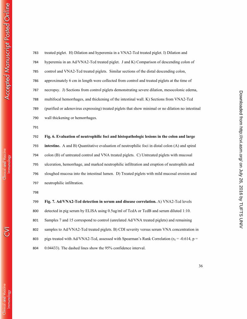
treated piglet. H) Dilation and hyperemia in a VNA2-Tcd treated piglet. I) Dilation and 783
hyperemia in an Ad/VNA2-Tcd treated piglet. J and K) Comparison of descending colon of 784
control and VNA2-Tcd treated piglets. Similar sections of the distal descending colon, 785
approximately 6 cm in length were collected from control and treated piglets at the time of 786
necropsy. J) Sections from control piglets demonstrating severe dilation, mesocolonic edema, 787
multifocal hemorrhages, and thickening of the intestinal wall. K) Sections from VNA2-Tcd 788
(purified or adenovirus expressing) treated piglets that show minimal or no dilation no intestinal 789
wall thickening or hemorrhages. 790
791
Fig. 6. Evaluation of neutrophilic foci and histopathologic lesions in the colon and large 792
intestine. A and B) Quantitative evaluation of neutrophilic foci in distal colon (A) and spiral 793
colon (B) of untreated control and VNA treated piglets. C) Untreated piglets with mucosal 794
ulceration, hemorrhage, and marked neutrophilic infiltration and eruption of neutrophils and 795
sloughed mucosa into the intestinal lumen. D) Treated piglets with mild mucosal erosion and 796
neutrophilic infiltration. 797
798
Fig. 7. Ad/VNA2-Tcd detection in serum and disease correlation. A) VNA2-Tcd levels 799
detected in pig serum by ELISA using 0.5ug/ml of TcdA or TcdB and serum diluted 1:10. 800
Samples 7 and 15 correspond to control (unrelated Ad/VNA treated piglets) and remaining 801
samples to Ad/VNA2-Tcd treated piglets. B) CDI severity versus serum VNA concentration in 802
pigs treated with Ad/VNA2-Tcd, assessed with Spearman’s Rank Correlation (rS = -0.614, p = 803
0.04433). The dashed lines show the 95% confidence interval. 804
on July 26, 2016 by TU
FT
S U
NIV
http://cvi.asm.org/
Dow
nloaded from