Clin. Vaccine Immunol. doi:10.1128/CVI.00022-07 Bannantine...
37
Bannantine et al. 1 2 4 Development and Characterization of Monoclonal Antibodies and Aptamers Against Major Antigens of Mycobacterium avium subsp. 6 paratuberculosis 8 10 John P. Bannantine 1* , Thomas J. Radosevich 1 , Judith R. Stabel 1 , Srinand Sreevatsan 2 , 12 Vivek Kapur 3 and Michael L. Paustian 1 14 16 1 National Animal Disease Center, USDA-ARS, Ames, Iowa, USA 2 Veterinary Population Medicine Department, 3 Departments of Microbiology and 18 Biomedical Genomics, University of Minnesota, St. Paul, Minnesota, USA 20 22 24 26 *Corresponding Author: National Animal Disease Center ARS – USDA 28 2300 North Dayton Avenue Ames, Iowa 50010 30 Telephone (515) 663-7340 FAX: 515-663-7458 32 Email: [email protected] 34 36 38 Running title: M. paratuberculosis antibodies and aptamers 40 42 Keywords: Mycobacterium paratuberculosis, Johne’s disease, diagnostics, antibodies, 44 bacterial detection ACCEPTED Copyright © 2007, American Society for Microbiology and/or the Listed Authors/Institutions. All Rights Reserved. Clin. Vaccine Immunol. doi:10.1128/CVI.00022-07 CVI Accepts, published online ahead of print on 7 March 2007 on July 8, 2018 by guest http://cvi.asm.org/ Downloaded from
-
Upload
nguyenmien -
Category
Documents
-
view
214 -
download
0
Transcript of Clin. Vaccine Immunol. doi:10.1128/CVI.00022-07 Bannantine...

Bannantine et al. 1
2
4
Development and Characterization of Monoclonal Antibodies and
Aptamers Against Major Antigens of Mycobacterium avium subsp. 6
paratuberculosis
8 10
John P. Bannantine1*, Thomas J. Radosevich1, Judith R. Stabel1, Srinand Sreevatsan2, 12 Vivek Kapur3 and Michael L. Paustian1
14 16
1National Animal Disease Center, USDA-ARS, Ames, Iowa, USA 2Veterinary Population Medicine Department, 3Departments of Microbiology and 18
Biomedical Genomics, University of Minnesota, St. Paul, Minnesota, USA 20 22 24 26 *Corresponding Author: National Animal Disease Center
ARS – USDA 28 2300 North Dayton Avenue Ames, Iowa 50010 30 Telephone (515) 663-7340 FAX: 515-663-7458 32 Email: [email protected] 34 36 38 Running title: M. paratuberculosis antibodies and aptamers 40 42 Keywords: Mycobacterium paratuberculosis, Johne’s disease, diagnostics, antibodies, 44 bacterial detection
ACCEPTED
Copyright © 2007, American Society for Microbiology and/or the Listed Authors/Institutions. All Rights Reserved.Clin. Vaccine Immunol. doi:10.1128/CVI.00022-07 CVI Accepts, published online ahead of print on 7 March 2007
on July 8, 2018 by guesthttp://cvi.asm
.org/D
ownloaded from

Bannantine et al. 2
ABSTRACT 2
Specific antibodies, available in unlimited quantities, have not been produced against 4
Mycobacterium avium subsp. paratuberculosis, the bacterium that causes Johne’s disease
(JD). To fill this gap in JD research, monoclonal antibodies (mAbs) against M. avium 6
subsp. paratuberculosis were produced from BALB/c mice immunized with a whole-cell
extract of M. avium subsp. paratuberculosis. A total of ten hybridomas were obtained 8
producing monoclonal antibodies to proteins ranging from 25-85 kDa. All mAbs showed
some degree of cross reactivity when analyzed against a panel of whole cell protein 10
lysates comprising seven different mycobacterial species. The mAbs were characterized
by several methods, which included isotype analysis, specificity, epitope, reactivity in 12
immunoblot assays and by electron microscopy. The identity of the antigens that bind
two selected monoclonal antibodies was determined by screening a M. avium subsp. 14
paratuberculosis-lambda phage expression library. This approach revealed that 9G10
detects MAP1643 (isocitrate lyase) and 11G4 detects MAP3840 (70-kDa heat shock 16
protein), two proteins present in high relative abundance in M. avium subsp.
paratuberculosis. The epitopes for 11G4 were mapped to the N-terminal half of 18
MAP3840 whereas 9G10 binds to the C-terminal half of MAP1643. Aptamers, nucleic
acids that bind specific protein sequences, were also generated against the hypothetical 20
protein encoded by MAP0105c and tested for binding to M. avium subsp.
paratuberculosis as well as other mycobacteria. These detection reagents may be 22
beneficial in many JD research applications.
24
ACCEPTED
on July 8, 2018 by guesthttp://cvi.asm
.org/D
ownloaded from

Bannantine et al. 3
INTRODUCTION
The genus Mycobacterium comprises a diverse group of animal and human pathogens as 2
well as saprophytes, many of which are ubiquitous in the environment. M. avium subsp.
paratuberculosis is a member of the Mycobacterium avium complex (MAC) and an 4
animal pathogen that is highlighted by the large financial burden on the dairy industry
due to Johne’s disease (JD). Extrapolated figures from the 1996 NAHMS dairy survey 6
suggest that the cost of this disease was over $200 million US dollars per year (25). The
growing recognition of M. avium subsp. paratuberculosis infection in wildlife species is 8
of considerable concern since it may impact our ability to control or eradicate JD from
domesticated animals (10, 11). 10
Despite the research difficulties and economic consequences of JD, there are very
few reports describing specific, antigen-based detection reagents for M. avium subsp. 12
paratuberculosis. With the exception of a single study published nearly ten years ago
(24), the scientific literature is silent on the subject of M. avium subsp. paratuberculosis 14
mAbs and their use in JD research. Very recently, single-chain antibodies were selected
by cloning heavy and light chains from sheep with JD (6). This effort has resulted in two 16
very promising recombinant antibodies; however, the M. avium subsp. paratuberculosis
proteins these antibodies react with remain unknown. The overall lack of detection 18
reagents for M. avium subsp. paratuberculosis is in stark contrast to other bacterial
pathogens of cattle such as Brucella or Mycobacterium bovis where scores of mAbs are 20
available to researchers (7, 9, 19, 21, 23, 32).
Against this background, recent changes have modified the research landscape in 22
JD. Within the United States, there has been a national consortium, entitled the Johne’s
ACCEPTED
on July 8, 2018 by guesthttp://cvi.asm
.org/D
ownloaded from

Bannantine et al. 4
Disease Integrated Program (JDIP; www.jdip.org), which has identified high research
priorities and knowledge gaps necessary to combat JD. Similar JD research consortiums 2
have also recently formed in Europe and New Zealand. One of the priorities identified by
JDIP is to develop specific detection reagents such as monoclonal antibodies (mAbs) for 4
M. avium subsp paratuberculosis. More than just their obvious application in diagnosis
of JD, mAbs are a critical reagent in cell biology and pathogenesis studies, including 6
macrophage-pathogen interactions, Luminex and magnetic bead technologies as well as
histopathology studies. MAbs that detect specific M. avium subsp paratuberculosis 8
proteins are ideal for incorporation into diagnostic assays such as those already developed
for Campylobacter (8) and E. coli (16). Furthermore, mAbs have application in 10
histopathological examination of infected tissues, typically the lamina propria of the
intestine, where acid-fast staining has historically been used to demonstrate the presence 12
of M. avium subsp paratuberculosis, albeit with low sensitivity and specificity (30).
The results of this study have identified and characterized novel mAbs against M. 14
avium subsp paratuberculosis with potential use in several JD research applications. We
demonstrate here the specificity, subcellular location, and utility by electron microscopy 16
for each mAb developed. In addition, we identified the corresponding M. avium subsp
paratuberculosis proteins detected by 2 of these mAbs. 18
MATERIALS AND METHODS 20
Mycobacterial antigens. The National Animal Disease Center’s mycobacterial 22
culture collection served as the source of all strains used in this study (Table 1). M. avium
ACCEPTED
on July 8, 2018 by guesthttp://cvi.asm
.org/D
ownloaded from

Bannantine et al. 5
subsp. paratuberculosis ATCC19698 is the type strain, M. avium subsp. paratuberculosis
Linda is a human isolate and all other M. avium subsp paratuberculosis are cattle isolates, 2
including K-10, the sequenced strain (20). All mycobacteria were cultivated in
Middlebrook 7H9 media supplemented with OADC (Hardy Diagnostics, Santa Maria, 4
CA). In the case where M. avium subsp paratuberculosis was cultivated, mycobactin J (2
mg/L; Allied Monitor, Fayette, MO) was added to the Middlebrook-OADC media. 6
Whole-cell sonicated extracts of mycobacterial species and isolates were prepared for use
as antigens in immunoassays as described previously (31). Sonicated extracts were 8
centrifuged at 50,000xg for 1 hr. The pellet was resuspended in an equal volume of
phosphate-buffered saline (PBS; 150 mM NaCl, 10 mM NaPO4, pH 7.4) and thereafter 10
used as the membrane enriched fraction. The supernatant was collected and used as the
cytosol enriched fraction. All samples were assayed for protein content (BioRad protein 12
assay) and were stored at -20° C.
Construction of expression clones producing recombinant M. avium subsp 14
paratuberculosis proteins, were obtained using detailed methods previously described (4)
and briefly mentioned below. All recombinant fusion proteins contained maltose-binding 16
protein (MBP) as the tag used in affinity purification. The MBP fusion was produced by
cloning the M. avium subsp paratuberculosis gene of interest into the pMAL-c2 18
expression vector (New England Biolabs, Beverly, MA). The entire reading frame or
partial reading frame was amplified using AmpliTaq-Gold DNA polymerase (Applied 20
Biosystems, Branchburg, NJ) and purified M. avium subsp paratuberculosis K-10
genomic DNA as template. The upstream and downstream oligonucleotides for each 22
amplification are listed in Table 2. The vector and amplification product were each
ACCEPTED
on July 8, 2018 by guesthttp://cvi.asm
.org/D
ownloaded from

Bannantine et al. 6
digested with XbaI and HindIII and purified from 1% agarose gels by Gene Clean
(Bio101). Ligation of these products yielded in-frame fusions between the malE gene in 2
the vector and the M. avium subsp paratuberculosis reading frame. After overnight
ligation at 16 oC, the products were transformed into competent E. coli DH5α 4
(Invitrogen). Constructs from selected transformants in each experiment were
authenticated by DNA sequencing. Each fusion protein was overexpressed and purified 6
by maltose affinity chromatography using an amylose resin supplied by New England
Biolabs. Detailed methods used for the induction and affinity purification of MBP/MAP 8
fusion proteins are previously described (4).
Production of monoclonal antibodies. Monoclonal antibodies were produced 10
using standard methods (13). Briefly, BALB/c mice were immunized three times
intraperitoneally with a sonicated protein lysate of M. avium subsp paratuberculosis K-10 12
(100 µg per injection) suspended in 0.5 ml of PBS (pH 7.3) at 14-day intervals. The
antigen was emulsified in Freund’s incomplete adjuvant for all immunizations. Humoral 14
immune responses of each mouse were evaluated by preparative immunoblot analysis
using the sonicated antigen. Cell fusions with splenic lymphocytes and myeloma cells 16
were performed on the best responder mouse. Positive antibody secreting hybridomas
were identified by immunoblot screening with culture supernatant. Stable secreting 18
hybridomas were immunotyped using isotype kit I from Pierce (Rockford, IL).
Immunoblot assay. Polyacrylamide gel electrophoresis was performed using 20
12% (w/v) polyacrylamide gels. Electrophoretic transfer of proteins onto pure
nitrocellulose was accomplished with the Bio-Rad Trans Blot Cell (Bio-Rad 22
Laboratories, Richmond, CA) with sodium phosphate buffer (25 mM, pH 7.8) at 0.8 A
ACCEPTED
on July 8, 2018 by guesthttp://cvi.asm
.org/D
ownloaded from
Bannantine et al. 7
for 90 min. After transfer, filters were blocked with PBS (pH 7.4) plus 2% bovine serum
albumin (BSA) and 0.1% Tween 20, termed PBS-BSA. Culture supernatants or 2
monoclonal antibodies were diluted in PBS-BSA and exposed to the blot at room
temperature for 2 h. After three washes in PBS plus 0.1 % Tween-20, blots were 4
incubated for 1.5 h in goat anti-mouse-peroxidase (Pierce) diluted 1:20,000 in PBS-BSA.
The blots were again washed three times as described above and developed for 6
chemiluminesence using SuperSignal detection reagents (Pierce). For preparative
immunoblots, 12% SDS-PAGE gels were cast with only a single lane (trough) across the 8
top of the gel in addition to a notch for the protein size standards. A 100-µl aliquot of M.
avium subsp paratuberculosis whole-cell protein lysate (0.1 mg total) was loaded into the 10
single, long lane and subjected to electrophoresis. Blotting was then carried out as
described immediately above. The complete panel of monoclonal antibodies was 12
evaluated on preparative immunoblots placed in a slot-blot device (BioRad) such that
individual culture supernatants could be loaded into independent slots on the same blot. 14
This method enabled the most direct comparison of antigen sizes detected by the
respective mAbs. 16
Aptamer selection against recombinant MAP0105c. An aptamer library that
consisted of a randomized 40-mer DNA sequence flanked by two known 28-mer primer 18
binding sites (5’-TTTGGTCCTTGTCTTATGTCCAGAATGC-N40-
ATTTCTCCTACTGGGATAGGTGGATTAT-3’: where N40 represents 40 random 20
nucleotides with equimolar A, C, G and T) was synthesized (Integrated DNA technology,
Inc., Coralville, IA). Recombinant MBP/MAP proteins, 218-4 and 218-9, which 22
represent the N-terminal and C-terminal halves of MAP0105c respectively, served as
ACCEPTED
on July 8, 2018 by guesthttp://cvi.asm
.org/D
ownloaded from
Bannantine et al. 8
targets for combinatorial library selection. The aptamer library was enriched for
candidates unique to each protein through a counter SELEX protocol using standard 2
lateral flow chromatography methods as described (29). In brief, the aptamer library was
exposed to MBP prior to 218-4 or 218-9 so as to exclude any candidates that bound to the 4
MBP affinity tag. After 6 rounds of Counter-SELEX with each protein, the aptamer
library was screened for cross-reactivity. All selected candidates appeared to bind to 6
218-4 and 218-9 alone. However, aptamers selected against each of these molecules
cross-reacted. Thus a second round of counter-SELEX was applied whereby aptamers 8
exposed to 218-4 were subsequently exposed to 218-9. These manipulations were
expected to remove any cross-reacting aptamers. After 6 additional rounds of SELEX, 10
50 clones from libraries derived from the 2 proteins were cloned and sequenced. Several
candidate aptamers were identified for specificity analysis. Aptamers with little or no 12
cross-reactivities between the two proteins were identified and selected for further
studies. Dot blots using the aptamers were performed as follows. A 5 x 5 inch NC 14
membrane strip (PROTRAN, Schleicher & Schuell Inc., Keene, NH.) was cut and
marked with a pencil for orientation. Positive and negative controls consisted of a 1 µl 16
aliquot of 10 µM any biotinylated oligonucleotide spotted on the NC membrane strip and
cross-linked under UV light for 5 min (positive control) as well as 1 µl of purified bovine 18
serum albumin (0.05 mg/ml; negative control). Test dots of 1 µl of E. coli cell lysate
suspension (E. coli negative control), 1 µl of 1 mg/ml MBP-LacZ (MBP Negative 20
control), 1 µl of 1 mg/ml MBP-218-4, and 1 µl of 1 mg/ml MBP-218-9 were also spotted
on the strip and allowed to air-dry. Strips were processed for immunoblot analysis as 22
described in the preceding section.
ACCEPTED
on July 8, 2018 by guesthttp://cvi.asm
.org/D
ownloaded from

Bannantine et al. 9
Epitope mapping of selected recombinant proteins. The full-length proteins of
MAP1643 (aceAB) and MAP3840 (dnaK) as well as the N-terminal and C-terminal 2
halves of each were produced as a fusion with the maltose binding protein (MBP) using
the method described under mycobacterial antigens. The primers for these truncated and 4
full-length constructs are shown in Table 2. These recombinant proteins were used in
immunoblot analysis to determine if the location of antibody binding was at the N-6
terminal or C-terminal half of each protein.
Electron microscopy. All fixation and staining procedures were conducted at 8
room temperature. Mycobacterial bacilli were fixed for 2-4 h in 2.5% glutaraldehyde in
0.1 M Cacodylate buffer, pH 7.4. Fixed cells were washed in the same buffer three times 10
and were postfixed in 1% OsO4 in 0.1 M cacodylate buffer, pH 7.4, for 2 h. After
washing in the same buffer, cells were incubated with 30% ethanol for 10 min. The cells 12
were further dehydrated with a graded series of ethanol and embedded in epoxy resin
(Embed 812). Ultrathin sections for immunoelectron microscopy were washed in buffer 14
15 min three times and etched with saturated sodium metaperiodate for 15 min. Cells
were then blocked with 5% BSA for 30 min at room temperature. Cells were treated with 16
each monoclonal antibody (diluted 1:40) in the blocking solution for 2 h at room
temperature. Cells were washed in Tris buffer containing 0.1% Tween 20 and 0.1% BSA 18
four times for 10 min each and then incubated with goat anti-mouse IgG conjugated to
colloidal gold (10 nm diameter) in Tris buffer for 2 h. Immunolabeled sections were 20
washed in Tris buffer four times and fixed with 1% glutaraldehyde in Tris for 10 min.
All ultrathin sections were double stained with uranyl acetate and Reynolds lead citrate 22
and then observed under a Philips 410 microscope.
ACCEPTED
on July 8, 2018 by guesthttp://cvi.asm
.org/D
ownloaded from

Bannantine et al. 10
RESULTS 2
Monoclonal antibodies against M. avium subsp paratuberculosis whole cell 4
homogenates. To obtain mAbs against M. avium subsp paratuberculosis proteins, six-
week-old female BALB/C mice were immunized with a sonicated protein lysate of M. 6
avium subsp paratuberculosis K-10 as described in Materials and Methods. Ten
hybridomas were identified using this immunization regimen. By immunoblot analysis, 8
the hybridomas reacted with a variety of proteins of different sizes from approximately
25 kDa to 85 kDa as shown in Figure 1. However, the mAbs designated 11F6 and 9G10 10
each reacted with a protein of similar size at approximately 85 kDa (lanes 1 and 7 of
Figure 1) and mAbs 13A4 and 11G4 also reacted with a similar sized protein at 12
approximately 66 kDa (lanes 3 and 6 of Figure 1). MAb 5A10 initially reacted with a
single band in the 45-kDa region; however, subsequent tissue culture supernatant 14
obtained from this hybridoma showed no reactivity (Figure 1, lane 2) indicating this
hybridoma had stopped secreting antibody. The rest of the culture supernatants reacted 16
strongly with a single, well-defined band (Figure 1, lanes 1, 3-7, 10) or two bands (Figure
1, lanes 8, 9). Because mAb 5A10 was not a stably secreting hybridoma, only the 18
remaining nine mAbs were used in further experiments. A preliminary characterization
of each stable mAb was performed by defining isotype and antigen size. The isotype 20
specificity of each mAb is presented in Table 3 along with the estimated size of the M.
avium subsp paratuberculosis proteins they bind. 22
ACCEPTED
on July 8, 2018 by guesthttp://cvi.asm
.org/D
ownloaded from
Bannantine et al. 11
Identification of M. avium subsp paratuberculosis antigens that bind selected
mAbs. As shown in Figure 1, several mAbs were obtained from mice immunized with a 2
whole cell homogenate of M. avium subsp paratuberculosis. However, the M. avium
subsp paratuberculosis antigens that react with these mAbs are unknown. Therefore, 4
four of the mAbs were used to screen an M. avium subsp paratuberculosis-lambda phage
expression library developed previously (5). No positive plaques were obtained with 6
mAbs 12C9 and 14G3; however, positive plaques were obtained when screening the
library with 11G4 and 9G10. DNA sequencing of the subcloned lambda phage inserts 8
revealed open reading frames for MAP3840 (dnaK) and MAP1643 (aceAb) that react
with 11G4 and 9G10 mAbs, respectively. In order to conclusively demonstrate that these 10
two mAbs reacted with the identified gene products, the MAP3840 and MAP1643 coding
sequences were cloned and expressed in Escherichia coli. The purified recombinant 12
fusion proteins representing MAP3840 and MAP1643 were analyzed by immunoblot,
which showed these gene products were detected by the mAbs (Figure 2). AceAb is a 14
probable isocitrate lyase enzyme that is involved in the glyoxylate cycle (14). DnaK is
the 70 kDa heat shock protein (34). 16
Mapping multiple monoclonal antibodies to MAP3840 and MAP1643. Once
the identity of the antigens that reacted with mAbs 9G10 and 11G4 were discovered, 18
further experimentation quickly revealed that additional independently isolated mAbs
13A4 and 11F6 also detected MAP3840 and MAP1643, respectively (Table 3 and Figure 20
2). One-dimensional SDS-PAGE separation of fractionated lysates followed by excision
and mass spectroscopy of prominent Coomassie-stained bands shows that both the 22
MAP3840 and MAP1643 gene products are present in high relative abundance in M.
ACCEPTED
on July 8, 2018 by guesthttp://cvi.asm
.org/D
ownloaded from

Bannantine et al. 12
avium subsp paratuberculosis (data not shown). Therefore, because of the antigenicity
and high relative abundance of MAP1643 and MAP3840 gene products, it was not 2
surprising to obtain multiple monoclonal antibodies to these proteins. To determine if
these mAbs detected the same epitopes or distinct epitopes within the same protein, the 4
N-terminal and C-terminal halves of each protein were cloned and purified from
recombinant E. coli. These proteins, along with the corresponding full-length proteins 6
were then analyzed by immunoblot with each of the respective mAbs. Both 11F6 and
9G10 bind to the C-terminal half of AceAb as shown in Figure 2. Furthermore, 11G4 8
detects the N-terminal half of DnaK, but 13A4 will only detect the full-length protein
(Figure 2). These data suggest that 11G4 and 13A4 may bind to distinct epitopes within 10
DnaK; however, because 11F6 and 9G10 both bind to the C-terminal half of the protein,
it remains inconclusive if the epitope is shared or distinct. 12
Specificity of anti-mycobacterial mAbs. Antibodies were next screened by
immunoblot with whole-cell lysate preparations of nine mycobacterial species and subsp 14
including M. avium subsp avium (M. avium), M. avium subsp silvaticum, M. avium subsp
paratuberculosis, M. scrofulaceum, M. abscessus, M. bovis, M. phlei, M. intracellulare 16
and M. kansasii. All nine stable monoclonal antibodies identified in Figure 1 reacted to
all three of the M. avium subsp paratuberculosis isolates, which included K-10, 18
ATCC19698 and the isolate Linda, recovered from a patient with Crohn’s disease (Figure
3). Although the 11F6 immunoblot is not shown, reactivity identical to that observed for 20
9G10 and 14G11 was obtained using this monoclonal antibody. MAb 14D4 showed the
most unexpected immunoblot reactivity with bands of widely varying sizes detected 22
among the different mycobacterial species (Figure 3). Surprisingly, 14D4 was also the
ACCEPTED
on July 8, 2018 by guesthttp://cvi.asm
.org/D
ownloaded from

Bannantine et al. 13
only antibody that did not detect either of the M. avium isolates, but reacted with the
more distantly related mycobacteria, such as M. phlei and M. bovis. While all antibodies 2
showed some degree of cross-reactivity with other mycobacterial species, mAb 12C9
showed strong reactivity with the two M. avium subsp paratuberculosis bovine isolates 4
(Figure 3, lanes 5 and 9) and weaker reactivity with the Crohn’s isolate Linda (Figure 3,
lane 11). The 4B6 antibody detected a protein that was the most conserved among the 6
mycobacteria as a band of similar size was observed in every species and subspecies
tested (Figure 3). 8
Location of M. avium subsp paratuberculosis antigens that bind mAbs.
Antigen localization was determined by immunoblot analysis of mAbs against 10
membrane-enriched and cytosol-enriched fractionated lysates prepared as described in
Materials and Methods. Note that 12C9, 14D4, 14G3 and 14G11 all bind to an 12
unidentified M. avium subsp paratuberculosis protein that is primarily localized in the
cell membrane fraction (Figure 4). Whereas mAbs 4B6, 9G10, 11G4 (Figure 4), 11F6 14
and 13A4 (not shown) all detect proteins that are present predominantly in the
cytoplasmic fraction. The protein detected by 14G3 may form a dimer complex in the 16
membrane-enriched fraction as a protein band approximately twice the size of the
monomer was also detected (Figure 4). 18
Detection of M. avium subsp paratuberculosis bacilli by immunoelectron
microscopy using mAbs developed in this study. Each of the mAbs were next 20
evaluated to determine reactivity levels with mycobacterial bacilli by immunoelectron
microscopy. Of all the mAbs tested, 14D4 and 9G10 showed the most immunogold 22
labeling, which indicates antibody binding to M. avium subsp paratuberculosis (Figure
ACCEPTED
on July 8, 2018 by guesthttp://cvi.asm
.org/D
ownloaded from
Bannantine et al. 14
5). Although 4B6 reacted strongly with all mycobacteria tested by immunoblot, no
reactivity by immunoelectron microscopy was observed, suggesting that either the 2
processing for microscopy damaged the epitope or perhaps it is masked by another
protein(s). None of the labeling patterns were distinct enough to confirm the subcellular 4
locations of the antigens they bind, although 14D4 seems to label mostly at the periphery
of the bacilli (Figure 5). This labeling pattern is consistent with what was observed by 6
immunoblot of fractionated lysates probed with 14D4 (Figure 4).
Development of aptamers to the M. avium subsp paratuberculosis 8
hypothetical protein encoded by MAP0105c. In an attempt to obtain a more specific
reagent to detect M. avium subsp paratuberculosis, aptamers were screened to identify 10
those that bind to the MAP0105c gene product recombinantly produced in E. coli.
Aptamers are short nucleic acid sequences that bind specific protein epitopes with high 12
affinity (27). MAP0105c is a gene reported to be unique to M. avium subsp
paratuberculosis (2). In that study, MAP0105c was designated gene 218, because the 14
project was published prior to annotation of the M. avium subsp paratuberculosis K-10
genome (20). Three aptamers, designated 43, 93 and 94, were obtained following 16
repeated screening and enrichments with the recombinant protein. Immunoblot analysis
with these aptamers showed binding to the correct sized protein, but reactivity was also 18
seen in most of the mycobacteria analyzed (Figure 6A). This cross reactivity with other
mycobacteria prompted a re-examination of MAP0105c by BLAST analysis. This 20
analysis still suggested that the nucleotide sequence of MAP0105c is present only in M.
avium subsp paratuberculosis and not other bacteria, including mycobacteria; however, 22
BLAST analysis with the translated sequence did show hits in Streptomyces (30%
ACCEPTED
on July 8, 2018 by guesthttp://cvi.asm
.org/D
ownloaded from

Bannantine et al. 15
identity), Frankia (22% identity) and Rhodococcus (28% identity). MAP0105c was also
produced as N-terminal and C-terminal halves (designated 218-4 and 218-9 respectively) 2
and dot blot analysis with these truncated proteins shows that all three aptamers detected
the N-terminal half (Figure 6B). Furthermore, all aptamers reacted more strongly with 4
the native protein when compared to the denatured protein.
6
DISCUSSION
8
The predominant difficulties in working with M. avium subsp paratuberculosis –
the slow growth, underdeveloped technology for genetic modification, and 10
intramacrophage location during infection, combined with a lack of knowledge regarding
the 1,810 M. avium subsp paratuberculosis hypothetical and unknown proteins presents 12
challenges that require unique approaches. The production of mAbs, such as those in this
study, is one critical tool currently lacking in JD research. The absence of these reagents 14
has blocked the progress of unique research approaches directed at controlling this
disease. 16
All mAbs and aptamers produced in this study cross-react with one or more
species of mycobacteria. This result is not surprising given the high genetic similarity M. 18
avium subsp paratuberculosis shares with other members of the MAC complex (26) and
highlights the challenges encountered in developing subspecies specific detection 20
reagents for this pathogen. However, what is surprising is that mAb 14D4 did not react
with M. avium subsp avium, which is most closely related to M. avium subsp 22
paratuberculosis and yet this same mAb reacted with the more distantly related species
ACCEPTED
on July 8, 2018 by guesthttp://cvi.asm
.org/D
ownloaded from

Bannantine et al. 16
M. bovis and even M. phlei. These findings highlight the need to broadly test any new
detection reagent across a large number of mycobacterial species. Furthermore, even if 2
specificity is demonstrated within the mycobacterial genus, additional tests may be
necessary with species outside the genus. 4
In an effort to obtain a more specific detection reagent for M. avium subsp
paratuberculosis, aptamers that bind to the MAP0105c gene product were obtained. 6
Nucleic acid similarity searches and PCR surveys have previously suggested that this
gene is present uniquely in M. avium subsp paratuberculosis (2). The three aptamers 8
identified in these studies all detected the N-terminal half of MAP0105c. However,
aptamer 94 binds to nearly all mycobacteria tested (Figure 6A), suggesting that 10
MAP0105c has conserved epitopes. Because the aptamers clearly react with the E. coli
expressed fusion protein representing MAP0105c (Figure 6B), we are confident they bind 12
specifically to the native MAP0105c protein produced by M. avium subsp
paratuberculosis. The unexpected cross reactivity prompted a BLAST analysis of the 14
nonredundant protein database with MAP0105c, which shows that the nucleic acid
sequence is present uniquely in M. avium subsp paratuberculosis; however, the translated 16
sequence has similarity to a hypothetical protein from Streptomyces avermitilis with a
30% amino acid identity and a hypothetical protein from Frankia species (22% identity). 18
Nonetheless, there was still no bioinformatic evidence to explain the cross-reactivity with
other mycobacterial species observed in this study (Figure 6A). Taken together, these 20
results suggest that MAP0105c does have conserved epitopes and at least sections of the
gene product should not be considered M. avium subsp paratuberculosis-specific. 22
ACCEPTED
on July 8, 2018 by guesthttp://cvi.asm
.org/D
ownloaded from

Bannantine et al. 17
To identify the corresponding antigens, four mAbs were chosen and used to
screen a M. avium subsp paratuberculosis-lambda phage expression library. It is 2
interesting to note that only mAbs 11G4 and 9G10, both detecting proteins present in the
cytoplasm of M. avium subsp paratuberculosis, were identified in screening of the 4
expression library. The other 2 mAbs, 12C9 and 14D4, used in the screening
experiments reacted with proteins in the mycobacterial cell membrane fraction (Figure 4). 6
It is likely that the membrane proteins detected by mAbs 12C9 and 14D4 are not readily
cloned, expressed or are underrepresented in the lambda library. An alternative strategy 8
to identify these membrane proteins is to combine affinity purification with the mAbs to
capture the native M. avium subsp paratuberculosis protein and analyze the 10
antibody:antigen complex by tandem mass spectroscopy. This method would enable
limited sequence identification of peptides from the captured antigen thus overcoming 12
cloning and expression obstacles.
Heat shock proteins, which belong to families of widely conserved proteins found 14
in prokaryotes and eukaryotes, are commonly immunodominant antigens recognized
following infection with many bacterial pathogens (1, 17, 18, 35). The M. avium subsp 16
paratuberculosis DnaK, encoded by MAP3840, appears to be immunodominant as well
since two of nine immortalized B cell cultures were obtained secreting antibody to this 18
protein. While this cytoplasmically located antigen is not unique to M. avium subsp
paratuberculosis, it is present in high relative abundance in mycobacterial bacilli cultured 20
in Middlebrook 7H9 media, which may also account for the identification of more than
one mAb. The same is also true for AceAb encoded by MAP1643. 22
ACCEPTED
on July 8, 2018 by guesthttp://cvi.asm
.org/D
ownloaded from

Bannantine et al. 18
Although aceAb is considered a metabolic gene encoding the isocitrate lyase
enzyme used in the glyoxylate cycle, it has been shown to be upregulated in M. avium 2
infected macrophages (28) implicating it in virulence as well. In M. tuberculosis H37Rv,
the isocitrate lyase gene actually consists of two overlapping genes (aceAa at 1104 bp 4
and aceAb at 1197 bp) that share a single base pair at the 3’ end of aceAa and the 5’ end
of aceAb, but is a single 2300 bp gene in M. tuberculosis CDC1551 (12). In M. avium 6
subsp paratuberculosis, aceAb appears to be a single open reading frame 2289 base pairs
in length sandwiched by two hypothetical genes, MAP1642 and 1644 (20). 8
As an initial step in defining the antigenic structure of DnaK and AceAb, the
epitopes were mapped to either the N-terminal or C-terminal halves of the proteins. Both 10
11F6 and 9G10 detected the C-terminal half of AceAb, suggesting that this half may
contain more B cell epitopes or perhaps a single dominant epitope. In contrast, 11G4 12
detects the N-terminal half of DnaK while 13A4 does not react with either half of DnaK
but it will only detect the full-length protein. These data suggest that 11G4 and 13A4 14
recognize distinct epitopes. The exact reason why 13A4 detects only the full-length
protein is unclear; however, one possibility is that the epitope is at the center of the 16
protein and by producing the two halves, the epitope is no longer intact. A more likely
possibility is that the mAb detects a conformational or discontinuous epitope that is 18
disrupted when only one half of the protein is represented.
From this study, four mAbs were identified that react with proteins present in the 20
membrane fraction of M. avium subsp paratuberculosis. Thus these proteins may even
be surface exposed and hence immune targets for the host. Although these potential 22
surface proteins remain to be identified, the mAbs are nonetheless useful in many
ACCEPTED
on July 8, 2018 by guesthttp://cvi.asm
.org/D
ownloaded from

Bannantine et al. 19
applications. Pathogenesis studies should include mAbs that bind surface molecules
because they may block infection of cultured epithelial cells (3) or facilitate uptake and 2
entry into macrophages (15). Results from these studies would determine if the proteins
are important in adherence or invasion of epithelial cells that line the bovine or ovine 4
intestine. More practically, these novel mAbs may be used to purify and concentrate M.
avium subsp paratuberculosis from environmental samples such as water or bulk milk 6
tank samples via immunomagnetic separation technologies (22, 33). Confident
identification of M. avium subsp paratuberculosis might also be obtained with these 8
mAbs on tissues from Crohn’s disease patients. Furthermore, mAbs developed in this
study can be used to identify the potential surface proteins on M. avium subsp 10
paratuberculosis, making them strong candidates for subunit vaccines. Finally, they also
provide a way to check the quality of fractionated protein preparations such as 12
membrane-enriched or cytoplasmic-enriched lysates used in proteomic studies.
It is important to have mAbs against proteins that are located on the cell surface 14
or cell membrane for ease of bacilli detection in downstream applications. While mAbs
to a cytoplasmic protein may be important in highly focused studies on particular 16
proteins, they may not be as effective at detecting bacilli in diagnostic or general research
settings. Not all mAbs are used solely for detection purposes, however. Further 18
delineation of the epitopes recognized by both antibodies as well as T cells will be
important in understanding the immunopathological conditions caused by infection with 20
M. avium subsp paratuberculosis. The mAbs described here may be extremely useful
reagents for such studies. 22
24
ACCEPTED
on July 8, 2018 by guesthttp://cvi.asm
.org/D
ownloaded from

Bannantine et al. 20
ACKNOWLEDGEMENTS
2
We thank Janis K. Hansen (NADC) and Paul Kapke (Iowa State University) for excellent
technical assistance. This work was supported by USDA-Agricultural Research Service 4
and USDA-NRI-CAP grant (JDIP).
6 8 REFERENCES 10 1. Ausiello, C. M., R. Palazzo, F. Spensieri, G. Fedele, R. Lande, A. 12
Ciervo, G. Fioroni, and A. Cassone. 2005. 60-kDa heat shock protein of
Chlamydia pneumoniae is a target of T-cell immune response. J Biol 14
Regul Homeost Agents 19:136-140.
2. Bannantine, J. P., E. Baechler, Q. Zhang, L. Li, and V. Kapur. 2002. 16
Genome Scale Comparison of Mycobacterium avium subsp.
paratuberculosis with Mycobacterium avium subsp. avium Reveals 18
Potential Diagnostic Sequences. J Clin Microbiol 40:1303-1310.
3. Bannantine, J. P., J. F. Huntley, E. Miltner, J. R. Stabel, and L. E. 20
Bermudez. 2003. The Mycobacterium avium subsp. paratuberculosis 35
kDa protein plays a role in invasion of bovine epithelial cells. 22
Microbiology 149:2061-2069.
4. Bannantine, J. P., and M. L. Paustian. 2006. Identification of diagnostic 24
proteins in Mycobacterium avium subsp paratuberculosis by a whole
genome analysis approach, p. 185-196. In L. O'Connor (ed.), Diagnostic 26
bacteriology protocols, Second ed. Humana Press, Totowa, New Jersey.
ACCEPTED
on July 8, 2018 by guesthttp://cvi.asm
.org/D
ownloaded from

Bannantine et al. 21
5. Bannantine, J. P., and J. R. Stabel. 2001. Identification of two
Mycobacterium avium subsp paratuberculosis gene products differentially 2
recognised by sera from rabbits immunised with live mycobacteria but not
heat-killed mycobacteria. J Med Microbiol 50:795-804. 4
6. Berger, S., D. Hinz, J. P. Bannantine, and J. F. Griffin. 2006. Isolation
of High-Affinity Single-Chain Antibodies against Mycobacterium avium 6
subsp. paratuberculosis Surface Proteins from Sheep with Johne's
Disease. Clin Vaccine Immunol 13:1022-1029. 8
7. Bowden, R. A., J. M. Verger, M. Grayon, and A. Cloeckaert. 1997.
Rapid identification of rough Brucella isolates by a latex coagglutination 10
assay with the 25-kilodalton outer membrane protein and rough-
lipopolysaccharide-specific monoclonal antibodies. Clin Diagn Lab 12
Immunol 4:611-614.
8. Brooks, B. W., J. Devenish, C. L. Lutze-Wallace, D. Milnes, R. H. 14
Robertson, and G. Berlie-Surujballi. 2004. Evaluation of a monoclonal
antibody-based enzyme-linked immunosorbent assay for detection of 16
Campylobacter fetus in bovine preputial washing and vaginal mucus
samples. Vet Microbiol 103:77-84. 18
9. Cloeckaert, A., H. S. Debbarh, M. S. Zygmunt, and G. Dubray. 1996.
Production and characterisation of monoclonal antibodies to Brucella 20
melitensis cytosoluble proteins that are able to differentiate antibody
responses of infected sheep from Rev. 1 vaccinated sheep. J Med 22
Microbiol 45:206-213.
ACCEPTED
on July 8, 2018 by guesthttp://cvi.asm
.org/D
ownloaded from

Bannantine et al. 22
10. Corn, J. L., E. J. Manning, S. Sreevatsan, and J. R. Fischer. 2005.
Isolation of Mycobacterium avium subsp. paratuberculosis from Free-2
Ranging Birds and Mammals on Livestock Premises. Appl Environ
Microbiol 71:6963-6967. 4
11. Daniels, M. J., M. R. Hutchings, and A. Greig. 2003. The risk of disease
transmission to livestock posed by contamination of farm stored feed by 6
wildlife excreta. Epidemiol Infect 130:561-568.
12. Fleischmann, R. D., D. Alland, J. A. Eisen, L. Carpenter, O. White, J. 8
Peterson, R. DeBoy, R. Dodson, M. Gwinn, D. Haft, E. Hickey, J. F.
Kolonay, W. C. Nelson, L. A. Umayam, M. Ermolaeva, S. L. Salzberg, 10
A. Delcher, T. Utterback, J. Weidman, H. Khouri, J. Gill, A. Mikula,
W. Bishai, W. R. Jacobs Jr, Jr., J. C. Venter, and C. M. Fraser. 2002. 12
Whole-genome comparison of Mycobacterium tuberculosis clinical and
laboratory strains. J Bacteriol 184:5479-5490. 14
13. Harlow, E., and D. Lane (ed.). 1988. Anitbodies: A Laboratory Manual,
First ed. Cold Spring Harbor Laboratory Press, Cold Spring Harbor, New 16
York.
14. Honer Zu Bentrup, K., A. Miczak, D. L. Swenson, and D. G. Russell. 18
1999. Characterization of activity and expression of isocitrate lyase in
Mycobacterium avium and Mycobacterium tuberculosis. J Bacteriol 20
181:7161-7167.
ACCEPTED
on July 8, 2018 by guesthttp://cvi.asm
.org/D
ownloaded from

Bannantine et al. 23
15. Hostetter, J., R. Kagan, and E. Steadham. 2005. Opsonization effects
on Mycobacterium avium subsp. paratuberculosis--macrophage 2
interactions. Clin Diagn Lab Immunol 12:793-796.
16. Kerr, P., H. Chart, D. Finlay, D. A. Pollock, D. P. MacKie, and H. J. 4
Ball. 2001. Development of a monoclonal sandwich ELISA for the
detection of animal and human Escherichia coli O157 strains. J Appl 6
Microbiol 90:543-549.
17. Koets, A., A. Hoek, M. Langelaar, M. Overdijk, W. Santema, P. 8
Franken, W. Eden, and V. Rutten. 2006. Mycobacterial 70 kD heat-
shock protein is an effective subunit vaccine against bovine 10
paratuberculosis. Vaccine 24:2550-2559.
18. Koets, A. P., V. P. Rutten, M. de Boer, D. Bakker, P. Valentin-12
Weigand, and W. van Eden. 2001. Differential changes in heat shock
protein-, lipoarabinomannan-, and purified protein derivative-specific 14
immunoglobulin G1 and G2 isotype responses during bovine
Mycobacterium avium subsp. paratuberculosis infection. Infect Immun 16
69:1492-1498.
19. Kuchinka, G. D., C. O. Thoen, and V. Moennig. 1990. Production and 18
partial characterization of monoclonal antibodies to the neotype strain of
Mycobacterium bovis. Am J Vet Res 51:1608-1615. 20
20. Li, L., J. P. Bannantine, Q. Zhang, A. Amonsin, B. J. May, D. Alt, N.
Banerji, S. Kanjilal, and V. Kapur. 2005. The complete genome 22
ACCEPTED
on July 8, 2018 by guesthttp://cvi.asm
.org/D
ownloaded from

Bannantine et al. 24
sequence of Mycobacterium avium subsp paratuberculosis. Proc Natl
Acad Sci U S A 102:12344-12349. 2
21. Mertens, P., D. Walgraffe, T. Laurent, N. Deschrevel, J. J. Letesson,
and X. De Bolle. 2001. Selection of phage-displayed peptides recognised 4
by monoclonal antibodies directed against the lipopolysaccharide of
Brucella. Int Rev Immunol 20:181-199. 6
22. Metzger-Boddien, C., D. Khaschabi, M. Schonbauer, S. Boddien, T.
Schlederer, and J. Kehle. 2006. Automated high-throughput 8
immunomagnetic separation-PCR for detection of Mycobacterium avium
subsp. paratuberculosis in bovine milk. Int J Food Microbiol 110:201-10
208.
23. Morris, J. A., C. J. Thorns, and J. Woolley. 1985. The identification of 12
antigenic determinants on Mycobacterium bovis using monoclonal
antibodies. J Gen Microbiol 131:1825-1831. 14
24. Mutharia, L. M., W. Moreno, and M. Raymond. 1997. Analysis of
culture filtrate and cell wall-associated antigens of Mycobacterium 16
paratuberculosis with monoclonal antibodies. Infect Immun 65:387-394.
25. Ott, S. L., S. J. Wells, and B. A. Wagner. 1999. Herd-level economic 18
losses associated with Johne's disease on US dairy operations. Prev Vet
Med 40:179-192. 20
26. Paustian, M. L., V. Kapur, and J. P. Bannantine. 2005. Comparative
genomic hybridizations reveal genetic regions within the Mycobacterium 22
ACCEPTED
on July 8, 2018 by guesthttp://cvi.asm
.org/D
ownloaded from

Bannantine et al. 25
avium complex that are divergent from Mycobacterium avium subsp.
paratuberculosis isolates. J Bacteriol 187:2406-2415. 2
27. Proske, D., M. Blank, R. Buhmann, and A. Resch. 2005. Aptamers--
basic research, drug development, and clinical applications. Appl 4
Microbiol Biotechnol 69:367-374.
28. Sturgill-Koszycki, S., P. L. Haddix, and D. G. Russell. 1997. The 6
interaction between Mycobacterium and the macrophage analyzed by two-
dimensional polyacrylamide gel electrophoresis. Electrophoresis 18:2558-8
2565.
29. Takemura, K., P. Wang, I. Vorberg, W. Surewicz, S. A. Priola, A. 10
Kanthasamy, R. Pottathil, S. G. Chen, and S. Sreevatsan. 2006. DNA
aptamers that bind to PrP(C) and not PrP(Sc) show sequence and structure 12
specificity. Exp Biol Med (Maywood) 231:204-214.
30. Thoresen, O. F., K. Falk, and O. Evensen. 1994. Comparison of 14
immunohistochemistry, acid-fast staining, and cultivation for detection of
Mycobacterium paratuberculosis in goats. J Vet Diagn Invest 6:195-199. 16
31. Waters, W. R., J. M. Miller, M. V. Palmer, J. R. Stabel, D. E. Jones,
K. A. Koistinen, E. M. Steadham, M. J. Hamilton, W. C. Davis, and J. 18
P. Bannantine. 2003. Early induction of humoral and cellular immune
responses during experimental Mycobacterium avium subsp. 20
paratuberculosis infection of calves. Infect Immun 71:5130-5138.
32. Weynants, V., D. Gilson, A. Cloeckaert, A. Tibor, P. A. Denoel, F. 22
Godfroid, J. N. Limet, and J. J. Letesson. 1997. Characterization of
ACCEPTED
on July 8, 2018 by guesthttp://cvi.asm
.org/D
ownloaded from
Bannantine et al. 26
smooth lipopolysaccharides and O polysaccharides of Brucella species by
competition binding assays with monoclonal antibodies. Infect Immun 2
65:1939-1943.
33. Whan, L., H. J. Ball, I. R. Grant, and M. T. Rowe. 2005. Development 4
of an IMS-PCR assay for the detection of Mycobacterium avium ssp.
paratuberculosis in water. Lett Appl Microbiol 40:269-273. 6
34. Young, D. B., and T. R. Garbe. 1991. Heat shock proteins and antigens
of Mycobacterium tuberculosis. Infect Immun 59:3086-3093. 8
35. Zugel, U., and S. H. Kaufmann. 1999. Immune response against heat
shock proteins in infectious diseases. Immunobiology 201:22-35. 10
12
14 16 18 20 22 24 26 28 30 32 34
ACCEPTED
on July 8, 2018 by guesthttp://cvi.asm
.org/D
ownloaded from
Bannantine et al. 27
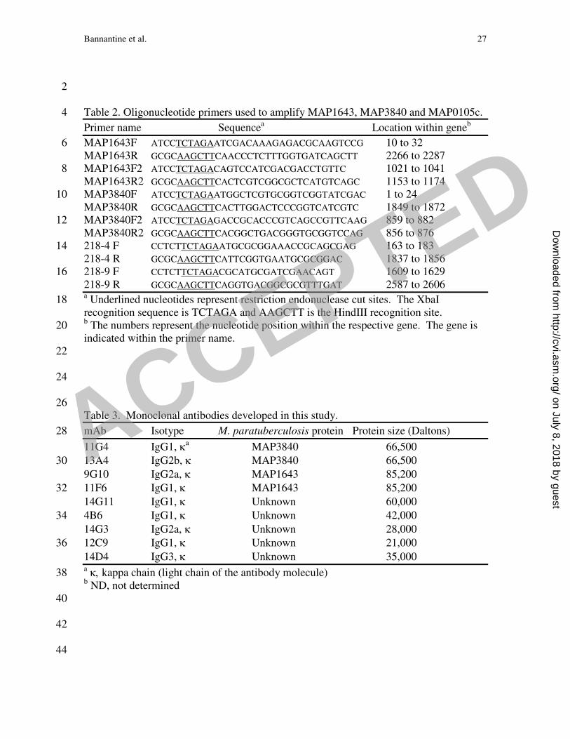
2 Table 2. Oligonucleotide primers used to amplify MAP1643, MAP3840 and MAP0105c. 4
Primer name Sequencea Location within geneb
MAP1643F ATCCTCTAGAATCGACAAAGAGACGCAAGTCCG 10 to 32 6 MAP1643R GCGCAAGCTTCAACCCTCTTTGGTGATCAGCTT 2266 to 2287 MAP1643F2 ATCCTCTAGACAGTCCATCGACGACCTGTTC 1021 to 1041 8 MAP1643R2 GCGCAAGCTTCACTCGTCGGCGCTCATGTCAGC 1153 to 1174 MAP3840F ATCCTCTAGAATGGCTCGTGCGGTCGGTATCGAC 1 to 24 10 MAP3840R GCGCAAGCTTCACTTGGACTCCCGGTCATCGTC 1849 to 1872 MAP3840F2 ATCCTCTAGAGACCGCACCCGTCAGCCGTTCAAG 859 to 882 12 MAP3840R2 GCGCAAGCTTCACGGCTGACGGGTGCGGTCCAG 856 to 876 218-4 F CCTCTTCTAGAATGCGCGGAAACCGCAGCGAG 163 to 183 14 218-4 R GCGCAAGCTTCATTCGGTGAATGCGCGGAC 1837 to 1856 218-9 F CCTCTTCTAGACGCATGCGATCGAACAGT 1609 to 1629 16 218-9 R GCGCAAGCTTCAGGTGACGGCGCGTTTGAT 2587 to 2606 a Underlined nucleotides represent restriction endonuclease cut sites. The XbaI 18 recognition sequence is TCTAGA and AAGCTT is the HindIII recognition site. b The numbers represent the nucleotide position within the respective gene. The gene is 20 indicated within the primer name. 22 24 26 Table 3. Monoclonal antibodies developed in this study.
mAb Isotype M. paratuberculosis protein Protein size (Daltons) 28
11G4 IgG1, κa MAP3840 66,500
13A4 IgG2b, κ MAP3840 66,500 30
9G10 IgG2a, κ MAP1643 85,200
11F6 IgG1, κ MAP1643 85,200 32
14G11 IgG1, κ Unknown 60,000
4B6 IgG1, κ Unknown 42,000 34
14G3 IgG2a, κ Unknown 28,000
12C9 IgG1, κ Unknown 21,000 36
14D4 IgG3, κ Unknown 35,000 a κ, kappa chain (light chain of the antibody molecule) 38 b ND, not determined 40 42 44
ACCEPTED
on July 8, 2018 by guesthttp://cvi.asm
.org/D
ownloaded from

Bannantine et al. 28
2 FIGURE LEGENDS 4 Figure 1. Immunoblot analysis of M. avium subsp paratuberculosis whole-cell lysates
with hybridoma culture supernatants containing monoclonal antibodies. Ten hybridoma 6
culture supernatants were loaded onto independent lanes or slots and analyzed in parallel
on a preparative slot immunoblot containing M. avium subsp paratuberculosis 8
homogenates separated by SDS-PAGE. Antibodies bound to M. avium subsp
paratuberculosis proteins ranging in size from 25 kDa to 95 kDa. Lanes: 1, 11F6; 2, 10
5A10; 3, 13A4; 4, 4B6; 5, 12C9; 6, 11G4; 7, 9G10; 8, 14D4; 9, 14G3 and 10, 14G11.
Protein size standards are indicated in kilodaltons in the left and right margins. 12
Figure 2. Identification of M. avium subsp paratuberculosis proteins detected by 14
selected mAbs and localization of epitopes to regions of MAP1643 and MAP3840 gene
products. For determination of the approximate locations of mAb epitopes in the 16
primary sequence of these M. avium subsp paratuberculosis proteins, purified
recombinant peptides representing the full length (lane 2), N-terminal half (lane 3) and C-18
terminal half (lane 4) of each protein were probed by immunoblotting with selected
mAbs as indicated beneath each blot. These results show that 9G10 and 11F6 react with 20
AceAb and both mAbs detect an epitope on the C-terminal half of the protein. Similarly,
11G4 and 13A4 both react with DnaK; however, 13A4 only reacts with the full-length 22
protein and 11G4 detects an epitope on the N-terminal half of DnaK. The blots probed
with α-MBP detect all the proteins present and indicate their relative amounts and 24
ACCEPTED
on July 8, 2018 by guesthttp://cvi.asm
.org/D
ownloaded from

Bannantine et al. 29
positions within each blot. The MBP-LacZ protein control protein is present in lane 5.
No reactivity is observed with the protein size standards (lane 1). 2
Figure 3. Evaluation of monoclonal antibodies against whole-cell homogenates from 4
several mycobacterial species. Immunoblot analysis shows that reactivity of each mAb is
observed with more than just M. avium subsp paratuberculosis lysates. Lane assignments 6
for A: lane 1: M. silvaticum; lane 2: M. scrofulaceum; lane 3: M. abscessus; lane 4: M.
avium subsp paratuberculosis K-10; lane 5: M. avium (TMC702); lane 6: M. bovis (strain 8
95-1315); lane 7: M phlei; lane 8: M. bovis BCG; lane 9: M. avium subsp
paratuberculosis ATCC19698; lane 10: M. avium (TMC715); lane 11: M. avium subsp 10
paratuberculosis (Linda); lane 12: M. intracellulare; lane 13: M. kansasii. Lane
assignments for B: lane 1, M. silvaticum; lane 2, M. scrofulaceum; lane 3, M. abscessus; 12
lane 4, M. avium subsp paratuberculosis K-10; lane 5, M. avium (TMC702); lane 6, M.
bovis (strain 95-1315); lane 7, M phlei; lane 8, M. avium subsp paratuberculosis 14
ATCC19698; lane 9, M. avium (TMC715); lane 10, M. avium subsp paratuberculosis
(Linda); lane 11, M. intracellulare; lane 12, M. kansasii. Kilodalton size standards are 16
indicated in the left margin and the mAb used is indicated in the right margin.
18
Figure 4. Localization of antigens in fractionated M. avium subsp paratuberculosis cell
lysates. Equal amounts (0.5 µg/lane) of cell lysates from membrane enriched M. avium 20
subsp paratuberculosis K-10 (lane M) and cytoplasmic enriched fractions of M. avium
subsp paratuberculosis K-10 (lane C) were loaded onto SDS-PAGE gels and analyzed by 22
immunoblot using selected monoclonal antibodies indicated beneath each blot. Three
ACCEPTED
on July 8, 2018 by guesthttp://cvi.asm
.org/D
ownloaded from
Bannantine et al. 30
mAbs detected proteins in the cytoplasmic enriched fraction and four mAbs detected
proteins present in the membrane-enriched fraction. 2
Figure 5. Immunogold labeling of M. avium subsp paratuberculosis bacilli with selected 4
mAbs. Bacilli were cultured and processed for immunoelectron microscopy as described
in materials and methods. The mAb used is indicated in the lower left corner of each 6
image. Magnification for all images except 9G10 is 104,000X. The 9G10 image was
magnified 112,000X. 8
Figure 6. Immunoblot (A) and dot blot (B) analysis of aptamers to MAP0105c. (A) The 10
immunoblot containing mycobacterial whole-cell sonicated extracts was exposed to
aptamer 94. Lane assignments: 1: M. silvaticum; 2, M. scrofulaceum; 3, M. abscessus; 4, 12
M. avium subsp paratuberculosis K-10; 5, M. avium (tmc702); 6, M. bovis; 7, M. phlei; 8,
M. bovis BCG; 9, M. avium subsp paratuberculosis ATCC19698; 10, M. avium 14
(tmc715); 11, M. avium subsp paratuberculosis (Linda); 12, M. intracellulare; 13, M.
kansasii. Size standards are indicated in kilodaltons in the left margin. (B) The dot blot 16
was exposed to the three aptamers, which are indicated above the blots. Proteins spotted
to the membrane are indicated in the left margin and state of the proteins is indicated in 18
the right margin. Abbreviations: MBP=maltose binding protein fused to the α-peptide of
LacZ; 218-4=an MBP fusion containing the N-terminal half of MAP0105c; 218-9=an 20
MBP fusion containing the C-terminal half of MAP0105c; K-10=a whole cell lysate of
M. avium subsp paratuberculosis K-10; Avium=a whole cell lysate of M. avium tmc715. 22
ACCEPTED
on July 8, 2018 by guesthttp://cvi.asm
.org/D
ownloaded from

Table 1. Mycobacterial isolates used in this study.
Isolate Organism Host Location Reference or source
K-10 M. avium subsp. paratuberculosis Bovine feces (20)
19698 M. avium subsp. paratuberculosis Bovine feces ATCC 19698
6100 M. avium subsp. paratuberculosis Human ileum ATCC 43015
187 M. avium subsp. paratuberculosis Bovine ileum Recent clinical isolate, NADC
523 M. avium subsp. paratuberculosis Bovine ileum
803 M. avium subsp. paratuberculosis Bovine ileum
6009 M. avium subsp. avium Bovine ATCC 35716 (TMC715)
6003 M. avium subsp. avium Chicken ATCC 35713 (TMC702)
6006 M. avium subsp. silvaticum Roe deer V1-72
6076 M. abscessus ATCC 19977
M. bovis Bovine lymph node ATCC 19210
M. bovis BCG Pasteur Bovine milk ATCC 35734
6081 M. kansasii Human ATCC 12478
6010 M. intracellulare Swine ATCC 35773
6083 M. phlei ATCC 11758
6077 M. scrofulaceum Human lymph node
ACCEPTED on July 8, 2018 by guest
http://cvi.asm.org/
Dow
nloaded from


![[Julie a. Bannantine] Fundamentals of Metal Fatigu(BookFi.org)-1](https://static.fdocuments.in/doc/165x107/55cf989b550346d033989e08/julie-a-bannantine-fundamentals-of-metal-fatigubookfiorg-1.jpg)