Clase 1_ Reglamento e Introducción
29
Objetivos, reglamento, evaluaciones y contenidos. Capitulo 1: Fisiología General y Neurofisiología 1. Introducción: Medio interno, compartimientos, homeostasis, sistemas de control. Cristian Vilos, TM, PhD Human physiology is the study of how the human body functions, with emphasis on specific cause-and-effect mechanisms. Knowledge of these mechanisms has been obtained experimentally through applications of the scientific method.
-
Upload
nataly-valdes-dewitt -
Category
Documents
-
view
251 -
download
0
description
Primera clase de fisiologia
Transcript of Clase 1_ Reglamento e Introducción

Objetivos, reglamento, evaluaciones y contenidos.Capitulo 1: Fisiología General y Neurofisiología 1. Introducción: Medio interno, compartimientos, homeostasis, sistemas de control.
Cristian(Vilos,(TM,(PhD
Human physiology is the study of how the human body functions, with emphasis on specific cause-and-effect mechanisms. Knowledge of these mechanisms has been obtained experimentally through applications of the scientific method.

FACULTAD DE CIENCIAS BIOLÓGICAS DEPARTAMENTO DE CIENCIAS BIOLÓGICAS 1. Identificación de la asignatura. CURSO : FISIOLOGÍA GENERAL CÓDIGO : BIO 372 TIPO DE ACTIVIDAD : TEÓRICO HORAS SEMANALES : 4 HORAS 2. Competencia(s) alcanzada(s) al finalizar el curso. Generales - Conocer el funcionamiento normal de los distintos sistemas del organismo humano y sus mecanismos de
regulación e integración. Específicas - Adquirir una visión integrada del funcionamiento normal del organismo. - Conocer el lenguaje fisiológico con orientación clínica. - Incentivar un pensamiento reflexivo-crítico y científico. 3. Contenidos. CAPÍTULO 1: FISIOLOGÍA GENERAL Y NEUROFISIOLOGÍA 25%
1. Fisiología y homeostasis 2. Transporte a través de membrana 3. Características y funcionamiento de las células excitables 4. Sinapsis 5. Contracción muscular 6. Sistema nervioso autónomo (SNA) 7. Transducción sensorial, sistemas sensoriales y sentidos especiales 8. Sistema motor 9. Funciones cerebrales superiores
CAPÍTULO 2: SISTEMA ENDOCRINO 20% 1. Hormonas hipotalámicas e hipofisiarias 2. Regulación endocrina del crecimiento. Prolactina. 3. Glándula suprarrenal 4. Glándula tiroides 5. Regulación endocrina de la glicemia 6. Regulación endocrina de la calcemia 7. Función gonadal
FACUDEPA
1. Iden
CURSCÓDISECC 2. ProCristia 3. Hor
. 4. Cro
Fech
MAR
Jueve
Vierne
Jueve
Vierne
ULTAD DE ARTAMEN
ntificación d
SO : FIGO : BI
CIÓN : 2
ofesor an Vilos Orti
rario: Jueve Vierne
onograma d
ha
RZO
s 12
ObjeCap1.Intsiste
es 13 2.- M
s 19 3. CPote4. Si
es 20
5. Chorm6. Cnerv
CIENCIASTO DE CIE
ANEXO D
de la Asigna
ISIOLOGIAIO 372; NR
iz
es 10:20-12:0es 08:30-10:
de la asignat
etivos, reglapitulo 1: Fisitroducción: emas de cont
Membranas b
Característicaencial de reinapsis.
Comunicaciónmonas.
Característicavioso autóno
S BIOLÓGIENCIAS BIO
DE PROGR
atura
A GENERARC 1250
00 (Módulos10 (Módulo
tura
amento, evaluiología GenMedio interntrol.
biológicas y
as y funcionaeposo y poten
n-intercelula
as y funcionomo.
ICAS OLÓGICA
RAMA 2015
AL
s 3 y 4) s 1 y 2)
Contenido
uaciones y cneral y Neurno, comparti
y mecanismo
amiento de lncial de acci
ar. Receptore
nes del sistem
AS
5 – I SEME
o
contenidos. rofisiologíaimientos, ho
os de transpo
as células exión.
es a neurotra
ma nervioso.
STRE
omeostasis,
orte.
xcitables.
ansmisores y
Sistema
Prof
C
C
C
y
C
fesor
CV
CV
CV
CV
Profesores, Cristian Vilos, Luis Constandil

FACULTAD DE CIENCIAS BIOLÓGICAS DEPARTAMENTO DE CIENCIAS BIOLÓGICAS 1. Identificación de la asignatura. CURSO : FISIOLOGÍA GENERAL CÓDIGO : BIO 372 TIPO DE ACTIVIDAD : TEÓRICO HORAS SEMANALES : 4 HORAS 2. Competencia(s) alcanzada(s) al finalizar el curso. Generales - Conocer el funcionamiento normal de los distintos sistemas del organismo humano y sus mecanismos de
regulación e integración. Específicas - Adquirir una visión integrada del funcionamiento normal del organismo. - Conocer el lenguaje fisiológico con orientación clínica. - Incentivar un pensamiento reflexivo-crítico y científico. 3. Contenidos. CAPÍTULO 1: FISIOLOGÍA GENERAL Y NEUROFISIOLOGÍA 25%
1. Fisiología y homeostasis 2. Transporte a través de membrana 3. Características y funcionamiento de las células excitables 4. Sinapsis 5. Contracción muscular 6. Sistema nervioso autónomo (SNA) 7. Transducción sensorial, sistemas sensoriales y sentidos especiales 8. Sistema motor 9. Funciones cerebrales superiores
CAPÍTULO 2: SISTEMA ENDOCRINO 20% 1. Hormonas hipotalámicas e hipofisiarias 2. Regulación endocrina del crecimiento. Prolactina. 3. Glándula suprarrenal 4. Glándula tiroides 5. Regulación endocrina de la glicemia 6. Regulación endocrina de la calcemia 7. Función gonadal

CAPÍTULO 3: SISTEMA DIGESTIVO 13% 1. Organización y control del sistema digestivo 2. Motilidad gastrointestinal 3. Secreciones gastrointestinales 4. Digestión y absorción de nutrientes, electrolitos y agua
CAPÍTULO 4: SISTEMA CARDIOVASCULAR 13% 1. Función cardiaca 2. Hemodinamia 3. Función vascular 4. Microcirculación 5. Regulación del gasto cardiaco 6. Regulación de la presión arterial
CAPÍTULO 5: SANGRE 4% 1. Hematopoyesis 2. Hemostasia
CAPÍTULO 6: SISTEMA RESPIRATORIO 12% 3. Estructura y función del sistema respiratorio 4. Mecánica de la respiración 5. Ventilación 6. Difusión y transporte de gases 7. Regulación de la respiración
CAPÍTULO 7: SISTEMA RENAL 13% 1. Organización del sistema renal 2. Líquidos corporales 3. Filtración glomerular y flujo sanguíneo renal 4. Función tubular 5. Concentración y dilución de la orina 6. Balance de sodio y potasio 7. Equilibrio ácido-base
4. Evaluación.
La evaluación del curso consta de 3 pruebas solemnes (PS1, PS2 y PS3, respectivamente) de selección múltiple (alternativas).
La nota de presentación a examen resultará del promedio ponderado de las tres pruebas solemnes, según los siguientes valores: PS1, 20%; PS2, 40%; PS3, 40% La nota final del curso se obtendrá según las siguientes ponderaciones: Nota de presentación a examen: 70% Nota de examen: 30% 5. Bibliografía.
� R Berne y M Levy. Fisiología. España: Elsevier. � Guyton y Hall. Tratado de Fisiología Médica. España: Elsevier. � Johnson LR (Ed) Essential Medical Physiology. USA: Lippincott-Raven Publishers. � Costanzo L. Fisiología. España: Elsevier. � Silverthorn. Fisiología Humana. Edit. Médica Panamericana. � Ganong. W.F. Fisiología Médica. México: Editorial Manual Moderno.
Se recomienda en lo posible las últimas ediciones de la bibliografía señalada.

FACUDEPA
1. Iden
CURSCÓDISECC 2. ProCristia 3. Hor
. 4. Cro
Fech
MAR
Jueve
Vierne
Jueve
Vierne
ULTAD DE ARTAMEN
ntificación d
SO : FIGO : BI
CIÓN : 2
ofesor an Vilos Orti
rario: Jueve Vierne
onograma d
ha
RZO
s 12
ObjeCap1.Intsiste
es 13 2.- M
s 19 3. CPote4. Si
es 20
5. Chorm6. Cnerv
CIENCIASTO DE CIE
ANEXO D
de la Asigna
ISIOLOGIAIO 372; NR
iz
es 10:20-12:0es 08:30-10:
de la asignat
etivos, reglapitulo 1: Fisitroducción: emas de cont
Membranas b
Característicaencial de reinapsis.
Comunicaciónmonas.
Característicavioso autóno
S BIOLÓGIENCIAS BIO
DE PROGR
atura
A GENERARC 1250
00 (Módulos10 (Módulo
tura
amento, evaluiología GenMedio interntrol.
biológicas y
as y funcionaeposo y poten
n-intercelula
as y funcionomo.
ICAS OLÓGICA
RAMA 2015
AL
s 3 y 4) s 1 y 2)
Contenido
uaciones y cneral y Neurno, comparti
y mecanismo
amiento de lncial de acci
ar. Receptore
nes del sistem
AS
5 – I SEME
o
contenidos. rofisiologíaimientos, ho
os de transpo
as células exión.
es a neurotra
ma nervioso.
STRE
omeostasis,
orte.
xcitables.
ansmisores y
Sistema
Prof
C
C
C
y
C
fesor
CV
CV
CV
CV
LC
LC

Jueves 26 7. Funciones superiores del sistema nervioso. CV
Viernes 27 8. Sistema motor. Reflejos. Contracción muscular. CV
ABRIL
Jueves 2 9.Sistemas sensoriales.Somestesia.Nocicepción. CV
Jueves 9 10.Sentidos especiales Visión y audición. CV
Viernes 10 Capitulo 2: Sistema Endocrino 1. Eje hipotálamo-hipófisis. 2. Regulación endocrina del crecimiento. Prolactina CV
Jueves 16
3. Glándula tiroides. 4. Regulación endocrina de la calcemia. CV
Viernes 17 5. Glándula Suprarrenal.
CV
Jueves 23 6. Páncreas endocrino y regulación hormonal de la glicemia.
CV
Viernes 24
7. Función Gonadal
CV
Sábado 25 SOLEMNE I (20%)
Fisiología general y Neurofisiología Módulos 2-3-4
CV
Jueves 30 Capitulo 3: Sistema Digestivo 1. Organización del sistema digestivo. 2.Motilidad gastrointestinal.
CV
MAYO
Jueves 7 3. Secreciones gastrointestinales.
CV
Viernes 8 4. Digestión y absorción de nutrientes, agua y electrolitos.
CV
Jueves 14
Capitulo 4: Sangre 1. Composición de la sangre. Funciones. Hematopoyesis.Grupos sanguíneos. 2. Hemostasia y coagulación.
CV
Viernes 15
Capitulo 5: Sistema Cardiovascular 1. Organización y funciones generales. Electrofisiología cardíaca.
Ciclo cardíaco. 2. Regulación del gasto cardíaco. 3. Hemodinamia.
CV
Viernes 22 Sin actividades
Sábado 23 SOLEMNE II (40%) Endocrino, Digestivo y Sangre
CV
LC
LC
LC
LC
Jueves 26 7. Funciones superiores del sistema nervioso. CV
Viernes 27 8. Sistema motor. Reflejos. Contracción muscular. CV
ABRIL
Jueves 2 9.Sistemas sensoriales.Somestesia.Nocicepción. CV
Jueves 9 10.Sentidos especiales Visión y audición. CV
Viernes 10 Capitulo 2: Sistema Endocrino 1. Eje hipotálamo-hipófisis. 2. Regulación endocrina del crecimiento. Prolactina CV
Jueves 16
3. Glándula tiroides. 4. Regulación endocrina de la calcemia. CV
Viernes 17 5. Glándula Suprarrenal.
CV
Jueves 23 6. Páncreas endocrino y regulación hormonal de la glicemia.
CV
Viernes 24
7. Función Gonadal
CV
Sábado 25 SOLEMNE I (20%)
Fisiología general y Neurofisiología Módulos 2-3-4
CV
Jueves 30 Capitulo 3: Sistema Digestivo 1. Organización del sistema digestivo. 2.Motilidad gastrointestinal.
CV
MAYO
Jueves 7 3. Secreciones gastrointestinales.
CV
Viernes 8 4. Digestión y absorción de nutrientes, agua y electrolitos.
CV
Jueves 14
Capitulo 4: Sangre 1. Composición de la sangre. Funciones. Hematopoyesis.Grupos sanguíneos. 2. Hemostasia y coagulación.
CV
Viernes 15
Capitulo 5: Sistema Cardiovascular 1. Organización y funciones generales. Electrofisiología cardíaca.
Ciclo cardíaco. 2. Regulación del gasto cardíaco. 3. Hemodinamia.
CV
Viernes 22 Sin actividades
Sábado 23 SOLEMNE II (40%) Endocrino, Digestivo y Sangre
CV

Módulos 2-3-4
Jueves 28 4. Microcirculación. 5. Regulación de la presión arterial.
CV
Viernes 29 Capitulo 6: Sistema Respiratorio 1. Generalidades y función. 2. Mecánica respiratoria.
CV
JUNIO
Jueves 4 3. Ventilación pulmonar. 4. Difusión y transporte de gases.
CV
Viernes 5 5. Regulación de la respiración.
CV
Jueves 11 Capitulo 7: Sistema Renal 1. Organización del sistema renal. 2. Función renal, Filtración, VFG y FPR, medición de la VFG.
CV
Viernes 12 3. Función tubular, reabsorción, secreción y excreción. 4. Balance de agua. Concentración y dilución de la orina.
CV
Jueves 18 5. Balance del sodio y potasio. Regulación de la volemia.
CV
Viernes 19 6. Equilibrio ácido base.
CV
Sábado 20 SOLEMNE III (40%)
Cardiovascular, Respiratorio y Renal Módulos 2-3-4
CV
JULIO
Viernes 3 EXAMEN
Toda la materia Módulos 2-3-4
CV
Las fechas de las PruebasSolemnes y Examen no serán modificadas bajo ninguna circunstancia.Se exigirá la cédula de identidad para rendir las pruebas.
La nota de presentación a examen resultará del promedio ponderado de las tres Pruebas Solemnes, según los siguientes valores: PS1, 20%; PS2, 40%; PS3, 40%
La nota final del curso se obtendrá del promedio ponderado entre la nota de presentación a examen y la nota obtenida en esta última evaluación (nota de examen), según los siguientes valores: nota de presentación a examen, 70%; nota de examen, 30% 5. Requisitos de asistencia
La asistencia a las clases de cátedra es libre. Según el Reglamento de la Universidad: Artículo 46º: “La asistencia a pruebas,
exámenes, controles, exposiciones u otras actividades de evaluación programadas, será

– The Greek philosopher Aristotle (384–322 B.C.) speculated on the function of the human body.
– Erasistratus (304–250? B.C.), is considered the father of physiology because he attempted to apply physical laws to the study of human function.
– Galen (A.D. 130–201) wrote widely on the subject and was considered the supreme authority until the Renaissance.
– Physiology became a fully experimental science with the revolutionary work of the English physician William Harvey (1578–1657), who dem onstrated that the heart pumps blood through a closed system of vessels.
– The father of modern physiology is the French physiologist Claude Bernard (1813–1878), who observed that the milieu interieur (internal environment) remains remarkably constant despite changing conditions in the external environment. In a book entitled The Wisdom of the Body, published in 1932, the American physiologist Walter Cannon (1871–1945) coined the term homeostasis to describe this internal constancy.
History of Physiology

Table 1.1 | History of Twentieth- and Twenty-First-Century Physiology (two citations per decade)
1900 Karl Landsteiner discovers the A, B, and O blood groups.
1904 Ivan Pavlov wins the Nobel Prize for his work on the physiology of digestion.
1910 Sir Henry Dale describes properties of histamine.
1918 Earnest Starling describes how the force of the heart’s contraction relates to the amount of blood in it.
1921 John Langley describes the functions of the autonomic nervous system.
1923 Sir Frederick Banting, Charles Best, and John Macleod win the Nobel Prize for the discovery of insulin.
1932 Sir Charles Sherrington and Lord Edgar Adrian win the Nobel Prize for discoveries related to the functions of neurons.
1936 Sir Henry Dale and Otto Loewi win the Nobel Prize for the discovery of acetylcholine in synaptic transmission.
1939–47 Albert von Szent-Györgyi explains the role of ATP and contributes to the understanding of actin and myosin in muscle contraction.
1949 Hans Selye discovers the common physiological responses to stress.
1953 Sir Hans Krebs wins the Nobel Prize for his discovery of the citric acid cycle.
1954 Hugh Huxley, Jean Hanson, R. Niedergerde, and Andrew Huxley propose the sliding filament theory of muscle contraction.
1962 Francis Crick, James Watson, and Maurice Wilkins win the Nobel Prize for determining the structure of DNA.
1963 Sir John Eccles, Sir Alan Hodgkin, and Sir Andrew Huxley win the Nobel Prize for their discoveries relating to the nerve impulse.
1971 Earl Sutherland wins the Nobel Prize for his discovery of the mechanism of hormone action.
1977 Roger Guillemin and Andrew Schally win the Nobel Prize for discoveries of the brains’ production of peptide hormone.
1981 Roger Sperry wins the Nobel Prize for his discoveries regarding the specializations of the right and left cerebral hemispheres.
1986 Stanley Cohen and Rita Levi-Montalcini win the Nobel Prize for their discoveries of growth factors regulating the nervous system.
1994 Alfred Gilman and Martin Rodbell win the Nobel Prize for their discovery of the functions of G-proteins in signal transduction in cells.
1998 Robert Furchgott, Louis Ignarro, and Ferid Murad win the Nobel Prize for discovering the role of nitric oxide as a signaling molecule in the cardiovascular system.
2004 Linda B. Buck and Richard Axel win the Nobel Prize for their discoveries of odorant receptors and the organization of the olfactory system.
2006 Andrew Z. Fine and Craig C. Mello win the Noble Prize for their discovery of RNA interference by short, double-stranded RNA molecules.
Body, published in 1932, the American physiologist Walter Cannon (1871–1945) coined the term homeostasis to describe this internal constancy. Cannon further suggested that the many mechanisms of physiological regulation have but one purpose—the maintenance of internal constancy.
Most of our present knowledge of human physiology has been gained in the twentieth century. Further, new knowledge in the twenty-first century is being added at an ever more rapid pace, fueled in more recent decades by the revolutionary growth of molecular genetics and its associ-ated biotechnologies, and by the availability of more power-ful computers and other equipment. A very brief history of twentieth- and twenty-first-century physiology, limited by space to only two citations per decade, is provided in table 1.1 .
Most of the citations in table 1.1 indicate the winners of Nobel prizes. The Nobel Prize in Physiology or Medicine (a single prize category) was first awarded in 1901 to Emil Adolf von Behring, a pioneer in immunology who coined the term antibody and whose many other discoveries included the use of serum (containing antibodies) to treat diphtheria. Many scientists who might deserve a Nobel Prize never receive one, and the prizes are given for particular achieve-ments and not others (Einstein didn’t win his Nobel Prize in Physics for relativity, for example) and are often awarded many years after the discoveries were made. Nevertheless, the awarding of the Nobel Prize in Physiology or Medicine each year is a celebrated event in the biomedical commu-nity, and the awards can be a useful yardstick for tracking the course of physiological research over time.
5The Study of Body Function
fox78119_ch01_001-023.indd 5fox78119_ch01_001-023.indd 5 25/06/10 8:55 PM25/06/10 8:55 PM

organization of the human body. Your exploration of the humanbody will extend from atoms and molecules to the whole person.From the smallest to the largest, six levels of organization willhelp you to understand anatomy and physiology: the chemical,cellular, tissue, organ, system, and organismal levels of organiza-tion (Figure 1.1).
●1 Chemical level. This very basic level can be compared tothe letters of the alphabet and includes atoms, the smallest
1.2 LEVELS OF STRUCTURAL ORGANIZATION AND BODY SYSTEMS 3
units of matter that participate in chemical reactions, andmolecules, two or more atoms joined together. Certainatoms, such as carbon (C), hydrogen (H), oxygen (O), nitro-gen (N), phosphorus (P), calcium (Ca), and sulfur (S), are es-sential for maintaining life. Two familiar molecules found inthe body are deoxyribonucleic acid (DNA), the genetic mate-rial passed from one generation to the next, and glucose,commonly known as blood sugar. Chapters 2 and 25 focus onthe chemical level of organization.
Figure 1.1 Levels of structural organization in the human body.
The levels of structural organization are chemical, cellular, tissue, organ, system, and organismal.
Which level of structural organization is composed of two or more different types of tissues that work together to perform aspecific function?
6
3
4
5
1 CHEMICAL LEVEL
Atoms (C, H, O, N, P)
2 CELLULAR LEVEL
Molecule (DNA)
Smooth muscle cell
Smooth muscle tissue
ORGANISMAL LEVEL
SYSTEM LEVEL
Mouth
Liver
Gallbladder
Large intestine
Esophagus
Small intestine
Pancreas(behind stomach)
Stomach
Digestive system
Stomach
Epithelial tissue
EpithelialandconnectivetissuesORGAN LEVEL
TISSUE LEVEL
Smooth muscletissue layers
Pharynx (throat)
Salivary glands
JWCL316_c01_001-028.qxd 7/1/10 5:35 AM Page 3

Homeostasis: A Framework for Human Physiology 3
Muscle cells are specialized to generate the mechanical forces that produce movement. They may be attached through other structures to bones and produce movements of the limbs or trunk. They may be attached to skin, such as the muscles
producing facial expressions. They may also surround hollow cavities so that their contraction expels the contents of the cav-ity, as in the pumping of the heart. Muscle cells also surround many of the tubes in the body—blood vessels, for example—and their contraction changes the diameter of these tubes.
Nerve cells are specialized to initiate and conduct elec-trical signals, often over long distances. A signal may initiate new electrical signals in other nerve cells, or it may stimulate a gland cell to secrete substances or a muscle cell to contract. Thus, nerve cells provide a major means of controlling the activities of other cells. The incredible complexity of connec-tions between nerve cells underlies such phenomena as con-sciousness and perception.
Epithelial cells are specialized for the selective secre-tion and absorption of ions and organic molecules, and for protection. They are located mainly at the surfaces that cover the body or individual organs, and they line the walls of vari-ous tubular and hollow structures within the body. Epithelial cells, which rest on an extracellular protein layer called the basement membrane, form the boundaries between com-partments and function as selective barriers regulating the exchange of molecules. For example, the epithelial cells at the surface of the skin form a barrier that prevents most sub-stances in the external environment—the environment sur-rounding the body—from entering the body through the skin. Epithelial cells are also found in glands that form from the invagination of epithelial surfaces.
Connective tissue cells, as their name implies, con-nect, anchor, and support the structures of the body. Some connective tissue cells are found in the loose meshwork of cells and fi bers underlying most epithelial layers. Other types include adipose (fat-storing) cells, bone cells, red blood cells, and white blood cells.
TissuesMost specialized cells are associated with other cells of a simi-lar kind to form tissues. Corresponding to the four general categories of differentiated cells, there are four general classes of tissues: (1) muscle tissue, (2) nerve tissue, (3) epithelial tissue, and (4) connective tissue. The term tissue is used in different ways. It is formally defi ned as an aggregate of a single type of specialized cell. However, it is also commonly used to denote the general cellular fabric of any organ or structure—for example, kidney tissue or lung tissue, each of which in fact usually contains all four classes of tissue.
The immediate environment that surrounds each indi-vidual cell in the body is the extracellular fl uid. Actually, this fl uid is interspersed within a complex extracellular matrix consisting of a mixture of protein molecules and, in some cases, minerals, specifi c for any given tissue. The matrix serves two general functions: (1) It provides a scaffold for cellular attachments, and (2) it transmits information, in the form of chemical messengers, to the cells to help regulate their activ-ity, migration, growth, and differentiation.
The proteins of the extracellular matrix consist of fi bers—ropelike collagen fi bers and rubberband-like elas-tin fi bers—and a mixture of nonfi brous proteins that contain chains of complex sugars (carbohydrates). In some ways, the
Fertilized egg
Celldivisionand growth
Celldifferentiation
Specializedcell types
Tissues
Functionalunit(nephron)
Organ(kidney)
Organ system(urinary system)
Total organism(human being)
Epithelialcell
Connectivetissue cell
Nervecell
Musclecell
Ureter
Bladder
Urethra
Kidney
Figure 1–1Levels of cellular organization. The nephron is not drawn to scale.
wid4962X_chap01.indd 3wid4962X_chap01.indd 3 6/21/07 4:42:10 PM6/21/07 4:42:10 PM

Eder−Kaminsky−Bertram:Laboratory Atlas of Anatomy and Physiology, Sixth Edition
1. Histology Text 13© The McGraw−Hill Companies, 2009
H i s t o l o g y 11
Figure 1-24Hyaline Cartilage Artifactual vacuolationforms characteristic lacunae aroundchondrocyte cell bodies. From monkeytrachea. (!250)
Figure 1-25Elastic Cartilage Extracellular matrixcontains elastic fibers that confer elasticrecoil to this tissue. From monkey ear.(!250)
Matrix
Lacuna
Chondrocyte
Chondrocyte
Elastic fiber
Lacunae
Eder−Kaminsky−Bertram:Laboratory Atlas of Anatomy and Physiology, Sixth Edition
1. Histology Text 11© The McGraw−Hill Companies, 2009
H i s t o l o g y 9
Figure 1-19Dense Regular Connective Tissue Bandsof collagen fibers extending in regular,parallel rows resist mechanical stress mainlyalong course of fibers. Monkey tendon.(!250)
Figure 1-20Dense Irregular Connective Tissue Bandsof collagen running in irregular rows givemultidirectional tensile strength. Collagen-secreting fibroblasts appear throughout.(!100)
Figure 1-21Adipose Tissue Large, clear, polyhedralvacuoles dominate small, eccentricallylocated cell nuclei of adipocytes. Thevacuoles are filled with lipid (fat) molecules.Fine capillaries run through tissue. (!100)
Collagen fibers
Nucleus of fibroblast
Nuclei of fibroblasts
Collagen fibers
Vacuole
Nucleus
Capillary
Cartilago Adipocitos
Eder−Kaminsky−Bertram:Laboratory Atlas of Anatomy and Physiology, Sixth Edition
1. Histology Text 11© The McGraw−Hill Companies, 2009
H i s t o l o g y 9
Figure 1-19Dense Regular Connective Tissue Bandsof collagen fibers extending in regular,parallel rows resist mechanical stress mainlyalong course of fibers. Monkey tendon.(!250)
Figure 1-20Dense Irregular Connective Tissue Bandsof collagen running in irregular rows givemultidirectional tensile strength. Collagen-secreting fibroblasts appear throughout.(!100)
Figure 1-21Adipose Tissue Large, clear, polyhedralvacuoles dominate small, eccentricallylocated cell nuclei of adipocytes. Thevacuoles are filled with lipid (fat) molecules.Fine capillaries run through tissue. (!100)
Collagen fibers
Nucleus of fibroblast
Nuclei of fibroblasts
Collagen fibers
Vacuole
Nucleus
Capillary
Tejido Conectivo
Eder−Kaminsky−Bertram:Laboratory Atlas of Anatomy and Physiology, Sixth Edition
1. Histology Text 15© The McGraw−Hill Companies, 2009
H i s t o l o g y 13
Figure 1-29Pacinian (Lamellated) CorpuscleEncapsulated nerve ending found deep indermis and throughout interior of bodydetects pressure. (!25)
Figure 1-30Human Scalp with Hair Follicle Follicleroot, with sheath embedded in pale adiposetissue, has sebaceous glands surrounding itnear surface. (!10)
Capsule
Free nerve ending
Hair papilla
Hair follicle
Root sheath
Hair root
Sebaceous gland
Figure 1-31Detail of Sebaceous Gland Nucleatedgerminative cells at base of gland matureand accumulate lipid. At duct, theydegenerate and lyse to release their oilyproduct, sebum. (!100)
Sebaceous gland
Hair root
Hair follicle
Foliculo Capilar Cabello
Eder−Kaminsky−Bertram:Laboratory Atlas of Anatomy and Physiology, Sixth Edition
1. Histology Text 17© The McGraw−Hill Companies, 2009
H i s t o l o g y 15
Figure 1-35Red Bone Marrow Medullary cavity in thehead of long bones of the adult containsstem cells, precursors to red blood cells, andwhite blood cells and platelets. Human.(!250)
Figure 1-36Developing Bone at Epiphyseal PlateMiddle belt of cartilage undergoing primarycalcification is replaced by new bone.
Figure 1-37Detail of Epiphyseal Plate Epiphysealplate cartilage at right transforms into zonesof proliferating chondrocytes with primaryossification occurring on their calcifiedremnants. Newly formed bone appears at left. (!50)
Cartilage of epiphyseal plate
White blood cell precursors
Eosinophilic myelocyte
Myeloblast
Basophilic myelocyte
Neutrophilic stab cell
Neutrophil
Erythroblasts Proerythroblast ErythroblastsRed blood cell precursors
New spongy bone
Ossification Cells die Older cells Proliferating enlarge chondrocytes
• e µµ
Médula Ósea
Eder−Kaminsky−Bertram:Laboratory Atlas of Anatomy and Physiology, Sixth Edition
1. Histology Text 19© The McGraw−Hill Companies, 2009
H i s t o l o g y 17
Figure 1-41Cardiac Muscle (Longitudinal Section)Striated muscle fibers branch andanastomose at junctions marked by darkintercalated disks. (!250)
Figure 1-42Smooth Muscle (Longitudinal Section)Canoe- or spindle-shaped muscle cells lackstriations, and each has a single, elongatednucleus. (!250)
Figure 1-43Innervation of Skeletal Muscle: MotorEndplate Branching nerve bundleterminates to form the myoneuraljunctions. Nerve terminals release smallquantities of chemical neurotransmitter tostimulate muscle contraction.
Nucleus
Intercalated disk
Nucleus
Terminal branches of motor neuron
Synaptic bulb
Skeletal muscle fibers
Myoneural junction
Smooth muscle cell
Músculo Cardiaco
Eder−Kaminsky−Bertram:Laboratory Atlas of Anatomy and Physiology, Sixth Edition
1. Histology Text20 © The McGraw−Hill Companies, 2009
18 C H A P T E R 1
Figure 1-44Astrocytes (Neuroglia) Star-shapedsupporting cells of central nervous systemmodulate ionic environment. Cytoplasmicextensions make contact with blood vessel.Cat. (Silver stain; !280)
Figure 1-45Purkinje Cells (Neurons) Numerousbranched processes (dendrites) receiveinformation for processing. Single process(axon) sends information to other neurons.Human cerebellum. (!100)
Figure 1-46Pyramidal Cells Neurons from cerebralcortex directly receive information fromhundreds of other cells; send informationon to hundreds of others.(Fox-Golgi stain; !100)
Astrocyte
Blood vessel
Dendrites
Nucleus
Cell body
Axon
Dendrites
Axon
Astrocitos
Eder−Kaminsky−Bertram:Laboratory Atlas of Anatomy and Physiology, Sixth Edition
1. Histology Text22 © The McGraw−Hill Companies, 2009
20 C H A P T E R 1
Figure 1-50Myelinated Nerve Fibers (Cross Section)Central core stains dark; insulating myelinappears white. (!250)
Figure 1-51Spinal Cord, Lumbar Region (Cross Section)Top is dorsal, bottom is ventral. Light,central dot is central canal. Darkly stainingH-shaped region is gray matter of cell bodies;surrounding lighter material is composed ofmyelinated axons. Ventral horns of graymatter contain motor neurons; dorsal hornscontain cell bodies of sensory pathways.(!4)
Figure 1-52Retina Layered structure evident. Darkline of cells near top is pigment epithelium.Broad, striped region representsphotoreceptors (rods and cones), whosenuclei stain heavily immediately beneath.Below receptor nuclei lie synaptic regionand a layer of nuclei belonging to bipolarcells. Bipolar cell output synapses ontoganglion cells, only a few of which appearnear bottom. Axons of ganglion cells formoptic nerve. (!100)
White matter
Dorsal horn
Central canal
Ventral horn
Choroid
Pigmented epithelium
Rods and cones
Receptor nuclei
Bipolar cell nuclei
Ganglion cells
Axon
Myelin sheath
Neurilemma
Capillary
Médula Espinal
Eder−Kaminsky−Bertram:Laboratory Atlas of Anatomy and Physiology, Sixth Edition
1. Histology Text 25© The McGraw−Hill Companies, 2009
H i s t o l o g y 23
Figure 1-57aPituitary Gland The pituitary glandconsists of two components: the posteriorcomponent, or neurohypophysis (lightstain), consists of mainly nervous tissue,whereas the anterior component, oradenohypophysis (dark stain), consists of aglandular epithelium. (!10)
Islet of Langerhans
Exocrine cells of pancreas
Figure 1-57bPituitary Gland The cleft between theneurohypophysis and adenohypophysis isvisible in this view of the pituitary gland.(!100)
Figure 1-58Pancreas The pancreatic islet ofLangerhans cells forms the endocrineportion of the pancreas. Alpha cells secreteglucagon, beta cells secrete insulin, anddelta cells secrete somatostatin. Theexocrine portion of the pancreas secretesdigestive enzymes through a series of ducts.
Cleft
Neurohypophysis
Adenohypophysis
Pituitary gland
Cleft
Adenohypophysis
Neurohypophysis
Páncreas
Eder−Kaminsky−Bertram:Laboratory Atlas of Anatomy and Physiology, Sixth Edition
1. Histology Text26 © The McGraw−Hill Companies, 2009
24 C H A P T E R 1
Figure 1-59Adrenal Cortex Outer zone of roundedgroups of cells (zona glomerulosa) secretesmineralcorticosteroids (aldosterone).Middle zone of cells appearing in rows (zonafasciculata) secretes glucocorticosteroids.Innermost zone of cells arranged in ameshwork (zona reticularis) secretes mainlyandrogens. (!50)
Zona glomerulosa
Zona fasciculata
Zona reticularis
Figure 1-60Neutrophil Most numerous (65%) of the leukocytes, it is characterized by amultilobed nucleus and granular cytoplasm.Engages in phagocytosis. The Barr body is afeature found only in male blood samples.(Neutral dyes stain; !640)
Barr body
Nucleus
Sangre
Eder−Kaminsky−Bertram:Laboratory Atlas of Anatomy and Physiology, Sixth Edition
1. Histology Text 31© The McGraw−Hill Companies, 2009
H i s t o l o g y 29
Figure 1-71Lymph Node Outer cortex containingseveral follicles surrounds medulla, with itsnarrow, dark medullary cords. Notch is thehilum, through which blood and lymphaticvessels pass. (!5)
Follicle (germinal center)
Hilum
Medulla
Cortex
Figure 1-70Capillary with Red Blood Cells in Single FileCapillary wall is made of flattenedendothelial cells without complex tunics,a simple structure that facilitates theexchange of gases, nutrients, wastes, and hormones. (!400)
Endothelium
Red blood cell
Figure 1-69Detail of Arterial Wall Inner endothelialcells of tunica intima (left) lie on a basementmembrane. A thin layer of smooth musclecells and elastic tissue (lamina propria)throws this tunic into folds. The tunicamedia contains multiple layers of smoothmuscle cells regularly arranged. A wavyexternal elastic membrane separates thetunica media from the adventitia. (!250)
Tunica adventitia
Tunica media
Lamina propria
Tunica intima
External elastic membranePAred Arterial
Eder−Kaminsky−Bertram:Laboratory Atlas of Anatomy and Physiology, Sixth Edition
1. Histology Text34 © The McGraw−Hill Companies, 2009
32 C H A P T E R 1
Figure 1-77Details of Alveolus Squamous cellscompose alveolar wall, which is bordered by thin-walled blood vessels (upper left)containing erythrocytes. (!100)
Blood vessels
Free alveolar macrophage
Erythrocyte
Simple squamous epithelium
Figure 1-78Bronchiole Epithelial layer that lines thelumen is surrounded by layer of smoothmuscle, which regulates bronchiolardiameter. Round structures outside ofsmooth muscle layer are blood vessels.(!100)
Blood vessel
Smooth muscle
Lumen
Pseudostratified ciliated columnar epithelium
Figure 1-79Esophagus Surrounding the lumen,esophageal structure contains, in order, thefour basic layers of the alimentary canal:mucosa (composed of epithelium, the thicklamina propria, and dark muscularis),submucosa (light with spaces, blood vessels,and lymph channels), two thick layers ofthe muscularis (circular and longitudinal),and the thin, connective adventitia on thesurface. Cross section, human. (!3)
Stratified squamous epithelium
Lumen
Mucosa
Submucosa
Adventitia (serosa)
Muscularis
Esófago

Major Organs• Skin• Hair• Sweat glands• Nails
Functions• Protects against
environmental hazards
• Helps regulate body temperature
• Provides sensory information
Major Organs• Bones• Cartilages• Associated
ligaments• Bone marrow
Functions• Provides support
and protection for other tissues
• Stores calcium and other minerals
• Forms blood cells
Major Organs• Skeletal muscles
and associated tendons
Functions• Provides movement• Provides protection
and support for other tissues
• Generates heat that maintains body temperature
Major Organs• Brain• Spinal cord• Peripheral nerves• Sense organs
Functions• Directs immediate
responses to stimuli• Coordinates or
moderates activities of other organ systems
• Provides and interprets sensory information about external conditions
Major Organs• Pituitary gland• Thyroid gland• Pancreas• Adrenal glands• Gonads• Endocrine tissues in
other systems
Functions• Directs long-term
changes in the activities of other organ systems
• Adjusts metabolic activity and energy use by the body
• Controls many structural and functional changes during development
Major Organs• Heart• Blood• Blood vessels
Functions• Distributes blood
cells, water and dissolvedmaterials including nutrients, waste products, oxygen, and carbon dioxide
• Distributes heat and assists in control of body temperature
Integumentary Skeletal Muscular Nervous Endocrine Cardiovascular
The Organ Systems
Chemical and Molecular Levels
Atoms in combination
Complex protein moleculeProtein filaments
Heart musclecell
Cellular Level
Interacting atoms form molecules that combine in the protein filaments of a heart muscle cell. Such cells interlock, creating heart muscle tissue, which makes up most of the walls of the heart, a three-dimensional organ. The heart is only one componentof the cardiovascular system, which also includes the blood and blood vessels. The various organ systems must work together to maintain life at theorganism level.
8
Figure 1–1Spotlight Levels of Organization

Major Organs• Spleen• Thymus• Lymphatic vessels• Lymph nodes• Tonsils
Functions• Defends against
infection and disease
• Returns tissue fluids to the bloodstream
Major Organs• Nasal cavities• Sinuses• Larynx• Trachea• Bronchi• Lungs• Alveoli
Functions• Delivers air to alveoli
(sites in lungs where gas exchange occurs)
• Provides oxygen to bloodstream
• Removes carbon dioxide from bloodstream
• Produces sounds for communication
Major Organs• Teeth• Tongue• Pharynx• Esophagus• Stomach• Small intestine• Large intestine• Liver• Gallbladder• Pancreas
Functions• Processes and
digests food• Absorbs and
conserves water• Absorbs nutrients• Stores energy
reserves
Major Organs• Kidneys• Ureters• Urinary bladder• Urethra
Functions• Excretes waste
products from the blood
• Controls water balance by regulating volumeof urine produced
• Stores urine prior to voluntary elimination
• Regulates blood ion concentrations and pH
Major Organs• Testes• Epididymides• Ductus deferentia• Seminal vesicles• Prostate gland• Penis• Scrotum
Functions• Produces male sex
cells (sperm), suspending fluids, and hormones
• Sexual intercourse
Major Organs• Ovaries• Uterine tubes• Uterus• Vagina• Labia• Clitoris• Mammary glands
Functions• Produces female sex
cells (oocytes) and hormones
• Supports developing embryo from con- ception to delivery
• Provides milk to nourish newborn infant
• Sexual intercourse
Organismlevel
Organ systemlevel
Lymphatic Respiratory Digestive Urinary Male Reproductive Female Reproductive
Cardiac muscletissue
The heart
Thecardiovascular
system
Tissue Level
Organ Level
9

Chapter 1 An Introduction to Anatomy and Physiology 11
1
growth and development: It is responsible for the changes thattake place in your body as you mature and age.
Regardless of the system involved, the function of homeo-static regulation is always to keep the characteristics of the inter-nal environment within certain limits. A homeostatic regulatorymechanism consists of three parts: (1) a receptor, a sensor thatis sensitive to a particular stimulus or environmental change;(2) a control center, or integration center, which receives andprocesses the information supplied by the receptor, and sendsout commands; and (3) an effector, a cell or organ that re-sponds to the commands of the control center and whose activ-ity either opposes or enhances the stimulus. You are probablyalready familiar with comparable regulatory mechanisms, suchas the thermostat in your house or apartment (Figure 1–2a).
The thermostat is the control center; it receives informationabout room temperature from an internal or remote ther-mometer (a receptor). The dial on the thermostat establishes theset point, or desired value, which in this case is the temperatureyou select. (In our example, the set point is 22˚C, or about 72˚F.)The function of the thermostat is to keep room temperaturewithin acceptable limits, usually within a degree or so of the setpoint. In summer, the thermostat accomplishes this function bycontrolling an air conditioner (an effector). When the tempera-
ture at the thermometer rises above the acceptable range, thethermostat turns on the air conditioner, which then cools theroom (Figure 1–2b); when the temperature at the thermometerreturns to the set point, the thermostat turns off the air condi-tioner. The control is not precise; the room is large, and the ther-mostat is located on just one wall. Over time, the temperature inthe center of the room fluctuates around the set point. The es-sential feature of temperature control by thermostat can be sum-marized very simply: A variation outside the desired rangetriggers an automatic response that corrects the situation. Thismethod of homeostatic regulation is called negative feedback, be-cause an effector activated by the control center opposes, ornegates, the original stimulus. Negative feedback thus tends tominimize change, keeping variation in key body systems withinlimits that are compatible with our long-term survival.
Checkpoint17. Define homeostasis.18. Which general mechanism of homeostatic regulation
always involves the nervous or endocrine system?19. Why is homeostatic regulation important to an
organism?
See the blue Answers tab at the back of the book.
Roo
m te
mpe
ratu
re (!
C)
Time
22
Airconditioner
turns off
Airconditioner
turns on
20! 30! 40!
EFFECTOR
Normalconditiondisturbed
Normalconditionrestored
STIMULUS:Room temperature
rises
RESPONSE:Room temperature
drops
Informationaffects
Sendscommands
toAir conditionerturns on
CONTROL CENTER(Thermostat)
Normalrange
RECEPTOR
Thermometer
HOMEOSTASIS
Normal roomtemperature
a In response to input from a receptor (a thermometer), a thermostat (the control center) triggers an effector response (either an air conditioner or a heater) that restores normal temperature. In this case, when room temperature rises above the set point, the thermostat turns on the air conditioner, and the temperature returns to normal.
b With this regulatory system, room temperature fluctuates around the set point.
Figure 1–2 The Control of Room Temperature.

3737.2
36.7• Sweat glands in skin increase secretion• Blood vessels
in skin dilate
Informationaffects
Normaltemperature
disturbed
STIMULUS:Body temperature
rises
RESPONSE:Increased heat loss,body temperature
drops
Normaltemperature
restored
Thermoregulatorycenter in brain
CONTROLCENTER
EFFECTORS
Bod
y te
mpe
ratu
re (!
C)
Normalrange
Time
Sendscommands
to
Vesselsconstrict,sweating
decreases
Vesselsdilate,
sweatingincreases
Temperaturesensors in skin
andhypothalamus
RECEPTORS
HOMEOSTASIS
Normal bodytemperature
a Events in the regulation of body temperature, which are comparable to those shown in Figure 1–2. A control center in the brain (the hypothalamus) functions as a thermostat with a set point of 37°C. If body temperature exceeds 37.2°C, heat loss is increased through enhanced blood flow to the skin and increased sweating.
b The thermoregulatory center keeps body temperature fluctuating within an acceptable range, usually between 36.7 and 37.2°C.
Figure 1–3 Negative Feedback in the Control of Body Temperature. In negative feedback, a stimulus produces a response that opposesor negates the original stimulus.
treme responses. For example, suppose that the thermostat inFigure 1–2a was accidentally connected to a heater rather thanto an air conditioner. Now, when room temperature exceedsthe set point, the thermostat turns on the heater, causing a fur-ther rise in room temperature. Room temperature will continueto increase until someone switches off the thermostat, turns offthe heater, or intervenes in some other way. This kind of esca-lating cycle is often called a positive feedback loop.
In the body, positive feedback loops are typically foundwhen a potentially dangerous or stressful process must be com-pleted quickly before homeostasis can be restored. For exam-ple, the immediate danger from a severe cut is loss of blood,which can lower blood pressure and reduce the efficiency of theheart. The body’s response to blood loss is diagrammed inFigure 1–4. Blood clotting will be examined more closely inChapter 19. Labor and delivery, another example of positivefeedback in action, will be discussed in Chapter 29.
The human body is amazingly effective in maintaininghomeostasis. Nevertheless, an infection, an injury, or a geneticabnormality can sometimes have effects so severe that homeo-static mechanisms cannot fully compensate for them. One ormore characteristics of the internal environment may then bepushed outside normal limits. When this happens, organ sys-
Chapter 1 An Introduction to Anatomy and Physiology 13
1
tems begin to malfunction, producing a state known as illness,or disease. Chapters 5–29 devote considerable attention to themechanisms responsible for a variety of human diseases.
Systems Integration, Equilibrium,and HomeostasisHomeostatic regulation controls aspects of the internal envi-ronment that affect every cell in the body. No single organ sys-tem has total control over any of these aspects; such controlrequires the coordinated efforts of multiple organ systems. Inlater chapters we will explore the functions of each organ sys-tem and see how the systems interact to preserve homeostasis.Table 1–1 lists the roles of various organ systems in regulatingseveral important physiological characteristics that are subjectto homeostatic control. Note that in each case such regulationinvolves several organ systems.
A state of equilibrium exists when opposing processes orforces are in balance. In the case of body temperature, a state ofequilibrium exists when the rate of heat loss equals the rate ofheat production. Each physiological system functions to main-tain a state of equilibrium that keeps vital conditions within nor-mal limits. This is often called a state of dynamic equilibrium

14 UNIT 1 Levels of Organization
1
This escalating processis a positive feedbackloop that ends with the formation of a blood clot, which patches the vessel wall and stops the bleeding.
Blood clot
As clotting continues, each step releases chemicals that further accelerate the process.
Clottingaccelerates
Positivefeedback
loop
Chemicals
The chemicals start chainreactions in which cells,cell fragments, and solubleproteins in the blood begin to form a clot.
Damage to cells in the blood vessel wall releases chemicals that begin the process of blood clotting.
Chemicals
Figure 1–4 Positive Feedback: Blood Clotting.
Table 1–1 The Roles of Organ Systems in Homeostatic Regulation
Internal Stimulus Primary Organ Systems Involved Functions of the Organ Systems
Body temperature Integumentary systemMuscular systemCardiovascular systemNervous system
Heat lossHeat productionHeat distributionCoordination of blood flow, heat production, and heat loss
Body fluid compositionNutrient concentration
Oxygen, carbon dioxide levels
Levels of toxins and pathogens
Digestive systemCardiovascular systemUrinary systemSkeletal systemRespiratory systemCardiovascular systemLymphatic system
Nutrient absorption, storage, and releaseNutrient distributionControl of nutrient loss in the urineMineral storage and releaseAbsorption of oxygen, elimination of carbon dioxideInternal transport of oxygen and carbon dioxideRemoval, destruction, or inactivation of toxins and pathogens
Body fluid volume Urinary systemDigestive systemIntegumentary systemCardiovascular system and lymphaticsystem
Elimination or conservation of water from the bloodAbsorption of water; loss of water in fecesLoss of water through perspirationDistribution of water throughout body tissues
Waste product concentration Urinary systemDigestive systemCardiovascular system
Elimination of waste products from the bloodElimination of waste products by the liver in fecesTransport of waste products to sites of excretion
Blood pressure Cardiovascular systemNervous system and endocrine system
Pressure generated by the heart moves blood through blood vesselsAdjustments in heart rate and blood vessel diameter can raise or lowerblood pressure

12 Chapter 1
once given the appropriate stimulus, into the extracellular fl uid. They then diffuse to neighboring cells, some of which are their target cells. Note that, given this broad defi ni-tion, neurotransmitters could be classifi ed as a subgroup of paracrine agents, but by convention they are not. Paracrine agents are generally inactivated rapidly by locally existing enzymes so that they do not enter the bloodstream in large quantities.
There is one category of local chemical messengers that are not intercellular messengers—that is, they do not com-municate between cells. Rather, the chemical is secreted by a cell into the extracellular fl uid and then acts upon the very cell that secreted it. Such messengers are termed autocrine agents (see Figure 1–8). Frequently a messenger may serve both paracrine and autocrine functions simultaneously—that is, molecules of the messenger released by a cell may act locally on adjacent cells as well as on the same cell that released the messenger.
One of the most exciting developments in physiology today is the identifi cation of a growing number of paracrine/autocrine agents and the extremely diverse effects they exert. Their structures range from a simple gas such as nitric oxide to fatty acid derivatives such as the eicosanoids (Chapter 5), to peptides and amino acid derivatives. They tend to be secreted by multiple cell types in many tissues and organs. According to their structures and functions, they can be classifi ed into families. For example, one such family constitutes the growth factors, encompassing more than 50 distinct molecules, each of which is highly effective in stimulating certain cells to divide and/or differentiate.
Stimuli for the release of paracrine/autocrine agents are also extremely varied. These include not only local chem-ical changes, such as in the concentration of oxygen, but neurotransmitters and hormones as well. In these two latter
cases, the paracrine/autocrine agent often serves to oppose the effects the neurotransmitter or hormone induces locally. For example, the neurotransmitter norepinephrine strongly constricts blood vessels in the kidneys, but it simultaneously causes certain kidney cells to secrete paracrine agents that cause the same vessels to dilate. This provides a local nega-tive feedback, in which the paracrine agents keep the action of norepinephrine from becoming too intense. This, then, is an example of homeostasis occurring at a highly localized level.
A point of great importance must be emphasized here to avoid later confusion. A nerve cell, endocrine gland cell, and other cell type may all secrete the same chemical messenger. Thus, a particular messenger may sometimes function as a neurotransmitter, as a hormone, or as a paracrine/autocrine agent. Norepinephrine, for example, is not only a neurotrans-mitter in the brain, it is also produced as a hormone by cells of the adrenal glands.
All types of intercellular communication described so far in this section involve secretion of a chemical messenger into the extracellular fl uid. However, there are two important types of chemical communication between cells that do not require such secretion. In the fi rst type, which occurs via gap junc-tions (Chapter 3), molecules move from one cell to an adja-cent cell without ever entering the extracellular fl uid. In the second type, the chemical messenger is not actually released from the cell producing it but rather is located in the plasma membrane of that cell. When the cell encounters another cell type capable of responding to the message, the two cells link up via the membrane-bound messenger. This type of signal-ing, sometimes termed “juxtacrine”, is of particular impor-tance in the growth and differentiation of tissues as well as in the functioning of cells that protect the body against microbes and other foreign agents (Chapter 18).
Target cell
Hormone-secretinggland cell
Nerve cell
Hormone
Neurotransmitter
Nerveimpulse
Bloodvessel
Local cell Local cell
Paracrine agent Autocrine agent
Target cell
Neuron oreffector cell
Figure 1–8Categories of chemical messengers. With the exception of autocrine agents, all messengers act between cells—that is, intercellularly.
wid4962X_chap01.indd 12wid4962X_chap01.indd 12 6/21/07 4:42:15 PM6/21/07 4:42:15 PM

Homeostasis: A Framework for Human Physiology 13
Processes Related to HomeostasisAdaptation and AcclimatizationThe term adaptation denotes a characteristic that favors sur-vival in specifi c environments. Homeostatic control systems are inherited biological adaptations. An individual’s ability to respond to a particular environmental stress is not fi xed, however, but can be enhanced by prolonged exposure to that stress. This type of adaptation—the improved function-ing of an already existing homeostatic system—is known as acclimatization.
Let us take sweating in response to heat exposure as an example and perform a simple experiment. On day 1 we expose a person for 30 min to a high temperature and ask her to do a standardized exercise test. Body temperature rises, and sweating begins after a certain period of time. The sweating provides a mechanism for increasing heat loss from the body and thus tends to minimize the rise in body temperature in a hot environment. The volume of sweat produced under these conditions is measured. Then, for a week, our subject enters the heat chamber for 1 or 2 h per day and exercises. On day 8, her body temperature and sweating rate are again measured during the same exercise test performed on day 1. The striking fi nding is that the subject begins to sweat sooner and much more profusely than she did on day 1. As a consequence, her body temperature does not rise to nearly the same degree. The subject has become acclimatized to the heat. She has under-gone an adaptive change induced by repeated exposure to the heat and is now better able to respond to heat exposure.
The precise anatomical and physiological changes that bring about increased capacity to withstand change during acclimatization are highly varied. Typically, they involve an increase in the number, size, or sensitivity of one or more of the cell types in the homeostatic control system that mediate the basic response.
Acclimatizations are usually completely reversible. Thus, if the daily exposures to heat are discontinued, our subject’s sweating rate will revert to the preacclimatized value within a relatively short time. If an acclimatization is induced very early in life, however, at a critical period for development of a struc-ture or response, it is termed a developmental acclimatiza-tion and may be irreversible. For example, the barrel-shaped chests of natives of the Andes Mountains do not represent a genetic difference between them and their lowland compatri-ots. Rather, this is an irreversible acclimatization induced dur-ing the fi rst few years of their lives by their exposure to the high-altitude, low-oxygen environment. The increase in chest size refl ects the increase in lung size and function. The altered chest size remains even if the individual moves to a lowland environment later in life and stays there. Lowland persons who have suffered oxygen deprivation from heart or lung disease during their early years show precisely the same chest shape.
Biological RhythmsAs noted earlier, a striking characteristic of many body func-tions is the rhythmical changes they manifest. The most com-mon type is the circadian rhythm, which cycles approximately
once every 24 h. Waking and sleeping, body temperature, hor-mone concentrations in the blood, the excretion of ions into the urine, and many other functions undergo circadian varia-tion (Figure 1–9).
What have biological rhythms to do with homeostasis? They add an anticipatory component to homeostatic control systems, in effect a feedforward system operating without detectors. The negative-feedback homeostatic responses we described earlier in this chapter are corrective responses. They are initiated after the steady state of the individual has been perturbed. In contrast, biological rhythms enable homeostatic mechanisms to be utilized immediately and automatically by activating them at times when a challenge is likely to occur but before it actually does occur. For example, there is a rhythm in the urinary excretion of potassium—excretion is high during the day and low at night. This makes sense because we ingest potassium in our food during the day, not at night when we are asleep. Therefore, the total amount of potassium in the body fl uctuates less than if the rhythm did not exist.
A crucial point concerning most body rhythms is that they are internally driven. Environmental factors do not drive the rhythm but rather provide the timing cues important for entrainment, or setting of the actual hours of the rhythm. A classic experiment will clarify this distinction.
38Lights on Lights off
37
36
15
10
5
0
15
10
5
0
3
2
1
Urin
ary
pota
ssiu
m(m
M)
Plas
ma
cort
isol
( g/
100
ml)
Plas
ma
grow
thho
rmon
e (n
g/m
l)B
ody
tem
pera
ture
(°C
)
Figure 1–9Circadian rhythms of several physiological variables in a human subject with room lights on (open bars at top) for 16 h and off (blue bars at top) for 8 h. Growth hormone and cortisol are hormones that regulate metabolism.Adapted from Moore-Ede and Sulzman.
wid4962X_chap01.indd 13wid4962X_chap01.indd 13 6/21/07 4:42:16 PM6/21/07 4:42:16 PM

14 UNIT 1 Levels of Organization
1
This escalating processis a positive feedbackloop that ends with the formation of a blood clot, which patches the vessel wall and stops the bleeding.
Blood clot
As clotting continues, each step releases chemicals that further accelerate the process.
Clottingaccelerates
Positivefeedback
loop
Chemicals
The chemicals start chainreactions in which cells,cell fragments, and solubleproteins in the blood begin to form a clot.
Damage to cells in the blood vessel wall releases chemicals that begin the process of blood clotting.
Chemicals
Figure 1–4 Positive Feedback: Blood Clotting.
Table 1–1 The Roles of Organ Systems in Homeostatic Regulation
Internal Stimulus Primary Organ Systems Involved Functions of the Organ Systems
Body temperature Integumentary systemMuscular systemCardiovascular systemNervous system
Heat lossHeat productionHeat distributionCoordination of blood flow, heat production, and heat loss
Body fluid compositionNutrient concentration
Oxygen, carbon dioxide levels
Levels of toxins and pathogens
Digestive systemCardiovascular systemUrinary systemSkeletal systemRespiratory systemCardiovascular systemLymphatic system
Nutrient absorption, storage, and releaseNutrient distributionControl of nutrient loss in the urineMineral storage and releaseAbsorption of oxygen, elimination of carbon dioxideInternal transport of oxygen and carbon dioxideRemoval, destruction, or inactivation of toxins and pathogens
Body fluid volume Urinary systemDigestive systemIntegumentary systemCardiovascular system and lymphaticsystem
Elimination or conservation of water from the bloodAbsorption of water; loss of water in fecesLoss of water through perspirationDistribution of water throughout body tissues
Waste product concentration Urinary systemDigestive systemCardiovascular system
Elimination of waste products from the bloodElimination of waste products by the liver in fecesTransport of waste products to sites of excretion
Blood pressure Cardiovascular systemNervous system and endocrine system
Pressure generated by the heart moves blood through blood vesselsAdjustments in heart rate and blood vessel diameter can raise or lowerblood pressure

34 Chapter 2
LipidsLipids are substances that dissolve in nonpolar solvents, such as alcohol or acetone, but not in polar solvents, such as water. Lipids are composed mainly of carbon, hydrogen, and oxygen, but other ele-ments, such as phosphorus and nitrogen, are minor components of some lipids. Lipids contain a lower proportion of oxygen to carbon than do carbohydrates. Fats, phospholipids, eicosanoids, and steroids are examples of lipids. Fats are important energy-storage molecules. Energy from the chemical bonds of ingested foods can be stored in the chemical bonds of fat for later use as energy is needed. Fats also provide protec-tion by surrounding and padding organs, and under-the-skin fats act as an insulator to prevent heat loss. Th e building blocks of fats are glycerol (glis′er-ol) and fatty acids (fi gure 2.14). Glycerol is a three-carbon molecule with a hydroxyl (hı-drok′sil) group (—OH) attached to each carbon atom, and fatty
Table 2.3 Important Organic Molecules and Their Functions
Molecule Building Blocks Function Examples
Carbohydrate Monosaccharides Energy Monosaccharides (glucose, fructose) can be used as energy sources. Disaccharides (sucrose, lactose) and polysaccharides (starch, glycogen) must
be broken down to monosaccharides before they can be used for energy. Glycogen (polysaccharide) is an energy-storage molecule in muscles and in the liver.
Structure Ribose forms part of RNA and ATP molecules, and deoxyribose forms part of DNA.
Bulk Cellulose forms bulk in the feces.
Lipid Glycerol and fatty Energy Fats can be stored and broken down later for energy; per acids (for fats) unit of weight, fats yield twice as much energy as carbohydrates.
Structure Phospholipids and cholesterol are important components of plasma membranes.
Regulation Steroid hormones regulate many physiological processes. For example, estrogen and testosterone are responsible for many of the diff erences
between males and females. Prostaglandins help regulate tissue infl ammation and repair.
Insulation Fat under the skin prevents heat loss. Myelin surrounds nerve cells and electrically insulates the cells from one another.
Protein Amino acids Regulation Enzymes control the rate of chemical reactions. Hormones regulate many physiological processes. For example, insulin aff ects glucose transport
into cells.
Structure Collagen fi bers form a structural framework in many parts of the body. Keratin adds strength to skin, hair, and nails.
Energy Proteins can be broken down for energy; per unit of weight, they yield the same energy as carbohydrates.
Contraction Actin and myosin in muscle are responsible for muscle contraction.
Transport Hemoglobin transports oxygen in the blood. Plasma proteins transport many substances in the blood.
Protection Antibodies and complement protect against microorganisms and other foreign substances.
Nucleic acid Nucleotides Regulation DNA directs the activities of the cell.
Heredity Genes are pieces of DNA that can be passed from one generation to the next generation.
Protein synthesis RNA is involved in protein synthesis.
acids consist of a carbon chain with a carboxyl (kar-bok′sil) group attached at one end. A carboxyl group consists of both an oxygen atom and a hydroxyl group attached to a carbon atom (—COOH).
O O " "
—C—OH or HO—C—
Th e carboxyl group is responsible for the acidic nature of the mole-cule because it releases hydrogen ions into solution. Monoglycerides (mon-o-glis′er-ıdz) have one fatty acid, diglycer-ides (dı-glis′er-ıdz) have two fatty acids, and triglycerides (trı-glis′er-ıdz) have three fatty acids bound to glycerol (see fi gure 2.14). Triglycerides constitute 95% of the fats in the human body. Fatty acids diff er from one another according to the length and degree of saturation of their carbon chains. Most naturally occurring fatty acids contain 14–18 carbon atoms. A fatty acid is saturated if it
tat78194_ch02_021-044.indd Page 34 07/10/10 9:28 AM user-f463tat78194_ch02_021-044.indd Page 34 07/10/10 9:28 AM user-f463 /Volume/201/MHDQ233/tat78194_disk1of1/0073378194/tat78194_pagefiles/Volume/201/MHDQ233/tat78194_disk1of1/0073378194/tat78194_pagefile

Microvilli
Free ribosome
Cytoskeleton
LysosomeLysosome fusing withincoming phagocyticvesiclePhagocyticvesicle
Centrioles
CentrosomeProteasome
Nuclearenvelope
Peroxisome
Nucleolus
Ribosome
Rough endoplasmicreticulum
Smooth endoplasmicreticulum
CytoplasmPlasma membrane
Mitochondrion
Golgi apparatus
Secretory vesicles
Chromatin
Nuclear pore
Nucleoplasm
Nucleus
Cilia
Cell Structures and Their Functions 47
Figure 3.1 CellA generalized human cell showing the plasma membrane, nucleus, and cytoplasm with its organelles. Although no single cell contains all these organelles, many cells contain a large number of them.
Marker molecules are cell surface molecules that allow cells to identify one another or other molecules. Th ey are mostly glycopro-teins, which are proteins with attached carbohydrates, or glycolipids,which are lipids with attached carbohydrates. Marker molecules allow immune cells to distinguish between self-cells and foreign cells, such as bacteria or donor cells in an organ transplant. Intercellular recognition and communication are important because cells are not isolated entities.
Attachment proteins allow cells to attach to other cells or to extracellular molecules. Many attachment proteins also attach to intracellular molecules. Cadherins are proteins that attach cells to other cells. Integrins are proteins that attach cells to extracellular molecules. Because of their interaction with intracellular molecules, integrins also function in cellular communication. Transport proteins extend from one surface of the plasma membrane to the other and move ions or molecules across the plasma membrane. Transport proteins include channel proteins, carrier proteins, and ATP-powered pumps (see “Movement Through the Plasma Membrane,” next section). Channel pro-teins form membrane channels, which are like small pores extending from one surface of the plasma membranes to the other (see figure 3.2).
Receptor proteins are proteins or glycoproteins in the plasma membrane with an exposed receptor site on the outer cell surface, which can attach to specifi c chemical signals. Many receptors and the chemical signals they bind are part of intercellular communication systems that coordinate cell activities. One cell can release a chemical signal that moves to another cell and binds to its receptor. Th e bind-ing acts as a signal that triggers a response. Th e same chemical signal has no eff ect on other cells lacking the specifi c receptor molecule.
Enzymes are protein catalysts which increase the rate of chemi-cal reactions on either the inner or the outer surface of the plasma membrane. For example, some enzymes on the surface of cells in the small intestine promote the breakdown of dipeptides to form two single amino acids.
3 Defi ne intracellular, extracellular, and intercellular .4 How do the hydrophilic heads and hydrophobic tails of phospholipid
molecules result in a lipid bilayer?5 Describe the fl uid-mosaic model of the plasma membrane. What is
the function of cholesterol in plasma membranes?6 List fi ve kinds of plasma membrane proteins and state their functions.7 Defi ne glycolipid, glycoprotein, cadherin, and integrin .
tat78194_ch03_045-076.indd Page 47 09/10/10 9:40 AM user-f463tat78194_ch03_045-076.indd Page 47 09/10/10 9:40 AM user-f463 Volume/201/MHDQ233/tat78194_disk1of1/0073378194/tat78194_pagefilesVolume/201/MHDQ233/tat78194_disk1of1/0073378194/tat78194_pagefile

Glycoprotein
Glycolipid
Cholesterol
Cytoskeleton
Polar (hydrophilic)regionsof phospholipidmolecules
Nonpolar(hydrophobic)regionsof phospholipidmolecules
Membrane channel
(a)
Carbohydrate chains
Externalmembranesurface
Phospholipid bilayer
Internalmembranesurface
ProteinsEXTRACELLULAR FLUID
CYTOPLASM (Intracellular Fluid)
Cell Structures and Their Functions 49
Figure 3.2 Plasma Membrane(a) Fluid-mosaic model of the plasma membrane. The membrane is composed of a bilayer of phospholipids and cholesterol with proteins “fl oating” in the membrane. The nonpolar hydrophobic region of each phospholipid molecule is directed toward either the extracellular fl uid or cytoplasm. (b) Proteins at either surface of the lipid bilayer stain more readily than the lipid bilayer does and give each membrane the appearance of consisting of three parts: The two outer parts consist of proteins and the phospholipid heads, and the central part is composed of the phospholipid tails and cholesterol.
3.3 Movement Through the Plasma Membrane
Plasma membranes are selectively permeable, allowing some sub-stances, but not others, to pass into or out of the cells. Intracellular fl uid has a diff erent composition from extracellular fl uid, and the survival of cells depends on maintaining the diff erence. Substances such as enzymes, glycogen, and potassium ions are found at higher concentrations intracellularly; and Na+, Ca2+, and Cl− are found in greater concentrations extracellularly. In addition, nutrients must enter cells continually, and waste products must exit. Cells can main-tain proper intracellular concentrations of ions and molecules because of the permeability characteristics of plasma membranes and their ability to transport certain ions and molecules. Rupture of the membrane, alteration of its permeability characteristics, or inhibition
of transport processes disrupts the normal intracellular concentration of molecules and can lead to cell death. Ions and molecules move across plasma membranes by diff u-sion, osmosis, mediated transport, and vesicular transport.
8 Defi ne selectively permeable .9 List four ways that substances move across the plasma membrane.
DiffusionDiff usion is the tendency for ions and molecules to move from an area of higher concentration to an area of lower concentration in a solution. Lipid-soluble molecules, such as oxygen, carbon dioxide, and steroid hormones, readily diff use through plasma membranes by dissolving in the phospholipid bilayer. Most non-lipid-soluble
(b)
tat78194_ch03_045-076.indd Page 49 09/10/10 9:40 AM user-f463tat78194_ch03_045-076.indd Page 49 09/10/10 9:40 AM user-f463 Volume/201/MHDQ233/tat78194_disk1of1/0073378194/tat78194_pagefilesVolume/201/MHDQ233/tat78194_disk1of1/0073378194/tat78194_pagefile

Homeostasis: A Framework for Human Physiology 5
To sum up, the human body can be viewed as a complex society of differentiated cells that combine structurally and functionally to carry out the functions essential to the survival of the entire organism. The individual cells constitute the basic units of this society, and almost all of these cells individ-ually exhibit the fundamental activities common to all forms of life, such as metabolism and replication.
There is a paradox in this analysis, however. Why are the functions of the organ systems essential to the survival of the body when each cell seems capable of performing its own fun-damental activities? As described in the next section, the resolu-tion of this paradox is found in the isolation of most of the cells of the body from the external environment, and in the existence of a reasonably stable internal environment. The internal envi-ronment of the body refers to the fl uids that surround cells and exist in the blood. These fl uid compartments and one other—that which exists inside cells—are described next.
Body Fluid CompartmentsWater is present within and around the cells of the body, and within all the blood vessels. Collectively, the fl uid present in blood and in the spaces surrounding cells is called extracel-lular fl uid. Of this, only about 20–25 percent is in the fl uid portion of blood, the plasma, in which the various blood cells are suspended. The remaining 75–80 percent of the extracel-lular fl uid, which lies around and between cells, is known as the interstitial fl uid.
As the blood fl ows through the smallest of blood ves-sels in all parts of the body, the plasma exchanges oxygen, nutrients, wastes, and other metabolic products with the interstitial fl uid. Because of these exchanges, concentrations of dissolved substances are virtually identical in the plasma and interstitial fl uid, except for protein concentration. With this major exception—higher protein concentration in plasma than in interstitial fl uid—the entire extracellular fl uid may be
considered to have a homogeneous composition. In contrast, the composition of the extracellular fl uid is very different from that of the intracellular fl uid, the fl uid inside the cells. Maintaining differences in fl uid composition across the cell membrane is an important way in which cells regulate their own activity. For example, intracellular fl uid contains many different proteins that are important in regulating cellular events such as growth and metabolism. These proteins must be retained within the intracellular fl uid, and are not required in the other fl uid compartments.
In essence, the fl uids in the body are enclosed in com-partments. Figure 1–2 summarizes the volumes of the body fl uid compartments in terms of water, because water is by far the major component of the fl uids. Water accounts for about 55–60 percent of normal body weight in an adult male, and slightly less in a female. (Females generally have more body fat than do males, and fat has a low water content.) Two-thirds of the water is intracellular fl uid. The remaining one-third is extracellular. As described previously, 75–80 percent of this extracellular fl uid is interstitial fl uid, and 20–25 percent is plasma.
Compartmentalization is an important general principle in physiology. Compartmentalization is achieved by barriers between the compartments. The properties of the barriers determine which substances can move between compartments. These movements, in turn, account for the differences in com-position of the different compartments. In the case of the body fl uid compartments, plasma membranes that surround each cell separate the intracellular fl uid from the extracellular fl uid. Chapter 4 describes the properties of plasma membranes and how they account for the profound differences between intracellular and extracellular fl uid. In contrast, the two com-ponents of extracellular fl uid—the interstitial fl uid and the blood plasma—are separated by the cellular wall of the small-est blood vessels, the capillaries. Chapter 12 discusses how this barrier normally keeps 75–80 percent of the extracellular fl uid
CapillaryPlasma 3 L
Intracellular fluid28 L
Interstitial fluid11 L
(a)
Figure 1–2Fluid compartments of the body. Volumes are for an average 70-kg (154-lb) person. (a) The bidirectional arrows indicate that fl uid can move between any two adjacent compartments. Total body water is about 42 L, which makes up about 55–60 percent of body weight. (b) The approximate percentage of total body water normally found in each compartment.
(b)
Perc
ent o
f tot
al b
ody
wat
er
Plasma Interstitialfluid
Intracellularfluid
70
60
50
40
30
20
10 (7%)
(26%)
(67%)
wid4962X_chap01.indd 5wid4962X_chap01.indd 5 6/21/07 4:42:12 PM6/21/07 4:42:12 PM
Fluidos en el organismo

Concentrationgradient for sugar
Distilledwater
Sugarcube
3. Sugar molecules and water molecules are distributed evenly throughout the solution. Even though the sugar and water molecules continue to move randomly, an equilibrium exists, and no net movement occurs because no concentration gradient exists.
2. Sugar molecules (green areas) move down their concentration gradient (from an area of higher concentration toward an area of lower concentration) into the water.
1. When a sugar cube is placed into a beaker of water, there is a concentration gradient for sugar molecules from the sugar cube to the water that surrounds it. There is also a concentration gradient for water molecules from the water toward the sugar cube.
Membranechannel
Plasmamembrane
Specificnon-lipid-solublemolecules or ions
Non-lipid-solublemolecules or ions
Lipid-solublemolecules
32
1
1. Lipid-soluble molecules diffuse directly through the plasma membrane.
2. Most non-lipid-soluble molecules and ions do not diffuse through the plasma membrane.
3. Some specific non-lipid-soluble molecules and ions pass through membrane channels or other transport proteins.
50 Chapter 3
molecules and ions do not pass through the phospholipid bilayer. Instead, they pass through transport proteins, such as membrane channels (fi gure 3.3). Th e normal intracellular concentrations of many substances depend on diff usion, and some nutrients enter and some waste products leave cells by diff usion. For example, if the extracellular concentration of oxygen is reduced, not enough oxygen diff uses into the cell, and normal cell function cannot occur. Diff usion is also an important means of movement of substances through the extracellular and intracellular fl uids in the body.
Process Figure 3.3 Movement Through the Plasma Membrane
A few terms need to be defi ned in order to better understand diff usion. A solution is any mixture of liquids, gases, or solids in which the substances are uniformly distributed with no clear boundary between the substances. For example, a salt solution con-sists of salt dissolved in water, air is a solution containing a variety of gases, and wax is a solid solution of several fatty substances. Solutions are often described in terms of one substance dissolving in another: Th e solute (sol′ūt) dissolves in the solvent. In a salt solu-tion, water is the solvent and the dissolved salt is the solute. Sweat is a salt solution in which sodium chloride (NaCl) and other solutes are dissolved in water. A concentration diff erence occurs when the concentration of a solute is greater at one point than at another point in a solvent. For example, when a sugar cube is placed in a beaker of distilled, or pure, water, sugar dissolves into the water. Th ere is a greater concentration of sugar near the cube than away from it (fi gure 3.4, step 1). A concentration gradient is the concentration diff erence between two points divided by the distance between the two points. Diff usion results from the constant, random motion of molecules and ions in a solution. Because the sugar molecules move randomly, like Ping-Pong balls in a lottery drawing, the chances are greater that they will move from a higher to a lower concentration than from a lower to a higher concentration. Th e sugar molecules are said to diff use down, or with, their concentration gradient, from the area of higher sugar concen-tration to the area of lower sugar concentration (fi gure 3.4, step 2). At equilibrium, diff usion stops when the sugar molecules are uni-formly distributed throughout the solution such that the random movement of the sugar molecules in any one direction is balanced by an equal movement in the opposite direction (fi gure 3.4, step 3). Th e rate of diff usion is infl uenced by the magnitude of the concentration gradient. Th e greater the concentration gradient, the greater the number of solute particles moving from a higher to a lower solute concentration. Th e concentration gradient increases, or is said to be steeper, when the concentration diff erence between two points increases and/or the distance between them decreases.
Process Figure 3.4 Diffusion
tat78194_ch03_045-076.indd Page 50 09/10/10 9:40 AM user-f463tat78194_ch03_045-076.indd Page 50 09/10/10 9:40 AM user-f463 Volume/201/MHDQ233/tat78194_disk1of1/0073378194/tat78194_pagefilesVolume/201/MHDQ233/tat78194_disk1of1/0073378194/tat78194_pagefile
Coeficiente de Difusión (CD) es proporcional a la velocidad que la molécula que difunde puede alcanzar el medio que le rodea. En moléculas pequeñas el CD es inversamente proporcional a la raíz cuadrada del peso molecular. En moléculas grandes es inversamente proporcional a la raíz cúbica del peso molecular.
Cell Structures and Their Functions 51
10 Defi ne diff usion, solution, solute, and solvent . 11 What causes diff usion and how does it stop? 12 What is the concentration gradient and how is it related to diff usion? 13 How is the rate of diff usion aff ected by an increased concentration
gradient? How can the concentration gradient be increased?
Predict 1Urea is a toxic waste produced inside liver cells. It diff uses from those cells into the blood and is eliminated from the body by the kidneys. What would happen to the intracellular and extracellular concentration of urea if the kidneys stopped functioning?
OsmosisOsmosis (os-mō′sis, a thrusting) is the movement of water (a solvent) across a selectively permeable membrane, such as the plasma mem-brane. Water can move through the lipid bilayer of the plasma mem-brane. Rapid movement of water through the plasma membrane occurs through water channels, or aquaporins, in some cells, such as kidney cells. Osmosis is important to cells because large volume changes caused by water movement disrupt normal cell function. A selectively permeable membrane allows water, but not all the solutes dissolved in the water, to move through the membrane. Water moves across a selectively permeable membrane from a solution with a higher water concentration into a solution with a lower water con-centration. Solution concentrations, however, are defi ned in terms of solute concentrations, not in terms of water concentration (see appendix C). For example, adding salt (solute) to distilled water produces a salt (NaCl) solution. Th e concentration of the salt solution increases as more and more salt is added to the water. Proportionately, as the concentration of salt increases, the concentration of water decreases. Th erefore, water moves from a less concentrated solution, which has fewer solute molecules but more water molecules, into a more concentrated solution, which has more solute molecules but fewer water molecules (fi gure 3.5 and table 3.2). Osmotic pressure is the force required to prevent the movement of water by osmosis across a selectively permeable membrane. Osmotic pressure can be measured by placing a solution into a tube that is closed at one end by a selectively permeable membrane and immersing the tube in distilled water (see fi gure 3.5, step 1). Water molecules move by osmo-sis through the membrane into the tube, forcing the solution to move up the tube (see fi gure 3.5, step 2). As the solution rises, the weight of the column of water in the tube produces a pressure, called hydrostatic pressure, which moves water out of the tube back into the distilled water surrounding the tube (see fi gure 3.5, step 3). Net movement of water into the tube stops when the hydrostatic pressure in the tube causes water to move out of the tube at the same rate that it moves into the tube by osmosis. Th e osmotic pressure of the solution in the tube is equal to the hydrostatic pressure that prevents net movement of water into the tube. Th e greater the concentration of a solution, the greater is its osmotic pressure. Th is occurs because water moves from less concentrated solu-tions (less solute, more water) into more concentrated solutions (more solute, less water). Th e greater the concentration of a solution, the greater the tendency for water to move into the solution, and the greater the osmotic pressure must be to prevent that movement.
Th ree terms describe the relative osmotic concentration of solutions based on the number of solute particles, which can be ions, molecules, or a combination of ions and molecules. Th e num-ber, not the type, of solute particles determines osmotic pressure. Isosmotic (ı′sos-mot′ik) solutions have the same concentration of solute particles and the same osmotic pressure. If one solution has a greater concentration of solute particles, and therefore a greater osmotic pressure, than another solution, the fi rst solution is said to be hyperosmotic (hı′per-oz-mot′ik) compared with the more dilute solution. Th e more dilute solution, with the lower osmotic concentration and pressure, is hyposmotic (hı-pos-mot′ik), com-pared with the more concentrated solution.
Predict 2Solution A is hyperosmotic to solution B and, therefore, solution B is hyposmotic to solution A. If solutions A and B are separated by a selectively permeable membrane, will water move from the hyperosmotic solution into the hyposmotic solution, or vice versa? Explain.
Cells will swell, remain unchanged, or shrink when placed into a solution. When a cell is placed into a hypotonic (hı′po-ton′ik, hypo, under + tonos, tone) solution, the solution usually has a lower concentration of solutes and a higher concentration of water than the cytoplasm of the cell. Water moves by osmosis into the cell, causing it to swell. If the cell swells enough, it can rupture, a process called lysis (lı′sis, loosening) (fi gure 3.6a). When a cell is immersed in an isotonic (ı′so-ton′ik, iso, equal) solution, the concentrations of vari-ous solutes and water are the same on both sides of the plasma mem-brane. Th e cell therefore neither shrinks nor swells (fi gure 3.6b). In general, solutions injected into the blood or into tissues must be isotonic because swelling or shrinking disrupts normal cell function and can lead to cell death. When a cell is immersed in a hypertonic (hı′per-ton′ik, hyper, above) solution, the solution usually has a higher concentration of solutes and a lower concentration of water than the cytoplasm of the cell. Water moves by osmosis from the cell into the hypertonic solution, resulting in cell shrinkage (fi gure 3.6c).
14 Defi ne osmosis and osmotic pressure. As the concentration of a solution increases, what happens to its osmotic pressure and to the tendency for water to move into it?
15 Compare the osmotic pressure of isosmotic, hyperosmotic, and hyposmotic solutions.
16 Defi ne isotonic, hypertonic, and hypotonic solutions. Which type of solution causes cells to swell or shrink?
Mediated TransportMost non-lipid-soluble molecules and ions do not readily pass through the phospholipid bilayer (see fi gure 3.3). Transport proteins move these substances across the plasma membrane. Mediated transport is the process by which transport proteins mediate, or assist in, the movement of ions and molecules across the plasma membrane. Mediated transport has three characteristics: specifi city, competition, and saturation. Specifi city means that each transport protein moves particular molecules or ions, but not others. For
tat78194_ch03_045-076.indd Page 51 07/10/10 1:02 PM user-f463tat78194_ch03_045-076.indd Page 51 07/10/10 1:02 PM user-f463 /Volume/201/MHDQ233/tat78194_disk1of1/0073378194/tat78194_pagefiles/Volume/201/MHDQ233/tat78194_disk1of1/0073378194/tat78194_pagefile
Ley de FickTasa Difusion Neta= coef. difusión del soluto (cm2/seg) x área de la membrana (cm2)
diferencia de concentración en ambos lados de la membrana (moles/cm3)grosor de la membrana (cm)
x
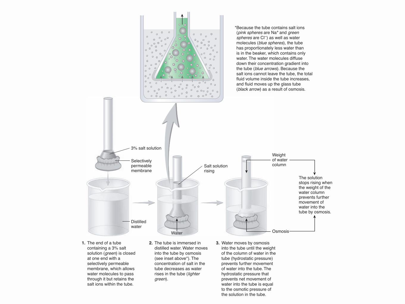
Because the tube contains salt ions*(pink spheres are Na+ and green spheres are Cl–) as well as water molecules (blue spheres), the tube has proportionately less water than is in the beaker, which contains only water. The water molecules diffusedown their concentration gradient into the tube (blue arrows). Because the salt ions cannot leave the tube, the total fluid volume inside the tube increases, and fluid moves up the glass tube (black arrow) as a result of osmosis.
Water
3% salt solution
Selectivelypermeablemembrane
Salt solutionrising
Distilledwater
3. Water moves by osmosis into the tube until the weight of the column of water in the tube (hydrostatic pressure) prevents further movement of water into the tube. The hydrostatic pressure that prevents net movement of water into the tube is equal to the osmotic pressure of the solution in the tube.
2. The tube is immersed in distilled water. Water moves into the tube by osmosis (see inset above*). The concentration of salt in the tube decreases as water rises in the tube (lighter green).
1. The end of a tube containing a 3% salt solution (green) is closed at one end with a selectively permeable membrane, which allows water molecules to pass through it but retains the salt ions within the tube.
The solution stops rising when the weight of the water column prevents further movement of water into the tube by osmosis.
Weightof watercolumn
Osmosis
52 Chapter 3
example, the transport protein that moves glucose does not move amino acids or ions. Competition occurs when similar molecules or ions can be moved by the transport protein. Although transport pro-teins exhibit specifi city, a transport protein may transport very similar substances. Th e substance in the greater concentration or the substance for which the transport protein is the most specifi c is moved across the plasma membrane at the greater rate. Saturation means that the rate of movement of molecules or ions across the membrane is limited by the number of available transport proteins. As the concentration of a
Process Figure 3.5 Osmosis
transported substance increases, more transport proteins become involved with transporting the substance, and the rate at which the substance is moved across the plasma membrane increases. Once the concentration of the substance is increased so that all the transport proteins are in use, the rate of movement remains constant, even though the concentration of the substance increases further. Th ree types of transport proteins—channel proteins, carrier proteins (transporters), and ATP-powered pumps—are involved in mediated transport.
tat78194_ch03_045-076.indd Page 52 09/10/10 9:41 AM user-f463tat78194_ch03_045-076.indd Page 52 09/10/10 9:41 AM user-f463 Volume/201/MHDQ233/tat78194_disk1of1/0073378194/tat78194_pagefilesVolume/201/MHDQ233/tat78194_disk1of1/0073378194/tat78194_pagefile

CHAPTER 1 General Physiological Concepts 3
generating organelles. The cytoplasmic organelles are held in position by filaments and microtubules, arising from the cen-trosomes, which are also important in the movement of chro-mosomes during cell division. Finally, the nucleus, also surrounded by a lipid bilayer membrane called the nuclear envelope, contains chromatin that is composed of DNA con-taining the nucleic acid code for cellular differentiation, func-tion, and replication. DNA contains the genes that encode mRNAs that are produced from DNA by transcription. Also contained within the nucleus is the nucleolus, which is the site of ribosome synthesis.
As you will learn in many chapters in this book, the cell membrane contains many different types of receptors that sense extracellular signals that are transduced into intracellu-lar signals. In addition, there are receptors within the cyto-
plasm and nucleus that respond to signals that enter the cell. Examples of such signals are steroid hormones such as estro-gen and testosterone that are lipophilic (“fat-loving”) and, as a result, can readily diffuse through the cell membrane to exert an intracellular action.
GENERAL STRUCTURE OF THE BODYFigure 1–3 is a diagrammatic representation of the human body. The organs (e.g., brain and heart) receive nutrients and eliminate waste products via the circulatory system. The heart is illustrated as two parts—right and left—as a functional
Central nervous system
Afferent and efferent nerves
Venousblood
Arterialblood
Food &waterintake
Right
Heart
Left
Tissues
Waste products
Nutrients
Endocrine glands
Hormones
Liver GI tract
Nutrients
Waste
Kidney
Waste
UrineFeces
Atmosphere
Lung
O2 CO2
Heart
O2 CO2
Synthesis
Metabolism
Reabsorption
Bile
Filtration
FIGURE 1–3 General organization of the major organs of the body. Arrows show the direction of blood flow and flux of gases, nutrients, hormones, and waste products.
Raff_Ch01_001-008.indd 3 11/26/10 9:50:32 AM
4 SECTION I Introduction
depiction even though it is actually one organ. The right side of the heart receives partially deoxygenated blood returning from the tissues and pumps blood to the lungs. In the lungs, oxygen diffuses into the blood from the gas phase for use in cellular respiration in the body, and carbon dioxide, a waste product of cellular respiration, is eliminated by diffusion from the blood into the gas phase. The left side of the heart receives oxygenated blood from the lung and pumps the blood into the arterial tree to perfuse the organs of the body. Nutrients, min-erals, vitamins, and water are taken in by the ingestion of food and liquids and absorption in the gastrointestinal (GI) tract. The liver, usually considered part of the GI system, processes substances absorbed into the blood from the GI tract, and also synthesizes new molecules such as glucose from precursors. Metabolic waste products are eliminated by the GI system in the feces and by the kidney in the urine. The two main integra-tive controllers of the internal environment are the nervous and endocrine systems. The nervous system is composed of the brain, spinal cord, sensory systems, and nerves. The endo-crine system is composed of ductless glands and scattered secretory cells distributed throughout the body that release hormones into the blood in response to metabolic, hormonal, and neural signals. It is the function of the nervous and endo-crine systems to coordinate the behavior and interactions of the organ systems described throughout this book.
Water is the most abundant molecule in the body, constitut-ing about 50–60% of the total body weight. All cells and organs exist in an aqueous environment. The intracellular water is the main component of the cytosol. Water is also the main compo-nent of the extracellular fluid. The extracellular fluid includes the interstitial fluid, which bathes the cells of the body, the blood plasma, which is the liquid component of the blood, cerebrospinal fluid, which is found only in the central ner-vous system, synovial fluid, which is found in joints such as the knee, and lymph, which is a liquid formed from interstitial fluid that flows back to the circulatory system via the lym-phatic system. There are significant differences in the compo-sition of intracellular and extracellular fluids that are very important in many aspects of cellular function (Table 1–1).
GENERAL PHYSICAL FACTORS AND CONCEPTSIt is not an accident that physiology and physics come from the same Greek word physis (nature). It is important that students of physiology understand the physical forces and factors that govern body function.
MEMBRANE TRANSPORTThere are several different mechanisms by which molecules cross the cell membrane either coming into or going out of the cell. These are all described in detail in Section 2. The sim-plest is diffusion in which the rate at which a molecule crosses
the cell membrane is governed by the concentration gradient and the ease with which each molecule can go through the cell membrane (permeability); energy expenditure is not directly required for diffusion, which is why it is sometimes called passive diffusion. There are also protein transporters located in the cell membrane that mediate facilitated diffu-sion of molecules that are too large or hydrophilic to perme-ate the membrane by simple diffusion. Facilitated diffusion does not require energy and moves molecules down a concen-tration gradient. By contrast, active transport is a process of moving molecules across a cell membrane against a concen-tration gradient; it can be thought of as a pump that uses energy to do work.
The movement of water molecules across the cell membrane also occurs by diffusion from a higher to a lower water “con-centration.” This is termed osmosis; water moves from a com-partment with fewer osmotically active particles (higher water concentration) to a compartment with more osmotically active particles (lower water concentration). Examples of osmotically active particles are ions such as sodium, potassium, and chlo-ride, and organic molecules such as glucose and amino acids.
BUFFERING AND pHOne of the most tightly controlled variables in the body is the hydrogen ion concentration of the intracellular and extracellu-lar fluids. This is because most proteins have optimal function within a very narrow range of pH. Remember that the pH is the negative logarithm (base 10) of the hydrogen ion concentration in molar units—when pH is low, the fluid is acidic; when pH is high, the fluid is alkaline. The body has several mechanisms for
TABLE 1–1 Composition of extracellular and intracellular fl uids.
Extracellular Concentration (mM)
Intracellular Concentration (mM)
Na+ 140 12
K+ 5 150
Ca2+ 1 0.0001
Mg2+ 1.5 12
Cl− 100 7
HCO3− 24 10
Amino acids 2 8
Glucose 4.7 1
Protein 0.2 4
The intracellular concentrations are slightly different for different tissues. The Ca2+ concentrations shown are the free, biologically active ions not bound to proteins. Total Ca2+ (bound plus free) are considerably higher in extracellular (2.5 mM) and intracellular (1.5 mM) fluids.Reproduced with permission from Widmaier EP, Raff H, Strang KT: Vander’s Human Physiology, 11th ed. McGraw-Hill, 2008.
Raff_Ch01_001-008.indd 4 11/26/10 9:50:34 AM
CAPÍTULO 3: SISTEMA DIGESTIVO 13% 1. Organización y control del sistema digestivo 2. Motilidad gastrointestinal 3. Secreciones gastrointestinales 4. Digestión y absorción de nutrientes, electrolitos y agua
CAPÍTULO 4: SISTEMA CARDIOVASCULAR 13% 1. Función cardiaca 2. Hemodinamia 3. Función vascular 4. Microcirculación 5. Regulación del gasto cardiaco 6. Regulación de la presión arterial
CAPÍTULO 5: SANGRE 4% 1. Hematopoyesis 2. Hemostasia
CAPÍTULO 6: SISTEMA RESPIRATORIO 12% 3. Estructura y función del sistema respiratorio 4. Mecánica de la respiración 5. Ventilación 6. Difusión y transporte de gases 7. Regulación de la respiración
CAPÍTULO 7: SISTEMA RENAL 13% 1. Organización del sistema renal 2. Líquidos corporales 3. Filtración glomerular y flujo sanguíneo renal 4. Función tubular 5. Concentración y dilución de la orina 6. Balance de sodio y potasio 7. Equilibrio ácido-base
4. Evaluación.
La evaluación del curso consta de 3 pruebas solemnes (PS1, PS2 y PS3, respectivamente) de selección múltiple (alternativas).
La nota de presentación a examen resultará del promedio ponderado de las tres pruebas solemnes, según los siguientes valores: PS1, 20%; PS2, 40%; PS3, 40% La nota final del curso se obtendrá según las siguientes ponderaciones: Nota de presentación a examen: 70% Nota de examen: 30% 5. Bibliografía.
� R Berne y M Levy. Fisiología. España: Elsevier. � Guyton y Hall. Tratado de Fisiología Médica. España: Elsevier. � Johnson LR (Ed) Essential Medical Physiology. USA: Lippincott-Raven Publishers. � Costanzo L. Fisiología. España: Elsevier. � Silverthorn. Fisiología Humana. Edit. Médica Panamericana. � Ganong. W.F. Fisiología Médica. México: Editorial Manual Moderno.
Se recomienda en lo posible las últimas ediciones de la bibliografía señalada.