Sacral Plexus & Nerves ANS Major arteries Accessory arteries Major veins Accessory veins
Upload
blanche-carrollCategory
view
231download
3
Circulatory SystemCirculatory System
Arteries, Veins, Capillaries, the Heart, Vessels
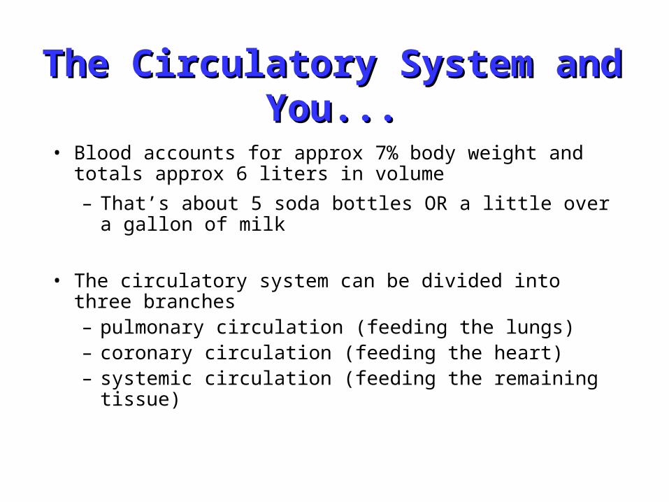
• Blood accounts for approx 7% body weight and totals approx 6 liters in volume
– That’s about 5 soda bottles OR a little over a gallon of milk
• The circulatory system can be divided into three branches– pulmonary circulation (feeding the lungs)– coronary circulation (feeding the heart)– systemic circulation (feeding the remaining tissue)
The Circulatory System and The Circulatory System and You...You...
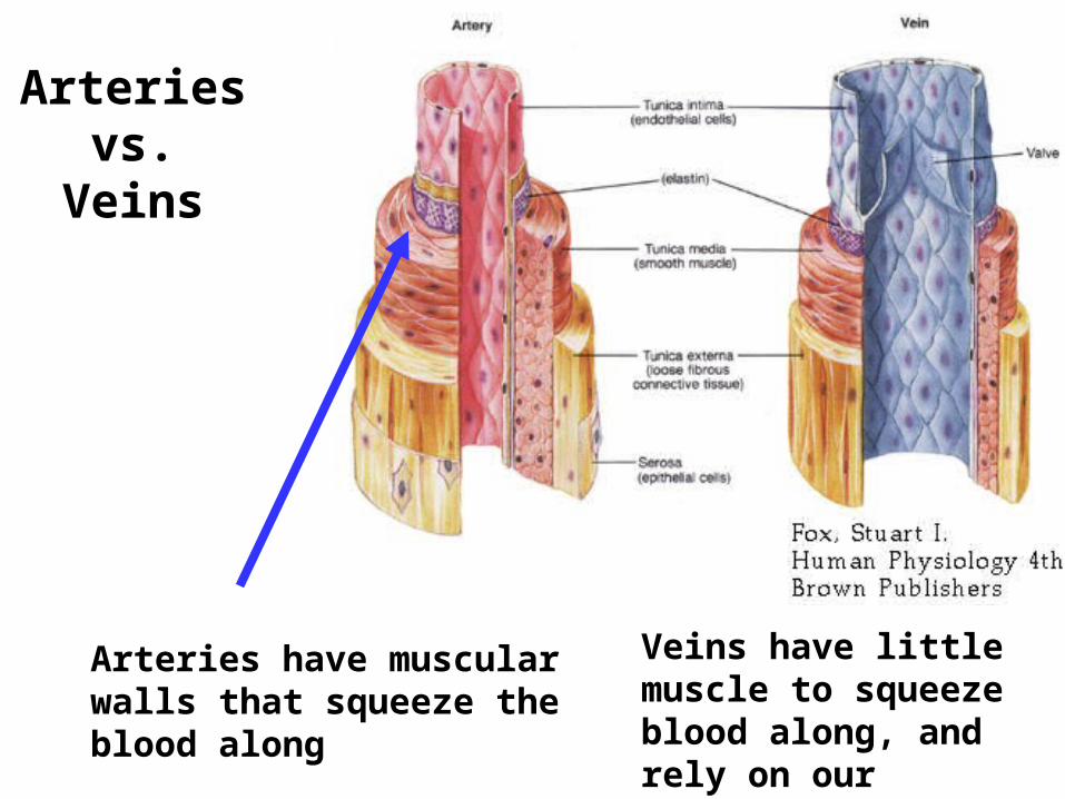
Arteries vs. Veins
Arteries have muscular walls that squeeze the blood along
Veins have little muscle to squeeze blood along, and rely on our skeletal muscles to do so.
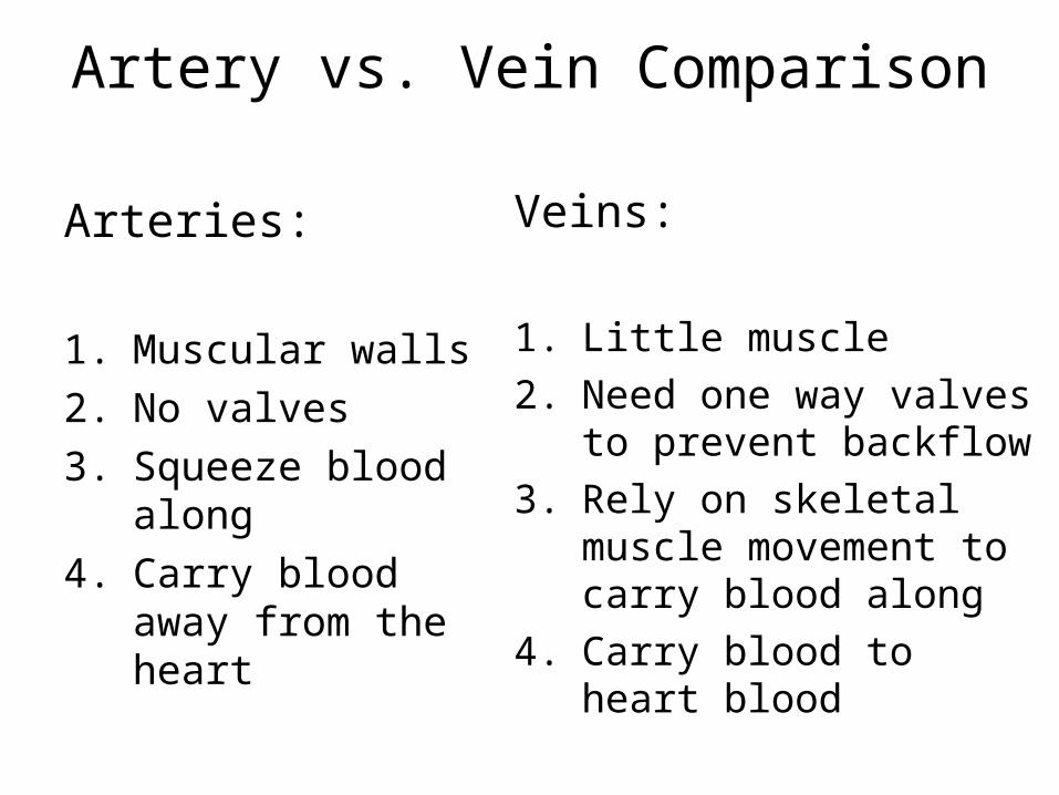
Artery vs. Vein Comparison
Arteries:
1. Muscular walls
2. No valves
3. Squeeze blood along
4. Carry blood away from the heart
Veins:
1. Little muscle
2. Need one way valves to prevent backflow
3. Rely on skeletal muscle movement to carry blood along
4. Carry blood to heart blood
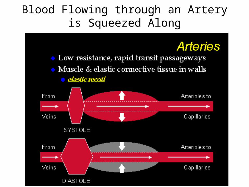
Blood Flowing through an Artery is Squeezed Along
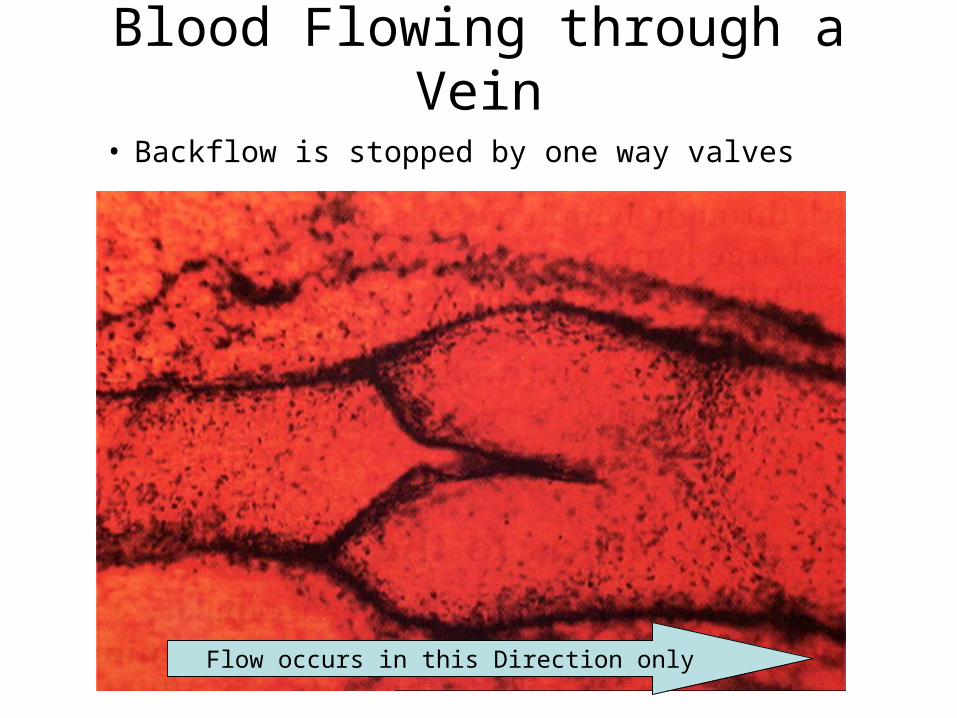
Blood Flowing through a Vein
• Backflow is stopped by one way valves
Flow occurs in this Direction only
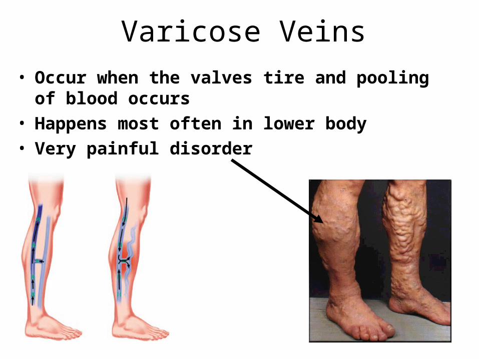
Varicose Veins
• Occur when the valves tire and pooling of blood occurs
• Happens most often in lower body
• Very painful disorder
bilateral varicosis with megaplasia of long saphenous vein and insufficiency of femoral vein.

• Extremely narrow and thin, and leaky– 5mm-10mm in diameter – 0.5mm wall thickness
• Composed of thin cells• Transfer oxygen and food to
tissues• Remove wastes, CO2
• Arterial Venuous Blood Blood
CapillariesCapillaries

Human Cardiovascular SystemHuman Cardiovascular System
The heart is located in the thoracic cavity between the lungs. It is The heart is located in the thoracic cavity between the lungs. It is surrounded by a protective pericardium—a double-walled sac surrounded by a protective pericardium—a double-walled sac with fluid between the two layers. with fluid between the two layers.
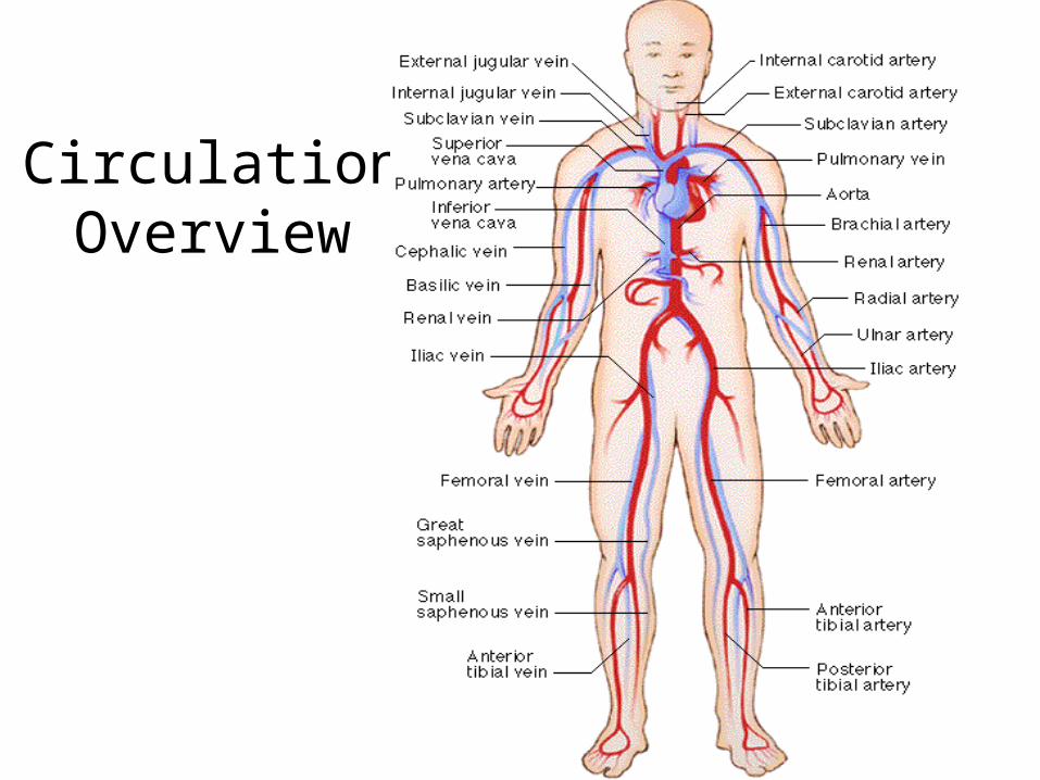
Circulation Overview
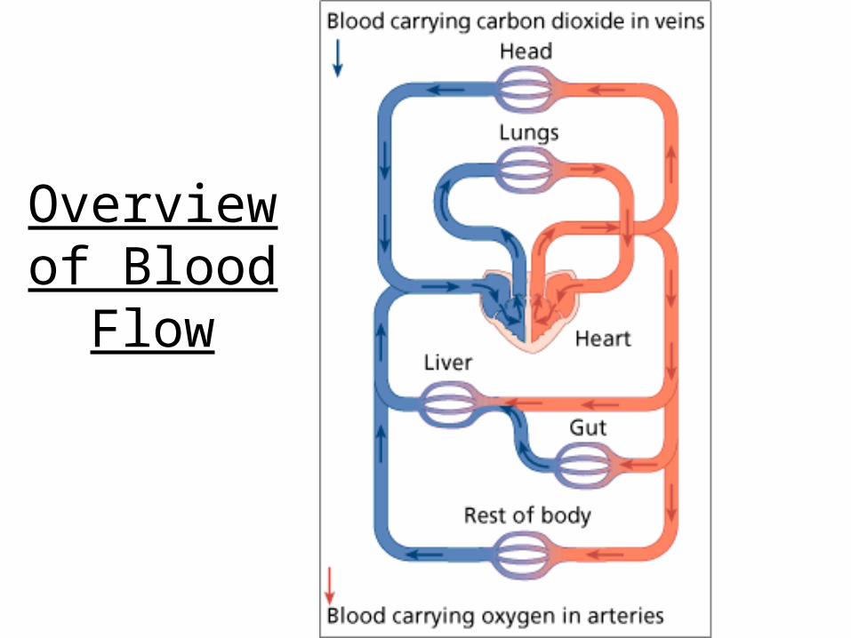
Overview of Blood Flow
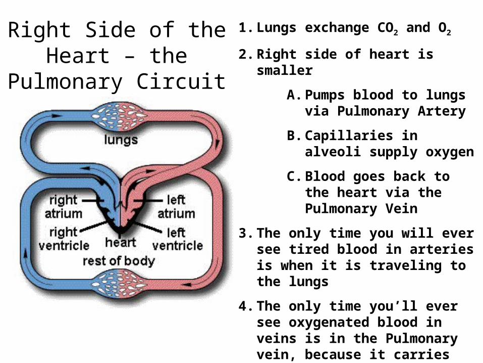
1. Lungs exchange CO2 and O2
2. Right side of heart is smaller
A. Pumps blood to lungs via Pulmonary Artery
B. Capillaries in alveoli supply oxygen
C. Blood goes back to the heart via the Pulmonary Vein
3. The only time you will ever see tired blood in arteries is when it is traveling to the lungs
4. The only time you’ll ever see oxygenated blood in veins is in the Pulmonary vein, because it carries the blood to the heart (as veins do) to be pumped to the rest of the body.
Right Side of the Heart – the Pulmonary Circuit
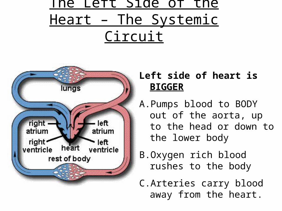
The Left Side of the Heart – The Systemic Circuit
Left side of heart is BIGGER
A. Pumps blood to BODY out of the aorta, up to the head or down to the lower body
B. Oxygen rich blood rushes to the body
C.Arteries carry blood away from the heart.
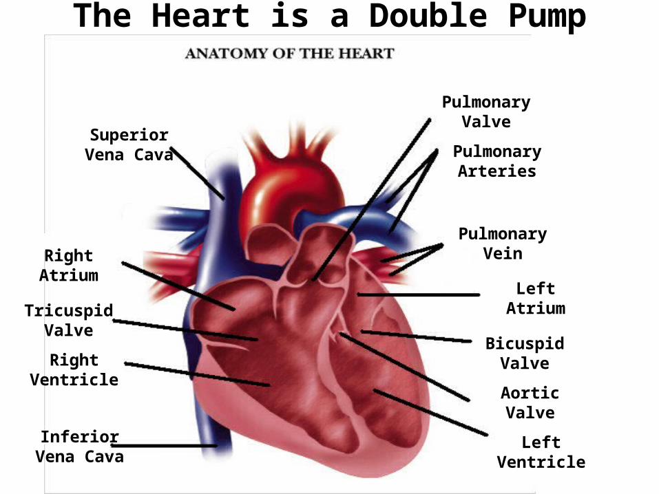
The Heart is a Double Pump
Superior Vena Cava
Inferior Vena Cava
Left Ventricle
Pulmonary Vein
Aortic Valve
Left Atrium
Bicuspid Valve
Pulmonary Arteries
Pulmonary Valve
Right Atrium
Tricuspid Valve
Right Ventricle
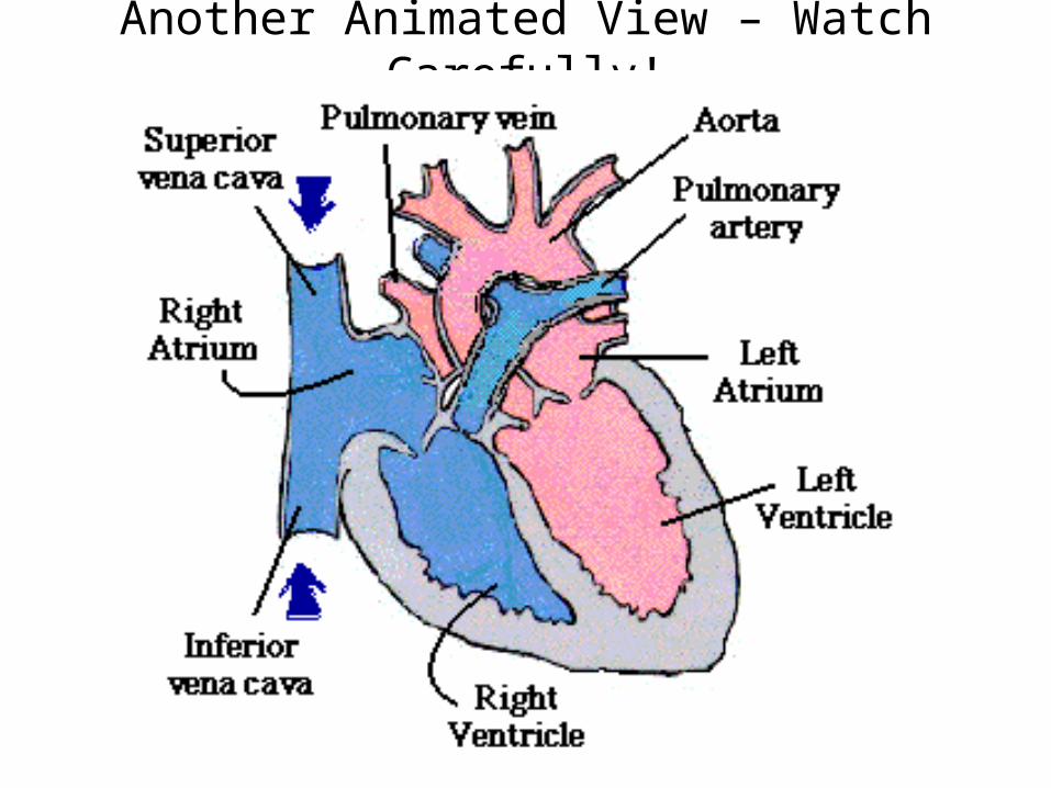
Another Animated View – Watch Carefully!

Check out the Atrioventricular Valve (tricuspid) Doing its Job Preventing Backflow between the Right Ventricle and Atrium

Heart Attacks
Occur because of coronary blockage due to cholesterol blockages which obstruct blood flow to the heart itself
Click the heart to see an animation --------->
4 Root Cause of Heart Attacks
1. Heredity – the liver cranks out cholesterol, and there are two types:
A. LDL – low density – bad, creates blockages
B. HDL – high density – good, scrubs our tubes
2. Lack of regular exercise
3. Unhealthy, high-fat diets
4. Generally being a couch potato does not help
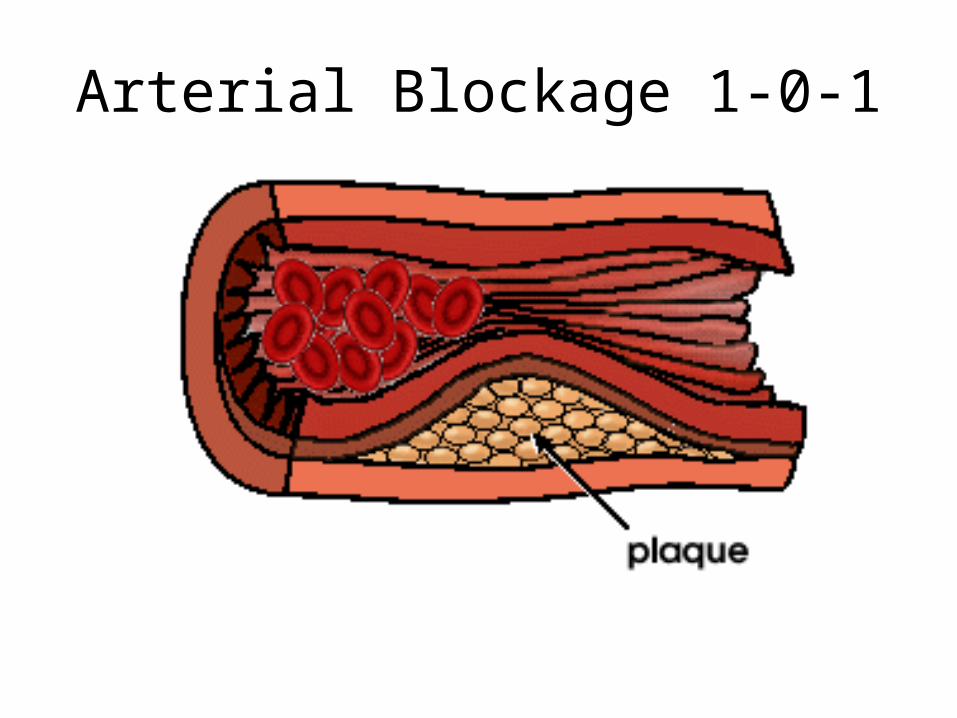
Arterial Blockage 1-0-1

Blood Flowing through an Artery is Squeezed Along
Blood Pressure
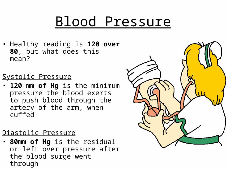
• Healthy reading is 120 over 80, but what does this mean?
Systolic Pressure• 120 mm of Hg is the minimum
pressure the blood exerts to push blood through the artery of the arm, when cuffed
Diastolic Pressure• 80mm of Hg is the residual or
left over pressure after the blood surge went through
Solutions to Blood Pressure• Reduce the volume of water in
the blood
• Water is attracted to salt, so reduce salt intake
• Exercise to shed excess water
• Eat healthier
• Record pressure regularly• Medication needed in extreme
cases
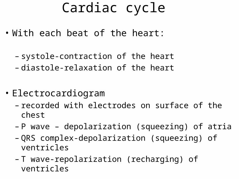
Cardiac cycle
• With each beat of the heart:
– systole-contraction of the heart– diastole-relaxation of the heart
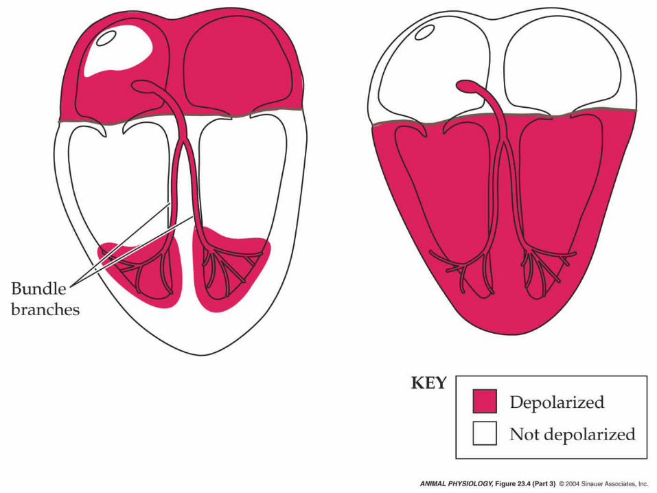
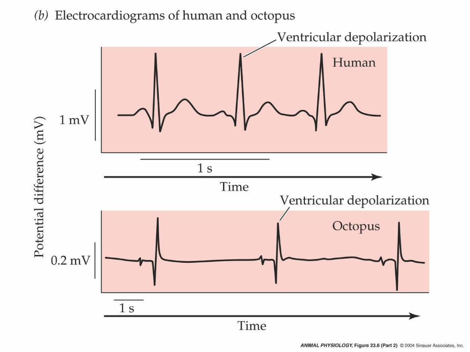
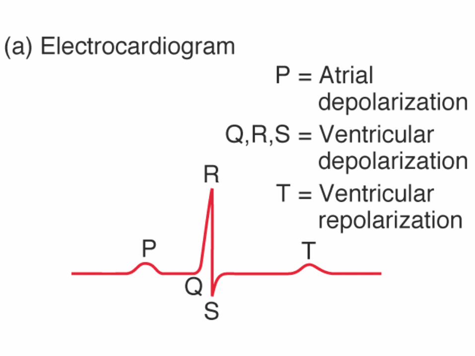
• Electrocardiogram– recorded with electrodes on surface of the chest– P wave – depolarization (squeezing) of atria– QRS complex-depolarization (squeezing) of
ventricles– T wave-repolarization (recharging) of ventricles
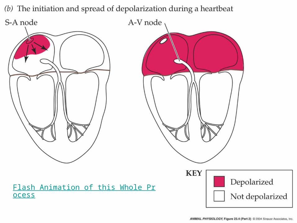
Flash Animation of this Whole Process

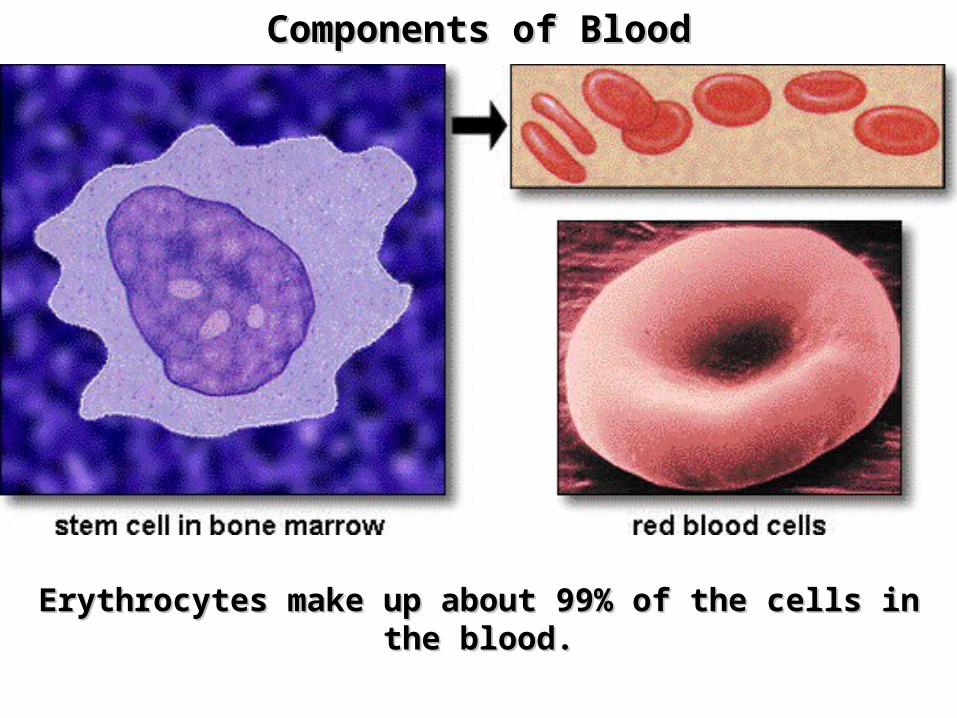
Components of BloodComponents of Blood
Erythrocytes make up about 99% of the cells in the blood.Erythrocytes make up about 99% of the cells in the blood.

Components of BloodComponents of Blood
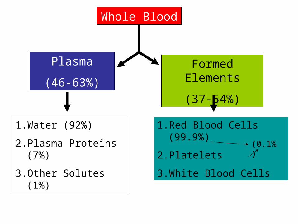
Whole Blood
Plasma
(46-63%)
Formed Elements
(37-54%)
1. Water (92%)
2. Plasma Proteins (7%)
3. Other Solutes (1%)
1. Red Blood Cells (99.9%)
2. Platelets
3. White Blood Cells
(0.1%)
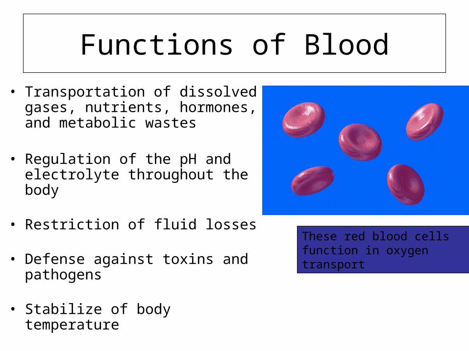
Functions of Blood
• Transportation of dissolved gases, nutrients, hormones, and metabolic wastes
• Regulation of the pH and electrolyte throughout the body
• Restriction of fluid losses
• Defense against toxins and pathogens
• Stabilize of body temperature
These red blood cells function in oxygen transport
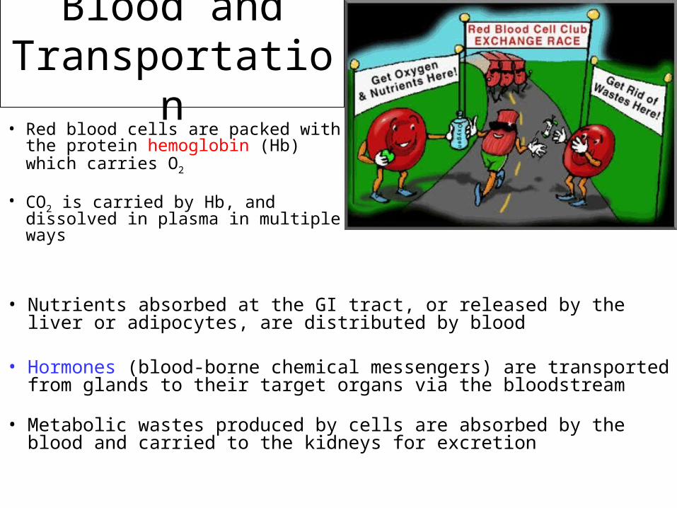
Blood and Transportation
• Red blood cells are packed with the protein hemoglobin (Hb) which carries O2
• CO2 is carried by Hb, and dissolved in plasma in multiple ways
• Nutrients absorbed at the GI tract, or released by the liver or adipocytes, are distributed by blood
• Hormones (blood-borne chemical messengers) are transported from glands to their target organs via the bloodstream
• Metabolic wastes produced by cells are absorbed by the blood and carried to the kidneys for excretion
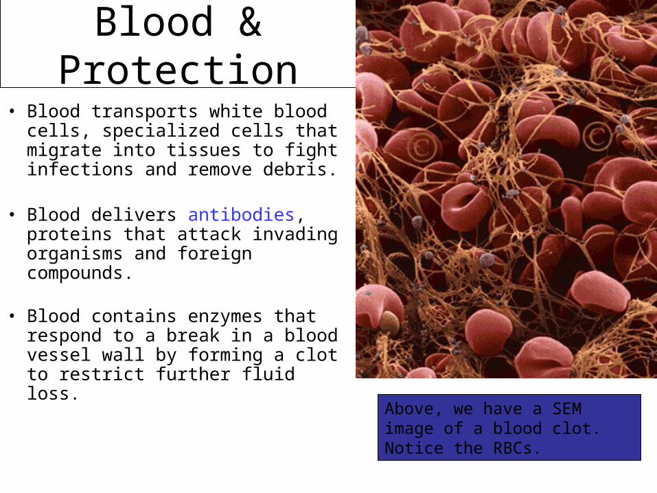
Blood & Protection
• Blood transports white blood cells, specialized cells that migrate into tissues to fight infections and remove debris.
• Blood delivers antibodies, proteins that attack invading organisms and foreign compounds.
• Blood contains enzymes that respond to a break in a blood vessel wall by forming a clot to restrict further fluid loss.
Above, we have a SEM image of a blood clot. Notice the RBCs.
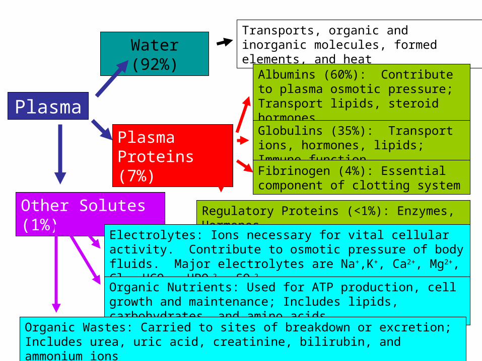
Plasma
Water (92%)Transports, organic and inorganic molecules, formed elements, and heat
Plasma Proteins (7%)
Albumins (60%): Contribute to plasma osmotic pressure; Transport lipids, steroid hormones
Globulins (35%): Transport ions, hormones, lipids; Immune function
Fibrinogen (4%): Essential component of clotting system
Regulatory Proteins (<1%): Enzymes, HormonesOther Solutes (1%)
Electrolytes: Ions necessary for vital cellular activity. Contribute to osmotic pressure of body fluids. Major electrolytes are Na+,K+, Ca2+, Mg2+, Cl-, HCO3
-, HPO42-, SO4
2-
Organic Nutrients: Used for ATP production, cell growth and maintenance; Includes lipids, carbohydrates, and amino acids
Organic Wastes: Carried to sites of breakdown or excretion; Includes urea, uric acid, creatinine, bilirubin, and ammonium ions
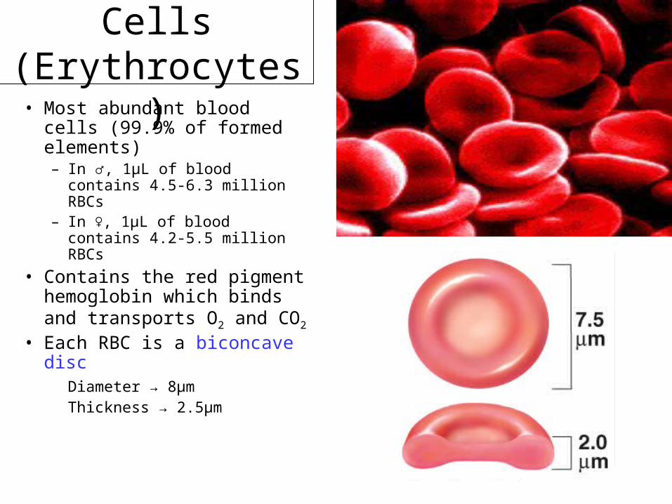
Red Blood Cells (Erythrocytes)
• Most abundant blood cells (99.9% of formed elements)– In ♂, 1µL of blood contains
4.5-6.3 million RBCs– In ♀, 1µL of blood contains
4.2-5.5 million RBCs
• Contains the red pigment hemoglobin which binds and transports O2 and CO2
• Each RBC is a biconcave disc
Diameter → 8µmThickness → 2.5µm

Interesting Facts about RBC’s
• Increased RBC production when O2 decreases- high altitude training
• Lack nucleus to carry more O2
• Life span of 120 days
• Broken down in liver (bilirubin into bile…)
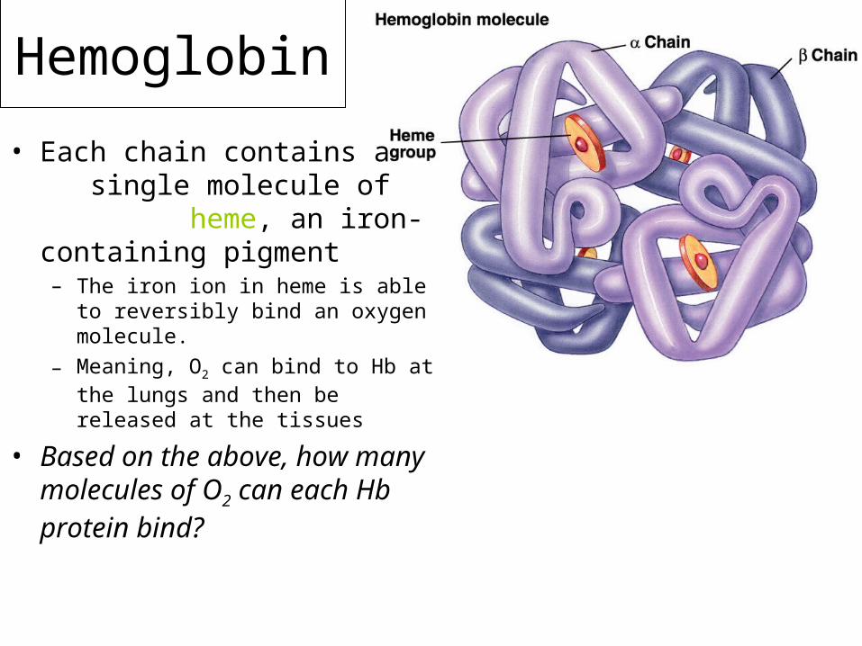
Hemoglobin
• Each chain contains a single molecule of heme, an iron-containing pigment– The iron ion in heme is able to
reversibly bind an oxygen molecule.
– Meaning, O2 can bind to Hb at the lungs and then be released at the tissues
• Based on the above, how many molecules of O2 can each Hb protein bind?
RBC Disorders
1. Anemia• Fe (Iron) deficiency• Pale apperance, and some weakness
2. Pernicous Anemia• Will cause low vitamin B12 absorption• RBC’s will not grow up / mature
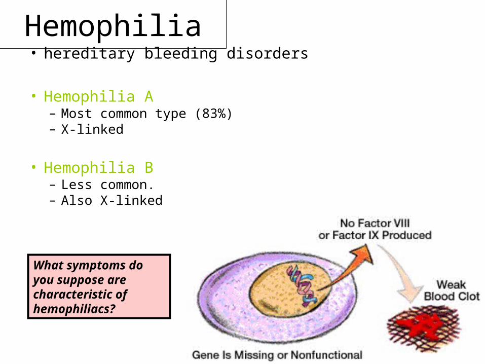
Hemophilia• hereditary bleeding disorders
• Hemophilia A– Most common type (83%)– X-linked
• Hemophilia B– Less common.– Also X-linked
What symptoms do you suppose are characteristic of hemophiliacs?
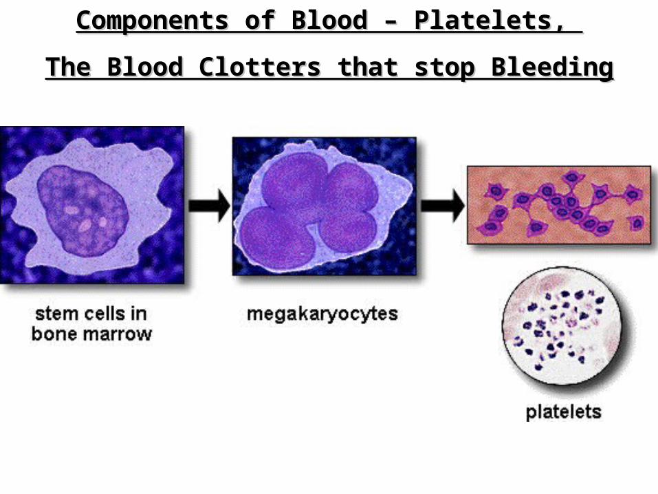
Components of Blood – Platelets, Components of Blood – Platelets,
The Blood Clotters that stop BleedingThe Blood Clotters that stop Bleeding
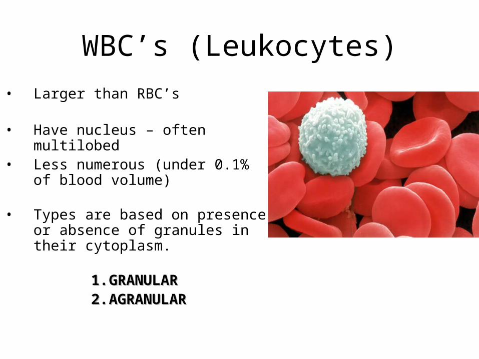
WBC’s (Leukocytes)
• Larger than RBC’s
• Have nucleus – often multilobed• Less numerous (under 0.1% of
blood volume)
• Types are based on presence or absence of granules in their cytoplasm.
1.1. GRANULARGRANULAR2.2. AGRANULARAGRANULAR
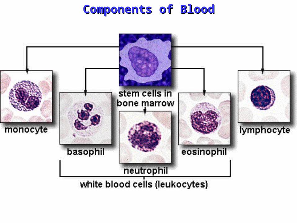
Components of BloodComponents of Blood
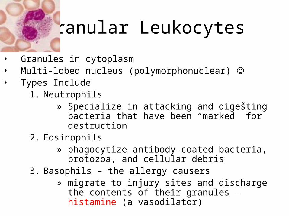
Granular Leukocytes
• Granules in cytoplasm• Multi-lobed nucleus (polymorphonuclear) • Types Include
1. Neutrophils » Specialize in attacking and digesting bacteria that
have been “marked” for destruction2. Eosinophils
» phagocytize antibody-coated bacteria, protozoa, and cellular debris
3. Basophils – the allergy causers» migrate to injury sites and discharge the contents
of their granules – histamine (a vasodilator)

Agranular Leukocytes
• No granules• The Explorers of the immune system
1. Lymphocytes (B&T) » Continuously migrate from the bloodstream thru
peripheral tissues and back into the bloodstream
» T cells: defend against foreign cells and tissues and coordinate the immune response
» B cells: produce and distribute antibodies that attack foreign materials
2. Monocytes – the largest WBC’s» Becomes a MACROPHAGE in tissue and
phagocytizes bacteria (munch)
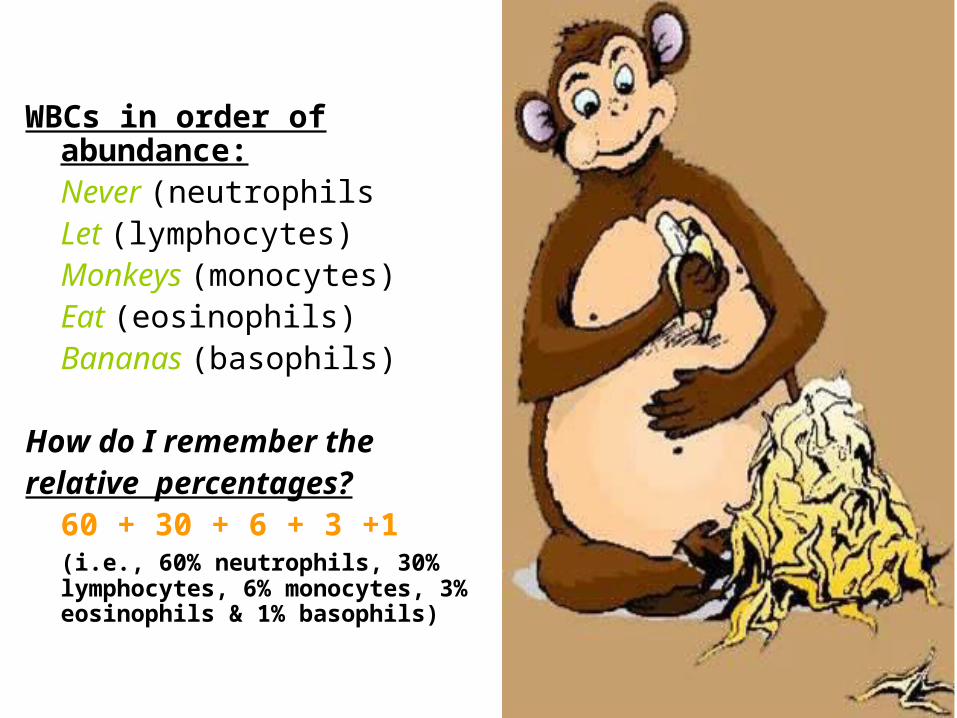
WBCs in order of abundance:Never (neutrophilsLet (lymphocytes)Monkeys (monocytes)Eat (eosinophils)Bananas (basophils)
How do I remember the relative percentages?
60 + 30 + 6 + 3 +1(i.e., 60% neutrophils, 30% lymphocytes, 6% monocytes, 3% eosinophils & 1% basophils)
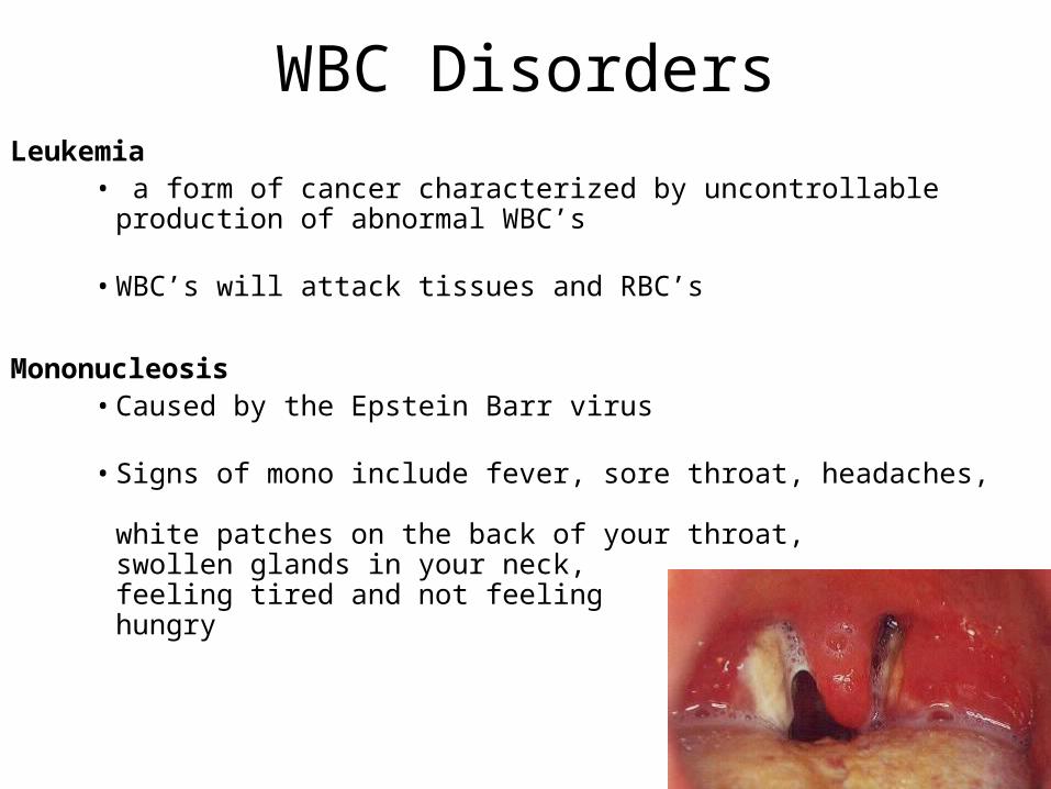
WBC DisordersLeukemia
• a form of cancer characterized by uncontrollable production of abnormal WBC’s
• WBC’s will attack tissues and RBC’s
Mononucleosis• Caused by the Epstein Barr virus
• Signs of mono include fever, sore throat, headaches, white patches on the back of your throat, swollen glands in your neck, feeling tired and not feeling hungry
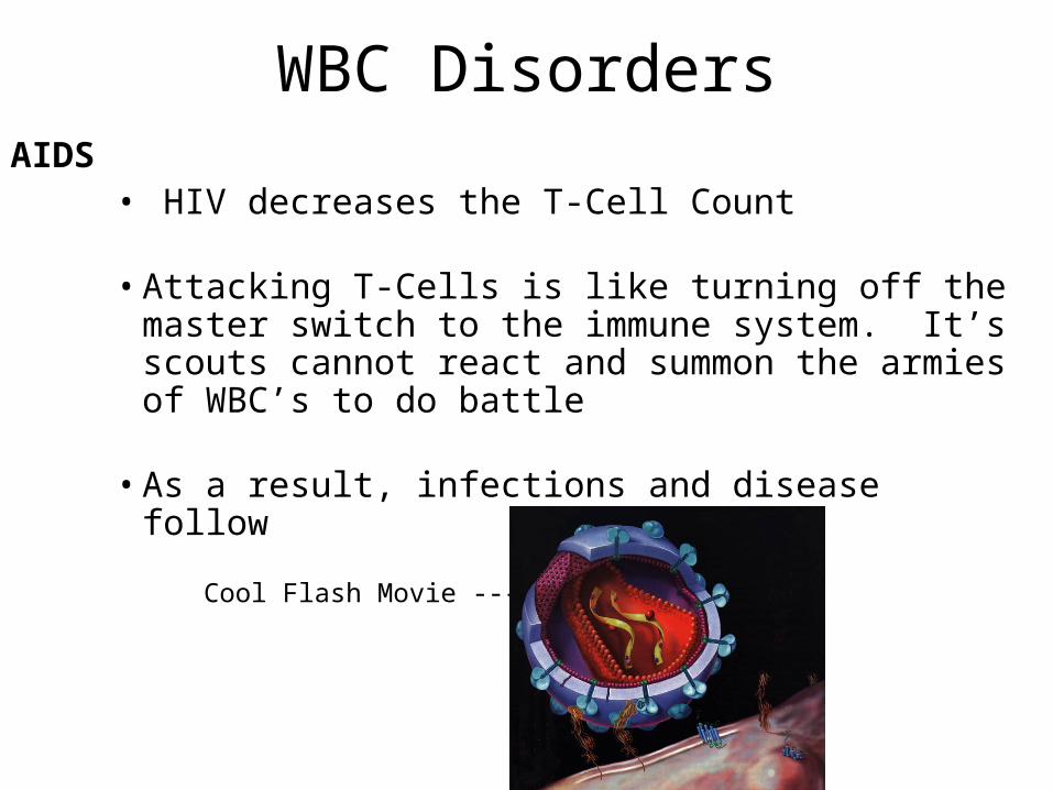
WBC DisordersAIDS
• HIV decreases the T-Cell Count
• Attacking T-Cells is like turning off the master switch to the immune system. It’s scouts cannot react and summon the armies of WBC’s to do battle
• As a result, infections and disease follow
Cool Flash Movie --- >
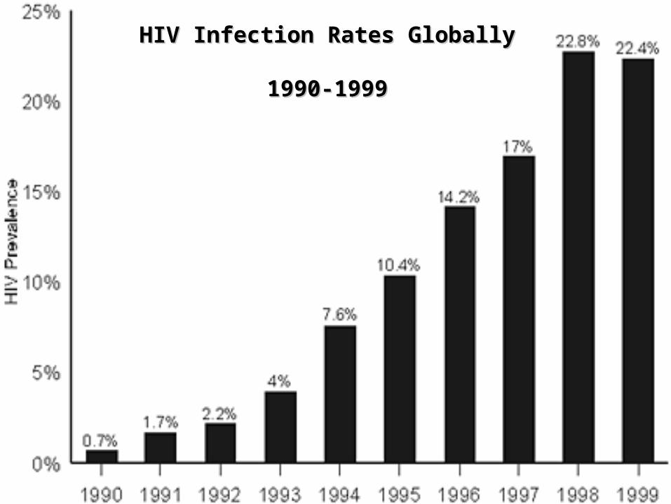
HIV Infection Rates Globally HIV Infection Rates Globally 1990-19991990-1999
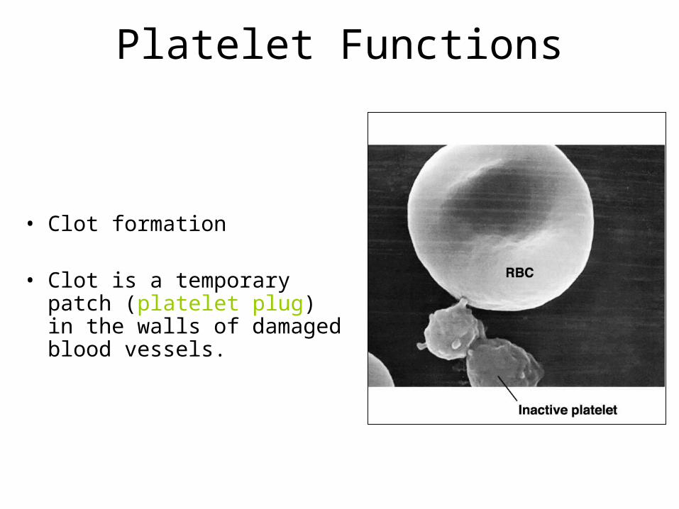
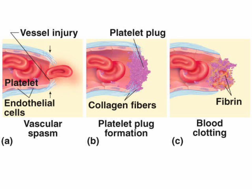
Platelet Functions
• Clot formation
• Clot is a temporary patch (platelet plug) in the walls of damaged blood vessels.
This Presentation Brought to you Couresy of the Hart Foundation