
Circular RNA ciRS-7 A Promising Prognostic Biomarker and a ... · cohorts, respectively)....
12
Biology of Human Tumors Circular RNA ciRS-7—A Promising Prognostic Biomarker and a Potential Therapeutic Target in Colorectal Cancer Wenhao Weng 1,2 , Qing Wei 3 , Shusuke Toden 1 , Kazuhiro Yoshida 1 , Takeshi Nagasaka 4 , Toshiyoshi Fujiwara 4 , Sanjun Cai 5,6 , Huanlong Qin 7 , Yanlei Ma 5,6 , and Ajay Goel 1 Abstract Purpose: Colorectal cancer is one of the most common malig- nancies worldwide. Recently, a novel circular RNA, ciRS-7, was proposed to be a potential miR-7 sponge. As miR-7, a putative tumor-suppressor, regulates the expression of several import- ant drivers of colorectal cancer, we analyzed the clinical signif- icance of ciRS-7 in colorectal cancer patients. Experimental Design: Initially, we evaluated the expression levels of ciRS-7 in a training cohort comprising of 153 primary colorectal cancer tissues and 44 matched normal mucosae. We subsequently confirmed its clinical relevance in an indepen- dent validation cohort (n ¼ 165), and evaluated the effect of ciRS-7 on miR-7, and its target genes EGFR and RAF1. Func- tional analyses were performed in cell lines and an animal model to support clinical findings. Results: Our data revealed that ciRS-7 was significantly upregulated in colorectal cancer tissues compared with matched normal mucosae (P ¼ 0.0018), and its overexpres- sion was associated with poor patient survival (P ¼ 0.0224 and 0.0061 in the training and validation cohorts, respective- ly). Multivariate survival analysis revealed that ciRS-7 emerged as an independent risk factor for overall survival (P ¼ 0.0656 and 0.0324 in the training and validation cohorts, respectively). Overexpression of ciRS-7 in HCT116 and HT29 cells led to the blocking of miR-7 and resulted in a more aggressive oncogenic phenotype, and ciRS-7 overexpres- sion permitted the inhibition of miR-7 and subsequent acti- vation of EGFR and RAF1 oncogenes. Conclusions: CiRS-7 is a promising prognostic biomarker in colorectal cancer patients and may serve as a thera- peutic target for reducing EGFR-RAF1 activity in colorectal cancer patients. Clin Cancer Res; 23(14); 3918–28. Ó2017 AACR. Introduction Colorectal cancer is a leading cause of tumor-associated mor- bidity and mortality worldwide, and its incidence continues to rise gradually (1). Although several critical events have been identified that play key roles during colorectal carcinogenesis (2, 3), only few of these molecular targets are clinically actionable. Wealth of published studies suggest that miRNAs play key roles in the development of various types of cancer, including colorectal cancer (4). Previous work from our group and others have highlighted that specific miRNAs contribute to colorectal cancer pathogenesis and can be used as biomarkers for diagnosis, prognosis, and metastasis prediction in colorectal cancer patients (5–9). One such miRNA that has aroused considerable interest recently is miR-7 (10–16). miR-7 is aberrantly expressed in several cancers and linked to many "oncogenic" pathways (17, 18). In colorectal cancer, miR-7 was demon- strated as a tumor suppressor, and its downregulation is cor- related with poor prognosis (19, 20). Considering the critical biological role of miR-7 in colorectal tumorigenesis and its clinical relevance as a prognostic biomarker, it is essential to better understand the underlying mechanisms in colorectal cancer, which still remains vastly unclear and unexplored. It has become increasingly clear that RNA transcripts share the same miRNA-binding sites can communicate with and regulate each other by competing for common pools of miRNA molecules (21), and thereby act as competing endogenous RNAs (ceRNA). Accumulating evidence has unveiled the impor- tance of ceRNA-mediated regulatory mechanisms in cancer pathogenesis. Recently, circular RNA ciRS-7 was shown to act as a ceRNA of miR-7: ciRS-7 can bind up to 73 copies of miR-7, allowing genes repressed by miR-7 to be reactivated during the development of brain (22). This circular RNA has also been 1 Center for Gastrointestinal Research; Center for Translational Genomics and Oncology, Baylor Scott & White Research Institute, Charles A. Sammons Cancer Center, Baylor University Medical Center, Dallas, Texas. 2 Department of Clinical Laboratory, Shanghai Tenth People's Hospital, Tongji University, Shanghai, China. 3 Department of Pathology, Shanghai Tenth People's Hospital, Tongji University, Shanghai, China. 4 Department of Gastroenterological Surgery and Surgical Oncology, Okayama University Graduate School of Medicine, Dentistry and Pharmaceutical Sciences, Okayama, Japan. 5 Department of Colorectal Surgery, Fudan University Shanghai Cancer Center, Shanghai, China. 6 Depart- ment of Oncology, Shanghai Medical College, Fudan University, Shanghai, China. 7 Department of Gastrointestinal Surgery, Shanghai Tenth People's Hospital Affiliated to Tongji University, Shanghai, China. Note: Supplementary data for this article are available at Clinical Cancer Research Online (http://clincancerres.aacrjournals.org/). W. Weng and Q. Wei contributed equally to this article. Corresponding Authors: Ajay Goel, Center for Gastrointestinal Research; and Center for Translational Genomics and Oncology, Baylor Scott & White Research Institute and Charles Sammons Cancer Center, Baylor University Medical Center, 3410 Worth Street, Suite 610, Dallas, TX 75246. Phone: 214-820-2603; Fax: 214- 818-9292; E-mail: [email protected]; and Yanlei Ma, Fudan University Shanghai Cancer Center, Fudan University, Shanghai 200032, China. Phone: 8621-6417-5590; Fax: 8621-5417-5590; E-mail: [email protected] doi: 10.1158/1078-0432.CCR-16-2541 Ó2017 American Association for Cancer Research. Clinical Cancer Research Clin Cancer Res; 23(14) July 15, 2017 3918 on June 8, 2020. © 2017 American Association for Cancer Research. clincancerres.aacrjournals.org Downloaded from Published OnlineFirst February 7, 2017; DOI: 10.1158/1078-0432.CCR-16-2541
Transcript of Circular RNA ciRS-7 A Promising Prognostic Biomarker and a ... · cohorts, respectively)....

Biology of Human Tumors
Circular RNA ciRS-7—A Promising PrognosticBiomarker and a Potential Therapeutic Target inColorectal CancerWenhao Weng1,2, Qing Wei3, Shusuke Toden1, Kazuhiro Yoshida1, Takeshi Nagasaka4,Toshiyoshi Fujiwara4, Sanjun Cai5,6, Huanlong Qin7, Yanlei Ma5,6, and Ajay Goel1
Abstract
Purpose:Colorectal cancer is one of the most commonmalig-nancies worldwide. Recently, a novel circular RNA, ciRS-7, wasproposed to be a potential miR-7 sponge. As miR-7, a putativetumor-suppressor, regulates the expression of several import-ant drivers of colorectal cancer, we analyzed the clinical signif-icance of ciRS-7 in colorectal cancer patients.
Experimental Design: Initially, we evaluated the expressionlevels of ciRS-7 in a training cohort comprising of 153 primarycolorectal cancer tissues and 44 matched normal mucosae. Wesubsequently confirmed its clinical relevance in an indepen-dent validation cohort (n ¼ 165), and evaluated the effect ofciRS-7 on miR-7, and its target genes EGFR and RAF1. Func-tional analyses were performed in cell lines and an animalmodel to support clinical findings.
Results: Our data revealed that ciRS-7 was significantlyupregulated in colorectal cancer tissues compared with
matched normal mucosae (P ¼ 0.0018), and its overexpres-sion was associated with poor patient survival (P ¼ 0.0224and 0.0061 in the training and validation cohorts, respective-ly). Multivariate survival analysis revealed that ciRS-7emerged as an independent risk factor for overall survival(P ¼ 0.0656 and 0.0324 in the training and validationcohorts, respectively). Overexpression of ciRS-7 in HCT116and HT29 cells led to the blocking of miR-7 and resulted in amore aggressive oncogenic phenotype, and ciRS-7 overexpres-sion permitted the inhibition of miR-7 and subsequent acti-vation of EGFR and RAF1 oncogenes.
Conclusions:CiRS-7 is a promising prognostic biomarkerin colorectal cancer patients and may serve as a thera-peutic target for reducing EGFR-RAF1 activity in colorectalcancer patients. Clin Cancer Res; 23(14); 3918–28. �2017AACR.
IntroductionColorectal cancer is a leading cause of tumor-associated mor-
bidity and mortality worldwide, and its incidence continues to
rise gradually (1). Although several critical events have beenidentified that play key roles during colorectal carcinogenesis(2, 3), only fewof thesemolecular targets are clinically actionable.Wealth of published studies suggest thatmiRNAs play key roles inthe development of various types of cancer, including colorectalcancer (4). Previous work from our group and others havehighlighted that specific miRNAs contribute to colorectal cancerpathogenesis and can be used as biomarkers for diagnosis,prognosis, and metastasis prediction in colorectal cancerpatients (5–9). One such miRNA that has aroused considerableinterest recently is miR-7 (10–16). miR-7 is aberrantlyexpressed in several cancers and linked to many "oncogenic"pathways (17, 18). In colorectal cancer, miR-7 was demon-strated as a tumor suppressor, and its downregulation is cor-related with poor prognosis (19, 20). Considering the criticalbiological role of miR-7 in colorectal tumorigenesis and itsclinical relevance as a prognostic biomarker, it is essential tobetter understand the underlying mechanisms in colorectalcancer, which still remains vastly unclear and unexplored.
It has become increasingly clear that RNA transcripts sharethe same miRNA-binding sites can communicate with andregulate each other by competing for common pools of miRNAmolecules (21), and thereby act as competing endogenousRNAs (ceRNA). Accumulating evidence has unveiled the impor-tance of ceRNA-mediated regulatory mechanisms in cancerpathogenesis. Recently, circular RNA ciRS-7 was shown to actas a ceRNA of miR-7: ciRS-7 can bind up to 73 copies of miR-7,allowing genes repressed by miR-7 to be reactivated during thedevelopment of brain (22). This circular RNA has also been
1Center for Gastrointestinal Research; Center for Translational Genomics andOncology, Baylor Scott &White Research Institute, Charles A. Sammons CancerCenter, Baylor University Medical Center, Dallas, Texas. 2Department of ClinicalLaboratory, Shanghai Tenth People's Hospital, Tongji University, Shanghai,China. 3Department of Pathology, Shanghai Tenth People's Hospital, TongjiUniversity, Shanghai, China. 4Department of Gastroenterological Surgery andSurgical Oncology, Okayama University Graduate School of Medicine, Dentistryand Pharmaceutical Sciences, Okayama, Japan. 5Department of ColorectalSurgery, Fudan University Shanghai Cancer Center, Shanghai, China. 6Depart-ment ofOncology, ShanghaiMedical College, FudanUniversity, Shanghai, China.7Department of Gastrointestinal Surgery, Shanghai Tenth People's HospitalAffiliated to Tongji University, Shanghai, China.
Note: Supplementary data for this article are available at Clinical CancerResearch Online (http://clincancerres.aacrjournals.org/).
W. Weng and Q. Wei contributed equally to this article.
Corresponding Authors: Ajay Goel, Center for Gastrointestinal Research; andCenter for Translational Genomics andOncology, Baylor Scott &White ResearchInstitute andCharles SammonsCancer Center, Baylor University Medical Center,3410Worth Street, Suite 610, Dallas, TX 75246. Phone: 214-820-2603; Fax: 214-818-9292; E-mail: [email protected]; and Yanlei Ma, Fudan UniversityShanghai Cancer Center, Fudan University, Shanghai 200032, China. Phone:8621-6417-5590; Fax: 8621-5417-5590; E-mail: [email protected]
doi: 10.1158/1078-0432.CCR-16-2541
�2017 American Association for Cancer Research.
ClinicalCancerResearch
Clin Cancer Res; 23(14) July 15, 20173918
on June 8, 2020. © 2017 American Association for Cancer Research. clincancerres.aacrjournals.org Downloaded from
Published OnlineFirst February 7, 2017; DOI: 10.1158/1078-0432.CCR-16-2541
linked to human disease by affecting miR-7 activity (22, 23),suggesting its role as a miR-7 regulator. However, to the best ofour knowledge, no studies have thus far interrogated thepathogenic role of ciRS-7 in cancer or its clinical relevance incolorectal cancer.
In the current study, we have made first attempts to fill this gapin knowledge to assess the molecular contribution of ciRS-7 incolorectal cancer.We specifically set out to investigate its relevanceas a prognostic biomarker and potential therapeutic target.Accordingly, we analyzed the expression level of ciRS-7 in neo-plastic tissues andmatchednormal tissues, followedby validationof our results in multiple, independent cohorts of colorectalcancer patients. In addition, we performed a systematic andcomprehensive functional analysis of ciRS-7 in colorectal cancer,its regulatory effect onmiR-7 activity in this disease, in a series of invitro experiments followed by validation of these results on tumorgrowth in xenograft animal models. We conclude that ciRS-7 is apromising prognostic biomarker for colorectal cancer patients,and therapeutic targeting of ciRS-7 maybe a potential strategy forthe management of colorectal cancer patients.
Materials and MethodsPatients and study design
This study included analysis of 448 specimens comprising of 90fresh frozen and 358 formalin-fixed, paraffin-embedded (FFPE)colorectal cancer tissues. For the analysis of clinical significance ofciRS-7 expression, wemeasured expression levels of ciRS-7 in 358FFPE samples which consisted of 318 FFPE tissues from primarycolorectal cancers, and 40 specimens from matched adjacentnormal mucosa (NM) tissues, obtained from colorectal cancerpatient cohorts that were enrolled at the Shanghai Tenth People'sHospital in China and Okayama University Medical Hospital inJapan. The baseline characteristics of these patient cohorts aredescribed in Supplementary Table S1. The study design consistedof an initial training cohort (Shanghai Tenth People's Hospital)and a subsequent validation cohort (OkayamaUniversityMedicalHospital). The details were shown in Supplementary Materialsand Methods. Written informed consent was obtained from allpatients and the study was approved by the institutional review
boards of all participating institutions. The median follow-uptime of colorectal cancer patients in the training cohort was 3.7years and was 5.1 years for the validation cohort. Patients treatedwith radiotherapy or chemotherapy before surgery were excludedfrom the study.
Quantitative reverse transcription PCRFor the genes and cirRS-7 expression analysis, High-Capacity
cDNA Reverse Transcription Kit (Applied Biosystems) and FastSYBR Green Master Mix (Applied Biosystems) were used. Therelative expression of target genes was determined by 2�DCt
method as described previously (24). Previously designedprimer sequences for U6 and ciRS-7 were used for quantitation(22, 25, 26). Other primer sequences used are shown inSupplementary Table S2. For miRNA analysis, qRT-PCR wasconducted using TaqMan MicroRNA Reverse Transcription Kit(Applied Biosystems) and TaqMan Universal PCR Master Mixkit (Applied Biosystems) according to the manufacturer'sinstructions. The details were shown in Supplementary Materi-als and Methods.
Cell lines, oligos, and plasmidsThe cell lines, oligos, and plasmids were shown in Supplemen-
tary Materials and Methods.
Transient transfection and construction of stable cell linesFor transient transfections, Lipofectamine 2000 (Invitrogen)
and Opti-MEM (Gibco) were used according to the manufac-turer's instructions. For the transfection studies, 30 nmol/L ofmiR-7 precursors were used for the overexpression of miR-7. Toinvestigate the suppressive effect of ciRS-7 onmiR-7with differentconcentrations, two different concentrations (30 nmol/L and 60nmol/L) of miR-7 precursors were used. For stable transfections,we first established ciRS-7 and negative vector stable–expressingHCT-116 and HT-29 cells using G148 selection methods asdescribed previously (27, 28). The stable cell lines were theninfected by miR-7 and negative control virus according to themanufacturer's instructions. The details were shown in Supple-mentary Materials and Methods.
Cell proliferation assay and colony formation assayThe details were shown in Supplementary Materials and
Methods.
Cell invasion, migration, and apoptosis assayMigration and invasion assays were performed using Boyden
chambers (Corning) using 8-mm pore membrane coated withMatrigel (for invasion assays) or without Matrigel (for migrationassays). For apoptosis assays, Muse Annexin V and dead cell kit(Millipore) were used according to the manufacturer's instruc-tions (the details were shown in Supplementary Materials andMethods).
Xenograft animal studiesMale athymic nude mice were obtained from Harlan Labora-
tories at 5 weeks of age and kept under controlled conditions(12-hour light and dark cycles). The animal protocol wasapproved by the Institutional Animal Care and Use Committeeof the Baylor Research Institute (Dallas, TX). The details wereshown in Supplementary Materials and Methods.
Translational Relevance
Recently, a novel circular RNA, ciRS-7, was proposed to be apotential miR-7 sponge, but the functional and clinical sig-nificance of this circular RNA in colorectal cancer remainsunexplored. Herein, we found ciRS-7 was significantly over-expressed in colorectal cancer tissues, and its upregulation wasassociated with poor patient survival. We further confirmed itsclinical relevance in another independent validation cohort.Functional assays identified overexpression of ciRS-7 inHCT116 and HT29 cells led to the blocking of miR-7 andresulted in amore aggressive oncogenic phenotype, whichwassubsequently validated in cell lines and a xenograft animalmodel. Collectively, we have firstly identified ciRS-7 as prom-ising prognostic biomarkers in colorectal cancer patients, andprovide novel evidence that therapeutic targeting of this cir-cular RNAmay be a potential treatment approach in colorectalcancer patients.
ciRS-7 in Colorectal Cancer
www.aacrjournals.org Clin Cancer Res; 23(14) July 15, 2017 3919
on June 8, 2020. © 2017 American Association for Cancer Research. clincancerres.aacrjournals.org Downloaded from
Published OnlineFirst February 7, 2017; DOI: 10.1158/1078-0432.CCR-16-2541

IHC and Western blottingFor IHC, the staining was performed using Dako envisionþ-
dual link system-HRP (DABþ; Dako) according to the manufac-turer's instructions. For Western immunoblotting, the followingprimary antibodies were used: rabbit anti-EGFR (1:1,000 dilu-tions; Cell Signaling Technology), rabbit anti-phospho-Akt(1:1,000 dilutions; Cell Signaling Technology), rabbit anti-phos-pho-p44/42 MAPK (Erk1/2; 1:1,000 dilutions; Cell SignalingTechnology), mouse anti-c-Raf (1:1,000 dilutions; 12552, CellSignaling Technology) and monoclonal mouse anti-b-actin(1:5,000 dilutions; Sigma-Aldrich). The details were shown inSupplementary Materials and Methods.
Statistical analysisAll statistical analyses were performed using GraphPad Prism
version 6.0 or Medcalc version 12.3 programs. Data wereexpressed as mean � SD. Statistical differences between groupswere determined by Wilcoxon signed rank test or the c2 test.Kaplan–Meier analysis and log-rank test was used to estimate andcompare survival, defined by the time from surgery until death(patients alivewere censored at the timeof their last follow-up), ofpatients with ciRS-7–positive and ciRS-7–negative primarytumors. The Cox proportional hazards models were used toestimate HRs for death. All P values were two-sided, and thoseless than 0.05 were considered statistically significant.
ResultsCiRS-7 is overexpressed in colorectal cancer
As no previous studies have evaluated the expression of ciRS-7 in colorectal cancer, we first measured its expression level in asubset of 40 matched pairs of cancer and normal mucosaspecimens from colorectal cancer patients by qPCR usingciRS-7 specific primers as described previously (ref. 22; Fig.1A). We found that ciRS-7 expression was significantly higher(2.4-fold increase, P ¼ 0.0018) in cancer versus normal tissues(Fig. 1B), suggesting its potential oncogenic role in colorectalcancer.
High ciRS-7 expression correlates with advanced tumor stage,tumor depth, and metastasis in colorectal cancer patients
We next examined the expression patterns of ciRS-7 in thetraining and validation cohorts of 318 colorectal cancer patientsrepresenting various clinical stages of the disease. In the trainingcohort, we categorized all patients into ciRS-7 high- and low-expression groups using the median ciRS-7 expression as the cut-off threshold in all colorectal cancer patients. Interestingly, ciRS-7expression was significantly higher in T4 stage patients (P ¼0.0179; Supplementary Table S1). Furthermore, ciRS-7 expres-sion in colorectal cancer stage II–IV patients was significantlyhigher than stage I patients (P ¼ 0.0020, Fig. 1C). To furtherconfirm the clinical significance of ciRS-7 in colorectal cancer, weused the cut-off value derived from the training cohort, to cate-gorize all patients into ciRS-7–high and -low expression groups,and analyzed the correlation between expression of ciRS-7 andclinicopathologic variables. Consistent with these findings, in thevalidation cohort, higher ciRS-7 expression was found in patientswith T4 disease (P¼ 0.0429) andmore advanced II–IV stages (P¼0.0002, Fig. 1D). In addition, high ciRS-7 expression was signif-icantly frequent in patients with lymph node involvement (P <0.0001) and distant metastasis (P¼ 0.0162). Taken together, our
data highlight the potential role of ciRS-7 as a novel, oncogenic,noncoding RNA that promotes the development of colorectalcancer.
High ciRS-7 expression is an important prognostic biomarkerin colorectal cancer patients
We investigated the prognostic impact of ciRS-7 expression intwo independent cohorts of colorectal cancer patients by time-to-event analysis using Kaplan–Meier estimations. High ciRS-7expression correlated with significantly poor overall survival inthe training cohort (log-rank test: P ¼ 0.0224, Fig. 1E), and thiscorrelation was subsequently confirmed in the validation cohort(log-rank test: P¼ 0.0061, Fig. 1F). Univariate regression analysesrevealed thatHRs for death in patientswith ciRS-7 high versus lowwere 2.07 and 2.69 along with corresponding, CI ¼ 1.0977–3.9023 and 1.2570–5.7405, P ¼ 0.0253 and 0.0108 in thetraining and validation cohorts, respectively.Multivariate survivalanalysis revealed that ciRS-7 emerged as an independent riskfactor for overall survival (HRs for death ¼ 1.8689 and 2.7262,CI¼1.0977–3.9023 and 1.0879–6.8315, P¼ 0.0656 and 0.0324in the training and validation cohorts, respectively; Supplemen-tary Table S3).Collectively, our data demonstrate that overexpres-sion of ciRS-7 has important clinical significance as a promisingprognostic biomarker in colorectal cancer patients.
CiRS-7 may serve as a potential therapeutic target throughregulation of tumor-suppressive miR-7 in colorectal cancer
Although themolecular roles of circular RNAs in cancer are stillevolving, it has been suggested that ciRS-7may function as amiR-7 sponge (22, 23, 29). Hence, we assumed that ciRS-7 maysuppress miR-7 activity and promote development of colorectalcancer. We ectopically overexpressed miR-7 together with ciRS-7in HCT-116 and HT-29 cells to evaluate the regulation of miR-7function by ciRS-7 (Supplementary Fig. S1). Although miR-7overexpression alone showed significant tumor-suppressive activ-ity (suppression of cell proliferation, migration, invasion, andinduction of apoptosis), ciRS-7 overexpression dramaticallyreduced miR-7 tumor-suppressive function (Fig. 2; Supplemen-tary Figs. S2 and S3), highlighting the novel observation for theability of ciRS-7 to inhibit tumor-suppressive function ofmiR-7 incolorectal cancer.
To further assess and confirm whether ciRS-7 promotes itsoncogenic potential through inhibition of miR-7 activity, weinoculated stable clones ofHCT-116 cells expressingmiR-7 alone,ciRS-7, alone or both subcutaneously into nude mice. As illus-trated in Fig. 3A and B, tumors in mice injected with miR-7–overexpressing cells grew significantly slower compared with thecontrols, ciRS-7–overexpressing or ciRS-7þmiR-7 double over-expressing tumors. Also, the average weight of miR-7–expressingtumors at the time of sacrifice was approximately half that of theother 3 groups. Likewise, miR-7/ciRS-7 double overexpressingtissues showed higher level of Ki67 and PCNA compared withmiR-7 (Fig. 3C), highlighting the ability of ciRS-7 to neutralize thetumor-suppressive effect ofmiR-7 and suggesting its potential roleas therapeutic target.
CiRS-7 activated EGFR/RAF1/MAPKpathway via suppression ofmiR-7 activity
As our data showed that ciRS-7 effectively quenched normalfunction of miR-7 to suppress colorectal tumorigenesis, we
Weng et al.
Clin Cancer Res; 23(14) July 15, 2017 Clinical Cancer Research3920
on June 8, 2020. © 2017 American Association for Cancer Research. clincancerres.aacrjournals.org Downloaded from
Published OnlineFirst February 7, 2017; DOI: 10.1158/1078-0432.CCR-16-2541

10-3
10-4
10-5
0
ciR
S-7
Rel
ativ
e to
U6
Normal Cancern = 40 n = 40
A
ChrX
140,783,578140,784,366
CDR1
140,783,176 140,784,660
Divergent primers ( )
ciRS-7
B
C
OS
%
100
80
60
40
200 1,000 2,000 3,000
Days after surgery
Low (n = 77)
High (n = 76)
*P = 0.0224HR = 2.07
D
OS
%
100
80
60
40
200 1,000 2,000 3,000 4,000
Days after surgery
Low (n = 76)
High (n = 89)
**P = 0.0061HR = 2.69
I II III IV
Stage
10-3
10-4
10-5
10-6
0
ciR
S-7
Rel
ativ
e to
U6
ciR
S-7
Rel
ativ
e to
U6
10-3
10-4
10-5
0I II III IV
Stage
E F
**
****
***
**
*
Figure 1.
CiRS-7 is overexpressed in colorectal cancer and correlates with poor prognosis. A, Schematic illustration of the ciRS-7 locus with specific divergent primers.B,Wilcoxon matched-pairs signed rank test showed ciRS-6 level is higher in colorectal cancer compared with adjacent normal tissues (P¼ 0.0018). The expressionlevel of ciRS-7 was examined in cancer tissues from colorectal cancer (CRC) patients with I–IV stage from training cohort (C) and validation cohort (D). Highlevel of ciRS-7 was found correlated with poor prognosis in training cohort (E) and validation cohort (F). Colorectal cancer patients were divided into high and lowexpression groups based upon median cut-off values established from training cohort. The overall survival (OS) analysis was performed by Kaplan–Meier analysisand log-rank method. (� , P < 0.05; �� , P < 0.01).
ciRS-7 in Colorectal Cancer
www.aacrjournals.org Clin Cancer Res; 23(14) July 15, 2017 3921
on June 8, 2020. © 2017 American Association for Cancer Research. clincancerres.aacrjournals.org Downloaded from
Published OnlineFirst February 7, 2017; DOI: 10.1158/1078-0432.CCR-16-2541

hypothesized that ciRS-7 may be responsible for enhancingthe expression levels of miR-7 targets by acting as a miR-7sponge and facilitating a more aggressive phenotype in colo-
rectal cancer patients. To prove our hypothesis, we first tested apanel of well-established miR-7 target genes (15, 16, 20, 30–37) in HCT-116 and HT-29 cells. Interestingly, we noticed a
A BciRS-7
miR
-7
–
– +
+
500
400
300
200
100
0
Col
ony
num
ber
NC ciRS-7 miR-7 ciRS-7/miR-70 1 2 3 4 5Days
0.8
0.6
0.4
0.2
0
OD
565
nm
NCmiR-7ciRS-7ciRS-7/miR-7
0 1 2 3 4 5Days
1.2
0.8
0.4
0
OD
565
nm
NCmiR-7ciRS-7ciRS-7/miR-7 500
400
300
200
100
0
Col
ony
num
ber
NC ciRS-7 miR-7 ciRS-7/miR-7
ciRS-7
miR
-7
–
– +
+
C D
Apo
ptot
ic c
ell %
40
30
20
10
040
30
20
10
0
HC
T-11
6H
T-29
E F
Cel
l num
ber/H
P
600
400
200
0
500400300200100
0
500400300200100
0
150
100
50
0
Migration Invasion
HC
T-11
6H
T-29
Figure 2.
CiRS-7 inhibits tumor-suppressive effects of miR-7 in vitro. MTT assay and colony formation assays were performed in HCT-116 (A and B) and HT-29 cells (C and D)with overexpression of miR-7 alone, ciRS-7 alone, or both (n ¼ 6, � , P < 0.05; �� , P < 0.01; independent t test was used to compare control and treated cells).E, Migration and Invasion assays showed miR-7 overexpression inhibited migration and ability of HCT-116 and HT-29 cells; such suppressive effect wasneutralized by ciRS-7 overexpression in colorectal cancer cells. F, CiRS-7 overexpression reduced apoptotic cells, whichwas induced bymiR-7 (n¼ 3, � , P < 0.05; �� ,P < 0.01; independent t test was used to compare control and treated cells).
Weng et al.
Clin Cancer Res; 23(14) July 15, 2017 Clinical Cancer Research3922
on June 8, 2020. © 2017 American Association for Cancer Research. clincancerres.aacrjournals.org Downloaded from
Published OnlineFirst February 7, 2017; DOI: 10.1158/1078-0432.CCR-16-2541

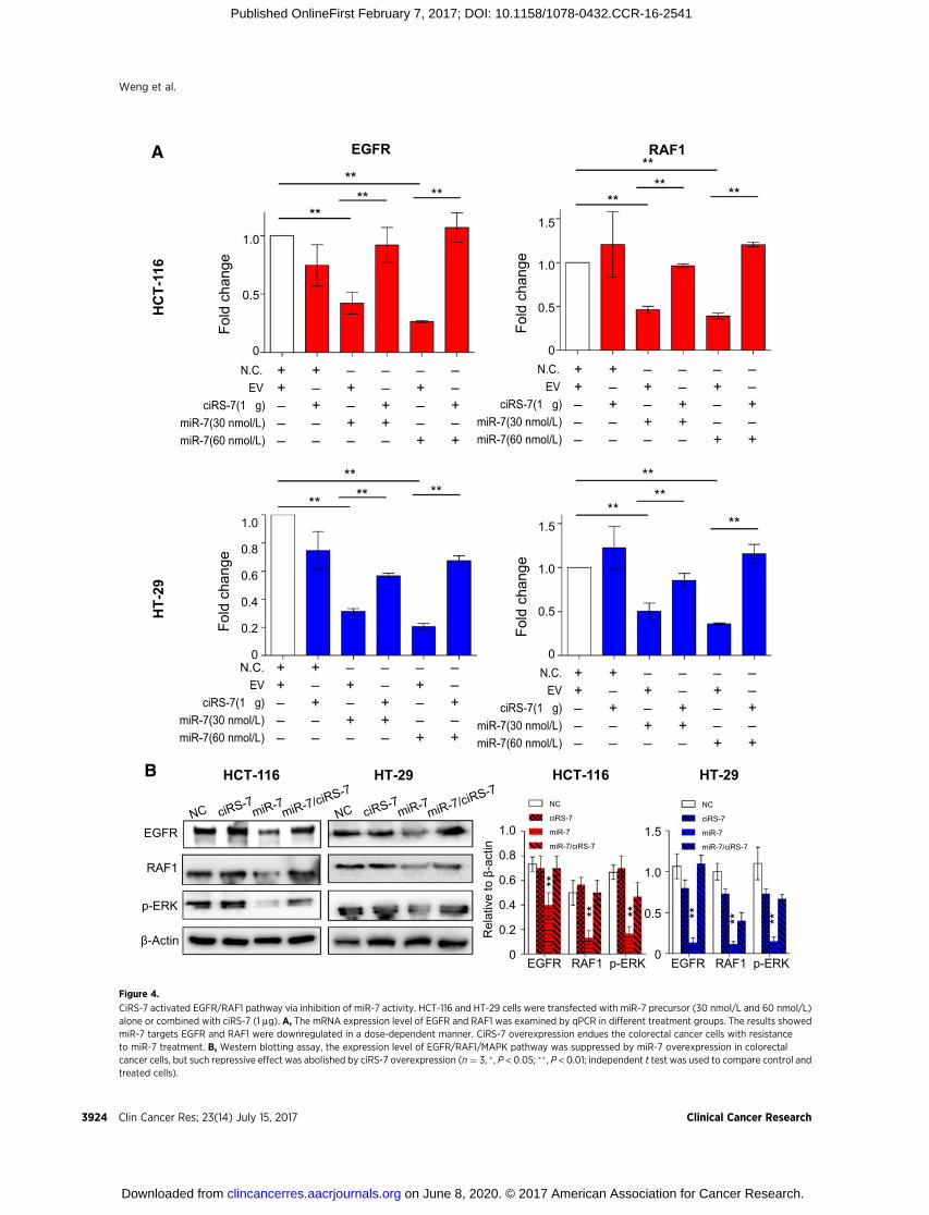
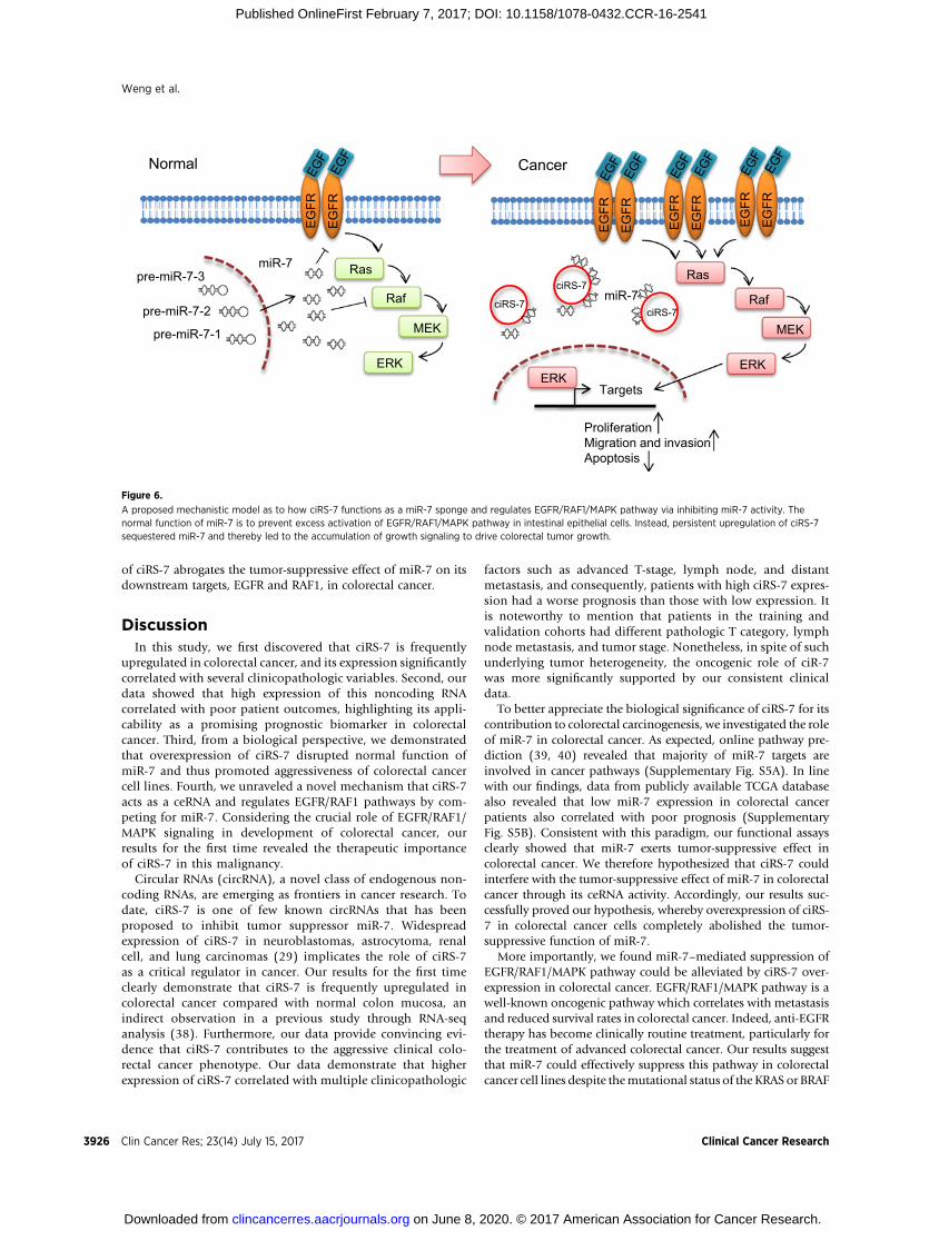
significantly decreased expression of EGFR and RAF1 subse-quent to overexpression of miR-7 (Supplementary Fig. S4).Considering the important role of EGFR/RAF1/MAPK pathwayin carcinogenesis, we deduced that ciRS-7 could be a majorcontributor for colorectal cancer development via its ceRNAactivity. The normal function of miR-7 is to prevent activationof EGFR/RAF1/MAPK pathway in intestinal epithelial cells.However, persistent upregulation of ciRS-7 sequestered miR-7 and thereby lead to the accumulation of growth signaling bydriving colorectal tumor growth.
To test our hypothesis,wemeasured EGFRandRAF1expressionin HCT-116 and HT-29 cells transfected with pre-miR-7 alone ortogether with ciRS-7. In line with our hypothesis, miR-7 over-expression led to decreased mRNA and protein expression levelsof EGFR and RAF1, while ciR-7 attenuated this inhibitory effect ofmiR-7 on EGFR and RAF1 expression (Fig. 4A). Likewise, inhibi-tionof ErkphosphorylationbymiR-7was also attenuated by ciRS-7, confirming that ciRS 7 activates EGFR/RAF1/MAPK pathway
through suppresion ofmiR-7 (Fig. 4B) in colorectal cancer, whichhas significant therapeutic implications for this malignancy.
To further validate our in vitro results that ciRS-7 regulatedEGFR1/RAF1/MAPK pathway through suppression of miR-7activity, we investigated the expression correlation betweenmiR-7, ciRS-7, and EGFR/RAF1 in colorectal cancer tissues. Wenoticed that ciRS-7 expressionwasnegatively correlatedwithmiR-7 expression in colorectal cancer (P¼ 0.0003), suggesting that theloss of function ofmiR-7 is not only due to its lowered expression,but also due to intimate association with upregulated ciRS-7expression in this malignancy (Fig. 5A). As expected, we alsofound miR-7 expression negatively correlated with EGFR (P ¼0.0014) and RAF1 (P < 0.0001) in cancer tissues, indicating thatmiR-7 suppress EGFR and RAF1 expression in CRC (Fig. 5B andC). Notably, we observed ciRS-7 overexpression was significantlyassociated with upregulation of EGFR (P < 0.0001) and RAF1 (P <0.0001) in colorectal cancer (Fig. 5D–F), highlighting the clinicalsignificance of these results, as these suggest that the upregulation
B
0 4 8 12 16 20 24 28 32 36
Days
15
12
9
6
3
0
Tum
or v
olum
e (c
m3 )
NCciRS-7miR-7ciRS-7+miR-7NC
miR-7
ciRS-7
ciRS-7+miR-7
A
ciRS-7/miR-7
miR-7
ciRS-7
NC0 0.05 0.10 0.15
Tumor weight (g)
C
NC
miR-7
ciRS-7miR-7
ciRS-7
Ki-67
×100 ×400
×100
×100
×400
×400
×400 ×100
PCNA
×400 ×100
×400 ×100
×400 ×100
×400 ×100
Figure 3.
CiRS-7 regulates miR-7 activity inxenograft animal models. StablytransfectedHCT-116 cells were inoculatedwith miR-7 alone, ciRS-7 alone, or bothsubcutaneously into nude mice. A and B,The tumor growth curve and averageweight of tumors at the time the animalswere sacrificed in different treatmentgroups (� , P < 0.05; �� , P < 0.01; paired ttest was used to compare control andtreated cells). C, Expression level of Ki67and PCNA in xenograft tissues fromdifferent treatment groups.
ciRS-7 in Colorectal Cancer
www.aacrjournals.org Clin Cancer Res; 23(14) July 15, 2017 3923
on June 8, 2020. © 2017 American Association for Cancer Research. clincancerres.aacrjournals.org Downloaded from
Published OnlineFirst February 7, 2017; DOI: 10.1158/1078-0432.CCR-16-2541
N.C.EV
ciRS-7(1 µg)miR-7(30 nmol/L)miR-7(60 nmol/L)
++–––
+–+––
–+–+–
––++–
–+––+
––+–+
Fold
cha
nge
1.0
0.8
0.6
0.4
0.2
0N.C.
EVciRS-7(1 µg)
miR-7(30 nmol/L)miR-7(60 nmol/L)
++–––
+–+––
–+–+–
––++–
–+––+
––+–+
Fold
cha
nge
1.5
1.0
0.5
0
A
N.C.EV
ciRS-7(1 µg)miR-7(30 nmol/L)miR-7(60 nmol/L)
++–––
+–+––
–+–+–
––++–
–+––+
––+–+
Fold
cha
nge
1.0
0.5
0
EGFR
N.C.EV
ciRS-7(1 µg)miR-7(30 nmol/L)miR-7(60 nmol/L)
++–––
+–+––
–+–+–
––++–
–+––+
––+–+
Fold
cha
nge
1.5
1.0
0.5
0
RAF1
HCT-116 HT-29B
β-Actin
RAF1
EGFR
p-ERK
NC
ciRS-7
miR-7
miR-7/ciRS-7
NC
ciRS-7
miR-7
miR-7/ciRS-7
Rel
ativ
e to
β-a
ctin
1.5
1.0
0.5
0
1.0
0.8
0.6
0.4
0.2
0EGFR RAF1 p-ERK EGFR RAF1 p-ERK
HCT-116 HT-29
HT-
29
H
CT-
116
Figure 4.
CiRS-7 activated EGFR/RAF1 pathway via inhibition of miR-7 activity. HCT-116 and HT-29 cells were transfected with miR-7 precursor (30 nmol/L and 60 nmol/L)alone or combined with ciRS-7 (1 mg). A, The mRNA expression level of EGFR and RAF1 was examined by qPCR in different treatment groups. The results showedmiR-7 targets EGFR and RAF1 were downregulated in a dose-dependent manner. CiRS-7 overexpression endues the colorectal cancer cells with resistanceto miR-7 treatment. B, Western blotting assay, the expression level of EGFR/RAF1/MAPK pathway was suppressed by miR-7 overexpression in colorectalcancer cells, but such repressive effect was abolished by ciRS-7 overexpression (n¼ 3, � , P < 0.05; �� , P < 0.01; independent t test was used to compare control andtreated cells).
Weng et al.
Clin Cancer Res; 23(14) July 15, 2017 Clinical Cancer Research3924
on June 8, 2020. © 2017 American Association for Cancer Research. clincancerres.aacrjournals.org Downloaded from
Published OnlineFirst February 7, 2017; DOI: 10.1158/1078-0432.CCR-16-2541

0 0.001 0.002miR-7
RA
F1
10
8
6
4
2
0
r = -0.571 **P = <0.0001(×
10-3
)r = -0.3317 **P = 0.0014
8
6
4
2
00 0.001 0.002
miR-7
EG
FR( ×
10-2
)12
10
8
6
4
2
0
ciR
S-7
0 0.001 0.002miR-7
r = -0.373**P = 0.0003
(×10
-2)
A B
r = 0.588**P = <0.0001
8
6
4
2
00 0.05 0.1 0.15
ciRS-7
EG
FR(×
10-2
) 10
8
6
4
2
0
r = 0.512 **P = <0.0001
0 0.05 0.1 0.15ciRS-7
(×10
-3)
RA
F1
-4 -2 0 2 4ciRS-7
6
3
0
-3
-6
Z-S
core
EGFRRAF1
C D
E F
Figure 5.
The correlation between ciRS-7, miR-7, and EGFR/RAF1 in colorectal cancer (CRC) tissues. A, ciRS-7 expression was negatively correlated with miR-7 expression incolorectal cancer. B and C, miR-7 expression negatively correlated with EGFR and RAF1 in colorectal cancer tissues. D–F, ciRS-7 overexpression was significantlyassociated with upregulation of EGFR and RAF1 [n ¼ 90, � , P < 0.05; �� , P < 0.01; Spearman rank correlation (r) was used for the correlation analysis].
www.aacrjournals.org Clin Cancer Res; 23(14) July 15, 2017 3925
ciRS-7 in Colorectal Cancer
on June 8, 2020. © 2017 American Association for Cancer Research. clincancerres.aacrjournals.org Downloaded from
Published OnlineFirst February 7, 2017; DOI: 10.1158/1078-0432.CCR-16-2541
of ciRS-7 abrogates the tumor-suppressive effect of miR-7 on itsdownstream targets, EGFR and RAF1, in colorectal cancer.
DiscussionIn this study, we first discovered that ciRS-7 is frequently
upregulated in colorectal cancer, and its expression significantlycorrelated with several clinicopathologic variables. Second, ourdata showed that high expression of this noncoding RNAcorrelated with poor patient outcomes, highlighting its appli-cability as a promising prognostic biomarker in colorectalcancer. Third, from a biological perspective, we demonstratedthat overexpression of ciRS-7 disrupted normal function ofmiR-7 and thus promoted aggressiveness of colorectal cancercell lines. Fourth, we unraveled a novel mechanism that ciRS-7acts as a ceRNA and regulates EGFR/RAF1 pathways by com-peting for miR-7. Considering the crucial role of EGFR/RAF1/MAPK signaling in development of colorectal cancer, ourresults for the first time revealed the therapeutic importanceof ciRS-7 in this malignancy.
Circular RNAs (circRNA), a novel class of endogenous non-coding RNAs, are emerging as frontiers in cancer research. Todate, ciRS-7 is one of few known circRNAs that has beenproposed to inhibit tumor suppressor miR-7. Widespreadexpression of ciRS-7 in neuroblastomas, astrocytoma, renalcell, and lung carcinomas (29) implicates the role of ciRS-7as a critical regulator in cancer. Our results for the first timeclearly demonstrate that ciRS-7 is frequently upregulated incolorectal cancer compared with normal colon mucosa, anindirect observation in a previous study through RNA-seqanalysis (38). Furthermore, our data provide convincing evi-dence that ciRS-7 contributes to the aggressive clinical colo-rectal cancer phenotype. Our data demonstrate that higherexpression of ciRS-7 correlated with multiple clinicopathologic
factors such as advanced T-stage, lymph node, and distantmetastasis, and consequently, patients with high ciRS-7 expres-sion had a worse prognosis than those with low expression. Itis noteworthy to mention that patients in the training andvalidation cohorts had different pathologic T category, lymphnode metastasis, and tumor stage. Nonetheless, in spite of suchunderlying tumor heterogeneity, the oncogenic role of ciR-7was more significantly supported by our consistent clinicaldata.
To better appreciate the biological significance of ciRS-7 for itscontribution to colorectal carcinogenesis, we investigated the roleof miR-7 in colorectal cancer. As expected, online pathway pre-diction (39, 40) revealed that majority of miR-7 targets areinvolved in cancer pathways (Supplementary Fig. S5A). In linewith our findings, data from publicly available TCGA databasealso revealed that low miR-7 expression in colorectal cancerpatients also correlated with poor prognosis (SupplementaryFig. S5B). Consistent with this paradigm, our functional assaysclearly showed that miR-7 exerts tumor-suppressive effect incolorectal cancer. We therefore hypothesized that ciRS-7 couldinterfere with the tumor-suppressive effect of miR-7 in colorectalcancer through its ceRNA activity. Accordingly, our results suc-cessfully proved our hypothesis, whereby overexpression of ciRS-7 in colorectal cancer cells completely abolished the tumor-suppressive function of miR-7.
More importantly, we found miR-7–mediated suppression ofEGFR/RAF1/MAPK pathway could be alleviated by ciRS-7 over-expression in colorectal cancer. EGFR/RAF1/MAPK pathway is awell-known oncogenic pathway which correlates with metastasisand reduced survival rates in colorectal cancer. Indeed, anti-EGFRtherapy has become clinically routine treatment, particularly forthe treatment of advanced colorectal cancer. Our results suggestthat miR-7 could effectively suppress this pathway in colorectalcancer cell lines despite themutational status of the KRAS or BRAF
EG
FR
EG
FR
Ras
Raf
MEK
ERK
pre-miR-7-1
pre-miR-7-2
pre-miR-7-3 miR-7
Normal
EG
FR
EG
FR
ciRS-7
ciRS-7
Ras
Raf
MEK
ERK ERK
Targets
Proliferation Migration and invasion Apoptosis
miR-7 ciRS-7
EG
FR
EG
FR
EG
FR
EG
FR
Cancer
Figure 6.
A proposed mechanistic model as to how ciRS-7 functions as a miR-7 sponge and regulates EGFR/RAF1/MAPK pathway via inhibiting miR-7 activity. Thenormal function of miR-7 is to prevent excess activation of EGFR/RAF1/MAPK pathway in intestinal epithelial cells. Instead, persistent upregulation of ciRS-7sequestered miR-7 and thereby led to the accumulation of growth signaling to drive colorectal tumor growth.
Clin Cancer Res; 23(14) July 15, 2017 Clinical Cancer Research3926
Weng et al.
on June 8, 2020. © 2017 American Association for Cancer Research. clincancerres.aacrjournals.org Downloaded from
Published OnlineFirst February 7, 2017; DOI: 10.1158/1078-0432.CCR-16-2541
oncogenes. Notably, KRAS and BRAF mutations are present inHCT-116 and HT-29 cells, respectively (41); however, mir-7 stillhas a strong inhibitory effect on the EGFR/RAF1/MAPK pathwaybecause miR-7 could successfully reduce expression levels of notonly EGFR but also another important MAPK member RAF1.RAF1, similar to BRAF, contributes to activation of MAPK path-way, but is rarely mutated in colorectal cancer (42), and a recentstudy showed inhibition of RAF1 as a promising therapeuticstrategy for BRAF- and KRAS-mutant cancers (43). These findingssuggestmiR-7 is a critical negative regulator of EGFR/RAF1/MAPKpathway. Interestingly, our results showed that ciRS-7 leads topersistent activation of EGFR/RAF1/MAPK pathway in colorectalcancer cells, regardless of treatment with low or high concentra-tionswithmiR-7 precursors supporting the hypothesis that ciRS-7enhances this key oncogenic pathway through inhibition of miR-7 activity (Fig. 6). Therefore, dual targeting ciRS-7 and miR-7could provide a new therapeutic strategy to suppress this onco-genic pathway for colorectal cancer patients.
In summary, this is the first study to systematically interrogatethe functional and clinical significance of ciRS-7 in colorectalcancer, and we provide comprehensive evidence that it acts as anovel oncogenic circRNA, as well as a prognostic biomarker incolorectal cancer. From a functional perspective, ciRS-7 impairsthe tumor-suppressive effects of miR-7 in colorectal cancer cellsand in xenograft animal model. Mechanistically, overexpressionof ciRS-7 enhanced EGFR/RAF1/MAPK pathway through inhibi-tion of miR-7 activity. We conclude that ciRS-7 is a promisingprognostic biomarker for colorectal cancer patients; therapeutictargeting of ciRS-7 maybe a potential treatment option forpatients with colorectal cancer.
Disclosure of Potential Conflicts of InterestNo potential conflicts of interest were disclosed.
Authors' ContributionsConception and design: A. GoelDevelopment of methodology: A. GoelAcquisition of data (provided animals, acquired and managed patients,provided facilities, etc.): S. Toden, K. Yoshida, T. Nagasaka, T. Fujiwara,H. Qin, A. GoelAnalysis and interpretation of data (e.g., statistical analysis, biostatistics,computational analysis): S. Cai, H. QinWriting, review, and/or revision of the manuscript: A. GoelAdministrative, technical, or material support (i.e., reporting or organizingdata, constructing databases): A. GoelStudy supervision: A. Goel
AcknowledgmentsThe authors thank Dr. Thomas B. Hansen for providing plasmids and
invaluable suggestions and guidance of this project.
Grant SupportThis work was supported by the grants R01 CA72851, CA181572,
CA184792, and U01 CA187956 from the National Cancer Institute, NIH, pilotgrants from the Baylor Sammons Cancer Center and Foundation, as well asfunds from the Baylor Research Institute. In addition, the current work wassupported by the grants from theNational Natural Science Foundation of China(no. 81372615), and the Shanghai Health System Outstanding Young TalentTraining Plan (no. XYQ2013118).
The costs of publication of this articlewere defrayed inpart by the payment ofpage charges. This article must therefore be hereby marked advertisement inaccordance with 18 U.S.C. Section 1734 solely to indicate this fact.
Received October 10, 2016; revised January 13, 2017; accepted January 28,2017; published OnlineFirst February 7, 2017.
References1. Jemal A, Bray F, Center MM, Ferlay J, Ward E, Forman D. Global cancer
statistics. CA Cancer J Clin 2011;61:69–90.2. Fearon ER, Vogelstein B. A genetic model for colorectal tumorigenesis. Cell
1990;61:759–67.3. Kinzler KW, Vogelstein B. Lessons from hereditary colorectal cancer. Cell
1996;87:159–70.4. Weng W, Feng J, Qin H, Ma Y, Goel A. An update on miRNAs as biological
and clinical determinants in colorectal cancer: a bench-to-bedsideapproach. Future Oncol 2015;11:1791–808.
5. Hur K, Toiyama Y, Okugawa Y, Ide S, Imaoka H, Boland CR, et al.Circulating microRNA-203 predicts prognosis and metastasis in humancolorectal cancer. Gut. 2017;66:654–65.
6. Okugawa Y, GradyWM, Goel A. Epigenetic alterations in colorectal cancer:emerging biomarkers. Gastroenterology 2015;149:1204–25.
7. Yamada A, Horimatsu T, Okugawa Y, Nishida N, Honjo H, Ida H, et al.Serum miR-21, miR-29a, and miR-125b are promising biomarkers forthe early detection of colorectal neoplasia. Clin Cancer Res 2015;21:4234–42.
8. Hur K, Toiyama Y, Schetter AJ, Okugawa Y, Harris CC, Boland CR, et al.Identification of a metastasis-specific microRNA signature in human colo-rectal cancer. J Natl Cancer Inst 2015;107:pii:dju492.
9. Han TS, Hur K, Xu G, Choi B, Okugawa Y, Toiyama Y, et al. MicroRNA-29cmediates initiation of gastric carcinogenesis by directly targeting ITGB1.Gut 2015;64:203–14.
10. KarsyM,ArslanE,MoyF.Current progress onunderstandingMicroRNAs inglioblastoma multiforme. Genes Cancer 2012;3:3–15.
11. Xiong S, Zheng Y, Jiang P, Liu R, Liu X,Qian J, et al. PA28gamma emerges asa novel functional target of tumour suppressor microRNA-7 in non-small-cell lung cancer. Br J Cancer 2014;110:353–62.
12. Giles KM, Brown RA, Epis MR, Kalinowski FC, Leedman PJ. miRNA-7–5pinhibits melanoma cell migration and invasion. Biochem Biophys ResCommun 2013;430:706–10.
13. Liu S, Zhang P, Chen Z, Liu M, Li X, Tang H. MicroRNA-7 downregulatesXIAP expression to suppress cell growth and promote apoptosis in cervicalcancer cells. FEBS Lett 2013;587:2247–53.
14. Webster RJ, Giles KM, Price KJ, Zhang PM, Mattick JS, Leedman PJ.Regulation of epidermal growth factor receptor signaling in human cancercells by microRNA-7. J Biol Chem 2009;284:5731–41.
15. Fang Y, Xue JL, Shen Q, Chen J, Tian L. MicroRNA-7 inhibits tumorgrowth and metastasis by targeting the phosphoinositide 3-kinase/Akt pathway in hepatocellular carcinoma. Hepatology 2012;55:1852–62.
16. Zhao X,DouW,He L, Liang S, Tie J, Liu C, et al.MicroRNA-7 functions as ananti-metastatic microRNA in gastric cancer by targeting insulin-like growthfactor-1 receptor. Oncogene 2013;32:1363–72.
17. Gu DN, Huang Q, Tian L. The molecular mechanisms and therapeuticpotential of microRNA-7 in cancer. Expert Opin Ther Targets 2015;19:415–26.
18. Zhao J, Tao Y, Zhou Y, Qin N, Chen C, Tian D, et al. MicroRNA-7:a promising new target in cancer therapy. Cancer Cell Int 2015;15:103.
19. Zhang N, Li X, Wu CW, Dong Y, Cai M, Mok MT, et al. microRNA-7 is anovel inhibitor of YY1 contributing to colorectal tumorigenesis. Oncogene2013;32:5078–88.
20. Suto T, Yokobori T, Yajima R, Morita H, Fujii T, Yamaguchi S, et al.MicroRNA-7 expression in colorectal cancer is associated with poor prog-nosis and regulates cetuximab sensitivity via EGFR regulation. Carcino-genesis 2015;36:338–45.
21. Cheng DL, Xiang YY, Ji LJ, Lu XJ. Competing endogenous RNA interplay incancer: mechanism, methodology, and perspectives. Tumour Biol2015;36:479–88.
22. Hansen TB, Jensen TI, Clausen BH, Bramsen JB, Finsen B, Damgaard CK,et al. Natural RNA circles function as efficient microRNA sponges. Nature2013;495:384–8.
ciRS-7 in Colorectal Cancer
www.aacrjournals.org Clin Cancer Res; 23(14) July 15, 2017 3927
on June 8, 2020. © 2017 American Association for Cancer Research. clincancerres.aacrjournals.org Downloaded from
Published OnlineFirst February 7, 2017; DOI: 10.1158/1078-0432.CCR-16-2541

23. XuH, Guo S, LiW, Yu P. The circular RNACdr1as, viamiR-7 and its targets,regulates insulin transcription and secretion in islet cells. Sci Rep2015;5:12453.
24. Okugawa Y, Toiyama Y, Toden S, Mitoma H, Nagasaka T, Tanaka K, et al.Clinical significance of SNORA42 as an oncogene and a prognosticbiomarker in colorectal cancer. Gut. 2017;66:107–17.
25. TianQ, Li Y,Wang F, Xu J, ShenY, Ye F, et al.MicroRNAdetection in cervicalexfoliated cells as a triage for human papillomavirus-positive women.J Natl Cancer Inst 2014;106:pii: dju241.
26. Xu J, Li Y, Wang F, Wang X, Cheng B, Ye F, et al. Suppressed miR-424expression via upregulation of target gene Chk1 contributes to the pro-gression of cervical cancer. Oncogene 2013;32:976–87.
27. Ueyama T, Kasahara H, Ishiwata T, Nie Q, Izumo S. Myocardin expressionis regulated by Nkx2.5, and its function is required for cardiomyogenesis.Mol Cell Biol 2003;23:9222–32.
28. Reuven NB, Antoku S, Weller SK. The UL12.5 gene product ofherpes simplex virus type 1 exhibits nuclease and strand exchangeactivities but does not localize to the nucleus. J Virol 2004;78:4599–608.
29. Hansen TB, Kjems J, Damgaard CK. Circular RNA and miR-7 in cancer.Cancer Res 2013;73:5609–12.
30. Glover AR, Zhao JT, Gill AJ, Weiss J, Mugridge N, Kim E, et al. microRNA-7as a tumor suppressor and novel therapeutic for adrenocortical carcinoma.Oncotarget 2015;6:36675–88.
31. Liu Z, Liu Y, Li L, Xu Z, Bi B, Wang Y, et al. MiR-7–5p is frequentlydownregulated in glioblastoma microvasculature and inhibits vascularendothelial cell proliferation by targeting RAF1. Tumour Biol 2014;35:10177–84.
32. ChangYL, ZhouPJ,Wei L, LiW, Ji Z, Fang YX, et al.MicroRNA-7 inhibits thestemness of prostate cancer stem-like cells and tumorigenesis by repressingKLF4/PI3K/Akt/p21 pathway. Oncotarget 2015;6:24017–31.
33. Meza-Sosa KF, Perez-Garcia EI, Camacho-Concha N, Lopez-Gutierrez O,Pedraza-Alva G, Perez-Martinez L. MiR-7 promotes epithelial cell
transformation by targeting the tumor suppressor KLF4. PLoS One2014;9:e103987.
34. Okuda H, Xing F, Pandey PR, Sharma S, Watabe M, Pai SK, et al. miR-7suppresses brain metastasis of breast cancer stem-like cells by modulatingKLF4. Cancer Res 2013;73:1434–44.
35. Li J, Zheng Y, Sun G, Xiong S. Restoration of miR-7 expression suppressesthe growth of Lewis lung cancer cells by modulating epidermal growthfactor receptor signaling. Oncol Rep 2014;32:2511–6.
36. Zhao JG, Men WF, Tang J. MicroRNA-7 enhances cytotoxicity induced bygefitinib in non-small cell lung cancer via inhibiting the EGFR and IGF1Rsignalling pathways. Contemp Oncol 2015;19:201–6.
37. Wang B, Sun F, Dong N, Sun Z, Diao Y, Zheng C, et al. MicroRNA-7 directlytargets insulin-like growth factor 1 receptor to inhibit cellular growth andglucose metabolism in gliomas. Diagn Pathol 2014;9:211.
38. Bachmayr-Heyda A, Reiner AT, Auer K, Sukhbaatar N, Aust S, Bachleitner-Hofmann T, et al. Correlation of circular RNA abundance with prolifer-ation–exemplified with colorectal and ovarian cancer, idiopathic lungfibrosis, and normal human tissues. Sci Rep 2015;5:8057.
39. Dweep H, Gretz N. miRWalk2.0: a comprehensive atlas of microRNA-target interactions. Nat Methods 2015;12:697.
40. Dweep H, Sticht C, Pandey P, Gretz N. miRWalk–database: prediction ofpossible miRNA binding sites by "walking" the genes of three genomes.J Biomed Inform 2011;44:839–47.
41. AhmedD, Eide PW, Eilertsen IA, Danielsen SA, EknaesM,HektoenM, et al.Epigenetic and genetic features of 24 colon cancer cell lines. Oncogenesis2013;2:e71.
42. Santarpia L, Lippman SM, El-Naggar AK. Targeting the MAPK-RAS-RAFsignaling pathway in cancer therapy. Expert Opin Ther Targets 2012;16:103–19.
43. Whittaker SR, Cowley GS, Wagner S, Luo F, Root DE, Garraway LA.Combined Pan-RAF and MEK inhibition overcomes multiple resistancemechanisms to selective RAF inhibitors. Mol Cancer Ther 2015;14:2700–11.
Clin Cancer Res; 23(14) July 15, 2017 Clinical Cancer Research3928
Weng et al.
on June 8, 2020. © 2017 American Association for Cancer Research. clincancerres.aacrjournals.org Downloaded from
Published OnlineFirst February 7, 2017; DOI: 10.1158/1078-0432.CCR-16-2541

2017;23:3918-3928. Published OnlineFirst February 7, 2017.Clin Cancer Res Wenhao Weng, Qing Wei, Shusuke Toden, et al. Potential Therapeutic Target in Colorectal Cancer
A Promising Prognostic Biomarker and a−−Circular RNA ciRS-7
Updated version
10.1158/1078-0432.CCR-16-2541doi:
Access the most recent version of this article at:
Material
Supplementary
http://clincancerres.aacrjournals.org/content/suppl/2017/02/07/1078-0432.CCR-16-2541.DC1
Access the most recent supplemental material at:
Cited articles
http://clincancerres.aacrjournals.org/content/23/14/3918.full#ref-list-1
This article cites 42 articles, 9 of which you can access for free at:
Citing articles
http://clincancerres.aacrjournals.org/content/23/14/3918.full#related-urls
This article has been cited by 2 HighWire-hosted articles. Access the articles at:
E-mail alerts related to this article or journal.Sign up to receive free email-alerts
Subscriptions
Reprints and
To order reprints of this article or to subscribe to the journal, contact the AACR Publications Department at
Permissions
Rightslink site. Click on "Request Permissions" which will take you to the Copyright Clearance Center's (CCC)
.http://clincancerres.aacrjournals.org/content/23/14/3918To request permission to re-use all or part of this article, use this link
on June 8, 2020. © 2017 American Association for Cancer Research. clincancerres.aacrjournals.org Downloaded from
Published OnlineFirst February 7, 2017; DOI: 10.1158/1078-0432.CCR-16-2541