Chronic suppurative osteomyelitis with proliferative ... · A panoramic radiograph was taken as an...
6
215 molar occurs very rarely. Only a few case reports have been published. The basic treatment strategy in these reports was removal of the impacted third molar. Further surgical inter- ventions varied from curettage and drainage to condylec- tomy, coronoidectomy, or extensive decortication. Chronic suppurative osteomyelitis can only be treated successfully by a combination of antimicrobial therapy and surgery for total elimination of the infected and necrotic bony tissue. This ap- proach can lead to complete cure of osteomyelitis and further resolution of proliferative periostitis. Early-onset primary chronic osteomyelitis (juvenile chronic osteomyelitis) differs from secondary chronic suppurative osteomyelitis, which is characterized by recurring periosteal reaction without com- plete resolution. II. Cases Report 1. Case 1 A 12-year-old girl was referred to the Department of Oral and Maxillofacial Surgery, Seoul National University Dental Hospital (Seoul, Korea), with a six-week history of recurrent pain and swelling of the right mandible, which had increased over the past month. Under the assumption that an infection was present, the patient had been treated with antibiotics for I. Introduction Osteomyelitis is an inflammatory condition of the bone and bone marrow. The pathogenesis of osteomyelitis of the jaw is predominantly odontogenic microorganisms. Bacterial in- fection of odontogenic origin can be in the form of a periapi- cal or periodontal abscess, pericoronitis, infected extraction wound, or fracture wound 1 . Bony pathology such as osteo- myelitis, trauma, and neoplastic disease can result in mild ir- ritation or low-grade infection that can affect the jaw and lead to peripheral subperiosteal bone deposition. The differential diagnoses of bony expansion also comprise Ewing’s sarcoma, fibrous dysplasia, osteogenic sarcoma, infantile cortical hy- perstosis, exostosis, calcifying hematoma, and osteoma. Osteomyelitis of the mandible due to an impacted third CASE REPORT Hoon Myoung Department of Oral and Maxillofacial Surgery, Dental Research Institute, School of Dentistry, Seoul National University, 101 Daehak-ro, Jongno-gu, Seoul 03080, Korea TEL: +82-2-2072-3059 FAX: +82-2-766-4948 E-mail: [email protected] ORCID: http://orcid.org/0000-0002-9984-8479 This is an open-access article distributed under the terms of the Creative Commons Attribution Non-Commercial License (http://creativecommons.org/licenses/by-nc/4.0/), which permits unrestricted non-commercial use, distribution, and reproduction in any medium, provided the original work is properly cited. CC Chronic suppurative osteomyelitis with proliferative periostitis related to a fully impacted third molar germ: a report of two cases Joonhyoung Park 1 , Hoon Myoung 1,2 1 Department of Oral and Maxillofacial Surgery, School of Dentistry, Seoul National University, 2 Dental Research Institute, Seoul National University, Seoul, Korea Abstract (J Korean Assoc Oral Maxillofac Surg 2016;42:215-220) In prolonged chronic osteomyelitis, chronic inflammation and low-grade infections can result in new periosteal bone formation. Chronic osteomyelitis with proliferative periostitis (traditionally termed Garré’s sclerosing osteomyelitis) mainly affects children and young adults. Here, we present two rare cases of an 11-year-old and a 12-year-old patient with suppurative chronic osteomyelitis with proliferative periostitis without any definitive infection source, such as dental caries or periodontitis. The source of infection was likely to be related to the development of a lower right third molar germ with follicular space widening. Management involved antibiotics and the removal of the third molar germ and surgical debridement. Disease remission and a normal appearance was observed at the six-month follow-up visit. Key words: Osteomyelitis, Juvenile periodontitis, Periostitis, Folliculitis [paper submitted 2015. 12. 28 / revised 2016. 3. 16 / accepted 2016. 3. 19] Copyright Ⓒ 2016 The Korean Association of Oral and Maxillofacial Surgeons. All rights reserved. http://dx.doi.org/10.5125/jkaoms.2016.42.4.215 pISSN 2234-7550 · eISSN 2234-5930
Transcript of Chronic suppurative osteomyelitis with proliferative ... · A panoramic radiograph was taken as an...
215
molar occurs very rarely. Only a few case reports have been
published. The basic treatment strategy in these reports was
removal of the impacted third molar. Further surgical inter-
ventions varied from curettage and drainage to condylec-
tomy, coronoidectomy, or extensive decortication. Chronic
suppurative osteomyelitis can only be treated successfully by
a combination of antimicrobial therapy and surgery for total
elimination of the infected and necrotic bony tissue. This ap-
proach can lead to complete cure of osteomyelitis and further
resolution of proliferative periostitis. Early-onset primary
chronic osteomyelitis (juvenile chronic osteomyelitis) differs
from secondary chronic suppurative osteomyelitis, which is
characterized by recurring periosteal reaction without com-
plete resolution.
II. Cases Report
1. Case 1
A 12-year-old girl was referred to the Department of Oral
and Maxillofacial Surgery, Seoul National University Dental
Hospital (Seoul, Korea), with a six-week history of recurrent
pain and swelling of the right mandible, which had increased
over the past month. Under the assumption that an infection
was present, the patient had been treated with antibiotics for
I. Introduction
Osteomyelitis is an inflammatory condition of the bone and
bone marrow. The pathogenesis of osteomyelitis of the jaw
is predominantly odontogenic microorganisms. Bacterial in-
fection of odontogenic origin can be in the form of a periapi-
cal or periodontal abscess, pericoronitis, infected extraction
wound, or fracture wound1. Bony pathology such as osteo-
myelitis, trauma, and neoplastic disease can result in mild ir-
ritation or low-grade infection that can affect the jaw and lead
to peripheral subperiosteal bone deposition. The differential
diagnoses of bony expansion also comprise Ewing’s sarcoma,
fibrous dysplasia, osteogenic sarcoma, infantile cortical hy-
perstosis, exostosis, calcifying hematoma, and osteoma.
Osteomyelitis of the mandible due to an impacted third
CASE REPORT
Hoon MyoungDepartment of Oral and Maxillofacial Surgery, Dental Research Institute, School of Dentistry, Seoul National University, 101 Daehak-ro, Jongno-gu, Seoul 03080, KoreaTEL: +82-2-2072-3059 FAX: +82-2-766-4948E-mail: [email protected]: http://orcid.org/0000-0002-9984-8479
This is an open-access article distributed under the terms of the Creative Commons Attribution Non-Commercial License (http://creativecommons.org/licenses/by-nc/4.0/), which permits unrestricted non-commercial use, distribution, and reproduction in any medium, provided the original work is properly cited.
CC
Chronic suppurative osteomyelitis with proliferative periostitis related to a fully impacted third molar germ: a report of two cases
Joonhyoung Park1, Hoon Myoung1,2
1Department of Oral and Maxillofacial Surgery, School of Dentistry, Seoul National University, 2Dental Research Institute, Seoul National University, Seoul, Korea
Abstract (J Korean Assoc Oral Maxillofac Surg 2016;42:215-220)
In prolonged chronic osteomyelitis, chronic inflammation and low-grade infections can result in new periosteal bone formation. Chronic osteomyelitis with proliferative periostitis (traditionally termed Garré’s sclerosing osteomyelitis) mainly affects children and young adults. Here, we present two rare cases of an 11-year-old and a 12-year-old patient with suppurative chronic osteomyelitis with proliferative periostitis without any definitive infection source, such as dental caries or periodontitis. The source of infection was likely to be related to the development of a lower right third molar germ with follicular space widening. Management involved antibiotics and the removal of the third molar germ and surgical debridement. Disease remission and a normal appearance was observed at the six-month follow-up visit.
Key words: Osteomyelitis, Juvenile periodontitis, Periostitis, Folliculitis[paper submitted 2015. 12. 28 / revised 2016. 3. 16 / accepted 2016. 3. 19]
Copyright Ⓒ 2016 The Korean Association of Oral and Maxillofacial Surgeons. All rights reserved.
http://dx.doi.org/10.5125/jkaoms.2016.42.4.215pISSN 2234-7550·eISSN 2234-5930
J Korean Assoc Oral Maxillofac Surg 2016;42:215-220
216
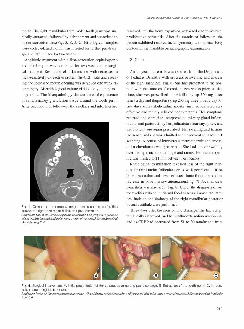
teomyelitis. The CT image showed widening of the follicular
space of the right mandibular third molar with definitive
cortical perforation on the buccal and lingual sides accompa-
nied by an outer cortical periosteal reaction.(Fig. 4) The at-
tenuation of bone marrow on the body of the right mandible
was increased. Pus formation was observed on the buccal
side of the third molar germ. Based on the clinical findings
and radiographic imaging, a provisional diagnosis of chronic
suppurative osteomyelitis of the mandible with proliferative
periostitis was made.
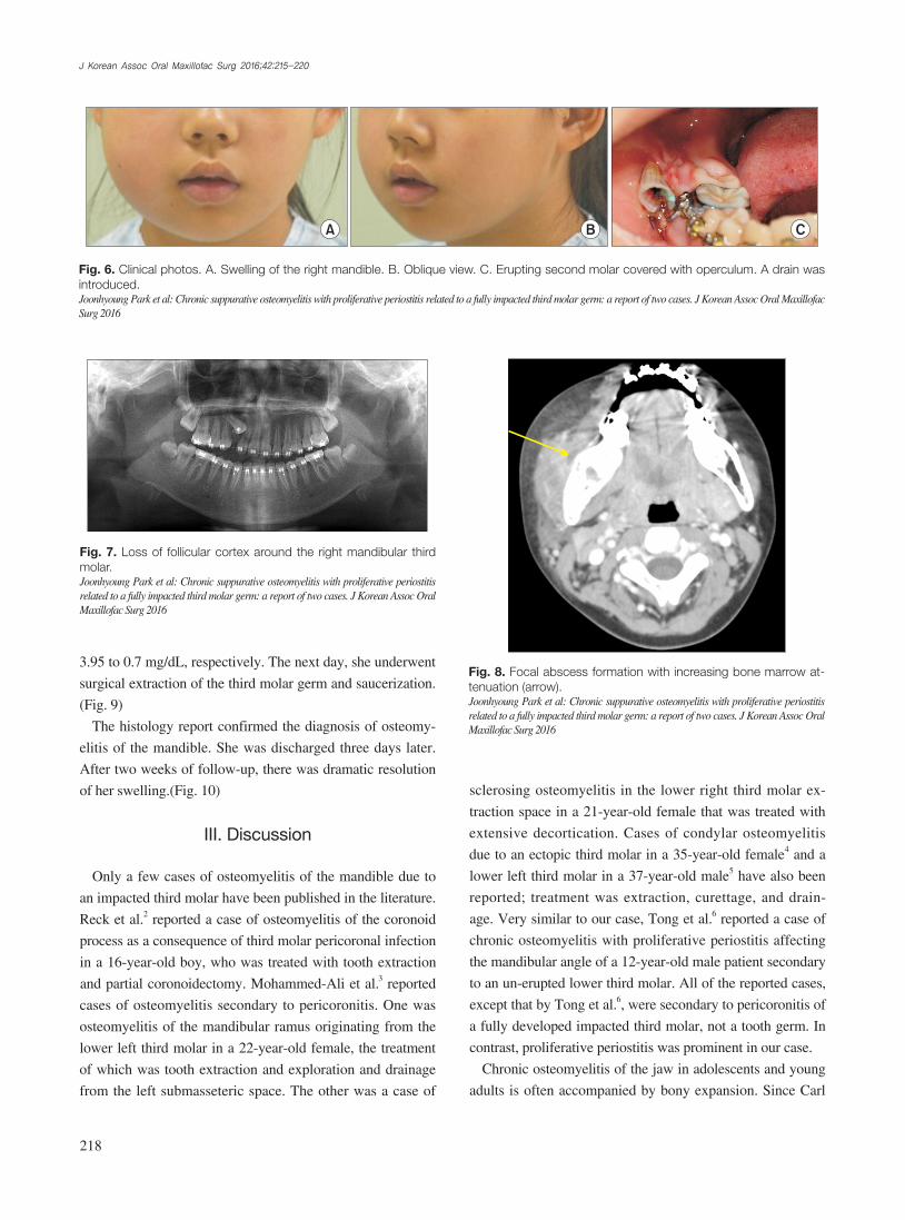
Surgical management including saucerization and germec-
tomy under general anesthesia were planned. First, the lesion
was drained through the cheek, and microbial cultures were
collected.(Fig. 5. A) An incision was made along the external
oblique ridge, and a mucoperiosteal flap was elevated. We
observed destruction of alveolar bone distal to the second
three weeks and underwent needle aspiration for drainage at
a local dental clinic.
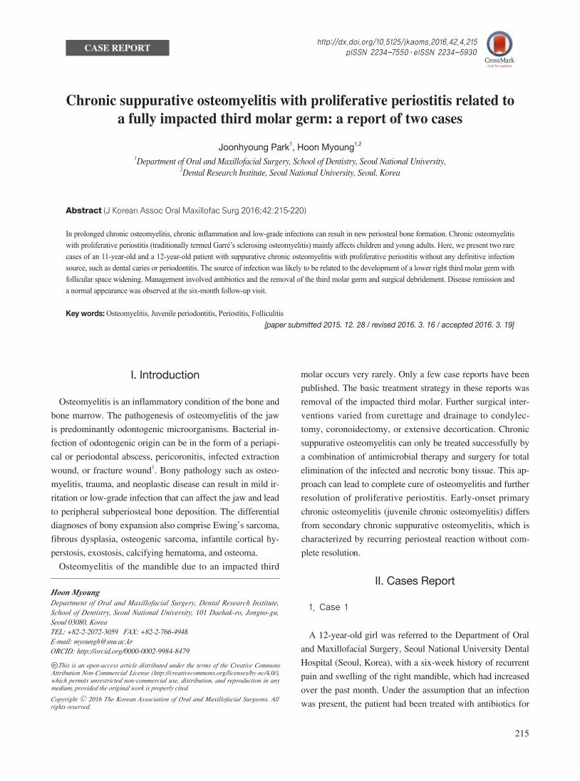
There was prominent right facial swelling, erythema, and
tenderness at the angle and inferior border of the mandible.
(Fig. 1) No cutaneous fistula was present on the first day.
Several days later, a cutaneous fistula had formed. There was
regional lymphadenopathy, but sensation of lower lip and
chin area was intact.
On intra-oral examination, the patient had full, well-main-
tained dentition with erupting second molars. There were no
caries lesions or periodontal pathology. No intra-oral drain-
ing sinus was found. She denied fevers or chills and difficulty
swallowing or talking. The maximal inter-incisal opening
was 15 mm due to guarding secondary to pain.
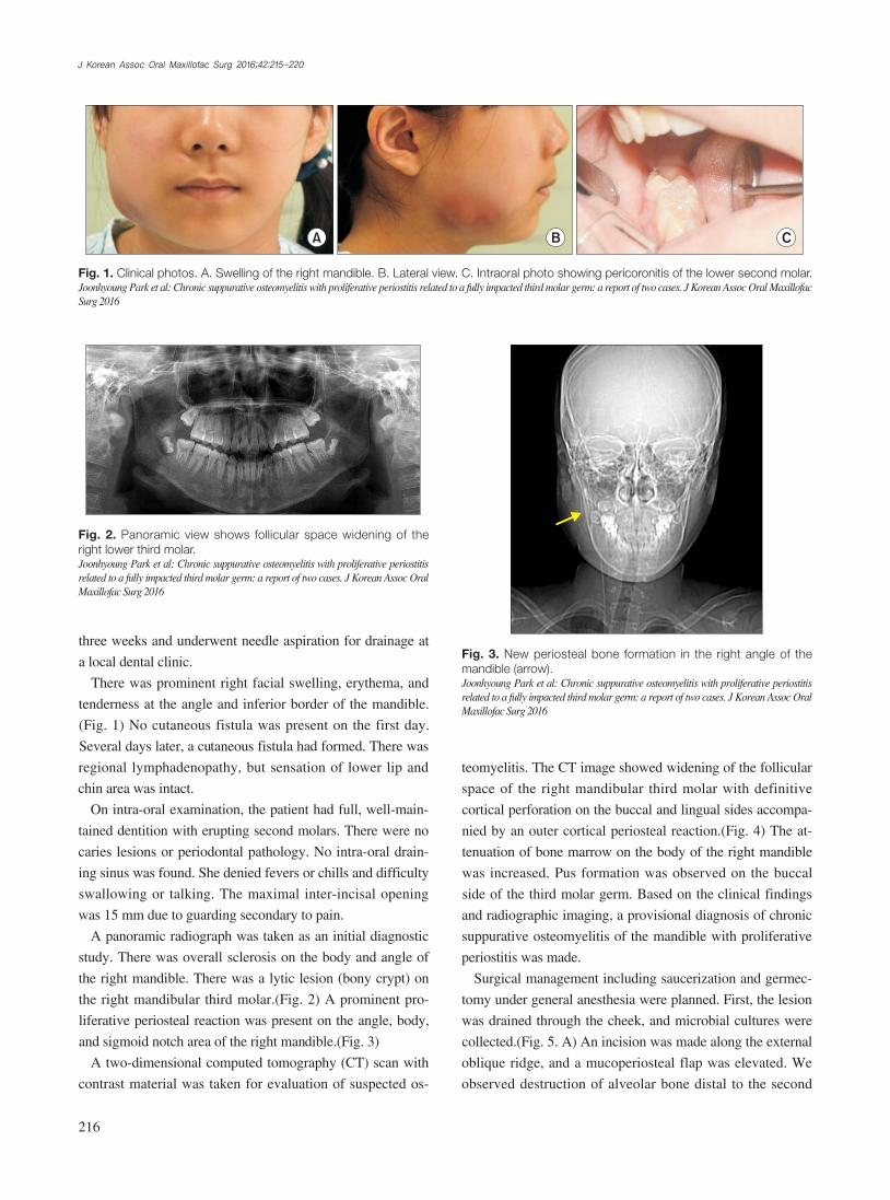
A panoramic radiograph was taken as an initial diagnostic
study. There was overall sclerosis on the body and angle of
the right mandible. There was a lytic lesion (bony crypt) on
the right mandibular third molar.(Fig. 2) A prominent pro-
liferative periosteal reaction was present on the angle, body,
and sigmoid notch area of the right mandible.(Fig. 3)
A two-dimensional computed tomography (CT) scan with
contrast material was taken for evaluation of suspected os-
Fig. 2. Panoramic view shows follicular space widening of the right lower third molar.Joonhyoung Park et al: Chronic suppurative osteomyelitis with proliferative periostitis related to a fully impacted third molar germ: a report of two cases. J Korean Assoc Oral Maxillofac Surg 2016
Fig. 3. New periosteal bone formation in the right angle of the mandible (arrow).Joonhyoung Park et al: Chronic suppurative osteomyelitis with proliferative periostitis related to a fully impacted third molar germ: a report of two cases. J Korean Assoc Oral Maxillofac Surg 2016
Fig. 1. Clinical photos. A. Swelling of the right mandible. B. Lateral view. C. Intraoral photo showing pericoronitis of the lower second molar.Joonhyoung Park et al: Chronic suppurative osteomyelitis with proliferative periostitis related to a fully impacted third molar germ: a report of two cases. J Korean Assoc Oral Maxillofac Surg 2016
A B C
Chronic osteomyelitis related to a fully impacted third molar germ
217
resolved, but the bony expansion remained due to residual
proliferative periostitis. After six months of follow-up, the
patient exhibited restored facial symmetry with normal bony
contour of the mandible on radiographic examination.
2. Case 2
An 11-year-old female was referred from the Department
of Pediatric Dentistry with progressive swelling and abscess
of the right mandible.(Fig. 6) She had presented to the hos-
pital with the same chief complaint two weeks prior. At that
time, she was prescribed amoxicillin syrup 250 mg three
times a day and ibuprofen syrup 200 mg three times a day for
five days with chlorhexidine mouth rinse, which were very
effective and rapidly relieved her symptoms. Her symptoms
returned and were then interpreted as salivary gland inflam-
mation and pariostitis by her pediatrician four days prior, and
antibiotics were again prescribed. Her swelling and trismus
worsened, and she was admitted and underwent enhanced CT
scanning. A course of intravenous metronidazole and amoxi-
cillin clavulanate was prescribed. She had tender swelling
over the right mandibular angle and ramus. Her mouth open-
ing was limited to 11 mm between her incisors.
Radiological examination revealed loss of the right man-
dibular third molar follicular cortex with peripheral diffuse
bone destruction and new periosteal bone formation and an
increase in bone marrow attenuation.(Fig. 7) Focal abscess
formation was also seen.(Fig. 8) Under the diagnosis of os-
teomyelitis with cellulitis and focal abscess, immediate intra-
oral incision and drainage of the right mandibular posterior
buccal vestibule were performed.
Three days after the incision and drainage, she had symp-
tomatically improved, and her erythrocyte sedimentation rate
and hs-CRP had decreased from 51 to 30 mm/hr and from
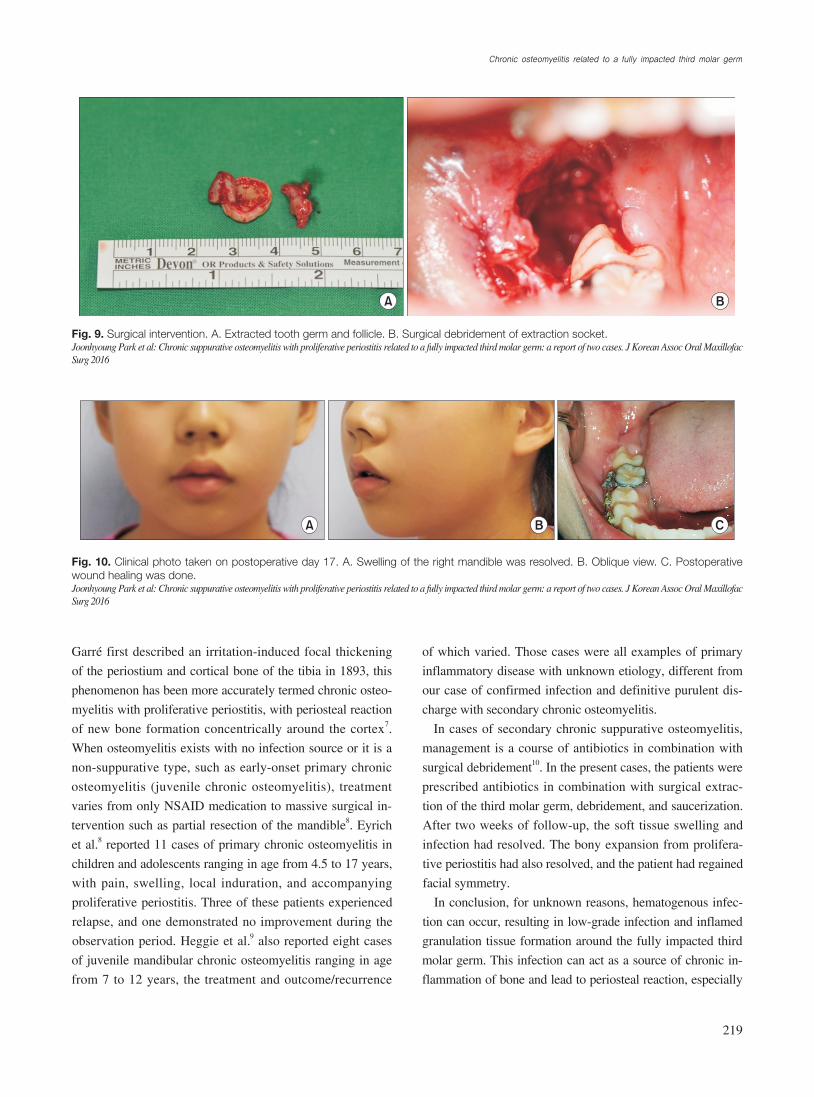
molar. The right mandibular third molar tooth germ was sur-
gically extracted, followed by debridement and saucerization
of the extraction site.(Fig. 5. B, 5. C) Histological samples
were collected, and a drain was inserted for further pus drain-
age and left in place for two weeks.
Antibiotic treatment with a first-generation cephalosporin
and clindamycin was continued for two weeks after surgi-
cal treatment. Resolution of inflammation with decreases in
high-sensitivity C-reactive protein (hs-CRP) rate and swell-
ing and increased mouth opening was achieved one week af-
ter surgery. Microbiological culture yielded only commensal
organisms. The histopathology demonstrated the presence
of inflammatory granulation tissue around the tooth germ.
After one month of follow-up, the swelling and infection had
A B C
Fig. 5. Surgical intervention. A. Initial presentation of the cutaneous sinus and pus discharge. B. Extraction of the tooth germ. C. Intraoral lesions after surgical debridement.Joonhyoung Park et al: Chronic suppurative osteomyelitis with proliferative periostitis related to a fully impacted third molar germ: a report of two cases. J Korean Assoc Oral Maxillofac Surg 2016
Fig. 4. Computed tomography image reveals cortical perforation around the right third molar follicle and pus formation.Joonhyoung Park et al: Chronic suppurative osteomyelitis with proliferative periostitis related to a fully impacted third molar germ: a report of two cases. J Korean Assoc Oral Maxillofac Surg 2016
J Korean Assoc Oral Maxillofac Surg 2016;42:215-220
218
sclerosing osteomyelitis in the lower right third molar ex-
traction space in a 21-year-old female that was treated with
extensive decortication. Cases of condylar osteomyelitis
due to an ectopic third molar in a 35-year-old female4 and a
lower left third molar in a 37-year-old male5 have also been
reported; treatment was extraction, curettage, and drain-
age. Very similar to our case, Tong et al.6 reported a case of
chronic osteomyelitis with proliferative periostitis affecting
the mandibular angle of a 12-year-old male patient secondary
to an un-erupted lower third molar. All of the reported cases,
except that by Tong et al.6, were secondary to pericoronitis of
a fully developed impacted third molar, not a tooth germ. In
contrast, proliferative periostitis was prominent in our case.
Chronic osteomyelitis of the jaw in adolescents and young
adults is often accompanied by bony expansion. Since Carl
3.95 to 0.7 mg/dL, respectively. The next day, she underwent
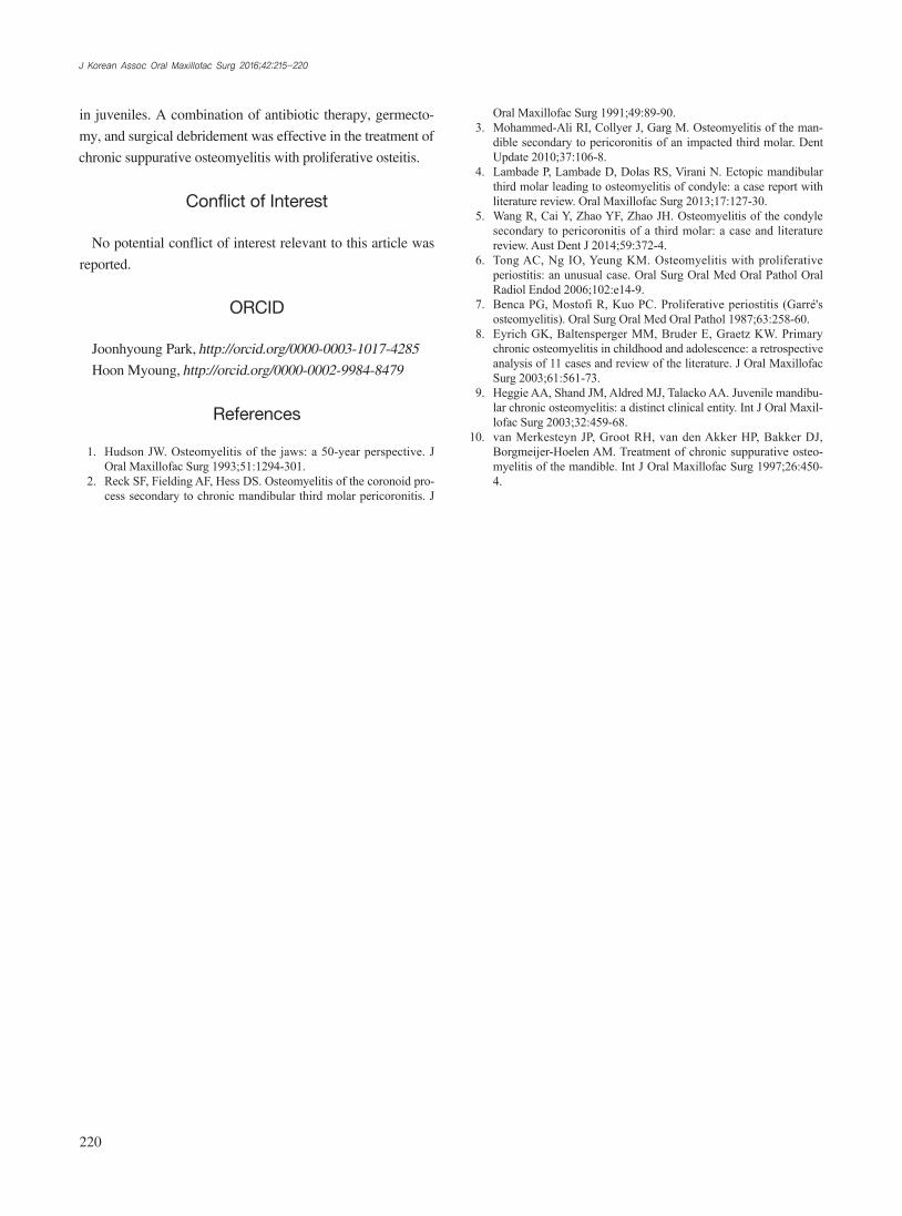
surgical extraction of the third molar germ and saucerization.
(Fig. 9)
The histology report confirmed the diagnosis of osteomy-
elitis of the mandible. She was discharged three days later.
After two weeks of follow-up, there was dramatic resolution
of her swelling.(Fig. 10)
III. Discussion
Only a few cases of osteomyelitis of the mandible due to
an impacted third molar have been published in the literature.
Reck et al.2 reported a case of osteomyelitis of the coronoid
process as a consequence of third molar pericoronal infection
in a 16-year-old boy, who was treated with tooth extraction
and partial coronoidectomy. Mohammed-Ali et al.3 reported
cases of osteomyelitis secondary to pericoronitis. One was
osteomyelitis of the mandibular ramus originating from the
lower left third molar in a 22-year-old female, the treatment
of which was tooth extraction and exploration and drainage
from the left submasseteric space. The other was a case of
A B C
Fig. 6. Clinical photos. A. Swelling of the right mandible. B. Oblique view. C. Erupting second molar covered with operculum. A drain was introduced.Joonhyoung Park et al: Chronic suppurative osteomyelitis with proliferative periostitis related to a fully impacted third molar germ: a report of two cases. J Korean Assoc Oral Maxillofac Surg 2016
Fig. 7. Loss of follicular cortex around the right mandibular third molar.Joonhyoung Park et al: Chronic suppurative osteomyelitis with proliferative periostitis related to a fully impacted third molar germ: a report of two cases. J Korean Assoc Oral Maxillofac Surg 2016
Fig. 8. Focal abscess formation with increasing bone marrow at-tenuation (arrow).Joonhyoung Park et al: Chronic suppurative osteomyelitis with proliferative periostitis related to a fully impacted third molar germ: a report of two cases. J Korean Assoc Oral Maxillofac Surg 2016
Chronic osteomyelitis related to a fully impacted third molar germ
219
of which varied. Those cases were all examples of primary
inflammatory disease with unknown etiology, different from
our case of confirmed infection and definitive purulent dis-
charge with secondary chronic osteomyelitis.
In cases of secondary chronic suppurative osteomyelitis,
management is a course of antibiotics in combination with
surgical debridement10. In the present cases, the patients were
prescribed antibiotics in combination with surgical extrac-
tion of the third molar germ, debridement, and saucerization.
After two weeks of follow-up, the soft tissue swelling and
infection had resolved. The bony expansion from prolifera-
tive periostitis had also resolved, and the patient had regained
facial symmetry.
In conclusion, for unknown reasons, hematogenous infec-
tion can occur, resulting in low-grade infection and inflamed
granulation tissue formation around the fully impacted third
molar germ. This infection can act as a source of chronic in-
flammation of bone and lead to periosteal reaction, especially
Garré first described an irritation-induced focal thickening
of the periostium and cortical bone of the tibia in 1893, this
phenomenon has been more accurately termed chronic osteo-
myelitis with proliferative periostitis, with periosteal reaction
of new bone formation concentrically around the cortex7.
When osteomyelitis exists with no infection source or it is a
non-suppurative type, such as early-onset primary chronic
osteomyelitis (juvenile chronic osteomyelitis), treatment
varies from only NSAID medication to massive surgical in-
tervention such as partial resection of the mandible8. Eyrich
et al.8 reported 11 cases of primary chronic osteomyelitis in
children and adolescents ranging in age from 4.5 to 17 years,
with pain, swelling, local induration, and accompanying
proliferative periostitis. Three of these patients experienced
relapse, and one demonstrated no improvement during the
observation period. Heggie et al.9 also reported eight cases
of juvenile mandibular chronic osteomyelitis ranging in age
from 7 to 12 years, the treatment and outcome/recurrence
A B
Fig. 9. Surgical intervention. A. Extracted tooth germ and follicle. B. Surgical debridement of extraction socket.Joonhyoung Park et al: Chronic suppurative osteomyelitis with proliferative periostitis related to a fully impacted third molar germ: a report of two cases. J Korean Assoc Oral Maxillofac Surg 2016
Fig. 10. Clinical photo taken on postoperative day 17. A. Swelling of the right mandible was resolved. B. Oblique view. C. Postoperative wound healing was done.Joonhyoung Park et al: Chronic suppurative osteomyelitis with proliferative periostitis related to a fully impacted third molar germ: a report of two cases. J Korean Assoc Oral Maxillofac Surg 2016
CBA
J Korean Assoc Oral Maxillofac Surg 2016;42:215-220
220
Oral Maxillofac Surg 1991;49:89-90.3. Mohammed-Ali RI, Collyer J, Garg M. Osteomyelitis of the man-
dible secondary to pericoronitis of an impacted third molar. Dent Update 2010;37:106-8.
4. Lambade P, Lambade D, Dolas RS, Virani N. Ectopic mandibular third molar leading to osteomyelitis of condyle: a case report with literature review. Oral Maxillofac Surg 2013;17:127-30.
5. Wang R, Cai Y, Zhao YF, Zhao JH. Osteomyelitis of the condyle secondary to pericoronitis of a third molar: a case and literature review. Aust Dent J 2014;59:372-4.
6. Tong AC, Ng IO, Yeung KM. Osteomyelitis with proliferative periostitis: an unusual case. Oral Surg Oral Med Oral Pathol Oral Radiol Endod 2006;102:e14-9.
7. Benca PG, Mostofi R, Kuo PC. Proliferative periostitis (Garré's osteomyelitis). Oral Surg Oral Med Oral Pathol 1987;63:258-60.
8. Eyrich GK, Baltensperger MM, Bruder E, Graetz KW. Primary chronic osteomyelitis in childhood and adolescence: a retrospective analysis of 11 cases and review of the literature. J Oral Maxillofac Surg 2003;61:561-73.
9. Heggie AA, Shand JM, Aldred MJ, Talacko AA. Juvenile mandibu-lar chronic osteomyelitis: a distinct clinical entity. Int J Oral Maxil-lofac Surg 2003;32:459-68.
10. van Merkesteyn JP, Groot RH, van den Akker HP, Bakker DJ, Borgmeijer-Hoelen AM. Treatment of chronic suppurative osteo-myelitis of the mandible. Int J Oral Maxillofac Surg 1997;26:450-4.
in juveniles. A combination of antibiotic therapy, germecto-
my, and surgical debridement was effective in the treatment of
chronic suppurative osteomyelitis with proliferative osteitis.
Conflict of Interest
No potential conflict of interest relevant to this article was
reported.
ORCID
Joonhyoung Park, http://orcid.org/0000-0003-1017-4285Hoon Myoung, http://orcid.org/0000-0002-9984-8479
References
1. Hudson JW. Osteomyelitis of the jaws: a 50-year perspective. J Oral Maxillofac Surg 1993;51:1294-301.
2. Reck SF, Fielding AF, Hess DS. Osteomyelitis of the coronoid pro-cess secondary to chronic mandibular third molar pericoronitis. J