Chronic methamphetamine treatment induces oxytocin ...epubs.surrey.ac.uk/806340/1/Zanos et al.,...
30
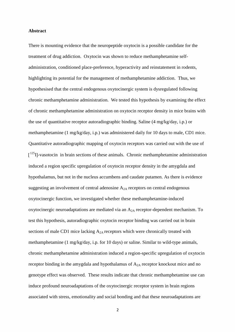
1 Chronic methamphetamine treatment induces oxytocin receptor up-regulation in the amygdala and hypothalamus via an adenosine A 2A receptor-independent mechanism Authors: Panos Zanos a , Sherie R. Wright a , Polymnia Georgiou a , Ji Hoon Yoo a , Catherine Ledent b , Susanna Hourani a , Ian Kitchen a , Raphaelle Winsky-Sommerer a and Alexis Bailey a a Sleep, Chronobiology & Addiction group, Department of Biochemistry & Physiology, Faculty of Health and Medical Sciences, University of Surrey, Guildford, GU2 7XH, Surrey, U.K. b Institut de Recherche Interdisciplinaire en Biologie Humaine et Moléculaire, Université Libre de Bruxelles, B- 1070, Belgium Correspondence: Alexis Bailey, BS.c, Ph.D Department of Biochemistry & Physiology Faculty of Health and Medical Sciences University of Surrey Guildford, GU27XH, Surrey, U.K Tel: +44 (0)1483682564 Fax: +44(0)1483 686401 email: [email protected]
Transcript of Chronic methamphetamine treatment induces oxytocin ...epubs.surrey.ac.uk/806340/1/Zanos et al.,...
1
Chronic methamphetamine treatment induces oxytocin receptor up-regulation in the
amygdala and hypothalamus via an adenosine A2A receptor-independent mechanism
Authors: Panos Zanosa, Sherie R. Wright
a, Polymnia Georgiou
a, Ji Hoon Yoo
a, Catherine
Ledentb, Susanna Hourani
a, Ian Kitchen
a, Raphaelle Winsky-Sommerer
a and Alexis Bailey
a
a Sleep, Chronobiology & Addiction group, Department of Biochemistry & Physiology,
Faculty of Health and Medical Sciences, University of Surrey, Guildford, GU2 7XH, Surrey,
U.K.
b Institut de Recherche Interdisciplinaire en Biologie Humaine et Moléculaire, Université
Libre de Bruxelles, B- 1070, Belgium
Correspondence: Alexis Bailey, BS.c, Ph.D
Department of Biochemistry & Physiology
Faculty of Health and Medical Sciences
University of Surrey
Guildford, GU27XH, Surrey, U.K
Tel: +44 (0)1483682564
Fax: +44(0)1483 686401
email: [email protected]
2
Abstract
There is mounting evidence that the neuropeptide oxytocin is a possible candidate for the
treatment of drug addiction. Oxytocin was shown to reduce methamphetamine self-
administration, conditioned place-preference, hyperactivity and reinstatement in rodents,
highlighting its potential for the management of methamphetamine addiction. Thus, we
hypothesised that the central endogenous oxytocinergic system is dysregulated following
chronic methamphetamine administration. We tested this hypothesis by examining the effect
of chronic methamphetamine administration on oxytocin receptor density in mice brains with
the use of quantitative receptor autoradiographic binding. Saline (4 mg/kg/day, i.p.) or
methamphetamine (1 mg/kg/day, i.p.) was administered daily for 10 days to male, CD1 mice.
Quantitative autoradiographic mapping of oxytocin receptors was carried out with the use of
125
I-vasotocin in brain sections of these animals. Chronic methamphetamine administration
induced a region specific upregulation of oxytocin receptor density in the amygdala and
hypothalamus, but not in the nucleus accumbens and caudate putamen. As there is evidence
suggesting an involvement of central adenosine A2A receptors on central endogenous
oxytocinergic function, we investigated whether these methamphetamine-induced
oxytocinergic neuroadaptations are mediated via an A2A receptor-dependent mechanism. To
test this hypothesis, autoradiographic oxytocin receptor binding was carried out in brain
sections of male CD1 mice lacking A2A receptors which were chronically treated with
methamphetamine (1 mg/kg/day, i.p. for 10 days) or saline. Similar to wild-type animals,
chronic methamphetamine administration induced a region-specific upregulation of oxytocin
receptor binding in the amygdala and hypothalamus of A2A receptor knockout mice and no
genotype effect was observed. These results indicate that chronic methamphetamine use can
induce profound neuroadaptations of the oxytocinergic receptor system in brain regions
associated with stress, emotionality and social bonding and that these neuroadaptations are

3
independent on the presence of A2A receptors. These results may at least partly explain some
of the behavioural consequences of chronic methamphetamine use.
Keywords: methamphetamine, oxytocin, A2A receptor, amygdala, hypothalamus
Abbreviations: Acb, nucleus accumbens; AOL, anterior olfactory nucleus-lateral; AOM,
anterior olfactory nucleus-medial; AOV, anterior olfactory nucleus-ventral; Amy, amygdala;
BNST, bed nucleus of stria terminalis; CA2/CA3, regions of the hippocampus; CgCx,
cingulate cortex; CPu, caudate-putamen; DR, dorsal raphe nucleus; FCx, frontal cortex; Hyp,
hypothalamus; KO, knockout; LS, lateral septum; MAP, methamphetamine; MS, medial
septum; NL, neural lobe of the pituitary; OB, olfactory bulb; OT, oxytocin; OTR, oxytocin
receptor; PAG, Periaqueductal gray; Pir Cx, piriform cortex; PVN, paraventricular
hypothalamic nucleus; Sol, Solitary tract nucleus; SC, spinal cord; SON, supraoptic
hypothalamic nucleus; Th, thalamus; Tu, olfactory tubercle; VDB, ventral limb of diagonal
band of Broca; VMH, ventromedial hypothalamus; WT, wild type
4
1. Introduction
Methamphetamine (MAP) is a widely abused drug and its use has become of public health
concern in many parts of the world. The relatively easy clandestine manufacture and
consequently low street cost of MAP means that its use has reached near-epidemic
proportions in the United States (see Gonzales et al. , 2010) and multiple cases of epidemic
MAP use in Japan have also been reported (see Wada, 2011). MAP is a highly addictive
psychostimulant drug (Woolverton et al. , 1984) that acutely stimulates dopamine release
and blocks its re-uptake from the synaptic cleft in the striatum (Krasnova and Cadet, 2009),
thus inducing euphoric and psychostimulant effects. Chronic use of MAP causes neurotoxic
effects and has been shown to induce emotional impairment, including social avoidance and
isolation, aggressiveness, depression, anxiety and can also induce psychotic behaviour in
humans (Grant et al. , 2012, McKetin et al. , 2011) and animal models (Clemens et al. , 2007,
Clemens et al. , 2004). Currently, pharmacotherapy for addiction to psychostimulant drugs
including MAP is limited (Ciccarone, 2011).
Although several mechanisms have been suggested, the molecular mechanism underpinning
the addictive properties of chronic methamphetamine use and its behavioural and emotional
consequences remain unclear. Accumulating evidence showing a strong interaction between
neuronal markers of social behaviour and those of stress and mood regulation (Dabrowska et
al. , 2011, Debiec, 2005, Di Simplicio et al. , 2009, Heinrichs and Gaab, 2007, Windle et al. ,
2004) and of drug reward/withdrawal (Liu et al. , 2011, Young et al. , 2011), suggest that the
“social” neuropeptide oxytocin (OT) and its receptor (OTR) may be implicated in the
modulation of at least some of these methamphetamine-induced effects. OT is primarily
synthesized by the magnocellular neurons of the supraoptic (SON) and paraventricular (PVN)
5
nuclei of the hypothalamus projecting to the posterior pituitary gland where it is stored in
vesicles and it is released into the bloodstream to exert its peripheral effects (Fig. 1).
Oxytocinergic neurons also project from the PVN and innervate brain regions associated with
drug-seeking behaviour, stress, mood, fear and emotionality such as the nucleus accumbens,
amygdala, septum and the bed nucleus of stria terminalis, where OTRs are located (Gimpl
and Fahrenholz, 2001) (Fig. 1). OT promotes social and sexual bonding and social memory
(Keverne and Curley, 2004) and has a marked anxiolytic, anti-aggressive and antidepressant
effect in humans when administered as an intranasal spray (Baumgartner et al. , 2008, Di
Simplicio et al., 2009, Kirsch et al. , 2005), or in animal models when administered centrally
or peripherally (Dabrowska et al., 2011, Windle et al., 2004). This has triggered a growing
interest in the involvement of OT in psychiatric conditions characterised by symptoms
associated with increased anxiety and social withdrawal, such as autism and schizophrenia
(Carter, 2007, Heinrichs and Gaab, 2007).
There is emerging evidence supporting the involvement of OT in the effects of a number of
drugs of abuse (Broadbear et al. , 2011, McGregor and Bowen, 2012, McGregor et al. , 2008,
Sarnyai, 2011). With respect to MAP, an inhibitory effect of OT has been demonstrated on
MAP-induced hyperlocomotion and conditioned place preference (CPP) (Qi et al. , 2008, Qi
et al. , 2009) and on dopamine release in the Acb (Qi et al., 2008) demonstrating a key role
for OT in the modulation of behavioural effects of MAP, possibly via a dopaminergic-
dependent mechanism. Moreover, central (i.c.v) OT administration abolished stress-induced
reinstatement of MAP seeking in mice (Qi et al., 2009) while peripheral OT administration
reduced MAP self-administration and MAP-induced reinstatement in rats (Carson et al. ,
2010a) supporting the oxytocinergic system as a novel pharmacotherapeutic target for MAP
6
relapse prevention. Interestingly, in rats, OT was also recently shown to reduce MAP-induced
Fos activation in neurons of the basal ganglia such as the subthalamic nucleus (STh) and the
Acb core (AcbC) (Carson et al. , 2010b), providing further mechanistic evidence behind OTs
beneficial effect on attenuating at least some of MAPs effects. Additionally, peripheral
administration or microinjections of OT into the STh and Acb core attenuated the formation
of methamphetamine-induced CPP (Baracz et al. , 2012). These findings provide further
support for a key role of the STh and Acb core in the inhibitory effects of OT on the
rewarding properties of methamphetamine.
Another mechanism underlining the beneficial effect of OT on modulating MAPs
behavioural effects may involve an interaction with adenosine A2A receptors. There is
emerging evidence suggesting an involvement of central adenosine A2A receptors on
endogenous oxytocinergic function in the brain. Indeed, the ubiquitous neuromodulator
adenosine has been shown to play an integral role in the neuroendocrine function of SON
neurons (vasopressin and oxytocin containing), via its action on A1 and A2A receptors. While
A1 activation suppresses neurons of the SON (Ponzio and Hatton, 2005), activation of A2A
receptors located on post-synaptic SON neurons and astrocytes causes depolarisation and
excitation of those neurons (Ponzio et al. , 2006). Although yet unknown, it is likely that A2A
receptors may facilitate the excitation of other oxytocinergic neurons (e.g. PVN projections to
the reward centers of the brain). Similarly to the effect of OT, activation of A2A receptors has
a prominent role in modulating MAP behavioural effects including attenuation of MAP-
induced hyperactivity (Shimazoe et al. , 2000) and stimulus discrimination (Munzar et al. ,
2002), at least partly by inhibiting MAP-induced elevation in dopamine output in the striatum
(Golembiowska and Zylewska, 1998, Yoshimatsu et al. , 2001). As a result, it is likely that
OT (endogenous and exogenous) might exert its modulatory effect on MAP-induced
7
behaviours via an A2A dependent mechanism. At the receptor level, while structural
interactions between OTR and A2A have yet to be identified, A2A receptors are well known to
be co-localised and form opposing functional interactions with dopamine D2 receptors in the
striatum (Ferre et al. , 2007, Ferre et al. , 2008). As OTR and D2 receptors have recently been
shown to co-localise and facilitate allosteric functional interactions in the nucleus accumbens
(McKetin et al., 2011), it is tempting to hypothesise that OTR (endogenous and exogenous)
may also interact with A2A in the striatum and this interaction might be involved in the
modulatory effect of OT on MAP-induced behavioural effects.
The outcomes of these studies have led us to hypothesise that the central endogenous
oxytocinergic system may be dysregulated following chronic methamphetamine use. We
tested this hypothesis by examining the effect of chronic methamphetamine administration on
oxytocin receptor density in mice brains with the use of quantitative receptor
autoradiographic binding. As there is evidence for interaction between A2A receptors and the
endogenous oxytocinergic system in the brain, we investigated whether possible
methamphetamine-induced oxytocinergic neuroadaptations are mediated via an A2A receptor-
dependent mechanism, with the use of A2A receptor knockout mice.
2. Methods and Materials
2.1 Animals
All animal care and experimental procedures complied with protocols approved by the UK
Home Office, Animals (Scientific Procedures) Act, 1986. All studies are reported in
accordance with the ARRIVE guidelines (McGrath et al. , 2010), and were designed to
8
minimise suffering and to reduce the number of animals used. 8-12 weeks old, male CD-1
mice were used in all experiments (total number: 24). The animals were individually housed
in a temperature-controlled environment, under a 12 h light/dark cycle (lights on: 7.00 am).
Food and water were available ad libitum. Wild type and adenosine A2A receptor knockout
(KO) mice, originally generated by Ledent et al., (1997) were obtained from the breeding
colony at the University of Surrey, as previously detailed (Bailey et al. , 2004a, Bailey et al. ,
2002a). Briefly, mutant mice were obtained from heterozygote breeding pairs, which
originated from the individual crossing of a number of heterozygote males from the existing
colony, each with a WT female of the CD-1 strain, purchased from an external supplier
(Charles River, UK). All mice were genotyped at weaning using a polymerase chain reaction
(PCR) based method. Tail tip samples were collected at 3 weeks of age, and DNA was
extracted using the DNeasy tissue kit, according to the manufacturer’s instructions
(QIAGEN, Germany).
2.2 Drug treatment
Mice were randomly divided into four groups; chronic saline-treated WT and KO and chronic
MAP-treated WT and KO animals. Chronic MAP-treated animals were injected
intraperitoneally (i.p.),once per day (11:00am), for 10 days with MAP (1mg/kg) (Sigma-
Aldrich, Poole, UK) in accordance with studies carried out by Liang et al., (2006), with minor
modifications. The chronic saline-treated group were administered with saline (4ml/kg body
weight, i.p.), , once per per day (11:00) for 10 days.
2.3 Oxytocin receptor autoradiography
Animals were killed via cervical dislocation 3 h following the last treatment injection (saline
or MAP) on Day 10 of the steady-dose MAP administration paradigm. Brains were
9
immediately removed and frozen in -25oC isopentane solution for 30 seconds and stored at
-80oC until use. Adjacent 20μm coronal brain sections were cut at 300μm distances at -21
°C
using a cryostat apparatus (Zeiss Microm 505E, Hertfordshire, U.K.) and thaw-mounted onto
gelatine-coated ice-cold microscope slides to define receptor binding levels from fore- to
hind-brain regions. Brain slides were stored at -20oC until use. OTR binding was carried out
on sections from chronic saline- and MAP-treated WT and A2A KO mice in accordance with
previously described methods (Jarrett et al. , 2006) with minor modifications. Sections were
rinsed twice for 10 min in a buffer solution (50mM Tris-HCl, pH 7.4, room temperature) to
remove endogenous oxytocin. Total binding was determined by incubating sections with
50pM 125
I-vasotocin (OVTA) (Perkin Elmer, 81.4 TBq/mmol) in an incubation buffer
medium (50mM Tris-HCl, 10mM MgCl2, 1mM EDTA, 0.1 % w/v bovine serum albumin,
0.05 % w/v bacitracin; Sigma-Aldrich, Poole, UK, pH 7.4 at room temperature). Adjacent
sections were incubated with [125
I]-OVTA (50 pM) in the presence of 50μM unlabelled (Thr4
,Gly7)-oxytocin (Bachem, Germany), to determine non-specific binding (NSB). Following a
radioligand binding period of 60 min, slides were rinsed for 3 x 5 min in ice cold rinse buffer
(50 mM Tris-HCl, 10 mM MgCl2, pH 7.4) followed by a 30 min wash in the ice cold rinse
buffer. That was followed by a 2 sec wash in ice cold distilled water. Slides were then dried
under a stream of cool air for 2 hours and stored in sealed containers with anhydrous calcium
sulphate (Drierite-BDH Chemicals, Dorset, U.K.) for 2 days. Slides were then apposed to
Kodak MR-1 films (Sigma-Aldrich, UK) in Hypercassettes with autoradiographic [14
C]
microscales of known radioactive concentration (GE Healthcare Life Sciences, Amersham,
U.K.) for 3 days and developed using 50% Kodak D19 developer (Sigma-Aldrich, Poole,
UK). Quantitative autoradiographic analysis of all structures were carried out by reference to
the mouse brain atlas of Frankin and Paxinos (1997) and binding was analysed as previously
described (Kitchen et al. , 1997), using MCID image analyser (Image Research, Ontario,
10
Canada). Briefly, optical density values, which were quantified from autoradiographic [ 14
C]
microscales of known radioactive concentration (GE Healthcare, UK), were entered with
their corresponding radioactivity values into a calibration table, and the relationship between
radioactivity and optical density was subsequently determined using the MCID software.
Sections for all treatment groups were processed in parallel and apposed to the same film at
the same time.
2.4 Confirmation of genotype with autoradiographic binding
To confirm the genotype of the animals used in the present studies, brain sections from the
aforementioned treated WT and KO mice were processed for A2A receptor binding according
to Bailey et al., (2004b), (2002b), (2003). Briefly, sections were pre-incubated for 30 min in
170 mM Tris-HCl buffer, containing 1 mM EDTA (pH 7.4, room temperature). Sections
were then incubated for 2 h at room temperature, in 170 mM Tris-HCl buffer, containing 10
nM [3H]CGS21680 (PerkinElmer; specific activity 30
Ci/mmol), 10 nM MgCl2, and 2 U/ml
adenosine deaminase (pH 7.4). Incubations were terminated by 3 x 5 min washes into ice
cold 50 mM Tris buffer (pH 7.4), followed by a rapid rinse in ice cold distilled water.
Radioligand bound sections were apposed to film for a period of 3 weeks and developed as
previously described above.
2.5 Statistical analysis
Three-way ANOVA for the factors treatment, genotype and brain region were used to
compare receptor binding levels in saline- and MAP- treated, WT and adenosine A2A receptor
KO animals. Moreover, two-way ANOVA was performed in each individual brain region for
factors ‘treatment” and ‘genotype’. Where ANOVA yielded significant main effects, post
hoc Bonferroni analysis was used to determine differences in radioligand binding between
treatment groups.
11
Statistical significance was set at level of <0.05. All data were analysed using the
Statistica V10 software (Statsoft Inc., France), and are presented as mean ± SEM of 5-6
animals/group.
3. Results
[3H]-CGS 21680 autoradiography showed restricted localisation of A2A receptors in the
striatum of WT brains (CPu, NAcb, olfactory tubercle) and a complete absence of A2A
receptor binding sites in the brains of homozygous A2A receptor knockout mice, confirming
the genotype of the animals used in this study (Fig. 2). The levels of A2A receptor binding in
the thalamus, hippocampus, septum, amygdala, hypothalamus and cortex was at the level of
background (see Fig. 2)
High levels of OTR binding (1.17-1.46fmol/mg) were observed within the olfactory nuclei,
moderate levels (0.33-0.85 fmol/mg) in the piriform cortex, septum, amygdala and
hypothalamus, and low levels (0.03-0.24fmol/mg) in the cingulate cortex, thalamus, Acb,
olfactory tubercle, caudate-putamen in all treatment groups (Fig. 3, Table 1). Threeway
ANOVA showed significant treatment effect (F[1,273]=13.22, p<0.001) and region effect
(F[14,273]=4.27, p<0.001). No genotype ( p>0.05) or genotype x treatment interaction effects
were observed (p>0.05). Two-way ANOVA for factors ‘treatment’ and ‘genotype’ in each
individual brain region revealed a significant treatment effect in the amygdala (F[1,19]=73.21,
p<0.001) and the hypothalamus (F[1,19]=28.08, p<0.001) but no effect of genotype was
observed in any of these brain regions (p>0.05). Chronic MAP treatment induced similar
degree of OTR upregulation in both WT (78%) and KO mice (54%) in the amygdala and
12
52% (WT) and 55% (KO) in the hypothalamus (Table 1). Chronic MAP administration
significantly increased OTR binding in both the amygdala (p<0.001) and the hypothalamus
(p<0.001) of WT and A2A KO mice compared with their respective saline controls
(Bonferroni post-hoc analysis). No genotype or other effect was observed in any other brain
regions analysed (p>0.05).
4. Discussion
This is the first study to demonstrate profound region-specific alterations of the OTR system
in the brain following chronic MAP administration, which may be involved in the modulation
of long-term behavioural and neurochemical adaptations taking place during and following
chronic drug use. We show that in both WT and A2A receptor KO mice, chronic MAP
induces a near 50-80 % increase of OTR binding in the hypothalamus and amygdala,
demonstrating that this dysregulation of the OTR system is not dependent on an A2A receptor
mediated mechanism.
The qualitative binding profile of the OTR in the brain of the CD-1 strain of mice used in the
present study is in accordance with Campbell et al. (2009) and Curley at al., (2009) who also
showed high levels of binding in the olfactory nuclei, septum, amygdala and endopiriform
and lower binding in the striatum, thalamus and hippocampus of mice. This distribution of
OTR is in sharp contrast to the monogamus prairie vole (Lim et al. , 2004) which shows very
high levels of OTR binding in striatal regions including the Acb, the caudate putamen and the
olfactory tubercle clearly indicating a striking species difference with respect to OTR
localization and density. It is generally accepted that these differences may underline species
differences in the ability to form pair bonds (Aragona et al. , 2003, Wang et al. , 1997).
13
Although dysregulations of the endogenous oxytocin system have been reported following
acute or repeated alcohol (Kovacs et al. , 1998), cocaine (Sarnyai, 1998, Sarnyai et al. ,
1992), GHB (van Nieuwenhuijzen et al. , 2010) , 3,4-methylenedioxymethamphetamine
(MDMA) (Thompson et al. , 2007) and morphine (Kovacs et al. , 1987) administration in
different brain regions of rodents, this is the first study to report any oxytocinergic
neuroadaptations following chronic MAP administration. The upregulation of the OTR
following chronic MAP treatment was localised in the amygadala and the hypothalamus, two
regions involved in stress regulation. This is in agreement with van Nieuwenhuijzen et al.,
(2009), (2010) who also showed an increase in hypothalamic OTR and OT mRNA levels
following repeated administration of other drugs of abuse (GBH and MDMA respectively) in
the rat; and an increase of OTR binding in the amygdala following treatment with another
psychostimulant (cocaine) in rat dams (Johns et al. , 2004). In contrast to our results, Jarrett
et al., (2006) observed a reduction in OTR binding density in the ventromedial hypothalamus
and failed to demonstrate an increase of OTR binding in the central nucleus of the amygdala
of postpartum rat dams repeatedly treated with cocaine. This discrepancy could reflect
differences in cocaine vs MAP effects, species, methodology and/or simply gender.
Nonetheless, the MAP induced upregulation observed in our study was profound (50-80 %)
and was replicated in genetically modified A2A receptor KO mice.
The significance of this MAP induced upregulation of OTRs is yet to be determined.
However, given the ability of OT to modulate MAP induced hyperactivity (Carson et al.,
2010a, Qi et al., 2008) and to attenuate MAP induced CPP (Qi et al., 2009) and stress- and
priming induced reinstatement of MAP self-administration (Carson et al., 2010a) or CPP (Qi
14
et al., 2009), it is highly likely that this oxytocinergic dysfunction may be underlining some
behavioural consequences of chronic MAP use. Indeed, it has been suggested that based on
the ability of OT to supress Fos activity in brain regions associated with motivation and
reward such as subthalamic nucleus (STh) and the Acb (Carson et al. , 2010c), and the ability
of OT to reduce MAP induced dopamine activity in Acb (Qi et al., 2008), OT may be
involved in the modulation of the incentive motivational properties of MAP. Based on these
observations, alterations of OTR would be been expected in the STh and the Acb following
chronic MAP treatment. The reason why these alterations were not observed in these regions
may be due to the relatively low expression of OTR in these regions in the CD1 mouse used
in the present study (see Fig. 3, Table 1). This does not, however, preclude the possibility that
profound changes in OT release occur in these brain regions following chronic MAP
administration, which may indeed lead to long-lasting morphological, functional and
behavioural changes.
Although alterations of OTR binding were not observed in brain regions traditionally
associated with drug dependence, it is important to highlight the possibility of OTR
dysregulation taking place in the amygdala in the present study, to influence dopaminergic
neurotransmission in the Acb and thus motivation. A clear connection between the Acb and
the amygdala has been proposed to control limbic function and motivation. For instance, it
has been suggested that the central amygdala affects tonic dopaminergic tone in the ventral
tegmental area, thereby regulating dopaminergic tone in the Acb and modulating the
incentive value of environmental stimuli (Phillips et al. , 2003). As a result the profound
upregulation of OTR system following chronic MAP administration in the amygdala may
affect dopamine levels in the Acb in order to modulate its addictive-related behavioural
effects.
15
The amygdala is a limbic structure which is involved in a number of facets of emotional
analysis (Phelps and LeDoux, 2005) and is a key component of social cognitive circuitry in
both animals and humans (Rosenfeld et al. , 2011). There is considerable evidence
demonstrating abnormalities in the amygdala following chronic MAP use (Kim et al. , 2011,
London et al. , 2004) which is likely to be at least partly responsible for the development of
anxiety, stress, depression (Dawe et al. , 2009, Goeldner et al. , 2011, Morley et al. , 2001,
Thompson et al. , 2004) and social cognitive deficits (Dawe et al., 2009, Volkow et al. ,
2011). Given that OT is known to stimulate oxytocinergic neurons in the amygdala (Carson
et al., 2010c) and that the amygdala is partly responsible for the prosocial, antidepressant and
anxiolytic properties of OT (Baumgartner et al., 2008, Debiec, 2005, Domes et al. , 2007,
Febo et al. , 2005, Febo et al. , 2009), it would be intriguing to speculate that the upregulation
of OTRs in this regions, following chronic MAP administration, may be involved in the
modulation of the behavioural consequences of chronic MAP use including depression,
anxiety and social deficits. Interestingly, Thompson et al., (2007) strongly implicated the
oxytocinergic system of the central medial amygdala in the prosocial effect of the drug
MDMA (“ecstasy”).
MAP induced upregulation of OTR observed in the present study is likely to be the result of
an endogenous central oxytocinergic deficiency which may represent a compensatory
homeostatic mechanism to modulate the anxiety, aggressiveness, depression and social deficit
and drug seeking motivation evident in MAP dependent individuals (Grant et al., 2012,
McKetin et al., 2011) and in animal models (Clemens et al., 2007, Clemens et al., 2004).
Thus, this OTR upregulation may play a protective role in modulating MAP dependence and
16
the consequences of chronic MAP use, which is in line with the efficacy of OT in decreasing
MAP self-administration (Carson et al., 2010a).
Interestingly, chronic MAP also induced an upregulation of OTR in the hypothalalamus,
another brain region associated with stress regulation. Given that OT is known to stimulate
oxytocinergic neurons in the hypothalamus (Carson et al., 2010c) and that the hypothalamus
is partly responsible for the antidepressant and anxiolytic properties of OT (Baumgartner et
al., 2008, Dabrowska et al., 2011, Di Simplicio et al., 2009, Kirsch et al., 2005, Windle et al.,
2004), it would be intriguing to speculate, as in the case of the amygdala, that the
upregulation of OTRs in this regions, following chronic MAP administration, may be
involved in the modulation of the behavioural consequences of chronic MAP use including
depression and anxiety. Further studies are needed to test the validity of this speculation.
Based on the evidence suggesting an involvement of central adenosine A2A receptors on the
endogenous oxytocinergic function in the brain (Ponzio et al., 2006), we hypothesised that
the neuroadaptations of the OTR system following chronic MAP administration may be
mediated by an A2A receptor dependent mechanism. However, as in WT mice, the profound
upregulation of OTR following chronic MAP administration was also observed in the
hypothalamus and amygdala of MAP-treated A2A KO mice clearly demonstrating a lack of
A2A involvement in this region-specific upregulation. The reason that we did not observe any
modulation of this MAP-induced upregulation in A2A KO mice might be due to the low
expression of A2A in these brain regions. Although, there is some evidence of A2A expression
in the amygdala (Hume et al. , 1996, Mak et al. , 2012) and hypothalamus (Kokoshka et al. ,
1998, Volkow et al. , 2009), our autoradiographic binding revealed that A2A receptors are

17
almost entirely restricted to the striatum (see Fig. 2). As a result, the lack of A2A receptors in
in those low A2A expressing regions is unlikely to influence OTR binding. This of course
does not preclude the possibility of A2A receptors affecting oxytocinergic function by
modulating endogenous OT release. Indeed, A2A have been shown to be localised
postsynaptically on SON neurons and that activation of those receptors on those neurons and
astrocytes causes depolarisation and excitation of oxytocin and vasopressin containing
neurons (Ponzio et al., 2006). It would be worth investigating if oxytocin release is affected
by A2A deletion in MAP-treated mice and whether A2A receptors are involved in the
modulatory properties of OT on MAP-induced behaviours, by modulating its release.
Interestingly, although A2A receptors functionally interact with D2 receptors restrictedly in the
striatum (Acb, CPu, see Fig. 2), a region where functional interactions between OTR and D2
receptors have been demonstrated (McKetin et al., 2011), no alteration of OTRs were
observed in the striatum of A2AKO mice suggesting a lack on A2A-OTR interaction in the
striatum. This may be due to very low levels of OTR in the caudate putamen and Acb of CD1
mice (Fig. 3). This of course does not exclude the possibility that striatal A2A receptors may
be affecting oxytocinergic function by modulating endogenous OT release in the nucleus
accumbens. Given the presence of strong interaction between D2 and OTR receptors in the
striatum (Romero-Fernandez et al. , 2012), it would be indeed worth investigating the role of
striatal A2A receptors on OT release in the Acb and whether this is involved in the modulatory
properties of OT on MAP-induced behaviours.
It is important to point out here that, despite its significant contribution towards
understanding the mechanisms underlyining the effects of drugs of abuse, full developmental

18
KO mouse technology does not come without limitations (see review by Yoo et al., (2012)).
One of the problems arising from studies in KO mice is the emergence of compensatory
adaptations of various systems in the mouse triggered by the gene deletion. For instance, an
upregulation of opioid receptors has been observed in preproenkephalin and preprodynorphin
KO mice (Clarke et al. , 2003). In the case of A2A KO, we showed no compensatory changes
in D2R, MOPr, DOPr, KOPr, 42 and 7 nAChR binding (Bailey et al., 2004a, Bailey et
al., 2002a, Campbell et al., 2009). No change in OTR was also observed in saline-treated A2A
KO vs WT in the present study. As a result, we can conclude that it is unlikely that the
upregulation of OTR in chronically MAP treated WT and A2A KO mice are caused by
compensatory neuroadaptations induced by A2A gene deletion, but rather to the effect of the
psychostimulant itself.
This is the first study to show profound alterations in the endogenous oxytocinergic system
following chronic MAP administration in specific brain regions. We showed that this MAP-
induced upregulation of OTR in the hypothalamus and amygdala does not depend on the
presence of A2A receptors. This may have implications for the delineation of the role of
endogenous OT system in modulating a number of MAP effects including social behaviour,
anxiety, depression and dependence.
5. Acknowledgments
Supported by the European Commission EC (LSHM-CT-2004-005166), FHMS studentship,
The Royal Society research grant (RG120556) and Spyroula and Soteris Zanos. The
sponsors had no involvement in the study design of the study and in the collection, analysis
19
and interpretation of the data nor in the writing of the report and the decision to submit this
article for publication. The authors report no conflict of interest.
6. References
Aragona BJ, Liu Y, Curtis JT, Stephan FK, Wang Z. A critical role for nucleus accumbens
dopamine in partner-preference formation in male prairie voles. J Neurosci. 2003;23:3483-
90.
Bailey A, Davis L, Lesscher HM, Kelly MD, Ledent C, Hourani SM, et al. Enhanced
morphine withdrawal and m-opioid receptor G-protein coupling in A2A adenosine receptor
knockout mice. J Neurochem. 2004a;88:827-34.
Bailey A, Hawkins RM, Hourani SM, Kitchen I. Quantitative autoradiography of adenosine
receptors in brains of chronic naltrexone-treated mice. Br J Pharmacol. 2003;139:1187-95.
Bailey A, Ledent C, Kelly M, Hourani SM, Kitchen I. Changes in spinal d- and k- opioid
systems in mice deficient in the A2A receptor gene. J Neurosci 2002a;22:9210-20.
Bailey A, Matthes H, Kieffer B, Slowe S, Hourani SM, Kitchen I. Quantitative
autoradiography of adenosine receptors and NBTI-sensitive adenosine transporters in the
brains and spinal cords of mice deficient in the mu-opioid receptor gene. Brain Res.
2002b;943:68-79.
Bailey A, Weber D, Zimmer A, Zimmer AM, Hourani SM, Kitchen I. Quantitative
autoradiography of adenosine receptors and NBTI-sensitive adenosine transporters in the
brains of mice deficient in the preproenkephalin gene. Brain Res. 2004b;1025:1-9.
Baracz SJ, Rourke PI, Pardey MC, Hunt GE, McGregor IS, Cornish JL. Oxytocin directly
administered into the nucleus accumbens core or subthalamic nucleus attenuates
methamphetamine-induced conditioned place preference. Behav Brain Res. 2012;228:185-93.
20
Baskerville TA, Douglas AJ. Dopamine and oxytocin interactions underlying behaviors:
potential contributions to behavioral disorders. CNS Neurosci Ther. 2010;16:92-123.
Baumgartner T, Heinrichs M, Vonlanthen A, Fischbacher U, Fehr E. Oxytocin shapes the
neural circuitry of trust and trust adaptation in humans. Neuron. 2008;58:639-50.
Broadbear JH, Tunstall B, Beringer K. Examining the role of oxytocin in the interoceptive
effects of 3,4-methylenedioxymethamphetamine (MDMA, 'ecstasy') using a drug
discrimination paradigm in the rat. Addict Biol. 2011;16:202-14.
Campbell P, Ophir AG, Phelps SM. Central vasopressin and oxytocin receptor distributions
in two species of singing mice. J Comp Neurol. 2009;516:321-33.
Carson DS, Cornish JL, Guastella AJ, Hunt GE, McGregor IS. Oxytocin decreases
methamphetamine self-administration, methamphetamine hyperactivity, and relapse to
methamphetamine-seeking behaviour in rats. Neuropharmacology. 2010a;58:38-43.
Carson DS, Hunt GE, Guastella AJ, Barber L, Cornish JL, Arnold JC, et al. Systemically
administered oxytocin decreases methamphetamine activation of the subthalamic nucleus and
accumbens core and stimulates oxytocinergic neurons in the hypothalamus. Addict Biol.
2010b;15:448-63.
Carson DS, Hunt GE, Guastella AJ, Barber L, Cornish JL, Arnold JC, et al. Systemically
administered oxytocin decreases methamphetamine activation of the subthalamic nucleus and
accumbens core and stimulates oxytocinergic neurons in the hypothalamus. Addict Biol.
2010c;15:448-63.
Carter CS. Sex differences in oxytocin and vasopressin: implications for autism spectrum
disorders? Behav Brain Res. 2007;176:170-86.
Ciccarone D. Stimulant abuse: pharmacology, cocaine, methamphetamine, treatment,
attempts at pharmacotherapy. Prim Care. 2011;38:41-58.
21
Clarke S, Zimmer A, Zimmer AM, Hill RG, Kitchen I. Region selective up-regulation of m-,
d- and k-opioid receptors but not opioid receptor-like 1 receptors in the brains of enkephalin
and dynorphin knockout mice. Neuroscience. 2003;122:479-89.
Clemens KJ, Cornish JL, Hunt GE, McGregor IS. Repeated weekly exposure to MDMA,
methamphetamine or their combination: long-term behavioural and neurochemical effects in
rats. Drug Alcohol Depend. 2007;86:183-90.
Clemens KJ, Van Nieuwenhuyzen PS, Li KM, Cornish JL, Hunt GE, McGregor IS. MDMA
("ecstasy"), methamphetamine and their combination: long-term changes in social interaction
and neurochemistry in the rat. Psychopharmacology (Berl). 2004;173:318-25.
Curley JP, Davidson S, Bateson P, Champagne FA. Social enrichment during postnatal
development induces transgenerational effects on emotional and reproductive behavior in
mice. Frontiers in behavioral neuroscience. 2009;3:25.
Dabrowska J, Hazra R, Ahern TH, Guo JD, McDonald AJ, Mascagni F, et al.
Neuroanatomical evidence for reciprocal regulation of the corticotrophin-releasing factor and
oxytocin systems in the hypothalamus and the bed nucleus of the stria terminalis of the rat:
Implications for balancing stress and affect. Psychoneuroendocrinology. 2011;36:1312-26.
Dawe S, Davis P, Lapworth K, McKetin R. Mechanisms underlying aggressive and hostile
behavior in amphetamine users. Curr Opin Psychiatry. 2009;22:269-73.
Debiec J. Peptides of love and fear: vasopressin and oxytocin modulate the integration of
information in the amygdala. Bioessays. 2005;27:869-73.
Di Simplicio M, Massey-Chase R, Cowen PJ, Harmer CJ. Oxytocin enhances processing of
positive versus negative emotional information in healthy male volunteers. J
Psychopharmacol. 2009;23:241-8.
22
Domes G, Heinrichs M, Glascher J, Buchel C, Braus DF, Herpertz SC. Oxytocin attenuates
amygdala responses to emotional faces regardless of valence. Biol Psychiatry. 2007;62:1187-
90.
Febo M, Numan M, Ferris CF. Functional magnetic resonance imaging shows oxytocin
activates brain regions associated with mother-pup bonding during suckling. J Neurosci.
2005;25:11637-44.
Febo M, Shields J, Ferris CF, King JA. Oxytocin modulates unconditioned fear response in
lactating dams: an fMRI study. Brain Res. 2009;1302:183-93.
Ferre S, Diamond I, Goldberg SR, Yao L, Hourani SM, Huang ZL, et al. Adenosine A2A
receptors in ventral striatum, hypothalamus and nociceptive circuitry implications for drug
addiction, sleep and pain. Prog Neurobiol. 2007;83:332-47.
Ferre S, Quiroz C, Woods AS, Cunha R, Popoli P, Ciruela F, et al. An update on adenosine
A2A-dopamine D2 receptor interactions: implications for the function of G protein-coupled
receptors. Curr Pharm Des. 2008;14:1468-74.
Franklin KBJ, Paxinos G. The mouse brain in stereotaxic coordinates: San Diego, Academic;
1997.
Fuxe K, Borroto-Escuela DO, Romero-Fernandez W, Ciruela F, Manger P, Leo G, et al. On
the role of volume transmission and receptor-receptor interactions in social behaviour: focus
on central catecholamine and oxytocin neurons. Brain Res. 2012;1476:119-31.
Gimpl G, Fahrenholz F. The oxytocin receptor system: structure, function, and regulation.
Physiol Rev. 2001;81:629-83.
Goeldner C, Lutz PE, Darcq E, Halter T, Clesse D, Ouagazzal AM, et al. Impaired emotional-
like behavior and serotonergic function during protracted abstinence from chronic morphine.
Biol Psychiatry. 2011;69:236-44.
23
Golembiowska K, Zylewska A. Agonists of A1 and A2A adenosine receptors attenuate
methamphetamine-induced overflow of dopamine in rat striatum. Brain Res. 1998;806:202-9.
Gonzales R, Mooney L, Rawson RA. The methamphetamine problem in the United States.
Annu Rev Public Health. 2010;31:385-98.
Grant KM, LeVan TD, Wells SM, Li M, Stoltenberg SF, Gendelman HE, et al.
Methamphetamine-associated psychosis. J Neuroimmune Pharmacol. 2012;7:113-39.
Heinrichs M, Gaab J. Neuroendocrine mechanisms of stress and social interaction:
implications for mental disorders. Curr Opin Psychiatry. 2007;20:158-62.
Hume SP, Lammertsma AA, Myers R, Rajeswaran S, Bloomfield PM, Ashworth S, et al. The
potential of high-resolution positron emission tomography to monitor striatal dopaminergic
function in rat models of disease. J Neurosci Methods. 1996;67:103-12.
Jarrett TM, McMurray MS, Walker CH, Johns JM. Cocaine treatment alters oxytocin
receptor binding but not mRNA production in postpartum rat dams. Neuropeptides.
2006;40:161-7.
Johns JM, Lubin DA, Walker CH, Joyner P, Middleton C, Hofler V, et al. Gestational
treatment with cocaine and fluoxetine alters oxytocin receptor number and binding affinity in
lactating rat dams. International journal of developmental neuroscience : the official journal
of the International Society for Developmental Neuroscience. 2004;22:321-8.
Keverne EB, Curley JP. Vasopressin, oxytocin and social behaviour. Curr Opin Neurobiol.
2004;14:777-83.
Kim YT, Song HJ, Seo JH, Lee JJ, Lee J, Kwon DH, et al. The differences in neural network
activity between methamphetamine abusers and healthy subjects performing an emotion-
matching task: functional MRI study. NMR in biomedicine. 2011;24:1392-400.
Kirsch P, Esslinger C, Chen Q, Mier D, Lis S, Siddhanti S, et al. Oxytocin modulates neural
circuitry for social cognition and fear in humans. J Neurosci. 2005;25:11489-93.
24
Kitchen I, Slowe S, Matthes H, Kieffer B. Quantitative autoradiographic mapping of m, d and
k opioid receptors in knockout mice lacking the m-opioid receptor gene. Brain Res.
1997;778:73-88.
Kokoshka JM, Vaughan RA, Hanson GR, Fleckenstein AE. Nature of methamphetamine-
induced rapid and reversible changes in dopamine transporters. Eur J Pharmacol.
1998;361:269-75.
Kovacs GL, Laczi F, Vecsernyes M, Hodi K, Telegdy G, Laszlo FA. Limbic oxytocin and
arginine 8-vasopressin in morphine tolerance and dependence. Exp Brain Res. 1987;65:307-
11.
Kovacs GL, Sarnyai Z, Szabo G. Oxytocin and addiction: a review.
Psychoneuroendocrinology. 1998;23:945-62.
Krasnova IN, Cadet JL. Methamphetamine toxicity and messengers of death. Brain Res Rev.
2009;60:379-407.
Ledent C, Vaugeois JM, Schiffmann SN, Pedrazzini T, El Yacoubi M, Vanderhaeghen JJ, et
al. Aggressiveness, hypoalgesia and high blood pressure in mice lacking the adenosine A2a
receptor. Nature. 1997;388:674-8.
Liang JH, Wang K, Sun HL, Han R. Potentiating effect of tramadol on methamphetamine-
induced behavioral sensitization in mice. Psychopharmacology. 2006;185:1-10.
Lim MM, Murphy AZ, Young LJ. Ventral striatopallidal oxytocin and vasopressin V1a
receptors in the monogamous prairie vole (Microtus ochrogaster). J Comp Neurol.
2004;468:555-70.
Liu Y, Young KA, Curtis JT, Aragona BJ, Wang Z. Social bonding decreases the rewarding
properties of amphetamine through a dopamine D1 receptor-mediated mechanism. J
Neurosci. 2011;31:7960-6.
25
London ED, Simon SL, Berman SM, Mandelkern MA, Lichtman AM, Bramen J, et al. Mood
disturbances and regional cerebral metabolic abnormalities in recently abstinent
methamphetamine abusers. Arch Gen Psychiatry. 2004;61:73-84.
Mak P, Broussard C, Vacy K, Broadbear JH. Modulation of anxiety behavior in the elevated
plus maze using peptidic oxytocin and vasopressin receptor ligands in the rat. J
Psychopharmacol. 2012;26:532-42.
McGrath JC, Drummond GB, McLachlan EM, Kilkenny C, Wainwright CL. Guidelines for
reporting experiments involving animals: the ARRIVE guidelines. Br J Pharmacol.
2010;160:1573-6.
McGregor IS, Bowen MT. Breaking the loop: oxytocin as a potential treatment for drug
addiction. Hormones and behavior. 2012;61:331-9.
McGregor IS, Callaghan PD, Hunt GE. From ultrasocial to antisocial: a role for oxytocin in
the acute reinforcing effects and long-term adverse consequences of drug use? Br J
Pharmacol. 2008;154:358-68.
McKetin R, Lubman DI, Lee NM, Ross JE, Slade TN. Major depression among
methamphetamine users entering drug treatment programs. Med J Aust. 2011;195:S51-5.
Meyer-Lindenberg A, Domes G, Kirsch P, Heinrichs M. Oxytocin and vasopressin in the
human brain: social neuropeptides for translational medicine. Nat Rev Neurosci.
2011;12:524-38.
Morley KC, Gallate JE, Hunt GE, Mallet PE, McGregor IS. Increased anxiety and impaired
memory in rats 3 months after administration of 3,4-methylenedioxymethamphetamine
("ecstasy"). Eur J Pharmacol. 2001;433:91-9.
Munzar P, Justinova Z, Kutkat SW, Ferre S, Goldberg SR. Adenosinergic modulation of the
discriminative-stimulus effects of methamphetamine in rats. Psychopharmacology.
2002;161:348-55.
26
Phelps EA, LeDoux JE. Contributions of the amygdala to emotion processing: from animal
models to human behavior. Neuron. 2005;48:175-87.
Phillips AG, Ahn S, Howland JG. Amygdalar control of the mesocorticolimbic dopamine
system: parallel pathways to motivated behavior. Neurosci Biobehav Rev. 2003;27:543-54.
Ponzio TA, Hatton GI. Adenosine postsynaptically modulates supraoptic neuronal
excitability. J Neurophysiol. 2005;93:535-47.
Ponzio TA, Wang YF, Hatton GI. Activation of adenosine A2A receptors alters postsynaptic
currents and depolarizes neurons of the supraoptic nucleus. Am J Physiol Regul Integr Comp
Physiol. 2006;291:R359-66.
Qi J, Yang JY, Song M, Li Y, Wang F, Wu CF. Inhibition by oxytocin of methamphetamine-
induced hyperactivity related to dopamine turnover in the mesolimbic region in mice.
Naunyn Schmiedebergs Arch Pharmacol. 2008;376:441-8.
Qi J, Yang JY, Wang F, Zhao YN, Song M, Wu CF. Effects of oxytocin on
methamphetamine-induced conditioned place preference and the possible role of
glutamatergic neurotransmission in the medial prefrontal cortex of mice in reinstatement.
Neuropharmacology. 2009;56:856-65.
Romero-Fernandez W, Borroto-Escuela DO, Agnati LF, Fuxe K. Evidence for the existence
of dopamine D2-oxytocin receptor heteromers in the ventral and dorsal striatum with
facilitatory receptor-receptor interactions. Mol Psychiatry. 2012.
Rosenfeld AJ, Lieberman JA, Jarskog LF. Oxytocin, dopamine, and the amygdala: a
neurofunctional model of social cognitive deficits in schizophrenia. Schizophrenia bulletin.
2011;37:1077-87.
Sarnyai Z. Oxytocin and neuroadaptation to cocaine. Prog Brain Res. 1998;119:449-66.
Sarnyai Z. Oxytocin as a potential mediator and modulator of drug addiction. Addict Biol.
2011;16:199-201.

27
Sarnyai Z, Vecsernyes M, Laczi F, Biro E, Szabo G, Kovacs GL. Effects of cocaine on the
contents of neurohypophyseal hormones in the plasma and in different brain structures in rats.
Neuropeptides. 1992;23:27-31.
Shimazoe T, Yoshimatsu A, Kawashimo A, Watanabe S. Roles of adenosine A(1) and A(2A)
receptors in the expression and development of methamphetamine-induced sensitization. Eur
J Pharmacol. 2000;388:249-54.
Thompson MR, Callaghan PD, Hunt GE, Cornish JL, McGregor IS. A role for oxytocin and
5-HT(1A) receptors in the prosocial effects of 3,4 methylenedioxymethamphetamine
("ecstasy"). Neuroscience. 2007;146:509-14.
Thompson MR, Li KM, Clemens KJ, Gurtman CG, Hunt GE, Cornish JL, et al. Chronic
fluoxetine treatment partly attenuates the long-term anxiety and depressive symptoms
induced by MDMA ('Ecstasy') in rats. Neuropsychopharmacology. 2004;29:694-704.
van Nieuwenhuijzen PS, Long LE, Hunt GE, Arnold JC, McGregor IS. Residual social,
memory and oxytocin-related changes in rats following repeated exposure to gamma-
hydroxybutyrate (GHB), 3,4-methylenedioxymethamphetamine (MDMA) or their
combination. Psychopharmacology (Berl). 2010;212:663-74.
van Nieuwenhuijzen PS, McGregor IS, Hunt GE. The distribution of gamma-
hydroxybutyrate-induced Fos expression in rat brain: comparison with baclofen.
Neuroscience. 2009;158:441-55.
Volkow ND, Baler RD, Goldstein RZ. Addiction: pulling at the neural threads of social
behaviors. Neuron. 2011;69:599-602.
Volkow ND, Fowler JS, Wang GJ, Baler R, Telang F. Imaging dopamine's role in drug abuse
and addiction. Neuropharmacology. 2009;56 Suppl 1:3-8.
Wada K. The history and current state of drug abuse in Japan. Ann N Y Acad Sci.
2011;1216:62-72.

28
Wang Z, Toloczko D, Young LJ, Moody K, Newman JD, Insel TR. Vasopressin in the
forebrain of common marmosets (Callithrix jacchus): studies with in situ hybridization,
immunocytochemistry and receptor autoradiography. Brain Res. 1997;768:147-56.
Windle RJ, Kershaw YM, Shanks N, Wood SA, Lightman SL, Ingram CD. Oxytocin
attenuates stress-induced c-fos mRNA expression in specific forebrain regions associated
with modulation of hypothalamo-pituitary-adrenal activity. J Neurosci. 2004;24:2974-82.
Woolverton WL, Cervo L, Johanson CE. Effects of repeated methamphetamine
administration on methamphetamine self-administration in rhesus monkeys. Pharmacology,
biochemistry, and behavior. 1984;21:737-41.
Yoo JH, Kitchen I, Bailey A. The endogenous opioid system in cocaine addiction: what
lessons have opioid peptide and receptor knockout mice taught us? Br J Pharmacol.
2012;166:1993-2014.
Yoshimatsu A, Shimazoe T, Kawashimo A, Shuto T, Doi Y, Fukumoto T, et al. Effects of
adenosine A1- and A2A-receptor agonists on enhancement of dopamine release from the
striatum in methamphetamine-sensitized rats. Jpn J Pharmacol. 2001;86:254-7.
Young KA, Gobrogge KL, Wang Z. The role of mesocorticolimbic dopamine in regulating
interactions between drugs of abuse and social behavior. Neurosci Biobehav Rev.
2011;35:498-515.
Figure legends
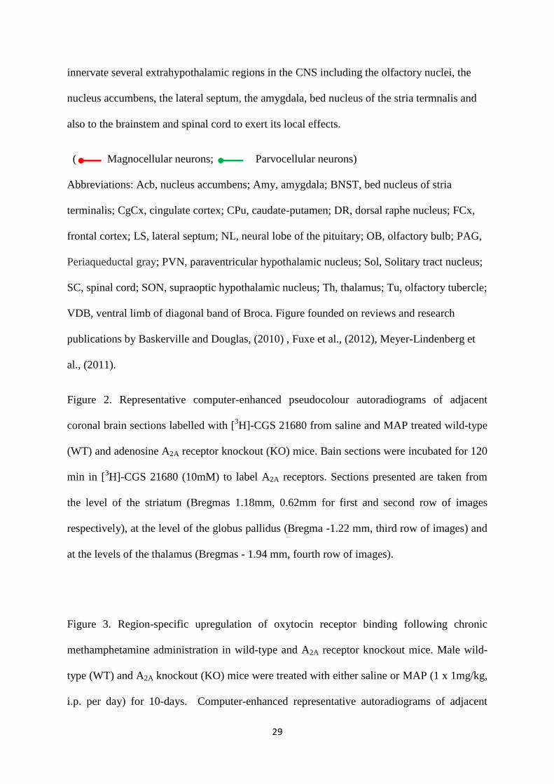
Figure 1. Prominent oxytocinergic projections in the brain of rodents. Oxytocin is primarily
synthesized in the magnocellular neurosecretory cells in the supraoptic (SON) and
paraventricular (PVN) nuclei; Oxytocin is then transported to the posterior pituitary gland
where it is stored in vesicles and is released into the bloodstream to exert its peripheral
effects. Moreover, OT is also synthesised in the parvocellular neurons in the PVN which
29
innervate several extrahypothalamic regions in the CNS including the olfactory nuclei, the
nucleus accumbens, the lateral septum, the amygdala, bed nucleus of the stria termnalis and
also to the brainstem and spinal cord to exert its local effects.
( Magnocellular neurons; Parvocellular neurons)
Abbreviations: Acb, nucleus accumbens; Amy, amygdala; BNST, bed nucleus of stria
terminalis; CgCx, cingulate cortex; CPu, caudate-putamen; DR, dorsal raphe nucleus; FCx,
frontal cortex; LS, lateral septum; NL, neural lobe of the pituitary; OB, olfactory bulb; PAG,
Periaqueductal gray; PVN, paraventricular hypothalamic nucleus; Sol, Solitary tract nucleus;
SC, spinal cord; SON, supraoptic hypothalamic nucleus; Th, thalamus; Tu, olfactory tubercle;
VDB, ventral limb of diagonal band of Broca. Figure founded on reviews and research
publications by Baskerville and Douglas, (2010) , Fuxe et al., (2012), Meyer-Lindenberg et
al., (2011).
Figure 2. Representative computer-enhanced pseudocolour autoradiograms of adjacent
coronal brain sections labelled with [3H]-CGS 21680 from saline and MAP treated wild-type
(WT) and adenosine A2A receptor knockout (KO) mice. Bain sections were incubated for 120
min in [3H]-CGS 21680 (10mM) to label A2A receptors. Sections presented are taken from
the level of the striatum (Bregmas 1.18mm, 0.62mm for first and second row of images
respectively), at the level of the globus pallidus (Bregma -1.22 mm, third row of images) and
at the levels of the thalamus (Bregmas - 1.94 mm, fourth row of images).
Figure 3. Region-specific upregulation of oxytocin receptor binding following chronic
methamphetamine administration in wild-type and A2A receptor knockout mice. Male wild-
type (WT) and A2A knockout (KO) mice were treated with either saline or MAP (1 x 1mg/kg,
i.p. per day) for 10-days. Computer-enhanced representative autoradiograms of adjacent
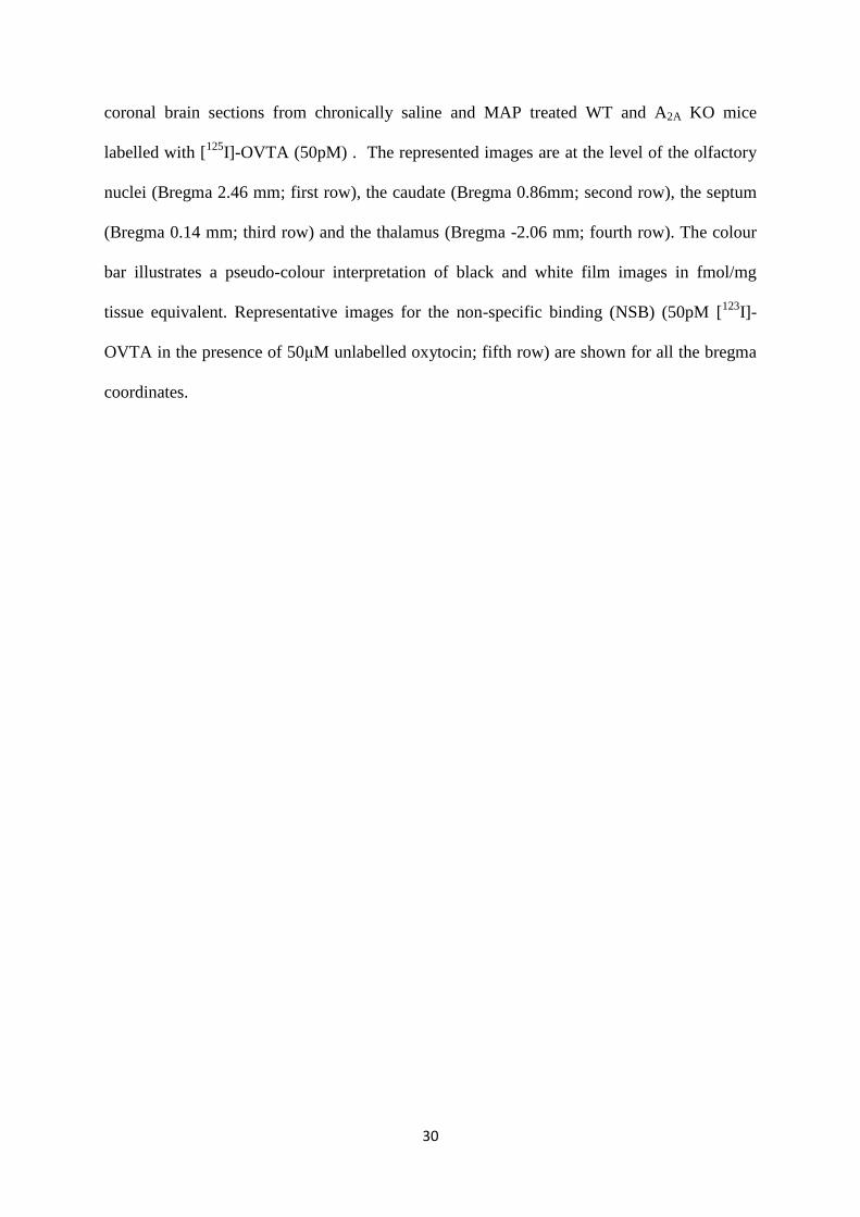
30
coronal brain sections from chronically saline and MAP treated WT and A2A KO mice
labelled with [125
I]-OVTA (50pM) . The represented images are at the level of the olfactory
nuclei (Bregma 2.46 mm; first row), the caudate (Bregma 0.86mm; second row), the septum
(Bregma 0.14 mm; third row) and the thalamus (Bregma -2.06 mm; fourth row). The colour
bar illustrates a pseudo-colour interpretation of black and white film images in fmol/mg
tissue equivalent. Representative images for the non-specific binding (NSB) (50pM [123
I]-
OVTA in the presence of 50μM unlabelled oxytocin; fifth row) are shown for all the bregma
coordinates.