Chitosan nanoparticles release nimodipine in response to ...
9
Contents lists available at ScienceDirect Neuropharmacology journal homepage: www.elsevier.com/locate/neuropharm Chitosan nanoparticles release nimodipine in response to tissue acidosis to attenuate spreading depolarization evoked during forebrain ischemia Orsolya M. Tóth a , Ákos Menyhárt a , Viktória Éva Varga a , Dóra Hantosi a , Orsolya Ivánkovits-Kiss a , Dániel Péter Varga a , Írisz Szabó a , László Janovák b , Imre Dékány b , Eszter Farkas a,∗ , Ferenc Bari a a Department of Medical Physics and Informatics, Faculty of Medicine & Faculty of Science and Informatics, University of Szeged, H-6720, Szeged, Korányi fasor 9, Hungary b University of Szeged, Interdisciplinary Excellence Centre, Department of Physical Chemistry and Materials Science, H-6720, Rerrich Béla tér 1, Szeged, Hungary HIGHLIGHTS • Tissue acidosis with cerebral ischemia was exploited for controlled drug release. • Nimodipine release from nanoparticles to low pH was shown in ischemic brain tissue. • Nimodipine delivered by nanoparticles improved perfusion in ischemic brain tissue. • Nimodipine delivered by nanoparticles inhibited spreading depolarization. • The applied chitosan nanoparticles did not activate microglia in the brain. ARTICLE INFO Keywords: Acidosis Cerebral blood flow Cerebral ischemia Nanomedicine Nimodipine Spreading depolarization ABSTRACT Stroke is an important cause of mortality and disability. Treatment options are limited, therefore the progress in this regard is urgently needed. Nimodipine, an L-type voltage-gated calcium channel antagonist dilates cerebral arterioles, but its systemic administration may cause potential side effects. We have previously constructed chitosan nanoparticles as drug carriers, which release nimodipine in response to decreasing pH typical of cer- ebral ischemia. Here we have set out to evaluate this nanomedical approach to deliver nimodipine selectively to acidic ischemic brain tissue. After washing a nanoparticle suspension with or without nimodipine (100 μM) on the exposed brain surface of anesthetized rats (n = 18), both common carotid arteries were occluded to create forebrain ischemia. Spreading depolarizations (SDs) were elicited by 1M KCl to deepen the ischemic insult. Local field potential, cerebral blood flow (CBF) and tissue pH were recorded from the cerebral cortex. Microglia activation and neuronal survival were evaluated in brain sections by immunocytochemistry. Ischemia-induced tissue acidosis initiated nimodipine release from nanoparticles, confirmed by the significant elevation of baseline CBF (47.8 ± 23.7 vs. 29.3 ± 6.96%). Nimodipine shortened the duration of both SD itself (48.07 ± 23.29 vs. 76.25 ± 17.2 s), and the associated tissue acidosis (65.46 ± 20.2 vs. 138.3 ± 66.07 s), moreover it enhanced the SD-related hyperemia (48.15 ± 42.04 vs. 17.29 ± 11.03%). Chitosan nanoparticles did not activate microglia. The data support the concept that tissue acidosis linked to cerebral ischemia can be employed as a trigger for targeted drug delivery. Nimodipine-mediated vasodilation and SD inhibition can be achieved by pH-responsive chitosan nanoparticles applied directly to the brain surface. 1. Introduction Even though ischemic stroke is increasingly more prevalent in the aging Western societies, treatment options with reasonable success rate are limited to systemic thrombolysis or endovascular thrombectomy, both feasible to conduct in a selected group of all stroke patients (Balami et al., 2013; Campbell et al., 2015). The development of other effective treatment options that may be beneficial to a large population https://doi.org/10.1016/j.neuropharm.2019.107850 Received 22 June 2019; Received in revised form 5 November 2019; Accepted 8 November 2019 ∗ Corresponding author. Department of Medical Physics and Informatics, Faculty of Medicine, and Faculty of Science and Informatics, University of Szeged, Korányi fasor 9, H-6720, Szeged, Hungary. E-mail address: [email protected] (E. Farkas). Neuropharmacology 162 (2020) 107850 Available online 09 November 2019 0028-3908/ © 2019 Elsevier Ltd. All rights reserved. T
Transcript of Chitosan nanoparticles release nimodipine in response to ...

Contents lists available at ScienceDirect
Neuropharmacology
journal homepage: www.elsevier.com/locate/neuropharm
Chitosan nanoparticles release nimodipine in response to tissue acidosis toattenuate spreading depolarization evoked during forebrain ischemia
Orsolya M. Tótha, Ákos Menyhárta, Viktória Éva Vargaa, Dóra Hantosia, Orsolya Ivánkovits-Kissa,Dániel Péter Vargaa, Írisz Szabóa, László Janovákb, Imre Dékányb, Eszter Farkasa,∗, Ferenc Baria
a Department of Medical Physics and Informatics, Faculty of Medicine & Faculty of Science and Informatics, University of Szeged, H-6720, Szeged, Korányi fasor 9,HungarybUniversity of Szeged, Interdisciplinary Excellence Centre, Department of Physical Chemistry and Materials Science, H-6720, Rerrich Béla tér 1, Szeged, Hungary
H I G H L I G H T S
• Tissue acidosis with cerebral ischemia was exploited for controlled drug release.
• Nimodipine release from nanoparticles to low pH was shown in ischemic brain tissue.
• Nimodipine delivered by nanoparticles improved perfusion in ischemic brain tissue.
• Nimodipine delivered by nanoparticles inhibited spreading depolarization.
• The applied chitosan nanoparticles did not activate microglia in the brain.
A R T I C L E I N F O
Keywords:AcidosisCerebral blood flowCerebral ischemiaNanomedicineNimodipineSpreading depolarization
A B S T R A C T
Stroke is an important cause of mortality and disability. Treatment options are limited, therefore the progress inthis regard is urgently needed. Nimodipine, an L-type voltage-gated calcium channel antagonist dilates cerebralarterioles, but its systemic administration may cause potential side effects. We have previously constructedchitosan nanoparticles as drug carriers, which release nimodipine in response to decreasing pH typical of cer-ebral ischemia. Here we have set out to evaluate this nanomedical approach to deliver nimodipine selectively toacidic ischemic brain tissue.
After washing a nanoparticle suspension with or without nimodipine (100 μM) on the exposed brain surfaceof anesthetized rats (n=18), both common carotid arteries were occluded to create forebrain ischemia.Spreading depolarizations (SDs) were elicited by 1M KCl to deepen the ischemic insult. Local field potential,cerebral blood flow (CBF) and tissue pH were recorded from the cerebral cortex. Microglia activation andneuronal survival were evaluated in brain sections by immunocytochemistry.
Ischemia-induced tissue acidosis initiated nimodipine release from nanoparticles, confirmed by the significantelevation of baseline CBF (47.8 ± 23.7 vs. 29.3 ± 6.96%). Nimodipine shortened the duration of both SD itself(48.07 ± 23.29 vs. 76.25 ± 17.2 s), and the associated tissue acidosis (65.46 ± 20.2 vs. 138.3 ± 66.07 s),moreover it enhanced the SD-related hyperemia (48.15 ± 42.04 vs. 17.29 ± 11.03%). Chitosan nanoparticlesdid not activate microglia.
The data support the concept that tissue acidosis linked to cerebral ischemia can be employed as a trigger fortargeted drug delivery. Nimodipine-mediated vasodilation and SD inhibition can be achieved by pH-responsivechitosan nanoparticles applied directly to the brain surface.
1. Introduction
Even though ischemic stroke is increasingly more prevalent in theaging Western societies, treatment options with reasonable success rate
are limited to systemic thrombolysis or endovascular thrombectomy,both feasible to conduct in a selected group of all stroke patients(Balami et al., 2013; Campbell et al., 2015). The development of othereffective treatment options that may be beneficial to a large population
https://doi.org/10.1016/j.neuropharm.2019.107850Received 22 June 2019; Received in revised form 5 November 2019; Accepted 8 November 2019
∗ Corresponding author. Department of Medical Physics and Informatics, Faculty of Medicine, and Faculty of Science and Informatics, University of Szeged, Korányifasor 9, H-6720, Szeged, Hungary.
E-mail address: [email protected] (E. Farkas).
Neuropharmacology 162 (2020) 107850
Available online 09 November 20190028-3908/ © 2019 Elsevier Ltd. All rights reserved.
T

of stroke patients is urgently needed.The focus of stroke therapy is the salvage of non-functional but vi-
able penumbra tissue surrounding the ischemic core beyond rescue.Spreading depolarization (SD) has been recognized as a central me-chanism of injury progression and of the conversion of penumbra tissueto the ischemic core (Nedergaard et al., 1996; Hossmann, 1996; Woitziket al., 2013; Hartings et al., 2017; Hertelendy et al., 2019). Delayedcerebral ischemia after aneurismal subarachnoid hemorrhage (SAH)has been also linked to SD (Dreier et al., 2006), and SD-inducedischemia (Dreier et al., 2009). Importantly, SD-induced ischemia wasfound to occur in the time window of the development of brain infarctsconfirmed by serial neuroimaging (Lückl et al., 2018).
Spreading depolarization has recently been considered as a pre-sumed target of pharmacological neuroprotective intervention (Hertleet al., 2012; Carlson et al., 2018). Next to the NMDA receptor blockerketamine (Sánchez-Porras et al., 2014; Reinhart and Shuttleworth,2018), the L-type voltage-gated calcium channel antagonist nimodipinehas been found effective to impede SD (Richter et al., 2002; Menyhártet al., 2018; Szabó et al., 2019), or to counteract the ischemia-relatedimpairment of the cerebral blood flow (CBF) response to SD (Dreieret al., 1998, 2002; Menyhárt et al., 2018; Szabó et al., 2019).
In clinical setting, nimodipine administered orally or intravenouslyhas become an approved agent to prevent delayed cerebral ischemiaafter aneurismal subarachnoid hemorrhage (SAH) (Pickard et al., 1989;van Gijn et al., 2007). Prophylactic treatment with nimodipine wasfound to reduce the frequency and severity of delayed ischemic stroke(Pickard et al., 1989; Dorhout Mees et al., 2007). In addition, someclinical trials have suggested that nimodipine may limit infarct growthin acute ischemic stroke, as well (Martinez-Vila et al., 1990; Fogelholmet al., 2000). Yet, the systemic administration of nimodipine is limitedby its suboptimal efficacy and the dose-dependent hypotension it mayproduce in some patients (Sandow et al., 2016). Also, the pharmaco-logical augmentation of the CBF response to SD at optimally perfusedbrain sites distant to the ischemic penumbra may cause steal effect, andultimately increase the risk of ischemic injury expansion (Pinard et al.,2002). All these drawbacks together justify the development of drugdelivery systems targeting selectively the tissue at risk of injury, toachieve neuroprotection without counterproductive side effects.
The use of biocompatible and biodegradable micro- or nanoparticlesthat may be selectively targeted to desired tissue sites has been recentlygaining increasing significance. Nimodipine, for instance, has been as-sociated to poly-D,L-lactide-co-glycolide (PLGA) microparticles andapplied intrathecally in order to achieve local, prolonged release of thedrug in experimental SAH (Hänggi et al., 2012), an approach latertaken to a clinical study, to be tested in patients surgically treated foraneurismal SAH (Etminan et al., 2015). Here we set out to explorewhether nimodipine can be targeted with nanoparticles selectively tobrain tissue affected by ischemia, to inhibit recurrent SD events per-ceived to be harmful to the survival of the penumbra. We have recentlyconstructed hydrophobized and pH- sensitive chitosan nanoparticles,which release nimodipine in response to an acidic pH shift (from pH 7.3to ~6.9), the drug release having been confirmed in suspension
(Janovák et al., 2018). Decreasing pH has been utilized as the trigger ofdrug release from nanoparticles, because ischemia produces local tissueacidosis, which is further worsened by the occurrence of SDs (Menyhártet al., 2017). Nanoparticles responsive to pH gradients were previouslydesigned for cancer therapy (Shen et al., 2008; Du et al., 2014), becausethe increased metabolism of glucose creates extracellular acidosis insolid tumors (Neri and Supuran, 2011), which can be exploited fortargeted drug delivery.
In the present study, we applied nimodipine associated to pH re-sponsive chitosan nanoparticles (d= 5.2 ± 1.1 nm) (Janovák et al.,2018) to the exposed cerebral cortex of rats to investigate (i) whethertissue acidosis caused by ischemia or SD occurrence initiates drug re-lease in the nervous tissue, and (ii) whether nimodipine administeredwith nanoparticles exerts its expected vasodilator and neuroprotectiveeffect against SD propagating over the penumbra. With encouragingdata in hand, we aim to build upon these results and design, in futurework, an approach for the systemic administration of the pH responsivechitosan nanoparticles.
2. Materials and methods
2.1. Surgical procedures
All experiments were approved by the National Food Chain Safetyand Animal Health Directorate of Csongrád County, Hungary. Theprocedures conformed to the guidelines of the Scientific Committee ofAnimal Experimentation of the Hungarian Academy of Sciences (up-dated Law and Regulations on Animal Protection: 40/2013. (II. 14.)Gov. of Hungary), following the EU Directive 2010/63/EU on theprotection of animals used for scientific purposes and reported incompliance with the ARRIVE guidelines.
Young adult, male Sprague-Dawley rats (Charles River Laboratories;n= 25, m=339 ± 37 g) were used in this study. Animals werehoused under a normal 12/12 h light/dark cycle and constant tem-perature (23 °C). Standard rodent chow and drinking water were sup-plied ad libitum. On the day of experiments, animals were anesthetizedwith isoflurane (1.5–2% in N2O:O2 70%: 30%) and allowed to breathespontaneously through a head mask. Body temperature was maintainedat 37 ± 0.5 °C by using a servo-regulated heating pad, feedback-con-trolled by a flexible rectal probe (Harvard Apparatus, Holliston, MA,U.S.A.). Atropine (0.1%, 0.05ml) was administered intramuscularlyshortly before the surgical procedures to avoid the production of airwaymucus. Lidocaine (1%) was administered topically before opening eachtissue layer. The left femoral artery was cannulated to monitor meanarterial blood pressure (MABP) continuously, and to collect samples forarterial blood gas analysis. Samples for blood gas analysis were takenprior to the start of the experimental protocol (physiological condition)and shortly after the evolution of the last SD event in a train (ischemiccondition) (Epoc Reader, Epocal, Ottawa, Canada). The depth of an-esthesia was regularly checked and controlled with the aid of MABP.Both common carotid arteries were carefully separated from the sur-rounding tissue and an occluder was looped around each vessel for later
Abbreviations
2VO bilateral common carotid artery occlusion (“two-vesselocclusion”)
aCSF artificial cerebrospinal fluidANOVA analysis of varianceAUC area under the curveBBB blood-brain barrierCBF cerebral blood flowDC direct currentIba1 ionized calcium binding adaptor molecule 1
LDF laser-Doppler flowLFP local field potentialMABP mean arterial blood pressureNeuN neuronal nuclear proteinNMDA N-methyl-D-aspartatePFA paraformaldehyderSD recurrent spreading depolarizationSAH subarachnoid hemorrhageSD spreading depolarizationSD1 the first spreading depolarization in a train of events
O.M. Tóth, et al. Neuropharmacology 162 (2020) 107850
2

induction of acute, incomplete, global forebrain ischemia. The head ofthe rats was fixed into a stereotactic frame and two cranial windows(~3×3 mm) were prepared on the right parietal bone (3mm caudal,5 mm lateral and 7mm caudal, 5 mm lateral from bregma) with a highprecision dental drill (ProLab Basic, Bien-Air Dental SA, Bienne,Switzerland) under saline cooling (n=18). The cortical surface wasexposed by the careful retraction of the dura mater in each cranialwindow. The caudal window was later used for evoking SDs, whereas apH sensitive microelectrode, a reference electrode recording local fieldpotential filtered in direct current (DC) mode, and an adjacent Laser-Doppler probe were positioned in the rostral window. The craniotomieswere regularly irrigated with artificial cerebrospinal fluid (aCSF; mMconcentrations: 126.6 NaCl, 3 KCl, 1.5 CaCl2, 1.2 MgCl2, 24.5 NaHCO3,6.7 urea, 3.7 glucose bubbled with 95% O2 and 5% CO2 to achieve aconstant pH of 7.4). In order to discriminate the degree of microgliaactivation caused by SDs themselves with respect to the impact of tre-panation alone, the cranial windows were created bilaterally in somerats (n=7).
2.2. Recording of extracellular pH, electrophysiological variables and localcerebral blood flow
Ion-sensitive microelectrodes were prepared according to Voipioand Kaila (1993). Glass capillary microelectrode tips (outer diameter:10–12 μm) were filled with a liquid H+ ion exchanger, and the shank ofthe microelectrode was backfilled with 150 mM NaCl + 40 mMHEPES + 20 mM NaOH. Each pH sensitive microelectrode was cali-brated in standard solutions of known pH (pH 8.05, 7.02, 6.2; cali-bration solution containing 150 mM NaCl + 40 mM HEPES). A saline-filled glass capillary electrode (20 μm outside tip diameter) was low-ered 800–1000 μm deep into the cerebral cortex to serve as referencefor the pH sensitive electrode, and to record DC potential. The tips ofthe two electrodes were positioned as near as possible. An Ag/AgClelectrode implanted subcutaneously in the neck served as commonground. Microelectrodes were connected to a custom-made dual-channel high input impedance electrometer (including AD549LH,Analog Devices, Norwood, MA, USA) via Ag/AgCl leads. The voltagesignal recorded by the reference electrode was subtracted from that ofthe pH sensitive microelectrode by dedicated differential amplifiers andassociated filter modules (NL106 and NL125, NeuroLog System, Digi-timer Ltd, United Kingdom), which yielded potential variations relatedto changes in extracellular [H+]. The recorded signals were then for-warded to an analog-to-digital converter (MP 150, Biopac Systems, Inc).Electric signals were continuously acquired at a sampling frequency of1 kHz. Extracellular pH changes were expressed in mV to be translatedto pH units offline, using least squares linear regression.
SD-associated changes in local CBF were recorded using Laser-Doppler flowmetry (LDF) by a Laser-Doppler needle probe (Probe 403connected to PeriFlux 5000; Perimed AB, Sweden). The probe waspositioned right above the cortical surface at the penetration site of theglass capillary electrodes with a micromanipulator, avoiding any largepial vessels. The signal was digitized and acquired, together with theDC potential and pH signals essentially as described above (MP 150 andAcqKnowledge 4.2.0, Biopac Systems, Inc. USA). The completed pre-paration was enclosed in a Faraday cage.
2.3. Pharmacological treatment and experimental protocol
Chitosan nanoparticle suspension – either loaded with nimodipine(an L-type voltage-gated calcium channel blocker), or devoid of thepharmacon (i.e. vehicle) – was prepared in aCSF according to Janováket al. (2018). The nanoparticle suspension with encapsulated drug wasexpected to release nimodipine in response to a pH shift from physio-logical (~pH 7.35) to acidic (~pH 6.75) (Janovák et al., 2018). Thepharmacological treatment was applied to rats with ipsilateral cranialwindows only (n=18). The rostral cranial window was incubated with
the nanoparticle suspension including nimodipine at a concentrationfound previously effective when given in solution (Szabó et al., 2019)(100 μM; n=10) or vehicle (n= 8). Suspensions were refreshed every10min until the termination of the experiment. The craniotomies inpreparations with bilateral cranial windows were regularly irrigatedwith aCSF alone (n=4). Animal selection for treatment was random byalternating treatment as the experimental work proceeded.
After a baseline period of 15min, the suspension was washed to thebrain surface and refreshed every 10min. Fifteen minutes later, in-complete, global forebrain ischemia was induced by occluding bothcommon carotid arteries permanently (“2-vessel occlusion”, 2VO).Occluders were pulled on until resistance was felt and were fixed inplace. Successful 2VO was confirmed by an immediate, sharp drop ofthe LDF-signal displayed live. Fifteen minutes after 2VO onset, three SDevents were evoked by placing a 1M KCl-soaked cotton ball on theexposed surface at an inter-SD interval of 15min. The cotton ball wasremoved immediately after the successful elicitation of each SD.
In some rats equipped with bilateral craniotomies (n=3), aCSF wasapplied to one hemisphere, and nanoparticle suspension (i.e. vehicle) tothe other, and 2VO was subsequently not imposed. This allowed thelater evaluation of potential microglia activation by the direct exposureof the cerebral cortex to chitosan nanoparticles. Like in the 2VO rats,aCSF or the nanoparticle suspension were refreshed every 10min untilthe termination of the experiments.
2.4. Histology
At the end of the experimental protocol, 1–1.5 h after ischemia in-duction, animals (n=16) were transcardially perfused in deep an-esthesia with physiological saline followed by 4% paraformaldehyde(PFA). The brains were removed and postfixed overnight in 4% PFA.Coronal forebrain sections of 20 μm thickness were cut with a vi-bratome (Leica VT 1000S; Leica Microsystems, Wetzlar, Germany).Microglia were labeled with Iba1 (rabbit anti- Iba-1 primary antibody,1:3000, 019–19741, Fujifilm Wako Chemicals Europe GmbH, Neuss,Germany) in order to explore whether the experimental procedures orthe topical application of the nanoparticle suspension by itself inducedmicroglia activation. To estimate neuronal loss due to SD, and neuro-protection offered by nimodipine, neurons in free floating slices werelabeled for the marker NeuN with immunohistochemistry (rabbit anti-NeuN primary antibody; 1:300, ab177487, AbCam, Cambridge, UK).Endogenous peroxidase activity was blocked with 5% H2O2, the non-specific protein binding sites were blocked with 5% normal goat serum(Merck, Kenilworth, New Jersey, USA) and the slices were permeabi-lized with Triton X-100 (Merck, Kenilworth, New Jersey, USA). Colorreaction was developed with a Polink-2 Plus HRP Detection Kit forrabbit primary antibody with DAB (diaminobenzidine) chromogen (GBILabs, Bothell, WA, USA). The slices were mounted on microscopic slideswith Eukit® (Merck, Kenilworth, New Jersey, USA) and digitally re-corded with a microscope slide scanner (Zeiss Mirax Midi Slide Scanner,Carl Zeiss MicroImaging GmbH, Jena, Germany) operated by aCaseViewer software (3D Histech Ltd., Budapest, Hungary). The slideswere evaluated with ImageJ (Wayne Rasband, NIH, Bethesda, USA)software.
Microglial activation was characterized with a ramification indexcalculated according to previously established principles (Faulkneret al., 2011; Varga et al., 2018). In each animal, 3 coronal brain sliceswere selected for the analysis. In each slice, 3 photomicrographs weretaken at 20x magnification along the depth of the parietal cortex inboth hemispheres. A 126× 126 μm grid was placed on each photo-micrograph. Microglial branches/grid intersections (B) as well as mi-croglial cell bodies within the grid (CBD) were counted manually withthe Cell Counter plugin of ImageJ. The ramification index (RI) wascalculated according to the following formula: RI=B2/CBD. Sinceactivated microglia are characterized by the retraction of their pro-cesses (i.e. amoeboid shape), the high ramification index corresponds to
O.M. Tóth, et al. Neuropharmacology 162 (2020) 107850
3

the resting state of microglia, while low ramification index reflectsmicroglial activation.
Neuronal loss in the ipsi- and contralateral parietal cortex wascharacterized by the estimation NeuN-positive immunolabeling in acortical area of a standard size. In each animal, 3 coronal brain sliceswere selected for the analysis. In each slice, 2 photomicrographs weretaken at 5x magnification along the depth of the parietal cortex in bothhemispheres. After masking binary images in ImageJ, the relative sur-face covered by NeuN-positive cell bodies was expressed.
2.5. Data processing and analysis
All variables (i.e., extracellular pH, DC potential, LDF signal andMABP) were simultaneously acquired, displayed live, stored and ana-lyzed using a personal computer equipped with a dedicated software(AcqKnowledge 4.2 for MP150, Biopac Systems, Inc., USA). For offlineanalysis, all variables were down sampled to 1 Hz. Full data analysiswas conducted for animals whose cerebrocortical tissue pH varied inthe physiological range prior to the application of nanoparticles andduring baseline (n= 9).
Data were evaluated separately for the first SD (SD1), and recurrentSD (rSD) events, because of the differences in the kinetics of the SD-associated CBF response. Changes in local CBF were expressed relativeto baseline by using the average CBF value preceding ischemia induc-tion (100%) and the mean recorded biological zero obtained after ter-minating the experiment (0%) as reference points. The SD-associatedhyperemia was then characterized by; (i) the amplitude of the peakhyperemia, (ii) duration of peak hyperemia at half amplitude and (iii)the magnitude (i. e. area under the curve, AUC) of the hyperemic re-sponse. SDs were analyzed by the following parameters: (i) amplitudeof depolarization, (ii) duration of depolarization at half amplitude, (iii)AUC of the negative DC shift, (iv) rate of depolarization and (v) re-polarization. Extracellular pH signal indicative of SD was characterizedby (i) amplitude of the transient acidosis, (ii) duration at half ampli-tude, (iii) AUC of the pH response, and (iv) rate of acidosis and (v)recovery.
All recordings were first screened for events suitable for compre-hensive analysis. Animals with alkalotic baseline brain pH over 7.5were excluded from the analysis.
Data are given as mean ± standard deviation (stdev). The softwareSPSS was used for statistical analysis (IBM SPSS Statistics for Windows,Version 22.0, IBM Corp.). A repeated measures, a one-way or a two-wayanalysis of variance (ANOVA) model was used, dependent on the typeof data set. Levels of significance were defined as p < 0.05*. Distinctstatistical methods are provided in detail in each Figure legend.
3. Results
3.1. Evidence for drug release from nanoparticles
Variables determined with arterial blood gas analysis were in thephysiological range prior to the initiation of the experimental protocol(pO2=144.4 ± 24.4 mmHg, pCO2=39.8 ± 9.9mmHg, bloodpH=7.36 ± 0.03), with a shift to higher pCO2 and lower pH values45–50min after ischemia onset (pO2= 128.0 ± 29.3mmHg,pCO2=48.7 ± 14.2 mmHg, blood pH=7.28 ± 0.06).
Nimodipine in solution, applied to the brain surface as done here,had been shown to increase local CBF significantly (Szabó et al., 2019).CBF elevation was, therefore, taken as a reliable read-out of the efficacyof nimodipine treatment, and was expected to confirm drug releasefrom nanoparticles. In the current experiments, local CBF remainedlevel during the incubation period, prior to ischemia onset (99.2 ± 2.6vs. 99.9 ± 3.0%, 30min after vs. before the application of nimodipineassociated to nanoparticles), in the face of invariable tissue pH (pH7.29 ± 0.22 vs. 7.28 ± 0.18, 30min after vs. before the application ofnimodipine associated to nanoparticles) ((a) and (b) in Fig. 1). Ischemia
induction produced a sharp drop of CBF to 29.4 ± 10.2%, and anacidic tissue pH shift to 7.06 ± 0.30 ((c) in Fig. 1). From this point on,CBF sampled prior to SD events increased in the nimodipine group, andwas higher than in the vehicle group, particularly prior to rSDs(47.8 ± 23.7 vs. 29.3 ± 6.96%, nimodipine vs. vehicle) ((f) in Fig. 1),which were triggered subsequent to the transient reduction of tissue pHto 6.71 ± 0.29 with SD1 ((e) in Fig. 1). At the same time, the expectedrelease of nimodipine from the nanoparticles did not exert any dis-cernible impact on MABP (e.g. 96.7 ± 14.3 vs. 92.0 ± 10.9 mmHg,nimodipine vs. vehicle, after ischemia induction – and thus the initia-tion of drug release) ((d) in Fig. 1).
3.2. Spreading depolarization
Spreading depolarization events were experimentally triggeredunder global forebrain ischemia to evaluate the potential impact ofnimodipine delivered with nanoparticles on the kinetics of SD, and theassociated tissue pH variation and CBF response (Figs. 2–4). The first SD(SD1) and the recurrent SDs (rSD) were analyzed apart, because ofknown differences in the kinetics of SD associated CBF response andtissue pH variation (Farkas et al., 2008; Menyhárt et al., 2017).
SD occurrence was confirmed by the transient negative shift of theDC potential (Fig. 2A). The analysis of the DC potential signature of SDsdemonstrated that nimodipine applied in the nanoparticle suspension
Fig. 1. Variation of baseline cerebral blood flow (CBF; upper scatter plot with yaxis to the left) with respect to tissue pH (bar chart with y axis to the right), andmean arterial pressure (MABP; lower scatter plot) at selected points of the ex-perimental protocol: (a) before treatment initiation; (b) after treatment initia-tion; (c) minimum after ischemia induction; (d) prior to the first spreadingdepolarization (SD1); (e) pH minimum with SD1; (f) prior to recurrent SD (rSD)events. Note that CBF becomes higher in the nimodipine group compared to thevehicle group following the tissue pH drop with ischemia induction.Representative traces under the bar chart (tissue pH: blue, CBF: black) obtainedfrom an animal treated with nimodipine show typical signal variation over theexperimental protocol. Data are given as mean± stdev. Statistical analysis re-lied on one-way analysis of variance (ANOVA) paradigm for CBF and tissue pH,and a repeated measures model for MABP. The level of significance was definedas p < 0.05*. (For interpretation of the references to colour in this figurelegend, the reader is referred to the Web version of this article.)
O.M. Tóth, et al. Neuropharmacology 162 (2020) 107850
4

Fig. 2. The impact of nimodipine delivered with nanoparticles on first and recurrent spreading depolarization events (SD1, rSDs). A, Traces demonstrate the negativedirect currant (DC) potential shift indicative of rSD events. Each trace is the average of a number of individual events taken from separate animals, and are presentedas mean ± stdev (n = 9/7). B, Rate of repolarization of the negative DC potential shift with SD events. C, Duration of the negative DC potential shift with SD events,measured at half amplitude. Data are given as mean ± stdev, sample size (number of events) is shown in each bar. Statistical analysis relied on one-way analysis ofvariance (ANOVA) paradigm. The level of significance was defined as p < 0.05*.
Fig. 3. The impact of nimodipine delivered with nanoparticles on the tissue pH response related to the first and recurrent spreading depolarization events (SD1,rSDs). A, Traces demonstrate the kinetics of the pH response to rSD events. Each trace is the average of a number of individual events taken from separate animals,and are presented as mean ± stdev (n = 4/6). B, Rate of return from tissue acidosis. C, Duration of tissue acidosis taken at half amplitude. D, Magnitude (i. e. areaunder the curve, AUC) of tissue acidosis. Data are given as mean ± stdev, sample size (number of events) is shown in each bar. Statistical analysis relied on one-wayanalysis of variance (ANOVA) paradigm. The level of significance was defined as p < 0.05*.
Fig. 4. The impact of nimodipine delivered with nanoparticles on the cerebral blood flow (CBF) response to the first and recurrent spreading depolarization events(SD1, rSDs). A, Traces demonstrate the kinetics of CBF response to rSD events. Each trace is the average of a number of individual events, and are presented asmean ± stdev (n = 9/14). B, Amplitude of peak hyperemia. The base of each bar in the chart is set to the CBF level preceding SD events. C, Magnitude (i. e. areaunder the curve, AUC) of peak hyperemia. Data are given as mean ± stdev, sample size (number of events) is shown in each bar. Statistical analysis relied on one-way analysis of variance (ANOVA) paradigm. The level of significance was defined as p < 0.05*.
O.M. Tóth, et al. Neuropharmacology 162 (2020) 107850
5

facilitated the rate of repolarization of rSD events in particular(0.8 ± 0.523 vs. 0.279 ± 0.153mV/s, nimodipine vs. vehicle)(Fig. 2B). Accordingly, the duration of rSDs shortened significantly withrespect to control (48.07 ± 23.29 vs. 76.25 ± 17.2 s, nimodipine vs.vehicle) (Fig. 2C).
Tissue pH variations associated with SD events started with a rapid,short alkaline shift followed by a longer-lasting, dominant, transientacidosis (Fig. 3A) (Menyhárt et al., 2017). Tissue pH did not fully re-cover and remained typically mildly acidic after SD1 compared to laterrSDs (pH 7.14 ± 0.29 vs. 7.23 ± 0.28, prior to rSDs vs. prior to SD1),but the difference was statistically not significant. Nimodipine treat-ment had no measurable impact on the initial alkaline shift, but itmodified the kinetics of the subsequent transient acidosis. As such, ni-modipine delivered by nanoparticles facilitated the rate of return fromthe acidic shift with rSDs (0.01 ± 0.006 vs. 0.005 ± 0.002 pH unit/s,nimodipine vs. vehicle) (Fig. 3B) and shortened the duration of acidosiswith rSDs (65.46 ± 20.2 vs. 138.3 ± 66.07 s, nimodipine vs. vehicle)(Fig. 3C). As a consequence, the magnitude of acidosis expressed as areaunder the curve was substantially reduced in the nimodipine comparedto vehicle group (25.75 ± 10.69 vs. 49.46 ± 23.38 pH unit*s, ni-modipine vs. vehicle) (Fig. 3C).
The SD related CBF response in anesthetized rats includes an initialtransient hypoperfusion, followed by a peak hyperemia and is con-cluded by a long lasting oligemia (Ayata and Lauritzen, 2015; Menyhártet al., 2017; Szabó et al., 2019). The initial hypoperfusion proved to bedetectable only occasionally (Fig. 4A), therefore the analysis focused onthe peak hyperemic element of the CBF response (Fig. 4). As expected,nimodipine delivered by nanoparticles significantly enhanced the am-plitude (48.15 ± 42.04 vs. 17.29 ± 11.03%, nimodipine vs. vehicle)and the magnitude of peak hyperemia in response to rSDs(4604.43 ± 2572.3 vs. 2368.05 ± 1324.71%*s, nimodipine vs. ve-hicle) (Fig. 4B-C).
3.3. Histology
In order to explore whether the chitosan nanoparticles used here
might trigger neuroinflammatory reaction (a potential unfavorable sideeffect of the drug delivery approach), we estimated microglial activa-tion in immuno-stained brain sections. Microglia immunolabeled forIba1 appeared to be activated in the cerebral cortex ipsilateral to theinitiation of SD, as shown by their sparser processes and rounded,amoeboid shape (Fig. 5A). Microglia activation was quantitatively ex-pressed by a ramification index (Varga et al., 2018) representing thedensity of microglial processes. The ramification index was remarkablyreduced in the ipsilateral compared to the contralateral somatosensorycortex (e.g. 398 ± 203 vs. 1118 ± 300, ipsi-vs. contralateral to SDinduction in the vehicle group) (Fig. 5A). The trepanation procedureactivated microglia (480 ± 211 vs. 740 ± 394, trepanated vs. non-trepanated contralateral to SD induction; p < 0.048*), but the hemi-sphere-specific reduction of the ramification index was also attributedto SD, because it was clearly observed in rats with bilateral craniotomy,as well (201 ± 102 vs. 483 ± 244, ipsi-vs. contralateral to SD in-duction; p < 0.002**). The application of the nanoparticle suspensionalone (vehicle), or incorporating nimodipine did not reduce the rami-fication index any further compared to aCSF-rinsed preparations ofcerebral ischemia (443 ± 208 vs. 398 ± 203 vs. 284 ± 107, ni-modipine vs. vehicle vs. aCSF; ipsilateral) (Fig. 5A). In naïve ratsequipped with bilateral craniotomy, one hemisphere treated with aCSFand the other with vehicle (i.e. nanoparticle suspension), the microgliaramification index was also found to be similar under the two condi-tions (389 ± 126 vs. 475 ± 172, vehicle vs. aCSF, p < 0.247). Thus,the administration of nanoparticles on the cortical surface did notproduce a detectable potentiation of microglia activation additional toany of the interventions (i.e. trepanation or ischemia induction).
We labeled viable neurons with NeuN immunocytochemistry toestimate (i) the degree of early neurodegeneration SD might cause inthe acute phase of global forebrain ischemia, and (ii) the potentialneuroprotection achieved by nimodipine (Fig. 5B). We screened thesomatosensory cortex (i.e. over the striatum) distant to the site of SDelicitation (i.e. over the hippocampus), with the aim to exclude areasfrom the analysis, in which neurodegeneration might have been causedby topical KCl application to trigger SD. In some animals, the reduced
Fig. 5. The impact of the topical application of the nanoparticle suspension with or without nimodipine on microglia activation and neuronal viability. A, Microgliaimmunolabeled for Iba1 appeared to be activated in the cerebral cortex ipsilateral to the initiation of spreading depolarization (SD) and craniotomy. The applicationof the nanoparticle suspension alone (vehicle), or incorporating nimodipine did not trigger additional microglia activation compared to aCSF-rinsed preparations(aCSF). Microglia activation was expressed by a ramification index representing the density of microglial processes. B, The relative area covered by NeuN-im-munolabeled neurons expressed early signs of neurodegeneration in the cerebral cortex ipsilateral to the initiation of SD and craniotomy. Nimodipine did not preventneurodegeneration in a statistically meaningful manner. Statistical analysis relied on a two-way analysis of variance (ANOVA) paradigm. The level of significancewas defined as p < 0.05*.
O.M. Tóth, et al. Neuropharmacology 162 (2020) 107850
6
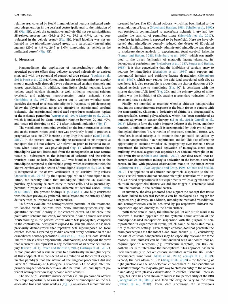
relative area covered by NeuN-immunolabeled neurons indicated earlyneurodegeneration in the cerebral cortex ipsilateral to the initiation ofSD (Fig. 5B), albeit the quantitative analysis did not reveal significantSD-related neuron loss (26.9 ± 5.0 vs. 29.1 ± 4.7%, ipsi-vs. con-tralateral in the vehicle group) (Fig. 5B). NeuN labeling was not en-hanced in the nimodipine-treated group in a statistically meaningfulmanner (29.0 ± 4.8 vs. 26.9 ± 5.0%, nimodipine vs. vehicle in theipsilateral cortex) (Fig. 5B).
4. Discussion
Nanomedicine, the application of nanotechnology with ther-apeutical purpose offers drug delivery targeted selectively to desiredsites, and with the potential of controlled drug release (Boulaiz et al.,2011; Patra et al., 2018). Nimodipine inhibits calcium influx to vascularsmooth muscle cells through L-type voltage gated calcium channels andcauses vasodilation. In addition, nimodipine blocks neuronal L-typevoltage gated calcium channels, as well, mitigates neuronal calciumoverload, and achieves neuroprotection under ischemic stress(Scriabine et al., 1989). Here we set out to explore whether nano-particles designed to release nimodipine in response to pH decreasingbelow the physiological range are effective in experimental cerebralischemia. The experimental model used reproduces conditions typicalof the ischemic penumbra (Astrup et al., 1977; Menyhárt et al., 2017),which is indicated by tissue perfusion ranging between 20 and 40%,and tissue pH dropping to 6.9–7.1 after ischemia induction (Fig. 1).
The vasodilator nimodipine (washed on the brain surface in solutionand at the concentration used here) was previously found to produce aprogressive baseline CBF increase during drug incubation (Szabó et al.,2019). In the present study, nimodipine associated to pH-responsivenanoparticles did not achieve CBF elevation prior to ischemia induc-tion, when tissue pH was physiological (Fig. 1), which confirms thatnimodipine was not dissociated from the nanoparticles at near neutraltissue pH. Subsequent to the induction of ischemia and the relatedtransient tissue acidosis, baseline CBF was found to be higher in thenimodipine compared to the vehicle group, which is consistent with theknown cerebrovascular action of nimodipine (Harper et al., 1981), andis interpreted as the in vivo verification of pH-sensitive drug release(Janovák et al., 2018). By the topical application of nimodipine in so-lution, we recently found that nimodipine inhibited SD evolution,possibly by blocking calcium entry to nerve cells, and augmented hy-peremia in response to SD in the ischemic rat cerebral cortex (Szabóet al., 2019). The present findings (Figs. 2 and 4) are fully consistentwith the data previously gathered, and substantiate the efficacy of drugdelivery with pH-responsive nanoparticles.
To further evaluate the neuroprotective potential of the treatment,we labeled viable neurons with NeuN immunocytochemistry andquantified neuronal density in the cerebral cortex. At this early timepoint after ischemia induction, we observed in some animals less denseNeuN staining in the parietal cortex where SDs propagated, comparedto the contralateral hemisphere exposed to ischemia alone. It has beenpreviously demonstrated that repetitive SDs superimposed on focalcerebral ischemia created by middle cerebral artery occlusion in the ratexacerbated neurodegeneration (Back et al., 1996). Our data stand inline with these earlier experimental observations, and support the viewthat recurrent SDs represent a key mechanism of ischemic cellular in-jury (Dreier, 2011; Dreier and Reiffurth, 2015; Hartings et al., 2017).Nimodipine did not rescue neurons to a statistically meaningful degreeat this endpoint. It is considered as a limitation of the current experi-mental paradigm that the nature of the surgical procedures did notfavor the follow-up of histological alterations 24 h or days after theprimary impact, when ischemia-related neuronal loss and signs of po-tential neuroprotection may become more obvious.
The use of pH-sensitive microelectrodes in our preparation offeredthe unique opportunity to assess the impact of nimodipine on the SD-associated transient tissue acidosis (Fig. 3), an action of nimodipine not
screened before. The SD-related acidosis, which has been linked to theaccumulation of lactate (Mutch and Hansen, 1984; Scheller et al., 1992)was previously contemplated to exacerbate ischemic injury and jeo-pardize the survival of penumbra tissue (Menyhárt et al., 2017),therefore its inhibition is expected to be beneficial. Here we have ob-served that nimodipine potently reduced the degree of SD-relatedacidosis. Similarly, intravenously administered nimodipine was shownto moderate tissue acidosis in experimental focal cerebral ischemia(Berger and Hakim, 1988; Bielenberg et al., 1990), which was attrib-uted to the direct facilitation of metabolic lactate clearance, in-dependent of perfusion rate (Bielenberg et al., 1987; Berger and Hakim,1988). It is thus conceivable that the inhibition of calcium entry toneurons by nimodipine (Scriabine et al., 1989) may support mi-tochondrial function and oxidative lactate degradation (Bielenberget al., 1987), which may reduce the acid load associated with SD, asseen here. It is also reasonable to argue that the shorter duration of SD-related acidosis due to nimodipine (Fig. 3C) is consistent with theshorter duration of SD itself (Fig. 2C), and the primary effect of nimo-dipine was the inhibition of SD, causing a secondary reduction of theassociated tissue acidosis.
Finally, we intended to examine whether chitosan nanoparticlesmay induce a neuroimmune response at the brain tissue in contact withthe nanoparticles. Chitosan, a derivative of chitin, is a biocompatible,biodegradable, natural polysaccharide, which has been considered asimmune adjuvant in cancer therapy (Li et al., 2013; Carroll et al.,2016). Microglia form the active immune defense of the brain, and theirreaction to inflammatory stimuli is accompanied by their typical mor-phological alteration (i.e. retraction of processes, amoeboid form). We,therefore, labeled microglia to estimate their potential activation bychitosan nanoparticles in our experimental model. This also offered theopportunity to examine whether SD propagating over ischemic tissuepotentiates the ischemia-related activation of microglia, since accu-mulating evidence suggest that repetitive SDs activate microglia in in-tact brain tissue (Shibata and Suzuki, 2017). Our data reveal that re-current SDs do potentiate microglia activation in the ischemic cerebralcortex, in line with previous observations made in the intact cortex(Gehrmann et al., 1993; Caggiano and Kraig, 1996; Shibata and Suzuki,2017). The application of chitosan nanoparticle suspension to the ex-posed cortical surface did not enhance microglia activation with respectto aCSF rinsed preparations in our experiments, which suggests that thechitosan nanoparticles themselves did not trigger a detectable localimmune reaction in the cerebral cortex.
In summary, the data generated here support the concept that tissueacidosis linked to cerebral ischemia can be employed as a trigger fortargeted drug delivery. In addition, nimodipine-mediated vasodilationand neuroprotection can be achieved by pH-responsive chitosan na-noparticles applied directly to the brain surface.
With these data in hand, the ultimate goal of our future work is toconceive a feasible approach for the systemic administration of thenimodipine-loaded nanoparticle suspension with the purpose of neu-roprotection in experimental stroke, which could be translated realis-tically to clinical settings. Even though chitosan does not penetrate thebrain parenchyma via the intact blood-brain barrier (BBB), consideringthe use of chitosan nanoparticles may be especially relevant for threereasons. First, chitosan can be functionalized with antibodies that re-cognize specific receptors (e.g. transferrin receptors) on BBB en-dothelial cells to internalize the nanospheres. This approach has beenused successfully to deliver caspase inhibitors across the BBB underexperimental conditions (Aktaş et al., 2005; Yemişci et al., 2012).Second, the breakdown of BBB (Jiang et al., 2018) – the loosening oftight junctions or the non-selective enhancement of transendothelialvesicular transport – may also give way to chitosan entry to the nervoustissue along with plasma extravasation in cerebral ischemia. Interest-ingly, SD itself has been shown to increase the permeability of the BBB(Sadeghian et al., 2018), and facilitate drug delivery to the brain(Cottier et al., 2018). These data encourage the intravenous
O.M. Tóth, et al. Neuropharmacology 162 (2020) 107850
7

administration of the drug carrier nanoparticles. Finally, much researcheffort has been dedicated to evaluate the efficacy of targeting drugs tothe brain by intranasal chitosan administration, which offers anotheralternative route (Casettari and Illum, 2014). Taken together, our cur-rent data may form a basis for the development of smart drug deliverysystems selectively targeted to the sensitive penumbra zone in ischemicstroke.
Declaration of competing interest
The authors report no competing interests.
Acknowledgements
This work was supported by grants from the National ResearchDevelopment and Innovation Office of Hungary (No. K120358,K111923 and PD128821); the Ministry of Human Capacities of Hungary(No. UNKP-18-3-I-SZTE-26) the Szeged Scientists Academy Program ofthe Foundation for the Future of Biomedical Sciences in Szeged, im-plemented with the support of the Ministry of Human Capacities ofHungary (No. 34232-3/2016/INTFIN); the Economic Development andInnovation Operational Programme in Hungary co-financed by theEuropean Union and the European Regional Development Fund (No.GINOP-2.3.2-15-2016-00060); the EU-funded Hungarian grant No.EFOP-3.6.1-16- 2016-00008, and the Ministry of Human Capacities,Hungary grant 20391-3/2018/FEKUSTRAT.
Appendix A. Supplementary data
Supplementary data to this article can be found online at https://doi.org/10.1016/j.neuropharm.2019.107850.
References
Aktaş, Y., Yemisci, M., Andrieux, K., Gürsoy, R.N., Alonso, M.J., Fernandez-Megia, E.,Novoa-Carballal, R., Quiñoá, E., Riguera, R., Sargon, M.F., Celik, H.H., Demir, A.S.,Hincal, A.A., Dalkara, T., Capan, Y., Couvreur, P., 2005. Development and braindelivery of chitosan-PEG nanoparticles functionalized with the monoclonal antibodyOX26. Bioconjug. Chem. 16 (6), 1503–1511.
Astrup, J., Symon, L., Branston, N.M., Lassen, N.A., 1977. Cortical evoked potential andextracellular K+ and H+ at critical levels of brain ischemia. Stroke 8 (1), 51–57.
Ayata, C., Lauritzen, M., 2015. Spreading depression, spreading depolarizations, and thecerebral vasculature. Physiol. Rev. 95 (3), 953–993.
Back, T., Ginsberg, M.D., Dietrich, W.D., Watson, B.D., 1996. Induction of spreadingdepression in the ischemic hemisphere following experimental middle cerebral arteryocclusion: effect on infarct morphology. J. Cereb. Blood Flow Metab. 16 (2),202–213.
Balami, J.S., Hadley, G., Sutherland, B.A., Karbalai, H., Buchan, A.M., 2013. The exactscience of stroke thrombolysis and the quiet art of patient selection. Brain 136 (Pt12), 3528–3553.
Berger, L., Hakim, A.M., 1988. Calcium channel blockers correct acidosis in ischemic ratbrain without altering cerebral blood flow. Stroke 19 (10), 1257–1261.
Bielenberg, G.W., Burniol, M., Rösen, R., Klaus, W., 1990. Effects of nimodipine on infarctsize and cerebral acidosis after middle cerebral artery occlusion in the rat. Stroke 21(12 Suppl. l), IV90–IV92.
Bielenberg, G.W., Haubruck, H., Krieglstein, J., 1987. Effects of calcium entry blockeremopamil on postischemic energy metabolism of the isolated perfused rat brain. J.Cereb. Blood Flow Metab. 7 (4), 489–496.
Boulaiz, H., Alvarez, P.J., Ramirez, A., Marchal, J.A., Prados, J., Rodríguez-Serrano, F.,Perán, M., Melguizo, C., Aranega, A., 2011. Nanomedicine: application areas anddevelopment prospects. Int. J. Mol. Sci. 12 (5), 3303–3321.
Caggiano, A.O., Kraig, R.P., 1996. Eicosanoids and nitric oxide influence induction ofreactive gliosis from spreading depression in microglia but not astrocytes. J. Comp.Neurol. 369 (1), 93–108.
Campbell, B.C.V., Donnan, G.A., Lees, K.R., Hacke, W., Khatri, P., Hill, M.D., Goyal, M.,Mitchell, P.J., Saver, J.L., Diener, H.C., Davis, S.M., 2015. Endovascular stentthrombectomy: the new standard of care for large vessel ischaemic stroke. LancetNeurol. 14 (8), 846–854.
Carlson, A.P., Abbas, M., Alunday, R.L., Qeadan, F., Shuttleworth, C.W., 2018. Spreadingdepolarization in acute brain injury inhibited by ketamine: a prospective, rando-mized, multiple crossover trial. J. Neurosurg. 25, 1–7.
Carroll, E.C., Jin, L., Mori, A., Muñoz-Wolf, N., Oleszycka, E., Moran, H.B.T., Mansouri, S.,McEntee, C.P., Lambe, E., Agger, E.M., Andersen, P., Cunningham, C., Hertzog, P.,Fitzgerald, K.A., Bowie, A.G., Lavelle, E.C., 2016. The vaccine adjuvant chitosanpromotes cellular immunity via DNA sensor cGAS-STING-dependent induction of
type I interferons. Immunity 44 (3), 597–608.Casettari, L., Illum, L., 2014. Chitosan in nasal delivery systems for therapeutic drugs. J.
Control. Release 190, 189–200.Cottier, K.E., Galloway, E.A., Calabrese, E.C., Tome, M.E., Liktor-Busa, E., Kim, J., Davis,
T.P., Vanderah, T.W., Largent-Milnes, T.M., 2018. Loss of blood-brain barrier in-tegrity in a KCl-induced model of episodic headache enhances CNS drug delivery.eNeuro 5 (4).
Dorhout Mees, S.M., Rinkel, G.J., Feigin, V.L., Algra, A., van den Bergh, W.M.,Vermeulen, M., van Gijn, J., 2007. Calcium antagonists for aneurysmal subarachnoidhaemorrhage. Cochrane Database Syst. Rev. 18 (3), CD000277.
Dreier, J.P., 2011. The role of spreading depression, spreading depolarization andspreading ischemia in neurological disease. Nat. Med. 17 (4), 439–447.
Dreier, J.P., Körner, K., Ebert, N., Görner, A., Rubin, I., Back, T., Lindauer, U., Wolf, T.,Villringer, A., Einhäupl, K.M., Lauritzen, M., Dirnagl, U., 1998. Nitric oxide scaven-ging by hemoglobin or nitric oxide synthase inhibition by N-nitro-L-arginine inducescortical spreading ischemia when K+ is increased in the subarachnoid space. J.Cereb. Blood Flow Metab. 18 (9), 978–990.
Dreier, J.P., Major, S., Manning, A., Woitzik, J., Drenckhahn, C., Steinbrink, J., Tolias, C.,Oliveira-Ferreira, A.I., Fabricius, M., Hartings, J.A., Vajkoczy, P., Lauritzen, M.,Dirnagl, U., Bohner, G., Strong, A.J., COSBID study group, 2009. Cortical spreadingischaemia is a novel process involved in ischaemic damage in patients with aneur-ysmal subarachnoid haemorrhage. Brain 132 (Pt 7), 1866–1881.
Dreier, J.P., Reiffurth, C., 2015. The stroke-migraine depolarization continuum. Neuron86 (4), 902–922.
Dreier, J.P., Windmüller, O., Petzold, G., Lindauer, U., Einhäupl, K.M., Dirnagl, U., 2002.Ischemia triggered by red blood cell products in the subarachnoid space is inhibitedby nimodipine administration or moderate volume expansion/hemodilution in rats.Neurosurgery 51 (6), 1457–1465 discussion 1465-1467.
Dreier, J.P., Woitzik, J., Fabricius, M., Bhatia, R., Major, S., Drenckhahn, C., Lehmann,T.N., Sarrafzadeh, A., Willumsen, L., Hartings, J.A., Sakowitz, O.W., Seemann, J.H.,Thieme, A., Lauritzen, M., Strong, A.J., 2006. Delayed ischaemic neurological deficitsafter subarachnoid haemorrhage are associated with clusters of spreading depolar-izations. Brain 129 (Pt 12), 3224–3237.
Du, J.Z., Mao, C.Q., Yuan, Y.Y., Yang, X.Z., Wang, J., 2014. Tumor extracellular acidity-activated nanoparticles as drug delivery systems for enhanced cancer therapy.Biotechnol. Adv. 32 (4), 789–803.
Etminan, N., Macdonald, R.L., Davis, C., Burton, K., Steiger, H.J., Hänggi, D., 2015.Intrathecal application of the nimodipine slow-release microparticle system eg-1962for prevention of delayed cerebral ischemia and improvement of outcome after an-eurysmal subarachnoid hemorrhage. Acta Neurochir. Suppl. 120, 281–286.
Farkas, E., Pratt, R., Sengpiel, F., Obrenovitch, T.P., 2008. Direct, live imaging of corticalspreading depression and anoxic depolarisation using a fluorescent, voltage-sensitivedye. J. Cereb. Blood Flow Metab. 28 (2), 251–262.
Faulkner, S., Bainbridge, A., Kato, T., Chandrasekaran, M., Kapetanakis, A.B., Hristova,M., Liu, M., Evans, S., De Vita, E., Kelen, D., Sanders, R.D., Edwards, A.D., Maze, M.,Cady, E.B., Raivich, G., Robertson, N.J., 2011. Xenon augmented hypothermia re-duces early lactate/N-acetylaspartate and cell death in perinatal asphyxia. Ann.Neurol. 70 (1), 133–150.
Fogelholm, R., Erilä, T., Palomäki, H., Murros, K., Kaste, M., 2000. Effect of nimodipineon final infarct volume after acute ischemic stroke. Cerebrovasc. Dis. 10 (3),189–193.
Gehrmann, J., Mies, G., Bonnekoh, P., Banati, R., Iijima, T., Kreutzberg, G.W., Hossmann,K.A., 1993. Microglial reaction in the rat cerebral cortex induced by corticalspreading depression. Brain Pathol. 3 (1), 11–17.
Hänggi, D., Perrin, J., Eicker, S., Beseoglu, K., Etminan, N., Kamp, M.A., Heiroth, H.J.,Bege, N., Macht, S., Frauenknecht, K., Sommer, C., Kissel, T., Steiger, H.J., 2012.Local delivery of nimodipine by prolonged-release microparticles-feasibility, effec-tiveness and dose-finding in experimental subarachnoid hemorrhage. PLoS One 7 (9)e42597.
Harper, A.M., Craigen, L., Kazda, S., 1981. Effect of the calcium antagonist, nimodipine,on cerebral blood flow and metabolism in the primate. J. Cereb. Blood Flow Metab. 1(3), 349–356.
Hartings, J.A., Shuttleworth, C.W., Kirov, S.A., Ayata, C., Hinzman, J.M., Foreman, B.,Andrew, R.D., Boutelle, M.G., Brennan, K.C., Carlson, A.P., Dahlem, M.A.,Drenckhahn, C., Dohmen, C., Fabricius, M., Farkas, E., Feuerstein, D., Graf, R.,Helbok, R., Lauritzen, M., Major, S., Oliveira-Ferreira, A.I., Richter, F., Rosenthal,E.S., Sakowitz, O.W., Sánchez-Porras, R., Santos, E., Schöll, M., Strong, A.J., Urbach,A., Westover, M.B., Winkler, M.K., Witte, O.W., Woitzik, J., Dreier, J.P., 2017. Thecontinuum of spreading depolarizations in acute cortical lesion development: ex-amining Leão's legacy. J. Cereb. Blood Flow Metab. 37 (5), 1571–1594.
Hertelendy, P., Varga, D.P., Menyhárt, Á., Bari, F., Farkas, E., 2019. Susceptibility of thecerebral cortex to spreading depolarization in neurological disease states: the impactof aging. Neurochem. Int. 127, 125–136.
Hertle, D.N., Dreier, J.P., Woitzik, J., Hartings, J.A., Bullock, R., Okonkwo, D.O., Shutter,L.A., Vidgeon, S., Strong, A.J., Kowoll, C., Dohmen, C., Diedler, J., Veltkamp, R.,Bruckner, T., Unterberg, A.W., Sakowitz, O.W., Cooperative Study of Brain InjuryDepolarizations (COSBID), 2012. Effect of analgesics and sedatives on the occurrenceof spreading depolarizations accompanying acute brain injury. Brain 135 (Pt 8),2390–2398.
Hossmann, K.A., 1996. Periinfarct depolarizations. Cerebrovasc. Brain Metab. Rev 8 (3),195–208.
Janovák, L., Turcsányi, Á., Bozó, É., Deák, Á., Mérai, L., Sebők, D., Juhász, Á., Csapó, E.,Abdelghafour, M.M., Farkas, E., Dékány, I., Bari, F., 2018. Preparation of novel tissueacidosis-responsive chitosan drug nanoparticles: characterization and in vitro releaseproperties of Ca2+ channel blocker nimodipine drug molecules. Eur. J. Pharm. Sci.123, 79–88.
O.M. Tóth, et al. Neuropharmacology 162 (2020) 107850
8

Jiang, X., Andjelkovic, A.V., Zhu, L., Yang, T., Bennett, M.V.L., Chen, J., Keep, R.F., Shi,Y., 2018. Blood-brain barrier dysfunction and recovery after ischemic stroke. Prog.Neurobiol. 163–164, 144–171.
Li, X., Min, M., Du, N., Gu, Y., Hode, T., Naylor, M., Chen, D., Nordquist, R.E., Chen, W.R.,2013. Chitin, chitosan, and glycated chitosan regulate immune responses: the noveladjuvants for cancer vaccine. Clin. Dev. Immunol. 2013, 387023.
Lückl, J., Lemale, C.L., Kola, V., Horst, V., Khojasteh, U., Oliveira-Ferreira, A.I., Major, S.,Winkler, M.K.L., Kang, E.J., Schoknecht, K., Martus, P., Hartings, J.A., Woitzik, J.,Dreier, J.P., 2018. The negative ultraslow potential, electrophysiological correlate ofinfarction in the human cortex. Brain 141 (6), 1734–1752.
Martínez-Vila, E., Guillén, F., Villanueva, J.A., Matías-Guiu, J., Bigorra, J., Gil, P.,Carbonell, A., Martínez-Lage, J.M., 1990. Placebo-controlled trial of nimodipine inthe treatment of acute ischemic cerebral infarction. Stroke 21 (7), 1023–1028.
Menyhárt, Á., Farkas, A.E., Varga, D.P., Frank, R., Tóth, R., Bálint, A.R., Makra, P., Dreier,J.P., Bari, F., Krizbai, I.A., Farkas, E., 2018. Large-conductance Ca2+-activated po-tassium channels are potently involved in the inverse neurovascular response tospreading depolarization. Neurobiol. Dis. 119, 41–52.
Menyhárt, Á., Zölei-Szénási, D., Puskás, T., Makra, P., Tóth, O.M., Szepes, B.É., Tóth, R.,Ivánkovits-Kiss, O., Obrenovitch, T.P., Bari, F., Farkas, E., 2017. Spreading depolar-ization remarkably exacerbates ischemia-induced tissue acidosis in the young andaged rat brain. Sci. Rep. 7 (1), 1154.
Mutch, W.A., Hansen, A.J., 1984. Extracellular pH changes during spreading depressionand cerebral ischemia: mechanisms of brain pH regulation. J. Cereb. Blood FlowMetab. 4 (1), 17–27.
Nedergaard, M., 1996. Spreading depression as a contributor to ischemic brain damage.Adv. Neurol. 71, 75–83 discussion 83-84.
Neri, D., Supuran, C.T., 2011. Interfering with pH regulation in tumours as a therapeuticstrategy. Nat. Rev. Drug Discov. 10 (10), 767–777.
Patra, J.K., Das, G., Fraceto, L.F., Campos, E.V.R., Rodriguez-Torres, M.D.P., Acosta-Torres, L.S., Diaz-Torres, L.A., Grillo, R., Swamy, M.K., Sharma, S., Habtemariam, S.,Shin, H.S., 2018. Nano based drug delivery systems: recent developments and futureprospects. J. Nanobiotechnol. 16 (1), 71.
Pickard, J.D., Murray, G.D., Illingworth, R., Shaw, M.D., Teasdale, G.M., Foy, P.M.,Humphrey, P.R., Lang, D.A., Nelson, R., Richards, P., et al., 1989. Effect of oral ni-modipine on cerebral infarction and outcome after subarachnoid haemorrhage:British aneurysm nimodipine trial. BMJ 298 (6674), 636–642.
Pinard, E., Nallet, H., MacKenzie, E.T., Seylaz, J., Roussel, S., 2002. Penumbral micro-circulatory changes associated with peri-infarct depolarizations in the rat. Stroke 33,606–612.
Reinhart, K.M., Shuttleworth, C.W., 2018. Ketamine reduces deleterious consequences ofspreading depolarizations. Exp. Neurol. 305, 121–128.
Richter, F., Ebersberger, A., Schaible, H.G., 2002. Blockade of voltage-gated calcium
channels in rat inhibits repetitive cortical spreading depression. Neurosci. Lett. 334(2), 123–126.
Sadeghian, H., Lacoste, B., Qin, T., Toussay, X., Rosa, R., Oka, F., Chung, D.Y., Takizawa,T., Gu, C., Ayata, C., 2018. Spreading depolarizations trigger caveolin-1-dependentendothelial transcytosis. Ann. Neurol. 84 (3), 409–423.
Sánchez-Porras, R., Santos, E., Schöll, M., Stock, C., Zheng, Z., Schiebel, P., Orakcioglu,B., Unterberg, A.W., Sakowitz, O.W., 2014. The effect of ketamine on optical andelectrical characteristics of spreading depolarizations in gyrencephalic swine cortex.Neuropharmacology 84, 52–61.
Sandow, N., Diesing, D., Sarrafzadeh, A., Vajkoczy, P., Wolf, S., 2016. Nimodipine dosereductions in the treatment of patients with aneurysmal subarachnoid hemorrhage.Neurocritical Care 25 (1), 29–39.
Scheller, D., Kolb, J., Tegtmeier, F., 1992. Lactate and pH change in close correlation inthe extracellular space of the rat brain during cortical spreading depression. Neurosci.Lett. 135 (1), 83–86.
Scriabine, A., Schuurman, T., Traber, J., 1989. Pharmacological basis for the use of ni-modipine in central nervous system disorders. FASEB J. 3 (7), 1799–1806.
Shen, Y., Tang, H., Radosz, M., Van Kirk, E., Murdoch, W.J., 2008. pH-responsive na-noparticles for cancer drug delivery. Methods Mol. Biol. 437, 183–216. https://doi.org/10.1007/978-1-59745-210-6_10.
Shibata, M., Suzuki, N., 2017. Exploring the role of microglia in cortical spreading de-pression in neurological disease. J. Cereb. Blood Flow Metab. 37 (4), 1182–1191.
Szabó, Í., Tóth, O.M., Török, Z., Varga, D.P., Menyhárt, Á., Frank, R., Hantosi, D., Hunya,Á., Bari, F., Horváth, I., Vigh, L., Farkas, E., 2019. The impact of dihydropyridinederivatives on the cerebral blood flow response to somatosensory stimulation andspreading depolarization. Br. J. Pharmacol. 176 (9), 1222–1234.
van Gijn, J., Kerr, R.S., Rinkel, G.J., 2007. Subarachnoid haemorrhage. Lancet 369(9558), 306–318.
Varga, V., Németh, J., Oláh, O., Tóth-Szűki, V., Kovács, V., Remzső, G., Domoki, F., 2018.Molecular hydrogen alleviates asphyxia-induced neuronal cyclooxygenase-2 expres-sion in newborn pigs. Acta Pharmacol. Sin. 39 (8), 1273–1283.
Voipio, J., Kaila, K., 1993. Interstitial PCO2 and pH in rat hippocampal slices measured bymeans of a novel fast CO2/H(+)-sensitive microelectrode based on a PVC-gelledmembrane. Pflüg. Arch. 423 (3–4), 193–201.
Woitzik, J., Hecht, N., Pinczolits, A., Sandow, N., Major, S., Winkler, M.K., Weber-Carstens, S., Dohmen, C., Graf, R., Strong, A.J., Dreier, J.P., Vajkoczy, P., COSBIDstudy group, 2013. Propagation of cortical spreading depolarization in the humancortex after malignant stroke. Neurology 80 (12), 1095–1102.
Yemişci, M., Gürsoy-Özdemir, Y., Caban, S., Bodur, E., Capan, Y., Dalkara, T., 2012.Transport of a caspase inhibitor across the blood-brain barrier by chitosan nano-particles. Methods Enzymol. 508, 253–269.
O.M. Tóth, et al. Neuropharmacology 162 (2020) 107850
9