Childhood Tuberculosis and Malnutrition 2012
29
Accepted Manuscript 1 © The Author 2012. Published by Oxford University Press on behalf of the Infectious Diseases Society of America. All rights reserved. For Permissions, please e-mail: [email protected] Childhood Tuberculosis and Malnutrition Devan Jaganath, 1 Ezekiel Mupere, 2* For the Tuberculosis Research Unit at Case Western Reserve University Cleveland Ohio, USA 1 David Geffen School of Medicine at the University of California, Los Angeles, California, USA 2 Uganda Makerere University-Case Western Reserve University Research Collaboration, Kampala, Uganda * Corresponding Author: [email protected] Journal of Infectious Diseases Advance Access published October 2, 2012
Transcript of Childhood Tuberculosis and Malnutrition 2012
Acce
pted
Man
uscr
ipt
1
© The Author 2012. Published by Oxford University Press on behalf of the Infectious Diseases Society
of America. All rights reserved. For Permissions, please e-mail: [email protected]
Childhood Tuberculosis and Malnutrition
Devan Jaganath,1 Ezekiel Mupere,
2* For the Tuberculosis Research Unit at Case
Western Reserve University Cleveland Ohio, USA
1David Geffen School of Medicine at the University of California, Los Angeles,
California, USA
2Uganda Makerere University-Case Western Reserve University Research Collaboration,
Kampala, Uganda
*Corresponding Author: [email protected]
Journal of Infectious Diseases Advance Access published October 2, 2012

Acce
pted
Man
uscr
ipt
2
Abstract
Despite the burden of both malnutrition and tuberculosis (TB) in children worldwide,
there are few studies on the mechanisms that underlie this relationship. From available
research, it appears that malnutrition is a predictor of tuberculosis disease and is
associated with worse outcomes. This is supported through several lines of evidence,
including the role of vitamin D receptor genotypes, malnutrition’s effects on immune
development, respiratory infections among malnourished children, and limited work
specifically on paediatric tuberculosis and malnutrition. Nutritional supplementation has
yet to suggest significant benefits on the course of TB in children. There is a critical need
for research on childhood tuberculosis, specifically on how nutritional status affects the
risk and progression of tuberculosis and whether nutritional supplementation improves
clinical outcomes or prevents disease.
Acce
pted
Man
uscr
ipt
3
Introduction
Tuberculosis (TB) remains a significant source of morbidity and mortality among
children in resource–limited settings. Of the nine million new TB infections each year,
11% are in children [1]. Malnutrition is also highly prevalent in children living in TB
endemic countries and contributes to 2.2 million deaths in children under five years of
age globally [2]. Poverty, overcrowding, food insecurity, and HIV further set the stage
for both malnutrition and poor infection control.
Although the World Health Organization (WHO) states that malnutrition is a significant
risk factor for childhood TB [1], there are limited studies to explain the mechanisms
underlying this association. This may be due to the challenges in diagnosing paediatric
TB, difficulty in establishing a causal role of malnutrition on TB, and an overall low
research priority because of the limited infectivity of children. We will review four lines
of support that serve as the foundation of our understanding of the interaction between
paediatric TB and nutritional status, namely 1) Gene polymorphisms relating vitamin
metabolism to risk of TB, 2) Studies investigating immune development among
malnourished children, 3) Associations between malnutrition and respiratory tract
infections in children, and 4) Associations between nutritional status and TB in both
animal models and children. Taken together, the evidence suggests that malnutrition
affects genetic expression and immune function that predisposes children to tuberculosis
progression, and the resulting disease and inflammatory response further worsens the
nutritional state. Because of this devastating cycle, understanding the mechanisms that
Acce
pted
Man
uscr
ipt
4
contribute to this precise interaction in children is essential to addressing both epidemics
and ascertaining whether nutritional interventions will be of benefit.
Methods
References were identified through searches on PubMed, Cochrane, Web of Knowledge,
and Google Scholar. Inclusion criteria were articles in English related to risk factors,
aetiology and management of tuberculosis in relation to nutritional status. PubMed
searches included the terms “tuberculosis,” “malnutrition [MeSH]”, “nutritional status
[MeSH],” “infection,” “pulmonary infection,” “respiratory tract infections [MeSH],”
“milk, human/immunology,” and “micronutrient.” Searches were completed with and
without the limits “All Infant: birth–23 months, All Child: 0–18 years.” The Cochrane
database was searched with the terms “malnutrition” and “tuberculosis.” Web of
Knowledge search terms included “malnutrition” and “tuberculosis,” and was limited to
paediatrics. Lastly, Google Scholar searches included “malnutrition tuberculosis
children,” “micronutrient deficiency tuberculosis,” “malnutrition respiratory infections
children” and “micronutrient tuberculosis.”Articles relevant to the topic were reviewed
and included in our discussion, as were significant articles cited by these papers.
Genetics of Malnutrition and TB
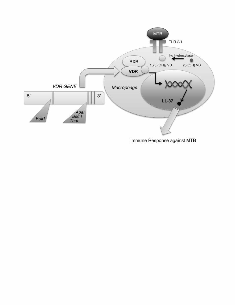
There is growing evidence that genes related to vitamin metabolism contribute to
susceptibility to TB. Specifically, vitamin D provides an exciting example of how
genetics may underlie risk of TB disease (Figure 1). The vitamin D receptor (VDR) is a
soluble nuclear receptor found in many immune cells, and is believed to play a role in
Acce
pted
Man
uscr
ipt
5
cytokine secretion patterns, maturation of dendritic cells, and effector and regulatory T
cell function [3]. Several VDR gene polymorphisms have been found that can impact TB
risk and outcomes, including BsmI, TaqI, and ApaI at the 3’ end of VDR, and FokI on
exon site 2 (Figure 1) [4]. For example, the TaqI Tt and ApaI AA genotype are associated
with improved response to therapy and faster time to sputum conversion in TB patients
[4]. On the other hand, TaqI tt, TaqI Bb, TaqI Ff, and BsmI bb have been associated with
an increased risk of TB [4]. Risk or protection may be influenced by ethnic background; a
recent meta–analysis was performed on a variety of populations and found that the FokI
ff genotype was most significant in the Asian population, while there was no effect in
Africans or South Americans [5]. Population differences may be related to a variety of
factors, including vitamin D status and HIV and TB prevalence rates.
Vitamin D itself is also essential for downstream genetic expression important for the
immune response against Mycobacterium tuberculosis. In the innate immune system,
activation of Toll–Like Receptor (TLR) 2/1 by Mycobacterium tuberculosis antigen
presentation leads to the expression of VDR and 1–α vitamin D hydroxylase [4]. The
hydroxylase converts 25(OH) vitamin D to its active form 1,25 (OH)2vitamin D, which
binds to VDR. They then form a heterodimer with Retinoid X Receptor (RXR), creating a
complex that translocates to the nucleus to regulate gene transcription (Figure 1). A key
protein formed is LL–37, a member of the cathelicidin family, known to have
antimicrobial effects against Mycobacterium tuberculosis while recruiting other immune
cells to the site of infection [4]. Other functions for vitamin D and VDR are to regulate
Acce
pted
Man
uscr
ipt
6
antigen presentation and processing, phagocytosis, and IL–1 β and TNF–α production
essential for the immune response [3].
Studies on vitamin D and VDR genetics illustrate a novel gene–environment model that
can help to stratify TB risk. It should be noted though that these studies were all
conducted in adult TB populations. Further research is necessary to investigate how these
polymorphisms influence the risk of TB in children.
Immune development and malnutrition
Defense against Mycobacterium tuberculosis requires a complex immune response that
involves both innate and adaptive immunity [6]. However, in newborns, it is important to
appreciate that cell-mediated immunity is incomplete and they depend mostly on innate
immunity and maternal antibodies [7]. Yet, even innate immunity is impaired; evidence
suggests that newborns have reduced function in antigen presenting cells (APCs),
neutrophils, and TLRs, and decreased blood complement levels [7]. Moreover, adaptive
immunity is thought to be skewed to a helper T cell (Th) 2 type response, potentially as a
way to reduce a pro-inflammatory reaction, decrease an allo-immune response against the
mother, and promote tolerance of harmless new antigens such as gut flora and food [7].
However, this also places them at considerable risk against intracellular organisms,
including TB, that depend on a Th1 response [6, 7]. Nutrition plays an essential role to
develop the appropriate innate and Th1 immune responses against TB [8].
Acce
pted
Man
uscr
ipt
7
The mucosal lining is an early site of defense against TB, where bacterial products trigger
TLR signaling pathways in dendritic cells and macrophages to release cytokines and
bactericidal defensins and cathelicidins [6, 9]. After birth, a neonate leaves the sterile
uterine environment to be rapidly exposed to foreign antigens, which has a large impact
on the formation of mucosal innate immunity. Furthermore, the colonization of
commensal flora is thought to compete with pathogenic bacteria while shaping TLR
responses in the child [7]. Growing evidence demonstrates early nutrition, in the form of
breast milk, has immune properties that modulate inflammation while promoting innate
immunity in the mucosa [7]. Breast milk contains a variety of important immune factors
including lysozyme, defensins, lactoferrin, soluble CD14, cytokines, complement, and
antiviral lipids [10]. Milk trigylcerides, when partially digested from lipases, become
monoglyceride and free fatty acid, which can be toxic to many pathogens [10]. Breast
milk is also a large source of glycans, which serves as substrate for fermentation and
colonization of commensal bacteria, and inhibit pathogen-binding to the mucosal surface
[10]. Studies have shown that limited or non–exclusive breastfeeding is associated with
an increased risk of respiratory infections [11-13]. A prospective study in Brazil showed
that neonates with acute viral bronchiolitis and a shorter length of exclusive breastfeeding
had worse clinical outcomes including increased use of oxygen and longer hospital stay
[12]. Therefore, maternal nutrition and her ability to confer protection have large
implications on the development of a functional innate immune system in the child.
While innate immunity is important against TB, adaptive immunity, in particular the Th1-
type response is critical against this intracellular bacterium [6]. The thymus plays an

Acce
pted
Man
uscr
ipt
8
important role in T lymphocyte maturation and is greatly affected by prenatal and early
child nutrition [8]. Studies have shown that low birth weight or being born in the
“hungry” season is associated with decreased thymus size [14, 15]. Echography
demonstrates thymic atrophy in malnourished children and is associated with higher
infant mortality due to infections [16]. Protein deficiency has especially been implicated;
children with protein energy malnutrition (PEM) have reduced thymic size, and tissue
samples demonstrate apoptosis of cortical thymocytes, microenvironment changes around
lymphoid tissue and epithelial cells, and a decrease in thymulin hormone production and
thymocyte proliferation [16, 17]. Inadequate zinc intake may also contribute to thymic
dysfunction; mice placed on a zinc deficient diet for 4 weeks only retain 25% of their
original thymus size [16]. In addition to its role in innate immunity, breast milk is also
associated with thymus size, correlated with the level of IL-7 in the milk [15]. Moreover,
glutamine is one of the most abundant proteins in breast milk, and supplementation in
early-weaned mice inoculated with Bacillus Calmette-Guérin (BCG) demonstrate
increased peripheral and lymph leukocyte and lymphocytes [18]. Thus, T cell maturation
is directly related to nutritional status essential for the TB response.
While our understanding of immune development grows, several questions still remain
regarding its role in the defense against TB. For example, despite immature immunity,
why do we see a robust Th1 type response to BCG vaccine? This suggests that children
are able to overcome this polarization towards Th2 responses through an unknown
mechanism [7]. Regardless of our limitations, however, we are able to note that breast
milk, protein and micronutrients have significant roles in the development of innate and
Acce
pted
Man
uscr
ipt
9
cell–mediated immunity, and that these factors are critical for the TB response. While
studies have not been conducted to determine how dysfunction in immune development
impacts risk of paediatric TB, this evidence supports that without adequate early
nutrition, appropriate immune development is greatly impaired and places the child at
considerable risk.
Nutrition and the Child with Respiratory Infections
Respiratory infections are among the largest contributors of morbidity and mortality in
children. Because of the high frequency, it provides an opportunity to study how
malnutrition impacts the outcomes and risk of diseases spread by respiratory droplets,
such as TB.
Malnutrition has been associated with increased risk of respiratory infections. A
prospective trial in Bangladeshi children found that being underweight increased the risk
of an upper respiratory infection by 13%, and being wasted increased it by 20% [19].
Moreover, a prospective 10-month study in nomadic Kenyan children found that wasting
predicted risk of acute respiratory infections in the wet season [20]. Malnutrition is also a
significant predictor of mortality in children with pneumonia, attributing to 52.3% of
pneumonia-related deaths [21].
Several mechanisms may underlie the increased risk and severity of respiratory infections
in malnourished children. A prospective study on neonates from the Netherlands found
that cord blood vitamin D levels predicted risk of RSV bronchiolitis in the first year of
Acce
pted
Man
uscr
ipt
10
life [22]. Zinc levels were also found to be significantly lower in Bangladeshi children
with a lower respiratory tract infection and PEM [23]. In addition, leptin deficiency has
been implicated; it is structurally similar to cytokines such as IL–6 and IL–11, and the
long isoform of the leptin receptor OB–Rb is similar to the cytokine receptor family
gp130 [24]. Leptin leads to the secretion of several cytokines, and animal models
demonstrate that elevated leptin during starvation prevents lymphoid tissue atrophy. An
ex–vivo study of T–lymphocytes from malnourished children in Mexico found that
incubation with leptin lead to decreases in IL–4 and IL–10 production, and increases in
IL–2 and IFN–γ, suggesting a shift to the Th1 response [24]. Thus, malnourished children
with respiratory infections may have deficits in cell–mediated immunity and the Th1 type
response, both critical for TB immunity.
Because respiratory infections are prevalent in children, a large body of evidence has
emerged on risk factors for infection and poor outcomes, including nutritional status.
While there are limitations in translating the risk of one pathogen to another, it has been
observed that poor nutrition is associated with severe deficits in immunity, both innate
and cell–mediated. Thus, until further research is conducted on childhood TB, we can
learn from past studies in children that suggest that malnutrition significantly worsens the
risk and severity of respiratory disease.
Nutrition and the Child with TB Disease
Although a third of the world is infected with tuberculosis, there is only a 10% lifetime
risk of progression to disease in HIV-uninfected individuals [9]. Malnutrition is thought
Acce
pted
Man
uscr
ipt
11
to contribute to this progression in children, through possible mechanisms as described
above. However, it is difficult to disentangle this process in vivo, for once the child has
active disease, the resulting inflammatory and immune response increases metabolic rate,
affects synthetic pathways (a so-called “anabolic block”), and impacts absorption,
distribution and excretion of nutrients, which altogether promotes malnutrition [25]. This
is supported with evidence that shows TB therapy significantly improves anthropometric
status and micronutrient levels [25, 26]. Thus, while cross sectional studies demonstrate
nutritional deficiencies in malnourished children with TB [27], they have a limited role in
describing the mechanisms underlying this relationship. Instead, it is more useful to
evaluate how the malnourished child develops an immune response against TB soon after
infection, and how any possible dysfunction may promote progression to active disease.
Consequently, we depend on experiments in which animals are exposed to a virulent
strain of TB, and studies in which malnourished children and animals are “infected” via
BCG vaccination.
Th-1 immunity against TB is impaired by malnutrition
Guinea pigs given a low protein diet and then exposed to Mycobacterium tuberculosis
have deficits in mounting an appropriate Th1–type cell–mediated response. This includes
decreased lymphocyte proliferation, higher IgG levels, and decreased cytokines such as
IL–2, TNF–α and IFN–γ [28]. In addition, there are increases in Fc–γ T cells and TGF–β,
considered to have a suppressive effect on function and T cell proliferation [28].
Consequently, these animals have evidence of worse disease, with higher bacillary load
in the lung and spleen [29]. Micronutrients deficiencies such as zinc and vitamin A are

Acce
pted
Man
uscr
ipt
12
also associated with greater bacterial load and worse lesions in the lung [30, 31]. More
recently, polyunsaturated fatty acids (PUFAs), in particular the anti-inflammatory omega
3 (n-3) fatty acids, have shown to decrease skin response, increase the bacterial load, and
reduce lymphocyte proliferation in TB-exposed guinea pigs [32]. There is mixed support
in mice studies; endogenous release of n-3 fatty acid from transgenic mice demonstrated
an increased bacterial load after TB inoculation, whereas exogenous supplementation
provided a level of protection against TB [33, 34]. Overall, though, we see that nutrition
has a profound effect on the Th-1 immune system’s ability to defend against TB soon
after infection, and thus predisposes the animal to disease progression.
Malnutrition and BCG Vaccination
Studies have shown that children who were vaccinated with BCG had significantly lower
tuberculin skin responses if they had severe protein deficiency [17, 35, 36]. While milder
forms of malnutrition may not have deficits in tuberculin response [37], a prospective
study among infants vaccinated at birth with BCG showed that mild or moderate
malnourished children still had a decrease in TB-associated cell-mediated immune
responses [38]. This is supported in animal studies, in which protein deficient animals
have a significantly impaired protection from BCG after TB exposure, as seen with
greater bacterial load in the lungs compared to nourished vaccinated animals [39]. This
deficit appears to be related to cell-mediated immunity, as it is associated with reduced
tuberculin skin reactivity and impaired IL-2, TNF-α and IFN-γ release from BCG
vaccinated, protein-deficient animals [28]. Re-nourishment of animals returns protection
Acce
pted
Man
uscr
ipt
13
similar to controls, suggesting that protein deficiency serves to affect the function, but not
acquisition, of the adaptive immune response against TB after BCG vaccination [29].
From in vivo studies of children and animals exposed to mycobacterium via inoculation
or vaccination, we see that a range of macro and micronutrients have direct effects on the
proper functioning of immune cells that would allow the child to either clear the infection
or drive it into a latent state. Consequently, this places children at risk for progression to
active disease and further worsening of their malnutrition.
Nutritional Supplementation as Adjuvant Therapy in Tuberculosis
Ultimately, we want to know if nutritional supplementation can improve immune
function and clinical outcomes in TB. Early ecological studies found that during times of
food restriction, such as war, TB morbidity rose significantly, and sharply declined after
food supplies returned [17]. However, clinical trials face large challenges, since TB
therapy will cause a rapid drop in bacillary load and improve nutritional status.
Consequently, this can overshadow any modest change after supplementation [40]. One
promising randomized trial among adults with TB in Indonesia found that
supplementation of zinc and vitamin A resulted in faster sputum conversion time and
resolution of lung lesions on chest X-ray [41]. However, more recently, the same group
was unable to repeat the results in a more malnourished population with a combined or
individual addition of vitamin A and zinc [42].
Acce
pted
Man
uscr
ipt
14
The few trials on nutritional supplementation for paediatric tuberculosis do not suggest a
significant benefit (Table 1) [43]. A study in Brazil showed that zinc supplementation at
the time of PPD placement in malnourished children increased the size of induration,
suggesting an improvement in cell–mediated immunity [44]. However, an in-vitro study
found that in HIV positive patients, zinc was unable to improve IFN-γ response or
increase lymphocyte levels after PPD stimulation [45]. Clinical trials have shown mixed
results. Hanekom et al. evaluated the response to vitamin A supplementation in 85 South
African children at baseline, six weeks and three months after initiation of TB therapy
[46]. Supplementation was not associated with a significant improvement in outcomes,
including weight change or improvement in respiratory symptoms. Morcos, et al.
conducted a small trial on vitamin D supplementation among children ages 1.5 to 13
years old, and noted clinical and radiographic improvement in the supplementation
group, but did not demonstrate differences in vitamin D levels or weight gain at the end
of therapy [47]. The most comprehensive trial was conducted recently by Mehta et al.,
among 255 children from Tanzania 6 weeks to 5 years of age with active TB [48]. The
children were randomized to receive a daily multivitamin or placebo for 8 weeks after
initiation of therapy. Overall, there was no difference in weight after 8 weeks, and there
was also no effect in terms of CD4, CD8 and CD3 T cell subsets.
In summary, there is insufficient evidence to support the use of macro or micronutrient
supplementation for children with active TB at this time. However, there are several
limitations in interpreting supplementation trials, including variable dose concentration,
adherence issues, and lack of complimenting food sources. In addition, while clinical
Acce
pted
Man
uscr
ipt
15
trials have been unable to demonstrate differences in micronutrient levels or nutritional
status at the conclusion of TB therapy, early in treatment individuals on supplements
have faster improvements in micronutrient levels and clinical indicators [46, 49]. A sub-
study of the Hanekom, et al., trial also found that vitamin A supplementation in children
may help in reducing soluble CD30 (sCD30) levels, suggesting a shift towards Th1 type
responses important against TB [50]. Thus, more comprehensive studies are required to
further elucidate how nutritional supplementation may benefit the risk and outcomes of
TB in children. Furthermore, while an early study in Harlem, New York, demonstrated
the preventative value of nutritional supplementation on TB prevalence among household
contacts, greater research is required on the role of nutrition on prevention of TB disease
[17, 40].
Conclusion
Studies on the role of nutrition on childhood tuberculosis are significantly limited. Yet,
we continue to classify malnourished children as high risk for TB, and support overall
nutritional supplementation for children with TB. However, the mechanisms underlying
the association between malnutrition and childhood TB remain unclear. Using the
available evidence, we suggest a model detailing the known information between
nutritional status, immune function, and risk of TB disease. However, it is equally
important to recognize the severe gaps in our knowledge (Table 2). We require greater
prospective studies that evaluate how nutritional status impacts the risk of TB, while
conducting further randomized controlled trials on the use of supplementation in TB
therapy. In resource–limited settings, TB in children is a major cause of morbidity and
Acce
pted
Man
uscr
ipt
16
mortality, and a large reservoir for continued transmission of infection. As our basic
understanding of the interaction between TB and malnutrition grows, it is important that
we seek to apply these advances to the welfare of this vulnerable population.
Footnote Page
Corresponding Author: Ezekiel Mupere, School of Medicine, College of Health Sciences,
Makerere University, Department of Paediatrics & Child Health, Clinical Research
Building, Ground Floor Room 9, Kampala, Uganda, Mobile Tel: +256 718 490 843, E–
mail: [email protected]
Alternate Corresponding Author: Devan Jaganath, David Geffen School of Medicine at
UCLA, 23615 Spires St. West Hills, CA 91304, Mobile Tel: +001 818 515 4313, E-mail:
Conflicts of Interest
The authors report no conflicts of interest.
Funding
This project has been funded in whole or in part by the Tuberculosis Research Unit
(TBRU), established with Federal funds from the United States National Institutes of
Allergy and Infectious Diseases & the United States National Institutes of Health and
Human Services, under Contract No. HHSN266200700022C / NO1-AI-70022
Acce
pted
Man
uscr
ipt
17
Acknowledgements
We would like to thank Dr. Robert Salata and Dr. Christina Lancioni for their critical
review of the article. We also greatly thank Dr. Henry Boom, Dr. Harriet Mayanja–Kizza,
Dr. Moses Joloba, the Makerere University– Case Western Reserve University Research
Collaboration and the Tuberculosis Research Unit for their guidance and support. The
authors report no conflicts of interest.
Authors’ Contributions
The authors contributed equally to the literature search and writing of the article. Tables
and figures were designed by DJ.

Acce
pted
Man
uscr
ipt
18
Figure Caption
Figure 1. The role of genetics in TB susceptibility. The Vitamin D Receptor (VDR) has
an integral role in the TB immune response through its binding with Vitamin D to induce
antimicrobial function via LL–37. Several polymorphisms in the VDR gene have also
been implicated in TB susceptibility. MTB = Mycobacterium tuberculosis, RXR=
Retinoid X Receptor, VD= Vitamin D, VDR=Vitamin D Receptor, TLR = Toll–like
Receptor
Acce
pted
Man
uscr
ipt
19
References
1. World Health Organization (WHO). Guidance for national tuberculosis
programmes on the management of tuberculosis in children. Geneva: World
Health Organization, 2006.
2. Black RE, Allen LH, Bhutta ZA, et al. Maternal and child undernutrition: global
and regional exposures and health consequences. Lancet. 2008; 371:243–60.
3. Walker VP, Modlin RL. The vitamin D connection to pediatric infections and
immune function. Pediatr Res. 2009; 65:106R–113R.
4. Luong K, Nguyen LT. Impact of vitamin D in the treatment of tuberculosis. Am J
Med Sci. 2011; 341:493–8.
5. Gao L, Tao Y, Zhang L, Jin Q. Vitamin D receptor genetic polymorphisms and
tuberculosis: updated systematic review and meta–analysis. Int J Tuberc Lung
Dis. 2010; 14:15–23.
6. Basu Roy R, Whittaker E, Kampmann B.Current understanding of the immune
response to tuberculosis in children. Curr Opin Infect Dis. 2012; 25:250-7.
7. Levy O.Innate immunity of the newborn: basic mechanisms and clinical
correlates. Nat Rev Immunol. 2007; 7:379-90.
8. Shennan DH, Kibel MA. Tuberculosis.In: Stanfield P, Brueton M, Chan M,
Parkin M, Waterston T, eds. Diseases of Children in the Subtropics and Tropics.
4th ed. London: Arnold, 1991:519-552.
9. Lawn SD, Zumla AI. Tuberculosis. Lancet. 2011; 378:57–72.
Acce
pted
Man
uscr
ipt
20
10. Newburg DS, Walker WA.Protection of the Neonate by the Innate Immune
System of Developing Gut and of Human Milk.Pediatr Res. 2007; 61:2-8.
11. Coles CL, Fraser D, Givon–Lavi N, et al. Nutritional status and diarrheal illness
as independent risk factors for alveolar pneumonia. Am J Epidemiol. 2005;
162:999–1007.
12. Dornelles CT, Piva JP, Marostica PJ. Nutritional status, breastfeeding, and
evolution of Infants with acute viral bronchiolitis. J Health Popul Nutr. 2007;
25:336–43.
13. Wayse V, Yousafzai A, Mogale K, Filteau S. Association of subclinical vitamin D
deficiency with severe acute lower respiratory infection in Indian children under 5
y. Eur J Clin Nutr. 2004; 58:563–7.
14. Moore SE, Prentice AM, Wagatsuma Y, et al.Early-life nutritional and
environmental determinants of thymic size in infants born in rural Bangladesh.
Acta Paediatr. 2009; 98:1168-75.
15. Ngom PT, Collinson AC, Pido-Lopez J, Henson SM, Prentice AM, Aspinall
R.Improved thymic function in exclusively breastfed infants is associated with
higher interleukin 7 concentrations in their mothers’ breast milk. Am J Clin Nutr.
2004; 80:722-8.
16. Savino W, Dardenne M, Velloso LA, Dayse Silva–Barbosa S. The thymus is a
common target in malnutrition and infection. Br J Nutr. 2007; 98:S11–6.
17. Scrimshaw NS, Taylor CE, Gordon JE.Interactions of nutrition and
infection.Monogr Ser World Health Organ. 1968;57:3-329.
Acce
pted
Man
uscr
ipt
21
18. Rogero MM, Tirapegui J, Vinolo MA, et al. Dietary Glutamine Supplementation
Increases the Activity of Peritoneal Macrophages and Hemopoiesis in Early-
Weaned Mice Inoculated with Mycobacterium bovis Bacillus Calmette-Guerin. J
Nutr. 2008; 138:1343-8.
19. Zaman K, Baqui AH, Yunus M, Sack RB, Chowdhury HR, Black RE.
Malnutrition, cell–mediated immune deficiency and acute upper respiratory
infections in rural Bangladeshi children. Acta Paediatr. 1997; 86:923–7.
20. Shell–Duncan B, Wood JW. The evaluation of delayed–type hypersensitivity
responsiveness and nutritional status as predictors of gastro–intestinal and acute
respiratory infection: a prospective field study among traditional nomadic Kenyan
children. J Trop Pediatr. 1997; 43:25–32.
21. Caulfield LE, de Onis M, Blössner M, Black RE. Undernutrition as an underlying
cause of child deaths associated with diarrhea, pneumonia, malaria, and measles.
Am J Clin Nutr. 2004; 80:193–8.
22. Belderbos ME, Houben ML, Wilbrink B, et al. Cord blood vitamin D deficiency
is associated with respiratory syncytial virus bronchiolitis. Pediatrics. 2011;
127:e1513–20.
23. Shakur MS, Malek MA, Bano N, Rahman M, Ahmed M. Serum and hair zinc in
severely malnourished Bangladeshi children associated with or without acute
lower respiratory infection. Indian J Pediatr. 2009; 76:609–14.
24. Rodríguez L, Graniel J, Ortiz R. Effect of leptin on activation and cytokine
synthesis in peripheral blood lymphocytes of malnourished infected children. Clin
Exp Immunol. 2007; 148:478–85.
Acce
pted
Man
uscr
ipt
22
25. Macallan DC. Malnutrition in Tuberculosis. Diagn Microbiol Infect Dis. 1999;
34:153-7.
26. Ramachandran G, Santha T, Garg R, et al.Vitamin A levels in sputum-positive
pulmonary tuberculosis patients in comparison with household contacts and
healthy ‘normals’. Int J Tuberc Lung Dis. 2004; 8:1130-3.
27. Williams B, Williams AJ, Anderson ST. Vitamin D deficiency and insufficiency
in children with tuberculosis. Pediatr Infect Dis J. 2008; 27:941–2.
28. Cegielski JP, McMurray DN. The relationship between malnutrition and
tuberculosis: evidence from studies in humans and experimental animals. Int J
Tuberc Lung Dis. 2004; 8:286–98.
29. McMurray DN, Mintzer CL, Tetzlaff CL, Carlomagno MA. The influence of
dietary protein on the protective effect of BCG in guinea pigs. Tubercle. 1986;
67:31–9.
30. Gopalan C. Importance of nutritional factors in tuberculosis. Indian J
Tuberculosis. 1957; 4:105–126.
31. McMurray DN, Carlomagno MA, Cumberland PA. Respiratory infection with
attenuated Mycobacterium tuberculosis H37Ra in malnourished guinea pigs.
Infect Immun. 1983; 39:793–9.
32. McFarland CT, Fan YY, Chapkin RS, Weeks BR, McMurray DN. Dietary
polyunsaturated fatty acids modulate resistance to Mycobacterium tuberculosis in
guinea pigs. J Nutr. 2008; 138:2123–8.
Acce
pted
Man
uscr
ipt
23
33. Bonilla DL, Fan YY, Chapkin RS, McMurray DN.Transgenic Mice Enriched in
Omega-3 Fatty Acids Are More Susceptible to Pulmonary Tuberculosis: Impaired
Resistance to Tuberculosis in fat-1 Mice. J Infect Dis. 2010;201:399-408.
34. Jordao L, Lengeling A, Bordat Y, et al.Effects of omega-3 and -6 fatty acids on
Mycobacterium tuberculosis in macrophages and in mice. Microbes Infect.
2008;10:1379-86.
35. Satyanarayana K, Bhaskaram P, Seshu VC, Reddy V. Influence of nutrition on
postvaccinial tuberculin sensitivity. Am J Clin Nutr. 1980; 33: 2334–7.
36. Harland PS, Brown RE. Tuberculin Sensitivity Following B.C.G. Vaccination In
Undernourished Children. East Afr Med J. 1965; 42:233-8.
37. Chadha VK, Jitendra R, Kumar P, Gupta J, Umadevi. Relationship of nutritional
status with tuberculin sensitivity. Indian J Pediatr. 2009; 76:605–7.
38. McMurray DN, Loomis SA, Casazza LJ, Rey H, Miranda R.Development of
impaired cell-mediated immunity in mild and moderate malnutrition.Am J Clin
Nutr. 1981;34:68-77.
39. McMurray DN, Carlomagno MA, Mintzer CL, Tetzlaff CL. Mycobacterium bovis
BCG vaccine fails to protect protein-deficient guinea pigs against respiratory
challenge with virulent Mycobacterium tuberculosis. Infect Immun. 1985;50:555-
9.
40. McMurray DN, Cegielski JP. The influence of nutrition on the risk and outcomes
of tuberculosis. In: Academy of Sciences of South Africa Consensus Panel on
Nutrition, HIV/AIDS, eds. HIV/AIDS, TB, and Nutrition: Scientific inquiry into
the nutritional influences on human immunity with special reference to HIV
Acce
pted
Man
uscr
ipt
24
infection and active TB in South Africa. Pretoria, South Africa: Academy of
Sciences of South Africa, 2007:153-169
41. Karyadi E, West CE, Schultink W, et al.A double-blind, placebo-controlled study
of vitamin A and zinc supplementation in persons with tuberculosis in Indonesia:
effects on clinical response and nutritional status. Am J Clin Nutr. 2002; 75:720-
7.
42. Pakasi TA, Karyadi E, Suratih NM, et al.Zinc and vitamin A supplementation
fails to reduce sputum conversion time in severely malnourished pulmonary
tuberculosis patients in Indonesia. Nutr J. 2010;9:41.
43. Sinclair D, Abba K, Grobler L, Sudarsanam TD. Nutritional supplements for
people being treated for active tuberculosis. Cochrane Database Syst Rev. 2011;
11:CD006086.
44. Cuevas LE, Almeida LM, Mazunder P, et al. Effect of Zinc on the tuberculin
response of children exposed to adults with smear–positive tuberculosis. Ann
Trop Paediatr. 2002; 22:313–9.
45. Green JA, Lewin SR, Wightman F, Lee M, Ravindran TS, Paton NI.A
randomised controlled trial of oral zinc on the immune response to tuberculosis in
HIV-infected patients. Int J Tuberc Lung Dis. 2005; 9:1378-84.
46. Hanekom WA, Potgieter S, Hughes EJ, Malan H, Kessow G, Hussey GD.
Vitamin A status and therapy in childhood pulmonary tuberculosis. J Pediatr.
1997; 131: 925–7.
47. Morcos MM, Gabr AA, Samuel S, et al. Vitamin D administration to tuberculosis
children and its value. Boll Chim Farm. 1998; 137:157–64.
Acce
pted
Man
uscr
ipt
25
48. Mehta S, Mugusi FM, Bosch RJ, et al. A randomized trial of multivitamin
supplementation in children with tuberculosis in Tanzania. Nutr J. 2011;10:120.
49. Das BS, Devi U, Mohan Rao C, Srivastava VK, Rath PK, Das BS.Effect of iron
supplementation on mild to moderate anaemia in pulmonary tuberculosis. Br J
Nutr. 2003; 90:541-50.
50. Hanekom WA, Hussey GD, Hughes EJ, Potgieter S, Yogev R, Check IJ.Plasma-
Soluble CD30 in Childhood Tuberculosis: Effects of Disease Severity, Nutritional
Status, and Vitamin A Therapy. Clin Diagn Lab Immunol. 1999; 6:204-8.
Acce
pted
Man
uscr
ipt
26
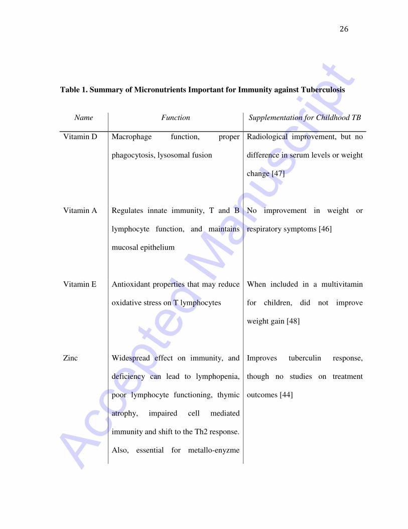
Table 1. Summary of Micronutrients Important for Immunity against Tuberculosis
Name Function Supplementation for Childhood TB
Vitamin D Macrophage function, proper
phagocytosis, lysosomal fusion
Radiological improvement, but no
difference in serum levels or weight
change [47]
Vitamin A Regulates innate immunity, T and B
lymphocyte function, and maintains
mucosal epithelium
No improvement in weight or
respiratory symptoms [46]
Vitamin E Antioxidant properties that may reduce
oxidative stress on T lymphocytes
When included in a multivitamin
for children, did not improve
weight gain [48]
Zinc Widespread effect on immunity, and
deficiency can lead to lymphopenia,
poor lymphocyte functioning, thymic
atrophy, impaired cell mediated
immunity and shift to the Th2 response.
Also, essential for metallo-enyzme
Improves tuberculin response,
though no studies on treatment
outcomes [44]

Acce
pted
Man
uscr
ipt
27
formation and creation of free radicals
Selenium
Cell and humoral immunity, utilized in
creation of metallo-enzymes
No known studies in children
Iron
Innate immunity such as neutrophil and
NK function, T cell maturation, and
deficiency can result in shift towards
Th2 response
No known studies in children
Acce
pted
Man
uscr
ipt
28
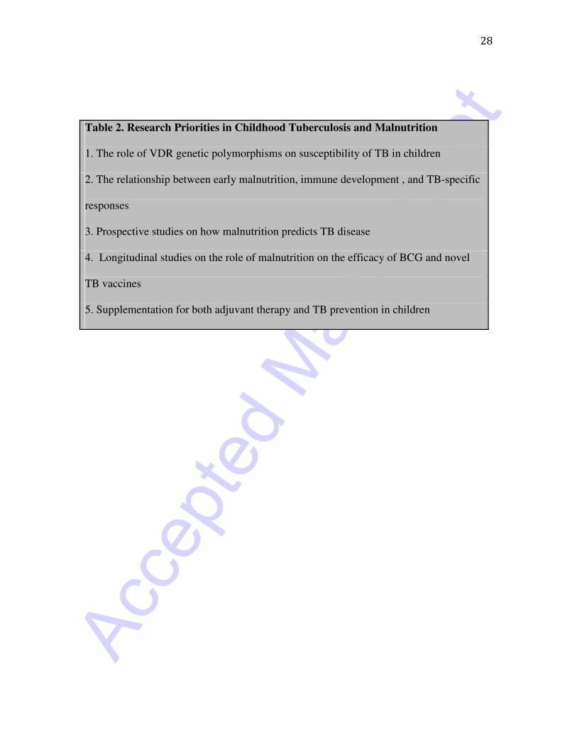
Table 2. Research Priorities in Childhood Tuberculosis and Malnutrition
1. The role of VDR genetic polymorphisms on susceptibility of TB in children
2. The relationship between early malnutrition, immune development , and TB-specific
responses
3. Prospective studies on how malnutrition predicts TB disease
4. Longitudinal studies on the role of malnutrition on the efficacy of BCG and novel
TB vaccines
5. Supplementation for both adjuvant therapy and TB prevention in children
!
RXR
MTB
25 (OH) VD 1,25 (OH)2 VD
1-α hydroxylase
LL-37
Immune Response against MTB
3’
FokI
VDR GENE
5’
ApaI BsmI
TaqI
TLR 2/1
Macrophage