Characterizing cancer cells with cancer stem cell - BioMed Central
12
RESEARCH Open Access Characterizing cancer cells with cancer stem cell-like features in 293T human embryonic kidney cells Bisrat G Debeb 1 , Xiaomei Zhang 2 , Savitri Krishnamurthy 3 , Hui Gao 4 , Evan Cohen 4 , Li Li 1 , Angel A Rodriguez 2 , Melissa D Landis 2 , Anthony Lucci 5 , Naoto T Ueno 6 , Fredika Robertson 7 , Wei Xu 1 , Lara Lacerda 1 , Thomas A Buchholz 1 , Massimo Cristofanilli 6 , James M Reuben 4 , Michael T Lewis 2 , Wendy A Woodward 1* Abstract Background: Since the first suggestion of prospectively identifiable cancer stem cells in solid tumors, efforts have been made to characterize reported cancer stem cell surrogates in existing cancer cell lines, and cell lines rich with these surrogates have been used to screen for cancer stem cell targeted agents. Although 293T cells were derived from human embryonic kidney, transplantation of these cells into the mammary fat pad yields aggressive tumors that self-renew as evidenced by serial xenograft passages through transplantation. Herein we fully characterize cancer stem cell-like features in 293T human embryonic kidney cells. Results: 293T cells can be readily cultured and passaged as spheres in serum-free stem cell promoting culture conditions. Cells cultured in vitro as three-dimensional spheres (3D) were shown to contain higher ALDH1 and CD44+/CD24- population compared to monolayer cells. These cells were also resistant to radiation and upregulate stem cell survival signaling including b-catenin, Notch1 and Survivin in response to radiation. Moreover, 3D spheres generated from the 293T cells have increased expression of mesenchymal genes including vimentin, n-cadherin, zeb1, snail and slug as well as pro-metastatic genes RhoC, Tenascin C and MTA1. In addition, microRNAs implicated in self-renewal and metastases were markedly reduced in 3D spheres. Conclusions: 293T cells exhibit a cancer stem cell-like phenotype when cultured as 3D spheres and represent an important research tool for studying the molecular and biological mechanisms of cancer stem cells and for testing and developing novel targets for cancer therapy. Background The cancer stem cell theory proposes that a subpopula- tion of cells, the cancer stem cells, exist in solid tumors as well as cancers of hematopoietic origin and constitute a reservoir of self-sustaining cells with the exclusive ability to self-renew and maintain the tumor. These can- cer stem cells have the capacity to both divide and expand the cancer stem cell pool and to differentiate into the heterogeneous non-tumorigenic cancer cell types that constitute the bulk of the cancer cells within the tumor [1]. Cancer stem cells have been proposed to play a role in tumorigenesis and metastasis [2-4] as well as in resistance to radiation and chemotherapy [5-9]. Thus, it may be necessary to target and eliminate these cells to eradicate cancers. Cancer stem cells have been identified in a mounting number of human malignancies. Using approaches developed in the hematopoietic malignancies, Clarke and colleagues demonstrated the existence of a subpo- pulation of tumorigenic cells (or tumor-initiating cells), isolated from breast cancer pleural effusions by limiting dilution transplantation of CD44+/CD24-/lineage- cells into the mammary fat pad of immunocompromised mice [10]..Importantly, Dontu et al. adapted a sphere culture system described in the central nervous system [11] by growing primary breast epithelial cells in serum- free medium containing EGF and FGF2 in suspension culture and demonstrated enrichment of the tumor- * Correspondence: [email protected] 1 Department of Radiation Oncology, The University of Texas MD Anderson Cancer Center, Houston, TX, USA Debeb et al. Molecular Cancer 2010, 9:180 http://www.molecular-cancer.com/content/9/1/180 © 2010 Debeb et al; licensee BioMed Central Ltd. This is an Open Access article distributed under the terms of the Creative Commons Attribution License (http://creativecommons.org/licenses/by/2.0), which permits unrestricted use, distribution, and reproduction in any medium, provided the original work is properly cited.
Transcript of Characterizing cancer cells with cancer stem cell - BioMed Central
RESEARCH Open Access
Characterizing cancer cells with cancer stemcell-like features in 293T human embryonickidney cellsBisrat G Debeb1, Xiaomei Zhang2, Savitri Krishnamurthy3, Hui Gao4, Evan Cohen4, Li Li1, Angel A Rodriguez2,Melissa D Landis2, Anthony Lucci5, Naoto T Ueno6, Fredika Robertson7, Wei Xu1, Lara Lacerda1,Thomas A Buchholz1, Massimo Cristofanilli6, James M Reuben4, Michael T Lewis2, Wendy A Woodward1*
Abstract
Background: Since the first suggestion of prospectively identifiable cancer stem cells in solid tumors, efforts havebeen made to characterize reported cancer stem cell surrogates in existing cancer cell lines, and cell lines rich withthese surrogates have been used to screen for cancer stem cell targeted agents. Although 293T cells were derivedfrom human embryonic kidney, transplantation of these cells into the mammary fat pad yields aggressive tumorsthat self-renew as evidenced by serial xenograft passages through transplantation. Herein we fully characterizecancer stem cell-like features in 293T human embryonic kidney cells.
Results: 293T cells can be readily cultured and passaged as spheres in serum-free stem cell promoting cultureconditions. Cells cultured in vitro as three-dimensional spheres (3D) were shown to contain higher ALDH1 andCD44+/CD24- population compared to monolayer cells. These cells were also resistant to radiation and upregulatestem cell survival signaling including b-catenin, Notch1 and Survivin in response to radiation. Moreover, 3D spheresgenerated from the 293T cells have increased expression of mesenchymal genes including vimentin, n-cadherin,zeb1, snail and slug as well as pro-metastatic genes RhoC, Tenascin C and MTA1. In addition, microRNAs implicatedin self-renewal and metastases were markedly reduced in 3D spheres.
Conclusions: 293T cells exhibit a cancer stem cell-like phenotype when cultured as 3D spheres and represent animportant research tool for studying the molecular and biological mechanisms of cancer stem cells and for testingand developing novel targets for cancer therapy.
BackgroundThe cancer stem cell theory proposes that a subpopula-tion of cells, the cancer stem cells, exist in solid tumorsas well as cancers of hematopoietic origin and constitutea reservoir of self-sustaining cells with the exclusiveability to self-renew and maintain the tumor. These can-cer stem cells have the capacity to both divide andexpand the cancer stem cell pool and to differentiateinto the heterogeneous non-tumorigenic cancer celltypes that constitute the bulk of the cancer cells withinthe tumor [1]. Cancer stem cells have been proposed toplay a role in tumorigenesis and metastasis [2-4] as well
as in resistance to radiation and chemotherapy [5-9].Thus, it may be necessary to target and eliminate thesecells to eradicate cancers.Cancer stem cells have been identified in a mounting
number of human malignancies. Using approachesdeveloped in the hematopoietic malignancies, Clarkeand colleagues demonstrated the existence of a subpo-pulation of tumorigenic cells (or tumor-initiating cells),isolated from breast cancer pleural effusions by limitingdilution transplantation of CD44+/CD24-/lineage- cellsinto the mammary fat pad of immunocompromisedmice [10]..Importantly, Dontu et al. adapted a sphereculture system described in the central nervous system[11] by growing primary breast epithelial cells in serum-free medium containing EGF and FGF2 in suspensionculture and demonstrated enrichment of the tumor-
* Correspondence: [email protected] of Radiation Oncology, The University of Texas MD AndersonCancer Center, Houston, TX, USA
Debeb et al. Molecular Cancer 2010, 9:180http://www.molecular-cancer.com/content/9/1/180
© 2010 Debeb et al; licensee BioMed Central Ltd. This is an Open Access article distributed under the terms of the Creative CommonsAttribution License (http://creativecommons.org/licenses/by/2.0), which permits unrestricted use, distribution, and reproduction inany medium, provided the original work is properly cited.

initiating population. This enriched population is suita-ble to demonstrate in vitro self-renewal and multi-line-age differentiation potential [12]. This assay has beenadapted to study cancer stem/progenitor cells in breastcancer cell lines as well as in colon, brain and pancreaticcancer tissues [11,13-18]. Another marker of the cancerstem cell population was recently established by Gines-tier et al. who showed that aldehyde dehydrogenase 1(ALDH1)-positive cells from human breast tumors werethe tumorigenic cancer-initiating population [19]. HighALDH activity has also been used to define cancer stemcell populations in other solid tumors including colon,liver and pancreatic cancers [20-22]. Numerous studiessuggest that cell lines can be useful in vitro tools tostudy these populations, and at this time the percentageof cancer stem cell markers has been characterized innumerous cancer cell lines [15,17]. These efforts haveled to the use of appropriately selected cell lines rich inthe surrogate phenotypes to screen for agents that targetthese cancer stem cell surrogates [23]; however thenumber of lines expressing each of the surrogatesreported thus far is few.We observed several key biologic features of pur-
ported cancer stem cells in human embryonic kidneycells 293T (also designated HEK 293T), a widely usedcell line derived through the sheared Adenovirus 5(Ad5) DNA transformation [24]. Most importantly,tumors from these cells self-renew as evidenced by serialxenograft passage through transplantation into thecleared mammary fat pad continued over eight genera-tions. Guan et al previously showed that primary HEK239T cells form tumors that could be inhibited byRbAP46 [25], and Shen et al. demonstrated that tumorforming efficiency had increased with increasing passageof these cells [26], but serial self-renewal has not beenreported. We hypothesized that these embryonic,tumorigenic cells may be useful to study cancer stemcells and add to the arsenal of cell lines with significantcancer stem cell surrogate representation to facilitatethe study of targeted agents against this population.Here we focused on the cancer stem cell surrogatesdescribed in breast tumor models and transplantedtumor cells into the cleared mammary fatpads to exam-ine in vivo tumor pathology. We report that 293T cellreadily form spheres that are enriched with cancer stemcell surrogates ALDH1 and CD44+/CD24- cells andexpress proteins associated with the epithelial-mesench-ymal transition and stem cell developmental pathways.They are also depleted in the microRNA that has beenimplicated in cancer stem cell self-renewal. We proposethat this is a valuable cell line to study cancer stem cellsurrogates identified in human cancers as well as theplasticity of transformed embryonic cells.
Results293T cells form spheres in stem cell promoting culturethat are enriched in cancer stem cell markersWhen 293T cells were transplanted into the cleared fatpads of 3 week old SCID/Beige mice, all (10/10) glandsdeveloped tumors at 14 days. These tumors were seriallypassaged through six transplant generations and arephenotypically stable. Hemotoxylin and eosin stainingwas performed and examined by a clinical breast pathol-ogist. Histology was indistinguishable from high gradeadenocarcinoma of the breast (although the histologicalappearance of high grade poorly differentiated tumor isnot specific only to breast tumors). All tumors hadevident tumor vasculature and expressed VEGF(Figure 1A). Necropsy was performed on all mice at thetime of sacrifice and no metastatic disease was identifiedin solid organs. Bone marrow aspirates from generationtwo mice were negative for malignant cells. Tumorswere negative for the epithelial markers e-cadherin andcytokeratin 19 while they were positive for vimentin andb-catenin (Figure 1B), features associated with poorlydifferentiated tumors.Given the in vivo observation that 293T tumors seri-
ally transplant and self-renew, we characterized the cellline for its cancer stem cell properties. 293T cells seededin the self-renewal promoting suspension culture condi-tions formed round spheres of varying sizes and thesespheres can be passaged to give a higher proportion ofsecondary spheres (Figure 2A, B). To characterizefurther the stem cell component of the spheres, flowcytometry was used to measure cells bearing the tumor-initiating phenotype CD44+/CD24- and ALDH activity.As shown in Figures 2C and 2D, CD44+/CD24- cellsrepresent an average of 4% ± 2% cells in 2D cultureand 22% ± 10% cells in 3D culture (5-fold increase,P = 0.003) and 3D spheres showed a 7-fold ALDH activ-ity compared to 2D cells (6% vs. 43%). Further, tumorsfrom 293T were dissociated into single cells andassessed for cancer stem cell surrogates using fresh cellsor cells propagated under sphere forming conditions.CD44+/CD24- cells comprised an average of 0.25%(N = 2) from primary tumors and 16.4% (N = 4) fromtumor derived sphere culture, a 65-fold increase. ALDHincreased comparably from 0.2% in the primary tumorto an average 17.5%. Individual tumor data are summar-ized in table 1.Using RT-PCR to examine the expression of embryo-
nic stem cell gene mRNAs in cells derived from spheres(3D) and compare them to cells maintained as adherentmonolayer culture we found 3D spheres selectivelyexpressed embryonic stem cell transcription factorsOct4, Nanog, Sox2 and Klf4 (Figure 3A) genes whoseexpression was associated with reprogramming of adult
Debeb et al. Molecular Cancer 2010, 9:180http://www.molecular-cancer.com/content/9/1/180
Page 2 of 12

fibroblasts into induced pluripotent stem cells [27-29].We also examined stem cell signaling and downstreameffectors in 2D versus 3D culture as a surrogate compar-ison of differentiated versus progenitor enriched culture.b-catenin, survivin and Rex-1 were upregulated in 3Dculture (Figure 3B) in protein lysates obtained from293T cells cultured as spheres vs. monolayer cells. Rex-1 is a target gene of the stemness marker Oct4 and thusits expression in the 3D spheres could be a functionalreadout of Oct4 activity. Moreover, the self-renewal fea-ture of the 293T spheres was assessed using the labelretaining fluorescent membrane dye, PKH-26. Self-renewal in sphere culture is expected to generate bothheterogeneous spheres containing self-renewing cellsand committed lineage specific spheres derived fromself-renewing cells. In spheres from 293T cells usingPKH-26 label retention as a marker for quiescent cells,spheres were identified after 4 passages that retain a sin-gle label-retaining cell (Figure 3C). Furthermore, immu-nostaining of 293T cells showed that the majority ofcells stain with the basal cytokeratin K14 while few cellsstain for the luminal marker K18 (Figure 3D) suggestingthat 293T cells have early differentiating cellcharacteristics.
Spheres generated from 293T cells are resistant totherapyFollowing the observation that 293T cells from thesphere culture are enriched in cancer stem cell markers,we examined differences in resistance to radiation- or
chemo-therapy between 2D and 3D cultures. Using the3D mammosphere/2D clonogenic assay method that hasbeen recently demonstrated by our lab [16], we foundthat cells in 3D culture are significantly more resistantto radiation than the same cells grown in 2D culture(Figure 4A) and second generation spheres are moreresistant than primary spheres (data not shown). More-over, b-catenin, Notch and survivin were all upregulatedin 3D culture cells 48 hours after 4 Gy of irradiationcompared to the unirradiated cells while Numb, a nega-tive regulator of Notch, is downregulated with radiation(Figure 4B). Similarly Retinoic acid, a compound thathas been used as a cancer chemotherapeutic agent, sig-nificantly reduced the number of colonies in monolayerculture but did not decrease sphere formation (Figure4C) indicating that the 3D cells can be resistant to che-motherapeutic agents.
Epithelial-Mesenchymal Transition (EMT) and pro-metastatic genes are upregulated in 3D spheres of293T cellsIn view of the recent findings that EMT increases thestem cell population in normal and malignant mammaryepithelial cells as well as in pancreatic and colorectalcancer cells [13,18], we investigated EMT gene expres-sion patterns by western blot in the 2D versus 3D cul-ture as a surrogate comparison of differentiated versusstem/progenitor enriched culture. Figure 5A shows thatthe mesenchymal gene markers n-cadherin, vimentinand zeb1 were upregulated in 293T cells cultured in
Figure 1 Transplantation of 293T cells into the cleared mammary fat pad of immunocompromised mice yields serially transplantingtumors. A) Skin overlying grossly involved tumor is necrotic and abnormal and tumors demonstrate gross tumor vasculature and are diffuselypositive for vascular endothelial growth factor (VEGF). B) By immunohistochemical analysis tumor xenografts are strongly vimentin positive,E-cadherin negative and beta-catenin positive at the membrane while K19, an epithelial marker is completely negative.
Debeb et al. Molecular Cancer 2010, 9:180http://www.molecular-cancer.com/content/9/1/180
Page 3 of 12

serum-free sphere promoting conditions vs. the standard2D culture conditions. Snail and slug, other EMT genes,were also upregulated in the 3D culture compared tothe 2D with RT-PCR (Figure 5B).We also assessed differences in 2D vs. 3D for the
expression pattern of genes implicated in promoting
invasion, migration and metastasis of cancer cells. RhoCis upregulated in 3D spheres of 293T cells as comparedto monolayer cells (Figure 5C). RhoC has been identifiedas an important player in metastasis [30,31] and itsexpression correlates with metastatic spread of varioustypes of carcinomas [32,33]. Similarly, Tenascin C and
Figure 2 293T cells form spheres in suspension culture that exhibit cancer stem cell phenotype. A, B) Sphere formation in 293T cellsseeded in self renewal promoting suspension culture conditions. Passage of 293T spheres gives rise to higher proportion of secondary spheres.C) Flow cytometric analysis of expression of CD24 and CD44 in 293T cells. Cells were cultured under adherent (2D) or tumor-initiating cellsenrichment condition (3D). There is an increase of CD44+/CD24- population in cells cultured in 3D vs. 2D culture conditions. D) Flow cytometricanalysis of ALDH activity showed that the 3D culture condition enriches for ALDH-positive cells compared to the 2D culture of 293T cells.
Table 1 Characterizing breast cancer stem cell surrogates in spheres derived from 293T tumors
Tumor ID CD44+/CD24- CD44-/CD24+ CD44+/CD24+ CD44-/CD24- ALDH+/-DEAB
293T 0.4 0.2 0 98.6 0.2
293T 0.1 37.4 0 52.6 0.2
293T Sphere -1 5.5 2.3 0 92.4 24.6
293T Sphere -2 17.4 1 0 80.2 27.7
293T Sphere -3 24.2 0.2 0 72.1 12.1
293T Sphere -4 18.3 3.2 0.1 78.5 5.6
Debeb et al. Molecular Cancer 2010, 9:180http://www.molecular-cancer.com/content/9/1/180
Page 4 of 12

MTA1 which are also associated with invasion andmigration of cancer cells [34-36] were upregulated in3D vs. 2D 293T cells (Figure 5C).
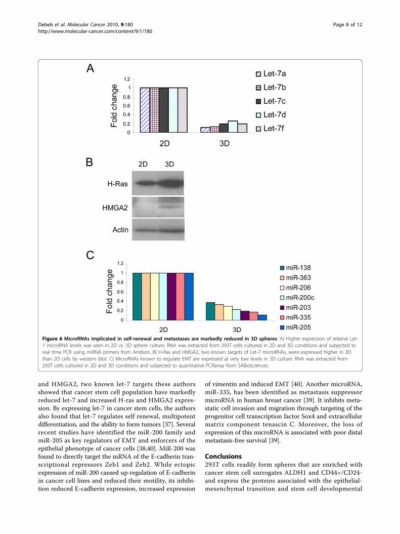
MicroRNAs implicated in self renewal and metastasis aremarkedly reduced in 3D spheresFollowing the findings that 3D spheres from 293T cellsexhibit the cancer stem cell phenotype as well as expressEMT and pro-metastatic genes, we examined for differ-ences in the expression of microRNAs that are associatedwith self-renewal and metastases. It has been demonstratedthat the let-7 microRNA family regulates the self-renewal ofbreast cancer stem cells [37]. In line with these findings, wefound that Let-7 microRNAs are highly reduced in 3D stemcell promoting cultures in 293T cells compared to the 2Dmonolayer cells (Figure 6A) while two known Let-7 targetgenes, H-Ras and HMGA2, were upregulated in 3D vs. 2D(Figure 6B). Likewise, the microRNAs miR-200c, miR-205,miR-206 and miR-335 previously reported to be involved ininvasion and metastasis [38-40] are reduced in the 3D stemcell promoting cultures in 293T cells. Moreover, miR-138,miR-203 and miR-363 previously uncharacterized in metas-tases are also downregulated in 3D vs. 2D cells (Figure 6C).Table 2 summarizes the miRNAs downregulated in 3D cul-ture of 293T cells and the validation of predicted genes bywestern blot or RT-PCR.
DiscussionWe propose 293T is a valuable tool to study cancerstem/progenitor features in that it rapidly generatesboth spheres and tumors that are readily serially pas-saged, and maintains significant populations of thetumor-initiating phenotypes described in the cancerstem cell literature. In this study, we observed thattransplanting 293T cells into the cleared mammary fatpads of immunocompromised mice resulted in seriallytransplantable rapidly growing tumors. Propagation ofspheres from tumor tissue or directly from the cell lineenriched for ALDH activity and CD44+/CD24- markersand expression of EMT markers as well as genes thatpromote stem cell survival. Papers characterizing similarfeatures in existing cancer cell lines have demonstrateda wide range in each of the purported cancer stem cellpopulations in these lines making the lines available toscreen simultaneously against each surrogate limited.The 293T cell line was developed through transforma-
tion of human embryonic kidney cells using the shearedAdenovirus 5 (Ad5) DNA [24]. It expresses genes per-mitting the replication of E1-deficient, non-replicatingadeno and adeno-associated viral vectors leading totheir common use of generating viral vectors for labora-tory and gene therapy use. From the initial report in1977, the transformation was surprisingly inefficient.
Figure 3 Spheres from 293T cells express embryonic transcription factors implicated in self-renewal. A) RT-PCR demonstrated higherexpression of embryonic stem cell specific genes including Oct4, Nanog, Sox2 and Klf4 in 3D spheres of 293T cells but no expression wasobserved in 2D monolayer cells. B) Genes implicated in promoting survival of cancer stem cells such as b-catenin, survivin and Rex-1 wereupregulated in 3D spheres vs. 2D monolayer cells. C) Serial passage for more than 4 weeks without additional PKH-26 label (Red) reveals serialdilution to a single long term label-retaining cell per sphere suggestive of asymmetric self-renewal. D) Immunocytochemistry reveals the majorityof 293T cells as keratin 14 positive.
Debeb et al. Molecular Cancer 2010, 9:180http://www.molecular-cancer.com/content/9/1/180
Page 5 of 12

Only two of eight transformations were successful withonly one of twenty cultures forming a single colony ineach case. Attempts to isolate these were not successful,however and a single transformed colony was observedin one dish not discarded 75 days after the originalexperiment. Partial serum starvation (2%) was used toselect for the transformed phenotype and eventually theisolated culture was expanded. The culture was reportedto contain cells of several morphologies of generallyepitheliod appearing cells. Injection of 2 × 10⁷ cells sub-cutaneously into nude mice yielded no tumors after6-8 weeks. Re-injection at that time resulted in tumors15-20 weeks after the first injection in 3/20 animals. Instark contrast, Shen et al [26] report that in cells pas-saged greater than 65 times, the tumorgenicity increasesto 100% with tumors formed in two weeks. We havereproduced these findings with 100% tumor formationwhen injected into the cleared mammary fat pads ofimmunocompromised mice and further report thatthese tumors are serially transplantable with similarly
aggressive biology and resembled poorly differentiated,high grade malignant primary tumor on histology. It’snoteworthy that there is controversy on the origins ofHEK 293T cells as it has been assumed that they weregenerated by transformation of either a fibroblastic,endothelial or epithelial cell all of which are abundantin kidney. However, the fact that the cells originatedfrom cultured kidney cells does not say much about theexact cellular origin of the HEK 293T, as embryonickidney cultures may contain small numbers of almostall cell types of the body. In fact, a study by Grahamand colleagues provided evidence that these cells andseveral other human cell lines generated by adenovirustransformation of human embryonic kidney cells havemany neuronal progenitor properties, suggesting thatthe adenovirus was taken up and transformed a neuro-nal lineage cell in the original kidney culture [41]. Thus,the spheres generated from 293T cells under stem-cellpromoting culture condition may have features observedin neurospheres.
Figure 4 Spheres generated from 293T cells are resistant to therapy. A) Clonogenic/mammosphere formation assay of 293T cells culturedunder 2D or 3D promoting culture conditions. Cells in 3D are more resistant to radiation than cells grown in 2D. Cells were grown in 2D or 3Dculture conditions and irradiated with single, increasing doses (0, 2, 4, 6 Gy) of radiation. B) Upregulation of genes implicated in radioresistanceincluding b-catenin, Notch and Survivin in 3D cells 48 hours after 4 Gy of irradiation. C) 3D cultured cells are also more resistant tochemotherapeutic drugs like retinoic acid compared to 2D. Similar results were obtained with Taxol (data not shown). Indicated concentrationsof drugs were added to the media until the number of colonies or spheres were counted.
Debeb et al. Molecular Cancer 2010, 9:180http://www.molecular-cancer.com/content/9/1/180
Page 6 of 12

Given the in vivo and histological findings, weexplored known cancer stem cell surrogates and signal-ing pathways described in various cancer studies andconfirm this cell line faithfully recapitulates thedescribed biology of purported cancer stem cells. Pre-vious studies showed that nuclear and cytoplasmicextracts from undifferentiated cells can reprogram 293Tcells into a stem cell-like phenotype with activation ofembryonic stem cell genes and outgrowth of sphericalstructures. As a result of treatment of 293T monolayercells with embryonic carcinoma cell extract, sphericalcolonies of 293T cells develop and induce Oct4 geneexpression which in turn induces the expression severalOct4-responsive genes like Sox2, Nanog and Rex1[42-44]. Consistent with these findings, our study hasfound activation of the pluripotency genes as well as for-mation of spheres in suspension culture suggesting thatthe stem cell promoting culture condition promotesdedifferentiation. The notion that cells with propertiesof cancer stem cells can be generated from differentiatedsomatic cells has been proposed in a recent study byDean and colleagues [45]. These authors demonstratedthat culturing retinoblastoma-deficient mouse embryo-nic fibroblasts in suspension culture form sphericalstructures, re-express embryonic stem cell markers andat least a subset of this population adopts characteristicsof cancer stem cells. Likewise, recent accounts fromWeinberg’s group [46] and others [47] suggest the possi-bility of reversion or reprogramming of differentiatedcancer cells into cancer stem cells.
Several studies from our lab have shown that breastcancer stem/progenitor cells are resistant to radiationcompared to bulk, differentiated cells [9,16,48,49]. Phil-lips and colleagues also demonstrated the radioresis-tance of putative breast cancer stem/progenitor cells bycomparing the radiosensitivity of cells derived from theCD44+/CD24-subpopulation of MCF-7 cell line grownas spheres vs. monolayers [8]. The resistance of cancerstem cells to radiation has been shown to be mediatedby the b-catenin [9,49] or Notch signaling pathway [8].Similar findings were reported in brain tumors demon-strating that CD133-expressing glioma stem cells arerelatively resistant to radiation in vivo and in culturecompared to non-stem cells [50]. Likewise, in our studythe stem cell-enriched population from the 293T cells ismuch more resistant and with increased expression ofb-catenin and Notch1 compared to the adherent popu-lation. Moreover, a study by Li et al demonstrated thatthe percentage of cancer stem cells, assayed by mammo-sphere forming efficiency, was significantly increasedafter chemotherapy highlighting the chemoresistant nat-ure of cancer stem cells [7].The molecular basis underlying the EMT process
involves multiple changes in expression and function ofproteins that include vimentin and cadherins [51-53].Acquired expression of vimentin by carcinoma cells oftensymbolizes mesenchymal-like cell transformation [52]while loss of E-cadherin or gain of N-cadherin on tumorcell surface is frequently observed in malignant carcino-mas and also correlated with enhanced aggressivenessand dedifferentiation [51]. Recent studies have demon-strated that induction of EMT using Twist and Snail intransformed mammary epithelial cells creates popula-tions that are highly enriched for cancer stem cells asevidenced by increased CD44+/CD24- expression, mam-mosphere formation and tumor seeding ability [18].These cells showed attributes of mesenchymal phenotypeincluding expression of vimentin, fibronectin and n-cad-herin. Similarly, in a recent study in pancreatic cell linesthe knockdown Zeb1, another EMT-inducer, affected notonly the initial formation of spheres but also led toreduction of sphere numbers in subsequent generationsindicating its role in self-renewal and maintenance of thestem cell phenotype[13]. In our current study, we foundhigher expression of these EMT transcription factors inspheres from 293T cells vs. monolayer cells in line withthe stem cell properties of 293T spheres.MicroRNAs are emerging as important regulators of
cellular differentiation and EMT. Recent findings haveassociated let-7 microRNAs with stem cell maintenanceand loss of let-7 in cancer has been reported to cause ded-ifferentiation and self-renewal of breast cancer cells [37].Consistent with our finding that the 293T spheres havemarkedly reduced Let-7 expression and increased H-ras
Figure 5 EMT and pro-metastatic genes are upregulated in 3Dspheres of 293T cells. A) 293T spheres show upregulation ofmarkers of the mesenchymal phenotype in the epithelial-mesenchymal transition such as higher n-cadherin, vimentin, andzeb-1 and low E-cadherin compared to 2D cells. B) RT-PCRdemonstrated higher expression of EMT promoting genes Snail andSlug in 3D sphere culture vs. 2D monolayer culture. C) Western blotdemonstrated upregulation of known proteins important inpromoting metastasis such as RhoC, Tenascin-C and MTA1 in 3Dspheres compared to 2D culture.
Debeb et al. Molecular Cancer 2010, 9:180http://www.molecular-cancer.com/content/9/1/180
Page 7 of 12
and HMGA2, two known let-7 targets these authorsshowed that cancer stem cell population have markedlyreduced let-7 and increased H-ras and HMGA2 expres-sion. By expressing let-7 in cancer stem cells, the authorsalso found that let-7 regulates self renewal, multipotentdifferentiation, and the ability to form tumors [37]. Severalrecent studies have identified the miR-200 family andmiR-205 as key regulators of EMT and enforcers of theepithelial phenotype of cancer cells [38,40]. MiR-200 wasfound to directly target the mRNA of the E-cadherin tran-scriptional repressors Zeb1 and Zeb2. While ectopicexpression of miR-200 caused up-regulation of E-cadherinin cancer cell lines and reduced their motility, its inhibi-tion reduced E-cadherin expression, increased expression
of vimentin and induced EMT [40]. Another microRNA,miR-335, has been identified as metastasis suppressormicroRNA in human breast cancer [39]. It inhibits meta-static cell invasion and migration through targeting of theprogenitor cell transcription factor Sox4 and extracellularmatrix component tenascin C. Moreover, the loss ofexpression of this microRNA is associated with poor distalmetastasis-free survival [39].
Conclusions293T cells readily form spheres that are enriched withcancer stem cell surrogates ALDH1 and CD44+/CD24-and express the proteins associated with the epithelial-mesenchymal transition and stem cell developmental
Figure 6 MicroRNAs implicated in self-renewal and metastases are markedly reduced in 3D spheres. A) Higher expression of relative Let-7 microRNA levels was seen in 2D vs. 3D sphere culture. RNA was extracted from 293T cells cultured in 2D and 3D conditions and subjected toreal time PCR using miRNA primers from Ambion. B) H-Ras and HMGA2, two known targets of Let-7 microRNAs, were expressed higher in 3Dthan 2D cells by western blot. C) MicroRNAs known to regulate EMT are expressed at very low levels in 3D culture. RNA was extracted from293T cells cultured in 2D and 3D conditions and subjected to quantitative PCRarray from SABiosciences.
Debeb et al. Molecular Cancer 2010, 9:180http://www.molecular-cancer.com/content/9/1/180
Page 8 of 12

pathways. They are also depleted in the microRNA thathas been implicated in cancer stem cell self-renewal. Ofparticular interest, these cells passage readily on serialtransplant into the cleared mammary fat pad of immu-nocompromised mice. We propose that this is a valuablecell line to study the cancer stem cell surrogates identi-fied in human breast cancer as well as the plasticity oftransformed embryonic cells.
MethodsCell cultureFor sphere formation, 293T cells were placed in ultralowattachment dishes (Corning, NY) in mammospheremedia as previously described by Dontu et al. [12].Briefly, single cells were seeded in 6-well ultralowattachment plates (Corning, NY) in serum free MEMmedia supplemented with 20 ng/ml bFGF, 20 ng/mlEGF, and B27 (Invitrogen). Adherent 293T cells weregrown on standard tissue culture plate in growth mediathat contains DMEM with 10% fetal bovine serum andpenicillin-streptomycin.
RT-PCR and miRNA analysisTotal RNA was isolated from cells 2D monolayer and 3Dspheres with TRIzol reagent according to the manufac-ture’s protocol. After treatment with DNase I (Ambion),two micrograms of the RNA samples were reverse-tran-scribed with random hexamers using Super Script IIIFirst-Strand Synthesis System from Invitrogen. Controlreactions excluded reverse transcriptase. PCR was donewith aliquots of the cDNA samples at the annealing tem-perature 60°C with the following primers: Oct4 forward5′-ccagtatcgagaaccgagtgag-3′, reverse 5′-gcatagtcgctgctt-gatcg-3′; Nanog forward 5′-gatgcctcacacggagactg-3′,reverse 5′-gctggggtaggtaggtgctg-3′; Sox2 forward 5′-tcca-cactcacgcaaaaacc-3′, reverse 5′-ttgcaaacttcctgcaaagc-3′;
Klf4 forward 5′-ttctcacctgtgtgggttcg-3′, reverse 5′-cctgggtcttgaggaagtgc-3′; Snail forward 5′-cggaagcctaacta-cagcgag-3′, reverse 5′-cttggcctcagagagctgg-3′; slugforward 5′-gatgcatattcggacccacac-3′, reverse 5′-cctcatgtttgtgcaggagag-3′; Gapdh forward 5′-cacccagaagactgtg-gatgg-3′, reverse 5′-ttctagacggcaggtcaggtc-3′. For micro-RNAs, small RNAs were extracted using a mirVanamiRNA isolation kit (Ambion, Austin, TX) following themanufacture′s protocol. Two micrograms of the totalRNA sample was reverse-transcribed with RT miRNAFirst Strand Kit (SABiosciences, Frederick, MD). ThecDNA was mixed with SYBR Green/ROX qPCR MasterMix and the mixture was added into a 96-well miRNAPCR Array (SABiosciences) that included primer pairsfor 88 human miRNAs. PCR was performed on 7300Real-Time PCR equipment (Applied Biosystems, Fostercity, CA). Threshold cycle (Ct) for each miRNA wasextracted using SDS2.3 software (Applied Biosystems)and data was analyzed using an Excel-based data analysissystem (SA Bioscencies) that is based on the ΔΔCtmethod. Normalization of the raw data was made to oneor more of the housekeeping genes in the Array.
ImmunoblottingCell extracts were prepared from 2D monolayer and 3Dsphere cultures of 293T cells using cell lysis buffer (CellSignaling, MA), supplemented with protease inhibitor(1 mM phenylmethanesulfonyl fluoride, PMSF). Proteinconcentrations were determined using the Bio-Rad Pro-tein Assay (Bio-rad, CA) and 40 ug of each sample wasseparated using 4-20% Tris-HCl polyacrylamide gel(Invitrogen, CA) and transferred onto a PVDF mem-brane from Bio-Rad. The membranes were blocked in5% milk powder in TBS-T for ~1 hour and were thenincubated with the following primary antibodies: anti-E-cadherin, anti-N-cadherin, and anti-Vimentin (BD
Table 2 MicroRNAs downregulated in 3D culture and their predicted target genes
miRNAs Predicted target genes (related to invasion and metastasis) Targets validated in 3D culture
miR-138 Vimentin Yes
RhoC Yes
Sox-4 No
Zeb2 No
miR-363 Snail Yes
Zeb-2 No
miR-206 Slug Yes
miR-200c Zeb-1 Yes
miR-203 Slug Yes
TGFb3 No
miR-205 Zeb-2 No
miR-335 TNC Yes
Sox-4 No
Debeb et al. Molecular Cancer 2010, 9:180http://www.molecular-cancer.com/content/9/1/180
Page 9 of 12

Biosciences); anti-zeb1 and anti-Rex1 (Abcam); anti-bcatenin and anti-survivin (Cell Signal); and anti-notch 1(Santa Cruz). Membranes were washed three times andincubated with the corresponding secondary antibodyconjugated with a horseradish peroxidase in 5% nonfatmilk at room temperature. After incubation with sec-ondary antibody, the membranes were washed threetimes and immunoreactivity was detected by enhancedchemiluminescence. Anti-actin and anti-vincullin(Sigma) antibodies were used as loading controls.
Flow CytometryFor analyzing the CD44/CD24 subpopulation, 293T cellscultured under sphere and adherent conditions wereharvested with 10 mM EDTA, centrifuged and resus-pended in phosphate buffered saline (PBS) (105 cells/ml). They were then incubated with FITC-conjugatedCD44 and PE-conjugated CD24 antibodies (BD Bios-ciences, San Diego, CA, USA) for 30 minutes at concen-trations recommended by the manufacturer. Cellsincubated in PBS, FITC or PE alone served as controls.Cell analysis for the expression of CD44 and CD24 wasperformed using a Beckman Coulter machine and thedata files were analyzed using FlowJo software (Treestar,Ashland, OR). For aldehyde dehydrogenase (ALDH)activity assay, the Aldefluor kit (Lonza) was usedaccording to the manufacturer’s instructions. Briefly,about 5 × 10⁵ 2D monolayer and 3D sphere cultures of293T cells were suspended in Aldefluor assay buffercontaining ALDH substrate and incubated for 30 min at37°C. As a negative control for each sample, a sample ofcells was incubated with 50 mmol/L of the specificALDH inhibitor diethylaminobenzaldehyde (DEAB).Aldefluor fluorescence was excited at 488 nm and fluor-escence emission was detected using a Beckman Coultermachine and the data files were analyzed using FlowJosoftware (Treestar, Ashland, OR).
Label retention assayCells were trypsinized, resuspendend in PBS, and labeledwith PKH26 (Sigma). Labeled cells were plated in sus-pension in ultralow attachment plates in mammosphereculture media [12]. After 7 days, spheres were harvested,enzymatically dissociated and subjected to serial pas-sages without further addition of PKH26. Intact spheresfrom the serial passages were subjected to PKH26 stain-ing and were viewed by confocal microscopy.
Clonogenic assay/3D sphere formation assayThis was conducted as we previously described [16].Briefly, monolayer cultures of 293T cells were trypsi-nized into single cells and were seeded into individualwells of a 6-well tissue culture plate (for 2D) or ultralowattachment plates (for 3D). Both 2D and 3D 6-well
plates containing seeded single cells were exposed toincreasing doses of irradiation 4 hrs after plating. 2Dplates were incubated for 14 days and colonies werestained with crystal violet while 3D cells were incubatedin mammosphere media for 7 days, the spheres werestained with MTT and those with a size of 50 uM werecounted using a Gelcount colony counter (OxfordOptronix, Oxford, UK). Survival curves were generatedusing Sigmaplot 8.0.
Transplantation2×106 fresh cells from monolayer culture were mixedwith matrigel (1:1), and injected directly into the epithe-lium-free cleared fat pads of three-week-old femaleSCID/Beige recipient mice [54]. Mice were palpatedweekly and tumor growth measured using calipers.When tumors reached 10 mm in diameter, they werecollected for further analysis. Fresh tumor fragmentswere minced into 1 mm3 fragments, and re-transplantedinto new SCID/Beige hosts for subsequent transplantgenerations, and expanded as transplantable tumortissue lines.
Immunostaining procedureImmunostaining was performed in a autostainer (VisionBiosystems, Norwell, MA) using Bond max protocol.The primary antibodies that were used in the studyincluded E-cadherin (HECD-1, 1:100, Zymed/Invitro-gen), b-catenin (1:500, BD Biosciences), VEGF (A-20;1:10, Santa Cruz Biotechnology) and Keratin 19 (RCK108, 1:50, Dako). The slides were dewaxed, rinsed withalcohol for dehydration and antigen retrieval was per-formed using citrate buffer. The slides were then treatedwith 3% H2O2, and primary antibodies were applied onthe tissue sections and incubated for 10 minutes. Theslides were subsequently treated with Poly-horseradishperoxidase, anti-mouse/rabbit IgGs. For visualization ofthe signal, 3, 3′diaminobenzidine (DAB) was used as thechromogen. Hematoxylin counterstaining was used forvisualization of the nucleus.
AcknowledgementsThe National Institute of Health R01CA138239-01;The State of Texas Grant forRare and Aggressive Cancers; The American Airlines Komen FoundationPromise Grant KGO81287; The University of Texas M.D. Anderson CancerCenter Institutional Research Grant; The University of Texas Health SciencesCenter KL2 RR024149.
Author details1Department of Radiation Oncology, The University of Texas MD AndersonCancer Center, Houston, TX, USA. 2Lester and Sue Smith Breast Center andDepartment of Molecular and Cellular Biology, Baylor College of Medicine,Houston, TX, USA. 3Department of Pathology, The University of Texas MDAnderson Cancer Center, Houston, TX, USA. 4Department ofHematopathology, The University of Texas MD Anderson Cancer Center,Houston, TX, USA. 5Department of Surgical Oncology, The University ofTexas MD Anderson Cancer Center, Houston, TX, USA. 6Department of Breast
Debeb et al. Molecular Cancer 2010, 9:180http://www.molecular-cancer.com/content/9/1/180
Page 10 of 12

Medical Oncology, The University of Texas MD Anderson Cancer Center,Houston, TX, USA. 7Department of Experimental Therapeutics, The Universityof Texas MD Anderson Cancer Center, Houston, TX, USA.
Authors’ contributionsBGD, XZ, AAR, MDL, WX carried out experiments; collection and/or assemblyof data by BGD, WX, AAR, MDL, HG, EC, LLi; data analysis and interpretationby BGD, LL, WX, SK, FR, JMR, AL, NTU, MTL, WAW; Financial support by WAW,MC, TAB, MTL; Manuscript writing by BGD, WAW; Final approval ofmanuscript by all authors.
Competing interestsThe authors declare that they have no competing interests.
Received: 29 April 2010 Accepted: 8 July 2010 Published: 8 July 2010
References1. Clarke MF, Dick JE, Dirks PB, Eaves CJ, Jamieson CH, Jones DL, Visvader J,
Weissman IL, Wahl GM: Cancer stem cells–perspectives on current statusand future directions: AACR Workshop on cancer stem cells. Cancer Res2006, 66:9339-9344.
2. Charafe-Jauffret E, Ginestier C, Iovino F, Tarpin C, Diebel M, Esterni B,Houvenaeghel G, Extra JM, Bertucci F, Jacquemier J, et al: Aldehydedehydrogenase 1-positive cancer stem cells mediate metastasis andpoor clinical outcome in inflammatory breast cancer. Clin Cancer Res2009, 16:45-55.
3. Li F, Tiede B, Massague J, Kang Y: Beyond tumorigenesis: cancer stemcells in metastasis. Cell Res 2007, 17:3-14.
4. Wicha MS: Cancer stem cells and metastasis: lethal seeds. Clin Cancer Res2006, 12:5606-5607.
5. Debeb BG, Xu W, Woodward WA: Radiation resistance of breast cancerstem cells: understanding the clinical framework. J Mammary Gland BiolNeoplasia 2009, 14:11-17.
6. Diehn M, Cho RW, Lobo NA, Kalisky T, Dorie MJ, Kulp AN, Qian D, Lam JS,Ailles LE, Wong M, et al: Association of reactive oxygen species levels andradioresistance in cancer stem cells. Nature 2009, 458:780-783.
7. Li X, Lewis MT, Huang J, Gutierrez C, Osborne CK, Wu MF, Hilsenbeck SG,Pavlick A, Zhang X, Chamness GC, et al: Intrinsic resistance of tumorigenicbreast cancer cells to chemotherapy. J Natl Cancer Inst 2008, 100:672-679.
8. Phillips TM, McBride WH, Pajonk F: The response of CD24(-/low)/CD44+breast cancer-initiating cells to radiation. J Natl Cancer Inst 2006,98:1777-1785.
9. Woodward WA, Chen MS, Behbod F, Alfaro MP, Buchholz TA, Rosen JM:WNT/beta-catenin mediates radiation resistance of mouse mammaryprogenitor cells. Proc Natl Acad Sci USA 2007, 104:618-623.
10. Al-Hajj M, Wicha MS, Benito-Hernandez A, Morrison SJ, Clarke MF:Prospective identification of tumorigenic breast cancer cells. Proc NatlAcad Sci USA 2003, 100:3983-3988.
11. Singh SK, Hawkins C, Clarke ID, Squire JA, Bayani J, Hide T, Henkelman RM,Cusimano MD, Dirks PB: Identification of human brain tumour initiatingcells. Nature 2004, 432:396-401.
12. Dontu G, Abdallah WM, Foley JM, Jackson KW, Clarke MF, Kawamura MJ,Wicha MS: In vitro propagation and transcriptional profiling of humanmammary stem/progenitor cells. Genes Dev 2003, 17:1253-1270.
13. Wellner U, Schubert J, Burk UC, Schmalhofer O, Zhu F, Sonntag A,Waldvogel B, Vannier C, Darling D, zur Hausen A, et al: The EMT-activatorZEB1 promotes tumorigenicity by repressing stemness-inhibitingmicroRNAs. Nat Cell Biol 2009, 11:1487-1495.
14. Ricci-Vitiani L, Lombardi DG, Pilozzi E, Biffoni M, Todaro M, Peschle C, DeMaria R: Identification and expansion of human colon-cancer-initiatingcells. Nature 2007, 445:111-115.
15. Charafe-Jauffret E, Ginestier C, Iovino F, Wicinski J, Cervera N, Finetti P,Hur MH, Diebel ME, Monville F, Dutcher J, et al: Breast cancer cell linescontain functional cancer stem cells with metastatic capacity and adistinct molecular signature. Cancer Res 2009, 69:1302-1313.
16. Debeb BG, Xu W, Mok H, Li L, Robertson F, Ueno NT, Reuben J, Lucci A,Cristofanilli M, Woodward WA: Differential radiosensitizing effect ofvalproic acid in differentiation versus self-renewal promoting cultureconditions. Int J Radiat Oncol Biol Phys 2010, 76:889-895.
17. Fillmore CM, Kuperwasser C: Human breast cancer cell lines contain stem-like cells that self-renew, give rise to phenotypically diverse progenyand survive chemotherapy. Breast Cancer Res 2008, 10:R25.
18. Mani SA, Guo W, Liao MJ, Eaton EN, Ayyanan A, Zhou AY, Brooks M,Reinhard F, Zhang CC, Shipitsin M, et al: The epithelial-mesenchymaltransition generates cells with properties of stem cells. Cell 2008,133:704-715.
19. Ginestier C, Hur MH, Charafe-Jauffret E, Monville F, Dutcher J, Brown M,Jacquemier J, Viens P, Kleer CG, Liu S, et al: ALDH1 is a marker of normaland malignant human mammary stem cells and a predictor of poorclinical outcome. Cell Stem Cell 2007, 1:555-567.
20. Huang EH, Hynes MJ, Zhang T, Ginestier C, Dontu G, Appelman H, Fields JZ,Wicha MS, Boman BM: Aldehyde dehydrogenase 1 is a marker for normaland malignant human colonic stem cells (SC) and tracks SCoverpopulation during colon tumorigenesis. Cancer Res 2009,69:3382-3389.
21. Ma S, Chan KW, Lee TK, Tang KH, Wo JY, Zheng BJ, Guan XY: Aldehydedehydrogenase discriminates the CD133 liver cancer stem cellpopulations. Mol Cancer Res 2008, 6:1146-1153.
22. Rasheed ZA, Yang J, Wang Q, Kowalski J, Freed I, Murter C, Hong SM,Koorstra JB, Rajeshkumar NV, He X, et al: Prognostic significance oftumorigenic cells with mesenchymal features in pancreaticadenocarcinoma. J Natl Cancer Inst 102:340-351.
23. Gupta PB, Onder TT, Jiang G, Tao K, Kuperwasser C, Weinberg RA,Lander ES: Identification of selective inhibitors of cancer stem cells byhigh-throughput screening. Cell 2009, 138:645-659.
24. Graham FL, Smiley J, Russell WC, Nairn R: Characteristics of a Human CellLine Transformed by DNA from Human Adenovirus Type-5. Journal ofGeneral Virology 1977, 36:59-72.
25. Guan LS, Li GC, Chen CC, Liu LQ, Wang ZY: Rb-associated protein 46(RbAp46) suppresses the tumorigenicity of adenovirus-transformedhuman embryonic kidney 293 cells. Int J Cancer 2001, 93:333-338.
26. Shen C, Gu M, Song C, Miao L, Hu L, Liang D, Zheng C: The tumorigenicitydiversification in human embryonic kidney 293 cell line cultured in vitro.Biologicals 2008, 36:263-268.
27. Takahashi K, Tanabe K, Ohnuki M, Narita M, Ichisaka T, Tomoda K,Yamanaka S: Induction of pluripotent stem cells from adult humanfibroblasts by defined factors. Cell 2007, 131:861-872.
28. Takahashi K, Yamanaka S: Induction of pluripotent stem cells from mouseembryonic and adult fibroblast cultures by defined factors. Cell 2006,126:663-676.
29. Wernig M, Meissner A, Foreman R, Brambrink T, Ku M, Hochedlinger K,Bernstein BE, Jaenisch R: In vitro reprogramming of fibroblasts into apluripotent ES-cell-like state. Nature 2007, 448:318-324.
30. Clark EA, Golub TR, Lander ES, Hynes RO: Genomic analysis of metastasisreveals an essential role for RhoC. Nature 2000, 406:532-535.
31. Hakem A, Sanchez-Sweatman O, You-Ten A, Duncan G, Wakeham A,Khokha R, Mak TW: RhoC is dispensable for embryogenesis and tumorinitiation but essential for metastasis. Genes Dev 2005, 19:1974-1979.
32. Kleer CG, Teknos TN, Islam M, Marcus B, Lee JS, Pan Q, Merajver SD: RhoCGTPase expression as a potential marker of lymph node metastasis insquamous cell carcinomas of the head and neck. Clin Cancer Res 2006,12:4485-4490.
33. Kondo T, Sentani K, Oue N, Yoshida K, Nakayama H, Yasui W: Expression ofRHOC is associated with metastasis of gastric carcinomas. Pathobiology2004, 71:19-25.
34. Hancox RA, Allen MD, Holliday DL, Edwards DR, Pennington CJ, Guttery DS,Shaw JA, Walker RA, Pringle JH, Jones JL: Tumour-associated tenascin-Cisoforms promote breast cancer cell invasion and growth by matrixmetalloproteinase-dependent and independent mechanisms. BreastCancer Res 2009, 11:R24.
35. Qian H, Lu N, Xue L, Liang X, Zhang X, Fu M, Xie Y, Zhan Q, Liu Z, Lin C:Reduced MTA1 expression by RNAi inhibits in vitro invasion andmigration of esophageal squamous cell carcinoma cell line. Clin ExpMetastasis 2005, 22:653-662.
36. Sarkar S, Nuttall RK, Liu S, Edwards DR, Yong VW: Tenascin-C stimulatesglioma cell invasion through matrix metalloproteinase-12. Cancer Res2006, 66:11771-11780.
Debeb et al. Molecular Cancer 2010, 9:180http://www.molecular-cancer.com/content/9/1/180
Page 11 of 12

37. Yu F, Yao H, Zhu P, Zhang X, Pan Q, Gong C, Huang Y, Hu X, Su F,Lieberman J, Song E: let-7 regulates self renewal and tumorigenicity ofbreast cancer cells. Cell 2007, 131:1109-1123.
38. Gregory PA, Bert AG, Paterson EL, Barry SC, Tsykin A, Farshid G, Vadas MA,Khew-Goodall Y, Goodall GJ: The miR-200 family and miR-205 regulateepithelial to mesenchymal transition by targeting ZEB1 and SIP1. NatCell Biol 2008, 10:593-601.
39. Tavazoie SF, Alarcon C, Oskarsson T, Padua D, Wang Q, Bos PD, Gerald WL,Massague J: Endogenous human microRNAs that suppress breast cancermetastasis. Nature 2008, 451:147-152.
40. Park SM, Gaur AB, Lengyel E, Peter ME: The miR-200 family determines theepithelial phenotype of cancer cells by targeting the E-cadherinrepressors ZEB1 and ZEB2. Genes Dev 2008, 22:894-907.
41. Shaw G, Morse S, Ararat M, Graham FL: Preferential transformation ofhuman neuronal cells by human adenoviruses and the origin of HEK293 cells. FASEB J 2002, 16:869-871.
42. Bru T, Clarke C, McGrew MJ, Sang HM, Wilmut I, Blow JJ: Rapid inductionof pluripotency genes after exposure of human somatic cells to mouseES cell extracts. Exp Cell Res 2008, 314:2634-2642.
43. Hansis C, Barreto G, Maltry N, Niehrs C: Nuclear reprogramming of humansomatic cells by xenopus egg extract requires BRG1. Curr Biol 2004,14:1475-1480.
44. Taranger CK, Noer A, Sorensen AL, Hakelien AM, Boquest AC, Collas P:Induction of dedifferentiation, genomewide transcriptionalprogramming, and epigenetic reprogramming by extracts of carcinomaand embryonic stem cells. Mol Biol Cell 2005, 16:5719-5735.
45. Liu Y, Clem B, Zuba-Surma EK, El-Naggar S, Telang S, Jenson AB, Wang Y,Shao H, Ratajczak MZ, Chesney J, Dean DC: Mouse fibroblasts lacking RB1function form spheres and undergo reprogramming to a cancer stemcell phenotype. Cell Stem Cell 2009, 4:336-347.
46. Gupta PB, Chaffer CL, Weinberg RA: Cancer stem cells: mirage or reality?Nat Med 2009, 15:1010-1012.
47. Meyer MJ, Fleming JM, Ali MA, Pesesky MW, Ginsburg E, Vonderhaar BK:Dynamic regulation of CD24 and the invasive, CD44posCD24negphenotype in breast cancer cell lines. Breast Cancer Res 2009, 11:R82.
48. Woodward WA, Debeb BG, Xu W, Buchholz TA: Overcoming RadiationResistance in Inflammatory Breast Cancer. Cancer 2010, 116:2840-2845.
49. Chen MS, Woodward WA, Behbod F, Peddibhotla S, Alfaro MP,Buchholz TA, Rosen JM: Wnt/beta-catenin mediates radiation resistanceof Sca1+ progenitors in an immortalized mammary gland cell line. J CellSci 2007, 120:468-477.
50. Bao S, Wu Q, McLendon RE, Hao Y, Shi Q, Hjelmeland AB, Dewhirst MW,Bigner DD, Rich JN: Glioma stem cells promote radioresistance bypreferential activation of the DNA damage response. Nature 2006,444:756-760.
51. Hazan RB, Phillips GR, Qiao RF, Norton L, Aaronson SA: Exogenousexpression of N-cadherin in breast cancer cells induces cell migration,invasion, and metastasis. J Cell Biol 2000, 148:779-790.
52. Kiemer AK, Takeuchi K, Quinlan MP: Identification of genes involved inepithelial-mesenchymal transition and tumor progression. Oncogene2001, 20:6679-6688.
53. Kim JB, Islam S, Kim YJ, Prudoff RS, Sass KM, Wheelock MJ, Johnson KR: N-Cadherin extracellular repeat 4 mediates epithelial to mesenchymaltransition and increased motility. J Cell Biol 2000, 151:1193-1206.
54. DeOme KB, Faulkin LJ Jr, Bern HA, Blair PB: Development of mammarytumors from hyperplastic alveolar nodules transplanted into gland-freemammary fat pads of female C3H mice. Cancer Res 1959, 19:515-520.
doi:10.1186/1476-4598-9-180Cite this article as: Debeb et al.: Characterizing cancer cells with cancerstem cell-like features in 293T human embryonic kidney cells. MolecularCancer 2010 9:180.
Submit your next manuscript to BioMed Centraland take full advantage of:
• Convenient online submission
• Thorough peer review
• No space constraints or color figure charges
• Immediate publication on acceptance
• Inclusion in PubMed, CAS, Scopus and Google Scholar
• Research which is freely available for redistribution
Submit your manuscript at www.biomedcentral.com/submit
Debeb et al. Molecular Cancer 2010, 9:180http://www.molecular-cancer.com/content/9/1/180
Page 12 of 12