BMC Cancer BioMed Central - Springer · BioMed Central Page 1 of 12 (page number not for citation...
12
BioMed Central Page 1 of 12 (page number not for citation purposes) BMC Cancer Open Access Research article High-resolution analysis of HLA class I alterations in colorectal cancer Jan Willem F Dierssen 1 , Noel FCC de Miranda 1 , Arend Mulder 2 , Marjo van Puijenbroek 1 , Willem Verduyn 2 , Frans HJ Claas 2 , Cornelis JH van de Velde 3 , Gert Jan Fleuren 1 , Cees J Cornelisse 1 , Willem E Corver 1 and Hans Morreau* 1 Address: 1 Department of Pathology, Leiden University Medical Center, Leiden, The Netherlands, 2 Department of Immunohemathology and Blood Transfusion, Leiden University Medical Center, Leiden, The Netherlands and 3 Department of Surgery, Leiden University Medical Center, Leiden, The Netherlands Email: Jan Willem F Dierssen - [email protected]; Noel FCC de Miranda - [email protected]; Arend Mulder - [email protected]; Marjo van Puijenbroek - [email protected]; Willem Verduyn - [email protected]; Frans HJ Claas - [email protected]; Cornelis JH van de Velde - [email protected]; Gert Jan Fleuren - [email protected]; Cees J Cornelisse - [email protected]; Willem E Corver - [email protected]; Hans Morreau* - [email protected] * Corresponding author Abstract Background: Previous studies indicate that alterations in Human Leukocyte Antigen (HLA) class I expression are frequent in colorectal tumors. This would suggest serious limitations for immunotherapy-based strategies involving T-cell recognition. Distinct patterns of HLA surface expression might conceal different immune escape mechanisms employed by the tumors and are worth further study. Method: We applied four-color multiparameter flow cytometry (FCM), using a large panel of alloantigen-specific anti-HLA-A and -B monoclonal antibodies, to study membranous expression of individual HLA alleles in freshly isolated colorectal cancer cell suspensions from 21 patients. Results: Alterations in HLA class I phenotype were observed in 8 (38%) of the 21 tumors and comprised loss of a single A or B alleles in 4 cases, and loss of all four A and B alleles in the other 4 cases. Seven of these 8 tumors were located on the right side of the colon, and those showing loss of both HLA-A and -B membranous expression were all of the MSI-H phenotype. Conclusion: FCM allows the discrimination of complex phenotypes related to the expression of HLA class I. The different patterns of HLA class I expression might underlie different tumor behavior and influence the success rate of immunotherapy. Background The high morbidity and mortality of colorectal cancer underscores the need for new, more effective adjuvant treatment strategies. One promising approach centers on boosting cell-mediated anti-tumor immune responses. Numerous tumor-associated peptides have been identi- fied that can be specifically targeted with vaccine-based immune therapy strategies [1]. Specifically, such strategies may be employed for colorectal tumors with a so-called microsatellite instability-high (MSI-H) phenotype [2,3]. Published: 02 October 2006 BMC Cancer 2006, 6:233 doi:10.1186/1471-2407-6-233 Received: 27 July 2006 Accepted: 02 October 2006 This article is available from: http://www.biomedcentral.com/1471-2407/6/233 © 2006 Dierssen et al; licensee BioMed Central Ltd. This is an Open Access article distributed under the terms of the Creative Commons Attribution License (http://creativecommons.org/licenses/by/2.0 ), which permits unrestricted use, distribution, and reproduction in any medium, provided the original work is properly cited.
Transcript of BMC Cancer BioMed Central - Springer · BioMed Central Page 1 of 12 (page number not for citation...
BioMed CentralBMC Cancer
ss
Open AcceResearch articleHigh-resolution analysis of HLA class I alterations in colorectal cancerJan Willem F Dierssen1, Noel FCC de Miranda1, Arend Mulder2, Marjo van Puijenbroek1, Willem Verduyn2, Frans HJ Claas2, Cornelis JH van de Velde3, Gert Jan Fleuren1, Cees J Cornelisse1, Willem E Corver1 and Hans Morreau*1Address: 1Department of Pathology, Leiden University Medical Center, Leiden, The Netherlands, 2Department of Immunohemathology and Blood Transfusion, Leiden University Medical Center, Leiden, The Netherlands and 3Department of Surgery, Leiden University Medical Center, Leiden, The Netherlands
Email: Jan Willem F Dierssen - [email protected]; Noel FCC de Miranda - [email protected]; Arend Mulder - [email protected]; Marjo van Puijenbroek - [email protected]; Willem Verduyn - [email protected]; Frans HJ Claas - [email protected]; Cornelis JH van de Velde - [email protected]; Gert Jan Fleuren - [email protected]; Cees J Cornelisse - [email protected]; Willem E Corver - [email protected]; Hans Morreau* - [email protected]
* Corresponding author
AbstractBackground: Previous studies indicate that alterations in Human Leukocyte Antigen (HLA) classI expression are frequent in colorectal tumors. This would suggest serious limitations forimmunotherapy-based strategies involving T-cell recognition. Distinct patterns of HLA surfaceexpression might conceal different immune escape mechanisms employed by the tumors and areworth further study.
Method: We applied four-color multiparameter flow cytometry (FCM), using a large panel ofalloantigen-specific anti-HLA-A and -B monoclonal antibodies, to study membranous expression ofindividual HLA alleles in freshly isolated colorectal cancer cell suspensions from 21 patients.
Results: Alterations in HLA class I phenotype were observed in 8 (38%) of the 21 tumors andcomprised loss of a single A or B alleles in 4 cases, and loss of all four A and B alleles in the other4 cases. Seven of these 8 tumors were located on the right side of the colon, and those showingloss of both HLA-A and -B membranous expression were all of the MSI-H phenotype.
Conclusion: FCM allows the discrimination of complex phenotypes related to the expression ofHLA class I. The different patterns of HLA class I expression might underlie different tumorbehavior and influence the success rate of immunotherapy.
BackgroundThe high morbidity and mortality of colorectal cancerunderscores the need for new, more effective adjuvanttreatment strategies. One promising approach centers onboosting cell-mediated anti-tumor immune responses.
Numerous tumor-associated peptides have been identi-fied that can be specifically targeted with vaccine-basedimmune therapy strategies [1]. Specifically, such strategiesmay be employed for colorectal tumors with a so-calledmicrosatellite instability-high (MSI-H) phenotype [2,3].
Published: 02 October 2006
BMC Cancer 2006, 6:233 doi:10.1186/1471-2407-6-233
Received: 27 July 2006Accepted: 02 October 2006
This article is available from: http://www.biomedcentral.com/1471-2407/6/233
© 2006 Dierssen et al; licensee BioMed Central Ltd.This is an Open Access article distributed under the terms of the Creative Commons Attribution License (http://creativecommons.org/licenses/by/2.0), which permits unrestricted use, distribution, and reproduction in any medium, provided the original work is properly cited.
Page 1 of 12(page number not for citation purposes)
BMC Cancer 2006, 6:233 http://www.biomedcentral.com/1471-2407/6/233
These tumors are characterized by a deficient DNA mis-match repair mechanism which results in an abundanceof frame shift mutations of (coding) microsatellite repeatsequences. The peptides produced by such frame shiftmutations can evoke specific anti-tumor immuneresponses [4-6]. Nevertheless, however promising theapproach, its success is dependent on the extent of opera-tional immune surveillance mechanisms in the tumor.
Human Leukocyte Antigen (HLA) class I molecules are ofmajor importance for cell-mediated anti-tumor immuneresponses. Expression of HLA class I/β2-microglobulin(β2-m) complexes carrying tumor-specific peptides is aprerequisite for adaptively matured cytotoxic T cells(CTLs) to be able to recognize tumor cells [7]. HLA class Iantigens are encoded by a family of highly polymorphicgenes, with each allele responsible for a different reper-toire of antigen presentation. Thus, even the loss of a sin-gle allele could potentially allow the escape from anantigen-specific anti-tumor response. Loss of expressionof HLA class I molecules has been frequently reported forcolorectal tumors [8-10]. This would therefore represent aserious limitation for vaccine-based anti-tumor therapies.
However, these studies have primarily been based onimmunohistochemical analyses (IHC) and therefore havea number of intrinsic limitations [11]. HLA staining byIHC is often strongly cytoplasmic, which could poten-tially obscure functionally relevant membranous co-expression and result in a false negative interpretation.Additionally, adequate study by IHC is hampered by thelimited choice of antibodies (Abs) available for the analy-sis of formalin-fixed, paraffin-embedded tissue andimportantly, despite the fact that the use of fresh frozentissue allows the employment of a higher number of Abs,complete panels of the latter are not yet readily available.Another important consideration is that certain HLA classI complexes also mediate inhibitory signals throughreceptors expressed by CTLs and natural killer (NK) cells,averting the so-called 'missing self' recognition [12,13].Hence, an effective tumor immune escape mechanismcould occur through a subtle alteration of the tumor cellHLA phenotype, circumventing both CTL and NK cellattack. Thus far, it has not been technically feasible tostudy these intricacies comprehensively, and the molecu-lar mechanisms proposed to generate HLA alterationshave been contradictory, as they could not always accountfor the observed phenotypic alterations.
In an attempt to overcome these limitations, we haveemployed four-color multiparameter flow cytometry(FCM) on freshly isolated tumor cells using a large panelof human alloantigen specific monoclonal antibodies(mAbs). This approach has numerous advantages: i) itallows for the study of complex phenotypes, ii) FCM is
much more sensitive than IHC, and iii) it is possible tostudy membranous HLA expression alone. Using the FCMtechnique we were able to distinguish two distinct pat-terns of altered HLA expression restricted to specific color-ectal tumor subsets.
MethodsPatient materialFresh tumor samples, macroscopically identified by apathologist (HM), were collected from 21 patients withsurgical resections between 2001 and 2003 at the depart-ment of Surgery of the Leiden University Medical Center.Peripheral blood samples were collected from 15 patientspre-operatively. There was no pre-selection of the patientsincluded in this series. All patients agreed to participateand provided written informed consent, and the studywas approved by the local ethical review committee.
HLA genotypingDNA was extracted from peripheral blood leucocytesusing a standard salting-out procedure. Patients wereHLA-A and -B genotyped using PCR-sequence-specific oli-gonucleotide probes (Dynal Biotech Ltd., Wirral, U.K.), asdescribed previously [14]. Patient HLA genotype is shownon Table 2.
Tissue dissociationMinced fresh tumor samples were weighed, cut in frag-ments of approximately 1 mm3, and incubated over nightat 4°C with 5 ml/g serum-free RPMI 1640 medium (Inv-itrogen, Paisley, UK) supplemented with 50 IU/ml peni-cillin-streptomycin (ICN Biomedicals Inc., Aurora), 128U/ml collagenase type I (Sigma-Aldrich, St. Louis, MO),100 U/ml hyaluronidase type V Sigma-Aldrich), and 250U/ml DNAse I (Sigma-Aldrich). The next day, the samesuspension was incubated for 90 min at 37°C followed bytwo washing steps with RPMI including 10% FCS (Invit-rogen) to block proteolysis, and Hank's Balanced SaltSolution (Sigma-Aldrich). Cells were incubated foranother 15 min at 37°C with 0.25% Trypsin (Invitrogen),1 mM EDTA in Hank's Balanced Salt Solution, chilled to4°C, and washed with RPMI with 10% FCS. Finally, thesuspension was filtered through a 100 μm sieve (Ver-seidag-Industrietextilien GmbH, Kempen, Germany) andused immediately for flow cytometry.
Anti-HLA antibodiesA large panel of human and mouse anti-HLA mAbs wasused for FCM (Table 1), some of which were previouslydescribed [15-17]. To establish the activity of the humanmAbs (hu-mAb), peripheral blood lymphocytes were iso-lated from multiparous women who had developed HLAalloantigen specific antibodies during pregnancy, and thelymphocytes transformed with Epstein Barr virus (EBV).HLA antibody producing EBV cell lines were stabilized by
Page 2 of 12(page number not for citation purposes)

BMC Cancer 2006, 6:233 http://www.biomedcentral.com/1471-2407/6/233
electrofusion and rigorous cloning [18]. The specificity ofhu-mAbs was determined by testing these antibodies witha large (n > 240) panel of HLA-typed peripheral bloodmononuclear cell suspensions in a conventional comple-ment dependent microcytotoxicity (CDC) assay [19]. Onthe basis of the mAb reactivity patterns, suitable HLAalloantigen-specific sub-panels were chosen for eachcolorectal cancer patient's HLA phenotype, as well as
proper negative isotype controls. Several mAbs were usedto address the same HLA allelic products to rule out arti-facts caused by differences in mAb in specificity/affinity.
Flow cytometryA seven-step, four-color staining procedure was per-formed as described previously [20]. Simultaneous labe-ling of HLA, β2m, DNA, and the intermediate filaments
Table 1: Monoclonal anti-human antibodies used for flow cytometry analysis of HLA Class I expression
mAb species isotype HLA specificity reference
116.5.28 M IgG2a Bw4 *126/39 M IgG3 Bw6 *A11.1M M IgG3 A11, A24 18BB7.2 M IgG2b A2, Aw68 18Bbm1 M IgG2b β 2-m 18GAP.A3 M IgG2a A3 18MA2.1 M IgG1 A2, B17 18SFR8.B6 R IgG2b Bw6 18W6/32 M IgG2a HLA -A,-B,-C 18BV 8E9 H IgM,κ A28, A33, A34 16BVK 5B10 H IgM,κ B8 †BVK 5C4 H IgM,κ A80, A9 21DMS 4G2 H IgG1,λ B62, B35 17FVS 4G4 H IgM,κ B15, B17, B5, B37, B16, B18, B35 16GK 31F12 H IgM,κ B13 16GV 5D1 H IgG1,λ A1, A9 16GVK 10H7 H IgM,λ B5, B35, B18, B37, B38, B14, B77, B72, B53 †GVK 4H11 H IgM,κ B35, B62, B5, B16, B18, B37, B53, B70, B14 †HDG 2G7 H IgG1,κ A19, B17, B63, B47 16HDG 8D9 H IgG1,λ B51, B35 16IN 2D12 H IgM,λ B15, B35, B21, B70 16JOK 3H5 H IgM,λ B40, B21, B13, B12, B41, B70 †KAL 3D5 H IgG1,λ B51, B52, B77 17KG 30A7 H IgM,λ B27, B12, B14, B49 18KLL 5E10 H IgG1,κ B51, B52 †MUS 4H4 H IgG1,λ Bw4 †Nie 44B8 H IgM,κ A10 16OK 1C9 H IgM,λ A3, A11, A33, A31, A26 18OK 2F3 H IgM,κ A3 16OK 2H12 H IgM,κ A11, A3, A36, A32, A1 16OK 3C8 H IgM,κ A3, A11, A32, A36, A31 16OK 4F10 H IgM,κ A1, A3, A11, A31, A33, A26, A29, A30 †OK 4F9 H IgM,κ A1, A36, A3, A11, A34, A66, A26, A29, A30, A31, A33 16OK 5A3 H IgM,λ A1, A3, A11, A24, A36 18OK 6H10 H IgM,κ B15, B21, B56, B35, B72 16OK 6H12 H IgM,κ B21, B56, B70, B35, B62 18ROU 9A6 H IgG3,λ B12, B13, B40, B21, B41 †SN 607D8 H IgG1,κ A2, A28 16SN 66E3 H IgM,κ A2, A28 16vD1F11 H IgM,λ B62, B35, B57, B21, B56, B70, B55 16VTM 1F11 H IgG1,κ B27, B7, B60 †VTM 4D9 H IgG1,κ B7, B27 †VTM 9A10 H IgG1,κ B7, B27 †WAR 5D5 H IgG1,κ B7, B27, B42, B55 †WIM 8E5 H IgG1,κ A1, A10, A11, A9, A29, A30, A31, A33 †WK 3D10 H IgM,κ A2, A3, A23, A31, B7, B13, B17, B21, B40, B62 †WK 4E3 H IgM,λ A locus (not A1, A24) †
M, Mouse; H, Human; R, Rat; *, kindly donated by Dr. K. Gelsthorpe, Sheffield, U.K.; †, this paper.
Page 3 of 12(page number not for citation purposes)
BMC Cancer 2006, 6:233 http://www.biomedcentral.com/1471-2407/6/233
keratin and vimentin enabled us to discriminate HLAexpression in keratin positive (tumor) epithelial cellsfrom those in vimentin positive 'normal' cells (leucocytesand fibroblasts). Importantly, we specifically analyzedmembranous expression since cells were only permeabi-lized after HLA immunostaining making the cytoplasminaccessible to the anti-HLA Abs. In 15 cases, we used acomplete panel of human and mouse anti-HLA mAbs,and in another 5 cases we used mouse mAbs (mu-mAb)because the HLA genotype was not known prior to tumorresection. Reagents used were biotinylated goat anti-mouse and anti-human mAbs (Southern BiotechnologyAssociates, Birmingham, AL), Streptavidin-allophycocy-anin (APC) (Molecular Probes, Eugene, OR), goat F(ab')2anti-mouse IgG1 and IgG2a-RPE, and goat F(ab')2 anti-mouse IgG2b or IgG1-FITC polyclonal antibodies (SBA),paraformaldehyde (Merck, Whitehouse Station, NJ), L-α-lysophosphatidylcholine (lysolecithin, Sigma-Aldrich),RNase (Sigma-Aldrich), and propidium iodide (Calbio-chem) were used. We used three different combinationsof anti-keratin and anti-vimentin mAbs to prevent cross-reactivity of the secondary reagents with the anti-HLAantibody. The first combined anti-keratin IgG1 mAbs M9,M20, and AE1/AE3 (Chemicon International Inc., Temec-
ula, CA) and anti-vimentin Ig2b mAb V9; the second com-bined anti-keratin IgG2a mAbs CAM5.2 (BD Biosciences,San Jose, CA) and anti-vimentin IgG1 mAb V9; and thethird combined anti-keratin IgG2a mAb CAM5.2 andanti-vimentin Ig2b mAb V9. Clones M9, M20, and V9were established at the Department of Pathology, LeidenUniversity Medical Center, The Netherlands, and are com-mercially available from ARA, Alphen a/d Rijn, the Neth-erlands. For each sample, measurements from 10,000–20,000 single cells were collected using a standard FACS-Calibur™ flow cytometer (BD Biosciences). Data were ana-lyzed using WinList 5.0 and ModFit 3.0 software (VeritySoftware House Inc., Topsham, ME). After electronic gat-ing on keratin and vimentin content, single parameter his-tograms were obtained for DNA content and HLAexpression. Geomean APC fluorescence intensity values(MFI) were subtracted by MFI values from proper negativecontrols. These ratios were used to calculate the relativeexpression value (REV) of keratin positive (ker+) cellscompared to vimentin positive (vim+) cells.
ImmunohistochemistryA standard three-step, indirect immunohistochemistrywas performed on consecutive 4 μm formalin-fixed, par-affin-embedded tissue sections mounted on aminopro-pylethoxysilane coated glass slides. Antigen retrieval wasperformed in boiling citrate 10 mM (pH 6.0) for 20 minfollowed by a peroxidase block with 0.03% hydrogen-per-oxide methanol and an endogenous avidin-binding activ-ity-block by incubation with avidin solution for 15 min(one chicken egg white resuspended in 100 ml PBS) and15 min in biotin solution (pasteurized cow milk), and di-aminobenzidine development. Antibodies used includedbiotinylated rabbit anti-mouse (DAKOCytomation, Glos-trup, Denmark), goat anti-rabbit (DAKOCytomation),biotinylated-peroxidase streptavidin complex (SABC;DAKOCytomation), anti-HLA-A heavy chain mAb HCA2,anti-HLA-B/C heavy chain mAb HC10 (supernatantkindly provided by Dr. J. Neefjes, NKI, Amsterdam, TheNetherlands and Dr. H. L. Ploegh, MIT, MA, at 1:400 and1:100 dilutions, respectively) [21,22], rabbit anti-β2-mpolyclonal Ab (A 072; DAKOCytomation; 167 μg/l), anti-MLH1 (clone G168-728; BD Biosciences Pharmingen; 20mg/l), and anti-PMS2 (clone A16-4; BD BiosciencesPharMingen, San Diego, CA, USA; 10 mg/l). Loss ofexpression was defined as complete loss of staining inboth the membrane and cytoplasm (HCA2, HC10, andanti-β2-m) or in the nucleus (anti-MLH1 and anti-PMS2)with concurrent expression in normal epithelium, stroma,or infiltrating leukocytes.
REVMFI HLA MFI negative controlMFI vim HLA
= + − ++ −
[ker ; ] [ker ; ][ ; ] MMFI vim negative control[ ; ]+
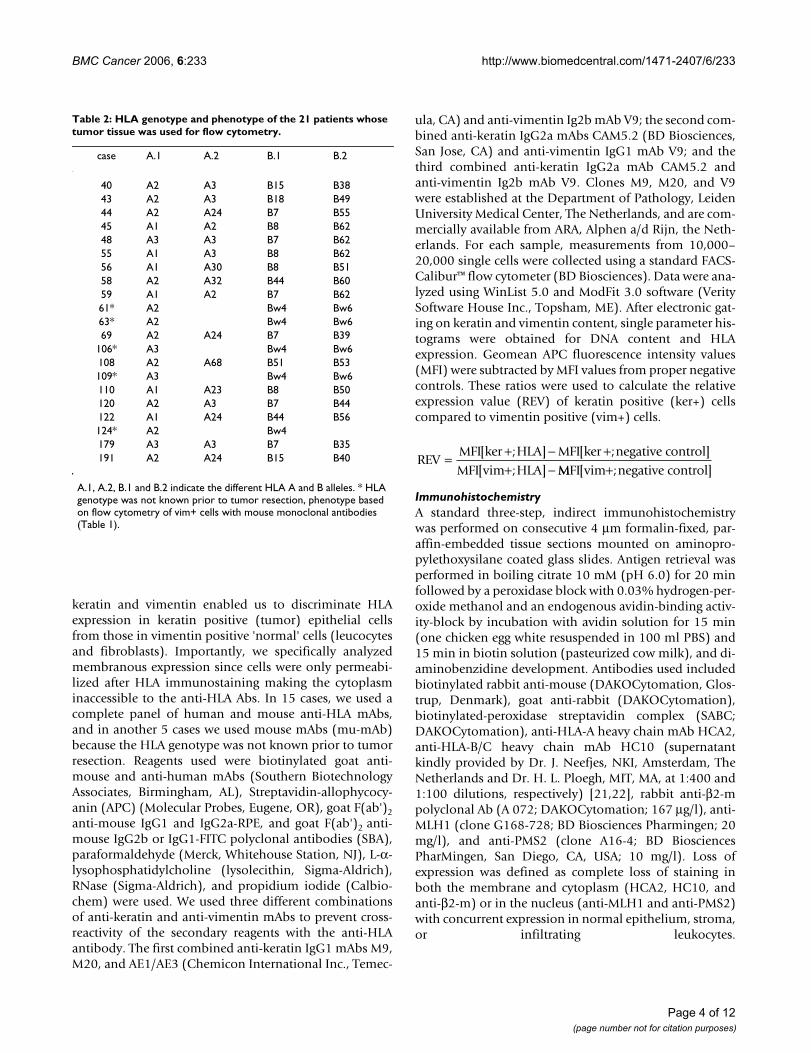
Table 2: HLA genotype and phenotype of the 21 patients whose tumor tissue was used for flow cytometry.
case A.1 A.2 B.1 B.2
40 A2 A3 B15 B3843 A2 A3 B18 B4944 A2 A24 B7 B5545 A1 A2 B8 B6248 A3 A3 B7 B6255 A1 A3 B8 B6256 A1 A30 B8 B5158 A2 A32 B44 B6059 A1 A2 B7 B6261* A2 Bw4 Bw663* A2 Bw4 Bw669 A2 A24 B7 B39
106* A3 Bw4 Bw6108 A2 A68 B51 B53109* A3 Bw4 Bw6110 A1 A23 B8 B50120 A2 A3 B7 B44122 A1 A24 B44 B56124* A2 Bw4179 A3 A3 B7 B35191 A2 A24 B15 B40
A.1, A.2, B.1 and B.2 indicate the different HLA A and B alleles. * HLA genotype was not known prior to tumor resection, phenotype based on flow cytometry of vim+ cells with mouse monoclonal antibodies (Table 1).
Page 4 of 12(page number not for citation purposes)
BMC Cancer 2006, 6:233 http://www.biomedcentral.com/1471-2407/6/233
Microsatellite instability analysisMicrosatellite instability (MSI) analysis was performedand scored as described previously [23] using the markersBAT25, BAT26, BAT40, D2S123, D5S346, D17S250,MSH3 and MSH6. β2m frameshift analysis was performedwith primers (available upon request) build around cod-ing repeats of the β2m gene; the (CT)4 repeat in exon 1and two (A)5 repeats in exon 2[24]. Genomic DNA ofboth normal and tumor tissue was isolated from 3 tissuecores of 0.6 mm diameter of formalin-fixed, paraffin-embedded material using the Chelex extraction method[25].
StatisticsValues of significance were calculated using the softwarepackage SPSS 10.0.7 (SPSS Inc., Chicago, IL, USA). Weperformed Pearson Chi-square tests or, when expectedcount was below 5, Fisher's exact 2-sided tests. The inde-pendent sample t-test was performed to test equality ofmeans of the age at operation.
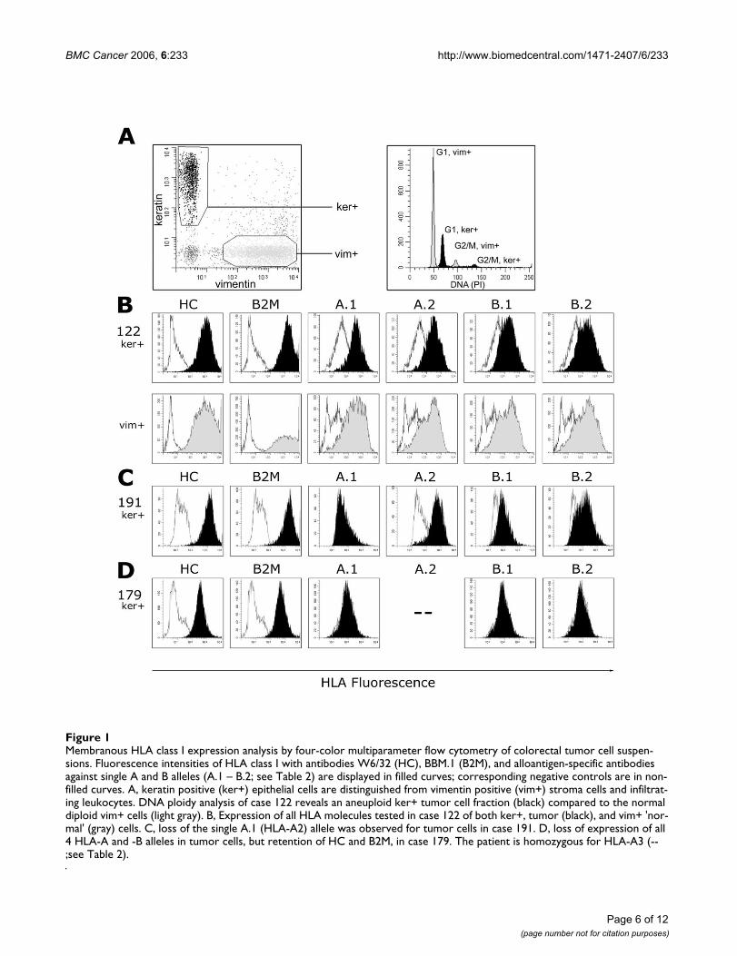
ResultsTwo patterns of alteration in HLA class I phenotype as observed by flow cytometryLoss of expression of HLA class I alleles was observed in 8of 21 (38%) colorectal cancer cases studied [see Addi-tional file 1]. Two distinct patterns of HLA loss were iden-tified; the loss of a single HLA-A or -B allele which wasobserved in 4 of 8 cases (cases 61, 63, 109, and 191), andthe loss of both HLA-A and -B alleles which was observedin the remaining 4 cases (cases 55, 56, 120, and 179).Importantly, in the latter group, membranous expressionof β2-microglobulin (β2-M) and remaining HLA class Iantigens was retained in the 4 tumors, but was diminishedas determined with the mAbs BBM.1 and W6/32. In thesesamples, the average Relative Expression Values (REV, seeMethods section) was 0.43 and 0.38, respectively, com-pared to the average REVS of 2.9 and 2.2, respectively, inthe other 17 cases (Figure 1) [see also Additional file 1].This would suggest the retention of at least one of theother HLA class I alleles, i.e. HLA-C, -E, -F or -G.
Alterations of HLA phenotype are found in specific subsets of colorectal cancerThe 7 of 8 cases with HLA alterations were all right sided(p = 0.024). Interestingly, all 5 MSI-H cases in the series(5/21) demonstrated HLA alterations [see Additional file1]. Four of the 5 cases demonstrated loss of HLA-A and -B(p = 0.028); in 1 of the 5 cases, we observed the loss of asingle HLA allele (case 191). Loss of expression of the het-erodimer of MLH1 and PMS2 was observed in the 4 MSI-H cases through IHC, and was not observed in any othercases [see Additional file 1]. These patients do not fulfillany criteria indicative of HNPCC are thus most likely spo-radic. Other clinicopathological features typical of spo-
radic MSI-H tumors [26] were also present; they occurredin elderly patients (mean age 75 years, p = 0.043), 3 of 4were located in the caecum (p = 0.019), and 3 of 4 wereperi-diploid (p = 0.053). The other MSI-H tumor (case120) was located in the sigmoid, occurring in the settingof the Hereditary Non-Polyposis Colorectal Cancer(HNPCC) syndrome. A mutation in the Mut-S-Homologue2 (MSH2) gene (G674R, exon 13 2020G>C) segregated inthis particular family, a variant that is probably patho-genic. The abrogation of MSH2 expression in this tumorwas confirmed by IHC. Since all the 4 MSI-H tumors thatlost HLA A and B expression showed diminished mem-branous expression of β2m we have screened the micros-atellite sequences of the corresponding gene but failed tofind any frameshift mutation.
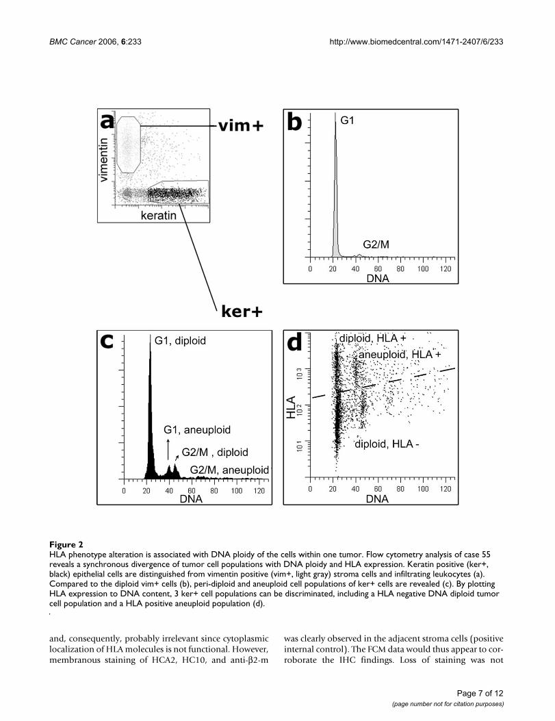
One case (case 55, MSI-H, multi-ploid) was particularlyintriguing. Ploidy studies indicated a peri-diploid tumorcell population as well as an aneuploid population (Fig-ure 2). Interestingly, while the aneuploid populationretained HLA expression for all HLA alleles examined, thediploid population was characterized by two populationsof cells; one population that retained HLA expression, andone population (the largest tumor cell population) thatdemonstrated a loss of expression of both HLA-A and -Balleles.
The 4 cases showing loss of both HLA-A and -B expressionhave not presented with lymph node or distant metastasesto date, while 3 of the 4 cases with loss of a single allele(including 1 MSI-H case) and 10 of the remaining 13cases without alterations of the HLA phenotype, didmetastasize (p = 0.035) to either lymph nodes or to dis-tant extranodal sites.
Comparison of FCM with conventional immunohistochemistryUsing FCM, we detected a significantly lower frequency ofHLA class I aberrations than previously reported [8-10,27]. Consequently, we decided to compare our FCMresults with those obtained using conventional IHC. Thehuman anti-HLA mAbs used for flow cytometry couldclearly not be used for the IHC analyses due to high back-ground staining caused by non-informative cross-reactiv-ity of the secondary anti-human antibody. Consequently,we chose to use a panel of mouse anti-HLA antibodies(HCA2, HC10, anti-β2-m) for the IHC analyses, althoughthese are not alloantigen specific. Loss of expression of asingle HLA allele could not be detected using these anti-bodies.
IHC of cases 55, 56, 120, and 179 (the 4 HLA-A and -Bnegative cases) showed complete absence of HCA2 andHC10 staining in tumor cells, and decreased anti-β2-mstaining which was primarily cytoplasmic (see Figure 3)
Page 5 of 12(page number not for citation purposes)
BMC Cancer 2006, 6:233 http://www.biomedcentral.com/1471-2407/6/233
Page 6 of 12(page number not for citation purposes)
Membranous HLA class I expression analysis by four-color multiparameter flow cytometry of colorectal tumor cell suspen-sionsFigure 1Membranous HLA class I expression analysis by four-color multiparameter flow cytometry of colorectal tumor cell suspen-sions. Fluorescence intensities of HLA class I with antibodies W6/32 (HC), BBM.1 (B2M), and alloantigen-specific antibodies against single A and B alleles (A.1 – B.2; see Table 2) are displayed in filled curves; corresponding negative controls are in non-filled curves. A, keratin positive (ker+) epithelial cells are distinguished from vimentin positive (vim+) stroma cells and infiltrat-ing leukocytes. DNA ploidy analysis of case 122 reveals an aneuploid ker+ tumor cell fraction (black) compared to the normal diploid vim+ cells (light gray). B, Expression of all HLA molecules tested in case 122 of both ker+, tumor (black), and vim+ 'nor-mal' (gray) cells. C, loss of the single A.1 (HLA-A2) allele was observed for tumor cells in case 191. D, loss of expression of all 4 HLA-A and -B alleles in tumor cells, but retention of HC and B2M, in case 179. The patient is homozygous for HLA-A3 (--;see Table 2).
BMC Cancer 2006, 6:233 http://www.biomedcentral.com/1471-2407/6/233
and, consequently, probably irrelevant since cytoplasmiclocalization of HLA molecules is not functional. However,membranous staining of HCA2, HC10, and anti-β2-m
was clearly observed in the adjacent stroma cells (positiveinternal control). The FCM data would thus appear to cor-roborate the IHC findings. Loss of staining was not
HLA phenotype alteration is associated with DNA ploidy of the cells within one tumorFigure 2HLA phenotype alteration is associated with DNA ploidy of the cells within one tumor. Flow cytometry analysis of case 55 reveals a synchronous divergence of tumor cell populations with DNA ploidy and HLA expression. Keratin positive (ker+, black) epithelial cells are distinguished from vimentin positive (vim+, light gray) stroma cells and infiltrating leukocytes (a). Compared to the diploid vim+ cells (b), peri-diploid and aneuploid cell populations of ker+ cells are revealed (c). By plotting HLA expression to DNA content, 3 ker+ cell populations can be discriminated, including a HLA negative DNA diploid tumor cell population and a HLA positive aneuploid population (d).
Page 7 of 12(page number not for citation purposes)

BMC Cancer 2006, 6:233 http://www.biomedcentral.com/1471-2407/6/233
Page 8 of 12(page number not for citation purposes)
Conventional HLA immunohistochemistry of formalin-fixed paraffin-embedded tissueFigure 3Conventional HLA immunohistochemistry of formalin-fixed paraffin-embedded tissue. Using HCA2 (HLA-A heavy chain), HC10 (HLA-B/C heavy chain) and anti-B2M antibodies, loss of HLA A and B expression as detected by flow cytometry (see Figures 1 and 2) could be confirmed. In the top panel, membranous staining (arrows) of B2M, HCA2, and HC10 is observed in the moderately differentiated sigmoid adenocarcinoma of case 122. In the bottom panel, loss of HLA-A and -B is illustrated for the mucinous caecum adenocarcinoma of case 179. Tumor B2M staining was mainly restricted to the cytoplasm (arrow), but some membranous expression of B2M was observed by FCM (see Figure 1). The HCA2 and HC10 staining was completely lost in tumor epithelium (t). Typically, the retained HLA expression of stroma cells (s) resulted in an inverted staining pattern in comparison to case 122. HE, haematoxylin and eosin staining. Pictures were made at 400 × magnification, HEs at 100×.
BMC Cancer 2006, 6:233 http://www.biomedcentral.com/1471-2407/6/233
observed in the remaining cases studied by IHC [see Addi-tional file 1]. A heterogeneous staining pattern, i.e. bothpositively and negatively staining tumor fields within thesame section, was frequently observed; such heterogeneityin membranous expression was not observed as discretepopulations (peaks) in our FCM data.
DiscussionWe used four-color multiparameter FCM to study com-plete HLA-A and -B phenotypes in colorectal tumors usinga large panel of alloantigen-specific mAbs. FCM allows thedetection of variation in expression over a dynamic rangeof 4 logarithmic decades and therefore FCM appears to bevery sensitive. Loss of membranous expression wasdetected in 38% (8 of 21 cases) of tumors in our study.Previous studies based on IHC demonstrated variablealterations in up to 73% of cases [8-10,27]. This discrep-ancy may be explained by the different technicalapproaches used and/or by the different composition ofthe groups under study (e.g. microsatellite stable/MSI-Hdistribution, tumor staging).
Except for one case (case 55) our FCM data did not pro-vide evidence for discrete intratumoral epithelial subpop-ulations of differing HLA expression as was observedusing IHC, although the lognormal distribution of themeasured fluorescence may obscure these variations inexpression. Nevertheless, the heterogeneous IHC stainingpatterns are difficult to interpret, and may be falselyregarded as loss of expression.
Using FCM, we characterized two distinctive alterations incolorectal tumors. We found 4 cases demonstrating loss ofa single HLA-A or B allele (alterations not identifiedthrough immunohistochemistry), and 4 cases demon-strating loss of expression of both the A and B alleles, butnot loss of all HLA class I molecules. The expression ofHLA I antigen complexes is a prerequisite for the optimaltargeting of tumor cells by CD8+ CTLs. However, NK cellsand CTLs also carry inhibitory receptors for specific HLAclass I alleles. Immunoselection thus relies on a delicatebalance between HLA allele loss and retention. Examplesof tumor immune escape due to such intricate alterationsof HLA phenotype have been described recently [16,28].In tumors showing loss of a single allele in our study, thisloss was restricted to HLA-A2 in 3 of the 4 cases. HLA-A2is not a ligand for inhibitory receptors. Those tumors mayhave effectively shed (HLA-A2) restricted epitopes andremained inhibitory to NK cell attack. Additionally, the 4cases demonstrating loss of both HLA-A and -B alleles(also applicable to the cases showing loss of a singleallele) retained expression of some total HLA class I mol-ecules, which was detected through the W6/32 antibodyand might indicate retention of HLA-C or other non clas-sical HLA molecules like HLA-G or -E.. Of these, HLA-E is
normally expressed in colon epithelial cells [29] and pre-dominantly function as inhibitors of NK-cell function[30]. These tumor cells may escape CTL attack and attackby a subset of NK cells. HIV-1 infected cells, for example,avoid both CTL and NK cell-mediated lysis through spe-cific HLA-A and -B downregulation caused by the HIV-1gene product nef [31]. These different phenotypic altera-tions may reflect differences in local immune selectiveforces between the tumor subsets. Such differences remainto be identified.
All but one of the tumors with an altered HLA phenotypewere located on the right side of the colon. The exceptionwas a left sided tumor arising in the setting of HNPCC.Furthermore, the loss of both HLA-A and -B alleles wasfound exclusively in tumors with the MSI-H phenotype.Mismatch repair deficiency has previously been associatedwith frequent mutations in the β2-m gene (B2M)[24,32-35]. This gene harbors multiple short tandem repeats thatare preferentially mutated, leading to total loss of mem-branous HLA class I expression. However, we did notobserve frame shift mutations in these repeats in the MSI-H tumors with HLA-A and -B loss, which is in line with theretention of membranous staining (although diminished)with the W6/32 and anti-β2-m mAbs. Yet, the commonloss of expression of HLA-A and -B in 3 of 4 sporadic MSI-H tumors, which is not due to B2M frame shift mutations,would suggest that another general mechanism may causethis altered phenotype.
Sporadic MSI-H tumors are usually relatively large, butrarely disseminate [3,36,37]. This favorable tumor behav-ior has been associated with an increased intra-epithelialCD8+ CTLs and CD57+ NK cells infiltrate when com-pared with the microsatellite stable tumors. CD4+ cellsare not frequently found in the intraepithelial infiltrate ofMSI-H tumors. [24,38-42]. The MSI of numerous codingmicrosatellites results in a large repertoire of immuno-genic frame shift peptides which can give rise to specificanti-tumor immune responses [4-6]. However, these infil-trating lymphocytes may lead to the selection of HLAalterations in tumor cells. High levels of infiltrate werepresent in the HLA-A and -B negative cases in this study asobserved by microscopy. Alternatively, the observed T cellinfiltrate could represent an innocent bystander effect.Previously, we identified loss of HLA-A and -B expressionin 6 of 88 cases of sporadic colorectal cancer using IHCthat, surprisingly, did not present with recurrences ormetastases during follow-up [27]. Although we could notdetermine the exact HLA phenotypic alteration at thattime, the previous cases resemble the 3 sporadic MSI-Hcases found in this study that lacked HLA-A and -B expres-sion and that have not presented with lymph node or dis-tant metastases to date. The NK cells that reside in thelymph and blood and that specifically kill metastasizing
Page 9 of 12(page number not for citation purposes)
BMC Cancer 2006, 6:233 http://www.biomedcentral.com/1471-2407/6/233
tumor cells (that lack total HLA class I expression) mayexplain the more favorable prognosis. However, the pos-tulated expression of the remaining non HLA-A and -Balleles by tumor cells may specifically inhibit these NKcells.
ConclusionFCM allows the discrimination of complex phenotypesrelated to the expression of HLA class I. The different pat-terns of HLA class I expression might underlie differenttumor behavior and influence the success rate of immu-notherapy.
Competing interestsThe author(s) declare that they have no competing inter-ests.
Authors' contributionsJWD – Performed the tumor cell isolation, the flowcytometry procedure, carried out the immunohistochem-istry assays, and performed the statistical analysis as wellas being involved in drafting the manuscript.
NM – Involved in the interpretation of the data, draftingof the manuscript, and critical revision.
AM – Supervised the production of the HLA allele specifichuman monoclonal antibodies.
MP – Performed the microsatellite instability analysis.
WV – Performed the HLA genotyping
FC – Supervised HLA genotyping
CV – Surgeon responsible for the resections of colorectalsamples used in the study
GJF – Contributed to the conception and design of thestudy, and to critical revision of the manuscript
CC – Contributed to the conception and design of thestudy, and critically reviewed the manuscript
WC – Supervised the technical procedures that involvedflow cytometry and the acquisition of data.
HM – Contributed to the conception and design of thestudy, and responsible for study
All authors read and approved the final manuscript.
Additional material
AcknowledgementsWe would like to thank Dr. K. Gelthorpe and Dr. J. Neefjes for the gener-ous gifts of antibodies, Dr. J. Frans Graadt van Roggen for his critical reading of the manuscript, Annemarie M.E.G. Voet-van den Brink and Graziella Kallenberg-Lantrua for providing the peripheral blood samples, Ronald L.P. Vlierberghe and Christa S. van Urk-van den Berg for technical assistance, Chantal Eijsink, Marrie J. Kardol and Marry E.I. Franke-van Dijk for human monoclonal HLA-antibody development, and the Rijnland Hospital Pathol-ogy Department for tissue specimens. The present study was supported by the Dutch Cancer Society (grant number 2000/2135).
References1. Van Der Bruggen P, Zhang Y, Chaux P, Stroobant V, Panichelli C,
Schultz ES, Chapiro J, Van Den Eynde BJ, Brasseur F, Boon T: Tumor-specific shared antigenic peptides recognized by human Tcells. Immunol Rev 2002, 188:51-64.
2. Ionov Y, Peinado MA, Malkhosyan S, Shibata D, Perucho M: Ubiqui-tous somatic mutations in simple repeated sequences reveala new mechanism for colonic carcinogenesis. Nature 1993,363:558-561.
3. Thibodeau SN, Bren G, Schaid D: Microsatellite instability in can-cer of the proximal colon. Science 1993, 260:816-819.
4. Linnebacher M, Gebert J, Rudy W, Woerner S, Yuan YP, Bork P, vonKnebel DM: Frameshift peptide-derived T-cell epitopes: asource of novel tumor- specific antigens. Int J Cancer 2001,93:6-11.
5. Ishikawa T, Fujita T, Suzuki Y, Okabe S, Yuasa Y, Iwai T, Kawakami Y:Tumor-specific immunological recognition of frameshift-mutated peptides in colon cancer with microsatellite insta-bility. Cancer Res 2003, 63:5564-5572.
Additional File 1HLA phenotype alterations and clinicopathological features of colorectal tumors and their mismatch repair status. These data provides the HLA phenotypes assessed by flow cytometry of the different tumors and their clinicopathological features and mismatch repair status. Clinicopathol-ogy: loc., tumor localization: A, colon ascendens; Ce, caecum; D, colon descendens; R, rectum; RS, rectosigmoid; S, sigmoid; modified Dukes stages [43]; F-U, follow-up (max. 2 years): DOD, dead of disease; DND, dead but not due to disease; M, distant metastasis; nr, no recurrences. MMR, mismatch repair status: MSI, microsatellite instability: H, MSI-High; S, microsatellite stable; *, HNPCC, hereditary non-polyposis color-ectal cancer; +, tumor epithelium staining positive as described in text; -, no staining of tumor cells, but staining of 'normal' cells, - +, heterogeneous staining of tumor cells; FCM, flow cytometry: A, aneuploid; D, diploid; M, multiploid; HLA FCM: Relative HLA Expression Values were calcu-lated from flow cytometry analyses as described in the methods section. Depicted in black are cases in which fluorescence intensity of ker+ cells was equal to the negative control (see Figure 1). A.1 – B.2, HLA-A and -B alleles as depicted in Table 2; HC, HLA heavy chain expression detected with the W6/32 antibody. †, HLA-A and -B negative and positive populations are present, therefore REV is not informative (see Figure 2); ‡, homozygous HLA-A genotype. HLA IHC, immunohistochemistry of HLA molecules: +, tumor epithelium staining positive as described in text; -, no staining of tumor cells, but staining of 'normal' cells; c, tumor epi-thelium staining restricted to the cytoplasm; -+, heterogeneous staining of tumor epithelium.Click here for file[http://www.biomedcentral.com/content/supplementary/1471-2407-6-233-S1.pdf]
Page 10 of 12(page number not for citation purposes)
http://www.ncbi.nlm.nih.gov/entrez/query.fcgi?cmd=Retrieve&db=PubMed&dopt=Abstract&list_uids=8505985
http://www.ncbi.nlm.nih.gov/entrez/query.fcgi?cmd=Retrieve&db=PubMed&dopt=Abstract&list_uids=8505985
http://www.ncbi.nlm.nih.gov/entrez/query.fcgi?cmd=Retrieve&db=PubMed&dopt=Abstract&list_uids=8505985
http://www.ncbi.nlm.nih.gov/entrez/query.fcgi?cmd=Retrieve&db=PubMed&dopt=Abstract&list_uids=8484122

BMC Cancer 2006, 6:233 http://www.biomedcentral.com/1471-2407/6/233
6. Saeterdal I, Bjorheim J, Lislerud K, Gjertsen MK, Bukholm IK, OlsenOC, Nesland JM, Eriksen JA, Moller M, Lindblom A, Gaudernack G:Frameshift-mutation-derived peptides as tumor-specificantigens in inherited and spontaneous colorectal cancer. ProcNatl Acad Sci U S A 2001, 98:13255-13260.
7. Townsend A, Bodmer H: Antigen recognition by class I-restricted T lymphocytes. Annu Rev Immunol 1989, 7:601-624.
8. Kaklamanis L, Gatter KC, Hill AB, Mortensen N, Harris AL, Krausa P,McMichael A, Bodmer JG, Bodmer WF: Loss of HLA class-I alle-les, heavy chains and beta 2-microglobulin in colorectal can-cer. Int J Cancer 1992, 51:379-385.
9. Cabrera T, Collado A, Fernandez MA, Ferron A, Sancho J, Ruiz-Cabello F, Garrido F: High frequency of altered HLA class I phe-notypes in invasive colorectal carcinomas. Tissue Antigens 1998,52:114-123.
10. Momburg F, Ziegler A, Harpprecht J, Moller P, Moldenhauer G, Ham-merling GJ: Selective loss of HLA-A or HLA-B antigen expres-sion in colon carcinoma. J Immunol 1989, 142:352-358.
11. Leong AS: Pitfalls in diagnostic immunohistology. Adv AnatPathol 2004, 11:86-93.
12. Ljunggren HG, Karre K: Host resistance directed selectivelyagainst H-2-deficient lymphoma variants. Analysis of themechanism. J Exp Med 1985, 162:1745-1759.
13. Long EO: Tumor cell recognition by natural killer cells. SeminCancer Biol 2002, 12:57-61.
14. Oudshoorn M, Doxiadis II, PM BL, Voorter CE, Verduyn W, Claas FH:Functional versus structural matching: can the CTLp test bereplaced by HLA allele typing? Hum Immunol 2002, 63:176-184.
15. Mulder A, Kardol M, Regan J, Buelow R, Claas F: Reactivity oftwenty-two cytotoxic human monoclonal HLA antibodiestowards soluble HLA class I in an enzyme-linked immuno-sorbent assay (PRA-STAT). Hum Immunol 1997, 56:106-113.
16. Demanet C, Mulder A, Deneys V, Worsham MJ, Maes P, Claas FH,Ferrone S: Down-regulation of HLA-A and HLA-Bw6, but notHLA-Bw4, allospecificities in leukemic cells: an escapemechanism from CTL and NK attack? Blood 2004,103:3122-3130.
17. Koopman LA, Mulder A, Corver WE, Anholts JD, Giphart MJ, ClaasFH, Fleuren GJ: HLA class I phenotype and genotype altera-tions in cervical carcinomas and derivative cell lines. TissueAntigens 1998, 51:623-636.
18. Mulder A, Kardol M, Blom J, Jolley WB, Melief CJ, Bruning H: Ahuman monoclonal antibody, produced following in vitroimmunization, recognizing an epitope shared by HLA-A2subtypes and HLA-A28. Tissue Antigens 1993, 42:27-34.
19. Mulder A, Kardol M, Blom J, Jolley WB, Melief CJ, Bruning JW: Char-acterization of two human monoclonal antibodies reactivewith HLA-B12 and HLA-B60, respectively, raised by in vitrosecondary immunization of peripheral blood lymphocytes.Hum Immunol 1993, 36:186-192.
20. Corver WE, Koopman LA, Mulder A, Cornelisse CJ, Fleuren GJ: Dis-tinction between HLA class I-positive and -negative cervicaltumor subpopulations by multiparameter DNA flow cytom-etry. Cytometry 2000, 41:73-80.
21. Stam NJ, Vroom TM, Peters PJ, Pastoors EB, Ploegh HL: HLA-A-and HLA-B-specific monoclonal antibodies reactive withfree heavy chains in western blots, in formalin-fixed, paraffin-embedded tissue sections and in cryo-immuno-electronmicroscopy. Int Immunol 1990, 2:113-125.
22. Stam NJ, Spits H, Ploegh HL: Monoclonal antibodies raisedagainst denatured HLA-B locus heavy chains permit bio-chemical characterization of certain HLA-C locus products.J Immunol 1986, 137:2299-2306.
23. de Leeuw WJ, van Puijenbroek M, Merx R, Wijnen JT, Brocker-Vriends AH, Tops C, Vasen H, Cornelisse CJ, Morreau H: Bias indetection of instability of the (C)8 mononucleotide repeat ofMSH6 in tumours from HNPCC patients. Oncogene 2001,20:6241-6244.
24. Kloor M, Becker C, Benner A, Woerner SM, Gebert J, Ferrone S,Doeberitz MV: Immunoselective pressure and human leuko-cyte antigen class I antigen machinery defects in microsatel-lite unstable colorectal cancers. Cancer Research 2005,65:6418-6424.
25. de Leeuw WJ, Dierssen J, Vasen HF, Wijnen JT, Kenter GG, Meijers-Heijboer H, Brocker-Vriends A, Stormorken A, Moller P, Menko F,Cornelisse CJ, Morreau H: Prediction of a mismatch repair gene
defect by microsatellite instability and immunohistochemi-cal analysis in endometrial tumours from HNPCC patients.J Pathol 2000, 192:328-335.
26. Jass JR, Whitehall VL, Young J, Leggett BA: Emerging concepts incolorectal neoplasia. Gastroenterology 2002, 123:862-876.
27. Menon AG, Morreau H, Tollenaar RA, Alphenaar E, van PuijenbroekM, Putter H, Janssen-Van Rhijn CM, Van De Velde CJ, Fleuren GJ,Kuppen PJ: Down-regulation of HLA-A expression correlateswith a better prognosis in colorectal cancer patients. LabInvest 2002, 82:1725-1733.
28. Giorda E, Sibilio L, Martayan A, Moretti S, Venturo I, Mottolese M,Ferrara GB, Cappellacci S, Eibenschutz L, Catricala C, Grammatico P,Giacomini P: The antigen processing machinery of class Ihuman leukocyte antigens: linked patterns of gene expres-sion in neoplastic cells. Cancer Res 2003, 63:4119-4127.
29. van den Elsen PJ, Holling TM, Kuipers HF, van der Stoep N: Tran-scriptional regulation of antigen presentation. Curr Opin Immu-nol 2004, 16:67-75.
30. Braud VM, Allan DS, McMichael AJ: Functions of nonclassicalMHC and non-MHC-encoded class I molecules. Curr OpinImmunol 1999, 11:100-108.
31. Cohen GB, Gandhi RT, Davis DM, Mandelboim O, Chen BK,Strominger JL, Baltimore D: The selective downregulation ofclass I major histocompatibility complex proteins by HIV-1protects HIV-infected cells from NK cells. Immunity 1999,10:661-671.
32. Bicknell DC, Rowan A, Bodmer WF: Beta 2-microglobulin genemutations: a study of established colorectal cell lines andfresh tumors. Proc Natl Acad Sci U S A 1994, 91:4751-4756.
33. Browning M, Petronzelli F, Bicknell D, Krausa P, Rowan A, Tonks S,Murray N, Bodmer J, Bodmer W: Mechanisms of loss of HLAclass I expression on colorectal tumor cells. Tissue Antigens1996, 47:364-371.
34. Bicknell DC, Kaklamanis L, Hampson R, Bodmer WF, Karran P:Selection for beta 2-microglobulin mutation in mismatchrepair-defective colorectal carcinomas. Curr Biol 1996,6:1695-1697.
35. Cabrera CM, Jimenez P, Cabrera T, Esparza C, Ruiz-Cabello F, Gar-rido F: Total loss of MHC class I in colorectal tumors can beexplained by two molecular pathways: beta2-microglobulininactivation in MSI-positive tumors and LMP7/TAP2 down-regulation in MSI-negative tumors. Tissue Antigens 2003,61:211-219.
36. Lothe RA, Peltomaki P, Meling GI, Aaltonen LA, Nystrom-Lahti M,Pylkkanen L, Heimdal K, Andersen TI, Moller P, Rognum TO:Genomic instability in colorectal cancer: relationship to clin-icopathological variables and family history. Cancer Res 1993,53:5849-5852.
37. Wright CM, Dent OF, Barker M, Newland RC, Chapuis PH, Bokey EL,Young JP, Leggett BA, Jass JR, Macdonald GA: Prognostic signifi-cance of extensive microsatellite instability in sporadic clin-icopathological stage C colorectal cancer. Br J Surg 2000,87:1197-1202.
38. Dolcetti R, Viel A, Doglioni C, Russo A, Guidoboni M, Capozzi E, Vec-chiato N, Macri E, Fornasarig M, Boiocchi M: High prevalence ofactivated intraepithelial cytotoxic T lymphocytes andincreased neoplastic cell apoptosis in colorectal carcinomaswith microsatellite instability. Am J Pathol 1999, 154:1805-1813.
39. Jass JR, Do KA, Simms LA, Iino H, Wynter C, Pillay SP, Searle J, Rad-ford-Smith G, Young J, Leggett B: Morphology of sporadic color-ectal cancer with DNA replication errors. Gut 1998,42:673-679.
40. Michael-Robinson JM, Biemer-Huttmann A, Purdie DM, Walsh MD,Simms LA, Biden KG, Young JP, Leggett BA, Jass JR, Radford-SmithGL: Tumour infiltrating lymphocytes and apoptosis are inde-pendent features in colorectal cancer stratified according tomicrosatellite instability status. Gut 2001, 48:360-366.
41. Young J, Simms LA, Biden KG, Wynter C, Whitehall V, Karamatic R,George J, Goldblatt J, Walpole I, Robin SA, Borten MM, Stitz R, SearleJ, McKeone D, Fraser L, Purdie DR, Podger K, Price R, Buttenshaw R,Walsh MD, Barker M, Leggett BA, Jass JR: Features of colorectalcancers with high-level microsatellite instability occurring infamilial and sporadic settings: parallel pathways of tumori-genesis. Am J Pathol 2001, 159:2107-2116.
42. Menon AG, Janssen-Van Rhijn CM, Morreau H, Putter H, TollenaarRA, Van De Velde CJ, Fleuren GJ, Kuppen PJ: Immune system and
Page 11 of 12(page number not for citation purposes)
http://www.ncbi.nlm.nih.gov/entrez/query.fcgi?cmd=Retrieve&db=PubMed&dopt=Abstract&list_uids=2469442
http://www.ncbi.nlm.nih.gov/entrez/query.fcgi?cmd=Retrieve&db=PubMed&dopt=Abstract&list_uids=2469442
http://www.ncbi.nlm.nih.gov/entrez/query.fcgi?cmd=Retrieve&db=PubMed&dopt=Abstract&list_uids=1592528
http://www.ncbi.nlm.nih.gov/entrez/query.fcgi?cmd=Retrieve&db=PubMed&dopt=Abstract&list_uids=1592528
http://www.ncbi.nlm.nih.gov/entrez/query.fcgi?cmd=Retrieve&db=PubMed&dopt=Abstract&list_uids=1592528
http://www.ncbi.nlm.nih.gov/entrez/query.fcgi?cmd=Retrieve&db=PubMed&dopt=Abstract&list_uids=9756399
http://www.ncbi.nlm.nih.gov/entrez/query.fcgi?cmd=Retrieve&db=PubMed&dopt=Abstract&list_uids=9756399
http://www.ncbi.nlm.nih.gov/entrez/query.fcgi?cmd=Retrieve&db=PubMed&dopt=Abstract&list_uids=2462591
http://www.ncbi.nlm.nih.gov/entrez/query.fcgi?cmd=Retrieve&db=PubMed&dopt=Abstract&list_uids=2462591
http://www.ncbi.nlm.nih.gov/entrez/query.fcgi?cmd=Retrieve&db=PubMed&dopt=Abstract&list_uids=3877776
http://www.ncbi.nlm.nih.gov/entrez/query.fcgi?cmd=Retrieve&db=PubMed&dopt=Abstract&list_uids=3877776
http://www.ncbi.nlm.nih.gov/entrez/query.fcgi?cmd=Retrieve&db=PubMed&dopt=Abstract&list_uids=3877776
http://www.ncbi.nlm.nih.gov/entrez/query.fcgi?cmd=Retrieve&db=PubMed&dopt=Abstract&list_uids=9455499
http://www.ncbi.nlm.nih.gov/entrez/query.fcgi?cmd=Retrieve&db=PubMed&dopt=Abstract&list_uids=9455499
http://www.ncbi.nlm.nih.gov/entrez/query.fcgi?cmd=Retrieve&db=PubMed&dopt=Abstract&list_uids=9455499
http://www.ncbi.nlm.nih.gov/entrez/query.fcgi?cmd=Retrieve&db=PubMed&dopt=Abstract&list_uids=9694355
http://www.ncbi.nlm.nih.gov/entrez/query.fcgi?cmd=Retrieve&db=PubMed&dopt=Abstract&list_uids=9694355
http://www.ncbi.nlm.nih.gov/entrez/query.fcgi?cmd=Retrieve&db=PubMed&dopt=Abstract&list_uids=7504327
http://www.ncbi.nlm.nih.gov/entrez/query.fcgi?cmd=Retrieve&db=PubMed&dopt=Abstract&list_uids=7504327
http://www.ncbi.nlm.nih.gov/entrez/query.fcgi?cmd=Retrieve&db=PubMed&dopt=Abstract&list_uids=7504327
http://www.ncbi.nlm.nih.gov/entrez/query.fcgi?cmd=Retrieve&db=PubMed&dopt=Abstract&list_uids=8391522
http://www.ncbi.nlm.nih.gov/entrez/query.fcgi?cmd=Retrieve&db=PubMed&dopt=Abstract&list_uids=8391522
http://www.ncbi.nlm.nih.gov/entrez/query.fcgi?cmd=Retrieve&db=PubMed&dopt=Abstract&list_uids=2088481
http://www.ncbi.nlm.nih.gov/entrez/query.fcgi?cmd=Retrieve&db=PubMed&dopt=Abstract&list_uids=2088481
http://www.ncbi.nlm.nih.gov/entrez/query.fcgi?cmd=Retrieve&db=PubMed&dopt=Abstract&list_uids=2088481
http://www.ncbi.nlm.nih.gov/entrez/query.fcgi?cmd=Retrieve&db=PubMed&dopt=Abstract&list_uids=3760563
http://www.ncbi.nlm.nih.gov/entrez/query.fcgi?cmd=Retrieve&db=PubMed&dopt=Abstract&list_uids=3760563
http://www.ncbi.nlm.nih.gov/entrez/query.fcgi?cmd=Retrieve&db=PubMed&dopt=Abstract&list_uids=8197130
http://www.ncbi.nlm.nih.gov/entrez/query.fcgi?cmd=Retrieve&db=PubMed&dopt=Abstract&list_uids=8197130
http://www.ncbi.nlm.nih.gov/entrez/query.fcgi?cmd=Retrieve&db=PubMed&dopt=Abstract&list_uids=8197130
http://www.ncbi.nlm.nih.gov/entrez/query.fcgi?cmd=Retrieve&db=PubMed&dopt=Abstract&list_uids=8795136
http://www.ncbi.nlm.nih.gov/entrez/query.fcgi?cmd=Retrieve&db=PubMed&dopt=Abstract&list_uids=8795136
http://www.ncbi.nlm.nih.gov/entrez/query.fcgi?cmd=Retrieve&db=PubMed&dopt=Abstract&list_uids=8994836
http://www.ncbi.nlm.nih.gov/entrez/query.fcgi?cmd=Retrieve&db=PubMed&dopt=Abstract&list_uids=8994836
http://www.ncbi.nlm.nih.gov/entrez/query.fcgi?cmd=Retrieve&db=PubMed&dopt=Abstract&list_uids=8994836
http://www.ncbi.nlm.nih.gov/entrez/query.fcgi?cmd=Retrieve&db=PubMed&dopt=Abstract&list_uids=8261392
http://www.ncbi.nlm.nih.gov/entrez/query.fcgi?cmd=Retrieve&db=PubMed&dopt=Abstract&list_uids=8261392
http://www.ncbi.nlm.nih.gov/entrez/query.fcgi?cmd=Retrieve&db=PubMed&dopt=Abstract&list_uids=8261392
http://www.ncbi.nlm.nih.gov/entrez/query.fcgi?cmd=Retrieve&db=PubMed&dopt=Abstract&list_uids=9659163
BMC Cancer 2006, 6:233 http://www.biomedcentral.com/1471-2407/6/233
Publish with BioMed Central and every scientist can read your work free of charge
"BioMed Central will be the most significant development for disseminating the results of biomedical research in our lifetime."
Sir Paul Nurse, Cancer Research UK
Your research papers will be:
available free of charge to the entire biomedical community
peer reviewed and published immediately upon acceptance
cited in PubMed and archived on PubMed Central
yours — you keep the copyright
Submit your manuscript here:http://www.biomedcentral.com/info/publishing_adv.asp
BioMedcentral
prognosis in colorectal cancer: a detailed immunohisto-chemical analysis. Lab Invest 2004, 84:493-501.
43. Gunderson LL, Sosin H: Areas of failure found at reoperation(second or symptomatic look) following "curative surgery"for adenocarcinoma of the rectum. Clinicopathologic corre-lation and implications for adjuvant therapy. Cancer 1974,34:1278-1292.
Pre-publication historyThe pre-publication history for this paper can be accessedhere:
http://www.biomedcentral.com/1471-2407/6/233/prepub
Page 12 of 12(page number not for citation purposes)
http://www.ncbi.nlm.nih.gov/entrez/query.fcgi?cmd=Retrieve&db=PubMed&dopt=Abstract&list_uids=4424091
http://www.ncbi.nlm.nih.gov/entrez/query.fcgi?cmd=Retrieve&db=PubMed&dopt=Abstract&list_uids=4424091