Characterization of Xanthomonas axonopodis pv. punicae ...
5
Madras Agric. J., 105 (4-6): 210-214, June 2018 *Corresponding author’s email: [email protected] Characterization of Xanthomonas axonopodis pv. punicae (Hingorani and Singh) Vauterin et al. and Isolation of Xap Specific Bacteriophage K.N. Harshitha* 1 , S.K. Manoranjitham 1 , E. Somasundaram 2 , L. Rajendran 1 and G. Karthikeyan 1 1 Department of Plant Pathology 2 Department of Sustainable Organic Agriculture, Tamil Nadu Agricultural University, Coimbatore - 641 003 Bacterial blight (BLB) caused by Xanthomonas axonopodis pv. punicae (Xap) has emerged as a potential threat in pomegranate cultivation in India. To know the present disease scenario and to identify the root cause for its epidemic, survey was conducted in five different localities of Tamil Nadu. Per cent disease severity (PDS) ranged from 1 to 36 per cent on leaves and zero to 40 per cent infection on fruits. Bacteriophage or bacteria killer specific to Xap-1 (Sennampatty) isolate was isolated from pond water after enrichment of water sample. Phage produced plaques of 2 mm, 1 mm and < 1mm size that coalesced after 18 h of plating which shows virulence and rapid phage multiplication. Disease severity on test plants were studied to know the degree of virulence and variability among the isolates with highest severity of 37.31 per cent documented in Xap-1 followed by Xap-3 (28.7%) and Xap-2 (24.3%). Physiological factors such as temperature, pH and their role in growth of Xap-1 were accessed. Maximum growth was observed at 28˚C (0.86 OD) followed by 30˚C (0.68 OD) and pH 7 (1.83 OD) and optimum at 7-8 pH for virulent (Xap-1) isolate. Molecular confirmation of Xap through 16S rRNA sequencing gave the amplicon of 1500 bp size and amplification through KKM5 and KKM6 further confirmed specific identity. Key words: Pomegranate, Bacterial blight, Bacteriophage, Physiological and molecular characterization Pomegranate (Punica granatum L.) belongs to the Punicaceae family which is one of the economically important fruit crops of India. Place of origin is believed to be Iran. In India it is cultivated over an area of 208.73 thousand ha with production of 2442.39 thousand metric tonnes (MT) and productivity of 11.70 MT. h -1 . Maharashtra occupying 2/3 rd of area followed by Karnataka and Gujarat, but highest productivity is recorded in Tamil Nadu with 27.43 t. ha -1 (“Horticulture statistics at a glance”). In Tamil Nadu the crop is cultivated in Erode, Dindigul, Coimbatore and Tirunelveli districts with major markets in Coimbatore and Chennai. Popular varieties extensively cultivated are Bhagwa, Ruby and Ganesh. In spite of these the fruit crop is attacked by several diseases such as anthracnose, Cercospora leaf spot and bacterial blight (BLB). Bacterial blight was first reported in Delhi by Hingorani and Mehta during 1952. Hingorani and Mehta (1952) first identified the causal organism as Xanthomonas campestris, later it was reclassified and renamed as Xanthomonas axonopodis pv. punicae (Xap). Symptomatology of BLB on leaf appears as small (2 to 5mm), irregular, prominent water soaked spots, which later develops necrotic center surrounded by water soaked margins and in the progressive stage of the disease the individual spots coalesce giving an eventual blighted appearance, lesions on stem are dark brown to black that leads to cracking and the breaking of branches. On fruits spots appears as irregular brown to black with Y or L shaped cracking or splitting of pericarp (Mondal et al., 2012). With this background information, the present study was carried out to isolate and study the Xanthomonas axonopodis pv. punicae pathogen in pomegranate from different regions of Tamil Nadu and also an attempt was made to isolate Xanthomonas axonopodis pv. punicae specific bacteriophage from pond water. Material and Methods For collection of symptoms and to know the bacterial blight of pomegranate severity in the field, survey was conducted from five different localities of Tamil Nadu. Survey was done from Sennampatty (12.1206˚N and 78.3814˚E) of Erode district, Thondamuthur (10.9899˚N and 76.8409˚E) of Coimbatore district, Tiruchengode (11.3782˚N and 77.8969˚E) of Nammakal district, Udumalpet (10.5846˚N and 77.2514˚E) of Tirupur district, TNAU orchard (11.0045˚N and 76.961632˚E) of Coimbatore district, where symptoms were collected from Bhagwa variety pomegranate plants and the per cent disease severity was determined using 0-6 disease severity scale given by ICAR-NRCP. Xanthomonas axonopodis pv. punicae culture Symptoms like blightening with oily ring on the leaves were selected and surface sterilized with 70 per cent ethanol further sterilized with 0.1 per cent mercuric chloride for 1 min. Followed by transferring
Transcript of Characterization of Xanthomonas axonopodis pv. punicae ...
Madras Agric. J., 105 (4-6): 210-214, June 2018
*Corresponding author’s email: [email protected]
Characterization of Xanthomonas axonopodis pv. punicae (Hingorani and Singh) Vauterin et al. and Isolation of Xap
Specific Bacteriophage
K.N. Harshitha*1, S.K. Manoranjitham1, E. Somasundaram2, L. Rajendran1 and G. Karthikeyan1 1Department of Plant Pathology
2Department of Sustainable Organic Agriculture, Tamil Nadu Agricultural University, Coimbatore - 641 003
Bacterial blight (BLB) caused by Xanthomonas axonopodis pv. punicae (Xap) has emerged as a potential threat in pomegranate cultivation in India. To know the present disease scenario and to identify the root cause for its epidemic, survey was conducted in five different localities of Tamil Nadu. Per cent disease severity (PDS) ranged from 1 to 36 per cent on leaves and zero to 40 per cent infection on fruits. Bacteriophage or bacteria killer specific to Xap-1 (Sennampatty) isolate was isolated from pond water after enrichment of water sample. Phage produced plaques of 2 mm, 1 mm and < 1mm size that coalesced after 18 h of plating which shows virulence and rapid phage multiplication. Disease severity on test plants were studied to know the degree of virulence and variability among the isolates with highest severity of 37.31 per cent documented in Xap-1 followed by Xap-3 (28.7%) and Xap-2 (24.3%). Physiological factors such as temperature, pH and their role in growth of Xap-1 were accessed. Maximum growth was observed at 28˚C (0.86 OD) followed by 30˚C (0.68 OD) and pH 7 (1.83 OD) and optimum at 7-8 pH for virulent (Xap-1) isolate. Molecular confirmation of Xap through 16S rRNA sequencing gave the amplicon of 1500 bp size and amplification through KKM5 and KKM6 further confirmed specific identity.
Key words: Pomegranate, Bacterial blight, Bacteriophage, Physiological and molecular characterization
Pomegranate (Punica granatum L.) belongs to the Punicaceae family which is one of the economically important fruit crops of India. Place of origin is believed to be Iran. In India it is cultivated over an area of 208.73 thousand ha with production of 2442.39 thousand metric tonnes (MT) and productivity of 11.70 MT. h-1. Maharashtra occupying 2/3rd of area followed by Karnataka and Gujarat, but highest productivity is recorded in Tamil Nadu with 27.43 t. ha-1 (“Horticulture statistics at a glance”). In Tamil Nadu the crop is cultivated in Erode, Dindigul, Coimbatore and Tirunelveli districts with major markets in Coimbatore and Chennai. Popular varieties extensively cultivated are Bhagwa, Ruby and Ganesh.
In spite of these the fruit crop is attacked by several diseases such as anthracnose, Cercospora leaf spot and bacterial blight (BLB). Bacterial blight was first reported in Delhi by Hingorani and Mehta during 1952. Hingorani and Mehta (1952) first identified the causal organism as Xanthomonas campestris, later it was reclassified and renamed as Xanthomonas axonopodis pv. punicae (Xap). Symptomatology of BLB on leaf appears as small (2 to 5mm), irregular, prominent water soaked spots, which later develops necrotic center surrounded by water soaked margins and in the progressive stage of the disease the individual spots coalesce giving an eventual blighted appearance, lesions on stem are dark brown to black that leads to cracking and
the breaking of branches. On fruits spots appears as irregular brown to black with Y or L shaped cracking or splitting of pericarp (Mondal et al., 2012). With this background information, the present study was carried out to isolate and study the Xanthomonas axonopodis pv. punicae pathogen in pomegranate from different regions of Tamil Nadu and also an attempt was made to isolate Xanthomonas axonopodis pv. punicae specific bacteriophage from pond water.
Material and Methods
For collection of symptoms and to know the bacterial blight of pomegranate severity in the field, survey was conducted from five different localities of Tamil Nadu. Survey was done from Sennampatty (12.1206˚N and 78.3814˚E) of Erode district, Thondamuthur (10.9899˚N and 76.8409˚E) of Coimbatore district, Tiruchengode (11.3782˚N and 77.8969˚E) of Nammakal district, Udumalpet (10.5846˚N and 77.2514˚E) of Tirupur district, TNAU orchard (11.0045˚N and 76.961632˚E) of Coimbatore district, where symptoms were collected from Bhagwa variety pomegranate plants and the per cent disease severity was determined using 0-6 disease severity scale given by ICAR-NRCP. Xanthomonas axonopodis pv. punicae culture
Symptoms like blightening with oily ring on the leaves were selected and surface sterilized with 70 per cent ethanol further sterilized with 0.1 per cent mercuric chloride for 1 min. Followed by transferring
211
the bits serially in sterile distilled water, then sterilized bits transferred in to Eppendorf tube containing sterile water and crushed; which allows the bacteria to diffuse in water for 10 min finally with the help of inoculation loop the diffused bacteria is streaked on the nutrient agar, plates incubated at 29-30˚ C for 48-72 h. Appearance of yellow convex or mucoid colonies can be observed on nutrient agar after 48 h. of incubation and the isolates were designated as Xap-1, Xap-2, Xap-3, Xap-4 and Xap-5, respectively (Raghuwanshi et al., 2013). Isolation of bacteriophage specific for Xanthomonas axonopodis pv. punicae
The isolation procedure of bacteriophage from pond water was done as described by (Chae et al., 2014). pond water (76.92˚E and 11.006˚N) of 5 ml was collected, to that equal volume of phage broth (peptone 10 g, yeast extract 5 g, sodium chloride 2.5 g, dipotassium hydrogen orthophosphate 8 g, water 1000 ml) and 4 to 5 ml of 48 hours old virulent Xap-1 broth was added and incubated overnight at room temperature. Consecutively, few drops of chloroform was mixed in the sample by keeping them in rotating shaker for 20 min followed by centrifugation of the incubated sample at 13,000 rpm for 10 min, supernatant was passed through 0.22 µm syringe filter. Phage filtrate was stored at 4˚C in complete darkness. Enumeration of phages was done by double agar overlay method proposed by Lark and Adams, 1953. Later for long term storage of the phages, single phage plaque were taken in the form discs and added to phage buffer, centrifuged and filter sterilized. Enumeration of phages by double-layer plaque assay
Enumeration is done by double agar overlay method as proposed by Lark and Adams (1953). Nutrient glucose agar (beef extract 3 g, peptone 5 g, sodium chloride 5 g, glucose 2.5 g, water 1000 ml) medium supplemented with 10 mM CaCl2 and kept in the water bath at 5560˚C. Nutrient glucose agar (Bottom agar) of 15 to 20 ml was poured on the sterilized plates and allowed to solidify. Sterilized nutrient glucose agar at 7 per cent agar (Top agar) concentration was amended with 10 mM CaCl2 and top agar was maintained at 45-50˚ C. After the incubation 100 µl of phage filtrate and 100 µl of overnight mass multiplied Xap-1 broth was mixed and poured above the bottom agar, plates were allowed to solidify for 20 min and incubated overnight at room temperature. Number and morphology of plaques was accessed after incubation of plates at room temperature. Pathogenicity test
Pathogenicity test was conducted in greenhouse conditions where one and half year old seedlings of susceptible variety “Bhagwa” were pruned 45 d before inoculation and well irrigated. Five isolates was tested for pathogenicity, one d prior to inoculation the pots were covered with polythene covers with holes to maintain more than 80 per cent relative humidity
required for successful infection (OD at 600nm = 0.3, equivalent to 108 cfu/ml). Bacterial isolates streaked on the medium was collected with the help of inoculation loop and mixed with sterile water and final volume was made to 100 ml; later the inoculum was sprayed on pomegranate plants during evening hours using hand sprayer (Peerajade et al., 2017) and finally covered with polythene cover, whereas the control plants were sprayed with sterile distilled water. Isolates showing high disease severity in test plants was considered as virulent and virulent isolate was tested for standardization of inoculation technique by three methods such as spray inoculation, infiltration and pin prick. Pathogenic variability among isolates was determined by symptom expression and lesion development up to 45th d after inoculation (Peerajade et al., 2017). Influence of temperature and pH on Xap-1 growth
The Xap-1 inoculated in Nutrient glucose broth (NGB) was placed in constant temperature at 10˚C, 28˚C, 29˚C, 30˚C, 35˚C, 37˚C and room temperature for 48 h with three replications. Mass multiplied broth was spectrophotometrically measured at 600 nm and minimum, maximum and optimum growth with varying temperatures was recorded. To study the optimum pH for the growth of the Xanthomonas axonopodis pv. punicae, the pH of the NGB medium was adjusted to a range of 2 to 9 with an increase value of one, inoculated with virulent isolate (Xap-1) and incubated at 28˚C for 48 h. OD values were spectrophotometrically measured and the pH showing highest growth was optimized as favorable pH. Amplification by 16S rRNA primers
Confirmation of conserved 16S rRNA region of Xanthomonas axonopodis pv. punicae by using 27F (5’-AGAGTTTGATCCTGGCTCAG-3’) and 1492R (5’-GGT TACCTTGTTACGACTT-3’) with PCR conditions at initial denaturation of 96°C for 2 min, followed by 35 cycles of denaturation at 94°C for 30 sec, annealing at 60°C for 1 min, extension at 72°C for 1 min and final extension at 72°C for 10 min in nexus gradient Mastercycler, shows the amplicon size of 1537 bp (Mondal et al., 2013). Amplification of gyrB specific region by specific primers
Specific primer amplification was carried out at 94oC for 7 min, followed by 30 cycles of 94oC for 30 sec, annealing of 57oC for 60 sec and extension 72oC for 1 min. Reactions were completed with a final extension step of 10 min at 72o C in nexus gradient Mastercycler by KKM5F (5’-GTTGATGCTGTTC ACCAGCG-3’) and KKM6R (5’-CATTCATTTCGCCCAAGCCC-3’) primers (Mondal et al., 2013).
Cultural and physiological factors required for Xanthomonas axonopodis pv. punicae, disease severity from pathogenicity test were analyzed by generalized univariate linear model. Effect of pH, temperature and per cent disease severity was analyzed using SPSS 16.0. The treatment means
212
were separated using the Fisher’s protected least significant difference (DMRT) (α = 0.05).
Results and Discussion
The survey report mentioned revealed that among the five locations, highest per cent disease severity
of blight was observed in Xap-1 (Sennampatty) showing maximum disease severity (PDS) of 36 per cent on leaves and 40 per cent on fruits, followed by Thondamuthur (Xap-2) region (13 % on leaves and 2 % on fruits). Third isolate (Xap-3) was collected from Tiruchengode field (8 and 2 PDS on leaves and fruits).
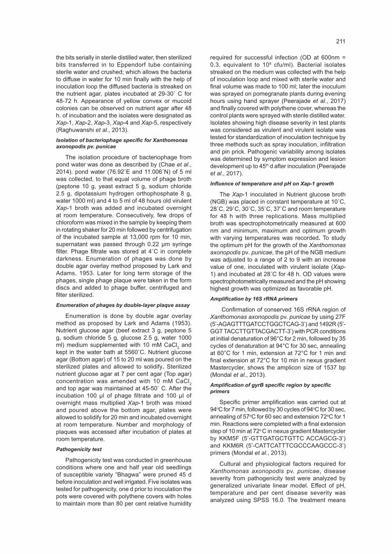
Table 1. Pathogenicity test of Xanthomonas axonopodis pv. punicae and symptom development on leaves up to 45 days
Isolates DAI for
symptom expression*
Disease severity
(%)5 DAI 15 DAI 25 DAI 35 DAI 45 DAI SE CD(0.05)
Xap-1 6 37.3 (6.11)a
0.0 (0.71)d
3.5 (2.00)c
5.00 (2.35)b
5.50 (2.45)a
5.52 (2.45)a 0.02 0.05
Xap-2 7 24.3 (4.93)c
0 (0.71)e
2.00 (1.58)d
3.00 (1.87)c
4.5 (2.24)b
4.97 (2.34)a 0.01 0.03
Xap-3 8 28.7 (5.35)b
0 (0.71)c
3.50 (2.00)b
3.50 (2.00)b
3.99 (2.12)a
4.00 (2.12)a 0.01 0.04
Xap-4 8 10.2 (3.19)d
0 (0.71)d
3.00 (1.87)c
3.50 (2.00)b
3.50 (2.00)b
4.00 2.12)a 0.01 0.03
Xap-5 8 6.8 (2.60)e
0 (0.71)d
2.00 (1.58)c
3.00 (1.87)b
3.50 (2.00)a
3.50 (2.00)a 0.01 0.03
SE 0.04
CD (0.05) 0.1 Mean of three replications; * DAI - Days after inoculation; values within parenthesis are square root transformation. Means in a column followed by same superscript letters are significantly different according to DMRT.
In Udumalpet (Xap-4) region disease severity was very low (2%) on leaves and no symptoms were recorded on fruits. Xap-5 isolate was collected from TNAU orchard plants with only few spots on leaves.
Lowest PDS was recorded from TNAU orchard because of fewer populations of plants, wider spacing and regular chemical sprays.
1
1.
Fig. 1. a- Minute oily spots seen in pomegranate susceptible variety “Bhagwa” inoculated plants; b- development of necrotic center with oily ring on leaves; c- lesion with yellow halo on upper surface; d- lesion on lower surface; e- shedding or defoliation of infected leaves.
a b
d e
c
Fig. 1. a- Minute oily spots seen in pomegranate susceptible variety “Bhagwa” inoculated plants; b- development of necrotic center with oily ring on leaves; c- lesion with yellow halo on upper surface; d- lesion on lower surface; e- shedding or defoliation of infected leaves.
213
Xap specific phages from pond water
Pond water phage sample designated as PE-1 contained circular plaques of 2 mm, 1 mm and <1 mm size appeared after 4-5 h of plating and merging of plaques were noticed after 18 h of plating. Plaques are the lytic zone produced by virulent bacteriophages on Xap-1 bacterial lawn, genomics behind this phage lytic principle is “endolysins” which are phage encoded peptidoglycan hydrolases that works in harmony with holins and together cause phage lysis after maturation of phages. Single plaque disc was separated with inoculation loop and added to phage buffer ( NaCL 5.8 g, MgSO4.7 H2O 2 g, 1M Tris HCl (pH 7.4) 50 ml, 2% gelatin 5 ml and water 1000 ml) and preserved at 4˚ C in complete darkness. Haq et al. (2012) isolated phage from stream water of plaque size ranging from 0.1 to 0.5 mm in diameter and with unique pinpoint morphology and well defined boundaries against Aeromonas punctata which is a gram negative, rod shaped bacterial cells. Phages are highly host specific and their efficacy against other bacteria can be tested by spot testing on respective bacterial lawn.
Pathogenicity
Five isolates were tested for pathogenicity, among them Xap-1 showed symptom 6th d after inoculation (DAI) as in Fig. 1 followed by Xap-2 on 7th DAI. Xap-3, Xap-4 and Xap-5 displayed oily spots after 8th d of inoculation. Disease severity from pathogenicity test is given in Table 1. Results revealed that highest severity was reported from Xap-1 (37.3%) isolate inoculated plants, followed by Xap-3 (28.7%) and Xap-2 (24.3%) thus Xap-1 was considered as virulent isolate and used for further experiments. Xap-1 was further tested by three inoculation methods expressed symptoms 7th d in spray inoculation, 12th d in infiltration and 12th d in pin prick method and per cent disease severity was recorded as 29.8, 15.2 and 14.6 respectively, whereas Sharma et al., 2017 findings showed that pathogenicity by spray inoculation displayed symptoms 14 d of post inoculation with highest incidence of 71 per cent and
severity of 67.8 per cent, 67.5 of disease incidence and 63.2 of disease severity in pin prick method and 19.0 per cent disease incidence and 92.7 per cent disease severity.
Xap-1 isolate showed no symptom up to 5th DAI. At 15th, 25th, 35th and 45th DAI lesion size was 3.5 mm, 5.00 mm, 5.00 mm and 5.52 mm, respectively. Isolate Xap-2 initially showed min 2 mm sized oily spot at 15 DAI and developed up to 4.97 mm lesion size. Xap-3 and Xap-4 isolates developed 4 mm lesion size at 45 DAI as shown in the Table 1. Xap-5 inoculated plants showed lesion development up to 3.5 mm size at 45 DAI, with increase in days after inoculation, lesion size increased which infers that positive correlation exists between lesion size and DAI.
Patil et al., 2017 reported the progression in the lesion; Xap-4 isolate had more lesion size of 4.3 mm after 18 d of inoculation followed by Xap-3 which showed 4.1 mm after 18 d and Xap-1 isolate showed 3.6 mm after 18 d of inoculation. Pathogenic variability among isolates is determined based on magnitude of infection on test plants, in the present findings Xap-1 showed high virulence by producing larger (5.52 mm) followed by Xap-3 (4.97 mm).
Colony morphology was yellow, mucoid, glossy and raised with fuscan or brown pigment production in YDCA medium. Patil et al. (2017) stated that the blighted leaves and fruits surface of around 2.5 mm size were crushed and loop full of suspension was streaked on nutrient agar plates which were incubated at 28± 2˚ C for 72 h. Typical yellow raised colonies were observed.
Best growth was recorded at 28˚ C (0.86 OD) and second highest growth was observed at 29˚ C (0.68 OD) which was statistically at par with 28˚ C, followed by 30˚ C (0.62 OD). Results inferred that optimum growth range for virulent isolate (Xap-1) was observed at 28˚ C to 30˚ C. Yenjerappa (2009) recorded the maximum growth of the Xap at 30º C. pH studies inferred that 7-8 pH is optimum for growth, with highest growth at pH 7 (1.83 OD) whereas
2
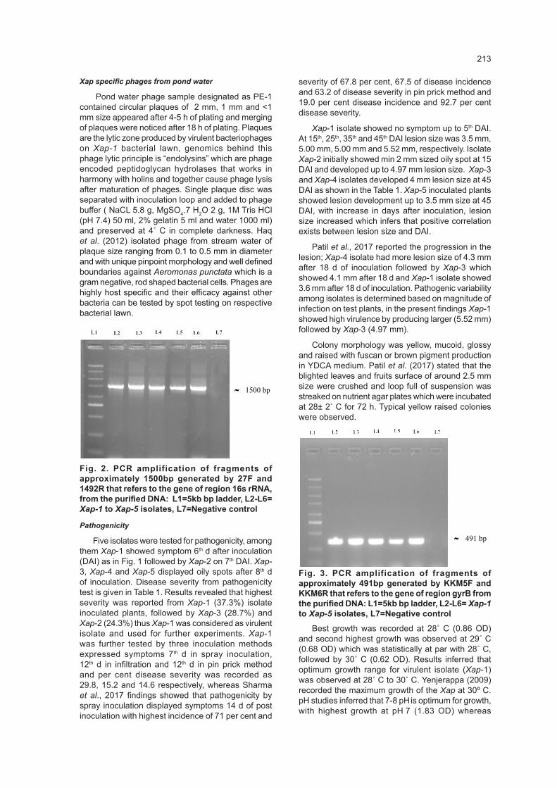
Fig. 2. PCR amplification of fragments of approximately 1500bp generated by 27F and 1492R that refers to the gene of region 16s rRNA, from the purified DNA: L1=5kb bp ladder, L2-L6= Xap-1 to Xap-5 isolates, L7=Negative control
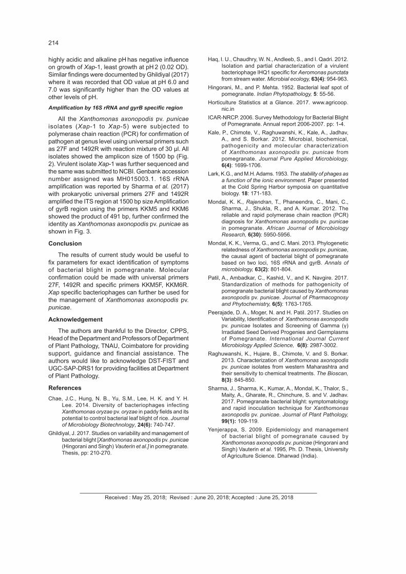
Fig. 3. PCR amplification of fragments of approximately 491bp generated by KKM5F and KKM6R that refers to the gene of region gyrB from the purified DNA: L1=5kb bp ladder, L2-L6= Xap-1 to Xap-5 isolates, L7=Negative control
11500 bp
491 bp
˜
˜
Fig. 2. PCR amplification of fragments of approximately 1500bp generated by 27F and 1492R that refers to the gene of region 16s rRNA, from the purified DNA: L1=5kb bp ladder, L2-L6= Xap-1 to Xap-5 isolates, L7=Negative control
2
Fig. 2. PCR amplification of fragments of approximately 1500bp generated by 27F and 1492R that refers to the gene of region 16s rRNA, from the purified DNA: L1=5kb bp ladder, L2-L6= Xap-1 to Xap-5 isolates, L7=Negative control
Fig. 3. PCR amplification of fragments of approximately 491bp generated by KKM5F and KKM6R that refers to the gene of region gyrB from the purified DNA: L1=5kb bp ladder, L2-L6= Xap-1 to Xap-5 isolates, L7=Negative control
11500 bp
491 bp
˜
˜
Fig. 3. PCR amplification of fragments of approximately 491bp generated by KKM5F and KKM6R that refers to the gene of region gyrB from the purified DNA: L1=5kb bp ladder, L2-L6= Xap-1 to Xap-5 isolates, L7=Negative control
214
highly acidic and alkaline pH has negative influence on growth of Xap-1, least growth at pH 2 (0.02 OD). Similar findings were documented by Ghildiyal (2017) where it was recorded that OD value at pH 6.0 and 7.0 was significantly higher than the OD values at other levels of pH. Amplification by 16S rRNA and gyrB specific region
All the Xanthomonas axonopodis pv. punicae isolates (Xap-1 to Xap-5) were subjected to polymerase chain reaction (PCR) for confirmation of pathogen at genus level using universal primers such as 27F and 1492R with reaction mixture of 30 µl. All isolates showed the amplicon size of 1500 bp (Fig. 2). Virulent isolate Xap-1 was further sequenced and the same was submitted to NCBI. Genbank accession number assigned was MH015003.1. 16S rRNA amplification was reported by Sharma et al. (2017) with prokaryotic universal primers 27F and 1492R amplified the ITS region at 1500 bp size Amplification of gyrB region using the primers KKM5 and KKM6 showed the product of 491 bp, further confirmed the identity as Xanthomonas axonopodis pv. punicae as shown in Fig. 3.
Conclusion
The results of current study would be useful to fix parameters for exact identification of symptoms of bacterial blight in pomegranate. Molecular confirmation could be made with universal primers 27F, 1492R and specific primers KKM5F, KKM6R. Xap specific bacteriophages can further be used for the management of Xanthomonas axonopodis pv. punicae.
Acknowledgement
The authors are thankful to the Director, CPPS, Head of the Department and Professors of Department of Plant Pathology, TNAU, Coimbatore for providing support, guidance and financial assistance. The authors would like to acknowledge DST-FIST and UGC-SAP-DRS1 for providing facilities at Department of Plant Pathology.
References Chae, J.C., Hung, N. B., Yu, S.M., Lee, H. K. and Y. H.
Lee. 2014. Diversity of bacteriophages infecting Xanthomonas oryzae pv. oryzae in paddy fields and its potential to control bacterial leaf blight of rice. Journal of Microbiology Biotechnology, 24(6): 740-747.
Ghildiyal, J. 2017. Studies on variability and management of bacterial blight [Xanthomonas axonopodis pv. punicae (Hingorani and Singh) Vauterin et al.] in pomegranate. Thesis, pp: 210-270.
Haq, I. U., Chaudhry, W. N., Andleeb, S., and I. Qadri. 2012. Isolation and partial characterization of a virulent bacteriophage IHQ1 specific for Aeromonas punctata from stream water. Microbial ecology, 63(4): 954-963.
Hingorani, M., and P. Mehta. 1952. Bacterial leaf spot of pomegranate. Indian Phytopathology, 5: 55-56.
Horticulture Statistics at a Glance. 2017. www.agricoop.nic.in
ICAR-NRCP. 2006. Survey Methodology for Bacterial Blight of Pomegranate. Annual report 2006-2007. pp: 1-4.
Kale, P., Chimote, V., Raghuwanshi, K., Kale, A., Jadhav, A., and S. Borkar. 2012. Microbial, biochemical, pathogenicity and molecular characterization of Xanthomonas axonopodis pv. punicae from pomegranate. Journal Pure Applied Microbiology, 6(4): 1699-1706.
Lark, K.G., and M.H. Adams. 1953. The stability of phages as a function of the ionic environment. Paper presented at the Cold Spring Harbor symposia on quantitative biology. 18: 171-183.
Mondal, K. K., Rajendran, T., Phaneendra, C., Mani, C., Sharma, J., Shukla, R., and A. Kumar. 2012. The reliable and rapid polymerase chain reaction (PCR) diagnosis for Xanthomonas axonopodis pv. punicae in pomegranate. African Journal of Microbiology Research, 6(30): 5950-5956.
Mondal, K. K., Verma, G., and C. Mani. 2013. Phylogenetic relatedness of Xanthomonas axonopodis pv. punicae, the causal agent of bacterial blight of pomegranate based on two loci, 16S rRNA and gyrB. Annals of microbiology, 63(2): 801-804.
Patil, A., Ambadkar, C., Kashid, V., and K. Navgire. 2017. Standardization of methods for pathogenicity of pomegranate bacterial blight caused by Xanthomonas axonopodis pv. punicae. Journal of Pharmacognosy and Phytochemistry, 6(5): 1763-1765.
Peerajade, D. A., Moger, N. and H. Patil. 2017. Studies on Variability, Identification of Xanthomonas axonopodis pv. punicae Isolates and Screening of Gamma (γ) Irradiated Seed Derived Progenies and Germplasms of Pomegranate. International Journal Current Microbiology Applied Science, 6(8): 2987-3002.
Raghuwanshi, K., Hujare, B., Chimote, V. and S. Borkar. 2013. Characterization of Xanthomonas axonopodis pv. punicae isolates from western Maharashtra and their sensitivity to chemical treatments. The Bioscan, 8(3): 845-850.
Sharma, J., Sharma, K., Kumar, A., Mondal, K., Thalor, S., Maity, A., Gharate, R., Chinchure, S. and V. Jadhav. 2017. Pomegranate bacterial blight: symptomatology and rapid inoculation technique for Xanthomonas axonopodis pv. punicae. Journal of Plant Pathology, 99(1): 109-119.
Yenjerappa, S. 2009. Epidemiology and management of bacterial blight of pomegranate caused by Xanthomonas axonopodis pv. punicae (Hingorani and Singh) Vauterin et al. 1995, Ph. D. Thesis, University of Agriculture Science. Dharwad (India).
Received : May 25, 2018; Revised : June 20, 2018; Accepted : June 25, 2018