
Characterization of the Precipitin Bands Detected in the
7
APPLIED MICROBIOLOGY, JUly 1974, p. 138-144 Copyright 0 1974 American Society for Microbiology Vol. 28, No. 1 Printed in U.S.A. Characterization of the Precipitin Bands Detected in the Immunodiffusion Test for Paracoccidioidomycosis ANGELA RESTREPO AND LUZ H. MONCADA Department of Microbiology and Parasitology, School of Medicine, University of Antioquia, Medellin, Colombia Received for publication 4 March 1974 In order to characterize the precipitin bands detected in the immunodiffusion test for paracoccidioidomycosis, a study was undertaken in 54 patients with the disease. On the basis of the pattern of known control sera, the three commonly observed lines of precipitate were designated as 1, 2, and 3 according to their location in the immunodiffusion plate. At time of diagnosis, 28 of the patients exhibited all three bands, 16 gave two bands, and 10 showed only one precipitin line. Over 50 of the sera with three bands had high complement fixation titers (above 1:512), whereas those with one band exhibited lower titers. A similar picture was obtained with the quantitative agar-gel techniques, where titers of 1:64 and above were more commonly observed in sera with three precipitin lines. Follow-up studies carried out in 18 patients revealed that band 3 disappeared first, followed by band 2, and, finally, by band 1. At the end of 2 to 3 years, 85.7% of the patients had lost band 3, 75% band 2, and only 27.7% band 1. Cross-reactions with histoplasmin were found in eight patients who gave the M precipitin line with this antigen. It was found that the latter band and our paracoccidioidin band 3 fused, producing lines of identity. Bands 1 and 2 were specific. The implications of these findings are discussed. In earlier publications, we have analyzed the characteristics of the immunodiffusion test for the serological diagnosis of paracoccidi- oidomycosis (9, 11, 12). It has been shown that a yeast-phase antigen from Paracoccidioides brasiliensis produced positive reactions in 95% of the cases (6, 11). One or two of the precipitin lines demonstrable at the time of diagnosis were gradually lost in the course of time. The antigen used was specific, and no precipitin bands were detected in patients with other mycoses (6). It was observed, however, that some patients with proven paracoccidioidomycosis reacted to the immunodiffusion test with histoplasmin (11). A more detailed study aimed towards stan- dardization of the technique was deemed neces- sary. The purpose of the present report is to characterize the precipitin bands, determining their frequency and tendency to disappear. Also, the relationship between the cross-react- ing histoplasmin band and the newly character- ized paracoccidiodin bands was investigated in an effort to select those which appear to be specific. MATERIALS AND METHODS Blood was obtained under fasting conditions from 54 newly diagnosed paracoccidioidomycosis patients. The serum was removed, preserved with merthiolate 1: 10,000, divided into 1.0-ml portions, and frozen for periods up to 3 years. Before testing, the required number of vials were thawed, and the serum was utilized the same day. A total of 140 serum specimens were available for the study. Serological follow-ups were possible in 18 patients, blood specimens being collected at the time of diagnosis, at 6 months, and 1, 2, and sometimes 3 years after establishment of the diagnosis. During the follow-up period, all patients received treatment with sulfa drugs or amphotericin B or both. The complement fixation (CF) test and the agar-gel immunodiffusion techniques were performed accord- ing to standard methods (9, 14). For the latter, both qualitative and quantitative procedures were utilized (12). The basic immunodiffusion pattern consisted of a central well surrounded by six lateral reservoirs; when cross-reactions were being investigated, the designs proposed by Huppert (5) were utilized. A single batch of the yeast paracoccidioidin, pre- pared according to techniques previously described, was utilized (13). The histoplasmin was obtained from the Center for Disease Control (CDC), Atlanta, Ga. Both antigens were utilized in the study of all serum specimens. For characterization purposes, six sera known to produce three precipitin bands with paracoccidioidin were pooled and used as a control. Subsequently, all sera were compared with this standard. The precipitin lines were designated as 1, 2, and 3, according to their 138
Transcript of Characterization of the Precipitin Bands Detected in the

APPLIED MICROBIOLOGY, JUly 1974, p. 138-144Copyright 0 1974 American Society for Microbiology
Vol. 28, No. 1Printed in U.S.A.
Characterization of the Precipitin Bands Detected in theImmunodiffusion Test for Paracoccidioidomycosis
ANGELA RESTREPO AND LUZ H. MONCADADepartment of Microbiology and Parasitology, School of Medicine, University of Antioquia,
Medellin, Colombia
Received for publication 4 March 1974
In order to characterize the precipitin bands detected in the immunodiffusiontest for paracoccidioidomycosis, a study was undertaken in 54 patients with thedisease. On the basis of the pattern of known control sera, the three commonlyobserved lines of precipitate were designated as 1, 2, and 3 according to theirlocation in the immunodiffusion plate. At time of diagnosis, 28 of the patientsexhibited all three bands, 16 gave two bands, and 10 showed only one precipitinline. Over 50 of the sera with three bands had high complement fixation titers(above 1:512), whereas those with one band exhibited lower titers. A similarpicture was obtained with the quantitative agar-gel techniques, where titers of1:64 and above were more commonly observed in sera with three precipitin lines.Follow-up studies carried out in 18 patients revealed that band 3 disappearedfirst, followed by band 2, and, finally, by band 1. At the end of 2 to 3 years, 85.7%of the patients had lost band 3, 75% band 2, and only 27.7% band 1.Cross-reactions with histoplasmin were found in eight patients who gave the Mprecipitin line with this antigen. It was found that the latter band and our
paracoccidioidin band 3 fused, producing lines of identity. Bands 1 and 2 were
specific. The implications of these findings are discussed.
In earlier publications, we have analyzed thecharacteristics of the immunodiffusion test forthe serological diagnosis of paracoccidi-oidomycosis (9, 11, 12). It has been shown that ayeast-phase antigen from Paracoccidioidesbrasiliensis produced positive reactions in 95%of the cases (6, 11). One or two of the precipitinlines demonstrable at the time of diagnosis weregradually lost in the course of time. The antigenused was specific, and no precipitin bands weredetected in patients with other mycoses (6). Itwas observed, however, that some patients withproven paracoccidioidomycosis reacted to theimmunodiffusion test with histoplasmin (11).A more detailed study aimed towards stan-
dardization of the technique was deemed neces-sary. The purpose of the present report is tocharacterize the precipitin bands, determiningtheir frequency and tendency to disappear.Also, the relationship between the cross-react-ing histoplasmin band and the newly character-ized paracoccidiodin bands was investigated inan effort to select those which appear to bespecific.
MATERIALS AND METHODSBlood was obtained under fasting conditions from
54 newly diagnosed paracoccidioidomycosis patients.
The serum was removed, preserved with merthiolate1: 10,000, divided into 1.0-ml portions, and frozen forperiods up to 3 years. Before testing, the requirednumber of vials were thawed, and the serum wasutilized the same day. A total of 140 serum specimenswere available for the study. Serological follow-upswere possible in 18 patients, blood specimens beingcollected at the time of diagnosis, at 6 months, and 1,2, and sometimes 3 years after establishment of thediagnosis. During the follow-up period, all patientsreceived treatment with sulfa drugs or amphotericinB or both.The complement fixation (CF) test and the agar-gel
immunodiffusion techniques were performed accord-ing to standard methods (9, 14). For the latter, bothqualitative and quantitative procedures were utilized(12). The basic immunodiffusion pattern consisted ofa central well surrounded by six lateral reservoirs;when cross-reactions were being investigated, thedesigns proposed by Huppert (5) were utilized.A single batch of the yeast paracoccidioidin, pre-
pared according to techniques previously described,was utilized (13). The histoplasmin was obtainedfrom the Center for Disease Control (CDC), Atlanta,Ga. Both antigens were utilized in the study of allserum specimens.
For characterization purposes, six sera known toproduce three precipitin bands with paracoccidioidinwere pooled and used as a control. Subsequently, allsera were compared with this standard. The precipitinlines were designated as 1, 2, and 3, according to their
138
VOL. 28, 1974 IMMUNODIFFUSION TEST FOR PARACOCCIDIOIDOMYCOSIS
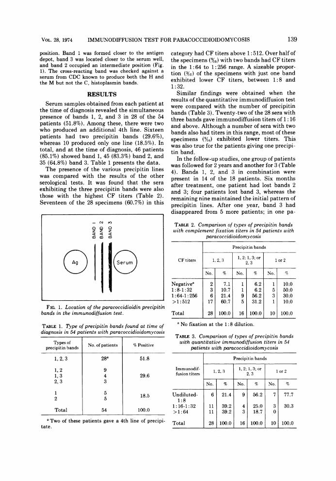
position. Band 1 was formed closer to the antigendepot, band 3 was located closer to the serum well,and band 2 occupied an intermediate position (Fig.1). The cross-reacting band was checked against a
serum from CDC known to produce both the H andthe M but not the C, histoplasmin bands.
RESULTS
Serum samples obtained from each patient atthe time of diagnosis revealed the simultaneouspresence of bands 1, 2, and 3 in 28 of the 54patients (51.8%). Among these, there were twowho produced an additional 4th line. Sixteenpatients had two precipitin bands (29.6%),whereas 10 produced only one line (18.5%). Intotal, and at the time of diagnosis, 46 patients(85.1%) showed band 1, 45 (83.3%) band 2, and35 (64.8%) band 3. Table 1 presents the data.The presence of the various precipitin lines
was compared with the results of the otherserological tests. It was found that the sera
exhibiting the three precipitin bands were alsothose with the highest CF titers (Table 2).Seventeen of the 28 specimens (60.7%) in this
- C'J re)
ZZ Z
Ag ~erum
FIG. 1. Location of the paracoccidioidin precipitinbands in the immunodiffusion test.
TABLE 1. Type of precipitin bands found at time ofdiagnosis in 54 patients with paracoccidioidomycosis
Types of No. of patients % Positiveprecipitin bands
1, 2,3 28a 51.8
1,2 91,3 4 29.62,3 3
2 5 18.5
Total 54 100.0
a Two of these patients gave a 4th line of precipi-tate.
category had CF titers above 1:512. Over half ofthe specimens (9I6) with two bands had CF titersin the 1:64 to 1: 256 range. A sizeable propor-tion (5/10) of the specimens with just one bandexhibited lower CF titers, between 1:8 and1:32.
Similar findings were obtained when theresults of the quantitative immunodiffusion testwere compared with the number of precipitinbands (Table 3). Twenty-two of the 28 sera withthree bands gave immunodiffusion titers of 1:16and above. Although a number of sera with twobands also had titers in this range, most of thesespecimens (9,46) exhibited lower titers. Thiswas also true for the patients giving one precipi-tin band.
In the follow-up studies, one group of patientswas followed for 2 years and another for 3 (Table4). Bands 1, 2, and 3 in combination werepresent in 14 of the 18 patients. Six monthsafter treatment, one patient had lost bands 2and 3; four patients lost band 3, whereas theremaining nine maintained the initial pattern ofprecipitin lines. After one year, band 3 haddisappeared from 5 more patients; in one pa-
TABLE 2. Comparison of types of precipitin bandswith complement fixation titers in 54 patients with
paracoccidioidomycosis
Precipitin bands
CF titers 1, 2, 3 1,2; 1,3; or Ior 22.3
No. % No. % No. %
Negativea 2 7.1 1 6.2 1 10.01:8-1:32 3 10.7 1 6.2 5 50.01:64-1:256 6 21.4 9 56.2 3 30.0> 1:512 17 60.7 5 31.2 1 10.0
Total 28 100.0 16 100.0 10 100.0
a No fixation at the 1:8 dilution.
TABLE 3. Comparison of types of precipitin bandswith quantitative immunodiffusion titers in 54
patients with paracoccidioidomycosis
Precipitin bands
Immunodif- 1, 1 2 ;1, 3; or 1 or 2fusion titers 2, 3
No. % No. % No. %
Undiluted- 6 21.4 9 56.2 7 77.71:8
1:16-1:32 11 39.2 4 25.0 3 30.3>1:64 11 39.2 3 18.7 0
Total 28 100.0 16 100.0 10 100.0
139

RESTREPO AND MONCADA
tient, band 2 also disappeared, but band 1continued to be present in 17 patients. Fromhere on, bands 2 and 3 were lost by most of theremaining patients; band 1, however, disap-peared only from four cases.
TABLE 4. Serological follow-up of patients withparacoccidioidomycosis and fate of the precipitin
bands
Precipitin bands at time of testingPatient
no. Timeof 6 months 1 year 2 years 3 yearsdiagnosis
1 1,2,3 1,2,3 1,2,3 1,2,3 1,2,32 1, 2,3 1, 2,3 1, 2,3 1, 2,3 1, 2, a3 1, 2,3 1, 2,3 1, 2,3 1, 2, - 1, - -4 1,2,3 1,2,3 1,2,- 1,-- 1,--5 1,2,3 1,2,3 1,2,- 1,-- 1,--6 1,2,3 1, -- 1, -- 1, -- 1, --7 1,2,3 1,2,3 1,2,3 1,2,3 1,2,38 1,2 1,2 1,2 1,- 1,-9 1 1 - -10 1 1 1 - NDb11 1, 2,3 1, 2,- 1, 2,- 1,-- ND12 1, 2,3 1, 2, - 1, 2,- 1, - - ND13 1,2,3 1,2,- 1,2,- - ND14 1, 2,3 1, 2,- 1, -- - ND15 1,2,3 1,2,3 1,2,- 1,-- ND16 1, 2, 1, 2 1, -- - ND17 1, 2,3 1, 2,3 1, 2,- 1, 2,- ND18 1, 2,3 1, 2,3 1, 2,- 1,-- NDa Precipitin line was lost (shown by dash)." ND, not done.
During the period of observation, band 3 waslost by 12 of the 14 patients (85.6%), band 2disappeared from 12 of the 16 patients (75.0%),and band 1 was lost only by five of the 18patients (27.7%). The sequence of events isillustrated in Fig. 2 and 3.Among the patients studied, there were eight
(14.8%) whose sera also produced a precipitinband with histoplasmin (M band). In six pa-tients this cross-reactivity was already presentin the first serum sample examined, and, in onecase, it persisted for the 3 years of the study.The remaining two patients acquired the cross-reactivity during the first 6 months of observa-tion. Three of the patients also exhibited com-plement-fixing antibodies with the Histoplasmacapsulatum antigen employed (Table 5). Ingeneral, these patients had high CF titers withparacoccidioidin, but the quantitative immuno-diffusion titers were not always elevated. All,however, had the three precipitin bands whenthe cross-reaction was detected. It must bestated that none of these patients had beengiven a skin test with histoplasmin.
Sera from these patients were compared inthe same agar-gel plate with 2 homologous anti-gen-antibody systems, namely, P. brasiliensisand H. capsulatum. It was observed that para-coccidioidin band 3 and histoplasmin band Mfused, producing lines of identity (Fig. 4).Bands 1 and 2 did not cross-react with thesoluble H. capsulatum antigen employed in thetest (Fig. 5).
F ia 2. Patient 10, follow-up. Central well has paracoccidioidin; peripheral wells have: + (left), control serawith bands 2 and 3; + (right), control serum with bands I and 2. Wells I, II, III, and IVhave patient's sera takenat diagnosis, 6 months, I and 2 years after treatment, respectively. In well IVnote that band 1 has disappeared.
140 APPL. MICROBIOL.

VOL 28, 1974 IMMUNODIFFUSION TEST FOR PARACOCCIDIOIDOMYCOSIS
Fic 3. Patient 3, follow-up. Central well has paracoccidioidin; peripheral wells have: + (left), control serumwith bands 1, 2, 3; + (right), control serum with bands 2, 3. Wells I, II, III, and IV have patient's sera taken atdiagnosis, 6 months, 1 and 3 years after treatment, respectively. In well IV note weak band 1 and absence ofother bands.
TABLE 5. Results of serological tests in eight patientswith paracoccidioidomycosis who reacted with both
paracoccidioidin and histoplasmin
Results of serological tests
Complement Immunodiffusionfixations
Patient H.no. H P. brasiliensis capsulatum
P. . bandsbrasi- cap-liensis SU- No.latum of Titera H M
bands
2 4.096 64 3 64 - +3 2.048 N5 3 8 - +4 1.024 8 3 128 - +11 512 N 3 Undiluted - +13 128 N 3 32 - +14 128 N 3 8 - +15 256 8 3 128 - +
a Reciprocal of the titer.'No fixation at the 1: 8 dilution.
DISCUSSIONWith the characterization of the precipitin
lines formed by patients with paracoccidi-oidomycosis, the results of the immunodiffusiontest can be better interpreted. In a new patientthe simultaneous presence of bands 1 and 2, or
even of band 1 alone, allows a presumptivediagnosis. Band 3, because of its cross-reactiv-
ity, should be interpreted judiciously, and pa-tients exhibiting only this line of precipitateshould be subjected to other mycological studiesbefore establishing a diagnosis.
In the patient undergoing treatment, one mayexpect a reduction in the number of precipitinbands. Band 3 should be lost in 1 to 2 years andband 2 in 2 to 3 years. Band 1 will probably bedetectable for extended periods of time, even inthe presence of substantial clinical improve-ment. Mention should be made of two of ourpatients (Table 4, no. 1, 7) who, in over 3 yearsof careful follow-up and treatment, did notchange their immunodiffusion patterns northeir CF titers ranging from 1:216 to 1:1024.Both patients exhibited pulmonary paracoc-cidioidomycosis but improved clinically andradiologically and had negative sputum cul-tures. If the serological reactivity indicates anactive infection, these patients would not beconsidered cured in spite of the improvementnoticed; perhaps their immune cellular mech-anisms were powerful enough to cope with thepersistent activity of the fungus, thus hinderingthe progress of the mycotic process.The value of the agar-gel immunodiffusion
techniques in the diagnosis of various pulmo-nary mycoses is well established (2). In histo-plasmosis and coccidioidomycosis, for instance,the precipitin bands have been properly charac-terized. In coccidioidomycosis, the F band,present in virtually all patients, is specific for
141

12RESTREPO AND MONCADA
F iG 4. Comparison of cross-reacting serum from a patient with paracoccidioidomycosis with standardizedreference patterns for paracoccidioidomycosis and histoplasmosis. Upper wells: L, paracoccidioidin; H,histoplasmin. Lower wells: SL, paracoccidioidomycosis antiserum; SH, histoplasmosis antiserum; 2, cross-reacting serum. In diagrammatical presentation below, note fusion of paracoccidioidin band 3 withhistoplasmin band M.
this mycotic disorder (5). In histoplasmosis, theH band and, to some extent, the M band areconsidered specific, and their joint presence isdiagnostically significant (2, 4). The contrary isthe case with band C which cross-reacts withantigens from Coccidiodes immitis (4).The results of the present study indicate that
some patients with paracoccidioidomycosis,who have not been skin-tested with histoplas-min, can react with this antigen, producing theM band. The fact that this band and our
paracoccidioidomycosis band 3 fuse to producea line of identity is of interest, as there are pos-sibilities for an erroneous diagnosis. If only his-toplasmin is utilized in the immunodiffusiontest, some patients with paracoccidioidomyco-sis might be misdiagnosed. If paracoccidiodin isalso employed, the presence of various precipi-tin lines with this antigen would clarify the pic-ture, because cross-reactions with histoplasminare apparently present only in patients exhibit-ing paracoccidiodin bands 1, 2, and 3. It is rec-
142 APPL. MWICROBIOL.
.6e4lei 6
X\

VOL. 28, 1974 IMMUNODIFFUSION TEST FOR PARACOCCIDIOIDOMYCOSIS
FIG 5. Nonidentity ofparacoccidioidin bands I and 2 and identity of band 3 with histoplasmin band. Upperwells contain paracoccidioidin (left) and histoplasmin (right). Lower wells: SL, paracoccidioidomycosisantiserum; SH, histoplasmosis antiserum; 4, cross-reacting serum. In diagrammatical presentation below, notefusion of histoplasmin M band with paracoccidioidin band 3.
ommended that laboratories processing samplesfrom patients living in areas endemic for histo-plasmosis and paracoccidioidomycosis make useof antigens derived from both etiologic agents.
As concerns the cross-reacting band, one ispuzzled by the fact that 35 patients producedband 3, but only eight cross-reacted with histo-plasmin. If band 3 is identical to band M, whydid the other 27 patients fail to react? A possibleexplanation would be that a relatively largequantity of the respective antibody is requiredto produce a distinct M band, whereas lesser
quantities are sufficient to form band 3. Per-haps an answer could be obtained by employinga more sensitive indicator such as the precipita-tion inhibition immunodiffusion technique.
Using immunoelectrophoretic techniques andantigens identical to the ones employed in thepresent study, various precipitin arcs (A, B, C,D, E) have been shown to occur in paracoccidi-oidomycosis (10). It is of interest that the Aband described in the latter study is detectedwith equal frequency and for the same longperiods of time as is band 1. Yarzabal (15),
143

144RESTREPO AND MONCADA
working with different antigenic preparationsbut also employing immunoelectrophoresis,found 25 arcs of precipitation; an arc, desig-nated E and characterized by its cathodicmigration, was considered completely specific,as it was absent from the sera of patients withother mycoses. We think that arc E is identicalto the previously described arc A. Thus, it ispossible that arc A, which appears to have thecharacteristics of band 1, may also be identicalto Yarzabal's specific arc E. More recently, andby means of the immunoelectroosmophoresis-immunodiffusion test (3), the presence of spe-cific precipitin lines located in the cathodiczone have been described, thus confirmingYarzabal's (15) findings.Although immunoelectrophoresis usually
gives more precise results than the immunodif-fusion test, the former is not easy to performand requires complex equipment. Conse-quently, its use is limited to specialized labora-tories. The immunodiffusion test, on the otherhand, is an extremely simple technique whichcan be adapted to the clinical laboratory.
It is expected that the results of the presentstudy will strengthen the value of the immuno-diffusion test in the diagnosis of paracoccidi-oidomycosis, especially now that more laborato-ries are starting to perform the test (1, 7, 8). It isalso hoped that in the future "reference rea-gents, tested for potency and specifity, (5)" canbe made available through international agen-cies so that results from various laboratories canbe compared.
ACKNOWLEDGMENTSSincere appreciation is expressed to Alberto Correa and
Carlos Jaramillo for their cooperation with the photographs.
LITERATURE CITED
1. Barbosa, W., J. R. Mendoca, and R. L. Oliveira. 1972.Imunologfa da Blastomicose Sul-Americana. Rev. Pat.
Trop. (Brazil) 1:393-402.2. Buechner, H. A., J. H. Seabury, C. C. Campbell, L. K.
George, L. Kaufman, and W. Kaplan. 1973. Thecurrent status of serologic, immunologic and skin testsin the diagnosis of pulmonary mycosis. Chest63:259-270.
3. Conti-Diaz, I. A., R. E. Somma-Moreira, E. Gezuele, A.de Gimenez, M. I. Pefia, and J. E. Mackinnon. 1973.Immunoelectroosmophoresis-immunodiffusion in para-coccidioidomycosis. Sabouraudia 11:39-41.
4. Heiner, D. C. 1958. Diagnosis of histoplasmosis usingprecipitin reactions in agar gel. Pediatrics 22:616-627.
5. Huppert, M. 1970. Standardization of immunologicalreagents, p. 243-252. Proc. Int. Symp. Mycoses. PAHOSci. Publ. no. 205.
6. Kaufman, L. 1972. Evaluation of serological tests forparacoccidioidomycosis: preliminary report, p.221-223. Proc. First Int. Symp. Paracoccidioidomyco-sis. PAHO Sci. Publ. no. 254.
7. Lazo, R. F. 1968. La inmunodifusi6n en el diagn6stico dela blastomicosis suramericana. Rev. Ecuator. Hig.Med. Trop. 25:253-260.
8. Negroni, R. 1968. Nuevos estudios sobre anttgenos paralas pruebas serol6gicas en la blastomicosis sud-americana. Dermatol. Ibero. Lat. Amer. 4:409-416.
9. Restrepo, A. 1966. La prueba de inmunodifusi6n en gel deagar en el diagn6stico de la paracoccidioidomicosis.Sabouraudia 4:223-230.
10. Restrepo, A., and E. Drouhet. 1970. Etude des anticorpsprecipitants dans la blastomycose sud-americaine parl'analyse immunoelectrophoretique des antigens de P.brasiliensis. Ann. Inst. Pasteur (Paris) 119:338-346.
11. Restrepo, A., and L. H. Moncada. 1970. Serologic proce-dures in the diagnosis of paracoccidioidomycosins, p.101-110. Proc. Int. Symp. Mycoses. PAHO Sci. Publ.no. 205.
12. Restrepo, A., and L. H. Moncada. 1972. Indirect fluores-cent antibody and quantitative agar-gel immunodiffu-sion tests for the serological diagnosis of paracoccidi-oidomycosis. Appl. Microbiol. 24:132-137.
13. Restrepo-Moreno, A., and J. D. Schneidau, Jr. 1967.Nature of the skin-reactive principle in culture filtratesprepared from Paracoccidioides brasiliensis. J. Bacte-riol. 93:1741-1748.
14. U.S. Department of Health, Education, and Welfare.1965. Standardized diagnostic complement fixationmethod and adptation to micro-test. U.S. Publ. HealthServ. Publ. no. 1228, Washington, D.C.
15. Yarzdbal, L. 1971. Anticuerpos precipitantes especfficosde la blastomicosis sudamericana, reveleados por in-munoelectroforesis. Rev. Inst. Med. Trop. Sao Paulo13:320-327.
144 APPL. MICROBIOL.


![arXiv:1601.01388v1 [astro-ph.CO] 7 Jan 2016of 6:9˙in a joint fit to data from bands 6 and 7; the effect of the subhalo is also detected in both bands individ-ually. We also derive](https://static.fdocuments.in/doc/165x107/5ea531317447731f0d2d82db/arxiv160101388v1-astro-phco-7-jan-2016-of-69in-a-joint-it-to-data-from.jpg)