Characterization of hydration products of mineral trioxide ... · PDF fileChemical analysis...
10
Characterization of hydration products of mineral trioxide aggregate J. Camilleri Department of Building and Civil Engineering, Faculty of Architecture and Civil Engineering, University of Malta, Malta Abstract Camilleri J. Characterization of hydration products of mineral trioxide aggregate. International Endodontic Journal, 41, 408– 417, 2008. Objective To characterize the hydration products of white mineral trioxide aggregate (MTA). Methodology Mineral trioxide aggregate, white Portland cement and bismuth oxide were evaluated using X-ray diffraction (XRD) analysis and Rietveld XRD. The cements were tested un-hydrated and after hydration and curing for 30 days at 37 °C. Analysis of hydrated cement leachate was performed weekly for five consecutive weeks from mixing using inductively coupled plasma atomic emission spectroscopy after which the cements were viewed under the scanning electron microscope to evaluate the cement micro- structure. Quantitative energy dispersive analysis with X-ray was performed and atomic ratios were plotted. Results Both Portland cement and MTA produced calcium silicate hydrate (C-S-H) and calcium hydroxide (CH) on hydration. The tricalcium aluminate levels were low for MTA which resulted in reduced produc- tion of ettringite and monosulphate. On hydration the bismuth level in the hydrated MTA decreased; bismuth oxide replaced the silica in the C-S-H and was leached out once the C-S-H decomposed with time. Both MTA and Portland cement released a high amount of calcium ions which decreased in amount over the 5-week period. Conclusions The hydration mechanism of MTA is different to that of Portland cement. In MTA the bismuth oxide is bound to the C-S-H and is leached out from the cement with time as the C-S-H decomposes. MTA produces a high proportion of calcium ions from CH a by-product of hydration and also by decomposi- tion of C-S-H. The release of calcium ions reduces with time. Keywords: hydration, mineral trioxide aggregate, Portland cement. Received 11 September 2007; accepted 30 October 2007 Introduction Mineral trioxide aggregate (MTA) is composed of ASTM Type 1 Portland cement with a 4:1 addition of bismuth oxide added for radio-opacity (Torabinejad & White 1995). The elemental constitution of MTA is similar to that of Portland cement (Estrela et al. 2000, Funteas et al. 2003, Asgary et al. 2004) and both cements are composed primarily of tri- and dicalcium silicate as verified by X-ray diffraction (XRD) analysis (Camilleri et al. 2005a). The levels of toxic chemicals in Portland cement are low and similar to those of MTA (Duarte et al. 2005). So it can be postulated that MTA can be replaced by a cheaper Portland cement. MTA is deficient in alumina suggesting that the material is not prepared in a rotary kiln as is customary for the manufacture of Portland cement (Camilleri 2007). Thus, although both MTA and Portland cement have similar major constituent elements there is a variation in the other compounds present. The low levels of alumina affect the production of ettringite and mono- sulphate usually formed on hydration of Portland cement (Camilleri 2007). The first publication on the chemical and physical properties of MTA (Torabinejad et al. 1995) did not report the presence of bismuth oxide in the material. Correspondence: Dr Josette Camilleri, Department of Building and Civil Engineering, Faculty of Architecture and Civil Engineering, University of Malta, Malta (Tel.: 00356 2340 2870; fax: 00356 21 330190; e-mail: josette.camilleri @um.edu.mt). doi:10.1111/j.1365-2591.2007.01370.x International Endodontic Journal, 41, 408–417, 2008 ª 2008 International Endodontic Journal 408
Transcript of Characterization of hydration products of mineral trioxide ... · PDF fileChemical analysis...

Characterization of hydration products of mineraltrioxide aggregate
J. CamilleriDepartment of Building and Civil Engineering, Faculty of Architecture and Civil Engineering, University of Malta, Malta
Abstract
Camilleri J. Characterization of hydration products of mineral
trioxide aggregate. International Endodontic Journal, 41, 408–
417, 2008.
Objective To characterize the hydration products of
white mineral trioxide aggregate (MTA).
Methodology Mineral trioxide aggregate, white
Portland cement and bismuth oxide were evaluated
using X-ray diffraction (XRD) analysis and Rietveld
XRD. The cements were tested un-hydrated and after
hydration and curing for 30 days at 37 �C. Analysis of
hydrated cement leachate was performed weekly for
five consecutive weeks from mixing using inductively
coupled plasma atomic emission spectroscopy after
which the cements were viewed under the scanning
electron microscope to evaluate the cement micro-
structure. Quantitative energy dispersive analysis with
X-ray was performed and atomic ratios were plotted.
Results Both Portland cement and MTA produced
calcium silicate hydrate (C-S-H) and calcium hydroxide
(CH) on hydration. The tricalcium aluminate levels
were low for MTA which resulted in reduced produc-
tion of ettringite and monosulphate. On hydration the
bismuth level in the hydrated MTA decreased; bismuth
oxide replaced the silica in the C-S-H and was leached
out once the C-S-H decomposed with time. Both MTA
and Portland cement released a high amount of
calcium ions which decreased in amount over the
5-week period.
Conclusions The hydration mechanism of MTA is
different to that of Portland cement. In MTA the
bismuth oxide is bound to the C-S-H and is leached out
from the cement with time as the C-S-H decomposes.
MTA produces a high proportion of calcium ions from
CH a by-product of hydration and also by decomposi-
tion of C-S-H. The release of calcium ions reduces with
time.
Keywords: hydration, mineral trioxide aggregate,
Portland cement.
Received 11 September 2007; accepted 30 October 2007
Introduction
Mineral trioxide aggregate (MTA) is composed of ASTM
Type 1 Portland cement with a 4 : 1 addition of
bismuth oxide added for radio-opacity (Torabinejad &
White 1995). The elemental constitution of MTA is
similar to that of Portland cement (Estrela et al. 2000,
Funteas et al. 2003, Asgary et al. 2004) and both
cements are composed primarily of tri- and dicalcium
silicate as verified by X-ray diffraction (XRD) analysis
(Camilleri et al. 2005a). The levels of toxic chemicals in
Portland cement are low and similar to those of MTA
(Duarte et al. 2005). So it can be postulated that MTA
can be replaced by a cheaper Portland cement. MTA is
deficient in alumina suggesting that the material is not
prepared in a rotary kiln as is customary for the
manufacture of Portland cement (Camilleri 2007).
Thus, although both MTA and Portland cement have
similar major constituent elements there is a variation
in the other compounds present. The low levels of
alumina affect the production of ettringite and mono-
sulphate usually formed on hydration of Portland
cement (Camilleri 2007).
The first publication on the chemical and physical
properties of MTA (Torabinejad et al. 1995) did not
report the presence of bismuth oxide in the material.
Correspondence: Dr Josette Camilleri, Department of Building
and Civil Engineering, Faculty of Architecture and Civil
Engineering, University of Malta, Malta (Tel.: 00356 2340
2870; fax: 00356 21 330190; e-mail: josette.camilleri
@um.edu.mt).
doi:10.1111/j.1365-2591.2007.01370.x
International Endodontic Journal, 41, 408–417, 2008 ª 2008 International Endodontic Journal408
The bismuth oxide seemed to have been added later to
enhance the radio-opacity of MTA. Bismuth oxide
affected the hydration mechanism of the MTA; it
formed part of the structure of the calcium silicate
hydrate (C-S-H) and also affected the precipitation of
calcium hydroxide (CH) in the hydrated paste
(Camilleri 2007). The levels of bismuth and calcium
leached out from MTA are as yet unknown. The level of
bismuth that is actually bound to the C-S-H gel
structure has also not been investigated.
The objective of this study was to characterize the
hydration products of white MTA and compare them to
those of white Portland cement.
Materials and methods
White MTA (Tulsa Dental Products, Tulsa, OK, USA;
Batch number: 756464/02) and white Portland
cement (Castle White Portland cement; BS EN 197-1:
2000, Type CEM 1; strength class 52,5N) were used in
this study.
Chemical analysis of the materials
Chemical analysis was performed on both hydrated and
un-hydrated MTA and Portland cement and bismuth
oxide (Fischer Scientific, Leicester, UK). MTA was
mixed with the liquid provided in capsules to produce
a liquid-to-MTA ratio of 0.5. One gram of white
Portland cement was mixed with 0.5 g distilled water,
to give a water/cement (w/c) ratio of 0.5. The two
pastes were cured at 37 �C in water for 30 days after
which they were washed with isopropyl alcohol to
desiccate them prior to performing the analysis.
X-ray diffraction analysis
For XRD analysis 20% rutile (Titanium dioxide; Sigma-
Aldrich, Gillingham, UK) was added to each material to
be used as internal standard. The hydrated cements
were crushed to a very fine powder using a mortar and
pestle. The phase analysis of both cements hydrated
and un-hydrated and bismuth oxide was performed
using XRD analysis. The diffractometer (Ital Structures
Compact 3K5, Riva del Garda, Italy) used Cu Karadiation at 30 mA and 40 kV. Samples were pre-
sented in powder form and were compacted on a
sample holder. The crystalline structure of the test
cement was determined by passing a beam of X-rays of
known wavelength into the specimen whilst rotating it
through an angle h. The intensity of X-rays from the
sample was measured by a detector. The detector was
rotated between 10� and 60� at 0.02� 2h per 0.5 s and
also between 45� and 55� at 0.005� 2h per 0.1 s.
Phase identification was accomplished by use of search-
match software utilizing the International Centre for
Diffraction Data (ICDD) database (International Centre
for Diffraction Data, Newtown Square, PA, USA).
Rietveld X-ray diffraction analysis
Materials under study were hydrated and un-hydrated
cements and bismuth oxide. Twenty per cent corun-
dum (Al2O3) relative to the weight of cement was
blended with the cement powder to serve as an internal
standard. This material was chosen because it does not
react with water and has no influence on the hydration
reaction. Rietveld analysis was performed on ground
sample. X-ray diffraction patterns were first obtained
with sufficient resolution for Rietveld analysis. The
detector was rotated at 0.017� 2h per 20 s.
Chemical analyses of cement leachate
The chemical analysis of the cement products released
into solution was performed using inductively coupled
plasma atomic emission spectroscopy (ICP-AES; Varian
Medical Systems, Palo Alto, CA, USA). MTA and
Portland cement were mixed as described in the
previous experiment, and cured for 3 days at 37 �C
and 100% humidity. They were then weighed and
placed in closed plastic sterile containers (Labplex,
Birmingham, UK) to which was added 100 mL of
deionized water. Containers filled with deionized water
were used as controls. The samples were allowed to
cure at 37 �C for 5 weeks. The water within the bottles
was changed on a weekly basis with the leachate water
being collected for analysis. The leachate was analysed
for aluminium, bismuth, calcium, iron, potassium,
magnesium, phosphorus, sodium and silicon. The
amount of leachate was calculated in lg/g by using
the following formula:
Amount of leachate per weight of cement
¼ amount of leachate per litre� 100
weight of cement pellet
Scanning electron microscopy of set leached
cements
After completing the leaching experiments for five
consecutive weeks the cements were immersed in
acetone for 30 days to remove any remaining water,
and then dried in a vacuum desiccator for 8 h. The
dried paste pieces were set in epoxy resin using vacuum
Camilleri Characterization of hydration products of mineral trioxide aggregate
ª 2008 International Endodontic Journal International Endodontic Journal, 41, 408–417, 2008 409
impregnation. The hardened resin block was sawn
(Labcut 1010; Agar scientific, Stansted, UK) and
ground under copious water irrigation using progres-
sively finer grits of abrasive paper to produce a flat
surface. Fresh resin was applied to the flat surface to fill
pores not filled with resin when originally embedded.
Finally, the hardened surface was reground and
polished. A thin conductive coating of evaporated
carbon was applied to the sections prior to examination
in the scanning electron microscopy (SEM; ISI SS40,
ISI, Tokyo, Japan). Analysis of microstructure of the
cements was performed qualitatively and quantitatively
by identifying and labelling the hydration products
viewed under the SEM in back scatter mode and also by
examining the sections in more detail by collecting a
series of 60 quantitative analyses (SAMx Numerix,
Levens, France) of the hydration products and plotting
the data as atomic ratios. Quantitative analysis was
carried out using X-ray standards obtained from
minerals for each element, with the exception of
bismuth. A bismuth standard was obtained from
bismuth oxide in uncured MTA. Oxygen was calculated
by stoichiometry. Atomic ratios were used rather than
the absolute values as the proportion of water present
could not be quantified. Plots of Al/Ca versus Si/Ca
were drawn.
Results
Chemical analysis of the materials
X-ray diffraction analysis
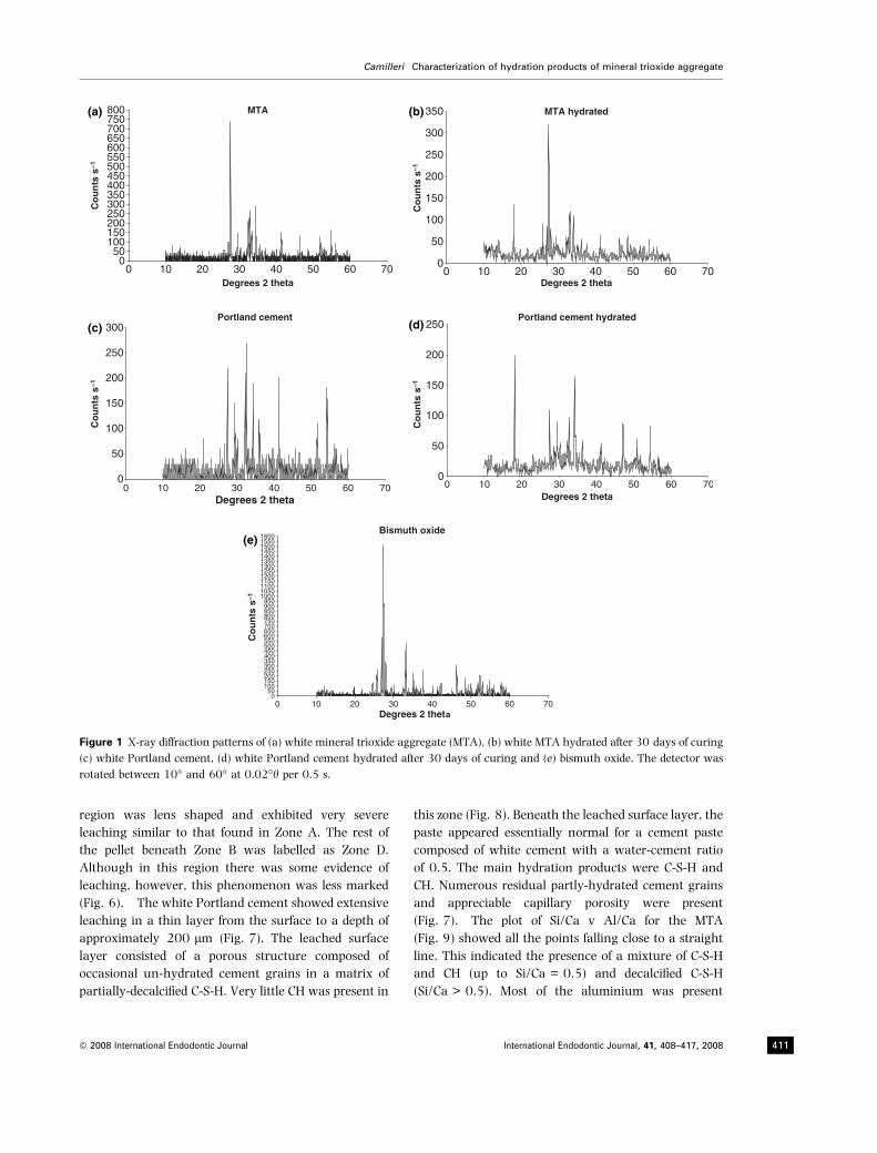
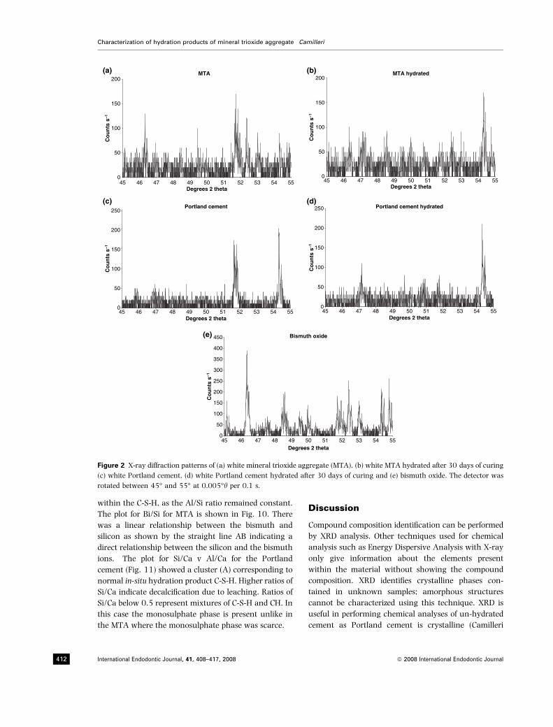
The XRD patterns between 10� and 60� 2h of the
cements and bismuth oxide are shown in Fig. 1 and the
diffraction patterns for the materials between 45� and
55� 2h are shown in Fig. 2. Both MTA and Portland
cement showed the major peaks of tricalcium silicate at
32.18, 32.54� 2h (Fig. 1a,c) and at 51.67� 2h(Fig. 2a,c). These peaks reduced in intensity in the
hydrated phases (Figs 1b,d and 2b,d). In addition both
hydrated materials had very strong peaks for CH at 18�2h (Fig. 1b,d) and 54.36� 2h (Fig. 2b,d). The bismuth
oxide had two very strong peaks at 27.30 and 46.30�2h (Figs 1e and 2e). The bismuth oxide peaks were
present in the MTA (Figs 1a,b and 2a,b) but not in the
Portland cement samples (Figs 1c,d and 2c,d). In the
hydrated MTA the bismuth oxide peak intensities at
27.30� 2h (Fig. 1b) and at 46.30� 2h (Fig. 2b) were
lower than in the un-hydrated paste (Figs 1a and 2a).
The bismuth oxide peak at 27.30� 2h coincided with
the rutile peak. Further peak superimposition occurred
at 54� 2h with major peaks for CH being superimposed
on rutile and bismuth oxide.
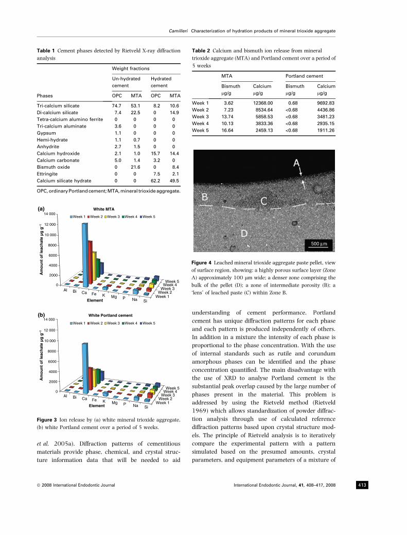
Rietveld x-ray diffraction analysis
The quantitative x-ray analysis performed on the
un-hydrated cements using the Rietveld diffractometer
are shown in Table 1. The MTA had a lower level of
tricalcium silicate and a higher level of dicalcium
silicate when compared to white Portland cement.
There was no tricalcium aluminate present in the MTA.
Tetracalcium alumino-ferrite was absent in both MTA
and Portland cement. Calcium sulphate levels were
either low or absent in MTA. MTA had a bismuth oxide
level of 21.6%. In the hydrated samples there was a
strong reduction in tricalcium silicate levels with an
increase in amorphous material level. CH and ettringite
were produced by both cements. The ettringite levels in
MTA were lower than that of Portland cement. The
amount of bismuth present in MTA was lower in the
hydrated cement. The Rietveld analysis however could
not make out where the bismuth was located.
Chemical analyses of cement leachate
The amount of elements leached out from white MTA is
shown in Fig. 3a and that leached out of white
Portland cement in Fig. 3b. Both cements showed the
same trends whereby calcium, potassium and sodium
levels decreased over the 5-week period, whilst the
magnesium and silicon increased. The levels of bismuth
leached out of the MTA increased over the 5-week
period. A comparison of calcium and bismuth ions
leached out of both materials is shown in Table 2.
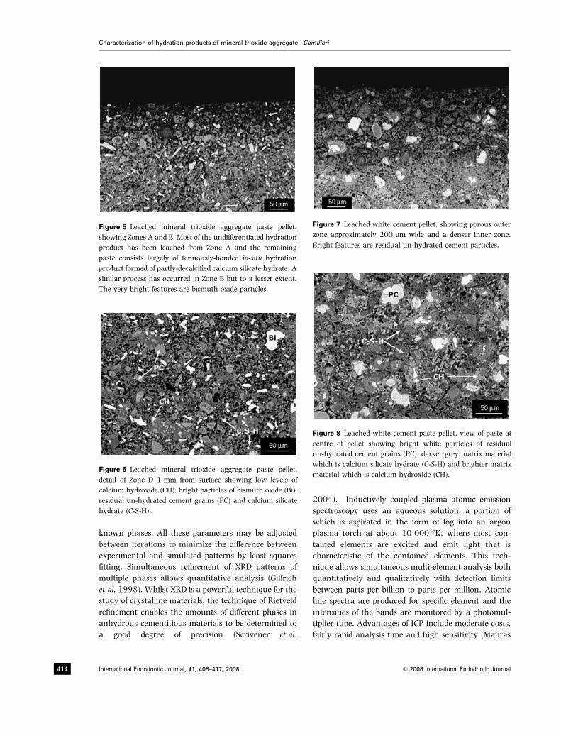
Scanning electron microscopy of set cements
The surface of the MTA pellet was more affected than
that of the Portland cement. Macroscopically the
surface of the MTA pellet showed an altered surface.
There was a change in colour and porosity of the
material. When viewed under the SEM the MTA paste
showed indications of leaching throughout most of the
pellet. The leaching was arbitrarily divided into various
zones namely Zone A, B, C, and D (Fig. 4). Zone A was a
very thin layer approximately 100 lm wide. This area
was strongly leached with no CH present and the C-S-H
was strongly decalcified (Figs 4 and 5). Zone B was
approximately 500 lm thick. This zone was also
strongly leached with loss of CH and decalcification of
C-S-H gel, but the leaching was less severe. Zone B
contained a distinct layer called Zone C (Fig. 4). This
Characterization of hydration products of mineral trioxide aggregate Camilleri
International Endodontic Journal, 41, 408–417, 2008 ª 2008 International Endodontic Journal410
region was lens shaped and exhibited very severe
leaching similar to that found in Zone A. The rest of
the pellet beneath Zone B was labelled as Zone D.
Although in this region there was some evidence of
leaching, however, this phenomenon was less marked
(Fig. 6). The white Portland cement showed extensive
leaching in a thin layer from the surface to a depth of
approximately 200 lm (Fig. 7). The leached surface
layer consisted of a porous structure composed of
occasional un-hydrated cement grains in a matrix of
partially-decalcified C-S-H. Very little CH was present in
this zone (Fig. 8). Beneath the leached surface layer, the
paste appeared essentially normal for a cement paste
composed of white cement with a water-cement ratio
of 0.5. The main hydration products were C-S-H and
CH. Numerous residual partly-hydrated cement grains
and appreciable capillary porosity were present
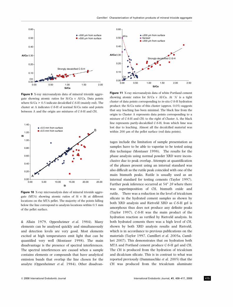
(Fig. 7). The plot of Si/Ca v Al/Ca for the MTA
(Fig. 9) showed all the points falling close to a straight
line. This indicated the presence of a mixture of C-S-H
and CH (up to Si/Ca = 0.5) and decalcified C-S-H
(Si/Ca > 0.5). Most of the aluminium was present
(a) (b)
(c) (d)
(e)
Figure 1 X-ray diffraction patterns of (a) white mineral trioxide aggregate (MTA), (b) white MTA hydrated after 30 days of curing
(c) white Portland cement, (d) white Portland cement hydrated after 30 days of curing and (e) bismuth oxide. The detector was
rotated between 10� and 60� at 0.02�h per 0.5 s.
Camilleri Characterization of hydration products of mineral trioxide aggregate
ª 2008 International Endodontic Journal International Endodontic Journal, 41, 408–417, 2008 411
within the C-S-H, as the Al/Si ratio remained constant.
The plot for Bi/Si for MTA is shown in Fig. 10. There
was a linear relationship between the bismuth and
silicon as shown by the straight line AB indicating a
direct relationship between the silicon and the bismuth
ions. The plot for Si/Ca v Al/Ca for the Portland
cement (Fig. 11) showed a cluster (A) corresponding to
normal in-situ hydration product C-S-H. Higher ratios of
Si/Ca indicate decalcification due to leaching. Ratios of
Si/Ca below 0.5 represent mixtures of C-S-H and CH. In
this case the monosulphate phase is present unlike in
the MTA where the monosulphate phase was scarce.
Discussion
Compound composition identification can be performed
by XRD analysis. Other techniques used for chemical
analysis such as Energy Dispersive Analysis with X-ray
only give information about the elements present
within the material without showing the compound
composition. XRD identifies crystalline phases con-
tained in unknown samples; amorphous structures
cannot be characterized using this technique. XRD is
useful in performing chemical analyses of un-hydrated
cement as Portland cement is crystalline (Camilleri
MTA(a) (b)
(c)
(e)
(d)
0
50
100
150
200
45 46 47 48 49 50 51 52 53 54 55Degrees 2 theta
Co
un
ts s
–1
MTA hydrated
0
50
100
150
200
45 46 47 48 49 50 51 52 53 54 55Degrees 2 theta
Co
un
ts s
–1
Portland cement
0
50
100
150
200
250
45 46 47 48 49 50 51 52 53 54 55Degrees 2 theta
Co
un
ts s
–1
Portland cement hydrated
0
50
100
150
200
250
45 46 47 48 49 50 51 52 53 54 55Degrees 2 theta
Co
un
ts s
–1
Bismuth oxide
0
50
100
150
200
250
300
350
400
450
45 46 47 48 49 50 51 52 53 54 55Degrees 2 theta
Co
un
ts s
–1
Figure 2 X-ray diffraction patterns of (a) white mineral trioxide aggregate (MTA), (b) white MTA hydrated after 30 days of curing
(c) white Portland cement, (d) white Portland cement hydrated after 30 days of curing and (e) bismuth oxide. The detector was
rotated between 45� and 55� at 0.005�h per 0.1 s.
Characterization of hydration products of mineral trioxide aggregate Camilleri
International Endodontic Journal, 41, 408–417, 2008 ª 2008 International Endodontic Journal412
et al. 2005a). Diffraction patterns of cementitious
materials provide phase, chemical, and crystal struc-
ture information data that will be needed to aid
understanding of cement performance. Portland
cement has unique diffraction patterns for each phase
and each pattern is produced independently of others.
In addition in a mixture the intensity of each phase is
proportional to the phase concentration. With the use
of internal standards such as rutile and corundum
amorphous phases can be identified and the phase
concentration quantified. The main disadvantage with
the use of XRD to analyse Portland cement is the
substantial peak overlap caused by the large number of
phases present in the material. This problem is
addressed by using the Rietveld method (Rietveld
1969) which allows standardization of powder diffrac-
tion analysis through use of calculated reference
diffraction patterns based upon crystal structure mod-
els. The principle of Rietveld analysis is to iteratively
compare the experimental pattern with a pattern
simulated based on the presumed amounts, crystal
parameters, and equipment parameters of a mixture of
Table 1 Cement phases detected by Rietveld X-ray diffraction
analysis
Phases
Weight fractions
Un-hydrated
cement
Hydrated
cement
OPC MTA OPC MTA
Tri-calcium silicate 74.7 53.1 8.2 10.6
Di-calcium silicate 7.4 22.5 0 14.9
Tetra-calcium alumino ferrite 0 0 0 0
Tri-calcium aluminate 3.6 0 0 0
Gypsum 1.1 0 0 0
Hemi-hydrate 1.1 0.7 0 0
Anhydrite 2.7 1.5 0 0
Calcium hydroxide 2.1 1.0 15.7 14.4
Calcium carbonate 5.0 1.4 3.2 0
Bismuth oxide 0 21.6 0 8.4
Ettringite 0 0 7.5 2.1
Calcium silicate hydrate 0 0 62.2 49.5
OPC, ordinary Portland cement; MTA, mineral trioxide aggregate.
Al Bi Ca Fe K Mg P Na SiWeek 1
Week 2Week 3
Week 4Week 5
0
2000
4000
6000
8000
10 000
12 000
14 000
Am
ou
nt
of
leac
hat
e µ
g g
–1
Element
White MTA(a)
(b)
Al Bi Ca Fe K Mg P Na SiWeek 1
Week 2Week 3
Week 4Week 5
0
2000
4000
6000
8000
10 000
12 000
14 000
Am
ou
nt
of
leac
hat
e µ
g g
–1
Element
White Portland cement
Week 1 Week 2 Week 3 Week 4 Week 5
Week 1 Week 2 Week 3 Week 4 Week 5
Figure 3 Ion release by (a) white mineral trioxide aggregate,
(b) white Portland cement over a period of 5 weeks.
Table 2 Calcium and bismuth ion release from mineral
trioxide aggregate (MTA) and Portland cement over a period of
5 weeks
MTA Portland cement
Bismuth
lg/g
Calcium
lg/g
Bismuth
lg/g
Calcium
lg/g
Week 1 3.62 12368.00 0.68 9692.83
Week 2 7.23 8534.64 <0.68 4436.86
Week 3 13.74 5858.53 <0.68 3481.23
Week 4 10.13 3833.36 <0.68 2935.15
Week 5 16.64 2459.13 <0.68 1911.26
Figure 4 Leached mineral trioxide aggregate paste pellet, view
of surface region, showing: a highly porous surface layer (Zone
A) approximately 100 lm wide; a denser zone comprising the
bulk of the pellet (D); a zone of intermediate porosity (B); a
‘lens’ of leached paste (C) within Zone B.
Camilleri Characterization of hydration products of mineral trioxide aggregate
ª 2008 International Endodontic Journal International Endodontic Journal, 41, 408–417, 2008 413
known phases. All these parameters may be adjusted
between iterations to minimize the difference between
experimental and simulated patterns by least squares
fitting. Simultaneous refinement of XRD patterns of
multiple phases allows quantitative analysis (Gilfrich
et al. 1998). Whilst XRD is a powerful technique for the
study of crystalline materials, the technique of Rietveld
refinement enables the amounts of different phases in
anhydrous cementitious materials to be determined to
a good degree of precision (Scrivener et al.
2004). Inductively coupled plasma atomic emission
spectroscopy uses an aqueous solution, a portion of
which is aspirated in the form of fog into an argon
plasma torch at about 10 000 �K, where most con-
tained elements are excited and emit light that is
characteristic of the contained elements. This tech-
nique allows simultaneous multi-element analysis both
quantitatively and qualitatively with detection limits
between parts per billion to parts per million. Atomic
line spectra are produced for specific element and the
intensities of the bands are monitored by a photomul-
tiplier tube. Advantages of ICP include moderate costs,
fairly rapid analysis time and high sensitivity (Mauras
Figure 6 Leached mineral trioxide aggregate paste pellet,
detail of Zone D 1 mm from surface showing low levels of
calcium hydroxide (CH), bright particles of bismuth oxide (Bi),
residual un-hydrated cement grains (PC) and calcium silicate
hydrate (C-S-H).
Figure 7 Leached white cement pellet, showing porous outer
zone approximately 200 lm wide and a denser inner zone.
Bright features are residual un-hydrated cement particles.
Figure 8 Leached white cement paste pellet, view of paste at
centre of pellet showing bright white particles of residual
un-hydrated cement grains (PC), darker grey matrix material
which is calcium silicate hydrate (C-S-H) and brighter matrix
material which is calcium hydroxide (CH).
Figure 5 Leached mineral trioxide aggregate paste pellet,
showing Zones A and B. Most of the undifferentiated hydration
product has been leached from Zone A and the remaining
paste consists largely of tenuously-bonded in-situ hydration
product formed of partly-decalcified calcium silicate hydrate. A
similar process has occurred in Zone B but to a lesser extent.
The very bright features are bismuth oxide particles.
Characterization of hydration products of mineral trioxide aggregate Camilleri
International Endodontic Journal, 41, 408–417, 2008 ª 2008 International Endodontic Journal414
& Allain 1979, Oppenheimer et al. 1984). Many
elements can be analysed quickly and simultaneously
and detection levels are very good. Most elements
excited at high temperatures emit light that can be
quantified very well (Montaser 1998). The main
disadvantage is the presence of spectral interferences.
The spectral interferences are caused when a sample
contains elements or compounds that have analytical
emission bands that overlap the line chosen for the
analyte (Oppenheimer et al. 1984). Other disadvan-
tages include the limitation of sample presentation as
samples have to be able to vaporize to be tested using
this technique (Montaser 1998). The results for the
phase analysis using normal powder XRD were incon-
clusive due to peak overlap. Attempts at quantification
of the phases present using an internal standard was
also difficult as the rutile peak coincided with one of the
main bismuth peaks. Rutile is usually used as an
internal standard for testing cements (Taylor 1997).
Further peak inference occurred at 54� 2h where there
was superimposition of CH, bismuth oxide and
rutile. There was a reduction in the level of tricalcium
silicate in the hydrated cement samples as shown by
both XRD analysis and Rietveld XRD as C-S-H gel is
amorphous thus does not produce any definite peaks
(Taylor 1997). C-S-H was the main product of the
hydration reaction as verified by Rietveld analysis. In
both hydrated cements there was a high level of CH,
shown by both XRD analysis results and Rietveld,
which is in accordance to previous publications on the
materials (Taylor 1997, Camilleri et al. 2005a, Camil-
leri 2007). This demonstrates that on hydration both
MTA and Portland cement produce C-S-H gel and CH.
The CH is produced from the hydration of tricalcium
and dicalcium silicate. This is in contrast to what was
reported previously (Dammaschke et al. 2005) that the
CH was produced from the tricalcium aluminate
0.60
0.50
0.40
Al/Ca 0.30
0.20
0.10
CH0.00
0.00 0.50 1.00Si/Ca
A
Strongly decalcified C-S-H
>500 µm from surface<500 µm from surface
1.50 2.00
Figure 9 X-ray microanalysis data of mineral trioxide aggre-
gate showing atomic ratios for Si/Ca v Al/Ca. Data points
where Si/Ca > 0.5 indicate decalcified C-S-H (mainly red). The
cluster at A indicates C-S-H of normal Si/Ca ratio and points
between A and the origin are mixtures of C-S-H and CH.
1.40
1.20
1.00
0.80
0.60
0.40
0.20
0.000.00
A
B
5.00
>0.5 mm from surface<0.5 mm from surface
10.00 15.00
Si
Bi
20.00 25.00
Figure 10 X-ray microanalysis date of mineral trioxide aggre-
gate (MTA) showing atomic ratios of Si v Bi at different
locations on the MTA pellet. The majority of the points falling
below the line correspond to analysis locations within 0.5 mm
of the pellet surface.
0.60
AFm
Strongly-decalcified C-S-H
ACH
>200 µm from surface
<200 µm from surfaceSeries20.50
Al/Ca
0.40
0.30
0.20
0.10
0.000.00 0.50 1.00
Si/Ca
1.50 2.00 2.50
Figure 11 X-ray microanalysis data of white Portland cement
showing atomic ratios for Si/Ca v Al/Ca. At ‘A’ is a tight
cluster of data points corresponding to in-situ C-S-H hydration
product; the Si/Ca ratio of this cluster (approx. 0.05) suggests
that any leaching has been minimal. The black line from the
origin to Cluster A represents data points corresponding to a
mixture of C-S-H and CH; to the right of Cluster A, the black
line represents partly-decalcified C-S-H, from which lime was
lost due to leaching. Almost all the decalcified material was
within 200 lm of the pellet surface (red data points).
Camilleri Characterization of hydration products of mineral trioxide aggregate
ª 2008 International Endodontic Journal International Endodontic Journal, 41, 408–417, 2008 415
hydrogenation. The Rietveld XRD also showed a low
level of tricalcium aluminate and gypsum in MTA
which is in accordance with a previous study (Camilleri
2007) where the level of tricalcium aluminate was
shown to be very low thus indicating that the cement is
not produced in a kiln but rather in a laboratory. The
levels of ettringite in the hydrated MTA were also much
lower than in the Portland cement. The low levels of
ettringite result form low levels of tricalcium aluminate
in the MTA powder. The results of SEM analysis further
confirm this as there was no monosulphate present in
MTA hydrated paste. The lack of aluminium caused the
reduced production of monosulphate in the hydrated
paste. This is again in accordance to a previous
publication (Camilleri 2007). The bismuth oxide
levels were 21.6% which is in accordance to manufac-
turer’s instructions which claim a 4 : 1 addition of
bismuth oxide to MTA powder. The bismuth oxide
peaks in the hydrated MTA were lower than those in
the un-hydrated powder. This was shown in both XRD
analysis and the Rietveld refinement demonstrating
that bismuth oxide does not act as filler in MTA but
rather takes an active part in the hydration mechanism
of the cement. This is again in accordance to previous
publications (Camilleri 2007). In the latter study it was
reported that bismuth oxide formed part of the struc-
ture of C-S-H and also affected the precipitation of CH in
the hydrated paste. The location of bismuth oxide in the
hydrated cement could not be determined using this
kind of analysis. The inductively coupled plasma
atomic absorption spectroscopy results showed that
the leachate had reducing levels of calcium with time.
This was further confirmed with the SEM results of
leached samples which showed extensive leaching of
calcium for both MTA and Portland cement. More
calcium was leached out from MTA than from Portland
cement. This was demonstrated in both the SEM study
where the leaching was more extensive in MTA than in
Portland cement and also from the ICP-AES results
where the calcium ion levels were higher for the MTA.
The loss of calcium from the cements is through
dissolution of CH and by progressive decalcification of
the C-S-H. The leached calcium is simultaneously
produced from the CH which, taking into consideration
the hydration equilibria is dissolved before the other
phases, and also from decomposition of C-S-H, which is
leached out at a slower rate (Taylor 1997). The
leaching also affects the ettringite and monosulphate
phases of the hydrated cement. CH is dissolved first
followed in sequence by monosulphate, ettringite and
C-S-H (Adenot & Buil 1992). The ettringite is dissolved
after the monosulphate phase as ettringite decomposes
at a lower pH than the monosulphate (Gabrisova et al.
1991). This is in accordance with the findings of the
present study where calcium leached out at fast rate,
whilst the silica showed a gradual rise in levels with
time due to decomposition of the C-S-H. The sodium
and potassium were leached early as they are not
bound to the C-S-H thus come in solution immediately.
The high levels of calcium leached out from the cement
account for the biocompatibility of MTA. Biocompati-
bility testing of cement leachables showed that the
elution made up of a high concentration of CH
produced during the hydration reaction induced cell
proliferation. Cell growth was poor when seeded in
direct contact with the test cements (Camilleri et al.
2005b). The MTA had a higher susceptibility to
leaching. Both pastes were made to water to powder
ratio of 0.5; however in the MTA this included the
bismuth oxide. High water to cement ratios increases
the susceptibility of the cement to leaching (Taylor
1997). The levels of bismuth and silica leached out of
MTA increased with time. This relationship was verified
by the SEM where a direct relationship between the
silica and bismuth was shown in the atomic ratio plots.
The bismuth does not act as filler in MTA but actively
takes part in the hydration reaction. The XRD analysis
showed a reduction in bismuth levels in the hydrated
paste showing that bismuth was being used up in the
hydration reaction. The bismuth replaced the silica in
the C-S-H (verified by the straight line in the atomic
ratio plot) then it is lost through leaching. This is again
in accordance to a previous publication which shows
that the bismuth actively took part in the hydration
mechanism (Camilleri 2007). Bismuth oxide is soluble
in acidic media (Lide 1998) thus, an acidic environ-
ment would likely increase the leaching of bismuth
from the material. It has been demonstrated that
bismuth oxide does not encourage cell growth and
proliferation (Camilleri et al. 2004). However, addition
of bismuth oxide to the Portland cement did not
interfere with the biocompatibility of the material.
Presumably the high concentration of calcium ions
released from the material make up for the lack of cell
proliferation on bismuth oxide.
Conclusions
Mineral trioxide aggregate and Portland cement are
primarily composed of tricalcium and dicalcium sili-
cate which on hydration produce C-S-H and CH.
Tricalcium aluminate levels in MTA were found to be
Characterization of hydration products of mineral trioxide aggregate Camilleri
International Endodontic Journal, 41, 408–417, 2008 ª 2008 International Endodontic Journal416
lower than those of Portland cement leading to
reduced production of ettringite and monosulphate
on hydration. The hydration mechanism of MTA is
different to that of Portland cement; in MTA the
bismuth oxide is bound to the C-S-H and is leached
out from the cement with time as the C-S-H decom-
poses. MTA produces a high proportion of calcium
ions from CH a by-product of hydration and also by
decomposition of C-S-H. The release of calcium ions
reduces with time.
Acknowledgements
The University of Malta Research Grant Committee for
funding. Heritage Malta for access to equipment and Mr
Lawrence Spiteri for his assistance.
References
Adenot F, Buil M (1992) Modelling of corrosion of the cement
paste by deionized water. Cement and Concrete Research 22,
489–96.
Asgary S, Parirokh M, Eghbal MJ, Brink F (2004) A compar-
ative study of white mineral trioxide aggregate and white
Portland cements using X-ray microanalysis. Australian
Endodontic Journal 30, 89–92.
Camilleri J (2007) Hydration mechanisms of mineral trioxide
aggregate. International Endodontic Journal 40, 462–70.
Camilleri J, Montesin FE, Papaioannou S, McDonald F, Pitt
Ford TR (2004) Biocompatibility of two commercial forms of
mineral trioxide aggregate. International Endodontic Journal
37, 699–704.
Camilleri J, Montesin FE, Brady K, Sweeney R, Curtis RV,
Pitt Ford TR (2005a) The constitution of mineral trioxide
aggregate. Dental Materials 21, 297–303.
Camilleri J, Montesin FE, Di Silvio L, Pitt Ford TR (2005b) The
chemical constitution and biocompatibility of accelerated
Portland cement for endodontic use. International Endodontic
Journal 38, 834–42.
Dammaschke T, Gerth HU, Zuchner H, Schafer E (2005)
Chemical and physical surface and bulk material charac-
terization of white ProRoot MTA and two Portland cements.
Dental Materials 21, 731–8.
Duarte MA, De Oliveira Demarchi AC, Yamashita JC, Kuga MC,
De Campos Fraga S (2005) Arsenic release provided by
MTA and Portland cement. Oral Surgery, Oral Medicine, Oral
Pathology, Oral Radiology and Endodontics 99, 648–50.
Estrela C, Bammann LL, Estrela CR, Silva RS, Pecora JD (2000)
Antimicrobial and chemical study of MTA, Portland cement,
calcium hydroxide paste, Sealapex and Dycal. Brazilian
Dental Journal 11, 3–9.
Funteas UR, Wallace JA, Fochtman EW (2003) A comparative
analysis of Mineral Trioxide Aggregate and Portland
cement. Australian Dental Journal 29, 43–4.
Gabrisova A, Havlika J, Sahu S (1991) Stability of calcium
sulphoaluminate hydrates in water solutions with various
pH values. Cement and Concrete Research 21, 1023–7.
Gilfrich JV, Cev Novan I, Jenkins R et al. (1998) Advances in
X-ray analysis. Vol 39. Proceedings of the 44th Annual
Conference on Applications of X-ray Analysis. Colorado:
University of Denver.
Lide DR (1998) CRC Handbook of Chemistry and Physics, 79th
edn. Abingdon, UK: CRC Press, Taylor & Francis, pp. 1–10.
Mauras Y, Allain P (1979) Determination of barium in water
and biological fluids by emission spectrometry with indirec-
tively-coupled plasma. Analityca Chimica Acta 110, 271–7.
Montaser A (1998) Inductively Coupled Plasma Mass Spectrom-
etry. New York: Ed. Wiley VCH.
Oppenheimer JA, Eaton AD, Leong LYC (1984) Multielemental
analytical techniques for hazardous waste analysis: The state of
art. Las Vegas NV: US Environmental Protection Agency,
Office of Research and Development, Environmental Mon-
itoring Systems Laboratory. EPA600484028.
Rietveld HM (1969) A profile refinement method for nuclear
and magnetic structure. Journal of Applied Crystallography 2,
65–71.
Scrivener KL, Fullmann T, Gallucci E, Walenta G, Bermejo E
(2004) Quantitative study of Portland cement hydration by
X-ray diffraction/Rietveld analysis and independent meth-
ods. Cement and Concrete Research 34, 1541–7.
Taylor HFW (1997) Cement Chemistry. London: Thomas
Telford.
Torabinejad M, White DJ (1995) Tooth filling material and use.
US Patent Number 5,769,638.
Torabinejad M, Hong CU, McDonald F, Pitt Ford TR (1995)
Physical and chemical properties of a new root-end filling
material. Journal of Endodontics 21, 349–53.
Camilleri Characterization of hydration products of mineral trioxide aggregate
ª 2008 International Endodontic Journal International Endodontic Journal, 41, 408–417, 2008 417