Characterization of Gastric Mucosa Biopsies Reveals ... · Rickards H, Wierup N, Bates GP,...
10
Characterization of Gastric Mucosa Biopsies Reveals Alterations in Huntington's Disease. Mccourt, Andy; O'Donovan, Kirsty L; Ekblad, Eva; Sand, Elin; Craufurd, David; Rosser, Anne; Sanders, David; Stoy, Nicholas; Rickards, Hugh; Wierup, Nils; Bates, Gillian P; Björkqvist, Maria; Quarrell, Oliver Published in: PLoS Currents DOI: 10.1371/currents.hd.858b4cc7f235df068387e9c20c436a79 Published: 2015-01-01 Link to publication Citation for published version (APA): Mccourt, A., O'Donovan, K. L., Ekblad, E., Sand, E., Craufurd, D., Rosser, A., ... Quarrell, O. (2015). Characterization of Gastric Mucosa Biopsies Reveals Alterations in Huntington's Disease. PLoS Currents, 7. DOI: 10.1371/currents.hd.858b4cc7f235df068387e9c20c436a79 General rights Copyright and moral rights for the publications made accessible in the public portal are retained by the authors and/or other copyright owners and it is a condition of accessing publications that users recognise and abide by the legal requirements associated with these rights. • Users may download and print one copy of any publication from the public portal for the purpose of private study or research. • You may not further distribute the material or use it for any profit-making activity or commercial gain • You may freely distribute the URL identifying the publication in the public portal Take down policy If you believe that this document breaches copyright please contact us providing details, and we will remove access to the work immediately and investigate your claim.
Transcript of Characterization of Gastric Mucosa Biopsies Reveals ... · Rickards H, Wierup N, Bates GP,...

LUND UNIVERSITY
PO Box 117221 00 Lund+46 46-222 00 00
Characterization of Gastric Mucosa Biopsies Reveals Alterations in Huntington'sDisease.
Mccourt, Andy; O'Donovan, Kirsty L; Ekblad, Eva; Sand, Elin; Craufurd, David; Rosser, Anne;Sanders, David; Stoy, Nicholas; Rickards, Hugh; Wierup, Nils; Bates, Gillian P; Björkqvist,Maria; Quarrell, OliverPublished in:PLoS Currents
DOI:10.1371/currents.hd.858b4cc7f235df068387e9c20c436a79
Published: 2015-01-01
Link to publication
Citation for published version (APA):Mccourt, A., O'Donovan, K. L., Ekblad, E., Sand, E., Craufurd, D., Rosser, A., ... Quarrell, O. (2015).Characterization of Gastric Mucosa Biopsies Reveals Alterations in Huntington's Disease. PLoS Currents, 7.DOI: 10.1371/currents.hd.858b4cc7f235df068387e9c20c436a79
General rightsCopyright and moral rights for the publications made accessible in the public portal are retained by the authorsand/or other copyright owners and it is a condition of accessing publications that users recognise and abide by thelegal requirements associated with these rights.
• Users may download and print one copy of any publication from the public portal for the purpose of private studyor research. • You may not further distribute the material or use it for any profit-making activity or commercial gain • You may freely distribute the URL identifying the publication in the public portalTake down policyIf you believe that this document breaches copyright please contact us providing details, and we will removeaccess to the work immediately and investigate your claim.

Characterization of Gastric Mucosa BiopsiesReveals Alterations in Huntington’s Disease
June 26, 2015 ·Huntington Disease
Tweet
McCourt AC, O'Donovan KL, Ekblad E, Sand E, Craufurd D, Rosser A, Sanders D, Stoy N,Rickards H, Wierup N, Bates GP, Björkqvist M, Quarrell O. Characterization of GastricMucosa Biopsies Reveals Alterations in Huntington’s Disease. PLOS Currents HuntingtonDisease. 2015 Jun 26 . Edition 1. doi:10.1371/currents.hd.858b4cc7f235df068387e9c20c436a79.
Citation
Authors
Andrew C McCourt
Kirsty L O'Donovan
Eva Ekblad
Elin Sand
David Craufurd
Anne Rosser
David Sanders
Nicholas Stoy
Hugh Rickards
Nils Wierup
Department of Experimental Medical Sciences, Brain Disease Biomarker Unit, Wallenberg Neuroscience Center,Lund University, Lund, Sweden.
Department of Clinical Genetics, Sheffield Children's Hospital, Sheffield Children's NHS Foundation Trust,Sheffield. South Yorkshire, UK.
Department of Experimental Medical Science, Lund University, Lund, Sweden.
Department of Neurogastroenterology, Experimental Medical Science, Lund University, Lund, Sweden.
University of Manchester, Manchester Academic Health Sciences Centre and Central Manchester UniversityHospitals NHS Foundation Trust, Manchester, UK.
School of Biosciences and Medicine, Cardiff University, Cardiff, Wakes, UK.
Department of Gastroenterology, Royal Hallamshire Hospital, Sheffield, South Yorkshire, UK.
Department of Systems Biologym University of Surrey, Guildford, Surrey, UK.
Department of Neuropsychiatry, University of Birmingham, Birmingham, West Midlands, UK.
Lund University Diabetes Centre, Lund University, Malmö, Sweden.
Characterization of Gastric Mucosa Biopsies Reveals Alterations ... http://currents.plos.org/hd/article/characterization-of-gastric-muc...
1 of 9 18/03/16 15:00
Weight loss is an important complication of Huntington’s disease (HD), however the mechanism forweight loss in HD is not entirely understood. Mutant huntingtin is expressed in the gastrointestinal (GI)tract and, in HD mice, mutant huntingtin inclusions are found within the enteric nervous system alongthe GI tract. A reduction of neuropeptides, decreased mucosal thickness and villus length, as well asgut motility impairment, have also been shown in HD mice. We therefore set out to study gastricmucosa of patients with HD, looking for abnormalities of mucosal cells using immunohistochemistry. Inorder to investigate possible histological differences related to gastric acid production, we evaluatedthe cell density of acid producing parietal cells, as well as gastrin producing cells (the endocrine cellcontrolling parietal cell function). In addition, we looked at chief cells and somatostatin-containing cells.In gastric mucosa from HD subjects, compared to control subject biopsies, a reduced expression ofgastrin (a marker of G cells) was found. This is in line with previous HD mouse studies showingreduction of GI tract neuropeptides.
This work was supported from a seed fund grant from the European Huntington’s Disease Network.The authors declare that no competing interests exist.
Most studies into the pathology of Huntington’s disease (HD) focus on the basal ganglia and cerebralcortex . However, mutant huntingtin is expressed throughout the body and abnormalities have beennoted in peripheral tissues, not considered secondary to neuronal damage .
Weight loss is one of the most common peripheral features of HD . The underlying mechanisms arenot, however, entirely known. Studies have indicated that weight loss is not secondary to inadequatenutrition, nor to hyperactivity . Studies have instead suggested that loss of body weight results fromchanges in metabolism and also that reduced absorption of nutrients along the intestinal tract mayplay a role . Work mostly performed in HD mouse models has demonstrated that tissues and organsthat are involved in nutrient absorption are affected .
In HD mouse models, huntingtin aggregates are abundantly present along the gastrointestinal tract .The R6/2 mouse, the most widely studied transgenic animal model of HD, exhibits loss of entericneuropeptides and altered gut motility . Gastrointestinal function has never been investigated in HDpatients, but there are indications that it may be affected. Patients are prone to suffer from gastritis andesophagitis .
We therefore set out to study the gastric mucosa, using gastric mucosal biopsies as a tool, to look for
Abstract
Funding Statement
Introduction
1
2,3,4
5,6
5
7
8
8
9
8
10
Gillian P. Bates
Maria Björkqvist
Oliver Quarrell
Department of Medical and Molecular Genetics, Kings College London, London, UK.
Department of Experimental Medical Science, Wallenberg Neuroscience Center, Lund University, Lund,Sweden.
Department of Clinical Genetics, Sheffield Children's Hospital, Sheffield Children's NHS foundation Trust,Sheffield, South Yorkshire, UK.
Characterization of Gastric Mucosa Biopsies Reveals Alterations ... http://currents.plos.org/hd/article/characterization-of-gastric-muc...
2 of 9 18/03/16 15:00

abnormalities of enteric neurons and mucosal cells.
Patient demographics
Patients with HD lose weight and have feeding difficulties. In some cases, this is managed by theinsertion of a percutaneous endoscopic gastrostomy (PEG) feeding tube. Ethical approval (MREC No.08/WSE02/66) was given to approach patients after a clinical decision to insert a PEG. Gastricbiopsies (from antrum and fundus/gastric body) were obtained from twelve HD subjects during theprocedure to insert the PEG. Using the total functional capacity (TFC) rating scale : 9 patients were atstage 5 (TFC = 0), one patient was at stage 4 (TFC = 1-2) and one patient was at stage 2 of thedisease (TFC = 7-10) and had a TFC of 7. The patients were in long-term care and the formal CAGlength report was not available for 8 patients (Table 1).
Control samples were obtained from 10 patients; 9 were being investigated for possible coeliacdisease, one for altered bowel habit; the gastric mucosa was considered normal by the endoscopist.Ethical approval, covering England and Wales, was granted by the South East Wales Research EthicsCommittee (08/WSE02/66) and confirmed in Scotland by the Scottish A Research Ethics Committee(08/MRE00/85). Written informed consent was obtained from all participants in this study.
Materials and methods
11
Characterization of Gastric Mucosa Biopsies Reveals Alterations ... http://currents.plos.org/hd/article/characterization-of-gastric-muc...
3 of 9 18/03/16 15:00
Immunohistochemistry
The gastric biopsies were fixed in formaldehyde and embedded in paraffin wax according to routineprocedures.
Antrum and fundus (gastric body) were cut into 7 µm thick sections using a microtome (LeicaSM2010R, Leica Biosystems Nussloch GmbH, Nussloch, Germany).
The different cell types were identified using immunohistochemistry; antrum sections – D-cells (anti-somatostatin antibody raised in rabbit; 1:3000 dilution, kind gift from Prof. J.J. Holst, CopenhagenUniversity, Denmark), G cells (anti-gastrin; 1:2000 dilution raised in rabbit, kind gift from Prof. J.E.Rehfeld, Copenhagen University, Denmark) and fundus (gastric body) sections – parietal cells(anti-H+/K+ ATPase antibody raised in mouse; 1:1000 dilution, kind gift from Prof. A.J. Smolka, UCLA,USA), chief cells (anti-pepsinogen antibody raised in swine, 1:1000 dilution, kind gift from Prof. P.T.Sangild, Copenhagen University, Denmark), endocrine cells (polyclonal anti-chromogranin A raised ingoat; 1:1000 dilution, Santa Cruz Biotechnology Inc., Santa Cruz, CA, USA). Antibodies were diluted inPBS containing 0.25% Triton X-100 and 0.25% bovine serum albumin. Prior to immunostaining,sections underwent antigen retrieval by boiling in citrate buffer using a microwave. Sections wereincubated with primary antibodies overnight at 4°C in the dark in a humid chamber. The next day,sections were incubated with the appropriate secondary antibodies for 1h at room temperature,followed by DAPI (1:2000, Sigma-Aldrich, Stockholm, Sweden) for 10 minutes: DyLightTM488-conjugated AffiniPure donkey anti-mouse, 1:1000, Jackson ImmunoResearch Laboratories Inc.,PA, USA; FITC-conjugated AffiniPure goat anti-swine, 1:100, BioNordika, Stockholm, Sweden;Cy2-conjugated AffiniPure donkey anti-rabbit, 1:300, Jackson ImmunoResearch; Cy2-conjugatedAffiniPure donkey anti-goat, 1:500, Jackson ImmunoResearch. Control incubations were also includedwithout the use of primary antibody; no staining was observed in these sections.
Immunofluorescence was examined using an epi-fluorescence microscope (Olympus BX53, Olympus,Tokyo, Japan) and digital images were acquired using a digital camera (Olympus DP73, Olympus,Tokyo, Japan). Section areas in the antrum with immunostaining against G cells were measured indigitized images using cellSens Dimensions 1.11 software (Olympus, Tokyo, Japan).
Cells were counted upon staining and expressed as total number of positive cells within the wholesection (G cells) and related to area of section, or total number of cells per visual field (parietal cells,chief cells and endocrine cells, 400 µm and D cells, 100 µm ).
Statistical analysis
All data were analysed using GraphPad Prism 6 (GraphPad Software Inc., San Diego, CA, USA). Dataare presented as mean ± SEM, with p < 0.05, one-tailed t-test considered as statistically significant.
Autolysis prevents the use of human post mortem tissue, therefore in this study, gastric biopsies wereobtained upon a clinical decision to insert a feeding tube. Control samples were obtained from patientsbeing investigated for a possible diagnosis of coeliac disease, however, these patients were not
Table 1
Patient demographics
Group N (M/F) Mean Age (Range)Control 10 (8/2) 55.5 (41-71)HD 12 (6/6) 55.8 (25-73)
2 2
Results and discussion
Characterization of Gastric Mucosa Biopsies Reveals Alterations ... http://currents.plos.org/hd/article/characterization-of-gastric-muc...
4 of 9 18/03/16 15:00
considered to have any gastric abnormality. This group of controls were chosen because it wasunlikely that they would have any neurodegenerative disease, although one patient had cerebellarataxia and was being investigated for a possible gluten enteropathy; his problems were eventuallyconsidered to be due to an excess intake of alcohol. One of the control patients was being investigatedfor altered bowel habit and found to have an oesophageal adenocarcinoma arising from a Barrett’smucosa. Only one patient was established to have coeliac disease.
In order to investigate possible histological differences related to gastric function, we usedimmunohistochemistry to evaluate the expression of cell specific markers of 2 exocrine cell types infundus (gastric body) sections, acid-producing parietal cells and pepsinogen producing chief cells aswell as markers of 2 endocrine cell types in antrum sections, gastrin producing cells and somatostatinproducing cells. We also stained fundus (gastric body) sections with chromogranin A, a protein foundin secretory vesicles of endocrine cells and neurons.
In line with previous HD mouse studies showing reduction of GI tract neuropeptides , usingimmunohistochemistry, we detected a reduction in the cell density of G-cells in antrum biopsies fromHD subjects compared to the control group (Figure 1). We also observed an increase in the celldensity of pepsinogen-producing chief cells of the fundus (gastric body) (Figure 1). Possibly, the lattercould play a role in the increased risk of gastritis/esophagitis in HD6 since increased levels of pepsin(the active form of pepsinogen) are associated with formation of peptic ulcers . There was nochange seen in the cell density of gastric acid producing parietal cells. Similarly, there was no changein the cell density of somatostatin-producing D cells, nor in the total number of endocrine cells (asrevealed by their chromogranin A expression) in the fundus (gastric body) (Table 2). Interestingly,gastrin is the hormone that upon food intake evokes acid secretion from the parietal cell, mediated bythe subsequent histamine release from the ECL cell, indicating that there might possibly be analteration in parietal cell stimulation in HD subjects.
8
12,13,14
Characterization of Gastric Mucosa Biopsies Reveals Alterations ... http://currents.plos.org/hd/article/characterization-of-gastric-muc...
5 of 9 18/03/16 15:00

Fig. 1: Representative images and cell counts
A, B, D, E, G, H, J, K, M and N show representative fluorescence microscope images of human stomachsections stained for various cell types. A, D, G, J and M are from control subjects, while B, E, H, K and Nare from HD patients. Panels C, F, I, L and O show cell counts for HD patients vs. control as indicated.Bars represent mean ± SEM. Scale bars: A, B, J, K – 50 µm; G, H, M, N – 20 µm; D, E – 10 µm. Visualfield areas analysed were 400 µm2 for parietal cells, chief cells and endocrine cells, and 100 µm2 for Dcells. No significant differences were found between HD patients and controls in fundus (gastric body)sections stained for the presence of parietal cells (A, B and C) or endocrine cells (G, H and I), nor antrumsections for the presence of D cells (M, N and O). Significant differences were observed in fundus (gastricbody) sections stained for chief cells (D, E and F), where HD patients had a greater cell density of positivecells, and antrum sections stained for gastrin producing G cells (J, K and L), with HD patients havingfewer counts than controls.
Characterization of Gastric Mucosa Biopsies Reveals Alterations ... http://currents.plos.org/hd/article/characterization-of-gastric-muc...
6 of 9 18/03/16 15:00

Characterization of Gastric Mucosa Biopsies Reveals Alterations ... http://currents.plos.org/hd/article/characterization-of-gastric-muc...
7 of 9 18/03/16 15:00
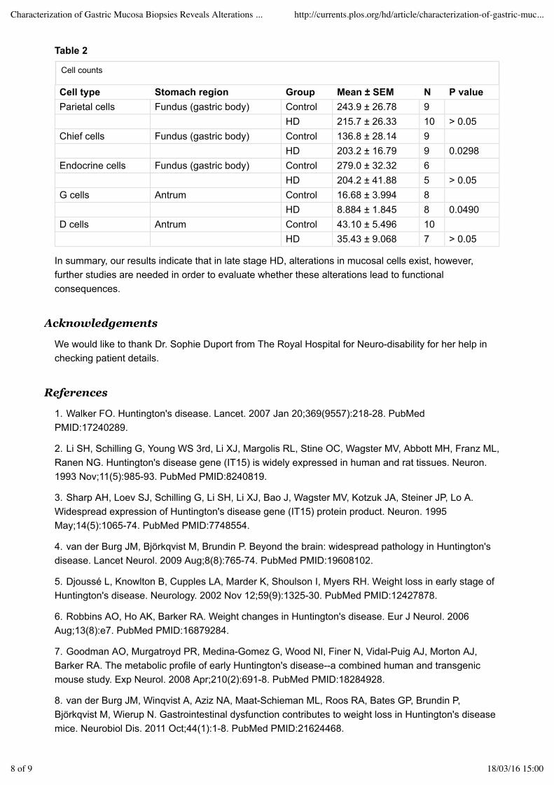
In summary, our results indicate that in late stage HD, alterations in mucosal cells exist, however,further studies are needed in order to evaluate whether these alterations lead to functionalconsequences.
We would like to thank Dr. Sophie Duport from The Royal Hospital for Neuro-disability for her help inchecking patient details.
1. Walker FO. Huntington's disease. Lancet. 2007 Jan 20;369(9557):218-28. PubMedPMID:17240289.
2. Li SH, Schilling G, Young WS 3rd, Li XJ, Margolis RL, Stine OC, Wagster MV, Abbott MH, Franz ML,Ranen NG. Huntington's disease gene (IT15) is widely expressed in human and rat tissues. Neuron.1993 Nov;11(5):985-93. PubMed PMID:8240819.
3. Sharp AH, Loev SJ, Schilling G, Li SH, Li XJ, Bao J, Wagster MV, Kotzuk JA, Steiner JP, Lo A.Widespread expression of Huntington's disease gene (IT15) protein product. Neuron. 1995May;14(5):1065-74. PubMed PMID:7748554.
4. van der Burg JM, Björkqvist M, Brundin P. Beyond the brain: widespread pathology in Huntington'sdisease. Lancet Neurol. 2009 Aug;8(8):765-74. PubMed PMID:19608102.
5. Djoussé L, Knowlton B, Cupples LA, Marder K, Shoulson I, Myers RH. Weight loss in early stage ofHuntington's disease. Neurology. 2002 Nov 12;59(9):1325-30. PubMed PMID:12427878.
6. Robbins AO, Ho AK, Barker RA. Weight changes in Huntington's disease. Eur J Neurol. 2006Aug;13(8):e7. PubMed PMID:16879284.
7. Goodman AO, Murgatroyd PR, Medina-Gomez G, Wood NI, Finer N, Vidal-Puig AJ, Morton AJ,Barker RA. The metabolic profile of early Huntington's disease--a combined human and transgenicmouse study. Exp Neurol. 2008 Apr;210(2):691-8. PubMed PMID:18284928.
8. van der Burg JM, Winqvist A, Aziz NA, Maat-Schieman ML, Roos RA, Bates GP, Brundin P,Björkqvist M, Wierup N. Gastrointestinal dysfunction contributes to weight loss in Huntington's diseasemice. Neurobiol Dis. 2011 Oct;44(1):1-8. PubMed PMID:21624468.
Table 2
Cell counts
Cell type Stomach region Group Mean ± SEM N P valueParietal cells Fundus (gastric body) Control 243.9 ± 26.78 9
HD 215.7 ± 26.33 10 > 0.05Chief cells Fundus (gastric body) Control 136.8 ± 28.14 9
HD 203.2 ± 16.79 9 0.0298Endocrine cells Fundus (gastric body) Control 279.0 ± 32.32 6
HD 204.2 ± 41.88 5 > 0.05G cells Antrum Control 16.68 ± 3.994 8
HD 8.884 ± 1.845 8 0.0490D cells Antrum Control 43.10 ± 5.496 10
HD 35.43 ± 9.068 7 > 0.05
Acknowledgements
References
Characterization of Gastric Mucosa Biopsies Reveals Alterations ... http://currents.plos.org/hd/article/characterization-of-gastric-muc...
8 of 9 18/03/16 15:00
9. Moffitt H, McPhail GD, Woodman B, Hobbs C, Bates GP. Formation of polyglutamine inclusions in awide range of non-CNS tissues in the HdhQ150 knock-in mouse model of Huntington's disease. PLoSOne. 2009 Nov 30;4(11):e8025. PubMed PMID:19956633.
10. Andrich JE, Wobben M, Klotz P, Goetze O, Saft C. Upper gastrointestinal findings in Huntington'sdisease: patients suffer but do not complain. J Neural Transm. 2009 Dec;116(12):1607-11. PubMedPMID:19771391.
11. Shoulson I, Fahn S. Huntington disease: clinical care and evaluation. Neurology. 1979Jan;29(1):1-3. PubMed PMID:154626.
12. Walker V, Taylor WH. Pepsin 1 secretion in chronic peptic ulceration. Gut. 1980 Sep;21(9):766-71.PubMed PMID:6776016.
13. Venables CW. Mucus, pepsin, and peptic ulcer. Gut. 1986 Mar;27(3):233-8. PubMedPMID:3084339.
14. Pearson JP, Ward R, Allen A, Roberts NB, Taylor WH. Mucus degradation by pepsin: comparisonof mucolytic activity of human pepsin 1 and pepsin 3: implications in peptic ulceration. Gut. 1986Mar;27(3):243-8. PubMed PMID:3084340.
Characterization of Gastric Mucosa Biopsies Reveals Alterations ... http://currents.plos.org/hd/article/characterization-of-gastric-muc...
9 of 9 18/03/16 15:00