Characteristics of surgical prosthetic heart valves and ... · Intraoperative surgical prosthetic...
12
Cite this article as: Durko AP, Head SJ, Pibarot P, Atluri P, Bapat V, Cameron DE et al. Characteristics of surgical prosthetic heart valves and problems around labelling: a document from the European Association for Cardio-Thoracic Surgery (EACTS)—The Society of Thoracic Surgeons (STS)—American Association for Thoracic Surgery (AATS) Valve Labelling Task Force. Eur J Cardiothorac Surg 2019;55:1025–36. Characteristics of surgical prosthetic heart valves and problems around labelling: a document from the European Association for Cardio-Thoracic Surgery (EACTS)—The Society of Thoracic Surgeons (STS)—American Association for Thoracic Surgery (AATS) Valve Labelling Task Force Andras P. Durko a,b , Stuart J. Head a , Philippe Pibarot c , Pavan Atluri d , Vinayak Bapat e , Duke E. Cameron f , Filip P.A. Casselman g , Edward P. Chen h , Gry Dahle i , Tjark Ebels j , John A. Elefteriades k , Patrizio Lancellotti l , Richard L. Prager m , Raphael Rosenhek n , Alan Speir o , Marco Stijnen p , Giordano Tasca q , Ajit Yoganathan r , Thomas Walther s and Ruggero De Paulis t, * (Task Force Chairman), EACTS–STS–AATS Valve Labelling Task Force a Department of Cardiothoracic Surgery, Erasmus University Medical Center, Rotterdam, Netherlands b Department of Cardiac Surgery, Medical and Health Science Centre, University of Debrecen, Debrecen, Hungary c Que ´ bec Heart and Lung Institute, Laval University, Quebec City, QC, Canada d Division of Cardiovascular Surgery, Department of Surgery, University of Pennsylvania, Philadelphia, PA, USA e Department of Cardiovascular Surgery, New York-Presbyterian/Columbia University Medical Center, New York, NY, USA f Division of Cardiac Surgery, Department of Surgery, Massachusetts General Hospital, Boston, MA, USA g Department of Cardiovascular and Thoracic Surgery, OLV Clinic, Aalst, Belgium h Division of Cardiothoracic Surgery, Department of Surgery, Emory University School of Medicine, Atlanta, GA, USA i Department of Cardiothoracic and Thoracic surgery, Rikshospitalet, Oslo University Hospital, Oslo, Norway j Department of Cardiothoracic Surgery, Amsterdam University Medical Center, Amsterdam, Netherlands k Department of Cardiothoracic Surgery, Yale University School of Medicine, New Haven, CT, USA l Department of Cardiology, GIGA Cardiovascular Sciences, University of Lie ` ge Hospital, Lie ` ge, Belgium m Department of Cardiac Surgery, University of Michigan Hospital, Ann Arbor, MI, USA n Department of Cardiology, Medical University of Vienna, Vienna, Austria o Department of Cardiac Surgery, Inova Heart and Vascular Institute, Falls Church, VA, USA p LifeTec Group, Eindhoven, Netherlands q Cardiovascular Department, Operative Unit of Cardiac Surgery, Hospital A. Manzoni, Lecco, Italy r Wallace H. Coulter Department of Biomedical Engineering, Georgia Institute of Technology/Emory School of Medicine, Atlanta, GA, USA s Department of Thoracic and Cardiovascular Surgery, University Hospital Frankfurt, Johann Wolfgang Goethe University Frankfurt, Frankfurt, Germany t Department of Cardiac Surgery, European Hospital, Rome, Italy * Corresponding author. Department of Cardiac Surgery, European Hospital, Via Portuense 700, 00149 Rome, Italy. Tel: +39-06-659 759; e-mail: [email protected] (R. De Paulis). Received 2 November 2018; received in revised form 8 January 2019; accepted 17 January 2019 Abstract Intraoperative surgical prosthetic heart valve (SHV) choice is a key determinant of successful surgery and positive postoperative outcomes. Currently, many controversies exist around the sizing and labelling of SHVs rendering the comparison of different valves difficult. To ex- plore solutions, an expert Valve Labelling Task Force was jointly initiated by the European Association for Cardio-Thoracic Surgery (EACTS), The Society of Thoracic Surgeons (STS) and the American Association for Thoracic Surgery (AATS). The EACTS–STS–AATS Valve Labelling Task Force, comprising cardiac surgeons, cardiologists, engineers, regulators and representatives from the International Organization for Standardization (ISO) and major valve manufacturers, held its first in-person meeting in February 2018 in Paris, France. This article was derived from the meeting’s discussions. The Task Force identified the following areas for improvement and clarification: reporting of physical dimensions and characteristics of SHVs determining and labelling of SHV size, in vivo and in vitro testing and report- ing of SHV haemodynamic performance and thrombogenicity. Furthermore, a thorough understanding of the regulatory background and the role of the applicable ISO standards, together with close cooperation between all stakeholders (including regulatory and standard- setting bodies), is necessary to improve the current situation. Cardiac surgeons should be provided with appropriate information to allow POSITION STATEMENT This article has been co-published with permission in the European Journal of Cardio-Thoracic Surgery, The Annals of Thoracic Surgery and The Journal of Thoracic and Cardiovascular Surgery. The articles are identical except for minor stylistic and spelling differences in keeping with each journal’s style. Either citation can be used when citing this article. V C 2019 European Association for Cardio-Thoracic Surgery, The Society of Thoracic Surgeons, and The American Association for Thoracic Surgery. Published by Oxford University Press and Elsevier Inc. European Journal of Cardio-Thoracic Surgery 0 (2019) 1–12 POSITION STATEMENT doi:10.1093/ejcts/ezz034 Downloaded from https://academic.oup.com/ejcts/advance-article-abstract/doi/10.1093/ejcts/ezz034/5487437 by Bibliotheque Fac de Medecine user on 14 May 2019
Transcript of Characteristics of surgical prosthetic heart valves and ... · Intraoperative surgical prosthetic...

Cite this article as: Durko AP, Head SJ, Pibarot P, Atluri P, Bapat V, Cameron DE et al. Characteristics of surgical prosthetic heart valves and problems around labelling:a document from the European Association for Cardio-Thoracic Surgery (EACTS)—The Society of Thoracic Surgeons (STS)—American Association for ThoracicSurgery (AATS) Valve Labelling Task Force. Eur J Cardiothorac Surg 2019;55:1025–36.
Characteristics of surgical prosthetic heart valves and problemsaround labelling: a document from the European Association for
Cardio-Thoracic Surgery (EACTS)—The Society of Thoracic Surgeons(STS)—American Association for Thoracic Surgery (AATS) Valve
Labelling Task Force
Andras P. Durkoa,b, Stuart J. Heada, Philippe Pibarotc, Pavan Atlurid, Vinayak Bapate, Duke E. Cameronf,
Filip P.A. Casselmang, Edward P. Chenh, Gry Dahlei, Tjark Ebelsj, John A. Elefteriadesk, Patrizio Lancellottil,
Richard L. Pragerm, Raphael Rosenhekn, Alan Speiro, Marco Stijnenp, Giordano Tascaq,
Ajit Yoganathanr, Thomas Walthers and Ruggero De Paulist,* (Task Force Chairman),
EACTS–STS–AATS Valve Labelling Task Force
a Department of Cardiothoracic Surgery, Erasmus University Medical Center, Rotterdam, Netherlandsb Department of Cardiac Surgery, Medical and Health Science Centre, University of Debrecen, Debrecen, Hungaryc Quebec Heart and Lung Institute, Laval University, Quebec City, QC, Canadad Division of Cardiovascular Surgery, Department of Surgery, University of Pennsylvania, Philadelphia, PA, USAe Department of Cardiovascular Surgery, New York-Presbyterian/Columbia University Medical Center, New York, NY, USAf Division of Cardiac Surgery, Department of Surgery, Massachusetts General Hospital, Boston, MA, USAg Department of Cardiovascular and Thoracic Surgery, OLV Clinic, Aalst, Belgiumh Division of Cardiothoracic Surgery, Department of Surgery, Emory University School of Medicine, Atlanta, GA, USAi Department of Cardiothoracic and Thoracic surgery, Rikshospitalet, Oslo University Hospital, Oslo, Norwayj Department of Cardiothoracic Surgery, Amsterdam University Medical Center, Amsterdam, Netherlandsk Department of Cardiothoracic Surgery, Yale University School of Medicine, New Haven, CT, USAl Department of Cardiology, GIGA Cardiovascular Sciences, University of Liege Hospital, Liege, Belgiumm Department of Cardiac Surgery, University of Michigan Hospital, Ann Arbor, MI, USAn Department of Cardiology, Medical University of Vienna, Vienna, Austriao Department of Cardiac Surgery, Inova Heart and Vascular Institute, Falls Church, VA, USAp LifeTec Group, Eindhoven, Netherlandsq Cardiovascular Department, Operative Unit of Cardiac Surgery, Hospital A. Manzoni, Lecco, Italyr Wallace H. Coulter Department of Biomedical Engineering, Georgia Institute of Technology/Emory School of Medicine, Atlanta, GA, USAs Department of Thoracic and Cardiovascular Surgery, University Hospital Frankfurt, Johann Wolfgang Goethe University Frankfurt, Frankfurt, Germanyt Department of Cardiac Surgery, European Hospital, Rome, Italy
* Corresponding author. Department of Cardiac Surgery, European Hospital, Via Portuense 700, 00149 Rome, Italy. Tel: +39-06-659 759;e-mail: [email protected] (R. De Paulis).
Received 2 November 2018; received in revised form 8 January 2019; accepted 17 January 2019
Abstract
Intraoperative surgical prosthetic heart valve (SHV) choice is a key determinant of successful surgery and positive postoperative outcomes.Currently, many controversies exist around the sizing and labelling of SHVs rendering the comparison of different valves difficult. To ex-plore solutions, an expert Valve Labelling Task Force was jointly initiated by the European Association for Cardio-Thoracic Surgery(EACTS), The Society of Thoracic Surgeons (STS) and the American Association for Thoracic Surgery (AATS). The EACTS–STS–AATS ValveLabelling Task Force, comprising cardiac surgeons, cardiologists, engineers, regulators and representatives from the InternationalOrganization for Standardization (ISO) and major valve manufacturers, held its first in-person meeting in February 2018 in Paris, France.This article was derived from the meeting’s discussions. The Task Force identified the following areas for improvement and clarification:reporting of physical dimensions and characteristics of SHVs determining and labelling of SHV size, in vivo and in vitro testing and report-ing of SHV haemodynamic performance and thrombogenicity. Furthermore, a thorough understanding of the regulatory background andthe role of the applicable ISO standards, together with close cooperation between all stakeholders (including regulatory and standard-setting bodies), is necessary to improve the current situation. Cardiac surgeons should be provided with appropriate information to allow
PO
SITI
ON
STA
TEM
ENT
This article has been co-published with permission in the European Journal of Cardio-Thoracic Surgery, The Annals of Thoracic Surgery and The Journal of Thoracic andCardiovascular Surgery.
The articles are identical except for minor stylistic and spelling differences in keeping with each journal’s style. Either citation can be used when citing this article.
VC 2019 European Association for Cardio-Thoracic Surgery, The Society of Thoracic Surgeons, and The American Association for Thoracic Surgery.Published by Oxford University Press and Elsevier Inc.
European Journal of Cardio-Thoracic Surgery 0 (2019) 1–12 POSITION STATEMENTdoi:10.1093/ejcts/ezz034
Dow
nloaded from https://academ
ic.oup.com/ejcts/advance-article-abstract/doi/10.1093/ejcts/ezz034/5487437 by Bibliotheque Fac de M
edecine user on 14 May 2019

for optimal SHV choice. This first article from the EACTS–STS–AATS Valve Labelling Task Force summarizes the background of SHV sizingand labelling and identifies the most important elements where further standardization is necessary.
Keywords: Aortic valve replacement • Mitral valve replacement • Prosthetic heart valve • Surgical prosthetic heart valve (SHV) •International Organization for Standardization (ISO) • International standard • Labelling • Sizing • Device approval • Regulation • Valve per-formance • Prosthesis–patient mismatch (PPM) • Objective performance criteria (OPC)
INTRODUCTION
Intraoperative prosthetic valve selection is a key determinant ofsurgical success; using the most appropriate surgical prostheticheart valve (SHV) minimizes the risks of surgery, maximizeshaemodynamic performance and optimizes long-term outcomes[1]. The final choice of an SHV, including appropriate size, is typ-ically made in the operating theatre. To facilitate an evidence-based SHV choice, sufficient appropriate information on SHVcharacteristics is required. Background information from medicalliterature is not available in the intraoperative setting, so SHVpackage labels and instructions for use (IFU) booklets are the pri-mary sources of information for the surgeon in the operatingtheatre.
In both the European Union (EU) and the USA, the quality andquantity of information provided with an SHV are regulated. TheInternational Organization for Standardization (ISO) plays an im-portant role in this process by providing a framework for regula-tory bodies [2]. Although the ISO 5840 standard (CardiovascularImplants—Cardiac Valve Prostheses) provides general conditionsfor testing SHVs for human implantation and defines operationaland labelling requirements, the current labelling situation is notoptimal. Simple definitions such as ‘labelled valve size’ are oftenunclear, and inconsistencies and controversies exist around thesizing and labelling of SHVs in relation to haemodynamic per-formance [3].
To resolve these issues, the European Association for Cardio-Thoracic Surgery (EACTS), The Society of Thoracic Surgeons(STS) and the American Association for Thoracic Surgery (AATS)set up the EACTS–STS–AATS Valve Labelling Task Force, involv-ing representatives of the 3 surgical societies, cardiologists, engi-neers, regulatory professionals and representatives from ISOand major valve-manufacturing companies. The discussionsduring the first in-person meeting of the Task Force (held inFebruary 2018, Paris, France) provided the core content of thisarticle.
This first article of the EACTS–STS–AATS Valve Labelling TaskForce is intended to provide an overview of important character-istics of SHVs relating to sizing and labelling, reviews currentpractices in these areas and identifies where improvements arenecessary. This article will be followed by an expert consensusdocument containing recommendations on SHV sizing andlabelling.
REGULATORY ASPECTS AND USE OF STANDARDSIN PROSTHETIC HEART VALVE LABELLING
In the EU and the USA, medical devices must demonstrate con-formity to the local legislations before they can be introduced tothe market. Assessment of conformity is defined as the evaluationof whether a certain device is safe and effective according to theapplicable regulations. This includes the assessment of the devicelabelling information, including the IFU and package labels. The
ISO standards play an important role in defining these device-specific requirements.
International Organization for Standardization andthe prosthetic heart valve standards
Technical standards are formal documents, defining uniform en-gineering criteria for technical systems. The ISO is an independ-ent, non-governmental organization consisting of nationalstandards bodies [4]. Manufacturers globally use ISO standardsduring product development and production. In addition, ISOstandards are widely utilized by regulatory bodies as a conform-ity assessment tool for market approval. The ISO standards areperiodically revised and updated in line with new innovations.
‘ISO 5840’ is a family of standards developed by a group ofprofessionals from engineering and medical backgrounds, includ-ing representatives of the medical device industry, regulators andclinicians. The current 2015 version (ISO 5840:2015,Cardiovascular Implants—Cardiac Valve Prostheses) consists of 3parts: ‘part 1: general requirements, part 2: surgically implantedheart valve substitutes and part 3: heart valve substitutesimplanted by transcatheter techniques’. ‘ISO 5840:2015’ providesrecommendations and requirements for preclinical and clinicalevaluations of SHVs [2], and it defines valve-related objective per-formance criteria according to linearized event rates of key safetyend points (thromboembolism, valve thrombosis, haemorrhage,paravalvular leakage and endocarditis) [2, 5, 6]. Furthermore, ISOprovides guidance on labelling by describing the informationthat should be available on the product labels and in the IFU,including detailed information on intended use, indications/con-traindications and warnings, and physical and performance char-acteristics [2].
Standards and prosthetic heart valve approval inthe European Union
In the EU, the regulatory framework provided by the EuropeanCommission sets the general requirements for the whole rangeof medical devices [7, 8]. More specific requirements are definedwithin ‘Common Specifications’ or in ‘Harmonized Standards’.The European Committee for Standardization (CEN) cooperateswith the ISO, and existing ISO standards can be adopted as‘Harmonized Standards’ endorsed by the European Commission[9]. Furthermore, technical standards are used as a tool to definethe generally acknowledged ‘state of the art’ in a certain field. Todefine ‘state of the art’, other documents such as consensusdocuments by professional societies are also taken into accountduring conformity assessment [8].
In the EU, conformity assessment of medical devices is per-formed by Notified Bodies. These are independent, third-partyorganizations appointed by Member States. Notified Bodies cangrant the CE (European Conformity) mark, which allows market-ing a product within the European Economic Area.
2 A.P. Durko et al. / European Journal of Cardio-Thoracic Surgery
Dow
nloaded from https://academ
ic.oup.com/ejcts/advance-article-abstract/doi/10.1093/ejcts/ezz034/5487437 by Bibliotheque Fac de M
edecine user on 14 May 2019

Standards and prosthetic heart valve approval inthe USA
In the USA, the medical device market is centrally regulated bythe Food and Drug Administration (FDA) through review and ap-proval of applications for new devices [10]. During the process ofapproving new SHVs, the use of ‘Consensus Standards’ by theFDA is voluntary [11]. The ISO 5840 standards are recognized bythe FDA and could serve as a guidance for manufacturers whensubmitting their applications for approval to the FDA [12].
DESIGN AND CHARACTERISTICS OF SURGICALPROSTHETIC HEART VALVES
Design and materials
The design and component materials of an SHV should: causeminimal harm to endothelial tissue and blood cells; pose minimalchances for platelet and thrombus deposition; be resistant tostructural wear and tear, mechanical failure and degradation; bebiochemically inert in a physiological milieu; and be suitable forsterilization [13]. Mechanical valve leaflets and their supportingframes are composed mostly of pyrolytic carbon. Bioprostheticvalves, however, have more variation in their component materi-als: both native valves from animals and valves manufacturedfrom animal pericardium are used (Table 1). Preparation techni-ques used for biological tissues aim to reduce immunogenicity[14], cross-link collagen and prevent calcification to delay valvedegeneration [15]. Polymeric heart valves might offer a relativelyinexpensive alternative to biological tissues, but their safety andeffectiveness in the clinical setting are yet to be proved [16, 17].Differences in long-term outcomes with various prostheseshave been identified, which may relate to specific design features[18, 19].
Materials used for the supporting frames of bioprostheticvalves vary in composition (Table 1), which is potentially relevantfor subsequent valve-in-valve transcatheter procedures, whenbioprosthetic valve frame fracture could enable insertion of alarger transcatheter prosthesis [20]. Importantly, some biopros-thetic SHVs are equipped with an expandable band, which isintended to facilitate controlled expansion of the valve supportstructure when subjected to radial force. Many SHVs areequipped with radiopaque markers or have an intrinsically radi-opaque support structure. This is useful when a valve-in-valveprocedure is planned, as it aids in positioning of the transcatheter
valve and provides information about the proximity of the SHVstrut to the coronary ostia. Currently, there is a considerable var-iety in radiopaque marking of bioprosthetic SHVs [21].
The ‘ISO 5840:2015’ standard contains recommendations onreporting the magnetic resonance imaging (MRI) safety designa-tion of the prosthesis in the accompanying IFU. However, it doesnot require detailed reporting of the materials used for SHVmanufacturing.
Physical dimensions
Physical dimensions of SHVs are closely related to their perform-ance, safety and ease of use, and can influence the choice ofprosthesis and the implantation technique. Dimensions aredefined in ‘ISO 5840:2015’ and should be provided in the IFU [2].However, reporting of these measurements in terms of dimen-sional definitions is not consistent, and the relationship betweenSHV physical dimensions and the ‘labelled valve size’ is unclear,creating confusion in the surgical community [3].
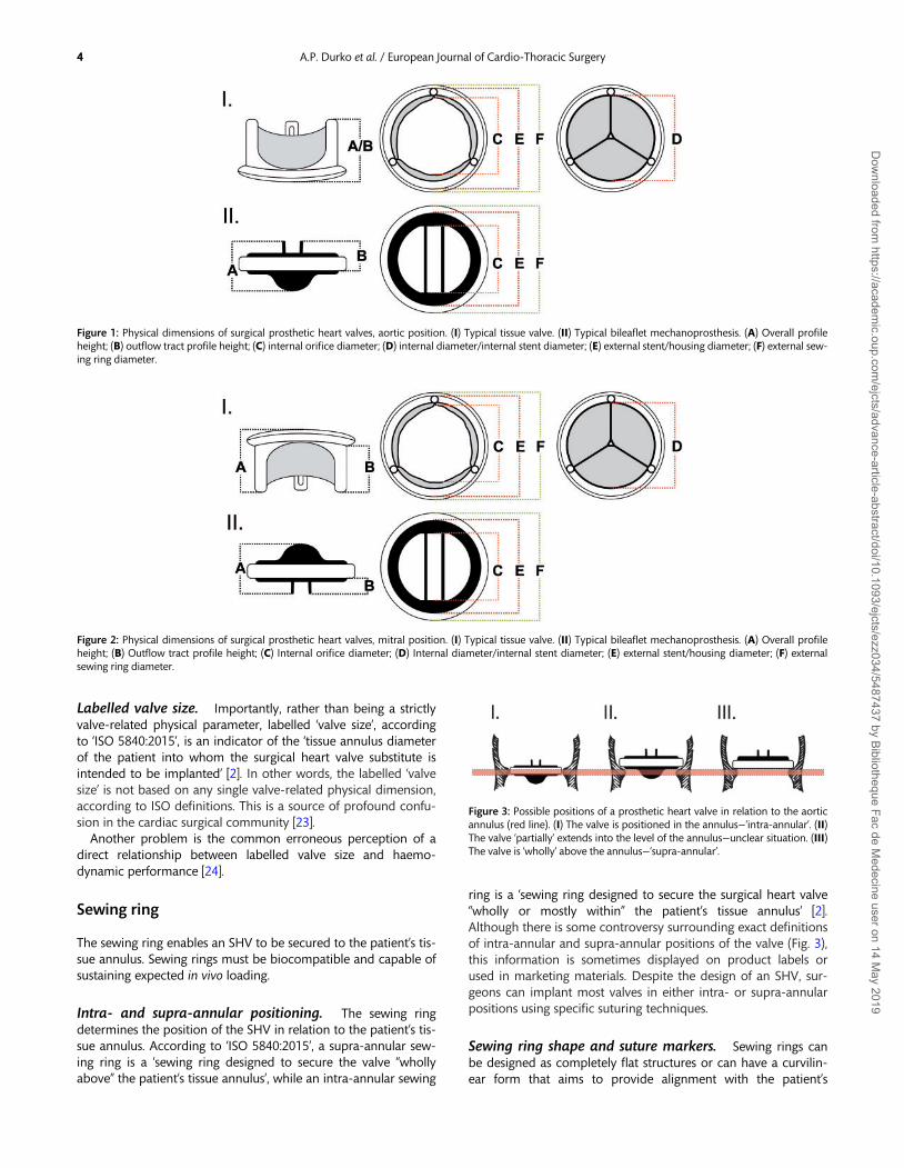
Axial dimensions. The fundamental axial dimensions of anSHV are the overall ‘profile height’ and ‘outflow tract profileheight’, the latter being the maximum distance that the heartvalve substitute extends axially into the outflow tract, measuredfrom the valve structure’s intended annular attachment, accord-ing to ‘ISO 5840:2015’ (Figs 1 and 2) [2].
Horizontal dimensions. The ISO-defined horizontal dimen-sions of an SHV are the ‘internal orifice diameter’ and the ‘exter-nal sewing ring diameter’ (Figs 1 and 2). According to ‘ISO5840:2015’, ‘internal orifice diameter’ is ‘the minimum diameterwithin a surgical heart valve substitute through which bloodflows’ [2], making it one of the most relevant physical dimensionscharacterizing SHV performance.
A widely used parameter when selecting a transcatheter pros-thesis during a valve-in-valve procedure is the ‘true internaldiameter’ (true ID) of the surgical bioprosthesis. This ‘true ID’ ismeasured by passing a Hegar dilator through the bioprostheticorifice and therefore closely corresponds to the ISO-defined ‘in-ternal orifice diameter’ [22]. Of note, manufacturers often report‘internal diameter (ID)’ or ‘stent internal diameter (stent ID)’,which is the ID of the stent (label F in Figs 1 and 2). Importantly,this ‘stent ID’ does not account for the space occupied by theprosthetic leaflets in the orifice of a bioprosthetic valve (e.g. thedifference between labels C and D in Figs 1 and 2).
Table 1: Component materials in stented surgical prosthetic heart valves
Stented bioprostheses Mechanoprostheses
Leaflets Native porcine valve and bovine pericardium Pyrolytic carbonSupporting frame/stent Titanium, Elgiloya and Delrinb Pyrolytic carbon and titaniumSewing ring Silicone rubber and Dacronc Dacron and Teflond
aCobalt–chromium–nickel–molybdenum alloy.bAcetal homopolymer.cPolyethylene terephthalate.dPolytetrafluoroethylene.
PO
SITI
ON
STA
TEM
ENT
3A.P. Durko et al. / European Journal of Cardio-Thoracic Surgery
Dow
nloaded from https://academ
ic.oup.com/ejcts/advance-article-abstract/doi/10.1093/ejcts/ezz034/5487437 by Bibliotheque Fac de M
edecine user on 14 May 2019
Labelled valve size. Importantly, rather than being a strictlyvalve-related physical parameter, labelled ‘valve size’, accordingto ‘ISO 5840:2015’, is an indicator of the ‘tissue annulus diameterof the patient into whom the surgical heart valve substitute isintended to be implanted’ [2]. In other words, the labelled ‘valvesize’ is not based on any single valve-related physical dimension,according to ISO definitions. This is a source of profound confu-sion in the cardiac surgical community [23].
Another problem is the common erroneous perception of adirect relationship between labelled valve size and haemo-dynamic performance [24].
Sewing ring
The sewing ring enables an SHV to be secured to the patient’s tis-sue annulus. Sewing rings must be biocompatible and capable ofsustaining expected in vivo loading.
Intra- and supra-annular positioning. The sewing ringdetermines the position of the SHV in relation to the patient’s tis-sue annulus. According to ‘ISO 5840:2015’, a supra-annular sew-ing ring is a ‘sewing ring designed to secure the valve “whollyabove” the patient’s tissue annulus’, while an intra-annular sewing
ring is a ‘sewing ring designed to secure the surgical heart valve“wholly or mostly within” the patient’s tissue annulus’ [2].Although there is some controversy surrounding exact definitionsof intra-annular and supra-annular positions of the valve (Fig. 3),this information is sometimes displayed on product labels orused in marketing materials. Despite the design of an SHV, sur-geons can implant most valves in either intra- or supra-annularpositions using specific suturing techniques.
Sewing ring shape and suture markers. Sewing rings canbe designed as completely flat structures or can have a curvilin-ear form that aims to provide alignment with the patient’s
Figure 1: Physical dimensions of surgical prosthetic heart valves, aortic position. (I) Typical tissue valve. (II) Typical bileaflet mechanoprosthesis. (A) Overall profileheight; (B) outflow tract profile height; (C) internal orifice diameter; (D) internal diameter/internal stent diameter; (E) external stent/housing diameter; (F) external sew-ing ring diameter.
Figure 2: Physical dimensions of surgical prosthetic heart valves, mitral position. (I) Typical tissue valve. (II) Typical bileaflet mechanoprosthesis. (A) Overall profileheight; (B) Outflow tract profile height; (C) Internal orifice diameter; (D) Internal diameter/internal stent diameter; (E) external stent/housing diameter; (F) externalsewing ring diameter.
Figure 3: Possible positions of a prosthetic heart valve in relation to the aorticannulus (red line). (I) The valve is positioned in the annulus—‘intra-annular’. (II)The valve ‘partially’ extends into the level of the annulus—unclear situation. (III)The valve is ‘wholly’ above the annulus—‘supra-annular’.
4 A.P. Durko et al. / European Journal of Cardio-Thoracic Surgery
Dow
nloaded from https://academ
ic.oup.com/ejcts/advance-article-abstract/doi/10.1093/ejcts/ezz034/5487437 by Bibliotheque Fac de M
edecine user on 14 May 2019

non-planar anatomical annulus. Suture markers on the sewingring are intended to facilitate implantation and correct orienta-tion of the SHV. Currently, there is a considerable variety in thenumber and position of suture markers.
Implantation aids. Several implantation aids are providedwith an SHV, including handles, rotators or systems to preventinadvertent suture looping and/or facilitating knot-tying.According to ‘ISO 5840:2015’, the use of these implantation aidsshould be described in the IFU.
Intraoperative sizing
The goal of intraoperative sizing is to determine the labelled sizeof the SHV that can be safely implanted into the patient. This in-formation, together with easy access to information about therelevant properties (e.g. haemodynamic performance, durability,thrombogenicity, etc.) of the particular SHV that would fit, makesoptimal intraoperative valve choice possible.
Sizers. Manufacturers provide a set of valve-related sizers foreach SHV model. Sizers are numbered according to the labelledsizes of the corresponding SHVs. Typically, sizers have 2 ends: acylindrical end (barrel) to measure the annulus and guide SHVselection based on the labelled valve size, and a replica mimick-ing the configuration of the prosthesis (Fig. 4). Sizing with a rep-lica after suture placement is particularly useful because both thepatient’s anatomy and the surgeon’s suturing technique influencethe SHV’s final position and affect ultimate sizing [25, 26].
Sizers and the labelled valve size. Given that the number-ing of sizers follows the labelled valve size, the sizer barrel shoulddetermine the diameter of the patient’s tissue annulus. Indeed,size measured using the sizer barrel is not intended to providedirect information regarding the physical dimensions of the cor-responding SHV [27]. However, numerous publications havedemonstrated significant differences between labelled valve sizeand the actual dimensions of the valve-related sizer barrel, caus-ing confusion in the surgical community [3, 28–30].
During labelling, the manufacturer determines which valve isrecommended for a measured tissue annulus, which is reflectedin the labelled valve size. However, clinical sizing can vary de-pending on the extent of annular debridement or surgeonaggressiveness when entering the sizer to the annulus. These vari-abilities make it challenging for manufacturers to determinewhich valve to recommend for implanting into a specific tissue
annulus diameter (i.e. to determine the labelled valve size).Although the actual tissue annulus diameter is easily determinedusing a Hegar dilator or a similar circular sizing tool, these incon-sistencies and challenges mean that optimal sizing is currentlybest performed using the set of sizers provided by the manufac-turer with the valve selected for implantation.
HAEMODYNAMIC PERFORMANCE OF SURGICALPROSTHETIC HEART VALVES
In vitro hydrodynamic performance testing
In vitro hydrodynamic testing is intended to assess the ability ofan SHV to enable forward flow and prevent reverse flow and isrequired for device approval. Although steady flow testers allowmanufacturers to measure forward flow and reverse flow (leak-age) across the SHV under controlled conditions, the testing en-vironment is very different from physiological conditions. ‘ISO5840:2015’ provides guidance for in vitro hydrodynamic perform-ance testing and defines flow hydrodynamic acceptance criteriafor pulsatile testing based on valve size and implant position [2].
Pulsatile testing. Pulsatile testing enables SHV performanceto be assessed under physiological flow and pressure conditionsthat are similar to those in which it is intended to function.Pulsatile testing enables measurement of flow and pressure drop(pressure gradient), calculation of the in vitro effective orifice area(EOA) and total regurgitant volume and fraction. In vitro EOA isderived from the mean pressure difference and forward flowmeasured across the open valve, while regurgitant fraction is thevolume of fluid that flows retrograde through the test valve as apercentage of forward flow. These parameters are defined in ‘ISO5840:2015’ [2]. In vitro EOA is calculated using the followingequation:
in vitro EOA =qV RMS
51:6 �ffiffiffiffiDpr
q ;
where EOA is measured in cm2, qV RMS is the root mean squareof forward flow (ml/s), Dp is the mean pressure difference(mmHg) and q is the test fluid density (g/cm3).
Pulse duplicator. Pulsatile testing is performed in a test ap-paratus commonly known as a ‘pulse duplicator’. ‘ISO 5840:2015’provides specifications for pulsatile testing to reduce variability intesting and reporting methods between testing centres. These in-clude specifications for the test apparatus (pulse duplicator),measurement equipment accuracy and test procedures [2].However, pulse duplicators are not perfect substitutes for humananatomy and the physiological conditions in which the SHV isintended to be used. Currently used pulse duplicators vary be-tween test centres and range from simple to sophisticated sys-tems with different degrees of mimicking of the human anatomy.These subtle differences in test environments have a profound ef-fect on the results of pulsatile testing. An inter-laboratory round-robin study of SHV in vitro pulsatile testing demonstrated consid-erable differences in results of hydrodynamic performance meas-ures in different test centres evaluating the same reference valves,using a common ISO-derived protocol. In this study, measures ofboth forward (EOA) and backward flow (regurgitant fraction)
Figure 4: Typical 2-ended valve sizer.
PO
SITI
ON
STA
TEM
ENT
5A.P. Durko et al. / European Journal of Cardio-Thoracic Surgery
Dow
nloaded from https://academ
ic.oup.com/ejcts/advance-article-abstract/doi/10.1093/ejcts/ezz034/5487437 by Bibliotheque Fac de M
edecine user on 14 May 2019
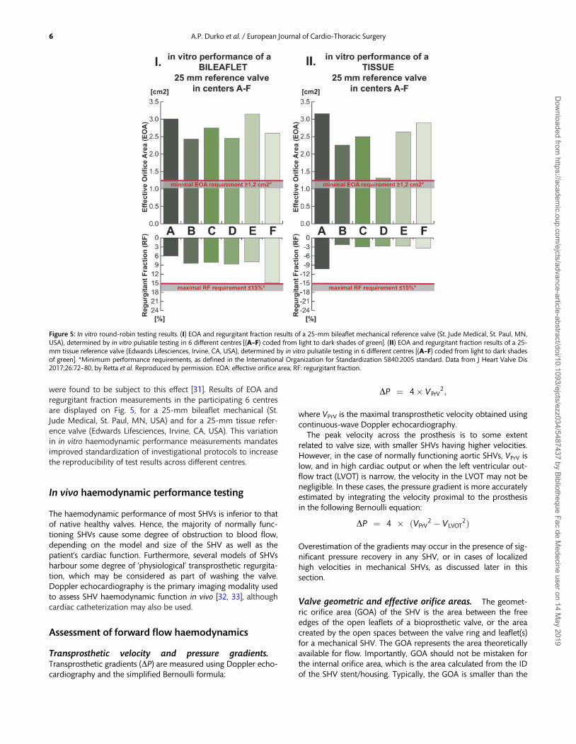
were found to be subject to this effect [31]. Results of EOA andregurgitant fraction measurements in the participating 6 centresare displayed on Fig. 5, for a 25-mm bileaflet mechanical (St.Jude Medical, St. Paul, MN, USA) and for a 25-mm tissue refer-ence valve (Edwards Lifesciences, Irvine, CA, USA). This variationin in vitro haemodynamic performance measurements mandatesimproved standardization of investigational protocols to increasethe reproducibility of test results across different centres.
In vivo haemodynamic performance testing
The haemodynamic performance of most SHVs is inferior to thatof native healthy valves. Hence, the majority of normally func-tioning SHVs cause some degree of obstruction to blood flow,depending on the model and size of the SHV as well as thepatient’s cardiac function. Furthermore, several models of SHVsharbour some degree of ‘physiological’ transprosthetic regurgita-tion, which may be considered as part of washing the valve.Doppler echocardiography is the primary imaging modality usedto assess SHV haemodynamic function in vivo [32, 33], althoughcardiac catheterization may also be used.
Assessment of forward flow haemodynamics
Transprosthetic velocity and pressure gradients.Transprosthetic gradients (DP) are measured using Doppler echo-cardiography and the simplified Bernoulli formula:
DP ¼ 4� VPrV2;
where VPrV is the maximal transprosthetic velocity obtained usingcontinuous-wave Doppler echocardiography.
The peak velocity across the prosthesis is to some extentrelated to valve size, with smaller SHVs having higher velocities.However, in the case of normally functioning aortic SHVs, VPrV islow, and in high cardiac output or when the left ventricular out-flow tract (LVOT) is narrow, the velocity in the LVOT may not benegligible. In these cases, the pressure gradient is more accuratelyestimated by integrating the velocity proximal to the prosthesisin the following Bernoulli equation:
DP ¼ 4 � ðVPrV2 � VLVOT
2Þ
Overestimation of the gradients may occur in the presence of sig-nificant pressure recovery in any SHV, or in cases of localizedhigh velocities in mechanical SHVs, as discussed later in thissection.
Valve geometric and effective orifice areas. The geomet-ric orifice area (GOA) of the SHV is the area between the freeedges of the open leaflets of a bioprosthetic valve, or the areacreated by the open spaces between the valve ring and leaflet(s)for a mechanical SHV. The GOA represents the area theoreticallyavailable for flow. Importantly, GOA should not be mistaken forthe internal orifice area, which is the area calculated from the IDof the SHV stent/housing. Typically, the GOA is smaller than the
Figure 5: In vitro round-robin testing results. (I) EOA and regurgitant fraction results of a 25-mm bileaflet mechanical reference valve (St. Jude Medical, St. Paul, MN,USA), determined by in vitro pulsatile testing in 6 different centres [(A–F) coded from light to dark shades of green]. (II) EOA and regurgitant fraction results of a 25-mm tissue reference valve (Edwards Lifesciences, Irvine, CA, USA), determined by in vitro pulsatile testing in 6 different centres [(A–F) coded from light to dark shadesof green]. *Minimum performance requirements, as defined in the International Organization for Standardization 5840:2005 standard. Data from J Heart Valve Dis2017;26:72–80, by Retta et al. Reproduced by permission. EOA: effective orifice area; RF: regurgitant fraction.
6 A.P. Durko et al. / European Journal of Cardio-Thoracic Surgery
Dow
nloaded from https://academ
ic.oup.com/ejcts/advance-article-abstract/doi/10.1093/ejcts/ezz034/5487437 by Bibliotheque Fac de M
edecine user on 14 May 2019
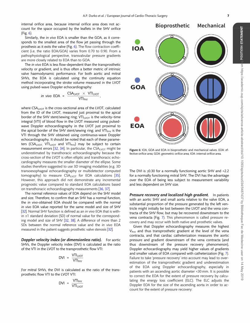
internal orifice area, because internal orifice area does not ac-count for the space occupied by the leaflets in the SHV orifice(Fig. 6).
Similarly, the in vivo EOA is smaller than the GOA, as it corre-sponds to the smallest area of the flow jet passing through theprosthesis as it exits the valve (Fig. 6). The flow contraction coeffi-cient (i.e. the ratio EOA/GOA) varies from 0.70 to 0.90. From apathophysiological perspective, transvalvular pressure gradientsare more closely related to EOA than to GOA.
The in vivo EOA is less flow-dependent than the transprostheticvelocity or gradient, and is thus often a better metric of intrinsicvalve haemodynamic performance. For both aortic and mitralSHVs, the EOA is calculated using the continuity equationmethod incorporating the stroke volume measured in the LVOTusing pulsed-wave Doppler echocardiography:
in vivo EOA =CSALVOT � VTILVOT
VTIPrV;
where CSALVOT is the cross-sectional area of the LVOT, calculatedfrom the ID of the LVOT, measured just proximal to the apicalborder of the SHV stent/sewing ring; VTILVOT is the velocity–timeintegral (VTI) of blood flow in the LVOT measured using pulsed-wave Doppler echocardiography in the LVOT just proximal tothe apical border of the SHV stent/sewing ring; and VTIPrV is theVTI through the SHV obtained using continuous-wave Dopplerechocardiography. It should be noted that each of these parame-ters (CSALVOT, VTILVOT and VTIPrV) may be subject to certainmeasurement errors [32, 34]. In particular, the CSALVOT might beunderestimated by transthoracic echocardiography because thecross-section of the LVOT is often elliptic and transthoracic echo-cardiography measures the smaller diameter of the ellipse. Somestudies therefore suggested to use 3D imaging modalities (e.g. 3Dtransoesophageal echocardiography or multidetector computedtomography) to measure CSALVOT for EOA calculations [35].However, this approach did not demonstrate any incrementalprognostic value compared to standard EOA calculations basedon transthoracic echocardiography measurements [36, 37].
The normal reference values of EOA depend on the SHV modeland size. Therefore, to confirm that an SHV has a normal function,the in vivo-obtained EOA should be compared with the normalin vivo EOA value reported for the same model and size of SHV[32]. Normal SHV function is defined as an in vivo EOA that is with-in ±1 standard deviation (SD) of normal value for the correspond-ing model and size of SHV [32, 38]. A difference of more than 2SDs between the normal reference value and the in vivo EOAmeasured in the patient suggests prosthetic valve stenosis [32].
Doppler velocity index (or dimensionless ratio). For aorticSHVs, the Doppler velocity index (DVI) is calculated as the ratioof the VTI in the LVOT to the transprosthetic flow VTI:
DVI =VTILVOT
VTIPrV
.For mitral SHVs, the DVI is calculated as the ratio of the trans-prosthetic flow VTI to the LVOT VTI:
DVI =VTIPrV
VTILVOT
The DVI is >_0.30 for a normally functioning aortic SHV and <2.2for a normally functioning mitral SHV. The DVI has the advantageover the EOA of being less subject to measurement variabilityand less dependent on SHV size.
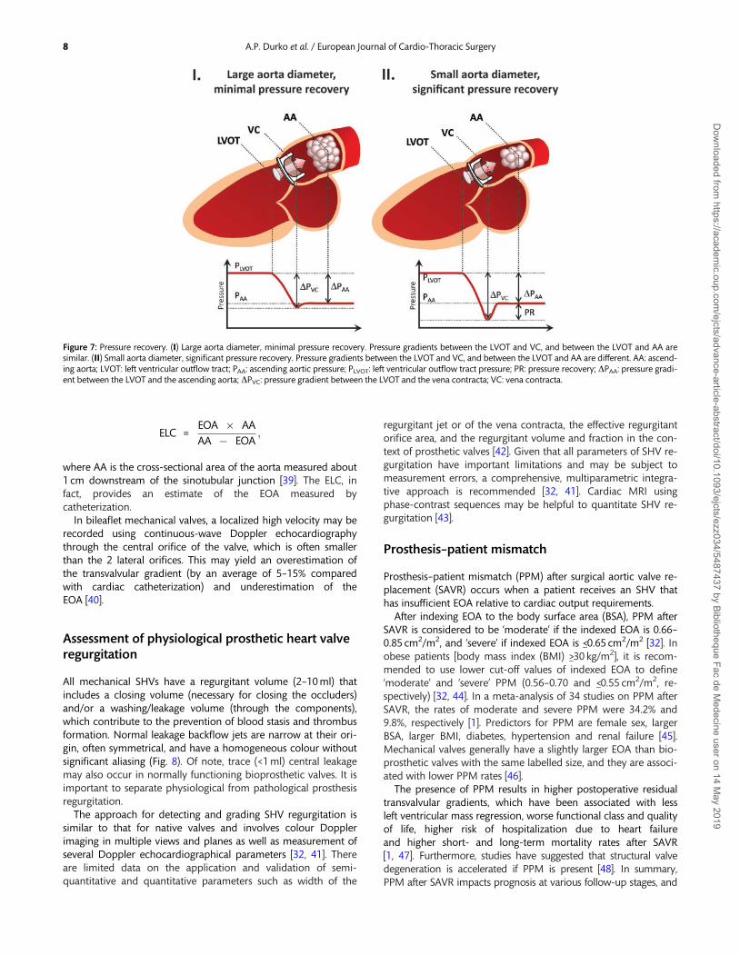
Pressure recovery and localized high gradient. In patientswith an aortic SHV and small aorta relative to the valve EOA, asubstantial proportion of the pressure generated by the left ven-tricle might initially be lost between the LVOT and the vena con-tracta of the SHV flow, but may be recovered downstream to thevena contracta (Fig. 7). This phenomenon is called pressure re-covery and may occur with both native and prosthetic valves.
Given that Doppler echocardiography measures the highestVPrV and thus transprosthetic gradient at the level of the venacontracta, and that cardiac catheterization measures the aorticpressure and gradient downstream of the vena contracta (andthus downstream of the pressure recovery phenomenon),Doppler echocardiography may yield higher values of gradientsand smaller values of EOA compared with catheterization (Fig. 7).Failure to take ‘pressure recovery’ into account may lead to over-estimation of the transprosthetic gradient and underestimationof the EOA using Doppler echocardiography, especially inpatients with an ascending aortic diameter <30 mm. It is possibleto correct the EOA for the extent of pressure recovery by calcu-lating the energy loss coefficient (ELC). The ELC adjusts theDoppler EOA for the size of the ascending aorta in order to ac-count for the extent of pressure recovery:
Figure 6: IOA, GOA and EOA in bioprosthetic and mechanical valves. EOA: ef-fective orifice area; GOA: geometric orifice area; IOA: internal orifice area.
PO
SITI
ON
STA
TEM
ENT
7A.P. Durko et al. / European Journal of Cardio-Thoracic Surgery
Dow
nloaded from https://academ
ic.oup.com/ejcts/advance-article-abstract/doi/10.1093/ejcts/ezz034/5487437 by Bibliotheque Fac de M
edecine user on 14 May 2019
ELC =EOA � AA
AA � EOA;
where AA is the cross-sectional area of the aorta measured about1 cm downstream of the sinotubular junction [39]. The ELC, infact, provides an estimate of the EOA measured bycatheterization.
In bileaflet mechanical valves, a localized high velocity may berecorded using continuous-wave Doppler echocardiographythrough the central orifice of the valve, which is often smallerthan the 2 lateral orifices. This may yield an overestimation ofthe transvalvular gradient (by an average of 5–15% comparedwith cardiac catheterization) and underestimation of theEOA [40].
Assessment of physiological prosthetic heart valveregurgitation
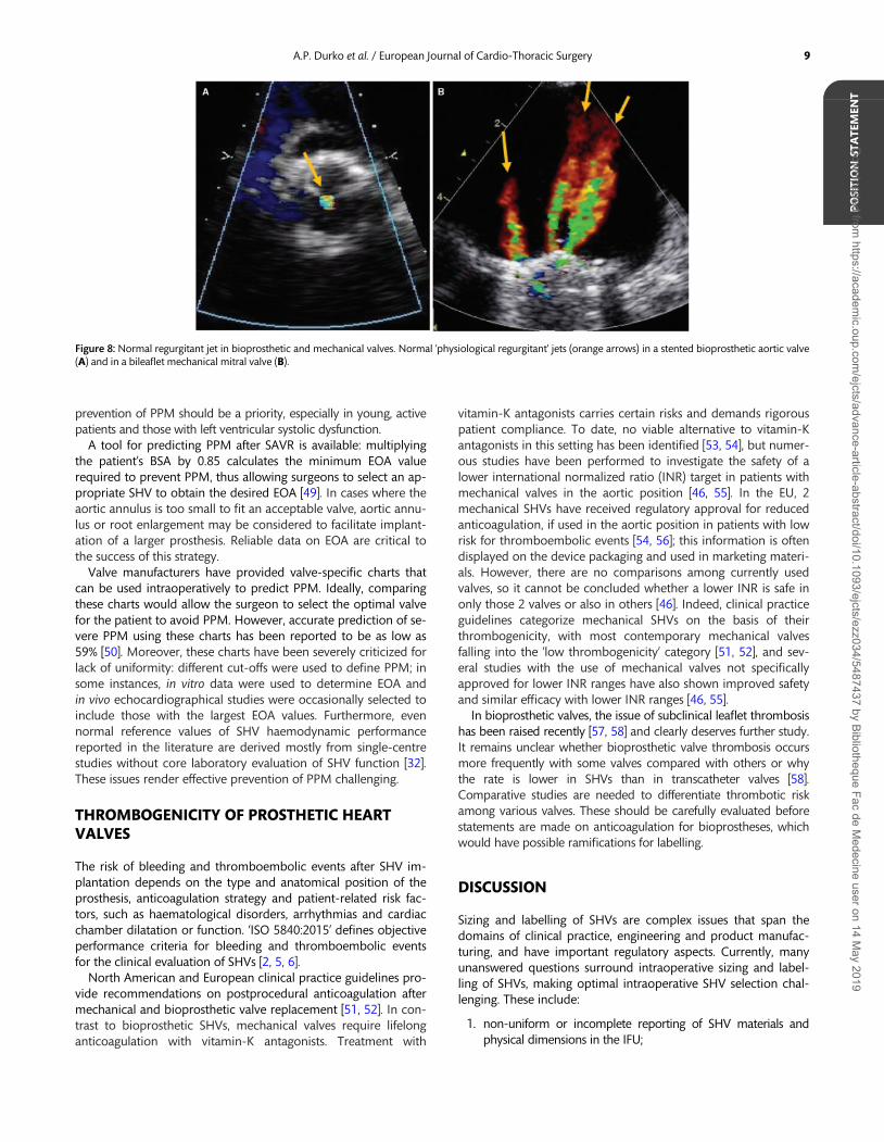
All mechanical SHVs have a regurgitant volume (2–10 ml) thatincludes a closing volume (necessary for closing the occluders)and/or a washing/leakage volume (through the components),which contribute to the prevention of blood stasis and thrombusformation. Normal leakage backflow jets are narrow at their ori-gin, often symmetrical, and have a homogeneous colour withoutsignificant aliasing (Fig. 8). Of note, trace (<1 ml) central leakagemay also occur in normally functioning bioprosthetic valves. It isimportant to separate physiological from pathological prosthesisregurgitation.
The approach for detecting and grading SHV regurgitation issimilar to that for native valves and involves colour Dopplerimaging in multiple views and planes as well as measurement ofseveral Doppler echocardiographical parameters [32, 41]. Thereare limited data on the application and validation of semi-quantitative and quantitative parameters such as width of the
regurgitant jet or of the vena contracta, the effective regurgitantorifice area, and the regurgitant volume and fraction in the con-text of prosthetic valves [42]. Given that all parameters of SHV re-gurgitation have important limitations and may be subject tomeasurement errors, a comprehensive, multiparametric integra-tive approach is recommended [32, 41]. Cardiac MRI usingphase-contrast sequences may be helpful to quantitate SHV re-gurgitation [43].
Prosthesis–patient mismatch
Prosthesis–patient mismatch (PPM) after surgical aortic valve re-placement (SAVR) occurs when a patient receives an SHV thathas insufficient EOA relative to cardiac output requirements.
After indexing EOA to the body surface area (BSA), PPM afterSAVR is considered to be ‘moderate’ if the indexed EOA is 0.66–0.85 cm2/m2, and ‘severe’ if indexed EOA is <_0.65 cm2/m2 [32]. Inobese patients [body mass index (BMI) >_30 kg/m2], it is recom-mended to use lower cut-off values of indexed EOA to define‘moderate’ and ‘severe’ PPM (0.56–0.70 and <_0.55 cm2/m2, re-spectively) [32, 44]. In a meta-analysis of 34 studies on PPM afterSAVR, the rates of moderate and severe PPM were 34.2% and9.8%, respectively [1]. Predictors for PPM are female sex, largerBSA, larger BMI, diabetes, hypertension and renal failure [45].Mechanical valves generally have a slightly larger EOA than bio-prosthetic valves with the same labelled size, and they are associ-ated with lower PPM rates [46].
The presence of PPM results in higher postoperative residualtransvalvular gradients, which have been associated with lessleft ventricular mass regression, worse functional class and qualityof life, higher risk of hospitalization due to heart failureand higher short- and long-term mortality rates after SAVR[1, 47]. Furthermore, studies have suggested that structural valvedegeneration is accelerated if PPM is present [48]. In summary,PPM after SAVR impacts prognosis at various follow-up stages, and
Figure 7: Pressure recovery. (I) Large aorta diameter, minimal pressure recovery. Pressure gradients between the LVOT and VC, and between the LVOT and AA aresimilar. (II) Small aorta diameter, significant pressure recovery. Pressure gradients between the LVOT and VC, and between the LVOT and AA are different. AA: ascend-ing aorta; LVOT: left ventricular outflow tract; PAA: ascending aortic pressure; PLVOT: left ventricular outflow tract pressure; PR: pressure recovery; DPAA: pressure gradi-ent between the LVOT and the ascending aorta; DPVC: pressure gradient between the LVOT and the vena contracta; VC: vena contracta.
8 A.P. Durko et al. / European Journal of Cardio-Thoracic Surgery
Dow
nloaded from https://academ
ic.oup.com/ejcts/advance-article-abstract/doi/10.1093/ejcts/ezz034/5487437 by Bibliotheque Fac de M
edecine user on 14 May 2019
prevention of PPM should be a priority, especially in young, activepatients and those with left ventricular systolic dysfunction.
A tool for predicting PPM after SAVR is available: multiplyingthe patient’s BSA by 0.85 calculates the minimum EOA valuerequired to prevent PPM, thus allowing surgeons to select an ap-propriate SHV to obtain the desired EOA [49]. In cases where theaortic annulus is too small to fit an acceptable valve, aortic annu-lus or root enlargement may be considered to facilitate implant-ation of a larger prosthesis. Reliable data on EOA are critical tothe success of this strategy.
Valve manufacturers have provided valve-specific charts thatcan be used intraoperatively to predict PPM. Ideally, comparingthese charts would allow the surgeon to select the optimal valvefor the patient to avoid PPM. However, accurate prediction of se-vere PPM using these charts has been reported to be as low as59% [50]. Moreover, these charts have been severely criticized forlack of uniformity: different cut-offs were used to define PPM; insome instances, in vitro data were used to determine EOA andin vivo echocardiographical studies were occasionally selected toinclude those with the largest EOA values. Furthermore, evennormal reference values of SHV haemodynamic performancereported in the literature are derived mostly from single-centrestudies without core laboratory evaluation of SHV function [32].These issues render effective prevention of PPM challenging.
THROMBOGENICITY OF PROSTHETIC HEARTVALVES
The risk of bleeding and thromboembolic events after SHV im-plantation depends on the type and anatomical position of theprosthesis, anticoagulation strategy and patient-related risk fac-tors, such as haematological disorders, arrhythmias and cardiacchamber dilatation or function. ‘ISO 5840:2015’ defines objectiveperformance criteria for bleeding and thromboembolic eventsfor the clinical evaluation of SHVs [2, 5, 6].
North American and European clinical practice guidelines pro-vide recommendations on postprocedural anticoagulation aftermechanical and bioprosthetic valve replacement [51, 52]. In con-trast to bioprosthetic SHVs, mechanical valves require lifelonganticoagulation with vitamin-K antagonists. Treatment with
vitamin-K antagonists carries certain risks and demands rigorouspatient compliance. To date, no viable alternative to vitamin-Kantagonists in this setting has been identified [53, 54], but numer-ous studies have been performed to investigate the safety of alower international normalized ratio (INR) target in patients withmechanical valves in the aortic position [46, 55]. In the EU, 2mechanical SHVs have received regulatory approval for reducedanticoagulation, if used in the aortic position in patients with lowrisk for thromboembolic events [54, 56]; this information is oftendisplayed on the device packaging and used in marketing materi-als. However, there are no comparisons among currently usedvalves, so it cannot be concluded whether a lower INR is safe inonly those 2 valves or also in others [46]. Indeed, clinical practiceguidelines categorize mechanical SHVs on the basis of theirthrombogenicity, with most contemporary mechanical valvesfalling into the ‘low thrombogenicity’ category [51, 52], and sev-eral studies with the use of mechanical valves not specificallyapproved for lower INR ranges have also shown improved safetyand similar efficacy with lower INR ranges [46, 55].
In bioprosthetic valves, the issue of subclinical leaflet thrombosishas been raised recently [57, 58] and clearly deserves further study.It remains unclear whether bioprosthetic valve thrombosis occursmore frequently with some valves compared with others or whythe rate is lower in SHVs than in transcatheter valves [58].Comparative studies are needed to differentiate thrombotic riskamong various valves. These should be carefully evaluated beforestatements are made on anticoagulation for bioprostheses, whichwould have possible ramifications for labelling.
DISCUSSION
Sizing and labelling of SHVs are complex issues that span thedomains of clinical practice, engineering and product manufac-turing, and have important regulatory aspects. Currently, manyunanswered questions surround intraoperative sizing and label-ling of SHVs, making optimal intraoperative SHV selection chal-lenging. These include:
1. non-uniform or incomplete reporting of SHV materials andphysical dimensions in the IFU;
Figure 8: Normal regurgitant jet in bioprosthetic and mechanical valves. Normal ‘physiological regurgitant’ jets (orange arrows) in a stented bioprosthetic aortic valve(A) and in a bileaflet mechanical mitral valve (B).
PO
SITI
ON
STA
TEM
ENT
9A.P. Durko et al. / European Journal of Cardio-Thoracic Surgery
Dow
nloaded from https://academ
ic.oup.com/ejcts/advance-article-abstract/doi/10.1093/ejcts/ezz034/5487437 by Bibliotheque Fac de M
edecine user on 14 May 2019

2. unclear definition of labelled valve size and inconsistencies be-tween sizer dimensions and labelled valve size;
3. non-uniform marking of SHV support structures;4. lack of robust information on in vivo haemodynamic perform-
ance in the IFU, and no information available regardinghaemodynamic performance on package labels;
5. lack of uniform tools backed by solid evidence to preventPPM; and
6. lack of good-quality, robust clinical data on SHVthrombogenicity.
This situation has persisted for decades and has received manycalls for action, but no uniform solution has been achieved todate.
Determining the right amount of information for intraopera-tive decision-making requires finding a delicate balance.Although currently available parameters on the package labelsprovide incomplete information regarding the most importantcharacteristics of the SHV, the inclusion of redundant or irrele-vant information would similarly create confusion in the surgicalcommunity.
Complex issues are best prioritized and solved through concen-trated efforts from all critical stakeholders [59]. The EACTS–STS–AATS Valve Labelling article Project has been initiated with this in-tention. The medical community requires clarity and should worktogether with valve manufacturers, regulatory bodies and the ISOgroup to achieve an optimal solution. This article has summarizedthe most important characteristics of SHVs and the background ofSHV labelling and is intended to pave the way for an EACTS–STS–AATS Expert Consensus Document that will include recommenda-tions on SHV sizing and labelling.
CONCLUSION
This joint EACTS–STS–AATS Labelling Task Force has identifiedseveral issues related to SHV sizing and labelling. These issuesshould be addressed to ensure that surgeons are provided withsufficient, appropriate and standardized information required foroptimal SHV choice.
ACKNOWLEDGEMENTS
The authors would like to acknowledge the help of Rianne Kalkman(EACTS Office) in coordinating the Task Force activities.
Funding
This work was supported by the European Association for Cardio-Thoracic Surgery (EACTS). We acknowledge the financial supportfrom the Netherlands Cardio Vascular Research Initiative: theDutch Heart Foundation, Dutch Federation of University MedicalCentres, the Netherlands Organisation for Health Research andDevelopment and the Royal Netherlands Academy of Sciences.
Conflict of interest: Vinayak Bapat served as a consultant forEdwards Lifesciences, Medtronic and 4Tech Inc.; Filip P.A.Casselman served as a consultant for Edwards Lifesciences andMedtronic; Edward P. Chen served as a consultant for CryoLife and
as a proctor for Medtronic; Philippe Pibarot has received researchgrant support from Edwards Lifesciences and Medtronic for echo-cardiography core laboratory analyses in transcatheter heart valves;Giordano Tasca reports speaker fees from St. Jude Medical; MarcoStijnen is an employee of LifeTec Group; Ruggero De Paulis servedas consultant for Edwards Lifesciences and Medtronic. All otherauthors declared no conflict of interest.
REFERENCES
[1] Head SJ, Mokhles MM, Osnabrugge RL, Pibarot P, Mack MJ, TakkenbergJJ et al. The impact of prosthesis-patient mismatch on long-term survivalafter aortic valve replacement: a systematic review and meta-analysis of34 observational studies comprising 27 186 patients with 133 141 pa-tient-years. Eur Heart J 2012;33:1518–29.
[2] ISO. International Standard, ISO 5840:2015. Cardiovascular Implants—Cardiac Valve Prostheses. International Organization for Standardization(ISO), 2015. www.iso.org (3 April 2018, date last accessed).
[3] Doenst T, Amorim PA, Al-Alam N, Lehmann S, Mukherjee C, Faerber G.Where is the common sense in aortic valve replacement? A review ofhemodynamics and sizing of stented tissue valves. J Thorac CardiovascSurg 2011;142:1180–7.
[4] ISO. ISO—International Organization for Standardization. 2018. https://www.iso.org/home.html (14 March 2018, date last accessed).
[5] Wu Y, Butchart EG, Borer JS, Yoganathan A, Grunkemeier GL. Clinicalevaluation of new heart valve prostheses: update of objective perform-ance criteria. Ann Thorac Surg 2014;98:1865–74.
[6] Head SJ, Mylotte D, Mack MJ, Piazza N, van Mieghem NM, Leon MBet al. Considerations and recommendations for the introduction of ob-jective performance criteria for transcatheter aortic heart valve deviceapproval. Circulation 2016;133:2086–93.
[7] The Council of the European Communities, Council Directive 93/42/EEC,http://eur-lex.europa.eu/LexUriServ/LexUriServ.do?uri=CONSLEG:1993L0042:20071011:en:PDF (3 April 2018, date last accessed)
[8] The European Parliament and the Council of the European Union,Regulation (EU) 2017/745 http://eur-lex.europa.eu/legal-content/ENG/TXT/PDF/?uri=CELEX:32017R0745&from=EN (3 April 2018, date lastaccessed).
[9] International Organization for Standardization (ISO), EuropeanCommittee for Standardization (CEN). Agreement on Technical Co-operation between ISO and CEN (Vienna Agreement). 2001. https://boss.cen.eu/ref/Vienna_Agreement.pdf (3 April 2018, date last accessed).
[10] U.S. Food and Drug Administration. FDA Organization: U.S. Departmentof Health and Human Services. 2018. https://www.fda.gov/AboutFDA/CentersOffices/default.htm (3 April 2018, date last accessed).
[11] U.S. Food and Drug Administration. Premarket Approval (PMA): U.S.Department of Health and Human Services. 2018. https://www.fda.gov/MedicalDevices/DeviceRegulationandGuidance/HowtoMarketYourDevice/PremarketSubmissions/PremarketApprovalPMA/ucm2007514.htm#data (3April 2018, date last accessed).
[12] U.S. Food and Drug Administration. Recognition and Use of ConsensusStandards, Guidance for Industry and FDA Staff. U.S. Food and DrugAdministration, 2007. https://www.fda.gov/downloads/medicaldevices/deviceregulationandguidance/guidancedocuments/ucm077295.pdf (3April 2018, date last accessed).
[13] Mohammadi H, Mequanint K. Prosthetic aortic heart valves: modelingand design. Med Eng Phys 2011;33:131–47.
[14] Konakci KZ, Bohle B, Blumer R, Hoetzenecker W, Roth G, Moser B et al.Alpha-Gal on bioprostheses: xenograft immune response in cardiac sur-gery. Eur J Clin Invest 2005;35:17–23.
[15] Flameng W, Rega F, Vercalsteren M, Herijgers P, Meuris B.Antimineralization treatment and patient-prosthesis mismatch are majordeterminants of the onset and incidence of structural valve degenerationin bioprosthetic heart valves. J Thorac Cardiovasc Surg 2014;147:1219–24.
[16] Scherman J, Bezuidenhout D, Ofoegbu C, Williams DF, Zilla P. TAVI forlow to middle income countries. Eur Heart J 2017;38:1182–4.
[17] Bezuidenhout D, Williams DF, Zilla P. Polymeric heart valves for surgicalimplantation, catheter-based technologies and heart assist devices.Biomaterials 2015;36:6–25.
[18] Glaser N, Jackson V, Franco-Cereceda A, Sartipy U. Survival after aorticvalve replacement with bovine or porcine valve prostheses: a systematic
10 A.P. Durko et al. / European Journal of Cardio-Thoracic Surgery
Dow
nloaded from https://academ
ic.oup.com/ejcts/advance-article-abstract/doi/10.1093/ejcts/ezz034/5487437 by Bibliotheque Fac de M
edecine user on 14 May 2019

review and meta-analysis. Thorac Cardiovasc Surg 2018; doi:10.1055/s-0038-1649513 [Epub ahead of print].
[19] Hickey GL, Bridgewater B, Grant SW, Deanfield J, Parkinson J, Bryan AJet al. National registry data and record linkage to inform postmarket sur-veillance of prosthetic aortic valve models over 15 Years. JAMA InternMed 2017;177:79–86.
[20] Allen KB, Chhatriwalla AK, Cohen DJ, Saxon JT, Aggarwal S, Hart A et al.Bioprosthetic valve fracture to facilitate transcatheter valve-in-valve im-plantation. Ann Thorac Surg 2017;104:1501–8.
[21] Bapat V, Mydin I, Chadalavada S, Tehrani H, Attia R, Thomas M. A guideto fluoroscopic identification and design of bioprosthetic valves: a refer-ence for valve-in-valve procedure. Catheter Cardiovasc Interv 2013;81:853–61.
[22] Bapat VN, Attia R, Thomas M. Effect of valve design on the stent internaldiameter of a bioprosthetic valve: a concept of true internal diameterand its implications for the valve-in-valve procedure. JACC CardiovascInterv 2014;7:115–27.
[23] Cevasco M, Mick SL, Kwon M, Lee LS, Chen EP, Chen FY. True externaldiameter better predicts hemodynamic performance of bioprostheticaortic valves than the manufacturers’ stated size. J Heart Valve Dis 2013;22:377–82.
[24] Ruzicka DJ, Hettich I, Hutter A, Bleiziffer S, Badiu CC, Bauernschmitt Ret al. The complete supraannular concept: in vivo hemodynamics of bo-vine and porcine aortic bioprostheses. Circulation 2009;120:S139–45.
[25] Tabata M, Shibayama K, Watanabe H, Sato Y, Fukui T, Takanashi S.Simple interrupted suturing increases valve performance after aorticvalve replacement with a small supra-annular bioprosthesis. J ThoracCardiovasc Surg 2014;147:321–5.
[26] Cameron D. Little things matter. J Thorac Cardiovasc Surg 2015;149:918–19.
[27] Christakis GT, Buth KJ, Goldman BS, Fremes SE, Rao V, Cohen G et al.Inaccurate and misleading valve sizing: a proposed standard for valvesize nomenclature. Ann Thorac Surg 1998;66:1198–203.
[28] Ruzicka DJ, Eichinger WB, Hettich IM, Bleiziffer S, Bauernschmitt R,Lange R. Hemodynamic performance of the new St. Jude Medical EpicSupra porcine bioprosthesis in comparison to the Medtronic Mosaic onthe basis of patient annulus diameter. J Heart Valve Dis 2008;17:426–33;discussion 34.
[29] Walther T, Falk V, Weigl C, Diegeler A, Rauch T, Autschbach R et al.Discrepancy of sizers for conventional and stentless aortic valveimplants. J Heart Valve Dis 1997;6:145–8.
[30] Bartels C, Leyh RG, Matthias Bechtel JF, Joubert-Hubner E, Sievers HH.Discrepancies between sizer and valve dimensions: implications forsmall aortic root. Ann Thorac Surg 1998;65:1631–3.
[31] Retta SM, Kepner J, Marquez S, Herman BA, S Shu MC, Grossman LW.In-vitro pulsatile flow measurement in prosthetic heart valves: an inter-laboratory comparison. J Heart Valve Dis 2017;26:72–80.
[32] Lancellotti P, Pibarot P, Chambers J, Edvardsen T, Delgado V, Dulgheru Ret al. Recommendations for the imaging assessment of prosthetic heartvalves: a report from the European Association of CardiovascularImaging endorsed by the Chinese Society of Echocardiography, theInter-American Society of Echocardiography and the BrazilianDepartment of Cardiovascular Imaging. Eur Heart J Cardiovasc Imaging2016;17:589–90.
[33] Zoghbi WA, Chambers JB, Dumesnil JG, Foster E, Gottdiener JS,Grayburn PA et al. Recommendations for evaluation of prosthetic valveswith echocardiography and Doppler ultrasound: a report from theAmerican Society of Echocardiography’s Guidelines and StandardsCommittee and the Task Force on Prosthetic Valves, developed in con-junction with the American College of Cardiology CardiovascularImaging Committee, Cardiac Imaging Committee of the American HeartAssociation, the European Association of Echocardiography, a registeredbranch of the European Society of Cardiology, the Japanese Society ofEchocardiography and the Canadian Society of Echocardiography,endorsed by the American College of Cardiology Foundation, AmericanHeart Association, European Association of Echocardiography, a regis-tered branch of the European Society of Cardiology, the JapaneseSociety of Echocardiography, and Canadian Society ofEchocardiography. J Am Soc Echocardiogr 2009;22:975–1014.
[34] Gaspar T, Adawi S, Sachner R, Asmer I, Ganaeem M, Rubinshtein R et al.Three-dimensional imaging of the left ventricular outflow tract: impacton aortic valve area estimation by the continuity equation. J Am SocEchocardiogr 2012;25:749–57.
[35] Kamperidis V, van Rosendael PJ, Katsanos S, van der Kley F, Regeer M, AlAmri I et al. Low gradient severe aortic stenosis with preserved ejection
fraction: reclassification of severity by fusion of Doppler and computedtomographic data. Eur Heart J 2015;36:2087–96.
[36] Mooney J, Sellers SL, Blanke P, Pibarot P, Hahn RT, Dvir D et al. CT-defined prosthesis-patient mismatch downgrades frequency and sever-ity, and demonstrates no association with adverse outcomes after trans-catheter aortic valve replacement. JACC Cardiovasc Interv 2017;10:1578–87.
[37] Clavel MA, Malouf J, Messika-Zeitoun D, Araoz PA, Michelena HI,Enriquez-Sarano M. Aortic valve area calculation in aortic stenosis by CTand Doppler echocardiography. JACC Cardiovasc Imaging 2015;8:248–57.
[38] Hahn RT, Leipsic J, Douglas PS, Jaber WA, Weissman NJ, Pibarot P et al.Comprehensive echocardiographic assessment of normal transcathetervalve function. JACC Cardiovasc Imaging 2018;
[39] Pibarot P, Garcia D, Dumesnil JG. Energy loss index in aortic stenosis:from fluid mechanics concept to clinical application. Circulation 2013;127:1101–4.
[40] Evin M, Pibarot P, Guivier-Curien C, Tanne D, Kadem L, Rieu R.Localized transvalvular pressure gradients in mitral bileaflet mechanicalheart valves and impact on gradient overestimation by Doppler. J AmSoc Echocardiogr 2013;26:791–800.
[41] Pibarot P, Hahn RT, Weissman NJ, Monaghan MJ. Assessment of para-valvular regurgitation following TAVR: a proposal of unifying gradingscheme. JACC Cardiovasc Imaging 2015;8:340–60.
[42] Fattouch K, Lancellotti P, Vannan MA, Speziale G (Eds). Advances inTreatments for Aortic Valve and Root Diseases. 1st edn. SpringerInternational Publishing, 2018. doi: 10.1007/978-3-319-66483-5.
[43] Ribeiro HB, Orwat S, Hayek SS, Larose E, Babaliaros V, Dahou A et al.Cardiovascular magnetic resonance to evaluate aortic regurgitation aftertranscatheter aortic valve replacement. J Am Coll Cardiol 2016;68:577–85.
[44] Mohty D, Dumesnil JG, Echahidi N, Mathieu P, Dagenais F, Voisine Pet al. Impact of prosthesis-patient mismatch on long-term survival afteraortic valve replacement: influence of age, obesity, and left ventriculardysfunction. J Am Coll Cardiol 2009;53:39–47.
[45] Dayan V, Vignolo G, Soca G, Paganini JJ, Brusich D, Pibarot P. Predictorsand outcomes of prosthesis-patient mismatch after aortic valve replace-ment. JACC Cardiovasc Imaging 2016;9:924–33.
[46] Head SJ, Celik M, Kappetein AP. Mechanical versus bioprosthetic aorticvalve replacement. Eur Heart J 2017;38:2183–91.
[47] Fallon JM, DeSimone JP, Brennan JM, O’Brien S, Thibault DP, DiScipioAW et al. The incidence and consequence of prosthesis-patient mis-match after surgical aortic valve replacement. Ann Thorac Surg 2018;106:14–22.
[48] Johnston DR, Soltesz EG, Vakil N, Rajeswaran J, Roselli EE, Sabik JF 3rdet al. Long-term durability of bioprosthetic aortic valves: implicationsfrom 12,569 implants. Ann Thorac Surg 2015;99:1239–47.
[49] Pibarot P, Dumesnil JG. Prosthesis-patient mismatch: definition, clinicalimpact, and prevention. Heart 2006;92:1022–9.
[50] Bleiziffer S, Eichinger WB, Hettich I, Guenzinger R, Ruzicka D,Bauernschmitt R et al. Prediction of valve prosthesis-patient mismatchprior to aortic valve replacement: which is the best method? Heart 2007;93:615–20.
[51] Falk V, Baumgartner H, Bax JJ, De Bonis M, Hamm C, Holm PJ et al. 2017ESC/EACTS Guidelines for the management of valvular heart disease. EurJ Cardiothorac Surg 2017;52:616–64.
[52] Nishimura RA, Otto CM, Bonow RO, Carabello BA, Erwin JP 3rd, FleisherLA et al. 2017 AHA/ACC focused update of the 2014 AHA/ACC Guidelinefor the management of patients with valvular heart disease: a report of theAmerican College of Cardiology/American Heart Association task force onclinical practice guidelines. J Am Coll Cardiol 2017;70:252–89.
[53] Eikelboom JW, Connolly SJ, Brueckmann M, Granger CB, Kappetein AP,Mack MJ et al. Dabigatran versus warfarin in patients with mechanicalheart valves. N Engl J Med 2013;369:1206–14.
[54] Puskas JD, Gerdisch M, Nichols D, Fermin L, Rhenman B, Kapoor D et al.Anticoagulation and antiplatelet strategies after On-X mechanical aorticvalve replacement. J Am Coll Cardiol 2018;71:2717–26.
[55] Koertke H, Zittermann A, Wagner O, Secer S, Sciangula A, Saggau Wet al. Telemedicine-guided, very low-dose international normalized ratioself-control in patients with mechanical heart valve implants. Eur Heart J2015;36:1297–305.
[56] Torella M, Aquila I, Chiodini P, Amarelli C, Romano G, Della Ratta EEet al. Low-dose anticoagulation after isolated mechanical aortic valve re-placement with Liva Nova Bicarbon prosthesis: a post hoc analysis ofLOWERING-IT Trial. Sci Rep 2018;8:8405.
PO
SITI
ON
STA
TEM
ENT
11A.P. Durko et al. / European Journal of Cardio-Thoracic Surgery
Dow
nloaded from https://academ
ic.oup.com/ejcts/advance-article-abstract/doi/10.1093/ejcts/ezz034/5487437 by Bibliotheque Fac de M
edecine user on 14 May 2019
[57] Makkar RR, Fontana G, Jilaihawi H, Chakravarty T, Kofoed KF, De BackerO et al. Possible subclinical leaflet thrombosis in bioprosthetic aorticvalves. N Engl J Med 2015;373:2015–24.
[58] Chakravarty T, Sondergaard L, Friedman J, De Backer O, Berman D,Kofoed KF et al. Subclinical leaflet thrombosis in surgical and transcath-eter bioprosthetic aortic valves: an observational study. Lancet 2017;389:2383–92.
[59] Kappetein AP, Head SJ, Genereux P, Piazza N, van Mieghem NM, BlackstoneEH et al. Updated standardized endpoint definitions for transcatheter aorticvalve implantation: the Valve Academic Research Consortium-2 consensusdocument. Eur J Cardiothorac Surg 2012;42:S45–S60.
APPENDIX: TASK FORCE MEMBERS
Cardiac surgeons
Ruggero De Paulis, European Hospital, Rome, Italy—Task Force chairmanPavan Atluri, University of Pennsylvania, Philadelphia, PA, USAVinayak Bapat, New York-Presbyterian/Columbia University Medical Center, NewYork, NY, USADuke E. Cameron, Massachusetts General Hospital, Boston, MA, USAFilip P.A. Casselman, OLV Clinic, Aalst, BelgiumEdward P. Chen, Emory University School of Medicine, Atlanta, GA, USAGry Dahle, Oslo University Hospital, Oslo, NorwayAndras P. Durko, Erasmus University Medical Center, Rotterdam, Netherlands
and Medical and Health Science Centre, University of Debrecen, Debrecen,Hungary
Tjark Ebels, University Medical Center Amsterdam, Amsterdam, NetherlandsJohn A. Elefteriades, Yale University School of Medicine, New Haven, CT, USAStuart J. Head, Erasmus University Medical Center, Rotterdam, NetherlandsA. Pieter Kappetein, Erasmus University Medical Center, Rotterdam, NetherlandsRichard L. Prager, University of Michigan Hospital, Ann Arbor, MI, USAAlan Speir, Inova Cardiac and Thoracic Surgery, Falls Church, VA, USAGiordano Tasca, Hospital A. Manzoni, Lecco, ItalyThomas Walther, Wolfgang Goethe University, Frankfurt, Germany
Cardiologists
Patrizio Lancellotti, University of Liege Hospital, Liege, BelgiumPhilippe Pibarot, Laval University, Quebec City, QC, CanadaRaphael Rosenhek, Medical University of Vienna, Vienna, Austria
Engineers
Jurgen de Hart, LifeTec Group, Eindhoven, NetherlandsMarco Stijnen, LifeTec Group, Eindhoven, Netherlands
ISO
Ajit Yoganathan, Georgia Institute of Technology/Emory School of Medicine,Atlanta, GA, USA
US food and drug administration
Nicole IbrahimJohn LaschingerChangfu Wu
Notified body
Giovanni Di Rienzo, TUV SUD, Munich, Germany
Competent authorities
Alexander McLaren, Medicines and Healthcare products Regulatory Agency,London, UK
Hazel Randall, Medicines and Healthcare products Regulatory Agency, London,UK
Industry representatives
Lisa Becker, Abbott, Chicago, IL, USAScott Capps, CryoLife, Kennesaw, GA, USABrian Duncan, LivaNova, London, UKChad Green, Abbott, Chicago, IL, USAJohn C. Hay, Medtronic, Minneapolis, MN, USAOrnella Ieropoli, LivaNova, London, UKAshwini A. Jacob, Edwards Lifesciences, Irvine, CA, USAEric Manasse, Abbott, Chicago, IL, USASalvador Marquez, Edwards Lifesciences, Irvine, CA, USAWilliam F. Northrup III, CryoLife Kennesaw, GA, USATim Ryan, Medtronic, Minneapolis, MN, USAWendel Smith, Edwards Lifesciences, Irvine, CA, USA
12 A.P. Durko et al. / European Journal of Cardio-Thoracic Surgery
Dow
nloaded from https://academ
ic.oup.com/ejcts/advance-article-abstract/doi/10.1093/ejcts/ezz034/5487437 by Bibliotheque Fac de M
edecine user on 14 May 2019