Earth’s Land Chapter 5 Lesson 1. Vocabulary Landform Crust Mantle Core.

CHAPTER V
THE MANTLE
PageAppeara.nce___________________________________________________________ 74Anatomy__ __ __ __ __ __ __ _ ____ __ __ ___ ___ __ __ __ _ 74
Rudimentary muscle of the ma.ntle___________________________________ 79Histology __ __ __ __ __ __ ___ __ __ __ ___ __ __ __ _____ __ __ 79
Connective tissue_________________________________________________ 79Muscles_ __ __ __ __ __ __ __ __ ___ __ __ ___ __ __ _ __ __ 83Blood vessels__ __ __ __ __ __ __ __ ___ __ __ ___ _ 84Epithelium, tentacles, a.ndnerves________________________________ 84Periostracal groove a.nd gla.nd_____________________________________ 86Subligamental ridge ____ __ __ __ __ __ _____ __ __ __ __ __ ___ __ __ __ __ 89
Functions of the ma.ntle______________________________________________ 90Formation a.nd calcification of sheIL_________________________________ 91
Theories of calcification___________________________________________ 94Cytological Identification of calclum______________________________ 101Sources of calclum________________________________________________ 103Mineralogy of calcium carbonate in mollusca.n shells______________ 103Rate of calclfication_______________________________________________ 104
Bibliography 107
The inner organs of all mollusks are coveredwith a soft and fleshy fold of tissue called the mantle or pallium (Latin for cloak or coverlet). Thestructure of the mantle is relatively simple: theorgan consists of a sheet of connective tissue containing muscles, blood vessels, and nerves and iscovered on both siaes by unicellular epithelium.Many blood cells invade and wander throughoutthe entire thickness of the mantle, infiltrating thespaces (sinuses) in the connective tissue, andcrawling through the epithelium to aggregate onthe outer surface of the mantle.
Although the principal role of the mantle is theformation of the shell and the secretion of theligament, the organ plays a major part in severalother functions. It receives sensory stimuli andconveys them to the nervous system and assistsin the shedding and dispersal of eggs duringspawning (see ch. XIV). The mantle also participates in respiration by providing direct exchange of gases between the surface tissues of theoyster and the surrounding water. It stores reserve materials (glycogen and lipids), secretes largequantities of mucus and, finally, aids in excretionby discarding blood cells loaded with wasteproducts.
APPEARANCE
The appearance of the mantle reflects the condition of the oyster. At the time of sexual maturity
74
it is a creamy-yellowish color. In oysters whichhave accumulated large amounts of glycogen withthe onset of the cold season the mantle is whiteand thick. In oysters of poor quality or in thosewhich have not yet recovered after spawning, themantle is so transparent that the brown or greenish color of the underlying digestive organ isclearly visible through the thin and watery tissue.Oysters in this condition are particularly suitablefor the study of muscles, blood vessels, and nerveswhich in good quality, "fat" oysters are coveredby a thick layer of reserve materials.
Pigment cells are concentrated along the freeedge of the mantle and in the tentacles in a bandvarying in color from light brown to jet black.Also, accumulation of copper in the blood cellsmay produce a distinct green coloration. Different intensities of pigmentation are often foundin oysters of identical origin growing together,and cannot be correlated with geographicallocation or type of bottom.
ANATOMYFor a detailed study of the mantle the oyster
should be fully narcotized by Epsom salt (seep. 65) or by refrigerating it overnight at a temperature of about 20 to 40 C. After the valves areforced apart and the body dissected along themedian plane, the two halves of the oyster areleft attached to their respective valves and themantle is preserved in its natural position by having a large quantity of fixing fluid poured over it.Portions of the mantle required for study are cutoff, stained, dehydrated, cleared, and mounted.In this way very satisfactory whole mounts canbe obtained.
The two lobes of the mantle are joined togetherat the dorsoposterior margin, and form a cap orhood which covers the mouth and the labialpalps (fig. 71). Along the anterior and ventralsides of the body the lobes are free and follow thecurvature of the shell. When the oyster opensits shell the mantles separate with the valves to
FISHERY BULLETIN: VOLUME 64, CHAPTER V

which they adhere, leaving a narrow openingbetween the two lobes through which sea watercan enter the mantle cavity. The edge of themantle, may, however, occupy various positions:it may extend parallel and beyond the edge of thevalves to leave a wide space between the twoopposing lobes, or it may bend inward almostperpendicular to the shell surface (fig. 74) toreduce or completely close the opening between thetwo lobes and thereby limit the access of waterto the mantle cavity. The behavior of themantle edge as a regulatory mechanism controllingthe flow of water through the mollusk will bediscussed later (p. 185). In a closed oyster themantle edge is located about midway betweenthe distal margin of the gills and the edge of theshell. Its position is marked by an impressioncalled the pallial line, which is less pronounced inthe oysters than in clams and some other bivalves.
At the ventroposterior end of the body the twoopposing lobes of the mantle join the gills to form
the delicate outside wall of the cloaca (figs. 72and 75, cl., f.). On the left side of the body themantle is joined to the visceral mass; on theright side it is separated from the visceral massby the promyal chamber. The fusion of themantle with the visceral mass and with the basesof gill plates forms the wall of the epibranchialchamber, which leads to the cloaca (fig. 75, cl.).The relative position of the epibranchial andpromyal chambers can be seen in the crosssection of the oyster made through the dorsalpart of the body (fig. 73, ep.br.ch.; pr.ch.).
An oblong slit between the two mantle lobes onthe dorsoposterior side of the body marks theopening of the promyal chamber. The inside ofthis chamber can be examined by completelynarcotizing the oyster and forcing its valves apartas far as possible without tearing the adductormuscle. Viewed from the posterior side thepromyal chamber in a relaxed oyster appears asan oval cavity (fig. 75) to the left of the adductor
J\ Millimeters
FIGURE 74.-Cross sections of the valves, mantle, gills, and adjacent portion of the visceral mass of C. virginica. In bothdiagrams the valves are open; the open pallial curtain (at left) permits free access of water to the mantle cavity; theclosed pallial curtain (at right) prevents water from entering the mantle cavity. The outer lobe adheres closelyto the valve and is not visible. Drawn from the photomicrographs of cross section of adult oyster. Bouin, hematoxylin-eosin.
THE MANTLE 75

oCent imeters
5
FIGURE 75.-Promyal chamber (at left) and cloaca (at right) viewed from the posterior side of a large oyster (C. virginica)completely relaxed by narcosis. Note the fusion of the two opposing lobes of the mantle, and the adductor muscle(in the middle). Drawn from life. Actual size. ad.m.-adductor muscle; cl.-cloaca; f.-fusion of mantle lobesand gills; pr.ch.-promyal chamber; r.-rectum.
muscle. The large round openings of the watertubes of the gills can be seen on the inner wall ofthe chamber. The rectum extends along theedge of the chamber, ending with a round anusadhering to the side of the adductor muscle; theopening of the cloaca lies to the right of the muscle.The water tubes emptying into the cloaca and thefusion of the mantle with the gill lamellae arealso clearly visible.
The most conspicuous components of the mantleare the radial muscles, the blood vessels, and thenerves (fig. 76). All these structures can beidentified in a piece of fresh tissue stretched overa glass slide and examined under strong illumination with a low-power microscope. For moredetailed study, it is necessary to prepare wholemounts or to section the preserved tissues.
The radial muscles extend from the place oftheir attachment to the visceral mass to the edgeof the mantle. At about two-thirds of theirlength from their base they begin a fanlike expansion toward the periphery before terminatingin the base of the tentacles. The majority ofthe muscles are accompanied along their lengthby nerves, blood vessels, and blood sinuses. Muchmore slender than the radial muscles are theconcentric muscular bands which parallel the freeedge of the mantle (not shown in fig. 76) and aremore abundant at its thickened distal edge.
Because of its strongly developed musculature,the mantle is highly contractile. It may stretcha considerable distance beyond the edge of thevalve, or withdraw inside the shell, and even rollup into a tube. Contraction of the radial muscleswill throw the inner surface of the mantle into
76
ridges which serve as temporary channels fordiscarding mucus and foreign particles accumulated on it. These movements may involveeither the entire surface of the mantle or only asmall portion of it, depending on the intensityof stimulation received by the tentacles.
The wide circumpallial artery (fig. 76, cp.a.)follows the entire periphery of the mantle. Atlow magnification it is usually visible as a widetubular structure with many branching vesselswhich communicate with the irregular spaces(blood sinuses) within the connective tissue. Alarge pulsating blood vessel, called the accessoryheart (ch. XI, fig. 236), is located in the anteroventral part in each lobe of the mantle. Thestructure and the function of this vessel are discussed in chapter XI, p. 254.
Just outward from the circumpallial artery runsthe circumpallial nerve, which also extends alongthe entire margin of the mantle. In whole mountpreparations seen under low power, the circumpallial nerve appears as a compact unbranchingband. Examination under high power, however,reveals a fine network of small nerves connectingthe circumpallial nerve with nerves and with thevisceral and cerebral ganglia. Since nerve fiberson the surface of the mantle and in the tentacleslead to the circumpallial nerve, stimuli receivedby the neuroreceptors of these areas are transmitted through the circumpallial nerve to theradial nerves and reach either the visceral or thecerebral ganglia.
The thick and muscular border of the mantleis divided into three lobes (fig. 77) which havebeen described in the literature as "folds" (Awati
FISH AND WILDLIFE SERVICE

0.1.
m.1.
cp.n.
cp.Q.
Centimeters
FIGURE 76.-Whole mount of a piece of mantle. Majorportion of connective tissue was removed by maceration.Safranin stain. Magnified about 10 times. bLv.blood vessels; cp.a.-circumpallial artery; cp.n.- circumpallial nerve; m.L-middle lobe; p.c.-pallial curtain (inner lobe); r.m.-radial muscle; o.L-outer lobeor shell lobe; t1-tentacles of inner lobe; t 2-tentaclesof middle lobe. Radial nerves surrounded by radialmuscles are not visible. Formalin 5 percent, hematoxylin.
and Rai, 1931), "reduplications" (Nelson, 1938;Pelseneer, 1906), "lamellae" (Hopkins, 1933),"lames" in French (Leenhardt, 1926), and"Klappe", in German (Rawitz, 1888). The term"reduplication" is misleading because the lobesare not formed by the duplication of the mantletissue, being comparable rather to a fringe orflounce at the margin of a soft material. To avoidconfusion the term marginal lobes is retained inthis text.
The mantle border of all the species of oystersstudied, namely, C. virginica, C. angulata, C.
THE MANTLE
733-851 Q.-.-<64-6
gigas, O. edulis, and O. lurida is divided intothree projecting lobes, the outer or shell lobe(sh.I.), the middle lobe (m.l.), and the inner lobeor pallial curtain (p.c.). Hopkins' statement(1933, p. 483) that "The border of the mantle(of C. gigas) divides into two lamellae, eachbearing a row of tentacles" is an obviousinaccuracy of description.
The outer or shell lobe (sh.I.) is narrow anddevoid of tentacles. It lies in contact with themargin of the shell and may be seen protrudingbeyond the edge of the valve during periods ofrapid growth. The middle and the inner lobeseach bear a row of sensitive and highly contractiletentacles.
The inner lobe or pallial curtain (fig. 77, p.c.)is especially broad and turned inward. In describing this structure in scallops Pelseneer namedit the "velum" (1906). Although that term hasbeen used by several investigators (Awati andRai, 1931; Dakin, 1909b) Nelson (1938) pointedout that the term "velum" is better known as theswimming organ of the pelecypod larvae andproposed to call the inner lobes of the mantlethe "pallial curtains". This term seems to beappropriate, but is used in this book in the singularsince there appears to be no advantage in thepIural recommended by Nelson.
The inner lobe may be projected into the mantlecavity (fig. 74). Depending on the degree ofcontraction of various sets of muscles the innerlobe assumes different angles in relation to themantle as a whole. In a fully relaxed molluskthe lobe of each side extends outward in the generalplane of the mantle and shell. In a contractedstate the lobes on both sides project inward almostat right angles to the surface of the mantle; inthis position the mantle borders touch and thetentacles of the two sides interlock, effectivelysealing the entrance to the mantle cavity. Thisfunction of the inner lobe was first described byRawitz in 1888 and was redescribed in 1933 byHopkins. As will be shown later (p. 304) thepallial curtain also plays an important role duringthe spawning of female oysters.
The deep furrow between the shell lobe andthe middle lobe is called the periostracal groove(fig. 77, per.gr.), the name referring to the secretion site of organic shell material by glandularcells concentrated in the deepest portion of thegroove and collectively known as the periostracalor conchiolin gland (c.gl). During the shell-
77

FIGURE 77.-Transverse section of the edge of the mantle of adult C. virginica. Bouin 3, hematoxylin-eosin. The outeror shell lobe at left faces the valve (not shown) and is bent as a result of fixation. The section passes between thetentacles of the inner lobe (pallial curtain); only the tentacle of the middle lobe (m.l.) is seen. c.gl.-conchiolin (orperiostracal) gland; conch.-sheet of conchiolin spread over the shell lobe ; cp.a.-circumpallial artery; cp.n.-circumpallial nerve; el.f.-elastic fibers; ep.-epithelium; l.m.-Iongitudinal muscles of tentacles; m.l.-middle lobe; ob.m.oblique muscles; p.c.-pallial curtain (inner lobe); per. gr.-periostracal groove; sh.l.-shell or outer lobe; tr.m.transverse muscles.
growing season, viscous yellowish material (fig.77, conch.) accumulates in the groove and gradually oozes out to the periphery of the outermantle lobe, where it solidifies into the periostracum. The groove between the middle lobe(m.l.) and the pallial curtain secretes mucus,which is gradually moved by ciliary currents tothe outer margin of the mantle and there discarded.
It has already been noted that the edges of themiddle and the inner lobe each bear a row ofhighly extensible, tapering tentacles; however,their arrangement and size in the two lobes aredifferent. Two types are clearly visible along theedge of the middle lobe: numerous short and slender tentacles, and less abundant long and stout
78
ones (fig. 76). The order of the tentacles followsa certain pattern, namely, each long tentacle issucceeded by a group of four to six small ones (tz).
The stout tentacles frequently occupy a positionslightly out of line with the small ones, being alittle nearer to the inner fold. The inner lobebears only the long and stout tentacles (t1).
There is great variation in the size of all thetentacles and in their pigmentation. Since theyare highly sensitive to touch and other stimuli andretract at the slightest disturbance, their relativesize can be observed only when they are completelyrelaxed. In fully narcotized adult oysters theratio between the numbers of tentacles on the
FISH AND WILDLIFE SERVICE

inner and middle lobes was found to vary from10:18 to 10:32.
It has not yet been definitely establishedwhether the two types of tentacles contain differentreceptors and thereforie respond to differentstimuli. According to Elsey (1935) the largetentacles of O. gigas are more sensitive to hydrocWoric acid than the small ones. Hopkins (1932)does not specify which row of tentacles was underobservation in his work on sensory stimulationof O. virginica. In my experiments (see p. 293)observations were made exclusively on the longtentacles of the inner lobe.
A narrow and slightly pigmented cylindricalstructure along the dorsal edge of the mantle(fig. 78) marks the position of the subligamentalridge, the organ which secretes the ligament.The ridge consists of a layer of specialized epithelium underlined by connective tissue. Largeblood vessels are found close to the base of theridge. Microscopic structure of the ridge is givenon p. 83.
RUDIMENTARY MUSCLE OF THEMANTLE
A small and sometimes hardly visible muscle islocated on the dorsal part of the mantle. Itslocation is sometimes marked by light violetpigmentation and by a shallow depression in thecorresponding part of the valve to which themuscle adheres. The attachment is weak, and inthe majority of oysters the muscle ~eparates fromthe valve when the valve is lift.ed. Leenhardt(1926) states, however, that in some O. edulis themuscles were so strongly attached to the shell thatthey could not be separated without rupturing themantle tissue. Examination of sections of themantle of O. virginica from the Woods Hole areaconvinced me that muscle fibers do not extendfrom one side to the other, but end in the connective tissue of the mantle. The muscle isapparently nonfunctional and morphologically isnot analogous to the anterior adductor of bivalves.Leenhardt (1926) considers the rudimentarymuscle of the mantle as a vestige of the larval footretractor which disappears during metamorphosis.Stenzel (1963) states that this mus61e is present inall the Ostreldae and calls it Quenstedt's muscle inhonor of its discoverer (Quenstedt, 1867).
HISTOLOGY
The mantle consists of connective tissue whichenvelops the muscles, blood vessels, and nerves
THE MANTLE
and is covered on both sides with the epithelium.
CONNECTIVE TISSUE
The most conspicuous structural element of theconnective tissue is the vesicular cell, characterizedby large globular or oval body and relatively smallnucleus without nucleoli. In zoological literaturethese cells appear under a variety of names andwere even incorrectly considered as lacunae(Leenhardt, 1926) and mucus cells (List, 1902).Well-developed membranes outline cell boundariessharply; the protoplasm within forms a delicatenetwork of fine granules. In preparations dehydrated with alcohol the inside of the vesicularcells appears almost empty, but in tissues treatedwith osmic acid and in frozen sections stainedwith Sudan II and other fat stains large globulesof lipids are seen to fill the inside of the cells (figs.79 and 80). Less abundant are the smallE'r roundcells with more compact protoplasm. They oftenoccur near small arteries (fig. 81, r.c.). Thefusiform cells (f.c.) with small bodies and ovalnuclei form long branching processes which anastomose and touch each other.
Examination of frozen sections of connectivetissue treated with toluidine blue or other metachromatic stains shows clearly the presence of acytoplasmic ground substance with a very finereticulum supporting various inclusions. Afterthe removal of glycogen this substance can bestained very deeply with periodic acid fuchsin(McMannus reagent )or with Hale stain which isused to test for acid polysaccharides of the hyaluronic acid type (Hale, 1946). The results of suchstaining reactions have been interpreted in theliterature as indicating the presence of mucopolysaccharides or mucoproteins. Histological methods are not entirely dependable (Meyer, 1957),but so far no chemical analyses of the connectivetissue of the mantle have been made. It is known,however, that acid mucopolysaccharides areamong the components of the ground substancesin mammalian tissues. It is very likely that theyare also present in the connective tissue of theoyster.
Elastic fibrils are scattered throughout theconnective tissue of the entire thickness of themantle but appear to be more abundant at the freeedge and in the layers underlying the surfaceepithelium (fig. 77, eLf.). Muscle fibers are alsovery abundant and will be discussed in detail later.
In some specimens the mantle may be thin andtransparent whereas in others it is thick and
79

Microns
FIGURE 78.-Longitudinal section of the subligamental ridge made at right angles to its dorsal surface. Bouin 3,hematoxylin-eosin. bl.v.-Iarge blood vessel; el.m.-basal elastic membrane; ep.-epithelium; m.-muscle fibers;pig.c.-pigment cells; po.-pockets between the epithelial cells; v.c.-vesicular cells.
80 FISH AND WILDLIFE SERVICE

FIGURE 79.-Vesicular cells of connective tissue from the mantle of an adult C. virginica surrounding the blood sinus.Blood cells crawl between the cells of connective tissue and penetrate into the sinus. Bouin 3, hematoxylin-eosin.
opaque. These changes in appearance usuallycoincide with seasonal cycles in the glycogen content of the connective tissue and with the progressive stages of gonad development.
The presence of glycogen can be easily demonstrated by treating the tissue with Lugol solution(1 percent iodine in 2 percent potassium iodidein water). Specific reagents used for the identification of glycogen, such as Best's carmine andBensley's modification of Bauer-Feulgen reagent(which stains glycogen granules red-violet), alsogive good results.
In the live oyster glycogen can be seen as smallcolloidal granules which ooze from the tissue underslight pressure. In preserved and stainedmaterial it appears in the form of granules orrods (fig. 82). The total amount of glycogen inthe connective tissue may be so great that theblood vessels and nerves of the mantle are COlll
pletely hidden under it and cannot be traced by
30oMicrons
FIGURE 80.-Vesicular cell of connective tissue wIth fatglobules. Frozen section. Sudan IT.
THE MANTLE 81

40Microns
~,.....-__ bl. c.
o
r. c.
FIGURE 8l.-Cross section of a small artery of the mantle. bl.c.-blood cells; e.lf.-elastic fibrils; end.-endothelium;f.c.-fusiform cells; r.c.-round cells; v.c.-vesicular cells. Kahle, hematoxylin-eosin.
··;::~,t·f;C·.·...::~.;.<;
~;..:;.:~;.;,?;~.::-::~~, .... , '.'t'
'..~...
oMicrons
30
FIGURE 82.-Two vesicular cells from the mantel of an adult C. vzrgznica. Left-the cell contains glycogen stainedwith Best's carmine; fat globules were dissolved in processing. Right-similar cell after fixation with Bouin 3; notecomplete absence of glycogen and fat, both dissolved during fixation and dehydration.
dissection. Such abundance of reserve materialled one of the earlier investigators (Creighton,1896, 1899) to conclude that its storage in theconnective tissue of lamellibranchs is a specialadaptation comparable to the storage of fat inthe connective tissues of vertebrates.
The quantity of glycogen stored in connectivetissue gradually decreases as the gonads of the
oyster increase in bulk. This was first reportedfor O. edulis by Pekelharing (1901) and confirmedby the more recent investigations of Bargeton(1942). Evidence presented in the latter workstrongly suggests that the growing sex cells utilizethe glycogen stored in the vesicular cells surrounding the gonad tubules, but cytological detailsof this process are still unknown and the problem
82 FISH AND WILDLIFE SERVICE

has not yet been studied from a biochemicalpoint of view.
After the disappearance of their containedglycogen the vesicular cells do not shrink orcollapse. A hypothesis was therefore advanced(Semichon, 1932) that the glycogen granules aresupported by a framework of a special substancewhich remains intact after the dissolution ofglycogen. It is claimed that this framework canbe revealed by staining with black anilin inks.The evidence for the existence of such a specialsubstance is not, however, convincing. In cellswith a moderate content of glycogen the lattercan be seen in close contact with the protoplasmicnetwork typical for vesicular cells. Furthermore,the walls of the vesicular cells are fairly rigid andthe cells retain their shape even when they areempty. The shrinkage of connective tissue frequently caused by changes in osmotic pressurewhen the salinity of the water surrounding theoyster is suddenly increased is not associatedwith the disappearance of glycogen.
The fat globules in vesicular cells vary greatlyin size and number, usually forming distinctvacuoles that are easily dislodged. The relationship between the fat and glycogen content of theoyster and the role of lipids in the physiology oflamellibranchs have not been studied.
1-1arge oval cells containing a brown pigment arescattered throughout the connective tissue of themantle. The pigment is not soluble either inacids or fat solvents. Its chemical nature andphysiological significance are not known.
Wandering blood cells are commonly seen inthe mantle. They crawl between the connectiveti:.;sue cells, aggregate in the vicinity of bloodvessels and blood sinuses (fig. 79), and are gradually discarded through the surface of the mantle.AR a rule, the oyster continually loses a certainamount of blood by diapedesis or bleeding. Anyexcess of heavy metals accumulated by bloodcells (see p. 390) is also discarded by this normalprocess.
MUSCLES
The radial muscles consist of large, regularlyspaced bands of fibers which extend almost theentire width of the mantle from the line of itsfusion with the visceral mass and with the adductor muscle to the free margin. For a study ofthe anatomy of the muscular system the connective tissue in which the bands are firmlyenclosed should be macerated in 1 percent potas-
THE MANTLE
sium hydroxide for about 24 hours. After beingwashed in distilled water the loosened tissues areremoved with a small stiff brush and fine forceps.
The radial muscle bands are composed oflarge bundles of unstriated fibers which begin tobranch toward the distal edge of the mantle aboutone-third of the distance from that edge. Atthis level the muscles appear fanlike and enterinto all three lobes, where they terminate.
The central part of a muscle band is usuallyoccupied by one or two radial nerves, althoughmuscles without a central nerve (figs. 83 and 84)do occur.
The contraction of the radial muscles pulls theentire mantle inside and throws its surface intoridges. Such a general reaction usually precedesthe contraction of the adductor muscle and theclosing of the valves. The contraction mayoccur spontaneously in response to some internalstimulus or it may develop as a result of externalirritation produced by chemicals, mechanical andelectrical shock, or sudden change in illumination.In response to a weak outside stimulus only asmall sector of the mantle contracts, making aslight V-shaped indentation along its periphery.This response mayor may not be followed bycontraction of the adductor muscle. Strongstimuli, as a rule, result in complete withdrawalof the mantle, contraction of the adductor muscle,and closing of the valves. Besides the largeradial bands there are many smaller bundles oftransverse fibers (fig. 77, tr.m.) extending diagonally across the thickness of the mantle, a welldeveloped system of longitudinal muscles (l.m.) ,and the oblique muscles (ob.m.) of the tentacles.
The longitudinal or concentric muscles followthe general outlines of the edge. They are moreabundant at the thickened distal edge of themantle but do not exhibit the definite patternof distribution apparent in the radial muscles.The transverse muscle fibers are more numerousin the pallial curtain (fig. 77, tr.m.) than in theother parts of the mantle. They are so arrangedthat the position of the curtain may be quicklychanged in response to external or internal stimuli.
All the muscle cells are of the smooth, nonstriated type with typical elongated nuclei. Insome bivalves the muscle fibers of the mantleappear to show a double oblique striation; thiswas shown to be an optical effect created by aseries of fine fibrillae spiralling around the largerfibers (Fol, 1888; Marceau, 1904). Muscle fibers
83

oMicrons
200
FIGURE 83.-Cross section of the radial muscle of the mantle of an adult C. virginica. The muscle completely surroundstwo nerves. Bouin 3, hematoxylin-eosin.
with true transverse striation, described in themantle of Pecten jacobaeus and P. opercularis(Dakin, 1909a), are not found in the oyster mantle.
BLOOD VESSELS
The principal blood vessels of the mantle(fig. 232 in ch. XI) are the circumpallial artery(cr.p.a.), which runs along its entire periphery andsends out many branches; the common pallialartery (co.p.a.); and a large pulsating vessel inthe anteroventral part of the mantle called theaccessory heart (fig. 236 in ch. XI). The lattercan be observed by dissecting the wall of theepibranchial chamber and spreading the cut tissues apart. The structure and function of thesevessels are discussed on page 253.
The small arteries and veins of the mantle canbe recognized easily by their histological charac-
84
teristics. The walls of the arteries have a thick,elastic, muscular layer lined with endothelium(fig. 81, end.). In the veins the elastic layer ismuch less developed and the endothelium absent(fig. 85). The sinuses (fig. 79) are irregularlyshaped spaces in the connective tissue. Sincethey have no walls of their own they cannot contract. The size of the opening or lumen may bereduced by growth of the surrounding vesicularcells and by accumulation of blood cells.
EPITHELIUM, TENTACLES, AND NERVES
Both sides of the mantle are covered by cylindrical epithelial cells set on an elastic basal membrane (fig. 77). Large goblet cells which secretemucus and cells containing eosinophile granulesare abundant on both sides of the mantle. Thecells of the side facing the pallial cavity are long
FISH AND WILDLIFE SERVICE

oMicrons
200
FIGURE 84. Cross section of the radial muscle of an adult C. virginica. The muscle is not accompanied by nerve. Bouin3, hematoxylin-eosin.
and ciliated; those on the outside under the valvesbear no cilia and are much shorter, in placesalmost cubical.
The two sides of the mantle perform differentfunctions. The inner side maintains ciliary currents, which in general move from the base of themantle to its edge and carry mucus and sedimentssettled from the water; this material is passed tothe margin of the shell to be discharged. Theepithelium of the outer side secretes the innerlayer of the shell, the so-called calcito-ostracum.
Although the ciliated epithelium of the edge ofthe mantle contains the same kind and proportionof cellular elements found in other parts of theorgan, the cilia at the border of the mantle areespecially powerful. The tentacles themselvesconsist of a core of connective tissue with associ-
THE MANTLE
ated blood vessels, elastic fibrils, and musclefibers which emerge from branches of the radialmuscles. On the outside the tentacles are coveredwith a single layer of ciliated epithelium to whichblack or brown pigment imparts a dark color.Special sense organs are absent but the tentacles,especially the long ones, are well supplied withner.ves branching out from the nerve which entersthe base of the tentacle and is itself connectedwith the nervous system of the mantle (fig. 86).
The circumpallial nerve provides communication between the tentacles and the radial nerves.The structure of this nerve resembles that of aganglion: numerous nerve cells of the types foundin visceral and other ganglia (see p. 288) occupythe periphery of the nerve; its center consists ofnerve bundles with occasional small ganglion cells.
85

FIGURE 86.-Innervation of the tentacles of the middlelobe. Formalin 5 percent, gold impregnation. Wholemount.
series of sections shows, however, that this appearance is caused by the invaginations of the innersurface of the lobe. The periostracal gland ispresent in all lamellibranchs and was the objectof many histological studies (Leenhardt, 1926;List, 1902; Moynier de Villepoix, 1895; Rassbach,1912; Rawitz, 1888).
There is a conspicuous difference in the appearance of the cells along the two sides of the groove.Those lining the outer lobe (fig. 88, left side) aredistended at the distal ends and taper toward thebase into slender rootlike processes which, according to Rawitz (1888) who described them in theoyster, penetrate the underlying connective tissue.1 was not able to reveal such rootlets in mymaterial. N one of these cells bear cilia, althoughthe distal part of the groove, not shown in figure88, is lined with ciliated epithelium. Typicalgoblet cells containing eosinophile granules,amoebocytes, and round mucus cells are presentin the epithelial layer of both sides of the groove.
0.3Mill imeters
o
100Microns
FWURE 85.-Transverse section of a small vein of themantle. Note the absence of endothelium and poorlydeveloped elastic layer. Bouin, hematoxylin-eosin.
Close nerve contact between the muscles andother organs of the mantle is maintained througha fine nerve network which can be made visibleby using the gold impregnation method (fig. 87).I have had no success in revealing it with vitalstains.
PERIOSTRACAL GROOVE AND GLAND
The narrow space between the middle and theouter lobes of the mantle edge, called the periostracal groove (fig. 77, per.gr.), is lined withciliated epithelium which is replaced at the bottom of the groove by glandular cells. The innermost part of the groove is called the periostracalgland (fig. 88), although it would have been moreappropriate to refer to it not as a gland but as asecretory epithelial surface (Maximow and Bloom,1930). This surface is covered with a singlelayer of glandular cells different in appearanceand structure from the epithelial cells of thedistal part of the groove. Unlike a true gland,it does not form a compact body extending underthe surface of the groove and it has no duct. Ontransverse sections of the mantle edge the glandsometimes appears as a round structure surrounded by connective tissue. Examination of a
86 FISH AND WILDLIFE SERVICE

oMicrons
30
FIGURE 87.-Small area of mantle showing nerve net. Formalin 5 percent, gold impregnation. Whole mount. Cameralucida drawing.
At the very bottom of the groove the tall epithelialcells are suddenly replaced by short cubical cells(fig. 88, right side) which extend a short distancealong the inner side of the groove.
The material secreted by the periostracal glandaccumulates at the bottom of the groove and inthe majority of my preparations appears toadhere to the cells of the outer side (left side offigure 88). This, however, is the result of shrinkage caused by dehydration during the processingof slides. In preparations mounted in glycerinthe conchiolin can be seen in close contact withthe epithelium of both sides of the groove.
The function of the periostracal gland is tosupply large quantities of the material requiredfor new shell growth at the edge of the valves.
THE MANTLE
The organic matrix (conchiolin) and foliatedlayers of calcite needed for increasing thickness ofthe valves, on the other hand, are secreted bythe epithelium covering the entire outer surfaceof the mantle and in close contact with the innersurface of the valve. The epithelium consists ofnonciliated cells which are cylindrical near thefree margin of the mantle but become flattenedand almost cubical in more proximal areas. Bothconchiolin-secreting and calcium-secreting cells arepresent in this epithelium but their cytologicaldifferentiation by means of staining reactions orby precipitation of calcium oxalate is not reliable.Mucus cells and oval cells containing eosinophilegranules also occur throughout the entire surfaceof the epithelial covering.
87

aMicrons
100
FIGURE 88.-Transverse section of the periostracal groove. The cells on the left side are distended with secretion; thoseat the very bottom are short, almost cUboid. The black mass at the bottom of the groove is conchiolin. Bouin,hematoxylin-eosin.
Owing to the presence of the conchiolin-secretingcells, the entire outer surface of the mantle issticky and adheres closely-to the inner surface ofthe shell. List (1902) advanced a theory, notwell supported by observation, that in theMytilidae the mantle adheres to the shell bymeans of fibrillae which originate in the myoepithelial cells and pass through the epithelium. Suchan arrangement is not found in C. virginica or inC. angulata, and according to Leenhardt (1926)does not exist in Mytilus.
The epithelial layer along both surfaces of themantle including its free edge contains alkalinephosphatase, an enzyme involved in the calcification of the shell. The presence of the enzymecan be demonstrated by the Gomori method
88
(Gomori, 1939, 1943) based on the formation ofinsoluble calcium phosphate as a result of phosphatase action on sodium glycero-phosphate andcalcium ions. Further treatment with 5 percentsilver nitrate (or with cobalt nitrate) convertsthe calcium phosphate to silver (or cobalt) phosphate which turns black after exposure to light.Both reagents gave satisfactory results in demonstrating the localization of the enzyme in theepithelium of the mantle. The strongest reaction,judged by the opacity and width of the blacklayer, was found to occur along the edges of themantle and in the area of the periostracal groove.Even the tips of the tentacles contained noticeableamounts of the enzyme (fig. 89). These obser-
FISH AND WILDLIFE SERVICE

I I I I
o .. Ow3Millimeter
FIGURE 89.-Localization of alkaline phosphatase in the mantle of C. virginica (longitudinal section). Photomicrographof a preparation treated by Gomori method.
vations are in full agreement with the resultsobtained by Bevelander (1952).
SUBLIGAMENTAL RIDGE
A small ridge marking the dorsal edge of themantle along the fusion of its two lobes is knownas the subligamental ridge. Its length in theanteroposterior direction corresponds exactly tothat of the ligament, which is secreted by theepithelial cells of the ridge. The base of theridge is flattened and rests on basic elastic membrane; the body of the ridge is semicylindrical incross section, its surface slightly undulating, ascan be seen from the longitudinal section shownin figure 78.
The histological structure of the ridge has beenstudied in }';lytilus (List, 1902; Tullberg, 1881),in Anodonta (Moynier de Villepoix, 1895;Rassbach, 1912), and in the Portuguese oysterCrassostrea (Gryphaea) angulata (Leenhardt, 1926).Leenhardt and Moynier de Villepoix call the
THE MANTLE
structure "bandelette paleale" (pallial strip) butthe term subligamental ridge seems to be moredescriptive.
In C. virginica the subligamental ridge is alwayswell developed and easily recognizable by itsshape and by its coloration, which is usuallydarker than that of the adjacent part of the mantle. The epithelium (fig. 78, ep.) covering theridge presents a. most striking picture. It consists of a layer of extremely tall and narrow cellsarranged in fanlike groups and set on a welldeveloped basal elastic membrane. The lengthof the cells varies from 50 to 200 p. depending onthe position they occupy within the layer. Thecells are very thin, with granular protoplasm andan oval-shaped nucleus. At the distal portionof the ridge the boundaries of the cells becomeindistinct and their protoplasm darker, presumably due to the concentration there of the organicmaterial which they secrete. The free surface of
89

FIGURE 90.-Discharge areas of the mantle. Note thetwo large lumps of the discarded material at the edge ofthe mantle which mark the boundaries of the principaldischarge area. Small arrows indicate the direction ofciliary currents. The clear path along the periphery ofthe mantle in the upper left side, indicated by shortarrows, marks the ciliary tract located over the circumpallial artery. Long arrow at right indicates the direction of exhalant current of the cloaca. Drawn from life.
it was sprinkled with powder, while in the samespecimen 5 to 10 minutes were required to clearthe lower (ventral) part. Although the ciliarycurrents along the posterior side of the mantlein the area adjacent to the cloaca are also directedfrom the base toward the periphery, this area isswept clear by an exhalant current from the gills(fig. 90, long arrow) which is much stronger thanthose produced by the mantle epithelium.
The currents along the anterodorsal part of themantle (upper left of the figure) adjacent to thelabial palps are directed at an acute angle to itsfree margin. There is also a well-defined tract ofciliary movement about 1.5 mm. wide parallel tothe edge of the mantle. Upon reaching the levelof the lower corners of the labial palps this current
the epithelium is not attached to the ligament aswas described by Moynier de Villepoix (1895).
At regular intervals the row of epithelial cellsis interrupted by oval-shaped pockets whichappear to be empty, with the exception of occasional amoebocytes and a few connective tissuecells. The significance of these pockets is notclear. The elastic membrane under the epithelium, thicker here than in the other parts ofthe mantle, includes many muscle fibers arrangedparallel to the length of the ridge (m.). Largeoval cells containing yellow-brownish granules(pig. c.) are abundant. The ridge is well suppliedwith blood through a large blood vessel (bI. v.),around which the connective tissue consists oftightly packed globular and spindle-shaped cells.Directly under the basal membrane of the ridge,however, the connective tissue of the mantle ismade up of large vesicular cells.
FUNCTIONS OF THE MANTLE
Ciliary currents along the inner surfaces of themantle form a definite pattern which may beeasily observed. If one valve of the oyster isremoved the corresponding mantle rolls up andexposes the gills and the inner surface of the mantle on the opposite side. In such a preparationthe intact lobe of the mantle remains fullystretched and the ciliary currents can be observedby sprinkling the surface with small quantities ofcarmine, colloidal carbon, powdered shell material,carborundum, or other powders insoluble in seawater. It is best to use very fine particles, suchas powdered mineral willemite and colloidalcarbon. Willemite phosphoresces a brilliant greenunder ultraviolet light, which makes it possibleto locate even the tiniest particles not otherwiserecognizable. As a source of ultraviolet light Iused a small Mineralight lamp. Hard and heavyparticles of this mineral may stimulate the ciliaby their weight, but this difficulty is avoided byusing colloidal carbon.
As can be seen from the diagram in figure 90,drawn from life, the general direction of the currents is from the base of the mantle to its periphery, with the ciliary motion strongest in theanterodorsal sector. In the large oyster (5 inchesin height) used for the drawing, this area extendedalong the margin of the mantle from the level ofthe labial palps approximately halfway down theanterior side. The upper part of the mantle wasusually completely cleared 2 or 3 minutes after
6Centimeters
90 FISH AND WILDLIFE SERVICE

turns sharply about 90 degrees across the mantle edge.This point marks the upper limit of the areathroughout which the material settled on themantle is discarded; in the oysters which receivedpowdered suspensions large lumps of particlesentangled in mucus are usually seen here. Asimilar accumulation of material ready to be discarded marks the anterior boundary of the discharge area which may vary in dimension andlocation in different oysters.
Along the ventral portion of the oyster bodythe ciliary currents sweep across the mantle atright angles to its edge and material is discardedalong the border. In this respect my observationsdiffer from those of Nelson (1938, p. 24), whothinks that the current in this area is directedalong the free margin toward the mouth. Sucha current was not present in the large oysters(0. virginica) I used in my experiments.
The existence of a special discharge area locatedin the zorie below the labial palps is biologicallysignificant. The so-called pseudofeces, or massesof discarded food particles and of detritusentangled in mucus, which accumulate in thisarea either slide over the free edge of the mantleand drop off or are forcibly ejected by the snappingof the valves. There is no doubt that the presenceof pseudofeces near the edge of the mantle stimulates the strong ejection reaction which constitutesone typical pattern of shell movement in theoyster (see p. 169).
FORMATION AND CALCIFICATION OFSHELL
The principal function of the mantle is theformation of the shell and its calcification. Thegreat structural complexity and intricate patternof pigmentation found in some species are producedby the mantle. The regulatory mechanisms responsible for this process are not known becausethe morphogenesis of molluscan shells has neverbeen studied experimentally. From observationson shell growth in some gastropods and lamellibranchs it is clear that the shape of the shell aswell as the pattern of pigmentation result fromthe position assumed by the edge of the mantleduring periods of shell secretion and from therate of deposition of calcium salts and pigments.
It can be easily observed in oysters, scallops,and other bivalves in which the edges of the mantleare not fused together that during periods ofgrowth the mantle extends a considerable distance
THE MANTLE
beyond the border of the shell. In some species iteven stretches far out and folds back over the outersurface of the valve. In this way, for instance,the mangrove oysters produce hooks or similarstructures by which they attach themselves tobranches of trees (fig. 5).
The differential rate of growth along the periphery of the shell as well as the formation of spines,nodes, ridges, and similar sculptural elements areboth caused by changes in the rate of depositionof shell material. Two distinct phases may bedistinguished in the shell-forming process: (1) themovements of the mantle which stretches andfolds itself in order to provide a matrix or moldupon which the shell is formed, and (2) thesecretion and deposition of the shell materialitself. It is probable that the circumpallial nerveplays a role in the first phase of the process bycontrolling the muscular activity of the mantle.Our present knowledge of the physiology andbiochemistry of shell secretion is inadequate topropose an explanation of the morphogeneticprocesses involved in shell formation. Theseprocesses are not haphazard but follow a definiteand predetermined course. This is self-evidentfrom the fact that the final shape of the shell hasdefinite mathematical characteristics (see p. 24)which can be attained only by orderly andregulated deposition of organic framework andmineral salts.
The first step in the formation of the oystershell is the secretion of conchiolin from the periostracal gland. This process can be easily observedby cutting off a small section of the edge of theupper valve and exposing the intact valve and theunderlying mantle of the opposite side. Under alow-power binocular microscope one can see aclear, viscous, and sometimes stringy substanceoozing out of the periostracal groove. Whilesecretion is taking place the edge of the mantleappears to be very active, expanding and retracting as successive layers of conchiolin are laiddown. Figure 91 shows the position of the mantleat the time of its retraction.
The newly deposited shell (n.sh.) extends outward along the plane of the valve; the edge of themantle (mn.e.) rolls upward; its outer lobe(o.mnJ.) is parallel to the plane of the valve,while the middle and inner lobe (m.l.) face theobserver. The tentacles of the inner lobe extenddown; those of the middle lobe are slightly contracted. The outer lobe underlies the sheet of
91

n.sh.
con.sft
m.L.f os.f}. mn. e.
i. L.
oMillimeters
3
FIGURE 91.-Small area of the mantle edge with the adjacent part of the newly secreted shell viewed from above. Themantle was exposed by cutting off a piece of the opposite valve, and the oyster was placed in sea water under abinocular microscope. con.sh.-conchiolin sheet; mn.e.-edge of the mantle; LL-inner lobe; n.sh.-new shell;o.mn.L-outer lobe of the mantle, contracted; p.os.g.-periostracal groove. Drawn from life. The position ofstructures at the edge of the mantle in relation to one another: the new shell area (n.sh.) marked on three sides by abroken line is in the plane of the drawing, next to it is the outer lobe (o.mn.L), then the conchiolin sheet (con.sh.),the middle lobe (m.L), and the inner lobe (LL) is at the top, nearest to the observer.
viscous conchiolin (conch.sh.) which oozes outfrom the periostracal groove (p.os.g.) between theouter and middle lobes. The distal edge of theconchiolin sheet (end of stippled area) indicatesthe previous maximal extension of the outer lobebefore the withdrawal of the mantle edge. Theentire group rests on the newly formed and alreadysolidified shell (n.sh.).
During the secretion of conchiolin the edge ofthe mantle frequently extends out and then withdraws to the position recorded in the drawing.At the time of expansion the outer lobe temporarilysupports the semiliquid conchiolin and by movingin and out spreads it over the shell. Because ofthis action the proximal part of the newly formedvalve receives a larger amount of conchiolin and
becomes thicker than the distal portion. Whensecretion is interrupted, the conchiolin layersbecome incorporated into the shell substance andthe conchiolin sheet as shown in figure 91 is nolonger visible.
The rate of secretion of the new shell varies atdifferent parts of the mantle edge. Quantitativedata are lacking, but observations made duringthe periods of more rapid growth in C. virginica(May to June and October to November in NewEngland waters) show that the area of newlyformed shell is always largest at the ventral sideof the valves near the principal axis of growth(fig. 92).
The organic matrix of the shell can be producedby the pallial epithelium at any place along the
92 FISH AND WILDLIFE SERVICE

FIGURE 92.-New shell growth formed during 1 year alongthe periphery of the valve of an adult oyster from LongIsland Sound planted in the Oyster River, Chatham,Mass. The newly formed shell is recognizable by zigzaglines of the material; its width is greatest along theventral edge.
entire outer surface of the mantle and is not restricted to the periostracal groove. Such secretion, first observed in pearl oysters (BjiSggild,1930), can be experimentally demonstrated in C.virginica. Oysters with one valve removed andthe edges of the mantle cut off above the periostracal groove secreted a new conchiolin layerover the entire surface of the exposed mantlewithin 5 days. Although the operated specimensremained alive in the laboratory tanks at WoodsHole over 3 weeks this conchiolin membraneremained uncalcified. In another experimentthree adult oysters were removed from their shellsand kept alive in sea water for 3 weeks. Theyformed rather thick coats of periostracum whichwas very lightly calcified. The repair of holesmade in oyster shells by boring snails and spongesalso shows that conchiolin is secreted by the entiresurface of the mantle. The damaged area is rapidly
oCentimeters 2
covered by a layer of organic material whichlater becomes calcified.
Soon after being secreted, the conchiolin becomes calcified. Progressive stages of this processcan be observed on the growing edge of the shell,or by inserting pieces of plastic or small glass coverslips between the edge of the mantle and the valveand removing them at regular intervals for inspection. The earliest stage of calcification isrecognized by the appearance of minute granulesof calcium salts, which become visible in polarizedlight as brightly sparkling dots (fig. 93). At thisearly stage the distribution of the granules (calcospherites) does not show any definite patternor arrangement. In a living oyster they can befound entangled in strands of mucus left on theconchiolin sheet by the back and forth movementsof the mantle edge. Within the next 24 to 48 hourstypical hexagonal crystals of calcite can be seen(fig. 94, black crosses). They gradually increasein size and present a picture of great brillianceand beauty in polarized light (fig. 95).
Distribution of calcospherites at the stage oftheir transformation into small calcite crystals onthe surface of the newly secreted shell (fig. 96)does not show any distinct orientation in relationto the growth axis of the shell. Some of the calcospherites are scattered over the entire field ofvision, while others are packed tightly betweenthe larger crystals (see large group of crystals atthe lower part of figure 96). Within the next 48hours the calcite crystals increase in size (fig. 97).In the final stage of shell formation the calcitecrystals become arranged in a distinct pattern toform the prismatic layer in which each unit is aprism oriented with its long axis at about a 90°angle to the edge of the shell (fig. 98). The formof the individual prisms varies greatly, some ofthem are even wedge-shaped and slightly curved.This can be observed after boiling a piece of shellin a strong sodium hydroxide solution to separatethe prisms (Schmidt, 1931).
Each calcite prism is surrounded by a capsuleof conchiolin. By dissolving the mineral in weakhydrochloric acid it is possible to obtain intactthe organic meshwork of the conchiolin layer.The walls of each capsule, as can be seen in figure99, are very thin and slightly iridescent. Since inthe earliest stages of shell formation the conchiolinsheet appears to be amorphous under the lightmicroscope, it is reasonable to assume that theorganic capsules of the calcite prisms are formed
THE MANTLE
733-851 0---64--7
93

FIGURE 93.-Small granules (calcospherites) in conchiolin shortly after its secretion by the mantle. Black and whiteenlargement of a Kodachrome photograph taken with polarized light.
by later deposition of conchiolin, the secretion ofwhich continues during calcification. The detailsof this prosess have not yet been described.
THEORIES OF CALCIFICATION
Studies of shell calcification fall into two majorcategories. One type of work places the emphasison the identification of calcium-secreting cells ororgans; the other approaches the problem fromthe biochemical point of view. It has been
94
generally accepted that calcium carbonate, separated from blood, is secreted as colloidal gel bycertain cells at the edge of the mantle and thatcrystallization takes place outside the cells(Crofts, 1929; Dakin, 1912; Kuyper, 1938) betweenthe conchiolin sheet and the mantle. Separationof calcium is not, however, confined to the surfacecells of the mantle. The calcium-secreting cellsmay be subepithelial, as in Patella (Davis andFleure, 1903). In the calcification of the epi-
FISH AND WILDLIFE SERVICE

Microns
FIGURE 94.-Calcite crystals of new oyster shell about 24 to 36 hours after its formation.of a Kodachrome photograph taken with polarized light.
200
Black and white enlargement
phragm of Helix pomatia (Prenant, 1924, 1928),the calcium is liberated by the leucocytes in theconnective tissue of the mantle. In the case ofpearl formation, Boutan (1923) has shown thatcalcareous deposits are formed by amoeboid cellswhich crawl through the mantle epithelium, whilethe latter secretes the concentric layers of theorganic matrix (conchiolin).
De Waele (1929) approached the calcificationproblem from the physiochemical point of view.
THE MANTLE
Working with Anodonta cygnea he has shown thatthe extrapallial fluid between the mantle and theshell is chemically identical with blood. Exposure of this fluid to air causes the formation of aprecipitate, which consists of a suspension ofcalcium spherules in protein solution. He therefore assumed the existence in the pallial fluid of ahypothetical compound consisting of protein,carbon dioxide, and calcium carbonate. Theescape of carbon dioxide would then cause the
95

~
I
.10Microns
200
FIGURE 95.-Early stage of the formation of prismatic layer. Photographed with polarized light.
that calcification ofwith the formationtricalcium-ci tra te
precipitation of calcium carbonate. Dotterweichand Elssner (1935) found, however, that calciumcarbonate crystals are formed in the extrapallialfluid of Anodonta only in an atmosphere containingless than 1.5 percent carbon. dioxide. In Helix,regeneration of the shell will take place in anatmosphere containing up to 15 percent of carbondioxide, according to Manigault (1933). Although the latter accepted De Waele's theory, hisown results seem to prove its inadequacy; andRobertson (1941) remarks that De Waele's hypothetical protein compound is without a realchemical basis. Furthermore there are other
96
discrepancies in De Waele's results which invalidate his theory. The calcospherites and theprotein precipitated from blood and from extrapallial fluid contained 50 percent organic matter,whereas the new shell contained only 4 percent ofit. To reconcile these facts it would be necessaryto assume that a great proportion of the organicmatter in the new shell must be reabsorbed. Theentire process as outlined by De Waele appears tobe highly improbable.
Steinhardt (1946) assumedthe oyster shell is associatedof citrate, probably the
FISH AND WILDLIFE SERVICE
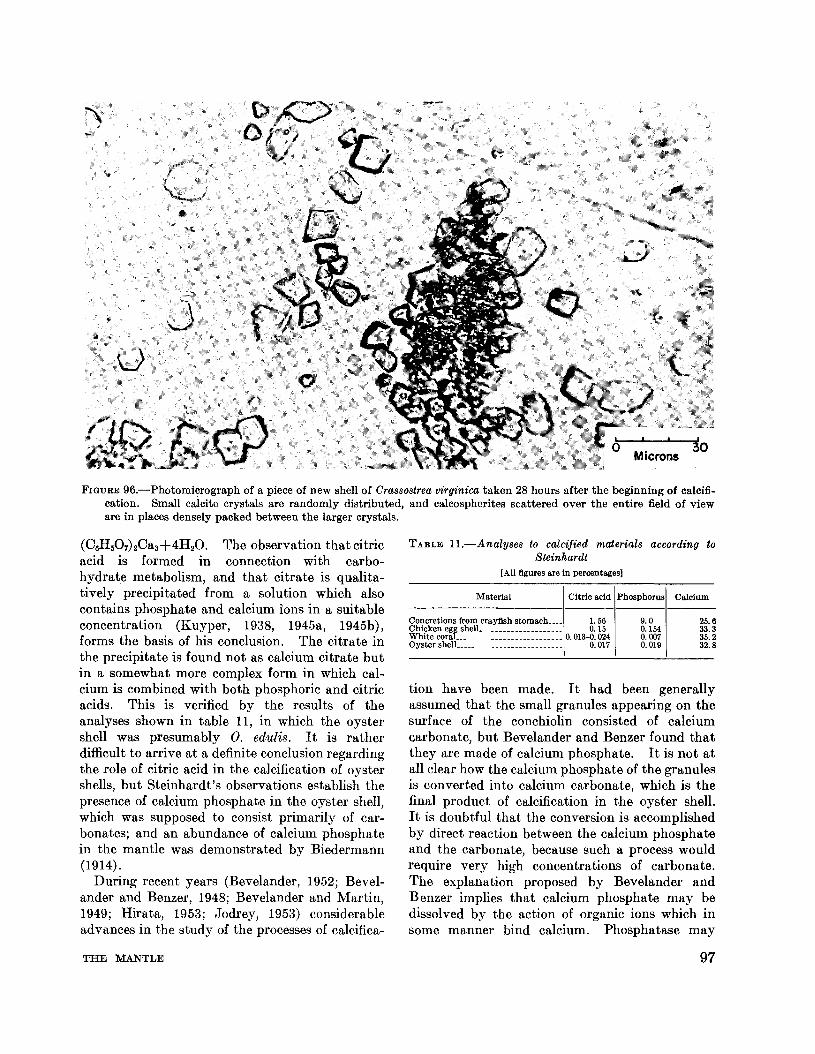
Microns
FIGURE 96.-Photomicrograph of a piece of new shell of Crassostrea virginica taken 28 hours after the beginning of calcification. Small caicite crystals are randomly distributed, and calcospherites scattered over the entire field of vieware in places densely packed between the larger crystals.
TABLE n.-Analyses to calcified materials according toSteinhardt
[All figures are In percentages]
tion have been made. It had been generallyassumed that the small granules appearing on thesurface of the conchiolin consisted of calciumcarbonate, but Bevelander and Benzer found thatthey are made of calcium phosphate. It is not atall clear how the calcium phosphate of the granulesis converted into calcium carbonate, which is thefinal product of calcification in the oyster shell.It is doubtful that the conversion is accomplishedby direct reaction between the calcium phosphateand the carbonate, because such a process wouldrequire very high concentrations of carbonate.The explanation proposed by Bevelander andBenzer implies that calcium phosphate may bedissolved by the action of organic ions which insome manner bind calcium. Phosphatase may
Citric acid Phosphorus Calcium
(06H507hOa3+4H20. The observation that citricacid is formed in connection with carbohydrate metabolism, and that citrate is qualitatively precipitated from a solution which alsocontains phosphate and calcium ions in a suitableconcentration (Kuyper, 1938, 1945a, 1945b),forms the basis of his conclusion. The citrate inthe precipitate is found not as calcium citrate butin a somewhat more complex form in which calcium is combined with both phosphoric and citricacids. This is verified by the results of theanalyses shown in table 11, in which the oystershell was presumably O. edulis. It is ratherdifficult to arrive at a definite conclusion regardingthe role of citric acid in the calcification of oystershells, but Steinhardt's observations establish thepresence of calcium phosphate in the oyster shell,which was supposed to consist primarily of carbonates; and an abundance of calcium phosphatein the mantle was demonstrated by Biedermann(1914).
During recent years (Bevelander, 1952; Bevelander and Benzer, 1948; Bevelander and Martin,1949; Hirata, 1953; Jodrey, 1953) considerableadvances in the study of the processes of calcifica-
Material
Concretions from crayfish stomach•••_ 1. 56Chicken egg sheIL . 0.15White coraL. 0.013-0.024Oyster sheIL.________________________ 0.017
9.00.1540.0070.019
25.633.335.232.8
THE MANTLE 97

FIGURE 97.-Calcite crystals deposited on a piece of conchiolin. Photomicrograph taken 38 hours after the secretion ofconchiolin has started.
contribute to this process by transferring phosphate to some substrate and removing the phosphate ions. This tentative explanation suggestsa number of biochemical studies that should bemade to obtain a better understanding of theprocess of calcification.
An important factor in the process of shellcalcification is the enzyme phosphatase, which isgenerally present in the ossifying cartilages ofyoung animals and in other tissues and organs in
98
which calcium is deposited. The action of theenzyme consists of hydrolysis of hexosemonophosphoric ester and glycerophosphoric ester andconsequent liberation of inorganic phosphate.The role of phosphatase in the shell formation ofmollusks was established by Manigault (1939),who found a direct correlation between pb,osphatase activity in the digestive diverticula,mantle, an4 blood and precipitation of calcium inthe shell. He concluded that phosphatase is
FISH AND WILDLIFE SERVICE

Millimeters
FIGURE 98.-Prismatic layer of the new growth of shell at the edge of the mantle. Four to five days old. Photomicrographof an unstained whole mount.
probably a transfer agent involved in the mobilization of calcium. The localization of this enzymealong the border of the mantle and in the surfaceepithelium of the oyster, shown by the Gomoritechnique (fig. 80), confirms the opinion ofManigault and of Bevela.nder that the phosphataseplays an active role in the calcification of oystershells.
During the last decade considemble advanceWllS made in studies of the metfibolic aspects ofshell formation. Hammen find Wilbur (1959)paid particulfir fittention to cfirbon dioxide conversion to shell carbonate llnd to the secretion ofconchiolin matrix in which the calcium carbonatecrystals are deposited b.v the oyster (C. virfjinica).The work of Jodrey ,wd Wilbur (1955) on highactivity of the enzyme oxalocetic deca.rbohylllSein the mantle tissue of this species suggested thfitthe deposition of cllrbonate may be relllted todecarboxylation reactions of the mnntle. Experi-
THE MANTLE
mentlll work conducted by Hllmmen llnd Wilburfit the Duke University Marine Laboratory atBeaufort, N.C., corrobornted this hypothesis.Living oysters and isolllted shells were placed for12 hours in sea wu.ter containing 240 microcuriesof NaHC I40 3 per liter. The rndioactivity of theshell surface was determined near the posteriormargin of the right valve and corresponding correction was made for seH absorption on the surface.By incubating pieces of oyster tissues in NaH0140 3
it was found that 0 14 is incorporated into organicficids of the mantle. \1ore than 90 percent of theradioactivity occurs in succinic and smalleramounts in fumaric and malic acids. The initialstep in the process is the fixation of carbon dioxideby propionic acid resulting in the formation ofsuccinic acid. Both acids were found in relativelyhigh concentrations in the shell forming tissuesof the o~'ster. The fact tl"tt in these experimentslabeled ll.mino acids were found in the radioactive
99

Microns200
FIGURE g9.-Photomicrograph of organic meshwork of prismatic layer of shell after decalcification in weak hydrochloricacid. Note that the outlines of the capsules retain the shape of the mineral prisms.
conchiolin of the shell indicate that carhon dioxidefixation also contributes to the syntheses of theorganic matrix or the shell.
Calcium enters the lllimtle directly from sea
100
wat,er, as was demonstrated by Jodrey (1953)using mantle-shell prepamtion und radioactiveCu''', and can be taken up through other parts ofthe mollusk and transported to the mantle. The
FISH AND WILDLIFE SERVICE

FIGURE lOO.-Calcium containing granules discharged bythe epithelial cells at the edge of the mantle of C.virginica. Neutral formalin 3 percent, alizarin.
and its color is affected by the presence of iron.Although these complications limit the usefulnessof alizarin as a reagent for the determination ofcalcium, I found that a 1 percent water solution ofalizarin S (sodium alizarin sulphonate) is probablythe best histochemical reagent for identification ofcalcium in the oyster mantle. It readily reactswith new deposits of calcium carbonate or calciumphosphate and forms compounds resistant to bothacids and alkalies.
To study the cytology of calcium secretion,the deposition of conchiolin and its calcificationwas stimulated by cutting off small pieces of shellalong the posterior margin of the oyster. Laboratory experience shows that such injury madeduring the warm season is rapidly repaired.Small pieces of the mantle border with the adheringand partly calcified conchiolin were excised and3 days later preserved in neutral formalin orabsolute ethyl alcohol. Sectioned tissues werestained with alizarin S and other reagents fordemonstration of calcium. The preparationsshowed a large number of alizarin stained globulesor granules, about 1.5 JJ. or less in diameter adheringto the surface of the mantle. Identical granuleswere found inSIde the goblet cells of the epitheliallayer along both sides of the mantle (fig. 100).
The results of the staining and other histo-
enzyme carbonic anhydrase which is present invarious mollusks may be expected to acceleratedeposition of calcium carbonate, and the rate ofdeposition is retarded by carbonic anhydraseinhibitors.
Complex metabolic cycles involved in shell formation have been reviewed by Wilbur (1960),and probable relations of carbon dioxide to shellconchiolin and carbonate deposition are shown byhim in a summary diagram (fig. C, p. 25 of Wilbur'spaper).
CYTOLOGICAL IDENTIFICATION OF CALCIUM
Several methods for the identification and localization of calcium salts in the oyster tissues areavailable, but none are completely reliable.Gomori (1939) suggests that soluble calcium couldbe demonstrated by treating the frozen sectionswith ammonium oxalate, the insoluble octahedralcrystals of calcium oxalate being easily recognized.The use of a fixative consisting of formalin andammonium oxalate was also proposed (Rahl,quoted from Gomori). Both methods tried inmy laboratory on sections of oyster mantle gaveunsatisfactory results. The difficulty is the dislodging of calcium-bearing granules and mucusduring sectioning, since the granules are easilycarried out 1;>y the knife's edge from their originallocation inside the cells to the outside of the epithelium. This difficulty can be avoided to a certainextent by double embedding the tissue in colloidinparaffin.
Indirect methods of Ca++ identification arebased on the use of heavy metals (silver, cobalt,copper, and iron). Because almost all insolublecalcium compounds in the tissues are eitherphosphate or carbonate, any procedure whichwould demonstrate the presence of these anionsmay be considered specific for calcium. When thesections are immersed in a solution of one of theheavy metals the corresponding metallic salt isformed at the sites of phosphate or carbonate.The reduction may be effected by exposing to lightif silver nitrate is used, or by immersing in appropriate reducing reagents (ammonium sulfide,acidified potassium ferricyanide). Identificationby staining of calcium is based on the formation ofinsoluble lacs with several hydroxyanthraquininedyes (alizarin sulfonic acid, purpurin, anthrapurpurin). Calcium deposited in the process of shellformation may, however, contain substances whichinterfere with the lac-forming reaction of alizarin.Also, the dye frequently fails to stain old deposits
oo 0 00 0 00
0 0 OOQo
o 0 0
o
o
o
oMicrons
40
THE MANTLE 101

!! ! !
6illimeters
FIGURE 101.-Photograph of crystals of a mixture of calcite and gypsum formed in the mantle cavity of C. virginica.
chemical reactions sho,w that the secretion ofcalcium is not confined to special sites but takesplace over the entire edge and outer surface ofthe mantle. The intensive coloration of thegranules by alizarin suggests tbat tbey containa considerable amount of calcium, probablybound in organic compounds of the globules.Amoebocytes present in the material secretedby the mantle also may be involved in tbe mobilization of calcium during the formation or repairof sbells.
Sometimes the mineral crystals formed by the
102
mantle are not incorporated in the conchiolin butaccumulate in tbe pallial cavity and are eventuallyejected. On several occasions fltirly lltrge qultntities of a white powdered material were found infront of the discharge areas of oysters which werekept in glass trays in running sea water in theiltbomtol'y. The materiltl consisted of crystltls(fig. 101) which, ltccording to the X-my ltnltlysiskindly pel'fo,med by Marie Lindberg of theGeochcmistry and Petrology Branch of theGeological Survey of the U.S. Department of theInterior, were found to consist of a mixture of
FISH AND WILDLIFE SERVICE

calcite and gypsum (hydrous calcium sulfate),with the latter present only as a minor constituent.The oysters appeared to be normal in every respectand showed good growth of shells. The presenceof gypsum is of interest since it is not a normalconstituent of oyster shell. What particulardisturbance in the calcium metabolism producedits formation is unknown.
SOURCES OF CALCIUM
It has been suggested (Pelseneer, 1920; Galtsoff,1938) that lamellibranchs may remove calciumdirectly from sea water. Pelseneer (1920) citesan example of a young Anodonta cygnea which in2 months removed all the calcium from 5 1. ofwater in which it was kept. Definite proof of thedirect absorption of calcium by the oyster mantleis given by the experiments with C. virginica(Jodrey) 1953) in which radioactive Ca45 was used.Calcium turnover was also studied by Hirata(1953) in mantle-shell preparations made by cutting off the adductor muscle and the visceralorgans, and leaving the intact mantles spread overtheir respective valves. The mantle remainedalive for several days and deposited the shellmaterial, although at a lower rate than does theintact oyster. Jodrey placed a mantle preparation in 500 ml. of aerated sea water with a Ca45
activity of 5.8 microcuries. At least part of thecalcium of the newly formed shell substance camedirectly from the sea water, and the deposition ofcalcite took place in tissue isolated from the circulatory and digestive systems. The experiments alsodemonstrated that the greater portion of calciumin the mantle appears to be inert. Only 2.5 percent of the total calcium content was renewedevery 24 minutes, the turnover being 0.6 mg. ofcalcium per minute per gram of mantle. Inaddition to entering the mantle directly calciumcan be taken up by other organs of the oyster andtransported to the mantle (Wilbur, 1960).
MINERALOGY OF CALCIUM CARBONATEIN MOLLUSCAN SHELLS
Calcium carbonate is known to occur in 12mineral forms (Prenant, 1924), but only three ofthese have been found in animals. In the shellsof mollusks, calcium carbonate usually occurs ascalcite and aragonite. There are many species inwhich both minerals occur together although indifferent parts of the shell. Prenant (1928), whocontributed much to the study of calcification,found that besides calcite and aragonite the animal
THE MANTLE
tissue may contain small spheres (sphaerolithes) ortiny needles of the mineral called "vaterite", afterthe mineralogist Vater who discovered it. Vaterite was reported to be present in the connectivetissue of certain gastropod mollusks, cestodes, andtrematodes, and in the fat tissue of insects (Diptera). Its presence in the tissues of the oysterhas not been reported.
The various forms of calcium carbonate secretedby animal tissue can be identified by their crystallographic properties, birefringence, density, andchemical reaction. Some of these distinctivecharacteristics are summarized in table 12, takenfrom Prenant (1924).
Impurities always present in material secretedby living forms can sometimes make the mineralogical identification of calcium carbonate doubtful.Calcite and aragonite can be distinguished bymeans of the polarizing microscope. Calcitecrystals examined under crossed nicols give abrilliant picture of various colors, and a distinctblack cross appears when the optical axis is alignedparallel to the axis of the microscope (fig. 94.)In the case of aragonite, hyperbolic arched linesappear instead of the black crosses. Exactidentification of minerals can of course be made byX-rays, but this method is rarely available to thebiologist.
Among various chemical identification methodsthe Meigen color reaction can be most easilyemployed (B¢ggild, 1930, p. 238). In a weaksolution of cobalt nitrate aragonite becomes violet,the intensity of coloration increasing as the solution is warmed. Calcite, however, remains paleblue even in a heated solution.
The conditions under which a mollusk secretescalcium carbonate in a specific mineralogical formare not at present understood. It is reasonableto presume that the organic matrix of the shell issomehow involved in this process. Roche, Ranson, and Eysseric-Lafon (1951) found that in theshells of mollusks consisting both of calcite andaragonite the conchiolin associated with the calciteof the prismatic layer had higher concentrationsof glycine and tyrosine than were present in thenacre of the same shell consisting of aragonite(see ch. II, p. 41). The causal relationship between the mineralogical forms of carbonate andamino acids of its conchiolin has not beendemonstrated.
A hypothesis that carbonic anhydrase, anenzyme present in the tissues of the mantle, plays
103

TABLE 12.-Distinctive properties of principal mineral forms of calcium carbonate found in invertebrates 1
Name Chemicalcomposition
Optical System Birefringence Index ofrefraction
Density Meigenreaction
Caicite CaCO. Rhomboedric. uniaxiaL Strong (0.172) 1.658-1.486 2.714Aragonite_______________ CaCO.____________ Monoclinic. biaxiaL Slightly weaker. (0.156)_ __ 1.686-1.530_____ 2.95Vaterite_________________ CaCO.____________ Sphaerolithes. optically negative______ Weak About 1.55 2.5-2.65Amorphous_____________ CaCO.____________ Isotropic______________________________ Near 1.5_______ 2.25-2.45Hydrated carbonate CaCO.6H,O Prisms or Monoclinic tablets Near 0.085 About 1.5 1. 777
Negative.Positive.
Do.
1 From the data published by Prenant. 1927.
2
2
6
o
TABLE 13.-Areas of new growth and rate of deposition ofshell material by C. virginica in mg. per day per em.!during April to June 1954, Woods Hole, Mass.
from colder waters contain varying amounts ofboth calcite and aragonite. This interestingecological observation does not, however, providea clue to the nature of the biochemical processeswhich control the predominance of one or anothercrystallization system.
RATE OF CALCIFICATION
The calcification rate of the left valve of C.'Virginica is significantly higher than that of theright one, as can be readily seen by examiningnewly formed shells. The calcareous materialdeposited by the left mantle is thicker andheavier than that deposited during the same timeby the right mantle (Galtsoff, 1955). I madethe following observations on shell growth rate ofadult C. virginica. After the new growth of shellalong the valve edge was carefully removed theoysters were placed in tanks abundantly suppliedwith running sea water. About 2 months laterthe areas of newly deposited shells on each valvewere measured with a planimeter, carefully removed from the shell, rinsed in distilled water,dried in air, and weighed. The results aresummarized in table 13. In every case theamount of calcified material deposited over aunit of area was considerably greater on the left
RatioArea Dec::;, D~S weight
Oysters of new Weight sit on un er oflert tshell per em.' per em.' obser- weight
payday vation of rlghvalve
------Five-year-old, Narragansett Bay Om.' Mg. Mg. No.Left valve___________________ 5.80 156.0 2.8 55 2.
Right valve _________________ 5.16 59.3 1.1Adult. Narragansett BayLeft valve___________________ 7.1 123.0 1.8 68 6.Right valve_________________ 7.7 19_ 9 0.3Adult. Narragansett Bay
2.9Left valve___________________ 6.1 74.2 1.09 68Right valve_________________ 8.8 25.5 0.37Two-year-old. New HampshireLeft. valve___________________ 3.68 163.8 2.98 55 3.Right valve__________________ 4.20 52.0 0.95Very old, New Hampshire
2.2Left valve _________________ 6.83 71. 2 1.3 55Right valve_________________ 7.35 33.0 0.6
an important role in the formation of calciumdeposits in molluscan shells has been advanced byStolkowski (1951). According to this theory theenzyme exerts its effect by orienting the calciumcarbonate molecules in the aragonite crystallattice. The action of carbonic anhydrase in thisadmittedly very complex process is not, however,satisfactorily explained and should be morethoroughly investigated before its role in theformation of aragonite or calcite in mollusk shellsis definitely established. In its present state thehypothesis fails to explain the existence of shellsin which both aragonite and calcite are present.Recently Stenzel (1963) reported that in the shellsof C. virginica aragonite covers the areas of atta,chment of the adductor muscle, the imprint ofQuenstedt's muscle, and is found in the ligament.
Another explanation of the formation of theless stable aragonite instead of calcite suggeststhat strontium and magnesium carbonates influence the formation of aragonite in shell. Somesupport to this idea is found in the fact that invitro the crystallization of aragonite is facilitatedby strontium and lead salts. This observationmade by Prenant (1924) apparently influencedTrueman's (1942) hypothesis that strontium,magnesium, and probably other salts found inliving mollusks influence the crystallization ofaragonite.
That there may be some correlation betweenthe predominance of the particular mineralogicalform of calcium carbonate and the temperatureof the surrounding water has recently beensuggested by some geologists. Through quantitative X-ray analysis of shells they have demonstrated that in certain polyclad worms (Serpulidae)and in some gastropods and pelecypods (Mytilus,Volsella, Pinctada, Anomia, and others) the concentration of aragonite in shells increases withincreasing temperatures (Epstein and Lowenstam,1953; Lowenstam, 1954). In Mytilus, for instance, only the shells of warm water species arecomposed entirely of aragonite, whereas those
104 FISH AND WILDLIFE SERVICE

valve (lower) than on the right one (upper),the difference varying from 2.2 to 6.2 times.
The rate of deposition of calcified material bythe surface of the mantle may also be studied byinserting between the mantle and shell smallpieces of plastic or other nontoxic material ofknown area and weight. Results obtained withthis method vary greatly. Observations madeon 16 adult oysters at Woods Hole during theperiod of August 9 to 20, 1953, show that in 15oysters the daily rate of shell deposition per squarecentimeter varied from 0.4 to 2.1 mg. Oneoyster deposited 14.2 mg. in 2 days or 7.1 mg. perday. The amounts of shell material depositedby 20 Narragansett Bay oysters kept in laboratorytanks for 68 days during the period of April toJune varied from 0.1 to 0.79 mg. of shell substanceper day cm. 2
• In some of these oysters thepresence of the plastic material induced pathological conditions which resulted in the formationof leathery capsules similar to the blisters fre,..quently found on the inside of shells near theadductor muscle. The formation of such blisterswas accompanied by deposition of calcite greatlyin excess of the rate of calcification under normalconditions.
Seasonal variation in rate of shell depositionover the inner surface of the valves was alsostudied, using 20 adult oysters for each set ofdeterminations. Observations were continuousfrom June 1954 until the end of February 1956.To avoid possible injury to the mantle while introducing pieces of plastic, the oysters were fullynarcotized in magnesium sulfate solution andinsertions made when the mantle was completelyrelaxed and did not respond to touch. Thin sheetsof plastic were cut into rectangular pieces 0.5 cm. 2
in area and weighed before inserting them underthe mantle, their weight varying from 5.5 to 6.0mg. Some of the pieces introduced were ejectedby the oysters, but losses were minimized when theinsertion was made under full narcosis. Thetreated oyster was then marked and placed on itsleft valve in a large tray supplied with runningsea water. The temperature of the water wasrecorded twice a day. Each set of 20 oysters waskept in water as long as the seasonal rise or fall ofwater temperature did not exceed 2.5° C.
To obtain measurable quantities of shell deposits the pieces of plastic were left inside theoysters for a longer time in winter and in August,after the completion of spawning, than during the
THE MANTLE
rest of the year. The number of days the oysterswith inserted pieces were left undisturbed variedas follows: from 10 to 16 days in April to July;from 25 to 30 days in August; from 13 to 18 daysin September to November; for 30 days in December; and 70 days in January to March. Observations were continued for 14 months. Noshell was formed in January to March except in afew oysters in which the mantle was injured duringinsertion. These samples were not included inthe data plotted in fig. 102. Laboratory observations showed that shell opening and feeding of theoysters at Woods Hole are as a rule temporarilyreduced after the discharge of sex products whichtakes place late in July and early in August.Unequal time intervals in observing shell deposition do not affect the validity of the results sincethe rates of shell formation shown in figure 102are expressed as weights of shell deposited percm. 2 in 1 day.
At the end of each period the oysters were removed, the pieces of plastic recovered, rinsed indistilled water, dried at 55° C., and weighed. Theresults summarized in figure 102 are shown as medians(Md.) of the rate of shell deposition per cm.2 perday, and as lower (QI) and upper <Qa) quartiles.
The curves show two periods of accelerated shellgrowth in Woods Hole water, one in May to Juneand another in October, and no shell growth during winter from December to the end of Aprilwhen the temperature of the water varied between1° and 2° C. These observations are in agreementwith many field data and with the experiences ofpractical oyster growers of the North Atlanticstates, who found that oysters grow more rapidlyin the spring and in the autumn and cease to growwhen the water temperature drops to about 5° C.The relatively low rate of shell deposition duringthe summer is attributable to the inhibitory effectof fully developed gonads. Observations frequently made in the Woods Hole laboratory showthat shell growth in the winter will begin within24 hours after the transfer of oysters from theharbor to much warmer sea water in the laboratory.
Under normal conditions no shell is depositedin winter. In several instances, however, largeamounts of shell material were secreted over anarea of the mantle which was apparently injuredby the insertion of plastic. One of these cases isshown in figure 103. In this oyster a heavy pocketof shell material was deposited on the valve over
105

z; 5
ti 25UJa:::>I-ji20UJ0.::!!
~ 15
a:UJI- 10;
o
2.)0-cto
ICIlo~ 0.5 Mel.o
MEDIAN
QI LOWER QUARTILE
Q3 UPPER QUARTILE
..J
..JW:z::CIl OL-_----O-__-'-__.1.......<O---.J"--_........L__-'-__-'---__L-_-..L__-'--,-,_...J...3>-:-:-:-,----~_:__--.l.-,__--'
II III IV V VI VII VIII IX X XII II
MONTHS 1955 1956
a:1&.10.
"'21.5
(,)
a:UJ0.
ci1.0::!!
FIGURE 102.-Seasonal changes in the rate of deposition of shell material over the inner surfaces of left (lower) valves.
the area occupied by a piece of plastic, and a shellridge was formed along the edge of the mantle,which was withdrawn a considerable distance backfrom its normal position. It can be deduced fromthese observations that injury to the mantle stimulates the shell secretion and that deposition may
take place even at low temperatures when normalshell growth is inhibited. This would indicatethat the enzymatic system involved in shell deposition is always present and may become activein response to pathological conditions in spite ofthe inhibitory effect of winter temperatures.
106 FISH AND WILDLIFE SERVICE

I I
o '3Centimeters
FIGURE 103.-Abnormal deposition of shell material alongthe edge of the mantle (black line) and over the piece ofplastic quadrangle, which was completely encapsulatedin a pocket of newly secreted shell. Mantle is shown bystippled area. Winter observation at Woods Hole.
BIBLIOGRAPHY
AWATI, P. R., and H. S. RAI.1931. Ostrea cucullata (the Bombay oyster). The
Indian Zoological Memoirs on Indian AnimalTypes, III. Methodist Publishing House, Lucknow, India, 107 pp.
BARGETON, MARIE.1942. Les variations saisonnil~resdu tissu conjonctif
vesiculeux de I'huttre. Bulletin Biologique de laFrance et de la Belgique, tome 76, pp. 175-191.
BENSLEY, CAROLINE M.1939. Comparison of methods for demonstrating
glycogen microscopically. Stain Technology, vol.14, No.2, pp. 47-52.
BEVELANDER, GERRIT.1952. Calcification in molluscs. III. Intake anddeposition of Ca45 and 1'32 in relation to shell formation. Biological BUlletin, vol. 102, No.1,pp.9-15.
BEVELANDER, GERRIT, and PAUL BENZER.1948. Calcification in marine molluscs. Biological
Bulletin, vol. 94, No.3, pp. 176-183.BEVELANDER, GERRIT, and JOSEPHINE MARTIN.
1949. Culture of mantle tissue of marine molluscs.[Abstract.] Anatomical Record, vol. 105, p. 614.
BIEDERMANN, W.1914. Physiologie der Stutz- und Skelettsubstanzen.
Hans Winterstein's Handbuch der Vergleichenden
THE MANTLE
Physiologie, Band 3, 1 hiUfte, 1 teil, pp. 319-1188.Gustav Fischer, Jena.
BIZIO, M. J.1866. Sur I'existence du glycogfme dans des animaux
invertebres. Comptes Rendus Hebdomadaires desSeances de l'Academie des Sciences, tome 62,pp. 675-678.
BLUNDSTONE, EDWIN RICHARDSON.1885. On the occurrence of glycogen as a constituent
of the vesicular cells of the connective tissue ofmolluscs. Proceedings of the Royal Society ofLondon, vol. 38, pp. 442-445.
Bj2lGGILD, O. B.1930. The shell structure of the mollusks. Det
Kongelige Danske Videnskabernes Selskabs Skrifter,9 raekke, bind II, part 2, pp. 233-326.
BOUTAN, LOUIS.1923. Nouvelle etude sur les perles naturelles et sur
les perles de culture. Annales des Sciences Naturelles, Zoologie, serie 10, tome 6, pp. 1-94.
CAMERON, G. R.1930. The staining of calcium. Journal of Pathology
and Bacteriology, vol. 33, part II, pp. 929-955.CHATIN, A., and A. MUNTY.
1895. Analyse des coquilles d'huttres. ComptesRendus Hebdomadaires des Seances de l'Academiedes Sciences, tome 120, pp. 531-534.
CREIGHTON, CHARLES.1896. Microscopic researches on the formative
property of glycogen. Part I. Physiological.Adam and Charles Black, London, 152 pp.
1899. Microscopic researohes on glycogen. Part II.Glycogen of snails and slugs in morphological andphysiological correspondence with the lymphsystem of vertebrates. Adam and Charles Blaok,London, 127 pp.
CROFTS, DORIS R.1929. Haliotis. L.M.B.C. Memoirs No. XXIX.
Proceedings and Transaotions of the LiverpoolBiologioal Society, vol. 43, appendix II, pp. 2-174.
DAKIN, WILLIAM J.1909a. Striped muscle in the mantle of lamelli
branohs. Anatomisoher Anzeiger, Band 34, pp.227-230.
1909b. Pecten, the edible soallop. L.M.B.C. Memoirs No. XVII. Proceedings and Transaotions ofthe Liverpool Biologioal Sooiety, vol. 23, pp. 333468.
1912. Buooinum (The whelk). L.M.B.C. MemoirsNo. XX. Prooeedings and Transactions of theLiverpool Biological Society, vol. 26, pp. 253-367.
DAVIS, J. R. AINSWORTH, and H. J. FLEURE.1903. Patella. L.M.B.C. Memoirs No. X. Pro
ceedings and Transactions of the Liverpool Biologioal Sooiety, vol. 17, pp. 193-268.
DEWAELE, A.1929. Le sang d'Anodonta cygnea et la formation de
la ooquille. Memoires Aoademie Royale de Belgique, Classe des Sciences, Collection in 40
, serie 2,tome 10, No.3, pp. 1-52.
DOTTERWEICH, HEINZ and, ERNST ELSSNER.1935. Die Mobilisierung des Sohalenkalkes fur die
Reaktionsregulation der Muscheln (Anodonta cyg-
107

nea). Biologisches Zentralblatt, Band 55, pp..138-163.
DUGAL, LOUIS-PAUL.1939. The use of the calcareous shell to buffer the
product of anaerobic glycolysis in Venus mercenaria.Journal of Cellular and Comparative Physiology,vol. 13, No.2, pp. 235-251.
DUGAl" LOUIS-PAUL, and LAURENCE IRVING.1937. Secretion de carbonate de calcium par lesVenus mercenaria fermees hermetiquement. Comptes Rendus Hebdomadaires des Seances et Memoiresde la Societe de Biologie et de ses Filiales et Associees,tome 124, pp. 526-528.
ELSEY, C. R.1935. On the structure and function of the mantle
and gill of Ostrea gigas (Thunberg) and Ostrealurida (Carpenter). Transactions of the RoyalSociety of Canada, third series, sec. 5, BiologicalSciences, vol. 29, pp. 131-160.
EpSTEIN, S., and H. A. LOWENSTAM.1953. Temperature-shell-growth relations of recent
and interglacial Pleistocene shoal-water biota fromBermuda. Journal of Geology, vol. 61, No.5, pp.424-438.
FLEMMING, WALTHER.1877. Ueber Bindesubstanz und Gefasswandung im
Schwellgewebe der Muscheln. Archiv fiir mikroskopische Anatomie, Band 13, pp. 818-867.
FOL, HERMANN.1888. Sur la structure microscopique des muscles des
Mollusques. Comptes Rendus Hebdomadaires desSeances de l'Academie des Sciences, tome 106, pp.306-308.
GALTSOFF, PAUL S.1934. The biochemistry of the invertebrates of the
sea. Ecological Monographs, vol. 4, No.4, pp.481-490.
1938. Sources of calcium for shell of Ostrea virginica.Nature, vol. 141, No. 3577, p. 922.
1955. Recent advances in the studies of the structureand formation of the shell of Crassostrea virginica.Proceedings of the National Shellfisheries Association, vol. 45, August 1954, pp. 116-135.
GOMORI, G.1939. Microtechnical demonstration of phosphatase
in tissue sections. Proceedings of the Society forExperimental Biology and Medicine, vol. 42, No.1, pp. 23-26.
1943. Calcification and phosphatase. AmericanJournal of Pathology, vol. 19, No.2, pp. 197-209.
1952. Microscopic histochemistry-principles andpractice. University of Chicago Press, Chicago,111., 273 pp.
HALE, C. W.1946. Histochemical demonstration of acid polysac
charides in animal tissue. Nature, vol. 157, No.3998, p. 802.
HAMMEN, CARL S., and KARL M. WILBUR.1959. Carbon dioxide fixation in marine inverte
brates. 1. The main pathway in the oyster.Journal of Biological Chemistry, vol. 234, No. 4-6,p. 1268-1271.
108
HAYASI, KAZUMASA.1938. Detection of calcium in molluscan mantles. 1.
Anodonta and Cristaria. Annotationes ZoologicaeJaponenses, vol. 17, No.2, pp. 95-103.
1939. Detection of calcium in molluscan mantles.II. Euhadra callizona amaliae kobelt. Annotationes Zoologicae Japonenses, vol. 18, No.1, pp. 1-9.
HEIDE, FRITZ.1924. Ueber den Vaterit. Centralblatt fiir Mineral
ogie, Geologie und Palaontologie, No. 21, pp. 641651.
HIRATA, ARTHUR A.1953. Studies on shell formation. II. A mantle
shell preparation for in vitro studies. BiologicalBulletin, vol. 104, No.3, pp. 394-397.
HOPKINS, A. E.1932. Sensory stimulation of the oyster, Ostrea vir
ginica, by chemicals. [U.S.] Bureau of Fisheries,Bulletin No.8, vol. 47, pp. 249-261.
1933. Experiments on the feeding behavior of theoyster, Ostrea gigas. Journal of Experimental Zoology, vol. 64, No.3, pp. 469-494.
JODREY, LOUISE H.1953. Studies on shell formation. III. Measure
ment of calcium deposition in shell and calciumturnover in mantle tissue using the mantle-shellpreparation and Ca45 • Biological Bulletin, vol. 104,No.3, pp. 398-407.
JODREY, LOUISE H., and KARL M. WILBUR.1955. Studies on shell formation. IV. The respira~
tory metabolism of the oyster mantle. BiologicalBulletin, vol. 108, No.3, pp. 346-358.
JOHNSTON, JOHN, R. E. MERWIN, and E. D. WILLIAMSON.1916. The several forms of calcium carbonate.
American Journal of Science, series 4, vol. 41, No.246, pp. 473-512.
KESSEL, ERWIN.1933. "Oeber die Schale von Viviparus viviparus L.
und Viviparus fasciatus MUll. Ein Beitrag zumStrukturnproblem der Gastropodenschale. Zeitschrift fUr Morphologie und 6klogie der Tiere,Band 27, pp. 129-198.
1940. "Ober Aufhebung von Formdoppelbrechungdurch Druck, gezeigt am Periostracum von Mytilusedulis. Zeftschrift fUr wissenschaftliche Mikroskopieund fUr mikroskopische Technik, Band 57, pp. 1924.
KOLLMANN, PROF.1877. Die Bindesubstanz der Acephalen. Archiv
fUr mikroskopische Anatomie, Band 13, pp. 558603.
KUYPER, ADRIAN C.1938. The quantitative precipitation of citric acid.
Journal of Biological Chemistry, vol. 123, No.2,pp. 405-407.
1945a. The chemistry of bone formation. 1. Thecomposition of precipitates formed from salt solutions. Journal of Biological Chemistry, vol. 159,No.2, pp. 411-416.
1945b. The chemistry of bone formation. II. Somefactors which affect the solubility of calcium phosphate in blood serum. Journal of Biological Chemistry, vol. 159, No.2, pp. 417-424.
FISH AND WILDLIFE SERVICE

LEENHARDT, HENRY.1926. Quelques etudes sur Gryphea angulata. (HuItre
du Portugal). Annales de l'Institut Oceanographique, nouvelle serie, tome 3, fascicule 1, pp.1-90.
LIST, THEODOR.1902. Die Mytiliden. Fauna und Flora des Golfes
von Neapel und der Angrenzenden Meeres-Abschnitte herausgegeben von der ZoologischenStation zu Neapel. 27 Monographie. R. FriedHinder und Sohn, Berlin, 312 pp.
LOWENSTAM, HEINZ A.1954. Factors affecting the aragonite: calcite ratios
in carbonate-secreting marine organisms. Journalof Geology, vol. 62, No.3, pp. 284-322.
MANIGAULT, PIERRE.1933. La tension du gaz carbonique, condition de la
regeneration de la coquille chez les Helix. Travauxde la Station Biologique de Roscoff, fascicule 11,pp.51-59.
1939. Recherches sur Ie calcaire chez les mollusques..Phosphatase et precipitation calcique. Histochimiedu calcium. Annales de 1'Institut Oceanographique,nouvelle serie, tome 18, fascicule 5, pp. 331-426.
MARCEAU, F.1904. Sur Ie mecanisme de la contraction des fibres
musculaires lisses dites a double striation obliqueou a fibrilles spiralees et en particulier de celles desmuscles adducteurs des Lamellibranches. ComptesRendus Hebdomadaires des Seances de I' Academiedes Sciences, tome 139, pp. 70-73.
MAXIMOW, ALEXANDER A., and WILLIAM BLOOM.1930. A text-book of histology. W. B. Saunders
Company, Philadelphia, Pa., 833 pp.MEYER, KARL.
1957. The chemistry of the mesodermal groundsubstances. The Harvey Lectures, series 51, pp.88-112.
MOYNIER de VILLEPOIX, R.1895. De la formation de la coquille dans les
mollusques. Comptes Rendus Hebdomadaires desSeances de l'Academie des Sciences, tome 120, pp.512-513.
NELSON, THURLOW C.1938. The feeding mechanism of the oyster. 1. On
the pallium and the branchial chambers of Ostreavirginica, O. edulis and O. angulata, with comparisonswith other species of the genus. Journal ofMorphology, vol. 63, No.1, pp. 1-61.
PEKELHARING, C. A.1901. Het bindweefsel bij de oester. Onderzoe
kingen, gedaan in het Physiologisch Laboratoriumder Utrechtsche Hoogeschool, Vijfde Reeks (Series5), III (vol. 3), Eerste Aflevering, pp. 227-239.
PELSENEER, PAUL.1906. Mollusca. In E. Ray Lankester (editor), A
treatise on zoology, Part V, 355 pp. Adam andCharles Black, London.
1920. A propos de la formation et de la compositionchimique de la coquille des mollusques. Annales dela Societe Royale Zoologique et Malacologique deBelgique, tome 51, pp. 70-74.
THE MANTLE
733-851 0-64--8
PRENANT, MARCEL.1924. Contributions a l'etude cytologique du calcaire.
1. Quelques formations calcaires du conjonctifchez les gasteropodes. Bulletin Biologique de laFrance et de la Belgique, tome 58, pp. 331-380.
1927. Les formes mineralogiques du calcaire chez lesHres vivants, et Ie probleme de leur determinisme.BiologicalReviews and Biological Proceedings of theCambridge Philosophical Society Biological Science,vol. 2, No.4, pp. 365-393.
1928. Contributions a l'etude cytologique du calcaire.IV. La vaterite chez les animaux. BulletinBiologique de la France et de la Belgique, tome 62,pp.21-50.
QUENSTEDT, F. A.1867. Handbuch der Petrefaktenxunde Laupp,
TUbingen, 2d ed., 5\)8 pp.RASSBACH RICHARD.
1912. Beitriige zur Kenntnis der Schale und Schalenregeneration von Anodonta cellensis Schrot. Zeitschrift fUr wissenschaftliche Zoologie, Band 103,pp. 363-448.
RAWITZ, BERNHARD.1888. Der Mantelrand der Acephalen, Erster Teil,
Ostreacea. Jenaische Zeitschrift fUr Naturwissenschaft, Neue Folge, Band 15, pp. 415-556.
ROBERTSON, JAMES D.1941. The function and metabolism of calcium in the
Invertebrata. Biological Reviews of the CambridgePhilosophical Society, vol. 16, No.2, pp. 106-133.
ROCHE, JEAN, GILBERT RANSON, and Marcelle EyssericLafon.
1951. Sur la composition des scleroproteines descoquilles des mollusques (conchiolines). ComptesRendus des Seances de la Societe de Biologie et deses Filiales, tome 145, pp. 1474-1477.
ROULE, LOUIS.1886. Sur quelques particularites histologiques des
Mollusques acephales. Comptes Rendus Hebdomadaires des Seances de l'Academie des Sciences,tome 103, pp. 936-938.
1888. Sur la structure des fibres musculaires appartenant aux muscles retracteurs des valves desMollusques lamellibranches. Comptes RendusHebdomadaires des Seances de l' Academie desSciences, tome 106, pp. 872-874.
SCHMIDT, W. J.1931. tJ"ber die Prismenschicht der Schale von
Ostrea edulis L. Zeitschrift fUr Morphologie und(jkologie der Tiere, Band 21, pp. 789-805.
SEMICHON, LOUIS.1932. Sur une matiere qui accompagne Ie glycogene
dans les cellules vesiculeuses des Mollusqueslamellibranches. Comptes Rendus Hebdomadairesdes Seances et Memoires de la Societe de Biologieet de ses Filiales et Associees, tome 111, pp. 785-786.
STEINHARDT, BRUNO.1946. The biological system calcium-phosphate
citrate. Acta Physiologica Scandinavica, vol. 12,pp. 381-384.
STENZEL, HENRY B.1963. Aragonite and calcite as constituents of adult
109

oyster shells. Science, vol. 142, No. 3589, p. 232233.
Oysters. In Treatise of invertebrate paleontology.Part N-Mollusca, 6 (Bivalvia). [In press.]
STOLKOWSKI, JOSEPH.
1951. Essai sur Ie determinisme des formes mineralogiques du calcaire chez les etres vivants (calcairescoquilliers). Annales de l'Institut Oceanographique,nouvelle serie, tome 26, fascicule 1, pp. 1-113.
TRUEMAN, E. R.1942. The structure and deposition of the shell of
Tellina tenuis. Journal of the Royal MicroscopicalSociety, series 3, vol. 62, pp. 69-92.
110
TULLBERG, TYCHO.
1881. Studien tiber den Bau und das Wachsthum desHummerpanzers und der Molluskenschalen.Kongliga Svenska Vetenskaps-akademiens Handlingar, series 4, band 19, No.3, pp. 3-57.
WILBUR, KARL M.1960. Shell structure and mineralization in molluscs.
In Reidar F. Sognnaes (editor), Calcification inbiological systems, A symposium presented at theWashington meeting of the American Associationfor the Advancement of Science, December 29, 1958,pp. 15-40. American Association for the Advancement of Science, Washington, D.C., PublicationNo. 64.
FISH AND WILDLIFE SERVICE