
CHAPTER I INTRODUCTION - Shodhgangashodhganga.inflibnet.ac.in/bitstream/10603/78574/7/07_chapter...
46
1 CHAPTER I INTRODUCTION 1.0 General consideration Nanotechnology and Nanoparticles are generally considered an invention of modern science. However they have a long history which can be thousands of years. The people however did not know what they were doing. In the last twenty years there has been a massive advance of nanomaterials in material science and nanotechnology. In this panorama silver and gold nanoparticles (AgNP and AuNP hereafter) are playing a protagonist role. The reason for AgNP and AuNP success lies in a favorable combination of physico-chemical properties and advances in chemical synthesis. The main characteristic of AgNP and AuNP is the Surface Plasmon Absorption (SPA), which has 10 5 - 10 6 larger extinction cross sections than ordinary molecular chromophores and is also more intense than that of other metal particles, due to the weak coupling to the interband transition. The frequency of silver and gold SPA can also be tuned from visible to near infrared, acting on the shape, size or nanoparticle assembly. Since the surface chemistry of AgNP and AuNP is very simple there has been a significant progress in AgNP and AuNP synthesis with tailored shape or size and has provided a gamut of tools with engineered properties, opening the access of nanotechnology to manifold applications. Furthermore AgNP and AuNP have a high chemical stability, photo stability and especially AuNP are nontoxic for living organisms. Their physiochemical stability, bright color and biocompatibility explain why the utilization of these particles is dated back to the 5 th century. 1.1 Historical Background Solutions of liquid gold have been mentioned by Egyptian and Chinese authors around 5 th century BC. In fact ancients believed in their metaphysical and healing powers 1 . Colloids of silver and gold have also been used in the Ancient Roman times to color glass with intense shades of yellow, red, or mauve, depending
Transcript of CHAPTER I INTRODUCTION - Shodhgangashodhganga.inflibnet.ac.in/bitstream/10603/78574/7/07_chapter...

1
CHAPTER I
INTRODUCTION
1.0 General consideration
Nanotechnology and Nanoparticles are generally considered an invention of
modern science. However they have a long history which can be thousands of years.
The people however did not know what they were doing. In the last twenty years there
has been a massive advance of nanomaterials in material science and nanotechnology.
In this panorama silver and gold nanoparticles (AgNP and AuNP hereafter)
are playing a protagonist role. The reason for AgNP and AuNP success lies in a
favorable combination of physico-chemical properties and advances in chemical
synthesis. The main characteristic of AgNP and AuNP is the Surface Plasmon
Absorption (SPA), which has 105 - 10
6 larger extinction cross sections than ordinary
molecular chromophores and is also more intense than that of other metal particles,
due to the weak coupling to the interband transition. The frequency of silver and gold
SPA can also be tuned from visible to near infrared, acting on the shape, size or
nanoparticle assembly. Since the surface chemistry of AgNP and AuNP is very simple
there has been a significant progress in AgNP and AuNP synthesis with tailored shape
or size and has provided a gamut of tools with engineered properties, opening the
access of nanotechnology to manifold applications. Furthermore AgNP and AuNP
have a high chemical stability, photo stability and especially AuNP are nontoxic for
living organisms. Their physiochemical stability, bright color and biocompatibility
explain why the utilization of these particles is dated back to the 5th
century.
1.1 Historical Background
Solutions of liquid gold have been mentioned by Egyptian and Chinese
authors around 5th
century BC. In fact ancients believed in their metaphysical and
healing powers1. Colloids of silver and gold have also been used in the Ancient
Roman times to color glass with intense shades of yellow, red, or mauve, depending

2
upon the concentration of the two metals. A fine example is the famous Lycurgus
cup2 (Fig. 1.1.1) in the British museum, which is dated 4
th century AD. For all the
middle ages Au colloids have also been used in medicines believing in their curative
properties for various diseases1.
Figure 1.1.1 The Lycurgus cup dates from the fourth century AD. In reflected light
(daylight) the glass appears to be green, but when light is transmitted from the inside
of the vessel it is red (by Courtesy of the British Museum)2.
In the 9th
century the Islamic world produced brightly colored luster porcelain3
using reducing metal oxide or metals in a vinegar solution and heating the dried
genista upto 6000 C. In the 15
th century Italian artisans in Gubbio and Deruta were
also able to produce brightly colored porcelain called luster. The luster contained
silver and silver copper alloy nanoparticles. In the 16th
century, the alchemist
Paracelsus claimed to have created a potion called “Arum Potabile” (Latin, Potable
Gold). In the 17th
century glass coloring process was further refined by Andrew
Cassius and John Kunchel by contriving “Purple of Cassius”1. This is a precipitate of
gold colloid and stannic hydroxide, which they have added to the base glass.
Michael Faraday4 around 1850 was the first to recognize that the red color of
gold colloid was due to the minute size of the gold particles. Graham5 first introduced
the word “colloid” in 1861. It was used to describe the very low sedimentation and
non crystalline state, appearances of aqueous solutions made up of compounds known
to be insoluble in water such as silver and gold. In its starting definition the term
colloid implied the suspension of a phase (solid or liquid) into a second phase and was
used for suspension that neither settled nor deposited spontaneously. These properties

3
have led Graham to postulate that these colloidal particles should be large enough
(above 1 nm) and of relatively weak size in order to settle out (below 1 m).
In 1898 Zsigmondy6 a
prepared the first colloidal gold in diluted solution. The
red color was the same as in the ‘Purple of Cassius”. Zsigmondy invented the ultra
microscope and with the help of this he was able to show that the colloidal gold was
due to the tiny gold particles. This laid the foundation for the modern colloid
chemistry. For this pioneering work, Zsigmondy was awarded the Nobel Prize for
Chemistry in 1925. Apart from Zsigmondy, Svedberg and Mie6 b
were also interested
in understanding synthesis and properties of colloidal gold. The surface plasmon
band was rationalized in a master publication by Mie in 1908.
1.2 Modern Approach
The fundamental work for synthesis of colloidal metal particles was first
developed by Turkevitch8 in 1951. He started a systematic study of AuNP synthesis
by various methods, using Transmission Electron Microscopy (TEM), for optimizing
the preparative conditions. This ultimately led to the commonly called “Turkevitch
Method” for preparation of colloids.
Nobel laureate Physicist, Richard Feynman9 a
in a lecture before the Annual
Meeting of the American Physical Society in December 1951 predicted that “there is
plenty of room at the bottom”. By these keywords, he pointed out that there is lot of
scope for research on materials of very small scale. By small scale he meant the
dimension of particles in the nanometer range. This attracted a large number of
scientists and researchers towards “nano”, which is still feebly discovered.
In 1974, the word, “Nanotechnology” was introduced by Taniguchi9b
, Japan.
In Greek, nanotechnology is derived from “nano” which means dwarf and
“technologies” which means systematic treatment of an art or craft. Nanotechnology
is based on the ability to explore the objects at the nanoscale and manipulate them
wherever possible. It can also be said to be a science and engineering of making
materials, functional structures and devices on the nanometer scale. In scientific terms
4
“Nano” means 10-9
meter, where 1 nm is equivalent to one thousandth of a
micrometer, 1 millionth of a millimeter and 1 billionth of a meter.
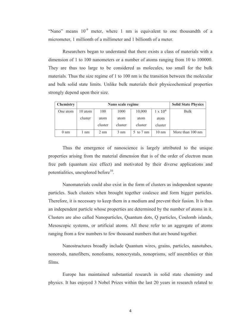
Researchers began to understand that there exists a class of materials with a
dimension of 1 to 100 nanometers or a number of atoms ranging from 10 to 100000.
They are thus too large to be considered as molecules, too small for the bulk
materials. Thus the size regime of 1 to 100 nm is the transition between the molecular
and bulk solid state limits. Unlike bulk materials their physicochemical properties
strongly depend upon their size.
Chemistry Nano scale regime Solid State Physics
One atom 10 atom
cluster
100
atom
cluster
1000
atom
cluster
10,000
atom
cluster
1 x !"
atom
cluster
Bulk
0 nm 1 nm 2 nm 3 nm 5 to 7 nm 10 nm More than 100 nm
Thus the emergence of nanoscience is largely attributed to the unique
properties arising from the material dimension that is of the order of electron mean
free path (quantum size effect) and motivated by their diverse applications and
potentialities, unexplored before10
.
Nanomaterials could also exist in the form of clusters as independent separate
particles. Such clusters when brought together coalesce and form bigger particles.
Therefore, it is necessary to keep them in a medium and prevent their fusion. It is thus
an independent particle whose properties are determined by the number of atoms in it.
Clusters are also called Nanoparticles, Quantum dots, Q particles, Coulomb islands,
Mesoscopic systems, or artificial atoms. All these refer to an aggregate of atoms
ranging from a few numbers to few thousand numbers that are bound together.
Nanostructures broadly include Quantum wires, grains, particles, nanotubes,
nonorods, nanofibers, nonofoams, nonocrystals, nonoprisms, self assemblies or thin
films.
Europe has maintained substantial research in solid state chemistry and
physics. It has enjoyed 3 Nobel Prizes within the last 20 years in research related to
5
nanotechnology. The first of this was for the discovery of Quantum Hall Effect,
awarded to Klaus Von Klitzing from the Max Planck Institute, Stuttgart, Germany in
1985. The second is for the discovery of the Scanning Tunneling Microscopy (STM)
awarded to Heinrich Roher and Gerd Binnig at IBM, Zurich, Switzerland. The third
was shared by Jean Marie Lehn, at the College of France in Paris in 1987, along with
two American Chemists (D. J. Cram, ULCA and C. J. Pederson, Dupont) for
stimulation and growth.
The spread of nanotechnology in the last 20 years is strictly due to the
improvement of synthesis techniques and characterization on the nanometer scale.
This has allowed the release of scientist’s innate curiosity towards a field generous of
new physical phenomena and synthetic opportunities. Thus nanotechnology,
nanoscience, nanostructures, nanoparticles and nanoclusters are most widely used
words in scientific literature.
1.3 Quantum Size Effect
The physical properties of nanomaterials are attracting increasing interest,
because they differ significantly from bulk properties as a result of surface or
quantum size effect10
. However, these properties are highly dependent on the size of
the particles and not on interactions between their surface, ligands, contaminants or a
support. This therefore requires the need to control precisely the size distribution of
the particles and their surface state. If a metal with bulk properties is reduced to a size
in the valence band the conduction band decreases respectively to such an extent that
electronic properties change dramatically. When a metal particle size is decreased, the
typical bulk properties like conductivity and magnetism begin to disappear or in other
words a quasi-continuous density of states is replaced by a discrete energy level
structure and band gap increases. The average energy level separation is comparable
to or larger than the experimental energy. The situation in a small molecular cluster is
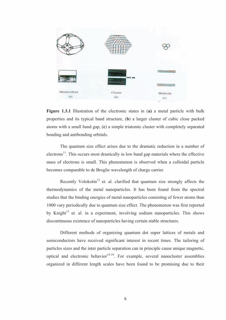
simple. Three metal atoms for instance form energetically well defined bonding and
antibonding molecular orbitals.
6
Figure 1.3.1 Illustration of the electronic states in (a) a metal particle with bulk
properties and its typical band structure, (b) a larger cluster of cubic close packed
atoms with a small band gap, (c) a simple triatomic cluster with completely separated
bonding and antibonding orbitals.
The quantum size effect arises due to the dramatic reduction in a number of
electrons11
. This occurs most drastically in low band gap materials where the effective
mass of electrons is small. This phenomenon is observed when a colloidal particle
becomes comparable to de Broglie wavelength of charge carrier.
Recently Volokotin12
et. al. clarified that quantum size strongly affects the
thermodynamics of the metal nanoparticles. It has been found from the spectral
studies that the binding energies of metal nanoparticles consisting of fewer atoms than
1000 vary periodically due to quantum size effect. The phenomenon was first reported
by Knight13
et. al. in a experiment, involving sodium nanoparticles. This shows
discontinuous existence of nanoparticles having certain stable structures.
Different methods of organizing quantum dot super lattices of metals and
semiconductors have received significant interest in recent times. The tailoring of
particles sizes and the inter particle separation can in principle cause unique magnetic,
optical and electronic behavior14-18
. For example, several nanocluster assemblies
organized in different length scales have been found to be promising due to their
7
potential application in diverse areas such as opto-electronic devices, single electron
transistors and chemical sensors17-22
.
Recently much attention has been paid to the synthesis and characterization of
bimetallic nanoparticles due to their catalytic, electronic, optical, structural and
thermal properties23-28
and subsequent technological applications as catalyst, sensors,
nanoelectronic devices29-34
and biosensors35
. The properties and applicability of
nanoparticles not only depend upon their size and length but also on the combination
of component metals (composition) and their fine structure as either an alloy or core
shell structures. Bimetallic nanoparticles with well defined alloy structures of noble
metals like Pt-Ru, Pt-Mo, Au-Ag, provide practical examples for the influence of
metals, compositions and their structure on their catalytic properties36-40
. Au-Ag
nanoparticles of alloy type structure exhibit high catalytic activity for low temperature
CO oxidation34,39
and aerobic oxidation of alcohol41
.
1.4 Properties of Metal and Metal oxide Nanoparticles
The dispersions of metal nanoclusters both mono and bimetallic one 13
, can be
easily obtained by the reduction of metal ions in water or organic solvent in the
presence of soluble polymers or ligands. The physical and the chemical properties of
metal nanoparticles depend upon several factors such as the particle size, structure,
the surface, the shape, the organization of the particles and their dispersity. Numerous
studies42-46
on size and shape dependent properties have been carried out in the last
two decades. The size dependent physical and chemical properties have been
reviewed extensively by many scientists47-49
.
a) Mechanical Properties – Mechanical Strength
The clusters interact fully with one another, yet the effect of cluster size is still
important. Clusters of metals and ceramics in the size range of 5 - 25 nm have been
consolidated to form ultrafine grained polycrystals that have mechanical properties
remarkably different from and frequently better than those of their conventional
coarse grained counterparts50
.

8
b) Melting Point
According to theoretical approach the van der Waals forces i.e. cohesive
forces between the atoms and molecules, affect the melting point of the substance.
When clustering of molecules or atoms takes place then depending upon the size of
the cluster the melting point varies. In general it is found that atomization energy per
atom for spherical cluster increases irregularly on going from 1 to 500 atoms.
Since the solid to liquid transitions begin at interfaces, a well known feature of
nanometric particles is the lower melting temperature with respect to the bulk. For
instance gold undergoes a decrease in the melting temperature35
of about 400 0C
going from 20 nm to 5 nm particles and of about 50 0C from bulk to 20 nm particles.
Melting temperature Tm depends on the particle size ‘d’ as,
Tm = A + #
$
%
Following, a general rule for physical properties dependent upon surface to
volume ratio, that can be described by using the size equations structure, as in the
above equation.
c) Colour
The colour of the metal nanoparticles in suspended state changes with the size.
As the size decreases band gap increases and thus the position of the fluorescence
band is shifted to the shorter wave length51
. Since ancient time gold was used to stain
glass with beautiful colours such as red, purple, burgundy52
. Faraday4 attributed this
colour to very finely divided colloidal gold or gold nanoparticles. Gold nanospheres
have a characteristic red colour, while silver nanospheres are yellow. The colour is
due to the collective oscillation of the electrons in the conduction band, known as
surface plasmon oscillation. The oscillation frequency is usually in the visible region
for silver and gold giving rise to the strong surface plasma resonance absorption. This
means that the origin of properties on the nanoscale is different for metal
nanoparticles than for semiconductor nanoparticles.

9
d) Optical Properties
The distinctive colours of colloidal gold and silver are due to a phenomenon
known as plasmon absorbance. Incident light creates oscillations in conduction
electrons on the surface of nanoparticles and electromagnetic radiation is absorbed.
The surface plasmon band (SPB) is due to the collective oscillations of the electrons
at the surface of the nanoparticles, which is correlated with the electromagnetic field
of the incoming light i.e. the excitation of the coherent oscillation of the conduction
band. The study of the SPB has remained an area of very active research from both
scientific and technological standpoints especially when the particles are embedded in
ionic matrices and glasses53, 5 4
. For instance a driving force for this interest is the
application to the photographic process55
. Thus the SPB provides a considerable body
of information on the development of the band structures in metals and has been the
subject of extensive study of optical spectroscopic properties of Au
nanoparticles43,54,56,57
.
According to Mie43
theory the total cross section composed of the SP
absorption and scattering is given as a summation of the overall electric and magnetic
oscillations. The resonances denoted as surface plasmons were described
quantitatively by solving Maxwell’s equation for spherical particles with the
appropriate boundary conditions. Mie’s theory attributes the plasmon band of
spherical particles to the dipole oscillations58
of the free electrons in the conduction
band occupying the energy states immediately above the free electrons in the
conduction band occupying the energy states immediately above Fermi energy level.
The SPB is absent for Au nanoparticles with core diameter less than 2 nm as well as
for bulk gold. The SPB maximum and bandwidth are also influenced by the particle
shape, medium, dielectric constant and temperature. The refractive index59,60
of the
solvent has been shown to induce a shift of the SPB as predicted by Mie theory. The
ligand shell alters the refractive index and causes either a red or blue shift, so that the
spectroscopic data obtained often deviate from the prediction of Mie theory that deals
with naked nanoparticles. The agreement with Mie theory is obtained only when the
shift induced by this ligand shell is taken into account. This shift is especially
significant with thiolate ligands, which are responsible for a strong ligand field
10
interacting with the surface electron cloud. In fact since all Au nanoparticles need
some kind of stabilizing ligands or polymer, the band energy is rarely exact as
predicted by Mie theory. If the shift of this stabilizer is not considered with the
elliptical particles, the SPB is shifted to higher wavelength as the spacing between
particles is reduced, and this shift is well described as an exponential function of the
gap between the two particles. A red shift for a polarization parallel to the long
particle axis and a blue shift for the orthogonal polarization are reported61
and
rationalized by a dipolar interaction mechanism. The optical thickness cloud can be
used as a measure of efficiency. This parameter is critically dependent on the particle
size and refractive index. Therefore impurities can be easily detected62
since the
refractive index of Au nanoparticles greatly differs from that of gold oxide or gold
chloride. Another influential parameter is the core charge60,62,63
. Excess electronic
charge causes shifts to higher energy, whereas electron deficiency causes shift to
lower energy. The SPB width was found to increase with decreasing size in the
intrinsic size region (mean diameter smaller than 25 nm) and also to increase with
increasing size in intrinsic size region (mean diameter larger than 25 nm). A small
temperature effect was also found. It was proposed that the dominant electronic
dephasing mechanism involves electron-electron interactions rather than electron
phonon coupling63
. The SPB position in Au@SiO2 (including films) was accurately
predicted. Thus it is possible to synthesize composite material with optical properties
that lie anywhere between those of transparent glass and those of metallic gold64-66.
A near field optical antenna effect was used to measure the line shape of these
SPB in single Au nanoparticle and the result was found to be in agreement with Mie
theory. Double peak shapes caused by electromagnetic coupling between close lying
particles were observed67
.
For rod like particles68
, the extinction maximum for incident electric fields
polarized along the long axis occurs at a longer wavelength than the !max for
polarization along the particle radius. Selective suppression69,70
of extinction of the
SPB was also observed. The coagulation71
(along with Oswald ripening) of Au
nanoparticles dispersed in organic liquids was dramatically accelerated by visible

11
light, and the process was shown to be wavelength dependent, UV irradiation caused
coalescence.
e) Fluorescence
Fluorescence studies of Au nanoparticles carried out under various
conditions72-74
, including femto second emission73
and steady state investigation of the
interaction between thiolate ligands and the gold core74
. Indeed resonant energy
transfer was observed in fluorescent ligand capped Au nanoparticles. This
phenomenon is of great interest in biophotonics75
and materials science76-78
. Both
radiative and nanoradiative rates critically depend on the size and shape of the Au
nanoparticles, the distance between the dye molecules, the orientation of the dipole
with respect to the dye-nanoparticles axis and the overlap of the molecule’s emission
with the nonaoparticles absorption spectrum79
. Orders of magnitude of higher
efficiencies were obtained with nanometer dimension metal samples80-82
. The
observed increase in the fluorescence83
yield reflected the suppression of the
nonradiative decay upon binding to Au nanoparticles. Visible luminescence has been
reported for water soluble Au nanoparticles, for which a hypothetical mechanism
involving 5 d10
- 6 sp1 interband transition has been suggested
72.
f) Electrochemical Properties
Electrochemical properties of metal nanoparticles are strongly size dependent,
in fact cluster made by few hundred or tens of atoms have been used to investigate the
transition from the metal like capacitive charging to the redox like charging. Upto 15
charging peaks were observed by differential pulse voltametry in case of Au (I).
The electro chemistry of ligand stabilized Au nanoparticles shows the
formation of core charge. Moreover, the quantized capacitance charging of Au
nanoparticles, self assembled monolayers on electrode surfaces could be rectified by
certain hydrophobic electrolyte ions in aqueous solution. Magneto electrochemistry of
Au nanoparticles with quantized capacitance charging shows the influence of
magnetic field on the electro chemistry of Au nanoparticles and in particular, the
effect of electron parity upon their charging states84
. Double layer capacitance was

12
obtained in aqueous media by differential pulse voltametry85
. Altogether,
electrochemistry86-90
has been used in several ways to fabricate and deposit Au
nanoparticles.
g) Electronic Properties
It was found that for, 2 D super lattices consisting of large (5 nm) Au
nanoparticles, the electronic behavior was dominated by the coulomb block effect at
low temperature, while the I-V response was ohmic at room temperature91
. TiO2
nanoparticles exhibited a blue coloration due to stored electrons within the particles
when subjected to UV. A partial disappearance92
of the blue color was seen upon
contact with Au nanoparticles as electrons were transferred from TiO2 to Au
nanoparticles. TiO2 films cast on conducting glass plates were modified, by adsorbing
Au nanoparticles (5 nm diameter) from toluene solution and selective formation of Au
islands and larger particles on the TiO2 surface was observed93
. The effect of spin-
orbit scattering on discrete energy level in Au nanoparticles and other nanoparticles
showed level-to-level fluctuations of the effective g-factor from zeeman splitting and
the statistics were found to be well characterized by random matrix predictions94
.
Energy transfer from a surface bound area near to the gold core was observed upon
photo irradiation95
.
In semiconductor the nanoscale becomes important due to the quantum
confinement of the electrons96
. As the particles size decreased below the Bohr radius
of the semiconductor material used, the electron becomes more confined in the
particle96
. This leads to an increase in the band gap energy. Furthermore the valence
and conduction bands break into quantized energy levels. The study by Bawendi97
, of
the CdSe spherical nanoparticles of various sizes illustrates the macroscopic
realization of quantized energy levels.
h) Chemical Properties
Gold and silver are known for being generally inert and especially gold for not
being attacked by O2 to a significant extent. This makes Au nanoparticles and Ag
nanoparticles stable in ordinary conditions98
. Gold nanoparticles are resistant to strong
oxidizing agents and strong acids however “aqua regia” or solutions containing I- or
13
CN- can immediately dissolve them. Both Au and Ag are reactive with sulphur, in
particular bulk silver often undergoes tanning due to the formation of an Ag2S surface
layer. In case of organic thiols, ligation to nanoparticle surface is particularly effective
for the contemporary presence of sigma type bond, in which sulphur is the electron
density donor and the metal atom is the acceptor, plus a " type bond in which metal
electrons are partially delocalised in molecular orbitals formed between the filled d
orbitals of the metal and the empty d orbitals of sulphur98
.
Solutions of Ag nanoparticles have applications as bactericidal agents because
the Ag+ ions interfere with bacterial metabolism. Since Ag nanoparticles are exposed
to a certain extent surface oxidation by atmospheric O2, sliver sols can release Ag+
ions with concentration sufficient to act as bactericides98
.
The significance of nanoscale quantum confinement of the electrons provides
visualization of the shift in the characteristics of the material depending upon the size
of nanoparticles. The electrons are not as free to move thus creating new samples for
applications hold a promise.
In a metal, electrons are highly delocalized over a large space (i.e. least
confined). As the separation between the valance and conduction bands vanishes it
renders the metal its conducting properties99
, whereas when the separation between
the valences and conduction band becomes comparable to or larger than KT, the metal
becomes semiconductor. When this confinement further increases the energy
separation, the metal becomes insulator. In noble metals the decrease in size below
the electron mean free path gives rise to intensive absorbance in the near UV visible
region.
i) Specific Surface Area
Nanoparticles present a higher surface to volume ratio with decreasing size of
the nanoparticles. Specific surface area is relevant for catalytic reactivity, electrical,
optical and thermal properties, although stabilization of the surface area, nanoparticles
topology (roughness) and interface with support material are also relevant aspects.
When a metal particle decreases in size to become a nanoparticle or a nanocrystal a

14
greater proportion of atoms is found at the surface. The metallic nanoclusters are built
up by hexagonal (HCP) or cubic (CCP) close packed atoms (nuclearities). As this is
the case in most bulk metals, where a central atom is surrounded by 1, 2, 3… closed
packed cells.
Table 1.4.1 Schematic illustration of metal nanocrystals, in closed-shell
configurations with their magic numbers of atoms and the relation between the total
number of atoms and the % of surface atoms100
.
Full Shell
Clusters
Total Numbers
of Atoms
Surface
Atoms (%)
1 shell 11 92
2 shells 55 76
3 shells 147 63
4 shells 309 52
5 shells 561 45
7 shells 1451 35
j) Thermal Properties
As compared to the study of other well known properties of the nanomaterials,
this study has seen slower progress. This is partially due to the difficulties of
experimentally measuring and controlling the thermal transport in nanoscale
dimensions. Atomic Force Microscope101
(AFM) has been introduced to measure the
thermal transport of nanostructures with nanometer scale having a high special
1 Shell 2 Shells
4 Shells 5 Shells7 Shells
3 Shells

15
resolution providing for a promising way to probe the thermal properties with
nanostructures. Recent advances in experiments have showed that certain
nanomaterials have extraordinary thermal properties as compared to their
macroscopic counterparts. This is attributed due to several factors such as the small
size, the special shape; the large interfaces modified the thermal properties of the
nanomaterials rendering them to quite different behavior. As the dimension goes
down to nanoscales, the size of the nanomaterial is comparable to the wavelength and
the mean free path of the photons, so that the phototransfer within the materials will
be changed significantly due to the photon confinement and quantization of photon
transport, resulting in modified thermal properties. For e.g. nanowires102
from silicon
have a much smaller thermal conductivity as compared to bulk silicon. Carbon
nanotubes103
have extreme high thermal conductivity in axial direction leaving high
anisotropy in the heat transports in the materials.
1.5 Synthesis of Metal nanoparticles
Nanoparticles have potentially useful size and shape dependent
properties104-107
. The synthesis of nanoparticles with desired size and shape has
therefore enormous importance, especially in the emerging field of
nanotechnology108-109
. This in fact demands development of a new general methods as
well as improvement over the existing ones. At present the nanoparticles are prepared
by number of physical and chemical methods.
The approaches110
for synthesis or preparation of nanoparticles fall under two
categories. These are “Bottom up” and “Top down”.
a) Bottom Up Synthesis
This involves the building of structures, atom by atom or molecule by
molecule. The formation of structures in this approach is governed by the polarity and
reactivity of the nanoparticle surface111
. This is derived from the fictionalization of
particle surface. The functionalities112
determine the binding capabilities of the
nanoparticles with other molecules, particles and surface. The bottom up approach for
fabrication of nanostructures from stable building blocks has become popular in
current science and engineering. This construction principle mimics biological

16
systems by exploiting the order inducing factors that are imminent to the system113
.
The bottom up methods may in future offer a number of potentially very attractive
advantages. These include experimental simplicity down to the atomic size scale, the
possibility of three dimensional assemblies and the potential for inexpensive mass
fabrication.
Bottom up approaches use single atoms or ions to grow clusters and
nanoparticles wet chemically of rather precise composition, size, and structure. In
order to prevent agglomeration, the nanosized entities have to be protected by
appropriate ligand molecules, serving as a protecting sphere. Thus special conditions
are required for the generation of identical species.
Figure 1.5.1 Bottom up approach for nanoparticle preparation.
b) Top Down Synthesis
This approach of synthesis involves starting with a larger piece of material and
etching, milling or machining a nanostructure from it by removing material (as, for
example in circuits on microchips). This can be done by using techniques such as
precision engineering and lithography, and has been developed and refined over the
last 30 years. Such methods offer reliability and device complexity, although they are
higher in energy usage, and produce more waste than bottom up methods. The
disadvantage of this method is the manipulation of small amounts of atoms at a time
resulting in low fabrication throughput.

17
Figure 1.5.2 Top down approach for nanoparticle preparation.
The various chemical and physical methods available for synthesis or
preparation of metal nanoparticles are discussed as follows.
a) Chemical Reduction Method
It is the most widely used method for nanoparticle synthesis. Metal ion in
aqueous solution is reduced by reducing agents such as hydroquinone, sodium
borohydride, hydrazine, sodium citrate, ascorbic acid etc. to produce nanoparticles.
An electric charge on the reductant particle was found to exert large effect on the
process of nanoparticle formation. The most highly dispersed and stable colloidal
nanoparticles are formed by using anionic reductant114
. All these wet chemical
approaches require the reduction of Ag+ or Au
+ ions and the chemisorption and
physisorption of ligands on the surface of metal nanoparticles to avoid their
coagulation and precipitation.
One of the best known technique is described by Turkivech115,116
, where an
aqueous solution is brought to boil and sodium citrate is used as both as a capping
material and reducing agent. The order of addition of the metal salt and sodium citrate
as well as the temperature, mole ratio of citrate to metal salt and volume of the
solution determined the size of the nanoparticle generated. This synthesis is easy,
quick and reproducible and generates mono dispersed population of nanoparticles
around 20 nm in diameter. This synthesis is widely used to generate metal
nanoparticles for many systems where 20 nm is a desirable diameter, or a seed for
further growth. The use of sodium citrate as a capping material allows easy surface
modification for a variety of applications. Sodium citrate is a weak reducing agent

18
which does not reduce the metal ion at room temperature. Thus the strength of the
reducing is controlled by the temperature of the solution.
A very popular technique of generating small gold nanoparticles has been
reported by Burst117, 118
. The technique is often referred to as creating monolayer
protected clusters, due to the formation of monolayer of thiol molecules on the surface
of the gold nanoparticles. These particles are extremely stable due to the high strength
of the gold–thiol bond. A two phase systems is used where the gold ion is first
transferred to the organic phase by employing a phase transfer agent, typically tetra
octyl ammonium bromide (TOAB). Then sodium borohydride is used to reduce the
gold ions. This reduction is accomplished slowly when the gold ions from the organic
phase come in contact with sodium borohydride from the aqueous phase. Then thiols
can be used to protect the gold ions after they are reduced. This technique is able to
glow clusters around 1.5 nm diameter that are very stable.
Other reducing agents that have been used to synthesis noble metal
nanoparticles are formamide119
, sucrose120
and glucose121
. Other wet chemical
approach exploit reduction of metal salts by organic solvents such as ethanol for gold
and dimethyl formamide for silver or polyols like ethylene glycol in presence of
protecting polymers as polyvinyl pyrrolidone or other stabilizing molecules122
.
b) Gamma ( ) Irradiation Technique
The radiation induced synthesis is one of the most promising strategies
because there are some important advantages to the use of the irradiation123
techniques, compared to the conventional chemical and photochemical methods.
These are :
1) The process is simple and clean.
2) The gamma ray irradiation has harmless future.
3) Controlled reduction of metal ions can be carried out, without using
excess reducing agent or producing undesired oxidation product of the
reductant.
4) The method provides metal nanoparticles in a fully reduced, highly
pure, highly stable state,
5) No disturbing impurities like metal oxides are produced.

19
Radiolytic reduction generally involves radiolysis of aqueous solutions that
provides an efficient method to reduce metal ions and form homo and hetero nuclear
clusters of metals. In the radiolytic method aqueous solutions are exposed to gamma
rays creating solvated electrons, e-aq. These solvated electrons in turn, reduce the
metal ions and the metal atoms eventually coalesce to form aggregates as depicted by
following reactions124
,
&'( radiation
e-aq
, H3O
+, H*, H2
, OH*, H2O2
e-aq
, + M
m+ M
(m – 1)+
e-aq + M
m+ M
0
nM0 M2 … Mn… Magg
The gamma irradiation based strategies have been used to synthesize
gold125, 126
and silver127
nanoparticles. Silver nitrate or hydrogen tetra chlorate aurate
was dissolved in water with 2 propanol and PVP to form primary solution with
predetermined concentration of the metal salts. The mixture solution was bubbled
with pure nitrogen for about 20 minutes to remove oxygen and then irradiated in field
of 60
Co, # ray source at certain irradiation dose rates. Dark brownish silver colloids or
reddish gold colloids were prepared. The addition of PVP128
and small amount of 2-
propanol did not lead to any thermal reduction of the Ag or Au salt. This technique
has also been used to prepare bimetallic alloy metal clusters of Ag-Pd129
.
c) Ultrasound Technique (Sonochemical reduction)
Ultrasonic irradiation treatment is done by means of high density ultrasonic
probe consisting of multiwave ultrasonic generator and barium titanate oscillator. This
technique has been proved useful for preparation of novel materials130
. The chemical
effect of ultrasound cavitation, that is formation, growth and implosive collapse of
bubbles in liquid produces unusual chemical environments131
.
Radical species are generated from water molecules due to the absorption of
ultrasound.
&'( altrasound radiation
H* OH* (sonolysis)
M 2+
+ 2H* M0 + 2H
+
nM0 (M
0)n (aggregates)

20
The OH*radical, produces hydrogen peroxide which oxidizes the metallic
cluster. In the presence of hydrogen the OH* radicals can be scavenged out. In this
method stable fine particles are produced in the presence of the protecting agent such
as surfactant or water soluble polymer.
Silver nanoparticles with a size of 5 nm have been prepared by this
technique132
. An argon / hydrogen environment produced more H* radicals and
prevented the oxidation of silver nanoparticles. The period of sonication was 90
minutes. Gold nanoparticles have also been synthesized by this method133-135
.
d) Laser Ablation Method
Wet chemical methods to prepare metal nanoparticles are based on the
reduction of suitable metal salts in the presence of stabilizers. Laser ablation136
on the
other hand is an alternative route to the chemical synthesis for the preparation of small
metal particles. The main advantage of laser ablation is the high purity of the
nanoparticles wherein pure bulk material and solvents can be used in the preparation.
Another advantage of this method includes its simplicity and versatility with respect
to metals or solvents in the absence of chemical reagents or ions in the final solution.
Furthermore it is shown that laser ablation137
of gold and silver produces stable
colloids.
Metal nanoparticles are produced by focusing Q switched Nd YAG laser
(1064 nm, 8 nsec. pulses) as the radiation source, on a metal wire. The laser used has
an energy output of 5 – 15 mJ. operated at 50 Hz. A 1064 laser beam is focused by a
lens with 50 cm focal length on the metal wire suspended in solvent. Typical ablation
time was 10 minutes.
e) Microwave Heating
Recently microwave irradiation as a heating method has been used in the
synthesis of nanoparticles. In principle this is a high temperature synthesis method.
The generation of nanoparticles originates from the heating effect, rather than the
energy of quantum of microwave. Polar molecules can be heated quickly under the
microwave irradiation. In comparison with conventional heating the microwave
synthesis is quite fast, simple, very energy efficient and also achieves uniform
21
nucleation in shorter crystallization time. In a microwave heating process one
precursor can be heated at much higher heating rates and reach a higher temperature
then its surrounding. A quick simple and energy efficient method based on
microwave138, 139
heating has been developed and is widely used in synthesis of solid
materials. Silver nanoparticles have been synthesized using the non conventional
reducing agent, N,N diphenyl benzamidine and the microwave irradiation method140
.
Different size silver nanoparticles of interesting colors were synthesized.
f) Photochemical Synthesis
Recently a radiolytical and chemical size controlled, with improved mono
dispersity via seed mediated growth of colloidal nanoparticles has been reported125
.
They have followed iterative growth method, i.e. particles were grown in the
immediately previous steps, were used as seeds in the next growth step. Here particles
of various size ranging from an average diameter of 5 –20 nm could be obtained
within a few minutes by UV irradiation at room temperature, in the presence of air.
Furthermore larger size particles (25 to 100 nm) were produced directly from the
original seed particles by varying the ratio of original [seed] to [Au (III)] and using
ascorbic acid as reductant. It should be noted that ascorbate ion is frequently used as a
reducing agent for reduction Au (III) or Ag (I) ions. In a variation of this method pH
dependent condition large spherical gold nanoparticles were synthesized directly from
Au (III) ions141
. There are other reports for synthesis by photochemical methods for
silver142,143
and gold144
nanoparticles.
g) Sol Gel Technique
Traditionally Sol-Gel processing refers to the hydrolysis and condensation of
alkoxide based precursor such as tetra ethyl orthosilicate. Disregarding the nature of
the precursor the Sol- Gel can be characterized by a series of distinct steps145
.
1) Formation of stable solution of alkoxide or solvated metal precursor (the Sol).
2) Gelation resulting from the formation of an oxide or alcohol bridged network
(the gel) by poly condensation or poly esterification that results in a dramatic
increase in the viscosity of the solution.

22
3) Ageing of the gel (syneresis) during which the poly condensation reactions
continue until the gel is transformed into a solid mass, accompanied by
contraction of the gel network and expulsion of the solvent from the gel pores.
4) Drying of the gel when water and other volatile liquids are removed from the
network. This process is complicated due to fundamental changes in the
structure of the gel.
5) Dehydration during which surface bound M–OH groups are removed, there by
stabilizing the gel against rehydration. This is achieved by calcining at
temperature of 800 0C.
6) Densification and decomposition of the gel at high temperature (T greater than
800 0C). The pores of the gel network collapse and remaining organic species
are volatilized. This step is normally reserved for preparation of dense
ceramics and glares.
The Sol-Gel processing of materials coupled with the inherent advantages of
the organically modified silicates make these matrices very attractive as supports and
stabilizers for nanometalic particles in both liquid and solid phases. The
nanobimetallic combinations145-147
such as Au-Pd, Au-Ag, Au-Pt and Ag-Pt have been
synthesized by this method.
h) Microemulsion Synthesis
The preparation of metal nanoparticles using the microemulsion system, where
ionic and non ionic surfactants are used has been well reported148
. Microemulsions are
colloidal “nanodispersions” of water in oil (W/O) or oil in water (O/W) stabilized by
surfactant film. In case of water in oil microemulsions the size of the water droplet is
varied by changing only one parameter W0 (W0 = Water to Surfactant molar ratio).
The size of the nanoparticles is controlled by the size of the droplets of the micro
emulsion.
A reverse micelle system149
has been used for the preparation of silver, gold
and silver gold bimetallic alloy nanoparticles. In the preparation non ionic surfactant,
Triton X 100 and cyclohexane as bulk phase and 1 hexanol as a co surfactant have
been used. The metallic and bimetallic nanoparticles are synthesized at room
temperature through the reduction of metal salts, using sodium borohydride as a

23
reducing agent. The nanoparticles synthesized by this technique are stable for about 6
months. Two reverse micelle system are prepared at room temperature, one contained
reducing agent and the other contained metal salt solutions, as a water domain in the
reverse micelle. Then the two reverse micelles are mixed with continuous stirring. A
change in the color occurred almost immediately after mixing the two reverse
micelles. The size of the nanoparticles was in the range of 5 to 50 nm. Using this
technique silver150,151
and gold152
nanoparticles have been prepared.
i) Templated Synthesis
The synthesis of nanoparticles on templates has generated an increasing
amount of attention in the past few years. Techniques on heterogeneous nucleation in
which seed crystals serve as nucleation site for further deposition and growth of
crystallites can, essentially be considered one of the simpler forms of a template
synthesis. This technique153,154
can be used to increase the average particle size of
nanoparticles such as aqueous Au (III) is deposited on colloidal Au or Ag+ is
deposited on colloidal Ag. This method can be also used for the synthesis of core shell
and onion structure such as deposition Pd, Bi, Sn, or In on colloidal Au155,156
. To
maintain the narrow size distribution of the nanoparticles care must be taken to ensure
that small particles do not nucleate from the solution during the deposition process.
Several new techniques have recently emerged to prevent this occurrence. Brown157
et. al. used hydroxyl amine for the seed mediated growth of colloidal Au, increasing
the average diameter from 12 to 50 nm. The use of hydroxylamine is critical to this
process.
j) Electrochemical Reduction method
Elucidating the electronic structure of metallic nanoparticles is attractive to
scientist. The development of preparative method to obtain enough amounts of the
monodispersed metallic nanoparticles is essential for the observation of different
analytical tool. Many researchers have developed the synthetic methods of
monodispersed metal nanoparticles in the presence of linear polymer158,159
,
ligands160,161
and so on. In general the control of nanoparticles size and shape
constitutes a preparative challenge. Some methodologies have been demonstrated by

24
chemical reduction methods either by adjusting the water contents at a certain
interconnected microemulsion phases for the production of rod like nanoparticles or
though using capping polymers in the preparation of cubic tetrahedral Pt.
nanoparticles162
.
Of all the methodologies developed for the production of metal nanoparticles
the electrochemical methods offers an alternative simple means within reverse
micelles in organic solvent systems. We now successfully apply it to the preparation
Ag and Au, mono and bimetallic nanoclusters within a suitable mixture of organic
solvents and have developed a unique synthetic route in preparing sufficient yield of
these nanoparticles. Our synthetic approach is to control the growth and particle size
by introducing size control reagent into the electrochemical systems in which
appropriate surfactants or ligands are employed. There act is a supporting electrolyte
and a stabilizer for the resulting nanoparticles. Other parameter for controlling the
size of the nanoparticels is current density in electrochemical reduction method.
Pure and size selective nanoparticles can be prepared by electrochemical
reduction method. The process makes the use of an expensive two electrode set up for
25 to 50 ml electrolyte solution. During the synthesis the bulk metal anode is oxidised
and converted into metal cations163
. These cations migrate to the cathode and the
reduction takes place with the formation of the metal atom in zero oxidation state.
The noble metal cluster in the 1 to 30 nm range has been prepared by this
method for fundamental and practical reason. They have unused electronic properties
and used in catalysis, photocatalysis and electrocatalysis (fuel cell technology). In
order to prevent agglomeration of metal particles in solution, synthesis is done in
presence of stabilizer such as ligand (phenanthroline), polymers (PVP) or surfactants
such as tetra alkyl ammonium salts.
The particle size of the nanostructured metal clusters can be controlled by
varying parameter such as types of ligands, by varying of surfactants, their
concentration, temperature, solvent and current density in the electrochemical
synthesis. “True control of particle size remains the most attractive goal for the
synthetic chemist”. The first report of stabilization of metal colloids by tetra alkyl
25
ammonium salt, Pt / acetyl trimethyl ammonium chloride is due to Gratzel164
in 1979.
Thereafter the research in the area of ammonium salt stabilized metal clusters was
intensified by Toshima165
et al. Recently Bollemen166
synthesized
stabilized metal cluster by reduction of metal salt with tri alkyl boron hydride.
Quaternary ammonium compounds are a group of ammonium salt in which organic
groups are substituted for all the four hydrogen of the original ammonium cation.
They have central nitrogen atom which is joined to four organic groups. The organic
group is mainly alkyl or aryl and the nitrogen can be a part of the ring system. They
show a variety of physical, chemical and biological properties and most compounds
are soluble in water and act as strong electrolyte.
In electrochemical synthesis, the choice of the tetra alkyl ammonium
surfactant is governed by the following constraints,
1. It should be adsorbed the metal surface to stabilize the synthesized
nanoparticles.
2. It should be highly soluble and dissociated in the solvent to play the role of a
electrolyte.
3. Hydrophylic- Liophilic balance of the chosen surfactant should be sufficiently
high to prevent any precipitation due to the presence of water, even in the
presence of small quantities in the medium.
The surfactant that statistics the above constraints, such as tetra ethyl
ammonium bromide (TEAB), tetra propyl ammonium bromide (TPAB), tetra butyl
ammonium (TBAB), tetra octyl ammonium bromide (TOAB) are used in the present
work and their structures are shown in the figure.
A controlled current electrolysis is used throughout the synthesis process.
Solvent selection is made (either single or mixture), depending on the solubility of the
ligand or surfactant. The basic principle is simple, viz, electrochemical reduction of
metal ion in the presence of surfactants is served as an electrolyte and as stabilizer.
The salient feature of the process includes high yield, absence of any side products,
easy isolation of the nanoparticles, variation and choice of ligands, and control of

26
particle size by controlling current density. The metal aggregates synthesized do not
coalesce during the process.
1.6 Applications of nanoparticles
Metal nanoparticles have many unique properties. These include large surface
to volume ratio, high surface reaction activity, high catalytic efficiency and strong
adsorptions ability. Thus they have many possible applications in areas such as non-
linear optical switching167
, immunoassay labeling168
and Raman spectroscopy169
enhancement. Thus the nanoparticles do not have only exceptional scientific
attractions but have increasing practical relevance in numerous disciplines like
Physics, Chemistry, Biology, Medicine and Material Science. The nanoparticles are
classified as two types i.e. Engineered or Non- Engineered. Engineered nanoparticles
are intentionally designed and created with physical properties tailored to meet the
need of specific application. They can be end product in themselves, as in the case of
quantum dots or pharmaceutical drugs, sensors for special purpose, or they can be
later incorporated, such as carbon black in rubber products.
a) Catalysis
Catalysis drives many reactions, with the ability to lower the activation energy
of the reaction, and thus increases the rate of reaction and the yield. The use of
nanoparticles as catalyst has increased as nanoparticles properties and reactions are
better understood. The possibility of using less material and have different properties
for different shapes of nanoparticles is very attractive. Nanoparticles catalysis has
been investigated170
for both homogenous and heterogeneous systems. Small clusters
are also found to be catalytically active171,172
, even for materials that display very
limited reactivity on the bulk scale. For example bulk gold is considered as a noble
metal and is unreactive in the bulk state. However, small clusters of gold have been
found to be catalytically active. Many possible explanations have been proposed for
the difference in activity between clusters and bulk gold. They include the electronic
and chemical properties of nanoparticles or the shape, size and oxidation state of the
nanoparticles. The surface support171
is also suggested to be responsible for the

27
catalytic activity. The crystal structure of gold has also been proposed to be important
in the catalytic property of gold.
The Au nanoparticles supported on Co3O4, Fe2O3 or TiO2 are highly active
catalyst under high dispersion for CO and H2 oxidation173,174
, NO reduction175
, water
gas shift reaction176
, CO2 hydrogenation177
and catalytic combustion of methanol178
.
Bimetallic nanoparticles are also applied to hydrogenation catalysts. PVP
stabilized Au–Pd179
bimetallic nanoparticles with various compositions are used as
catalysts for the selective hydrogenation of 1, 3 cyclooctodiene to cycloctane. In these
bimetallic nanoparticles, with palladium content of 80% showed the highest activity
which is greater than Pd nanoparticles themselves.
The greatest attraction of gold nanoparticles catalysis is their green
credentials. They offer the possibility of catalytic oxidations, that use atmospheric air
as the oxidant and forms pure water as the only by product. Recently the aerobic
oxidation of methanol to industrially important chemical, methylformate using an
unsupported nano-porus gold catalyst has been reported178
. The porus catalyst was
prepared by removing silver from gold-silver alloy, a process called dealloying.
b) Nanofluids
Nanofluids are engineered by dispersing nanometer size solid particles in
traditional heat transfer fluids to increase thermal conductivity and heat transfer
performance. The primary function of nanofluid is to dramatically enhance thermal
conductivity in conventional fluids. Since thermal conductivity of solid nanoparticles
is much higher than that of fluids the suspended particles are expected to increase the
thermal conductivity and heat transfer performance. In fact many factors (such as
shape, size, volume, fraction, thermal conductivity of nanoparticles, viscosity as well
as membrane of liquid molecules around the nanoparticles etc.) may influence the
thermal conductivity of nanofluids.
Experimental result should that the enhanced thermal conductivity ratio
increases with the increase of volume fraction of Al2O3 nanoparticles in
nanosuspensions180
. The results of Zhang181
showed that the effective thermal

28
conductivity and thermal diffusivity of Au / toulene, Al2O3 / water and CNT / water
nanofluids at different temp increases with the increase in the particle concentration,
the nanoparticle thermal conductivity and the ratio of the length to the diameter of
CNT. Another important factor that may influence the thermal conductivity of
nanofluids is the liquid solid interface. Liquid molecules close to the liquid solid
surface are known to form a layered structure. A liquid in contact with a solid
interface is more ordered than a bulk liquid. Since crystalline solids (which are
obviously ordered) display much better thermal transport than liquids, such a liquid
layering is expected to lead to a higher thermal conductivity182
.
c) Cosmetics
ZnO nanoparticles183
have received considerable attention due to their unique
properties. They are well known as UV blocking materials especially of light in the
UV region and as such are used widely in cosmetics, paints and fibers. But their high
catalytic activity in oxidation and photochemical reactions restrict their application as
UV blocking materials. Particle surface modification is regarded as an effective way
to restrain the ultrafine particles, from high oxidative and photochemical activity.
d) Industrial Applications
Silver is amongst the widely used metal in the world. Its metallic properties
make it a great conductor. Its antimicrobial property has also been exploited in several
applications. Reducing silver to nonosized particles helps to make the element highly
effective, making it more in demand for several uses in the industry of medicine and
technology. Currently there are many uses of silver nanoparticles in industries that
have resulted in a boost in its demand and production. Silver nanoparticles can be
used as an ingredient for water purifiers and even as an ingredient for inks used in
inkjets. A potential for AgNP industrial use is making it as coating on polyurethane
foams. The material is ideal as it can be washed, dried and stored for long period of
time and still retained same number of nanoparticles. The material is also tested as an
element for water filtration to eliminate bacteria. It is also possible to use it in the air
filtration, air quality management and industrial settings, as well as antibacterial
packing semiconductor nanoparticles.

29
e) Semiconductor nanoparticles
A decrease in the size of semi-conductor nanoparticles results in the shift of
the conduction and valence bands towards more negative and more positive potentials
respectively. It seems to be tempting to use the possibility of controlling the redox
properties by changing their size, in photocatalytic systems, water decomposition,
synthesis and destruction of organic compounds.
The small size of semi conductor nanoparticles can provide high efficiency of
detrapping of light generated electrons and holds the increasing probability of the
photocatalytic process on the surface of the conductor. The idea of using
nanoparticles for photocatalytic water decomposition was first proposed by
Henglein184
and Duonghong185
who demonstrated the possibility of photoredution of
methylviologen in colloidal solutions of semi-conductors. The photo reduction of
methyl viologen on the surface of nanoparticles was studied by laser flashed
photolysis, in colloidal solutions of TiO2185,186
and metal chalcogenides187
. When Cd-
Se is electrochemically deposited188
on electrically conducting glass, agglomerated Q
particles are formed and not a compact film. Such electrodes in photochemical cells
exhibit photocurrent arising from the Q particles. In order to absorb significant
quantities of the incident light 10-20 coatings of Q particles are deposited one over the
other, and thus the released electrons must be transported over many particles to the
back contact. Energy is lost with every jump of the electrons from one particle to
another, so the photocurrent and voltage attainable with such systems are relatively
low. This problem can be overcomed by depositing the Q particles on a porus TiO2
electrode, to which the electron are transferred after light absorption189
. Less than a
monolayer of particles is necessary for practically all the incident light to be absorbed.
f) Solar Cells
Electrochemical studies have demonstrated the electron storing properties of
gold nanoparticles and their ability to act as an electric relay on a nanotemplate
structure190-192
. Beneficial role of gold nanoparticles in photocurrent generation has
been reported by Kuwahara193
in tris 2, 8 bipyridilede ruthenium (II) vilogogen linked
thiol.

30
g) Nanodiagnostic
Molecular recognition is fundamental for the development of clinical
diagnostic tools and therapeutic modalities. Various organic molecules possessing
unique properties have been used to achieve the recognition of different targets (194).
The possibility of combining the ease of handling DNA based modification with
different modification strategies of nanomaterials has showed its applicability195
in
spectroscopy, electrochemistry, magnetism (imaging, purification and detecting and
others). The incorporation of nanomaterials (e.g. AuNP) facilitates signal
transduction, as the signal of recognition can be amplified by several orders of
magnitude, making recognition more effective. They can be modified according the
function of the designated DNA probe and make applications of functional DNA
more practical for molecular recognition in medical diagnostics by taking advantage
of the unusual interaction between the nanomaterials and the living system196
. This
increase in sensitivity and flexibility present numerous advantages197
over more
traditional procedure like fluorescence and chemi-luminescence.
The first report of DNA hybridization event with thiol-DNA modified gold
nanopaticle (Au nanoprobes) was made by Storhoff198
and co-workers and since then
several new approaches and applications has been reported.
h) Drug Delivery
In recent years nanocapsusles and nanospheres surrounded by a polymer
matrix into which the drug is physically and uniformly dispersed are used as drug
delivery devices. These have the ability to circulate for a prolonged period of time,
target a particular organ, as carriers199
of DNA in gene therapy and have the ability to
deliver proteins, particles and genes.
The advantages of using nanoparticles as delivery systems includes,
1) Particle size and surface characteristics can be easily manipulated to achieve
passive and active drug targeting after parental administration.
2) They control and sustain release of the drug during transportation and at the
site of localization, altering organic distribution of the drug and subsequent
31
clearance of the drug so as to achieve increase in drug efficacy and avoid side
effects.
3) Drug loading is relatively high and drugs can be incorporated into the system
without any chemical reaction thus preserving drug activity.
4) Site specific targeting can be achieved by attaching targeting ligands to the
surface of the particles or use of magnetic guidance.
5) The system can be used for various routes of administration including oral,
parental, intra-ocular etc.
The delivery of the drug Doxorubcin200,201
into the mononuclear phagocytic
system rich organs/ tissues like liver, spleen and lungs is helpful for treatment of
hepatic-carcinoma, hepatic metastasis, gynecological cancers, bronco pulmonary
tumors, myeleoma and leukemia. Bioactive molecules and vaccines based on peptides
and protein can also be encapsulated and delivered. The insulin loaded nanoparticles
were found to reduce blood glucose for a longer period202
. Polynucleotide vaccines
and therapeutic genes are also delivered to produce immune response203
and treatment
of bone healing204
respectively. Several drugs have also been delivered to the brain by
crossing the blood-brain barrier system, the nanoparticles, containing the drugs, were
transported to the brain by receptor mediated transcytosis205,206
.
i) Nanomedicines
Some of the challenges facing conventional therapies are poor bioavailability
and intrinsic toxicity. These have seriously compromised the therapeutic efficacy of
many otherwise beneficial drugs. Thus innovative nanodevices and nanostructures
have been developed for applications207-209
in the fields of diagnostics, biosensing,
therapeutics, drug delivery and targeting.
Drug delivery with nanotechnological products takes advantage of the patho-
physiological conditions and anatomical changes within diseases tissues to achieve
site specific and targeted delivery. The nanosystems are accumulated at higher
concentration thus enhancing bioavailability at the targeted site. This leads to less
systemic toxicity. The unique property of the nanosystem has helped to deliver drugs
to harder-to target sites such as the brain, where blood-brain barrier exists210
. Some

32
nanoparticles can penetrate smaller capillaries and are taken up by cells. Many of the
NP is known to be biocompatible, undetected by the immune system and
biodegradable. Additional some may possess unique optical and electrical properties
e.g. AuNP, making it possible to track their systemic movement and localization.
Recent advances210
in the use of nanoparticles in medicine include delivery of
antigens for vaccinations, gene delivery for treatment or prevention of genetic
disorders, in cardiac treatment, in dental care and orthopedic applications.
Chrysotherapy the use of gold in medicine has been practiced since antiquity.
Ancient cultures in Egypt, India and China have used gold to treat diseases like
smallpox, skin disorders, syphilis, and measles. In India, Ayurved an ancient medical
science makes use of various Bhasma. One of them Suvarna Bhasma (Gold Ash) is a
therapeutic form of gold metal nanoparticles. In the past few decades, several
organogold complexes have emerged with antitumor, antimicrobial, antimalarial and
anti-HIV activities. It is also used in the treatment of rheumatoid arthritis. Gold
nanoparticles of various shapes have been found to effective in this treatment.
Proteins grafted into gold nanoparticles have also been used to kill cancer cells210
.
The antibacterial effects of Ag salts have been noticed since antiquity211
, and
Ag is currently used to control bacterial growth in a variety of applications212,213
,
including dental work, catheters and burn wounds. It is well know that Ag ions and
Ag-based compounds are highly toxic to micro-organisms, showing strong biocide
effects on a number of bacterial species. The hybrids of Ag nanoparticles with
amphiphilic hyper branched molecules exhibit effective antimicrobial activity in
surface coating agents214,215
.
1.7 Scope of the present work
Nanomaterials have emerged as an area of current interest motivated by
potential applications of these materials in electronics, non-linear optics and magnetic.
Growth in the area has been facilitated by wide accessibility to tools necessary to
characterize these materials. Metal nanoparticles exhibit properties differing from the
bulk metal due to the quantum size effects, including novel electronic optical and
chemical behavior. The properties may be tuned via control of size shape inter-

33
particle spacing and dielectric environment, and methods to vary these have been
developed. In particular nanoparticles made from silver or gold with associated strong
plasmon resonance have increased interest. The plasmon properties of these
nanoparticles of these nanoparticles depend upon their size and shape. Silver
nanoparticles have a potential to eliminate bacteria and fungi, for cosmic and beauty
applications and have self cleaning properties. Gold nanoparticles have a wide variety
of potential applications, for instance, an effective drug delivery in cancerous tumor,
biomedicals, in a wide range of cosmetic and beauty applications. The bimetallic
nanoparticles are of wide interest since they lead to many interesting properties. They
are particularly important in the field of catalysis since they often exhibit better
catalytic activity than their nanometallic counterparts. Both Ag and Au have very
similar lattice constants and are completely miscible over the entire composition
range, hence single phase alloys can be achieved.
In the present work interest in the silver and gold monometallic, bimetallic
clusters and colloids is for fundamental and practical reasons. The particles in the
range of 1 to 10 nm have been seen to have unusual electronic properties which may
lead to new technologies based on advanced materials, e.g., quantum dots in the
miniaturaization of electronic devices. Some progress has been made to control the
particle size of the nanostructured metal clusters. However true control of the particle
size, still remains the most attractive goal in this field.
The electrochemical reduction method has been reported to synthesize metal
colloids and nanoparticles in the nanometer regime and that the particle size can be
controlled in a simple manner by the variation in the current density. Apart from this
important aspect the method is simple, easy to operate, cost effective, allows high
purity of the products, allow reproducible results, and is environment friendly since it
does not make use of toxic chemicals. Thus in the present study the method is selected
for the preparation of monometallic and bimetallic nanoclusters of silver and gold.
In the preparation of nanoclusters capping agents are required to stabilize them
and prevent their aggregation. There are various classes of capping agents available
for stabilization the surfactants are one such class. The surfactants used in the present

34
study are a group of tetra alkyl ammonium salt, which include TEAB, TPAB, TBAB
and TOAB. Except for TOAB there are very few reports regarding the use of other
surfactants. This efficiency in the synthesis of small size nanoparticles by this series
of the surfactant will be evaluated. The tetra alkyl ammonium ion forms a monolayer
around the nanoparticles and is weakly bonded with it. These have thus drawn an
attention for applications which require their labile (or temporary) monolayer binding
properties. Moreover these precipitates are soluble in nonpolar solvents.
The electrochemical reduction method for the synthesis of the nanoparticles
this can either in the form of water or non polar organic solvents. The non polar
organic solvent in the form of a mixture of acetonitirile and tetrahydroforan (4:1) is
used. The advantages in using the non polar organic solvent include control over the
size of the nanoparticles, monodispersity and the chemical nature of the nanoparticles
surface (via capping).
In order to understand the effect of the current density on the size of the
nanoparticles synthesized the current density can be varied. The electrochemical
reduction process will be carried out 6 and 10 mA / cm2 current density.
The samples synthesized in the present work will be subjected to
characterization using techniques like UV-visible spectroscopy, FTIR spectroscopy,
XRD analysis, XPS, SEM and TEM. These analytical techniques will help to
understand a number of important aspects of the synthesized nanoclusters. The UV-
visible study will help to understand the surface plasmon behavior of the
nanoparticles while the FTIR study can reveal if proper capping has been achieved.
The XRD analysis will give information regarding the size and the phase of the metal
nanoparticles. XPS analysis will help to give information regarding the chemical
content of the nanoparticles and the nature of interaction between the metal
nanoparticles and the capping agent. The SEM study will help to reveal the surface
nature of the nanoparticles. The TEM analysis will give information of the shape and
size of the nanoparticles.
Thus the present work can be pursued in a systematic and result oriented
manner with definite information being obtained for the mono metallic and bimetallic

35
nanoclusters of silver and gold capped with different capping agents like TEAB,
TPAB, TBAB and TOAB. Results can be obtained for the effect, on the variation of
the current density as well on the variation of the bukiness of the capping agent on the
synthesis of the nanoparticles.

36
References
1. Daniel M. C, Astruc D., Chemical Review 2004, 104, 293-346.
2. British Museum http:// www. britishmusem.org / explore // highlights/ highlit
objects /pe –mla / t / the_ lycurgus_cup.aspx.
3. Padeletti G., Fermo P., Applied Physics A, 2003, 76, 515-525.
4. Faraday M., Philos. Trans. Roy. Soc., 1857, 147, 145-181.
5. Graham T., Philos. Trans. Roy. Soc., 1861, 151, 183.
6. a ) Zsigmondy R., Liebigs Ann., 1898, 301, 367-87.
b ) Mie G., Ann. Phys., 1908, 25, 377.
7. Sharma V., Park K., Srinivasrao M., Materials Science and Engineering
Reports. 2009, 65 (1-3), 1-38.
8. Turkevich J., Stevenson J., Hiller, Discuss Faraday Sco., 1951, 11, 55-75.
9. a) Feynman R., Engineering and Science, Feb 1961, P.22.
b) Taniguchi N., Proc. Intl. Conf. Prod. Eng., Tokyo, Part II, Japan Society of
Precision Engineering, 1974
10. (a) Schmid G., Clusters and Colloids from Theory to Applications, VCH, New
York 1994.
(b) Haberland H., Ed., Clusters of Atoms and Molecules, Springer-Verblag,
New York 1994.
(c) Hapat M. A, Ed. Colloidal Gold, Principles, Methods and Applications,
Vol. 1, Academic Press, New York, 1998.
11. Haiperin W. P, Rev. Mod. Phys., 1986, 58, 533.
12. Volokotin Y., Smizig J., de Tong L. T., Schmid G., Vargaftik M. N., Moiseev
J. I., Nature, 1996, 384, 621.
13. Knight W. D., Clemenger K., de Heer W. A., Saunder W. A., Chou. M. Y.,
Cohen M., Phys. Rev. Lett., 1984, 52, 441
14. Collier C. P., Vossmeyer T., Heath R. J., Annu. Rev. Phys. Chem., 1998, 49,
371.
15. Harfenist S. A., Wang Z. L., Whetten R. L., Wezmar I. and Alvarez., J. Phys.
Chem., 1996, 100, 13904.

37
16. Elghanian R, Shorhott J. J., Munic R. C., Letsinger L. R. and Mirkin C. R.,
Science, 1997, 277, 1078.
17. Nakanishi T., Dhatani B. and Uosaki. J. Phys. Chem. B, 1998, 102, 1571.
18. Kagan C. R., Murray C. B., and Bawendi M., Phys. Rev. B, 1996, 54, 8623.
19. Guzelian A. A., Katari J. E. B., Kadavanich A. V., Banin U. and Hamad K., J.
Phys. Chem., 1996, 100, 7212.
20. Yin S. J. and Wang Z. L., Phys. Rev. Lett. 1997, 79, 2570.
21. Rao C. N. R., Kulkarni G. U., Thomas P. J and Edwards, Chem Soc Rev.,
2000, 29, 27.
22. Pethkar S. Aslam M. Mulla I. S., Ganeshan P. and Vijaymohanan K., J.
Master. Chem., 2001, 1710-1714.
23. Sanchez-Ramirez J. F., Jimenoz Perez J. L., Cruz Orea A., Gutierrez Fuentes
R., Bautista Hermanadez, and Pal U., J. of Nanosceince and Nanotechnology,
2006, 6, 3, 685 –690.
24. Espara R., Rasas G., Lopez Fuentes, et. al., Materials Characterization, 2007,
58, 8-9, 694-700.
25. Sanchez–Ramirez J. F., Vazquez-Lopez C. and Pal U., Superficies Vacio,
2002, 15, 16-18.
26. Rodriguez-Lopez, J. L. Montejano-Carrizales J. M., Pal U., et. al, Phys Rev
Lett., 2004, 92, 19.
27. Kubo R., Journal of Physical Society Japan, 1962, 17, 975 –986.
28. Sanchez-Ramirez J. F., Pal U., Nolasco-Hernandez L., Mendonza-Alvarez J.
and Pescador-Rojas J. A., Journal Nanomaterials, 2008, ID 620412.
29. Nutt O., Heck K.N., Alvarez P. and Wong M.S., Applied Catalysis, 2006, 69,
1-2, 115-125.
30. Sarkar A., Kapoor S. and Mukherjee T., Journal of Phys. Chem. B, 2005, 109,
16, 7698-7704.
31. Ye H., Crooks R.M., J. Am. Chem. Soc., 2007, 129, 12, 3627 – 2633.
32. Hernandez-Fernandez P., Rojas S., Ocon P., et.al., J. Phys. Chem. C, 2007,
111, 7, 2913-2923.
33. Sharma A. K. and Gupta B. D., Nanotechnology, 2006, 17, 1, 124-131.
38
34. Liu. J. H., Wang A.P., Chi Y.S., Lin H.P. and Mou C.Y., J. Phys. Chem. B,
2005, 109, 1, 40-43.
35. Bruda C. Chem X., Narayanan R., El-Sayeed M. A., Chemical Reviews, 2005,
105, 4, 1025-1102.
36. Basnayake R., Li Z., Katar S., et. al, Langmuir, 2006, 22, 25, 10446-10450.
37. Molanbroek A. M., Haukka S. and Clausen B. S., J. Phys. Chem. B., 1998,
102, 52, 10680-10689.
38. Lee S. A., Park K. W., Choi J. H, Kwon B. K. and Sung Y. E., Journal of the
Electrochemical Soc. 2002, 149, 10, 1299-1304.
39. Wang A Q., Liu J.H., Lin S.D and Mou C.Y., Journal of Catalysis, 2005, 233,
1, 186-197.
40. Esparza R., Ascencio J. A., Rosas G., Sanchez-Ramirez J. F., Pal U. and Perez
Campus R., Journal of Nanoscience and Nanotechnology, 2005, 5, 4, 641-647.
41. Chaki N. K., Tsunoyama H., Negishi Y. Sakurai H. and Tsukuda T. J. of Phys.
Chem. C, 2007, 111, 13, 4585-4888.
42. Hirai H., Toshima N., Tailored Metal Catalyst Ed. by Iwasawa Y. Reidel D.
Pub. Co., Dordrecht, 1986, 87.
43. Mie G., Ann Phys., 1908, 25, 377.
44. Gao H. R., Angelici R. J., J. Am. Chem. Soc., 1997, 119, 6973.
45. Hirai H., Chawanya H., Toshima, React. Polym.,1985, 3, 127.
46. Bigioni T. P., Whetten R. L., Day O., J. Phys. Chem B, 2000, 104, 6983.
47. Beverly K. C., Sampaio, J. F., Heath J. R., J. Phy. Chem. B, 2002, 106, 2131.
48. Lue J. T., J. Phys. Chem. Solid, 2001, 62, 1599.
49. Rao C. N. R., Kulkarni G.U., Thomas P. J., Edwards P. P., Chem. Eur. J.,
2002, 8, 29.
50. Nieman G. W., Weertman J. R., Siegel R.W., Scripta. Metall,, 1989, 23, 2013.
51. Weller H., Angew Chem. Int. Ed. Engl, 1993, 32, 41.
52. Daniel M.C., Astruc D., Chem Rev. 2004, 104, (1), 293, 306,
53. Mulvaney P., Langmuir, 1996, 12, 788-800.
54. James T. H. Ed. The Theory of the Photographic Process, 1977, 4th Ed.,
MacMilan Press, New York.
39
55. Vander Hulst H.C., Light scattering by small metal particles, 1975, Willey,
New York.
56. Kerker M., The Scattering of Light and other Electromagnetic Radiations,
1969, Academic Press, New York.
57. Bohren C. F., Huffman D. R., Absorption of light by small particles, Willey,
New York, 1983
58. Alvarez M. M., Khoury J. T., Schaff T. G., Shafigullin M. N., Vezmar I.
Whetten R. L., J. Phys. Chem. B, 1997, 101, 3706-3712.
59. Templeton A. C., Pietron J. J., Murray R. W., Mulvaney P., J. Phys. Chem. B,
2000, 104, 564-570.
60. El-Sayed, M. A., J. Phys. Chem. B, 1999, 103, 4212-4217.
61. Rechberger W, Hohenau A., Leithner A., Krenn J. R., Lamprecht B.,
Aussengg F. R., Opt. Commun., 2003, 220, 137-141.
62. Swanson N. L., Billard B. D., Nanotechnology, 2003, 14, 353- 357.
63. Ung T., Liz-Marzain, L. M., Mulvaney P., Colloids Surf : Physicochem. Eng.
Asp., 2002, 119-126.
64. Ung T., Giersig M., Dunstan O., Mulvaney P., Langmuir, 1997, 13, 1773-
1782.
65. Ung T., Liz-Marzan L. M., Mulvaney P., J. Phys. Chem. B, 2001, 105, 3441-
3452.
66. Klar T., Perner M., Grosse S., Von Plessen G., Spirki W., Feld Mann., Phys.
Rev Lett., 1998, 80, 4249-4252.
67. Al-Rawashdeh N. A. F., Foss C. A., Jr., Nanostruck. Master, 1997, 9, 383-386.
68. Linden S., Christ A., Kuhl J., Giessen H., Appl. Phys. B, 2001, 73, 311- 316.
69. Linden S., Kuhl J., Giessen H., Phys. Rev. Lett., 2001, 86, 4688-4691.
70. Ahmadi T. S., Logunov S. L., El-Sayed M. A., J. Phys. Chem. B, 1999, 100,
8059-8056.
71. Satoh N., Hasegawa H., Tsujii K., Kimura K., J. Phys. Chem., 1994, 98, 2143-
2147.
72. Hwang Y. N., Jeoung D. H., Shin H. J., Kim D., Jeoung S.C., Han S. H., Lee
J. S., Cho G., J. Phys. Chem. B, 2002, 106, 7581-7584.
73. Chen M. M. Y., Katz A., Langmuir, 2002, 18, 2413-2420.
40
74. Hu. J., Zhang J., Liu F., Kittredege K.
75. Imahori H., Fukuzumi S., Adv. Mater, 2001, 13, 1197-1199.
76. Imahori H., Ariumura M., Hanada T., Nishimura Y., Yamazaki I., Sakata Y.,
Fukuzumi S., J. Am. Chem. Soc., 2001, 123, 335- 336.
77. Lakowitez J. R., Principles of Fluorescence Spectroscopy, 1999, Kluwer, New
York.
78. Wilcoxon J. P., Martin J. E., Parsapour F., Wiedenaman B., Kelley D. D., J.
Chem., Phys., 1998, 108, 9137-9143.
79. Gu. T., Whitesell J. K., Fox M. A., Chem. Mate., 2003, 15, 1358-1366.
80. Mohamed M. B., Volkov V., Link S., El-Sayed M. A., Chem. Phys. Lett.,
2000, 317, 517-523.
81. Bigioni T. P., Whetten T. P., Dog O., J. Phys. Chem. B, 2000, 104, 6983-
6986.
82. Link S., El-Sayed M. A, Int. Rev. Phys. Chem. 2000, 19, 3, 409-453.
83. Sarathy V. K. Narayan K. S., Kim J., White J. O., Chem. Phys. Lett., 2000,
318, 543-548.
84. Chen S., Yang Y., J. Am. Chem. Soc., 2002, 124, 5280-5281.
85. Li D., Li J., Chem. Phys. Lett. 2003, 372, 668-673.
86. Yamada M., Kubo K., Nishihara H., Chem. Lett., 1999, 1335-1336.
87. Yamada M., Nishihara H., Dendimers and Nanoscience, 2003, Astruc D., Ed.,
Compte. Rendus Chimie, Elsevier, Paris.
88. Ogawa T., Kobayashi K., Masuda G., Takase T., Meeda S., Thin Solid Films,
2001, 393, 374-378.
89. Chen S., Deng F., Langmuir, 2002, 18, 8942-8948.
90. Yamada M., Nishihara H., Chem. Common., 2002, 2578-2579.
91. Kanchara M., Qumi Y., Sano T., Teranishi T., J. Am. Chem. Soc., 2003, 125,
8708.
92. Jakob M., Levanon H., Kamat P. V., Nano Lett., 2003, 3, 353-358.
93. Chandrashekaran N. Kamat P. V., J. Phys. Chem. B, 2000, 104, 1085-10857.
94. Petta J. R., Ralph D. C., Phys., Rev. Lett., 2001, 87, 26680-1-266801-4.
95. Dubertret B., Calame M., Libchaber A., J. Nat. Biotechnol., 2001, 19, 365-
370.

41
96. Jin R. C., Cao Y. W., Mirkin C. A., Kelly K.L., Schatz G. C., Zheng J. G.,
Science, 2001, 219, (5548), 1901-1903.
97. Dabbousi B. O., Rodriguez-Viejo J., Mikulec F. V., Heine J. R., Mattoussi H.,
Ober R., Jensen K. F., Bawendi M. G., J. Phys. Chem. B, 1997, 101, (46),
9463-9475.
98. Greenwood N. N., Earnshaw A., Chemistry of the Elements, 1977, Elsevier
Science, Oxford.
99. Toshima N., Takabasi T., Hirai H., Chem. Lett., 1985, 1245.
100.Klabunde K. J., Ed., Nanoscale Materials in Chemistry, 2001, John Wiley
and Sons Inc.
101.David G. C., Wayne K. F., Keneeth E. G., Gerald D. M., Arun M., Humphrey
J. M., Roberto M., and Simon R.P., Journal Applied Physics, 2003, 93, 793.
102.Deyu L., Yiying W., Philip K., Li S., Peiodong Y. and Aru M., Applied
Physics Letters, 2003, 83, 2934.
103.Dresselhaus M. S., Dresselhauss G. and Avouris P., 2001, Carbo Nanotubes
Synthesis, Structure, Properties, and Applications, Springer, Berlin.
104. Jana N. R., Sau T. L., Pal T., J. Phys. Chem. B, 1999, 103, 155-121.
105.Pal T., Sau T. K., and Jana N. R., Langmuir, 1997, 13, 1481-1482.
106.Henglein A., J. Phys Chem., 1993, 97, 5457-5471.
107.Schmid G., In Cluster and Colloids from Theory to Applications, 1994, VCH,
New York
108.Collier C.P., Vossmeyes T. and Heath J. R., Ann Rev. Phys., Chem., 1988,
49, 371-404.
109.Rao C. N. R., J. Mater. Chem., 1999, 9, 1-4.
110.http://www.nanotech.org.uk/findReport.htm, Nanotechnology and
Nanoscience. Opportunities and Uncertainties, 2004.
111.Shi W., Sahoo Y., Swihart M. T., Colloids and Surfaces A, 2004, 246, 109-
113.
112.Mucic R. C., Storhoff J. J., Mirkin C. A., Letsinger R. L., J. Am. Chem. Soc,
1998, 120, 12674.
113.Burst M. and Kiely C. J., Colloids and Surfaces A, 202, 175-186.

42
114.Savinova E. R., Chuvillan A. L., and Parmon V. N., J. Mol. Cat., 1988, 48,
217.
115.Turkevich J., Garton G., Stevenson P. C., J. Colloid Sci., 1954, 9,
(Supplement), 26 –35.
116. Turkevich J., Colloidal Gold Part I., Gold Bull., 1985, 18, (3), 86-91.
117.Brust, M., Fink J., Poethell D., Schiffrin D. J., Kiely C., J. Chem. Soc. Chem.
Comm., 1995, 1655-1656.
118. Brust M., Bethell D., Kiely C., Schiffrin D. J., Langmuir, 1998, 14, (19),
5425-5429.
119. Sarkar A., Kapoor S., Mukherjee T., J. Phys. Chem. B, 2005, 109, (16),
7698-7704.
120. Q, , Zhou H., Matsuda N., Honma I., Shimada K., Takatsu A., Kato K., J.
Phys. Chem. B., 2004, 108, (22), 7006-7011.
121. Raveendran P., Fu J., Wallen S. L., J. Am. Chem. Soc., 2003, 125, (46),
13940-13941.
122. Liz Marzan L.M., Materials Today, 2004, 2, 26-31.
123. Mostafavi M. O., Delcourt G. P., Radiat. Phys. Chem., 1993, 41, 453.
124. Marignier J. L., Belloni J., Delcourt M. O., Chevalier J. P., Nature, 1985,
317, 344.
125. Henglin A., Langmuir, 1999, 15, (20), 6738-6744.
126. Gachard E., Remita H., Khatouri J., Keita B., Nadjo L., Belloni J., New J.
Chem., 1998, 1257-1265.
127. Henglein A., Giersig M., J. Phys. Chem. B, 1999, 103, (44), 9533-9539.
128. Zhu Y., Qian Y., Zhang M., Chen Z., Lu B., Wang C., Material Letters,
1993, 17, 314.
129. Doudna C. M., Bertino M. E., Tokuhiro A. T., Langmuir, 2002, 18, 2134.
130. Mason T. T., Ed., Advances in Sonochemistry, Vol. I and II, 1990, JAF
Press, London.
131. Suslick K. S., Ed. Ultrasound. Its Chemical, Physical and Biological effects,
1988, VCH, Germany.
132. Pol V.G., Srivastva D. N., Palchik O., Palchik V., Slifkin M. A., Weiss A.
M., Gedanken A., Langmuir, 2002, 18, 3352-3357.

43
133. Okitsu K., Yue A., Tanabe S., Matsumoto H., Yobiko T., J. Phys. Chem. B.,
2001, 105, 9050-9056.
134. Chem. W., Cai W., Zhang L., Wang G., Zhang L., J. Coll. Int. Sci., 2001,
238, 291-295.
135. Caruso R. A., Ashokkkumar M., Grieser F., Langmuir, 2002, 18, 7831-7836.
136. Perez-Luna V. H., Aslan K., Betala P., In Nalwa H. S, Ed., Encyclopedia
Nanoscience and Nantechnology, Vol II, 2004, American Scientific
Publishers.
137. Neddersen J., Chumanov G. and Cotton T. M., Appl. Spectr., 1993, 47,
1959-1964.
138. Lewis L. N. Lewis N., J. Am. Chem. Soc., 1986, 108, 7228.
139. Li Q., Wei Y., Mater. Res. Bull., 1998, 33, 779.
140. Sharma S., Thakur M., Deb M. K., J of Experimental Nanoscience, 2007,
24, 251-256.
141. Goia D.V. and Matijevic E., Colloids and Surface A, 1999, 146, 139-152.
142. Henglin A., Chem. Mater, 1998, 10, 444-50.
143. Jin R., Cao Y. W., Mirkin C. A., Kelly K. L., Schatz G. C., Zheng J. G.,
Science, 1901, 294, 1901-1903.
144. Sau T. K., Pal A., Jana N. R., Wang Z. L., and Pal T., J of Nanoparticle
Research, 2001, 3, 257-261.
145. Bharati S., Fishelson N., Lev O., Langmuir, 1999, 15, 1929.
146. D’ Souza L., Sampath S., Langmuir, 2000, 16, 8510.
147. Devrajan S., Bera P., Sampath S., J of Colloid and Interface Science, 2005,
290, 117-129.
148. Lopez-Quintela M. A., Tojo C., Blanco M. C., Rio L. G., and Leis J. R.,
Current Opinion in Colloid and Interface Science, 2004, 9, 264.
149. Pal A., Shah S and Devi S., African Physical Rev., 2007, 1, Special Issue
(Micro fluids)
150. Talch A., Petit C. and Pileni M. P., Chem. Mater., 1997, 9 , 950.
151. Xu J., Han X., Liu H., Hu Y., Colloids and Surfaces A, 2006, 273, 179-183.
152. Lin J., Zhou W., O’Connor C. J., Mater. Lett., 2001, 49, 282.
153. Zhang Z., Han M., Chem. Phys. Lett, 2003, 374, 91.
44
154. Jana N. R., Gearheart L., Murphy C. J., J. Chem. Mater., 2001, 13, 2313.
155. Schmid G, Lehnert A., Malm J. O., Bovine J. O., Angew Chem. Int. Ed.,
1991, England,
156. Yu H., Gibbons P. C., Kelton K. M., Buhro W. E., J. Phys. Chem. Soc.,
2001, 123, 9198.
157. Brown K. R., Natan N., Langmuir, 1998, 44, 726.
158. Hirai H., Toshima N., Tailored metal catalysts, Ed. by Iwasawa Y., Reidel
D. Pub. Co.,Dordrecht, 1986.
159. Teranishi T., Nakata K., Miyake M., Toshima N., Chem. Lett., 1996, 277.
160. Schmid G., Morun B., Malm J. O., Angnew. Chem. Int. Ed. Eng. 1989, 28,
778.
161. Kiwi J., Gratzen M., J. Am. Chem. Soc., 1979, 101 (24), 7214.
162. Walder R., Chem. Ind., 1980, 260.
163. Reetz M. T., Helbig W., J. Am Chem. Soc., 1999, 116, 7401.
164. Kiwi J, Gratzel M., J. of Am. Chem. Soc., 1979, 101, (24), 7214.
165. Toshima N., Takahasi T., Hirai H., Chem. Lett., 1985, 1245.
166. Bollemen H., Brijoux W., Brinkmann R., Dinjus E., Joussen T., Corall B.,
Angew. Chem., 1991, 103, 1344.
167. Caratenutu G., Pepe G. P., Eur. Phys. J., 2000, B-16, 11-17.
168. Grubisha D. S., Libert R. J., Park H.Y., Driskell J., Portter M. D., Anal.
Chem., 2003, 75, 5936-5943.
169. Doerin W. E., Mie S.M., Anal. Chem., 2003, 75 (22), 6171-6176.
170. Narayanan R., El-Sayed M. A., J. Phys. Chem. B, 2005, 109. (26), 12663.
171. Chen M.S., Goodman D. W., Science, 2004, 306, (5694), 252- 255.
172. Valden M., Liai X., Goodman D. W., Science, 1998, 281, (5383), 1647.
173. Galvagno S., Parravano G., J. of Catal., 1978, 55, 178-190.
174. Haruta M., Kobayashi T., Sano H., Yamada N., Chem. Lett. 1987, 405-406.
175. Haruta M., Catal. Today, 1997, 36, 153-166.
176. Ueda A., Oshima , T., Haruta M., Applied Catalysts B, 1997, 12, 81-93.
177. Smftrrva D., Tabakova T., Ideikieva V., Christon P., Giovanoli R., Applied
Catalysis Part-A, 1998, 169, 9-14.
178. Sakurai H., Haruta M., Catal. Today, 1996, 29, 361, 365.
45
179. Toshima N., Haraba M., Yamazaki Y., Asakura K., J. Phys. Chem., 1992,
96, 9927.
180. Xie H., Wang J., Xi T., Liu Y., Ai F., J. Appl. Phys. 2002, 91, 4568-4572.
181. Zhang X., Gu H., Fuji M., J. Appl. Phys., 2006, 100 (044325), 1-5.
182. Leong K. C., Yang C., Murshad S. M. S., J. Nanopart Res., 2006, 8, 245.
183. Hotchandani S., Kumar P. V., J. Electrochem. Soc., 1991, 113, 2826.
184. Henglein A., J. Phys. Chem., 1982, 86, 2291.
185. Duonghong D. Ramsden J., Gratzen M., J. Am. Chem. Soc., 1982, 104,
2877
186. Harriman A., Porter G., Walters P., J. Photochem., 1982, 19, 183.
187. Green A., Mills A. J. Photochem. Photobiol. A, Chem., 1992, 64, 221.
188. Weller H., Angew, Chem. Int. Ed., 1993, 32, 41-53
189. Vogel R., Pohik K., Weller H., Chem. Phys. Lett., 1990, 7
190. Chen S., Murray R. W., J. Phys. Chem. B, 1999, 103, 9996.
191. Fishelson N., Shkrob I., Lev O., Gun J Modestov A. D., Langmuir, 2001.,
17, 403.
192. Baun T., Brust M., Bethell D., Schiffrin D. J., Langmuir, 1999, 15, 4866.
193. Kuwahara Y., Akiyam T., Yamada S., Langmuir, 2001, 17, 5714.
194. De-silva A. P., Gunarathe H. Q. N., Huxley A. J. M., McCoy C. P., et. al.,
Chem. Rev. 1997, 97, 1515, 1566.
195. Rosi N. L., Mirkin C. A., Chem. Rev., 2005, 105, 1547-1562.
196. Wang H., Yang R., Yang L., Tan W., ACS Nano, 2009, 3, 2451-2460.
197. Baptista P., Pereira E., Eaton P., Doria G., Mirinda A., et.al. Anal. Bioanal.
Chem, 2008, 391, 943-950
198. Mirkin C. A., Letsinger R. L., Starhoff J. J., Nature, 1996, 382, 607-609.
199. Mohanraj V. J., Chen Y., Tropical Journal of Pharmaceutical Research,
2006, 5 (1), 561-573.
200. Chiannnilkukhai N., Ammoury N., Caillou B., Devissaguet J.P, Couvreur,
Cancer Chemother. Pharmacol, 1990, 26, 122.
201. Moghimi S. M., Hunter A. C., Murray J. C., Pharmacol Rev., 2001, 53, 283
202. Damge C., Michel C., Courvreur P., Devissaguet J. P., J. Control Release,
1990, 13, 233-239.
46
203. Gurunathan S., Wu C., Freiday B., Current Opin. Inmulnol., 2000, 12, 442-
447.
204. Hedley M., Curley J., Urban R., Nat. Med., 1998, 4, 365-368.
205. Scherrmann J. M., Temsammani J., Int. Congress Series, 2005, 1277, 199.
206. Gabathuler R., Arthur G., Kennard M., Chen Q., Tsai S., Yang J., Int.
Congres Series, 2005, 1277, 171-184
207. Baron R., Willner B., Willner I., Chem. Commun., 2007, 28, 323-32
208. Heath J. R., Davis M. E., Annu. Rev. Med., 2008, 59, 251-265
209. Villalonga R., Cao R., Gragoso A., Chem. Rev., 2007, 107, 3088-116.
210. Chen P. C., Mwakwari S. C., Oyelere A. K., Nanotechnology Science and
Applications, 2008, 1, 45-66.
211. Silver S., Phung L. T., Annu. Rev. Microbiol, 1996, 50, 753-789.
212. Catauro M., Raucci M. G., De Gaetano F. D., Marotta A., J. Mater. Sci.
Mater Med. 2004, 15(7), 831-837.
213. Crabireee J. H., Burcheette R. J., Siddiqi R. A., Huen I. T., Handott L. L.,
Perit. Dial Int., 2003, 23 (4)
214. Aymonier C., Schlotterbeck, Antonietti L., Zacharias P., et. al., Chem.
Common. (Camb), 2002, 24, 3018.
215. Kim J. S., Kuk E.M.S., Kyeong Nam Y., Jong-Ho K., et.al, Nanomedicine:
Nanotechnology, Biology and Medicine, 2007, 3, 95-101.