CHAPTER 8 BONE MECHANICS - Semantic Scholar · · 2017-10-20CHAPTER 8 BONE MECHANICS Tony M....
24
8.1 CHAPTER 8 BONE MECHANICS Tony M. Keaveny University of California, San Francisco, California and University of California, Berkeley, California Elise F. Morgan University of California, Berkeley Oscar C. Yeh University of California, Berkeley 8.1 INTRODUCTION 8.1 8.5 MECHANICAL PROPERTIES OF 8.2 COMPOSITION OF BONE 8.2 TRABECULAR BONE 8.11 8.3 BONE AS A HIERARCHICAL 8.6 MECHANICAL PROPERTIES OF COMPOSITE MATERIAL 8.2 TRABECULAR TISSUE MATERIAL 8.16 8.4 MECHANICAL PROPERTIES OF 8.7 CONCLUDING REMARKS 8.17 CORTICAL BONE 8.6 REFERENCES 8.17 Bone is a complex tissue that is continually being torn down and replaced by biological remodeling. As the main constituent in whole bones (which as organs contain other tissues such as bone marrow, nerves, and blood vessels), the two types of bone tissue—cortical and trabecular bone—have the functional task of withstanding substantial stress during the course of locomotion and strenuous activities such as lifting heavy weights or fast running. Since bones are loaded both cyclically and statically, fatigue and creep responses are important aspects of their mechanical behavior. Indeed, there is evidence that a primary stimulus for bone remodeling is the repair of damage that accumu- lates from habitual cyclic loading. 1,2 With aging, however, the balance between bone loss and gain is disrupted, and bone deteriorates, leading to a variety of devastating clinical problems. In modern populations, fractures from osteoporosis are becoming increasingly common, the spine, hip, and wrist being the primary sites. Implantation of orthopedic prostheses for conditions such as disc degenera- tion and osteoarthritis require strong bone for optimal fixation, a difficult requirement for sites such as the aged spine or hip, where bone strength can be greatly compromised. The goal of this chapter is to summarize the highlights of what is known about the mechanical behavior of bone as a material. 8.1 INTRODUCTION Downloaded from Digital Engineering Library @ McGraw-Hill (www.digitalengineeringlibrary.com) Copyright © 2004 The McGraw-Hill Companies. All rights reserved. Any use is subject to the Terms of Use as given at the website. Source: STANDARD HANDBOOK OF BIOMEDICAL ENGINEERING AND DESIGN
Transcript of CHAPTER 8 BONE MECHANICS - Semantic Scholar · · 2017-10-20CHAPTER 8 BONE MECHANICS Tony M....
8.1
CHAPTER 8
BONE MECHANICS
Tony M. KeavenyUniversity of California, San Francisco, California andUniversity of California, Berkeley, California
Elise F. MorganUniversity of California, Berkeley
Oscar C. YehUniversity of California, Berkeley
8.1 INTRODUCTION 8.1 8.5 MECHANICAL PROPERTIES OF8.2 COMPOSITION OF BONE 8.2 TRABECULAR BONE 8.118.3 BONE AS A HIERARCHICAL 8.6 MECHANICAL PROPERTIES OFCOMPOSITE MATERIAL 8.2 TRABECULAR TISSUE MATERIAL 8.16
8.4 MECHANICAL PROPERTIES OF 8.7 CONCLUDING REMARKS 8.17CORTICAL BONE 8.6 REFERENCES 8.17
Bone is a complex tissue that is continually being torn down and replaced by biological remodeling.As the main constituent in whole bones (which as organs contain other tissues such as bone marrow,nerves, and blood vessels), the two types of bone tissue—cortical and trabecular bone—have thefunctional task of withstanding substantial stress during the course of locomotion and strenuousactivities such as lifting heavy weights or fast running. Since bones are loaded both cyclically andstatically, fatigue and creep responses are important aspects of their mechanical behavior. Indeed,there is evidence that a primary stimulus for bone remodeling is the repair of damage that accumu-lates from habitual cyclic loading.1,2 With aging, however, the balance between bone loss and gain isdisrupted, and bone deteriorates, leading to a variety of devastating clinical problems. In modernpopulations, fractures from osteoporosis are becoming increasingly common, the spine, hip, and wristbeing the primary sites. Implantation of orthopedic prostheses for conditions such as disc degenera-tion and osteoarthritis require strong bone for optimal fixation, a difficult requirement for sites suchas the aged spine or hip, where bone strength can be greatly compromised. The goal of this chapteris to summarize the highlights of what is known about the mechanical behavior of bone as a material.
8.1 INTRODUCTION
Downloaded from Digital Engineering Library @ McGraw-Hill (www.digitalengineeringlibrary.com)Copyright © 2004 The McGraw-Hill Companies. All rights reserved.
Any use is subject to the Terms of Use as given at the website.
Source: STANDARD HANDBOOK OF BIOMEDICAL ENGINEERING AND DESIGN
8.2 MECHANICS OF THE HUMAN BODY
With a focus on the behavior of human bone, we review the mechanics of cortical bone, trabecularbone, and trabecular tissue material. Rather than attempt to provide an encyclopedic review of theliterature, our intent is to provide a focused summary that will be most useful as input for biome-chanical analyses of whole bone and bone-implant systems.
At the nanometer scale, bone tissue is composed of inorganic and organic phases and water. On aweight basis, bone is approximately 60 percent inorganic, 30 percent organic, and 10 percent water,3
whereas on a volume basis, these proportions are about 40 percent, 35 percent, and 25 percent,respectively. The inorganic phase of bone is a ceramic crystalline-type mineral that is an impure formof naturally occurring calcium phosphate, most often referred to as hydroxyapatite: Ca10(PO4)6(OH)2.4
Bone hydroxyapatite is not pure hydroxyapatite because the tiny apatite crystals (2- to 5-nm-thick ×15-nm-wide × 20- to 50-nm-long plates) contain impurities such as potassium, magnesium, stron-tium, and sodium (in place of the calcium ions), carbonate (in place of the phosphate ions), andchloride or fluoride (in place of the hydroxyl ions). The organic phase of bone consists primarily oftype I collagen (90 percent by weight), some other minor collagen types (III and VI), and a varietyof noncollagenous proteins such as osteocalcin, osteonectin, osteopontin, and bone sialoprotein.5 Thecollagen molecules are arranged in parallel with each other head to tail with a gap or “hole zone” ofapproximately 40 nm between each molecule.6 Mineralization begins in the hole zones and extendsinto other intermolecular spaces, resulting in a mineralized fibril. The three-dimensional arrangementof collagen molecules within a fibril is not well understood. However, collagen fibrils in bone rangefrom 20 to 40 nm in diameter, suggesting that there are 200 to 800 collagen molecules in the crosssection of a fibril.
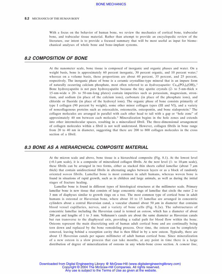
At the micron scale and above, bone tissue is a hierarchical composite (Fig. 8.1). At the lowest level(≈0.1-µm scale), it is a composite of mineralized collagen fibrils. At the next level (1- to 10-µm scale),these fibrils can be arranged in two forms, either as stacked thin sheets called lamellae (about 7 µmthick) that contain unidirectional fibrils in alternating angles between layers or as a block of randomlyoriented woven fibrils. Lamellar bone is most common in adult humans, whereas woven bone isfound in situations of rapid growth, such as in children and large animals, as well as during the initialstages of fracture healing.
Lamellar bone is found in different types of histological structures at the millimeter scale. Primarylamellar bone is new tissue that consists of large concentric rings of lamellae that circle the outer 2 to3 mm of diaphyses similar to growth rings on a tree. The most common type of cortical bone in adulthumans is osteonal or Haversian bone, where about 10 to 15 lamellae are arranged in concentriccylinders about a central Haversian canal, a vascular channel about 50 µm in diameter that containsblood vessel capillaries, nerves, and a variety of bone cells (Fig. 8.2a). The substructures ofconcentric lamellae including the Haversian canal is termed an osteon, which has a diameter of about200 µm and lengths of 1 to 3 mm. Volkmann’s canals are about the same diameter as Haversian canalsbut run transverse to the diaphyseal axis, providing a radial path for blood flow within the bone.Osteons represent the main discretizing unit of human adult cortical bone and are continually beingtorn down and replaced by the bone remodeling process. Over time, the osteon can be completelyremoved, leaving behind a resorption cavity that is then filled in by a new osteon. Typically, there areabout 13 Haversian canals per square millimeter of adult human cortical bone. Since mineralizationof a new osteon is a slow process that can take months, at any point in time there is a largedistribution of degree of mineralization of osteons in any whole-bone cross section. A cement line,
8.2 COMPOSITION OF BONE
8.3 BONE AS A HIERARCHICAL COMPOSITE MATERIAL
Downloaded from Digital Engineering Library @ McGraw-Hill (www.digitalengineeringlibrary.com)Copyright © 2004 The McGraw-Hill Companies. All rights reserved.
Any use is subject to the Terms of Use as given at the website.
BONE MECHANICS
BONE MECHANICS 8.3
which is a thin layer of calcified mucopolysaccharides with very little collagen and low mineralcontent,7 remains around the perimeter of each newly formed osteon. The cement line is thought torepresent a weak interface between the osteon and the surrounding interstitial bone.8 These weakinterfaces are thought to improve the fatigue properties of cortical bone by providing avenues fordissipation of energy during crack propagation.7
The bone matrix that comprises lamellar and woven bone contains another level of porosity onthe order of 5 to 10 µm that is associated with the bone cells (see Fig. 8.2a, b, c). Osteocytes, the mostcommon type of bone cell, are surrounded by a thin layer of extracellular fluid within smallellipsoidal holes (5 µm minor diameter, 7 to 8 µm major diameter) called lacunae, of which there areabout 25,000 per µm3 in bone tissue. The lacunae are generally arranged along the interfaces betweenlamellae. However, the lacunae also have a lower-scale porosity associated with them. Each osteocytehas dendritic processes that extend from the cell through tiny channels (≈0.5 µm diameter, 3 to 7 µmlong) called canaliculi to meet at cellular gap junctions with the processes of surrounding cells.*There are about 50 to 100 canaliculi per single lacuna and about 1 million per mm3 of bone.
At the highest hierarchical level (1 to 5 mm), there are two types of bone: cortical bone, whichcomes as tightly packed lamellar, Haversian, or woven bone; and trabecular bone, which comes as ahighly porous cellular solid. In the latter, the lamellae are arranged in less well-organized “packets”to form a network of rods and plates about 100 to 300 µm thick interspersed with large marrowspaces. Many common biological materials, such as wood and cork, are cellular solids.9
The distinction between cortical and trabecular bone is most easily made based on porosity.Cortical bone can be defined as bone tissue that has a porosity P of less than about 30 percent or,equivalently, a volume fraction Vf of greater than about 0.70 (Vf = 1 - P). Volume fraction is the ratioof the volume of actual bone tissue to the bulk volume of the specimen. In the field of bonemechanics, porosity measures usually ignore the presence of lacunae and canaliculi. Porosity of adult
FIGURE 8.1 The four levels of bone microstructure, from the level of mineralizedcollagen fibrils to cortical and trabecular bone. It is generally assumed that at theformer level, all bone is equal, although there may be subtle differences in thenature of the lamellar architecture and degree of mineralization between corticaland trabecular bone. (Adapted from Ref. 145.)
* Gap junctions are arrays of small pores in the cell membrane that make connections between the interiors of neighboringcells, allowing direct passage of small molecules such as ions from one cell to another.
Downloaded from Digital Engineering Library @ McGraw-Hill (www.digitalengineeringlibrary.com)Copyright © 2004 The McGraw-Hill Companies. All rights reserved.
Any use is subject to the Terms of Use as given at the website.
BONE MECHANICS

8.4 MECHANICS OF THE HUMAN BODY
human femoral cortical bone, for example, can vary from as low as 5 percent at age 20 up to almost30 percent above age 80.10 Porosity of trabecular bone can vary from 70 percent in the femoralneck11 up to about 95 percent in the elderly spine.12
Two other common measures of bone density in biomechanical studies are termed tissue andapparent densities. Tissue density ρtiss is defined as the ratio of mass to volume of the actual bonetissue. It is similar for cortical and trabecular bone, varies little in adult humans, and is about 2.0 g/cm3. Apparent density ρapp is defined as the ratio of the mass of bone tissue to the bulk volume of the
FIGURE 8.2 (a) Diagram of a sector of the shaft of a long bone showing the different types of cortical bone, trabecular bone, and the various channels.(From Figure 5–1d of Ref. 146.) (b) Environmental scanning electron micrograph of a fracture surface of a piece of cortical bone showing a fracturedlacuna at low (left) and high (right) magnifications. Note the elliptical shape of the lacuna and the multiple smaller canaliculi. (c) A schematicdepicting the interconnection of osteocytes (OC) via the cell processes that extend along the canaliculi and meet at gap junctions (GJ). Bone-liningcells (dormant osteoblasts, BLC) lie on each exposed bone surface, where osteoclasts (OCL) can be found removing bone as part of the ongoingremodeling process. A thin layer of bone fluid (BF) surrounds the cells and their processes.
Downloaded from Digital Engineering Library @ McGraw-Hill (www.digitalengineeringlibrary.com)Copyright © 2004 The McGraw-Hill Companies. All rights reserved.
Any use is subject to the Terms of Use as given at the website.
BONE MECHANICS
BONE MECHANICS 8.5
specimen, including the volume associated with the vascular channels and higher-level porosity.Volume fraction, tissue density, and apparent densities are related as follows:
Typically, mean values of apparent density of hydrated cortical bone are about 1.85 g/cm3, and thisdoes not vary much across anatomic sites or species. By contrast, the average apparent density oftrabecular bone depends very much on anatomic site. It is as low as 0.10 g/cm3 for the spine,13 about0.30 g/cm3 for the human tibia,14 and up to about 0.60 g/cm3 for the load-bearing portions of theproximal femur.11 After skeletal maturity (around ages 25 to 30), trabecular bone density decreasessteadily with aging, at a rate of about 6 percent per decade.15
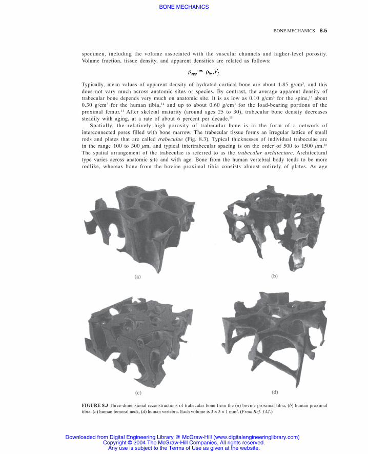
Spatially, the relatively high porosity of trabecular bone is in the form of a network ofinterconnected pores filled with bone marrow. The trabecular tissue forms an irregular lattice of smallrods and plates that are called trabeculae (Fig. 8.3). Typical thicknesses of individual trabeculae arein the range 100 to 300 µm, and typical intertrabecular spacing is on the order of 500 to 1500 µm.16
The spatial arrangement of the trabeculae is referred to as the trabecular architecture. Architecturaltype varies across anatomic site and with age. Bone from the human vertebral body tends to be morerodlike, whereas bone from the bovine proximal tibia consists almost entirely of plates. As age
FIGURE 8.3 Three-dimensional reconstructions of trabecular bone from the (a) bovine proximal tibia, (b) human proximaltibia, (c) human femoral neck, (d) human vertebra. Each volume is 3 × 3 × 1 mm3. (From Ref. 142.)
Downloaded from Digital Engineering Library @ McGraw-Hill (www.digitalengineeringlibrary.com)Copyright © 2004 The McGraw-Hill Companies. All rights reserved.
Any use is subject to the Terms of Use as given at the website.
BONE MECHANICS
8.6 MECHANICS OF THE HUMAN BODY
increases and volume fraction decreases, the architecture becomes increasingly rodlike, and these rodsbecome progressively thin and can be perforated. Quantification of trabecular architecture with theintent of understanding its role in the mechanical behavior of trabecular bone has been the subject ofintense research. In addition to calculating trabecular thickness, spacing, and surface-to-volume ratio,stereological and three-dimensional methods may be used to determine the mean orientation (maingrain axis) of the trabeculae, connectivity, and the degree of anisotropy.17 While earlier studies usedtwo-dimensional sections of trabecular bone to perform these architectural analyses,18,19 more recentinvestigations use three-dimensional reconstructions generated by micro-computed tomography andother high-resolution imaging techniques.16,20–22
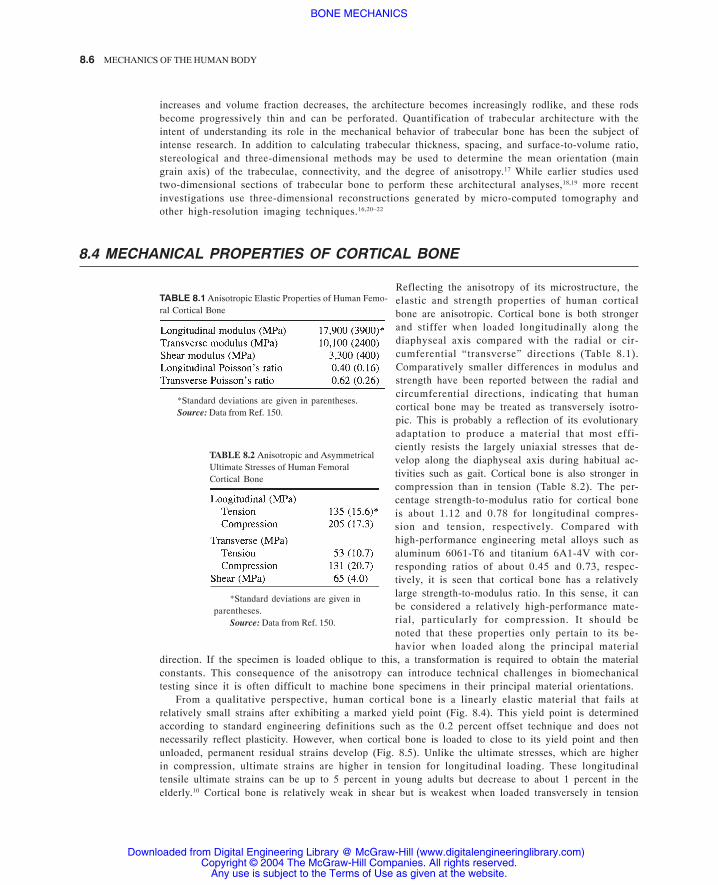
Reflecting the anisotropy of its microstructure, theelastic and strength properties of human corticalbone are anisotropic. Cortical bone is both strongerand stiffer when loaded longitudinally along thediaphyseal axis compared with the radial or cir-cumferential “transverse” directions (Table 8.1).Comparatively smaller differences in modulus andstrength have been reported between the radial andcircumferential directions, indicating that humancortical bone may be treated as transversely isotro-pic. This is probably a reflection of its evolutionaryadaptation to produce a material that most effi-ciently resists the largely uniaxial stresses that de-velop along the diaphyseal axis during habitual ac-tivities such as gait. Cortical bone is also stronger incompression than in tension (Table 8.2). The per-centage strength-to-modulus ratio for cortical boneis about 1.12 and 0.78 for longitudinal compres-sion and tension, respectively. Compared withhigh-performance engineering metal alloys such asaluminum 6061-T6 and titanium 6A1-4V with cor-responding ratios of about 0.45 and 0.73, respec-tively, it is seen that cortical bone has a relativelylarge strength-to-modulus ratio. In this sense, it canbe considered a relatively high-performance mate-rial, particularly for compression. It should benoted that these properties only pertain to its be-havior when loaded along the principal material
direction. If the specimen is loaded oblique to this, a transformation is required to obtain the materialconstants. This consequence of the anisotropy can introduce technical challenges in biomechanicaltesting since it is often difficult to machine bone specimens in their principal material orientations.
From a qualitative perspective, human cortical bone is a linearly elastic material that fails atrelatively small strains after exhibiting a marked yield point (Fig. 8.4). This yield point is determinedaccording to standard engineering definitions such as the 0.2 percent offset technique and does notnecessarily reflect plasticity. However, when cortical bone is loaded to close to its yield point and thenunloaded, permanent residual strains develop (Fig. 8.5). Unlike the ultimate stresses, which are higherin compression, ultimate strains are higher in tension for longitudinal loading. These longitudinaltensile ultimate strains can be up to 5 percent in young adults but decrease to about 1 percent in theelderly.10 Cortical bone is relatively weak in shear but is weakest when loaded transversely in tension
8.4 MECHANICAL PROPERTIES OF CORTICAL BONE
TABLE 8.1 Anisotropic Elastic Properties of Human Femo-ral Cortical Bone
*Standard deviations are given in parentheses.Source: Data from Ref. 150.
*Standard deviations are given inparentheses.
Source: Data from Ref. 150.
TABLE 8.2 Anisotropic and AsymmetricalUltimate Stresses of Human FemoralCortical Bone
Downloaded from Digital Engineering Library @ McGraw-Hill (www.digitalengineeringlibrary.com)Copyright © 2004 The McGraw-Hill Companies. All rights reserved.
Any use is subject to the Terms of Use as given at the website.
BONE MECHANICS

BONE MECHANICS 8.7
(see Table 8.2). An example of such loading is the circumferential or “hoop” stress that can developwhen large intramedullary tapered implants such as uncemented hip stems are impacted too far intothe diaphysis.
While it is often appropriate to assume average properties for cortical bone, as shown in Tables 8.1and 8.2, it may be necessary in some cases to account for the heterogeneity that can arise fromvariations in microstructural parameters such as porosity and percentage mineralization. Bothmodulus and ultimate stress can halve when porosity is increased from 5 to 30 percent10,23 (Fig. 8.6a).Small increases in percentage mineralization cause large increases in both modulus and strength (seeFig. 8.6b), and while this parameter does not vary much in adult humans,10 it can vary substantiallyacross species.24
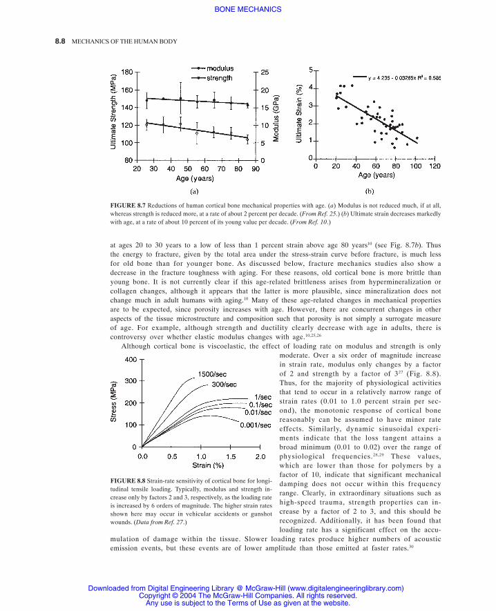
Aging also affects the mechanical properties of cortical bone. Tensile ultimate stress decreases at arate of approximately 2 percent per decade25 (Fig. 8.7a). Perhaps most important, tensile ultimate straindecreases by about 10 percent of its “young” value per decade, from a high of almost 5 percent strain
FIGURE 8.4 Typical stress-strain behavior for humancortical bone. The bone is stiffer in the longitudinaldirection, indicative of its elastic anisotropy. It is alsostronger in compression than in tension, indicative ofits strength asymmetry (modulus is the same in tensionand compression). (From Ref. 9.)
FIGURE 8.5 Creep response of cortical bone for three dif-ferent stress levels. When a low stress is applied to thebone, the strain remains constant over time, and there is nopermanent deformation after unloading. For stresses justbelow yield, strains increase with time at a constant rate,and a small permanent deformation exists after unloading.As the magnitude of the stress is increased, the rate ofcreep increases, and a larger permanent deformation existsafter unloading. (From Ref. 108.)
FIGURE 8.6 (a) Dependence of the ultimate tensile stress of human cortical bone on volume fraction (expressed as a percentage).Ages of the specimens were in the range 20 to 100 years. (From Ref. 10.) (b) Modulus versus calcium content (in mg/gm ofdehydrated bone tissue) for cortical bone taken from 18 different species. (From Ref. 24.)
Downloaded from Digital Engineering Library @ McGraw-Hill (www.digitalengineeringlibrary.com)Copyright © 2004 The McGraw-Hill Companies. All rights reserved.
Any use is subject to the Terms of Use as given at the website.
BONE MECHANICS
8.8 MECHANICS OF THE HUMAN BODY
at ages 20 to 30 years to a low of less than 1 percent strain above age 80 years10 (see Fig. 8.7b). Thusthe energy to fracture, given by the total area under the stress-strain curve before fracture, is much lessfor old bone than for younger bone. As discussed below, fracture mechanics studies also show adecrease in the fracture toughness with aging. For these reasons, old cortical bone is more brittle thanyoung bone. It is not currently clear if this age-related brittleness arises from hypermineralization orcollagen changes, although it appears that the latter is more plausible, since mineralization does notchange much in adult humans with aging.10 Many of these age-related changes in mechanical propertiesare to be expected, since porosity increases with age. However, there are concurrent changes in otheraspects of the tissue microstructure and composition such that porosity is not simply a surrogate measureof age. For example, although strength and ductility clearly decrease with age in adults, there iscontroversy over whether elastic modulus changes with age.10,25,26
Although cortical bone is viscoelastic, the effect of loading rate on modulus and strength is onlymoderate. Over a six order of magnitude increasein strain rate, modulus only changes by a factorof 2 and strength by a factor of 327 (Fig. 8.8).Thus, for the majority of physiological activitiesthat tend to occur in a relatively narrow range ofstrain rates (0.01 to 1.0 percent strain per sec-ond), the monotonic response of cortical bonereasonably can be assumed to have minor rateeffects. Similarly, dynamic sinusoidal experi-ments indicate that the loss tangent attains abroad minimum (0.01 to 0.02) over the range ofphysiological frequencies.28,29 These values,which are lower than those for polymers by afactor of 10, indicate that significant mechanicaldamping does not occur within this frequencyrange. Clearly, in extraordinary situations such ashigh-speed trauma, strength properties can in-crease by a factor of 2 to 3, and this should berecognized. Additionally, it has been found thatloading rate has a significant effect on the accu-
mulation of damage within the tissue. Slower loading rates produce higher numbers of acousticemission events, but these events are of lower amplitude than those emitted at faster rates.30
FIGURE 8.7 Reductions of human cortical bone mechanical properties with age. (a) Modulus is not reduced much, if at all,whereas strength is reduced more, at a rate of about 2 percent per decade. (From Ref. 25.) (b) Ultimate strain decreases markedlywith age, at a rate of about 10 percent of its young value per decade. (From Ref. 10.)
FIGURE 8.8 Strain-rate sensitivity of cortical bone for longi-tudinal tensile loading. Typically, modulus and strength in-crease only by factors 2 and 3, respectively, as the loading rateis increased by 6 orders of magnitude. The higher strain ratesshown here may occur in vehicular accidents or gunshotwounds. (Data from Ref. 27.)
Downloaded from Digital Engineering Library @ McGraw-Hill (www.digitalengineeringlibrary.com)Copyright © 2004 The McGraw-Hill Companies. All rights reserved.
Any use is subject to the Terms of Use as given at the website.
BONE MECHANICS
BONE MECHANICS 8.9
Multiaxial failure properties of cortical boneare not well understood, although it is clear thatsimple isotropic and symmetrical criteria suchas the von Mises are not capable of describingthe multiaxial strength properties of this tissue.The Tsai-Wu criterion, commonly used forfiber-reinforced composite materials, has beenapplied to cortical bone using both transverselyisotropic31 and orthotropic32 treatments. Thetransversely isotropic case works quite well foraxial-shear-loading configurations,31 but neitherthis case nor the orthotropic one has beenvalidated across the full range of multiaxialstresses. Regardless, this criterion accounts forthe difference in tensile and compressivestrengths, as well as the low shear strength withrespect to the tensile strength, and in this senseis the most suitable criterion currently available.
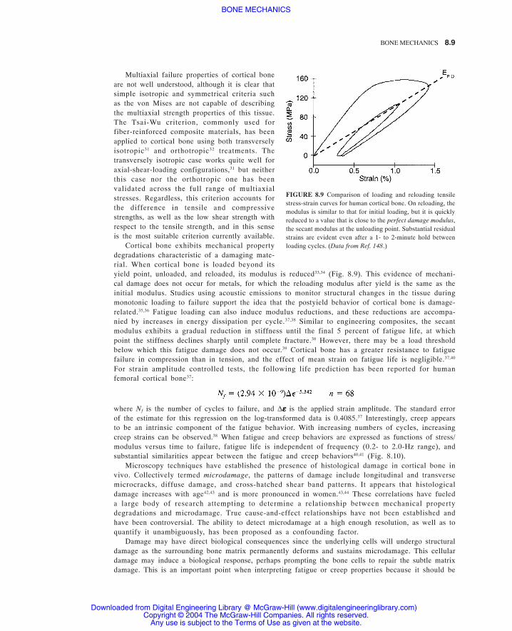
Cortical bone exhibits mechanical propertydegradations characteristic of a damaging mate-rial. When cortical bone is loaded beyond itsyield point, unloaded, and reloaded, its modulus is reduced33,34 (Fig. 8.9). This evidence of mechani-cal damage does not occur for metals, for which the reloading modulus after yield is the same as theinitial modulus. Studies using acoustic emissions to monitor structural changes in the tissue duringmonotonic loading to failure support the idea that the postyield behavior of cortical bone is damage-related.35,36 Fatigue loading can also induce modulus reductions, and these reductions are accompa-nied by increases in energy dissipation per cycle.37,38 Similar to engineering composites, the secantmodulus exhibits a gradual reduction in stiffness until the final 5 percent of fatigue life, at whichpoint the stiffness declines sharply until complete fracture.38 However, there may be a load thresholdbelow which this fatigue damage does not occur.39 Cortical bone has a greater resistance to fatiguefailure in compression than in tension, and the effect of mean strain on fatigue life is negligible.37,40
For strain amplitude controlled tests, the following life prediction has been reported for humanfemoral cortical bone37:
where Nf is the number of cycles to failure, and ∆εεεεε is the applied strain amplitude. The standard errorof the estimate for this regression on the log-transformed data is 0.4085.37 Interestingly, creep appearsto be an intrinsic component of the fatigue behavior. With increasing numbers of cycles, increasingcreep strains can be observed.38 When fatigue and creep behaviors are expressed as functions of stress/modulus versus time to failure, fatigue life is independent of frequency (0.2- to 2.0-Hz range), andsubstantial similarities appear between the fatigue and creep behaviors40,41 (Fig. 8.10).
Microscopy techniques have established the presence of histological damage in cortical bone invivo. Collectively termed microdamage, the patterns of damage include longitudinal and transversemicrocracks, diffuse damage, and cross-hatched shear band patterns. It appears that histologicaldamage increases with age42,43 and is more pronounced in women.43,44 These correlations have fueleda large body of research attempting to determine a relationship between mechanical propertydegradations and microdamage. True cause-and-effect relationships have not been established andhave been controversial. The ability to detect microdamage at a high enough resolution, as well as toquantify it unambiguously, has been proposed as a confounding factor.
Damage may have direct biological consequences since the underlying cells will undergo structuraldamage as the surrounding bone matrix permanently deforms and sustains microdamage. This cellulardamage may induce a biological response, perhaps prompting the bone cells to repair the subtle matrixdamage. This is an important point when interpreting fatigue or creep properties because it should be
FIGURE 8.9 Comparison of loading and reloading tensilestress-strain curves for human cortical bone. On reloading, themodulus is similar to that for initial loading, but it is quicklyreduced to a value that is close to the perfect damage modulus,the secant modulus at the unloading point. Substantial residualstrains are evident even after a 1- to 2-minute hold betweenloading cycles. (Data from Ref. 148.)
Downloaded from Digital Engineering Library @ McGraw-Hill (www.digitalengineeringlibrary.com)Copyright © 2004 The McGraw-Hill Companies. All rights reserved.
Any use is subject to the Terms of Use as given at the website.
BONE MECHANICS

8.10 MECHANICS OF THE HUMAN BODY
realized that no biological healing can occur during in vitro experiments. Thus the preceding fatiguecharacteristics are best considered as lower bounds on the in vivo fatigue life (see Fig. 8.10). It isunlikely that high-cycle (low-stress) fatigue failure occurs in vivo since the resulting fatigue damagewould be healed biologically before large enough cracks could develop that would cause overtfracture of the bone. However, it should also be noted that the increase in porosity associated with theinitial stages of the bone remodeling process may actually weaken the bone tissue even as the processattempts to strengthen it.
Fracture mechanics has been applied to cortical bone to determine its resistance to crack initiationand propagation. Various experimental techniques involving single-edge-notch (SEN), center-notched-cylindrical (CNC), and compact-tension (CT) specimens have been used to measure criticalstress intensity factor Kc and critical energy release rate Gc. Size requirements of standard fracturetoughness tests cannot be strictly met due to limitations on the size of available human tissue.Therefore, experimentally determined values of fracture toughness depend on specimen geometryand do not reflect a true material property. Additionally, plane-strain conditions and the associatedrelationships between Kc and Gc cannot be assumed. Theoretical developments45 and tests on largerbones (see Ref. 46 for review), such as bovine tibiae, have been used to determine correction factorsthat are used to account for specimen geometry. Comparisons of reported values should be made withcare because some studies do not attempt to correct for specimen geometry but rather report valuesin a comparative sense only consistent with the specific study.
Average values for Kc and Gc range from 2 to 6 MNm-3/2 and 50 to over 1000 N/m, respectively,for specimens oriented such that the crack propagated along the longitudinal axis of the long bone
FIGURE 8.10 Fatigue and creep behaviors of human corticalbone versus time to failure. For fatigue loading, the ordinate onthis graph can be converted to number of cycles by multiply-ing the time to failure by the frequency, which is typically onecycle per second for normal walking. Note that both creep andfatigue resistance are lower in tension, consistent with mono-tonic behavior. (Data from Refs. 37 and 41.)
TABLE 8.3 Fracture Toughness Values per Anatomic Site for Human Cor-tical Bone
Source: Data from Ref. 45.
Downloaded from Digital Engineering Library @ McGraw-Hill (www.digitalengineeringlibrary.com)Copyright © 2004 The McGraw-Hill Companies. All rights reserved.
Any use is subject to the Terms of Use as given at the website.
BONE MECHANICS

BONE MECHANICS 8.11
(Table 8.3). These values are similar, for example, to those of polystyrene. This orientation has beenfound to be the weaker in terms of crack resistance relative to the transverse orientation (ratio of1:1.75 for bovine bone46). Fracture toughness decreases with age in the diaphyses of long bones26,47 ata rate of 4.1 percent per decade in the femoral shaft.26 Fracture toughness in mode II loading can begreater by as much as fivefold than that in mode I.45,48 The most significant factors that are correlatedwith fracture toughness are age and a variety of porosity-related measures (including apparentdensity, water content, and osteon density).47,48
A number of studies have developed micromechanical models for the elastic and strengthproperties of cortical bone. This work has been motivated by observations that changes in themechanical properties with age and disease are accompanied by alterations in the tissuemicrostructure.49 While it is generally agreed on that bone behaves mechanically as a compositematerial, the complex hierarchy of composite structure has led to disagreements over which scale orscales are most relevant for a particular aspect of the mechanical behavior. For instance, ultrastructuralmodels have focused on the role of the mineral phase as reinforcement for the collagen matrix,50,51
whereas macrostructural models have treated the Haversian systems as fibers embedded in a matrix ofcement lines and interstitial lamellae.52–54 On an intermediate scale, the individual mineralized collagenfibers have been modeled as reinforcement for the noncollagenous mineralized matrix.55,56 Stillanother class of models has used a hierarchical approach to synthesize two or more of these differentlength scales.57–59 These modeling efforts rely extensively on accurate information regarding theconstituent and interface properties. Obtaining this information has proven challenging not onlybecause of the extremely small scale involved but also because, unlike the case with engineeringcomposites, isolating the different phases of bone tissue often involves processes that may alter theproperties being measured.60 Recent studies using nanoindentation,61,62 for example, have begun toaddress the issue of scale by providing the ability to measure the elastic modulus and microhardnessof various microstructures within cortical tissue. Thus efforts are ongoing to developmicromechanical constitutive models for cortical bone.
It is also of keen interest to determine the role of mechanical stimuli on bone cells and theinteraction between cell biology and bone mechanical properties. Biological processes that are activethroughout an individual’s lifetime can alter the bone tissue on many scales. It has often beensuggested that osteocytes act as sensors of mechanical loading and initiators of the bone-adaptationprocesses.63–66 Whether these processes add or remove bone, or whether they are activated at all, isthought to depend on the level of mechanical loading67 and on the presence or absence of tissuedamage.1,2 Many studies have suggested that strain or strain rate is the appropriate measure ofmechanical stimulus,68–70 although others have used stress or strain energy.71,72 In addition, othercharacteristics of the mechanical loading, such as mode, direction, frequency, duration, anddistribution, have also been identified as important. Using this collective empirical evidence, severaltheories of mechanical adaptation have been proposed (see Ref. 73 for a comprehensive treatment ofthis topic). When applied to finite-element models of whole bones, some of these theories have beenable to predict the density pattern of the trabecular bone in the proximal femur74,75 and the loss ofbone in the regions surrounding a hip implant.76 This remains an area of active research sinceexperimentally validated mechanobiological constitutive relations of the remodeling process have yetto be developed.
Although trabecular bone—also referred to as cancellous or spongy bone—is nonlinearly elastic evenat small strains,77 it is most often modeled as linearly elastic until yielding. It yields in compression atstrains of approximately 1 percent, after which it can sustain large deformations (up to 50 percentstrain) while still maintaining its load-carrying capacity. Thus trabecular bone can absorb substantialenergy on mechanical failure. A heterogeneous porous cellular solid, trabecular bone has anisotropicmechanical properties that depend on the porosity of the specimen as well as the architecturalarrangement of the individual trabeculae. Its apparent (whole-specimen) level properties also depend
8.5 MECHANICAL PROPERTIES OF TRABECULAR BONE
Downloaded from Digital Engineering Library @ McGraw-Hill (www.digitalengineeringlibrary.com)Copyright © 2004 The McGraw-Hill Companies. All rights reserved.
Any use is subject to the Terms of Use as given at the website.
BONE MECHANICS

8.12 MECHANICS OF THE HUMAN BODY
on the tissue-level material properties of the individual trabeculae. An overwhelming portion oftrabecular bone mechanics research has been devoted to improving our understanding of the relativecontributions and interplay of porosity, architecture, and tissue properties in the apparent levelproperties.
The elastic and strength properties of trabecular bone display substantial heterogeneity withrespect to donor age and health, anatomic site, loading direction (with respect to the principalorientation of the trabeculae), and loading mode. Both modulus and strength decrease with age,falling approximately 10 percent per decade15,78 (Fig. 8.11). Pathologies such as osteoporosis,osteoarthritis, and bone cancer are also known to affect mechanical properties.79,80 Young’s moduluscan vary 100-fold within a single epiphysis81 and 3-fold depending on loading direction.82–85
Typically, the modulus of human trabecular bone is in the range 10 to 3000 MPa depending on thepreceding factors; strength, which is linearly and strongly correlated with modulus,11,81,82 is generally2 orders of magnitude lower than modulus and is usually in the range 0.1 to 30 MPa.
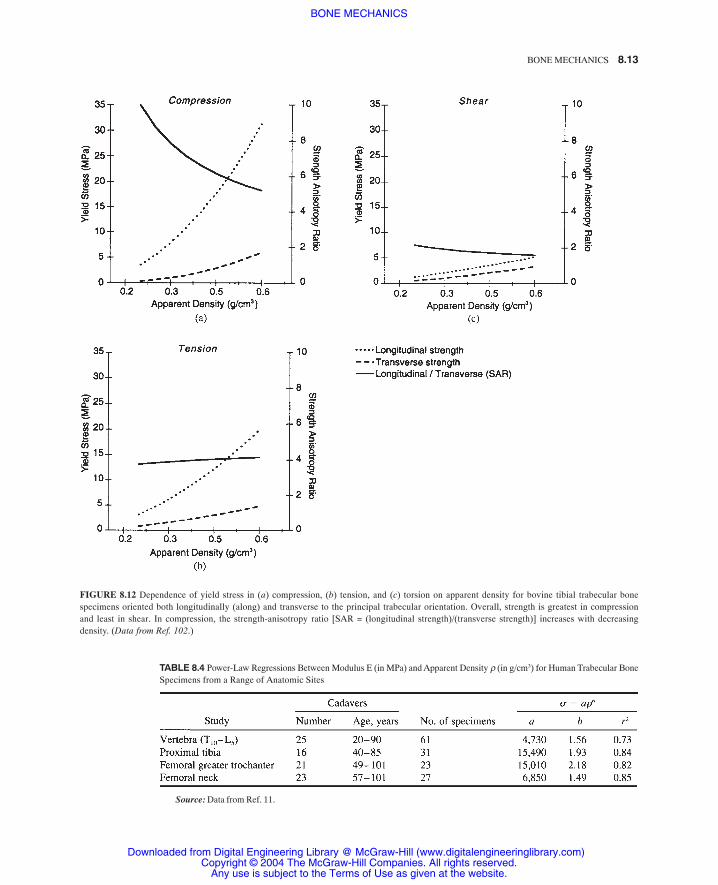
In compression, the anisotropy of trabecular bone strength increases with age78 and decreasingdensity (Fig. 8.12). The strength also depends on loading mode, being highest in compression andlowest in shear.86,87 Ratios of compressive to tensile strength and compressive to shear strength are notconstant but rather depend on modulus87 and density (see Fig. 8.12). Both modulus and strengthdepend heavily on apparent density, yet these relationships vary for different types of trabecular bonebecause of the anatomic site-, age-, and disease-related variations in trabecular architecture. Linearand power-law relationships* can be used to describe the dependence of modulus and compressivestrength on apparent density (Tables 8.4 and 8.5), with typical coefficients of determination (r2
values) in the range 0.5 to 0.9.Interestingly, the failure (yield and ultimate) strains of human trabecular bone have only a weak
dependence, if any, on apparent density and modulus.11,13,78,88–91 A recent study designed to test forintersite differences found that yield strains were approximately uniform within anatomic sites, withstandard deviations on the order of one-tenth the mean value, but mean values could vary acrosssites11 (Fig. 8.13). Thus, for analysis purposes, yield strains can be considered constant within sites butheterogeneous across sites. Regardless of anatomic site, however, yield stains are higher incompression than in tension.11 Ultimate strains are typically in the range of 1.0 to 2.5 percent.Evidence from experiment on bovine bone indicates that yield strains are also isotropic92,93 despitesubstantial anisotropy of modulus and strength.
FIGURE 8.11 Dependence of ultimate stress on age for trabecu-lar bone from the human vertebra and femur. For both anatomicsites, strength decreases approximately 10 percent per decade.(Data from Refs. 15 and 149.)
* Differences in the predictive power between the various linear and power laws are usually negligible within a singleanatomic site because the range of apparent density exhibited by trabecular bone is less than 1 order of magnitude.
Downloaded from Digital Engineering Library @ McGraw-Hill (www.digitalengineeringlibrary.com)Copyright © 2004 The McGraw-Hill Companies. All rights reserved.
Any use is subject to the Terms of Use as given at the website.
BONE MECHANICS
BONE MECHANICS 8.13
FIGURE 8.12 Dependence of yield stress in (a) compression, (b) tension, and (c) torsion on apparent density for bovine tibial trabecular bonespecimens oriented both longitudinally (along) and transverse to the principal trabecular orientation. Overall, strength is greatest in compressionand least in shear. In compression, the strength-anisotropy ratio [SAR = (longitudinal strength)/(transverse strength)] increases with decreasingdensity. (Data from Ref. 102.)
Source: Data from Ref. 11.
TABLE 8.4 Power-Law Regressions Between Modulus E (in MPa) and Apparent Density ρ (in g/cm3) for Human Trabecular BoneSpecimens from a Range of Anatomic Sites
Downloaded from Digital Engineering Library @ McGraw-Hill (www.digitalengineeringlibrary.com)Copyright © 2004 The McGraw-Hill Companies. All rights reserved.
Any use is subject to the Terms of Use as given at the website.
BONE MECHANICS

8.14 MECHANICS OF THE HUMAN BODY
The advent of high-resolution finite-element modeling94 has led to enormous progress in deter-mining elastic stiffness matrices, multiaxial failure behavior, and as will be seen later, trabecular tissueproperties. Finite-element models of individual specimens, developed using microcomputed tomogra-phy95,96 and other types of microscopic imaging,17,97 have been used to compute the full set of elasticconstants for specimens from multiple anatomic sites. Results indicate that trabecular bone can beconsidered orthotropic98,99 or, in some cases, transversely orthotropic.100 Poisson’s ratios, which aredifficult to measure experimentally for trabecular bone, range from 0.03 to 0.60.22,99
Given the complexity of in vivo loading conditions, there is a need to develop a multiaxial failurecriterion for trabecular bone. Results have been reported for bovine bone only.101–103 These studiesindicate that the von Mises criterion does not work well and that expression of the criterion in terms ofstrain (or nondimensional measures of stress divided by modulus) greatly simplifies the mathematicalform of the criterion since it eliminates the dependence of the criterion on specimen density. Criteriasuch as the Tsai-Wu criterion have only a limited ability to describe multiaxial failure of trabecular bone
TABLE 8.5 Power-Law Regressions Between Ultimate Stress σ (in MPa) and Apparent Density ρ (in g/cm3) for CompressiveLoading of Human Trabecular Bone Specimens from a Range of Anatomic Sites
FIGURE 8.13 Mean yield strain per anatomic site in both compression and ten-sion. Error bars indicate 1 standard deviation. Yield strain is only weakly depen-dent on apparent density for four of the groups, as indicated by the Pearson corre-lation coefficient r in the bar. Compressive yield strains are higher than tensileyield strains for each anatomic site. Intrasite scatter is on the order of one-tenth themean values. (From Ref. 11.)
Downloaded from Digital Engineering Library @ McGraw-Hill (www.digitalengineeringlibrary.com)Copyright © 2004 The McGraw-Hill Companies. All rights reserved.
Any use is subject to the Terms of Use as given at the website.
BONE MECHANICS

BONE MECHANICS 8.15
for arbitrary stress states. Coupling between normalstrengths in different directions (longitudinal versustransverse, for example) appears to be minimal.104 Atpresent, it is recommended for multiaxial failureanalysis that the tensile-compressive-shear-strengthasymmetry be recognized, as well as the strengthanisotropy. If properties are not available for a specificsite under analysis, failure strains should be used fromsites that have a similar density range and architecture.
When trabecular bone is loaded in compressionbeyond its elastic range, unloaded, and reloaded, itdisplays loss of stiffness and development of perma-nent strains105 (Fig. 8.14). In particular, it reloads withan initial modulus close to its intact Young’s modulusbut then quickly loses stiffness. The residual modulusis statistically similar to the perfect-damage modulus (asecant modulus from the origin to the point of unload-ing). In general, the reloading stress-strain curve tendsto reload back toward the extrapolated envelope of theoriginal curve. Phenomenologically, trabecular bonetherefore exhibits elements of both plasticity and damage. The magnitudes of stiffness loss % ∆E andresidual strain εRESIDUAL for human vertebral trabecular bone are highly correlated with the applied strainεTOTAL (all expressed in percent) in the initial overload:
Also, at any given strain, modulus on reloading is reduced more than strength:
where %∆S is the percentage change in strength, and ρAPP is the apparent density. These relations arefor applied strains on the order of 1 to 5 percent only; residual behavior for much greater appliedstrains has not yet been reported. The percent modulus reductions and residual strains do not dependon volume fraction because similar trends have been reported for bovine bone, which is much moredense and platelike.106,107 Furthermore, the trabecular behavior is qualitatively similar to that forcortical bone loaded in tension.34,108 This suggests that the dominant physical mechanisms for damagebehavior act at the nanometer scale of the collagen and hydroxyapatite.
Regarding time-dependent behavior, trabecular bone is only slightly viscoelastic when tested invitro, with both compressive strength and modulus being related to strain rate raised to a power of0.06.109,110 The stiffening effect of marrow is negligible except at very high strain rates (10 strains/s),although there is evidence that the constraining effects of a cortical shell may allow hydraulicstiffening of whole bones in vivo under dynamic loads.111 Minor stress relaxation has been shown tooccur112 and depends on the applied strain level,113 indicating that human trabecular bone isnonlinearly viscoelastic. Little else is known about its time-dependent properties, including creep andfatigue for human bone. Fatigue and creep studies on bovine bone have revealed the followingpower law relations between compressive normalized stress (stress divided by modulus, expressed inpercent) and time to failure (frequency for fatigue loading was 2 Hz):
Fatigue:
Creep:
Failure in these experiments was defined by a 10 percent reduction in secant modulus compared withthe initial Young’s modulus.
FIGURE 8.14 Compressive load-unload-reload behav-ior of human vertebral trabecular bone. Similar to corti-cal bone tested in tension, an initial overload causesresidual strains and a reloading curve whose modulusquickly reduces from a value similar to the intact modu-lus to a value similar to the perfect damage modulus.(From Ref. 105.)
Downloaded from Digital Engineering Library @ McGraw-Hill (www.digitalengineeringlibrary.com)Copyright © 2004 The McGraw-Hill Companies. All rights reserved.
Any use is subject to the Terms of Use as given at the website.
BONE MECHANICS
8.16 MECHANICS OF THE HUMAN BODY
It should be noted that the in vitro mechanical test methods most often used to date on trabecularbone are known to introduce substantial errors in the data as a result of the porous anisotropictrabecular structure. Damage preferentially occurs at the ends of machined specimens when they arecompressed between loading platens due to disruption of the trabecular network at the specimenboundaries.114–116 In addition, friction at the platen-specimen interface creates a triaxial stress state thatmay result in an overestimation of modulus.114,115,117 If strains are computed from the relativedisplacement of the platens, substantial systematic and random errors in modulus on the order of20 ± 12 percent can occur.118 Strength and failure strain data are also affected.119,120 The trabecularanisotropy indicates that experimental measurements of the orthotropic elastic constants should bedone in the principal material coordinate system. Since off-axis moduli are functions of multiplematerial elastic constants, substantial errors can be introduced from misalignment121 if not correctedfor. Much of the data in the literature have been obtained in various types of off-axis configurations,since specimens are often machined along anatomic rather than material axes. The difficulty ininterpreting these off-axis measurements is heightened when intersite and interstudy comparisons areattempted. For all these reasons, and since the in vitro test boundary conditions rarely replicate thosein vivo, interpretation and application of available data must be done with care.
An important development for structural analysis of whole bones is the use of quantitativecomputed tomography (QCT) to generate anatomically detailed models of whole bone122–127 or bone-implant128,129 systems. At the basis of such technology is the ability to use QCT to noninvasivelypredict the apparent density and mechanical properties of trabecular bone. Some studies havereported excellent predictions (r2 ≥ 0.75) of modulus and strength from QCT density information forvarious anatomic sites.82,89,130,131 Since the mechanical properties depend on volume fraction andarchitecture, it is important to use such relations only for the sites for which they were developed;otherwise, substantial errors can occur. Also, since QCT data do not describe any anisotropicproperties of the bone, trabecular bone is usually assumed to be isotropic in whole-bone and bone-implant analyses. Typically, in such structural analyses, cortical bone properties are not assigned fromQCT since it does not have the resolution to discern the subtle variations in porosity andmineralization that cause variations in cortical properties. In these cases, analysts typically assignaverage properties, sometimes transversely isotropic, to the cortical bone.
While most biomechanical applications at the organ level require knowledge of material properties atthe scales described earlier, there is also substantial interest in the material properties of trabeculartissue because this information may provide additional insight into diseases such as osteoporosis anddrug treatments designed to counter such pathologies. Disease- or drug-related changes could be mostobvious at the tissue rather than apparent or whole-bone level, yet only recently have researchersbegun to explore this hypothesis. This is so primarily because the irregular shape and small size ofindividual trabecula present great difficulties in measuring the tissue material properties.
Most of the investigations of trabecular tissue properties have addressed elastic behavior. Some ofthe earlier experimental studies concluded that the trabecular tissue has a modulus on the order of 1to 10 GPa.132–135 Later studies using combinations of computer modeling of the whole specimen andexperimental measures of the apparent modulus have resulted in a wide range of estimated values forthe effective modulus of the tissue (Table 8.6) such that the issue became controversial. However,studies using ultrasound have concluded that values for elastic modulus are about 20 percent lowerthan for cortical tissue.136,137 This has been supported by results from more recent nanoindentationstudies.61,62,138 The combined computer-experiment studies that successfully eliminated end artifacts inthe experimental protocols also found modulus values more typical of cortical bone than the muchlower values from the earlier studies.139 Thus an overall consensus is emerging that the elastic modulusof trabecular tissue is similar to, and perhaps slightly lower than, that of cortical bone.140 Regardingfailure behavior, it appears from the results of combined computational experimental studies that the
8.6 MECHANICAL PROPERTIES OF TRABECULAR TISSUEMATERIAL
Downloaded from Digital Engineering Library @ McGraw-Hill (www.digitalengineeringlibrary.com)Copyright © 2004 The McGraw-Hill Companies. All rights reserved.
Any use is subject to the Terms of Use as given at the website.
BONE MECHANICS

BONE MECHANICS 8.17
yield strains for trabecular tissue are similar to those for cortical bone, being higher in compressionthan in tension.139 Experimental studies on machined microbeams have shown that the fatigue strengthof trabecular tissue is lower than that of cortical tissue.141
The field of bone mechanics has evolved to a very sophisticated level where mechanical properties ofcortical and trabecular bone are available for many anatomic sites. Studies have also reported on theeffects of bone density, aging, and disease on these properties, enabling researchers to perform highlydetailed specimen-specific analyses on whole bone and bone-implant systems. We have reviewed heremuch of that literature. Our focus was on data for human bone, although we reported bovine datawhen no other reliable data were available. One important theme in bone mechanics is to account forthe substantial heterogeneity in bone properties that can occur for both cortical and trabecular bone,particularly for the latter. The heterogeneity results from aging, disease, and natural interindividualbiological variation and thus occurs longitudinally and cross-sectionally in populations. The hetero-geneity also exists spatially within bones. Successful structural analysis depends on appreciation ofthis heterogeneity so that appropriate material properties are used for the analysis at hand. Improvedunderstanding of the micromechanics and damage behaviors of bone is also leading to unique insightinto mechanisms of disease and their treatment as well as biological remodeling and tissue engineer-ing. While a number of excellent texts are available for more detailed study of these topics and manyof those presented here,73,142–144 it is hoped that this review will provide a concise basis for practicalengineering analysis of bone.
Support is gratefully acknowledged from NIH (AR41481, AR43784), NSF (BES-9625030), and TheMiller Institute for Basic Research in Science, Berkeley, Calif.
1. Burr, D. B., Forwood, M. R., Fyhrie, D. P., Martin, R. B., Schaffler, M. B., and Turner, C. H. (1997), Bone microdamage andskeletal fragility in osteoporotic and stress fractures, J. Bone Miner. Res. 12(1):6–15.
TABLE 8.6 Trabecular Tissue Moduli Using a Variety of Experimental and Computational Techniques
8.7 CONCLUDING REMARKS
ACKNOWLEDGMENTS
REFERENCES
Downloaded from Digital Engineering Library @ McGraw-Hill (www.digitalengineeringlibrary.com)Copyright © 2004 The McGraw-Hill Companies. All rights reserved.
Any use is subject to the Terms of Use as given at the website.
BONE MECHANICS
8.18 MECHANICS OF THE HUMAN BODY
2. Burr, D. B., Martin, R. B., Schaffler, M. B., and Radin, E. L. (1985), Bone remodeling in response to in vivo fatiguemicrodamage, J. Biomech. 18(3):189–200.
3. Gong, J. K., Arnold, J. S., and Cohn, S. H. (1964), Composition of trabecular and cortical bone, Anat. Rec. 149:325–332.
4. Lowenstam, H. A., and Weiner, S. (1989), On Biomineralization, Oxford University Press, New York.
5. Herring, G. (1972), The organic matrix of bone, in G. Bourne (ed.), The Biochemistry and Physiology of Bone, 2d ed., Vol.1, pp.127–189, Academic Press, New York.
6. Hodge, A. J., Petruska, J. A. (1963), Recent studies with the electron microscope on ordered aggregates of the tropocol-lagen molecule, in G. N. Ramachandran (ed.) Aspects of protein structure, Academic Press, London, pp 289–300.
7. Burr, D. B., Schaffler, M. B., and Frederickson, R. G. (1988), Composition of the cement line and its possible mechanicalrole as a local interface in human compact bone, J. Biomech. 21(11):939–945.
8. Lakes, R., and Saha, S. (1979), Cement line motion in bone, Science 204(4392):501–503.
9. Gibson, L. J., and Ashby, M. F. (1997), Cellular Solids: Structures & Properties, 2d ed. Pergamon Press, Oxford, U.K.
10. McCalden, R. W., McGeough, J. A., Barker, M. B., and Court-Brown, C. M. (1993), Age-related changes in the tensileproperties of cortical bone: The relative importance of changes in porosity, mineralization, and microstructure, J. BoneJoint Surg. 75A(8):1193–1205.
11. Morgan, E. F., and Keaveny, T. M., (2001), Dependence of yield strain of human trabecular bone on anatomic site,J. Biomech. 34(5):569–577.
12. Snyder, B. D., Piazza, S., Edwards, W. T., and Hayes, W. C. (1993), Role of trabecular morphology in the etiology of age-related vertebral fractures, Calcif. Tissue Int. 53S(1):S14-S22.
13. Kopperdahl, D. L., and Keaveny, T. M. (1998), Yield strain behavior of trabecular bone, J. Biomech. 31(7): 601–608.
14. Linde, F., Hvid, I., and Pongsoipetch, B. (1989), Energy absorptive properties of human trabecular bone specimens duringaxial compression, J. Orthop. Res. 7(3):432–439.
15. McCalden, R. W., McGeough, J. A., and Court-Brown, C. M. (1997), Age-related changes in the compressive strength ofcancellous bone: The relative importance of changes in density and trabecular architecture, J. Bone Joint Surg.79A(3):421–427.
16. Hildebrand, T., Laib, A., Müller, R., Dequeker, J., and Rüegsegger, P. (1999), Direct three-dimensional morphometricanalysis of human cancellous bone: microstructural data from spine, femur, iliac crest, and calcaneus, J. Bone Miner. Res.14(7):1167–1174.
17. Odgaard, A. (1997), Three-dimensional methods for quantification of cancellous bone architecture, Bone 20(4):315–328.
18. Mosekilde, L. (1989), Sex differences in age-related loss of vertebral trabecular bone mass and structure: Biomechanicalconsequences, Bone 10(6):425–432.
19. Whitehouse, W. J. (1974), The quantitative morphology of anisotropic trabecular bone, J. Microsc., 2:153–168.
20. Goldstein, S. A., Goulet, R., and McCubbrey, D. (1993), Measurement and significance of three-dimensional architectureto the mechanical integrity of trabecular bone, Calcif. Tissue Int. 53S(1):S127–S133.
21. Majumdar, S., Kothari, M., Augat, P., Newitt, D. C., Link, T. M., Lin, J. C., Lang, T., Lu, Y., and Genant, H. K. (1998), High-resolution magnetic resonance imaging: three-dimensional trabecular bone architecture and biomechanical properties,Bone 22(5):445–454.
22. Ulrich, D., Van Rietbergen, B., Laib, A., and Rueegsegger, P. (1999), The ability of three-dimensional structural indices toreflect mechanical aspects of trabecular bone, Bone 25(1):55–60.
23. Schaffler, M. B., and Burr, D. B., (1988), Stiffness of compact bone: Effects of porosity and density, J. Biomech.21(1):13–16.
24. Currey, J. D. (1988), The effect of porosity and mineral content on the Young’s modulus of elasticity of compact bone,J. Biomech. 21(2):131–139.
25. Burstein, A. H., Reilly, D. T., and Martens, M. (1976), Aging of bone tissue: Mechanical properties, J. Bone Joint Surg.58A(1):82–86.
26. Zioupos, P., and Currey, J. D. (1998), Changes in the stiffness, strength, and toughness of human cortical bone with age,Bone 22(1):57–66.
27. McElhaney, J. H., and Byars, E. F. (1965), Dynamic response of biological materials, in Proc. Amer. Soc. Mech. Eng., ASME65-WA/HUF-9:8, Chicago.
28. Lakes, R. S. (1982), Dynamical study of couple stress effects in human compact bone, J. Biomech. Eng. 104(1):6–11.
Downloaded from Digital Engineering Library @ McGraw-Hill (www.digitalengineeringlibrary.com)Copyright © 2004 The McGraw-Hill Companies. All rights reserved.
Any use is subject to the Terms of Use as given at the website.
BONE MECHANICS
BONE MECHANICS 8.19
29. Lakes, R. S., Katz, J. L., and Sternstein, S. S. (1979), Viscoelastic properties of wet cortical bone: I. Torsional and biaxialstudies, J. Biomech. 12(9):657–678.
30. Fischer, R. A., Arms, S. W., Pope, M. H., and Seligson, D. (1986), Analysis of the effect of using two different strain rateson the acoustic emission in bone, J. Biomech. 19(2):119–127.
31. Cezayirlioglu, H., Bahniuk, E., Davy, D. T., and Heiple, K. G. (1985), Anisotropic yield behavior of bone under combinedaxial force and torque, J. Biomech. 18(1):61–69.
32. Cowin, S. C. (1989), Bone Mechanics, CRC Press, Boca Raton, Fla.
33. Courtney, A. C., Hayes, W. C., and Gibson, L. J. (1996), Age-related differences in post-yield damage in human corticalbone: Experiment and model, J. Biomech. 29(11):1463–1471.
34. Fondrk, M. T., Bahniuk, E. H., and Davy, D. T. (1999), A damage model for nonlinear tensile behavior of cortical bone,J. Biomech. Eng. 121:533–541.
35. Wright, T. M., Vosburgh, F., and Burstein, A. H. (1981), Permanent deformation of compact bone monitored by acousticemission, J. Biomech. 14(6):405–409.
36. Zioupos, P., Currey, J. D., and Sedman, A. J. (1994), An examination of the micromechanics of failure of bone and antlerby acoustic emission tests and laser scanning confocal microscopy, Med. Eng. Phys. 16(3):203–212.
37. Carter, D. R., Caler, W. E., Spengler, D. M., and Frankel, V. H. (1981), Fatigue behavior of adult cortical bone: The influenceof mean strain and strain range, Acta Orthop. Scand. 52(5):481–490.
38. Pattin, C. A., Caler, W. E., and Carter, D. R. (1996), Cyclic mechanical property degradation during fatigue loading ofcortical bone, J. Biomech. 29(1):69–79.
39. Schaffler, M. B., Radin, E. L., and Burr, D. B. (1990), Long-term fatigue behavior of compact bone at low strain magnitudeand rate, Bone 11(5):321–326.
40. Carter, D. R., Caler, W. E., Spengler, D. M., and Frankel, V. H. (1981), Uniaxial fatigue of human cortical bone: The influenceof tissue physical characteristics, J. Biomech. 14(7):461–470.
41. Caler, W. E., and Carter, D. R. (1989), Bone creep-fatigue damage accumulation, J. Biomech. 22(6–7):625–635.
42. Frost, H. M. (1960), Presence of microscopic cracks in vivo in bone, Bull. Henry Ford Hosp. 8:27–35.
43. Schaffler, M. B., Choi, K., and Milgrom, C. (1995), Aging and matrix microdamage accumulation in human compact bone,Bone 17(6):521–525.
44. Norman, T. L., and Wang, Z. (1997), Microdamage of human cortical bone: Incidence and morphology in long bones, Bone20(4):375–379.
45. Brown, C. U., Yeni, Y. N., and Norman, T. L. (2000), Fracture toughness is dependent on bone location: A study of the femoralneck, femoral shaft, and the tibial shaft, J. Biomed. Mater. Res. 49(3):380–389.
46. Melvin, J. W. (1993), Fracture mechanics of bone, J. Biomech. Eng. 115(4B):549–554.
47. Yeni, Y. N., Brown, C. U., and Norman, T. L. (1998), Influence of bone composition and apparent density on fracturetoughness of the human femur and tibia, Bone 22(1):79–84.
48. Yeni, Y. N., Brown, C. U., Wang, Z., and Norman, T. L. (1997), The influence of bone morphology on fracture toughness ofthe human femur and tibia, Bone 21(5):453–459.
49. Landis, W. J. (1995), The strength of a calcified tissue depends in part on the molecular structure and organization of itsconstituent mineral crystals in their organic matrix, Bone 16(5):533–544.
50. Katz, J. L. (1971), Hard tissue as a composite material: I. Bounds on the elastic behavior, J. Biomech. 4(5):455–473.
51. Mammone, J. F., and Hudson, S. M. (1993), Micromechanics of bone strength and fracture, J. Biomech. 26(4–5):439–446.
52. Gottesman, T., and Hashin, Z. (1980), Analysis of viscoelastic behaviour of bones on the basis of microstructure,J. Biomech. 13(2):89–96.
53. Katz, J. L. (1981), Composite material models for cortical bone, in S. C. Cowin (ed.), Mechanical Properties of Bone:Proceedings of the Joint ASME-ASCE Applied Mechanics, Fluids Engineering, and Bioengineer ing Conference, Boulder,Colorado, June 22–24, 1981, Vol. 45, pp. 171–184, American Society of Mechanical Engineers, New York.
54. Krajcinovic, D., Trafimow, J., and Sumarac, D. (1987), Simple constitutive model for cortical bone, J. Biomech. 20(8):779–784.
55. Braidotti, P., Branca, F. P., and Stagni, L. (1997), Scanning electron microscopy of human cortical bone failure surfaces,J. Biomech. 30(2):155–162.
56. Pidaparti, R. M., Chandran, A., Takano, Y., and Turner, C. H. (1996), Bone mineral lies mainly outside collagen fibrils:Predictions of a composite model for osteonal bone, J. Biomech. 29(7):909–916.
Downloaded from Digital Engineering Library @ McGraw-Hill (www.digitalengineeringlibrary.com)Copyright © 2004 The McGraw-Hill Companies. All rights reserved.
Any use is subject to the Terms of Use as given at the website.
BONE MECHANICS
8.20 MECHANICS OF THE HUMAN BODY
57. Akiva, U., Wagner, H. D., and Weiner, S. (1998), Modelling the three-dimensional elastic constants of parallel-fibred andlamellar bone, J. Mater. Sci. 33(6):1497–1509.
58. Sasaki, N., Ikawa, T., and Fukuda, A. (1991), Orientation of mineral in bovine bone and the anisotropic mechanicalproperties of plexiform bone, J. Biomech. 24(1):57–61.
59. Wagner, H. D., and Weiner, S. (1992), On the relationship between the microstructure of bone and its mechanical stiffness,J. Biomech. 25(11):1311–1320.
60. McCutchen, C. W. (1975), Do mineral crystals stiffen bone by straitjacketing its collagen? J. Theor. Biol. 51(1):51–58.
61. Rho, J. Y., Tsui, T. Y., and Pharr, G. M. (1997), Elastic properties of human cortical and trabecular lamellar bone measuredby nanoindentation, Biomaterials 18(20):1325–1330.
62. Zysset, P. K., Guo, X. E., Hoffler, C. E., Moore, K. E., and Goldstein, S. A. (1999), Elastic modulus and hardness of corticaland trabecular bone lamellae measured by nanoindentation in the human femur, J. Biomech. 32(10):1005–1012.
63. Cowin, S. C., Moss-Salentijn, L., and Moss, M. L. (1991), Candidates for the mechanosensory system in bone, J. Biomech.Eng. 113(2):191–197.
64. Lanyon, L. E. (1993), Osteocytes, strain detection, bone modeling and remodeling, Calcif. Tissue Int. 53(Suppl. 1):S102-106; see also Discussion, pp. S106–107.
65. Marotti, G., Ferretti, M., Muglia, M. A., Palumbo, C., and Palazzini, S. (1992), A quantitative evaluation of osteoblast-osteocyte relationships on growing endosteal surface of rabbit tibiae, Bone 13(5):363–368.
66. Mullender, M. G., and Huiskes, R. (1995), Proposal for the regulatory mechanism of Wolff’s law, J. Orthop. Res. 13(4):503–512.
67. Frost, H. M. (1987), Bone “mass” and the “mechanostat”: A proposal, Anat. Rec. 219(1):1–9.
68. Biewener, A. A., Thomason, J., Goodship, A., and Lanyon, L. E. (1983), Bone stress in the horse forelimb during locomotionat different gaits: A comparison of two experimental methods, J. Biomech. 16(8):565–576.
69. Lanyon, L. E. (1984), Functional strain as a determinant for bone remodeling, Calcif. Tissue Int. 36(Suppl. 1):S56–61.
70. Turner, C. H., Owan, I., and Takano, Y. (1995), Mechanotransduction in bone: Role of strain rate, Am. J. Physiol. 269(3 Pt.1):E438–442.
71. Fyhrie, D. P., and Carter, D. R. (1986), A unifying principle relating stress to trabecular bone morphology, J. Orthop. Res.4(3):304–317.
72. Weinans, H., Huiskes, R., and Grootenboer, H. J. (1992) The behavior of adaptive bone-remodeling simulation models,J. Biomech. 25(12):1425–1441.
73. Martin, R. B., and Burr, D. B. (1989), Structure, Function, and Adaptation of Compact Bone, Raven Press, New York.
74. Carter, D. R., Orr, T. E., and Fyhrie, D. P. (1989), Relationships between loading history and femoral cancellous bonearchitecture, J. Biomech. 22(3):231–244.
75. Mullender, M. G., Huiskes, R., and Weinans, H. (1994), A physiological approach to the simulation of bone remodelingas a self-organizational control process, J. Biomech. 27(11):1389–1394.
76. Huiskes, R., Weinans, H., and van Rietbergen, R. (1992), The relationship between stress shielding and bone resorptionaround total hip stems and the effects of flexible materials, Clin. Orthop. 274:124–134.
77. Morgan, E. F., Yeh, O. C., Chang, W. C., and Keaveny, T. M. (2001), Non-linear behavior of trabecular bone at small strains,J. Biomech. Eng. 123(1):1–9.
78. Mosekilde, L., Mosekilde, L., and Danielsen, C. C. (1987), Biomechanical competence of vertebral trabecular bone inrelation to ash density and age in normal individuals, Bone 8(2):79–85.
79. Hipp, J. A., Rosenberg, A. E., and Hayes, W. C. (1992), Mechanical properties of trabecular bone within and adjacent toosseous metastases, J. Bone Miner. Res. 7(10):1165–1171.
80. Pugh, J. W., Radin, E. L., and Rose, R. M. (1974), Quantitative studies of human subchondral cancellous bone: Its relation-ship to the state of its overlying cartilage, J. Bone Joint Surg. 56A(2):313–321.
81. Goldstein, S. A., Wilson, D. L., Sonstegard, D. A., and Matthews, L. S. (1983), The mechanical properties of human tibialtrabecular bone as a function of metaphyseal location, J. Biomech. 16(12):965–969.
82. Ciarelli, M. J., Goldstein, S. A., Kuhn, J. L., Cody, D. D., and Brown, M. B. (1991), Evaluation of orthogonal mechanicalproperties and density of human trabecular bone from the major metaphyseal regions with materials testing and computedtomography, J. Orthop. Res. 9(5):674–682.
83. Goulet, R. W., Goldstein, S. A., Ciarelli, M. J., Kuhn, J. L., Brown, M. B., and Feldkamp, L. A. (1994), The relationshipbetween the structural and orthogonal compressive properties of trabecular bone, J. Biomech. 27(4):375–389.
Downloaded from Digital Engineering Library @ McGraw-Hill (www.digitalengineeringlibrary.com)Copyright © 2004 The McGraw-Hill Companies. All rights reserved.
Any use is subject to the Terms of Use as given at the website.
BONE MECHANICS

BONE MECHANICS 8.21
84. Linde, F., Pongsoipetch, B., Frich, L. H., and Hvid, I. (1990), Three-axial strain controlled testing applied to bone speci-mens from the proximal tibial epiphysis, J. Biomech. 23(11):1167–1172.
85. Townsend, P. R., Raux, P., Rose, R. M., Miegel, R. E., and Radin, E. L. (1975), The distribution and anisotropy of the stiffnessof cancellous bone in the human patella, J. Biomech. 8(6):363–367.
86. Ford, C. M., and Keaveny, T. M. (1996), The dependence of shear failure properties of bovine tibial trabecular bone onapparent density and trabecular orientation, J. Biomech. 29:1309–1317.
87. Keaveny, T. M., Wachtel, E. F., Ford, C. M., and Hayes, W. C. (1994), Differences between the tensile and compressivestrengths of bovine tibial trabecular bone depend on modulus, J. Biomech. 27:1137–1146.
88. Hansson, T. H., Keller, T. S., and Panjabi, M. M. (1987), A study of the compressive properties of lumbar vertebral trabe-culae: Effects of tissue characteristics, Spine 12(1):56–62.
89. Hvid, I., Bentzen, S. M., Linde, F., Mosekilde, L., and Pongsoipetch, B. (1989), X-ray quantitative computed tomography:The relations to physical properties of proximal tibial trabecular bone specimens, J. Biomech. 22(8–9):837–844.
90. Hvid, I., Jensen, N. C., Bunger, C., Solund, K., and Djurhuus, J. C. (1985), Bone mineral assay: Its relation to the mechanicalstrength of cancellous bone, Eng. Med. 14:79–83.
91. Rohl, L., Larsen, E., Linde, F., Odgaard, A., and Jorgensen, J. (1991), Tensile and compressive properties of cancellous bone,J. Biomech. 24(12):1143–1149.
92. Chang, W. C. W., Christensen, T. M., Pinilla, T. P., and Keaveny, T. M. (1999), Isotropy of uniaxial yield strains for bovinetrabecular bone, J. Orthop. Res. 17:582–585.
93. Turner, C. H. (1989), Yield behavior of bovine cancellous bone, J. Biomech. Eng. 111(3):256–260.
94. Van Rietbergen, B., Weinans, H., Huiskes, R., and Odgaard, A. (1995), A new method to determine trabecular bone elasticproperties and loading using micromechanical finite element models, J. Biomech. 28(1):69–81.
95. Kinney, J. H., Lane, N. E., and Haupt, D. L. (1995), In vivo three-dimensional microscopy of trabecular bone, J. Bone Miner.Res. 10(2):264–270.
96. Kuhn, J. L., Goldstein, S. A., Feldkamp, L. A., Goulet, R. W., and Jesion, G. (1990), Evaluation of a microcomputed tomog-raphy system to study trabecular bone structure, J. Orthop. Res. 8(6):833–842.
97. Beck, J. D., Canfield, B. L., Haddock, S. M., Chen, T. J. H., Kothari, M., and Keaveny, T. M. (1997), Three-dimensional imagingof trabecular bone using the computer numerically controlled milling technique, Bone 21:281–287.
98. Yang, G., Kabel, J., Van Rietbergen, B., Odgaard, A., Huiskes, R., and Cowin, S. (1999), The anisotropic Hooke’s law forcancellous bone and wood, J. Elasticity 53:125–146.
99. Zysset, P. K., Goulet, R. W., and Hollister, S. J. (1998), A global relationship between trabecular bone morphology andhomogenized elastic properties, J. Biomech. Eng. 120(5):640–646.
100. Odgaard, A., Kabel, J., an Rietbergen, B., Dalstra, M., and Huiskes, R. (1997), Fabric and elastic principal directions ofcancellous bone are closely related, J. Biomech. 30(5):487–495.
101. Fenech, C. M., and Keaveny, T. M. (1999), A cellular solid criterion for predicting the axial-shear failure properties oftrabecular bone, J. Biomech. Eng. 121:414–422.
102. Keaveny, T. M, Wachtel, E. F., Zadesky, S. P., and Arramon, Y. P. (1999), Application of the Tsai-Wu quadratic multiaxialfailure criterion to bovine trabecular bone, J. Biomech. Eng. 121:99–107.
103. Stone, J. L., Beaupre, G. S., and Hayes, W. C. (1983), Multiaxial strength characteristics of trabecular bone, J. Biomech.16(9):743–752.
104. Niebur, G. L., and Keaveny, T. M. (2001), Trabecular bone exhibits multiple failure surfaces in biaxial loading, Trans.Orthop. Res. Soc. 26:517.
105. Keaveny, T. M., Wachtel, E. F., and Kopperdahl, D. L. (1999), Mechanical behavior of human trabecular bone after overload-ing, J. Orthop. Res. 17:346–353.
106. Keaveny, T. M., Wachtel, E. F., Guo, X. E., and Hayes, W. C. (1994), Mechanical behavior of damaged trabecular bone,J. Biomech. 27(11):1309–1318.
107. Zysset, P. K., and Curnier, A. (1996), A 3D damage model for trabecular bone based on fabric tensors, J. Biomech.29(12):1549–1558.
108. Fondrk, M., Bahniuk, E., Davy, D. T., and Michaels, C. (1988), Some viscoplastic characteristics of bovine and humancortical bone, J. Biomech. 21(8):623–630.
109. Carter, D. R., and Hayes, W. C. (1977), The compressive behavior of bone as a two-phase porous structure, J. Bone JointSurg. 59A:954–962.
Downloaded from Digital Engineering Library @ McGraw-Hill (www.digitalengineeringlibrary.com)Copyright © 2004 The McGraw-Hill Companies. All rights reserved.
Any use is subject to the Terms of Use as given at the website.
BONE MECHANICS

8.22 MECHANICS OF THE HUMAN BODY
110. Linde, F., Norgaard, P., Hvid, I., Odgaard, A., and Soballe, K. (1991), Mechanical properties of trabecular bone: Dependencyon strain rate, J. Biomech. 24(9):803–809.
111. Ochoa, J. A., Sanders, A. P., Kiesler, T. W., Heck, D. A., Toombs, J. P., Brandt, K. D., and Hillberry, B. M. (1997), In vivoobservations of hydraulic stiffening in the canine femoral head, J. Biomech. Eng. 119(1):103–108.
112. Zilch, H., Rohlmann, A., Bergmann, G., and Kolbel, R. (1980), Material properties of femoral cancellous bone in axialloading: II. Time dependent properties, Arch. Orthop. Trauma Surg. 97(4):257–262.
113. Deligianni, D. D., Maris, A., and Missirlis, Y. F. (1994), Stress relaxation behaviour of trabecular bone specimens,J. Biomech. 27(12):1469–1476.
114. Linde, F., Hvid, I., and Madsen, F. (1992), The effect of specimen geometry on the mechanical behaviour of trabecular bonespecimens, J. Biomech. 25:359–368.
115. Odgaard, A., and Linde, F. (1991), The underestimation of Young’s modulus in compressive testing of cancellous bonespecimens, J. Biomech. 24(8):691–698.
116. Zhu, M., Keller, T. S., and Spengler, D. M. (1994), Effects of specimen load-bearing and free surface layers on the compres-sive mechanical properties of cellular materials, J. Biomech. 27(1):57–66.
117. Keaveny, T. M., Borchers, R. E., Gibson, L. J., and Hayes, W. C. (1993), Theoretical analysis of the experimental artifact intrabecular bone compressive modulus, J. Biomech. 26(4–5):599–607.
118. Keaveny, T. M., Pinilla, T. P., Crawford, R. P., Kopperdahl, D. L., and Lou, A (1997), Systematic and random errors incompression testing of trabecular bone, J. Orthop. Res. 15:101–110.
119. Keaveny, T. M., Borchers, R. E., Gibson, L. J., and Hayes, W. C. (1993), Trabecular bone modulus and strength can dependon specimen geometry, J. Biomech. 26:991–1000.
120. Keaveny, T. M., Guo, X. E., Wachtel, E. F., McMahon, T. A., and Hayes, W. C. (1994), Trabecular bone exhibits fully linearelastic behavior and yields at low strains, J. Biomech. 27(9):1127–1136.
121. Turner, C. H., and Cowin, S. C. (1988), Errors introduced by off-axis measurements of the elastic properties of bone,J. Biomech. 110:213–214.
122. Cody, D. D., Gross, G. J., Hou, F. J., Spencer, H. J., Goldstein, S. A., and Fyhrie, D. P. (1999), Femoral strength is betterpredicted by finite element models than QCT and DXA, J. Biomech. 32(10):1013–1020.
123. Keyak, J. H., Meagher, J. M., Skinner, H. B., and Mote, C. D., Jr. (1990), Automated three-dimensional finite elementmodelling of bone: a new method, J. Biomed. Eng. 12(5):389–397.
124. Keyak, J. H., Rossi, S. A., Jones, K. A., and Skinner, H. B. (1998), Prediction of femoral fracture load using automated finiteelement modeling, J. Biomech. 31:125–133.
125. Keyak, J. H., and Skinner, H. B. (1992), Three-dimensional finite element modelling of bone: effects of element size,J. Biomed. Eng. 14(6):483–489.
126. Lotz, J. C., Cheal, E. J., and Hayes, W. C. (1991), Fracture prediction for the proximal femur using finite element models:I. Linear analysis, J. Biomech. Eng. 113(4):353–360.
127. Lotz, J. C., Cheal, E. J., and Hayes, W. C. (1991), Fracture prediction for the proximal femur using finite element models:II. Nonlinear analysis, J. Biomech. Eng. 113(4):361–365.
128. Cheal, E. J., Spector, M, and Hayes, W. C. (1992), Role of loads and prosthesis material properties on the mechanics of theproximal femur after total hip arthroplasty, J. Orthop. Res. 10(3):405–422.
129. Keaveny, T. M., and Bartel, D. L. (1995), Mechanical consequences of bone ingrowth in a hip prosthesis inserted withoutcement, J. Bone Joint Surg. 77A:911–923.
130. Kopperdahl, D. L., and Keaveny, T. M. (2002), Quantitative computed tomography estimates of the mechanical propertiesof human vertebral trabecular bone, J. Orthop. Res. (in press).
131. Lotz, J. C., Gerhart, T. N., and Hayes, W. C. (1990), Mechanical properties of trabecular bone from the proximal femur: Aquantitative CT study, J. Comput. Assist. Tomogr. 14(1):107–114.
132. Choi, K., Kuhn, J. L., Ciarelli, M. J., and Goldstein, S. A. (1990), The elastic moduli of human subchondral, trabecular, andcortical bone tissue and the size-dependency of cortical bone modulus, J. Biomech. 23(11):1103–1113.
133. Kuhn, J. L., Goldstein, S. A., Choi, K., London, M., Feldkamp, L. A., and Matthews, L. S. (1989), Comparison of the trabecularand cortical tissue moduli from human iliac crests, J. Orthop. Res. 7(6):876–884.
134. Mente, P. L., and Lewis, J. L. (1989), Experimental method for the measurement of the elastic modulus of trabecular bonetissue, J. Orthop. Res. 7(3):456–461.
Downloaded from Digital Engineering Library @ McGraw-Hill (www.digitalengineeringlibrary.com)Copyright © 2004 The McGraw-Hill Companies. All rights reserved.
Any use is subject to the Terms of Use as given at the website.
BONE MECHANICS
BONE MECHANICS 8.23
135. Ryan, S. D., and Williams, J. L. (1989), Tensile testing of rodlike trabeculae excised from bovine femoral bone, J. Biomech.22(4):351–355.
136. Ashman, R. B., and Rho, J. Y. (1988), Elastic modulus of trabecular bone material, J. Biomech. 21(3): 177–181.
137. Rho, J. Y., Ashman, R. B., and Turner, C. H. (1993), Young’s modulus of trabecular and cortical bone material: Ultrasonicand microtensile measurements, J. Biomech. 26:111–119.
138. Turner, C. H., Rho, J., Takano, Y., Tsui, T. Y., and Pharr, G. M. (1999), The elastic properties of trabecular and cortical bonetissues are similar: Results from two microscopic measurement techniques, J. Biomech. 32(4):437–441.
139. Niebur, G. L., Feldstein, M. J., Yuen, J. C., Chen, T. J., and Keaveny, T. M. (2000), High-resolution finite element models withtissue strength asymmetry accurately predict failure of trabecular bone, J. Biomech. 33:1575–1583.
140. Guo, X. E., and Goldstein, S. A. (1997), Is trabecular bone tissue different from cortical bone tissue? Forma 12:185–196.
141. Choi, K., and Goldstein, S. A. (1992), A comparison of the fatigue behavior of human trabecular and cortical bone tissue,J. Biomech. 25(12):1371–1381.
142. Cowin, S. (2001), Bone Mechanics Handbook, 2d ed., CRC Press, Boca Raton, Fla.
143. Currey, J. (1984), The Mechanical Adaptations of Bones, Princeton University Press, Princeton, N.J.
144. Martin, R., Burr, D., and Sharkey, N. (1998), Skeletal Tissue Mechanics, Springer-Verlag, New York.
145. Wainwright, S., Gosline, J., and Biggs, W. (1976), Mechanical Design in Organisms, Halsted Press, New York.
146. Tortora, G. (1983), Principles of Human Anatomy 3d ed., Harper & Row, New York.
147. Ross, M., and Romrell, L. (1989), Histology, 2d ed., Williams & Wilkins, Baltimore.
148. Fondrk, M. T. (1989), An experimental and analytical investigation into the nonlinear constitutive equations of corticalbone, Ph.D. thesis, Case Western Reserve University, Cleveland.
149. Mosekilde, L., and Mosekilde, L. (1986), Normal vertebral body size and compressive strength: Relations to age and tovertebral and iliac trabecular bone compressive strength, Bone 7:207–212.
150. Reilly, D. T., and Burstein, A. H. (1975), The elastic and ultimate properties of compact bone tissue, J. Biomech. 8:393–405.
151. Linde, F., and Hvid, I. (1989), The effect of constraint on the mechanical behaviour of trabecular bone specimens,J. Biomech. 22(5):485–490.
152. Zysset, P. K., Guo, X. E., Hoffler, C. E., Moore, K. E., and Goldstein, S. A. (1998), Mechanical properties of human trabecularbone lamellae quantified by nanoindentation, Technol. Health Care 6(5–6):429–432.
153. Hou, F. J., Lang, S. M., Hoshaw, S. J., Reimann, D. A., and Fyhrie, D. P. (1998), Human vertebral body apparent and hard tissuestiffness, J. Biomech. 31(11):1009–1015.
154. Ladd, A. J., Kinney, J. H., Haupt, D. L., and Goldstein, S. A. (1998), Finite-element modeling of trabecular bone: Comparisonwith mechanical testing and determination of tissue modulus, J. Orthop. Res. 16(5): 622–628.
Downloaded from Digital Engineering Library @ McGraw-Hill (www.digitalengineeringlibrary.com)Copyright © 2004 The McGraw-Hill Companies. All rights reserved.
Any use is subject to the Terms of Use as given at the website.
BONE MECHANICS

Downloaded from Digital Engineering Library @ McGraw-Hill (www.digitalengineeringlibrary.com)Copyright © 2004 The McGraw-Hill Companies. All rights reserved.
Any use is subject to the Terms of Use as given at the website.
BONE MECHANICS