Chapter 5 Integumentary System - Mr. B's Science...
18
10/16/2014 1 5-1 Chapter 5 Integumentary System 5-2 Overview • Structures that are part of the integument – Skin – Hair – Nails – Glands • Overview of Functions – Protection – Sensation – Temperature regulation – Vitamin D production – Excretion – Immunity
Transcript of Chapter 5 Integumentary System - Mr. B's Science...

10/16/2014
1
5-1
Chapter 5
Integumentary System
5-2
Overview• Structures that are part of
the integument – Skin– Hair– Nails– Glands
• Overview of Functions– Protection– Sensation– Temperature regulation– Vitamin D production– Excretion– Immunity
10/16/2014
2
5-3
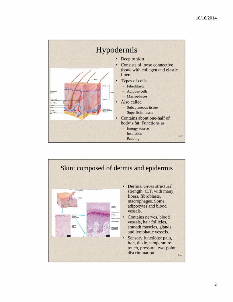
Hypodermis• Deep to skin• Consists of loose connective
tissue with collagen and elastic fibers
• Types of cells– Fibroblasts– Adipose cells– Macrophages
• Also called– Subcutaneous tissue– Superficial fascia
• Contains about one-half of body’s fat. Functions as– Energy source– Insulation– Padding
5-4
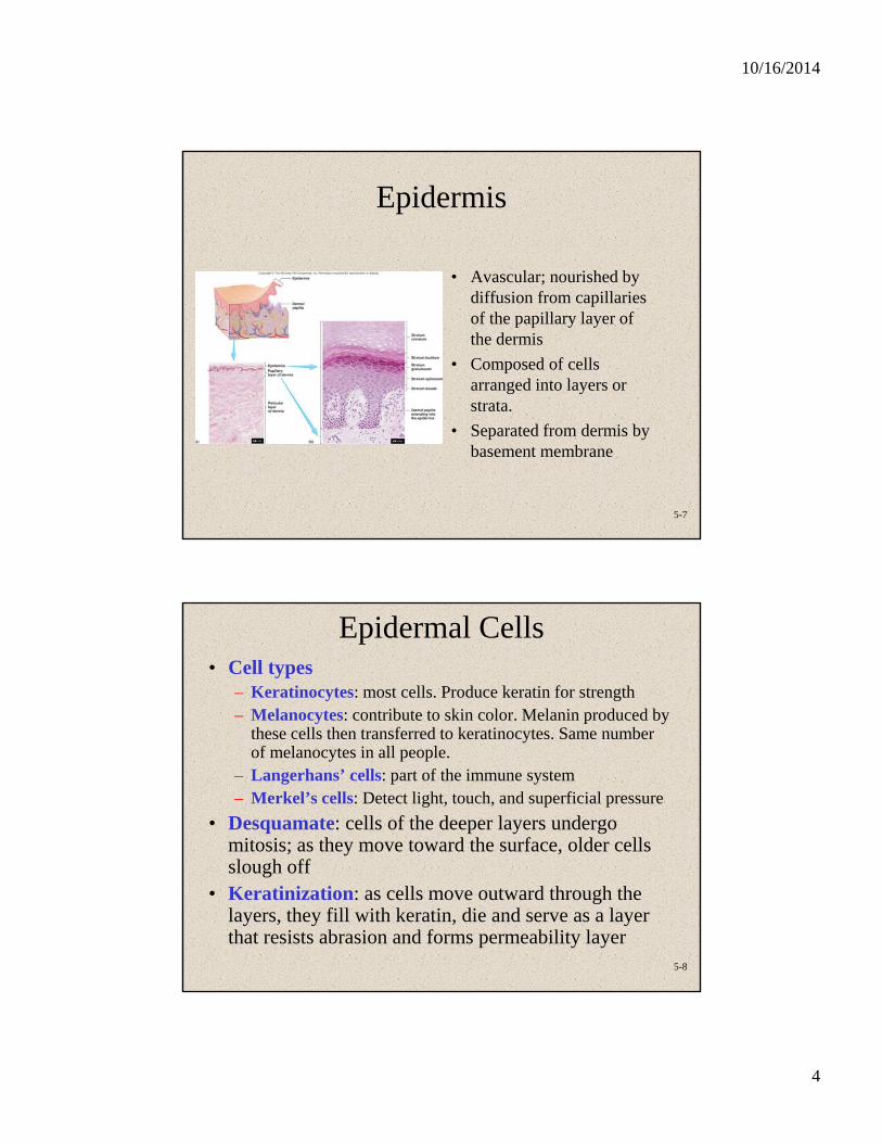
Skin: composed of dermis and epidermis
• Dermis. Gives structural strength. C.T. with many fibers, fibroblasts, macrophages. Some adipocytes and blood vessels.
• Contains nerves, blood vessels, hair follicles, smooth muscles, glands, and lymphatic vessels.
• Sensory functions: pain, itch, tickle, temperature, touch, pressure, two-point discrimination.

10/16/2014
3
5-5
Two Layers of the Dermis• Two layers variable in
thickness – Reticular: Deep (inner) 4/5
Dense irregular C.T. Collagen and elastic fibers. In the picture see: some adipose, hair follicles, nerves, oil glands, ducts of sweat glands, heat sensors.
– Papillary. Superficial (outer) 1/5. Areolar with lots of elastic fibers. Dermal papillae, capillary beds. Fingerprints. Whorls of ridges. Touch receptors (Meissner's), free nerve endings sensing pain
5-6
Cleavage (Tension) Lines and Striae
• Cleavage (tension) lines: elastin and collagen fibers oriented in some directions more than in others
• Important in surgery– If incision parallel to lines,
there is less gapping, faster healing, less scar tissue
• If skin is overstretched, striae (stretch marks) occur
10/16/2014
4
5-7
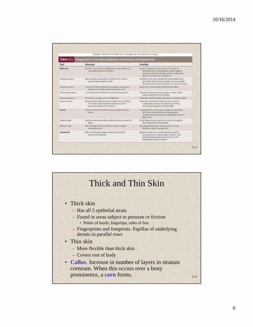
Epidermis
• Avascular; nourished by diffusion from capillaries of the papillary layer of the dermis
• Composed of cells arranged into layers or strata.
• Separated from dermis by basement membrane
5-8
Epidermal Cells• Cell types
– Keratinocytes: most cells. Produce keratin for strength– Melanocytes: contribute to skin color. Melanin produced by
these cells then transferred to keratinocytes. Same number of melanocytes in all people.
– Langerhans’ cells: part of the immune system– Merkel’s cells: Detect light, touch, and superficial pressure
• Desquamate: cells of the deeper layers undergo mitosis; as they move toward the surface, older cells slough off
• Keratinization: as cells move outward through the layers, they fill with keratin, die and serve as a layer that resists abrasion and forms permeability layer

10/16/2014
5
5-9
Epidermal Strata• Stratum basale (germinitivum)
– Deepest portion of epidermis and single layer. High mitotic activity and cells become keratinized
• Stratum spinosum– Limited cell division. Desmosomes. Lamellar bodies and
additional keratin fibers
• Stratum granulosum– Contains keratohyalin. In superficial layers nucleus and other
organelles degenerate and cell dies
• Stratum lucidum– Thin, clear zone. Found only in palms and soles
• Stratum corneum– Most superficial and consists of cornified cells
5-10
Epidermal Layers and Keratinization
10/16/2014
6
5-11
5-12
Thick and Thin Skin
• Thick skin– Has all 5 epithelial strata– Found in areas subject to pressure or friction
• Palms of hands, fingertips, soles of feet
– Fingerprints and footprints. Papillae of underlying dermis in parallel rows
• Thin skin– More flexible than thick skin– Covers rest of body
• Callus. Increase in number of layers in stratum corneum. When this occurs over a bony prominence, a corn forms.

10/16/2014
7
5-13
Skin Color: PigmentsDetermined by 3 factors: pigments
blood circulating through the skin, thickness of stratum corneum
• Pigments– Melanin: provides for protection
against UV light. Group of chemicals derived from aa tyrosine. Colored brown to black, may be yellowish or reddish
• Melanocytes. Processes extend between Keratinocytes.
• Albinism: deficiency or absence of pigment. Production determined by genetics, hormones, exposure to light
– Carotene: yellow pigment. From vegetables. Accumulates in stratum corneum, in adipose cells of dermis, and in hypodermis.
5-14
Skin Color: Blood and Stratum Corneum Thickness
• Blood circulating through the skin– Imparts reddish hue and increases during
blushing, anger, inflammation
– Cyanosis: blue color caused by decrease in blood oxygen content
• Thickness of stratum corneum

10/16/2014
8
5-15
Accessory Skin Structures: Hair • Found everywhere on human
body except palms, soles, lips, nipples, parts of external genitalia, and distal segments of fingers and toes
• Shaft protrudes above skin surface
• Root located below surface; base of root is the hair bulb
• Has 3 concentric layers– Medulla: Central axis– Cortex: Forms bulk of hair– Cuticle: Forms hair surface
5-16
Hair, cont.• Hair follicle
– Dermal root sheath: part of dermis that surrounds the epithelial root sheath
– Epithelial root sheath with internal and external parts.
• Internal part contains stratum basale that may remain after injury and supply a source of new epidermis
• When hairs are pulled out, internal part comes out and is visible as white bulb
• Hair bulb– Internal matrix is source of hair– Dermis projects into bulb and is
blood supply
10/16/2014
9
5-17
Hair Growth, Color, and Muscles• Hair Growth
– Cycles• Growth and resting stages
– Growth: cells added at base and hair elongates. Average rate is 0.3 mm/day
– Rest: follicle shortens and holds hair in place. Rest, then hair falls out of follicle. New hair begins.
– Regular hair loss means hair is being replaced. – Permanent hair loss: pattern baldness most common
cause
• Hair Color. Caused by varying amounts and types of melanin. Melanin can be black-brown and red
• Muscles. Arrector pili. Type of smooth muscle. – Muscle contraction causes hair to “stand on end”– Skin pushed up by movement of hair follicle
5-18
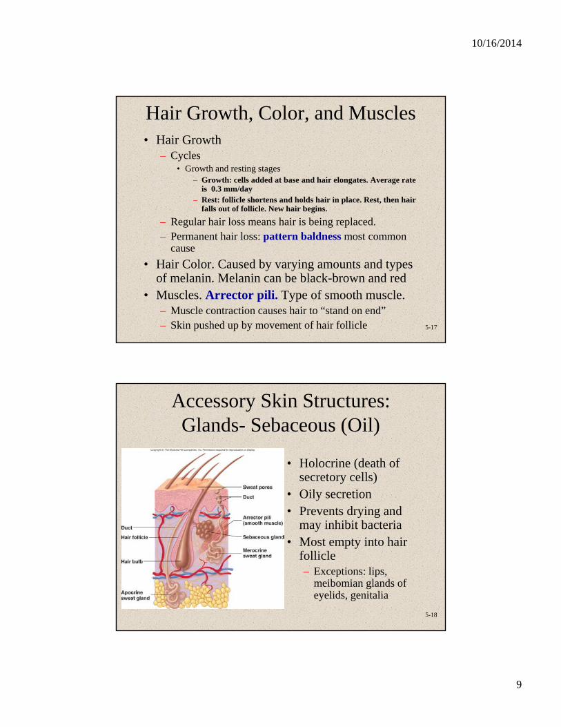
Accessory Skin Structures: Glands- Sebaceous (Oil)
• Holocrine (death of secretory cells)
• Oily secretion• Prevents drying and
may inhibit bacteria• Most empty into hair
follicle– Exceptions: lips,
meibomian glands of eyelids, genitalia
10/16/2014
10
5-19
Accessory Skin Structures: Glands- Sudoriferous (Sweat)
• Two types traditionally called apocrine and merocrine, but apocrine may secrete in a merocrine or holocrine fashion.
• Merocrine or eccrine. Most common. – Simple coiled tubular glands. – Open directly onto surface of skin. Have own pores. – Coiled part in dermis, duct exiting through epidermis. – Produce isotonic fluid (water and NaCl, but also excretory because sweat
includes ammonia, urea, uric acid and lactic acid). As fluid moves through duct, NaCl is moved by active transport back into the body. Final product is hyposmotic (hypertonic). Sweat.
– Numerous in palms and soles. Absent from margin of lips, labia minora, tips of penis, and clitoris.
• Apocrine. Active at puberty. – Compound coiled tubular, usually open into hair follicles superficial to
opening of sebaceous gland. – Secretion: organic compounds that are odorless but, when acted upon by
bacteria, may become odiferous.– Found in axillae, genitalia (external labia, scrotum), around anus.
5-20
Accessory Skin Structures: Glands- Ceruminous and
Mammary• Ceruminous glands: modified merocrine sweat
glands, external auditory meatus.– Earwax (cerumen). Composed of a combination of
sebum and secretion from ceremonious.
– Function- In combination with hairs, prevent dirt and insects from entry. Also keep eardrum supple.
• Mammary glands: modified apocrine sweat glands. Covered with reproductive chapter.

10/16/2014
11
5-21
Accessory Skin Structures: Nails • Anatomy
– Nail body: stratum corneum
– Eponychium or cuticle is corneum superficial to nail body, hyponychium is corneum beneath the free edge
– Matrix and nail bed: cells that give rise to the nail
– Nail root: extends
• Growth– Grow continuously unlike
hair– Fingernails grow 0.5-1.2
mm/day; faster than toenails
5-22
Skin Functions: Protection• Against abrasion, sloughing off of bacteria as
desquamation occurs.• Against microorganisms and other foreign
substances. Glandular secretions bacteriostatic and skin contains cells of the immune system.
• Melanin against UV radiation.• Hair on head is insulator and protection against
light, and from abrasion. Eyebrows keep sweat out of the eyes; eyelashes protect eyes from foreign objects. Hair in nose and ear against dust, bugs, etc.
• Nails protect ends of digits, self defense.• Acts as barrier to diffusion of water.
10/16/2014
12
5-23
Skin Functions: Sensation and Temperature Regulation
• Sensation: Pressure, temperature, pain, heat, cold, touch, movement of hairs.
• Temperature Regulation: sweating and radiation.
– Sweat causes evaporative cooling.
– Arterioles in dermis change diameter as temperature changes. More or less blood flows through the dermis.
5-24
Skin Functions: Vitamin D Production
• Begins here in skin; aids in Ca+2 absorption. • Vitamin D (calcitriol): hormone.
– Stimulates uptake of Ca and PO4 from intestines– Promotes Ca and PO4 release from bones– Reduces Ca loss from kidneys. – Increases blood Ca and PO4 levels.
• Functions of Ca– bone formation, growth, repair– clotting– nerve and muscle function.
• 7-dehydrocholesterol converts to cholecalciferol when exposed to UV radiation. Cholecalciferol released to blood and modified in the liver and kidneys to form calcitriol (active vitamin D).
• People in cold climates and those who cover the body can be deficient, but calcitriol can be absorbed through intestinal wall.
– Sources: dairy, liver, egg yolks, supplements.

10/16/2014
13
5-25
Skin Functions: Excretion
• Removal of waste products from the body. Sweat. Water, salt, urea, ammonia, uric acid.
• Insignificant when compared with kidneys.
24-26
Skin Cancer• Basal cell carcinoma – progresses slowly and
rarely spreads to other body parts
• Squamous cell carcinoma – more likely to spread to surrounding tissues
• Malignant melanoma – more aggressive and occurs anywhere – Most arise from melanocytes

10/16/2014
14
24-27
Skin Cancer: Basal Cell Carcinoma
• Signs and symptoms– New growth or sore that will not heal
– Waxy, smooth, red, pale, flat, or lumpy
– May or may not bleed
• Treatment: – Curettage and electrodessication Cryosurgery
– Mohs’ surgery Laser therapy
24-28
Skin Cancer: Squamous Cell Carcinoma
• Less common than basal cell carcinoma
• Found on face, lips, ears, and backs of hands
• Signs and symptoms and treatments are the same as for basal cell carcinoma

10/16/2014
15
24-29
Skin Cancer: Malignant Melanoma
• Signs and Symptoms– From melanocytes
– Appear on trunk, head, neck of men
– Appear on arms and legs of women
– Itches or bleeds
• Treatment– Surgery and biopsy
– Removal of lymph nodes
– Chemotherapy and radiation therapy
– Immunotherapy
24-30
Skin Cancer: Stages of Melanoma
Stage 0 Only found in epidermis
Stage I Spread to epidermis and dermis (1 to 2 mm thick)
Stage II 2 to 4 mm thick plus ulceration
Stage III Spread to one or more lymph nodes
Stage IV Spread to other body organs or lymph nodes far from original melanoma

10/16/2014
16
24-31
Skin Cancer: ABCD Rule
A Asymmetry: The mole should not become asymmetrical
B Border should not become irregular
C Color should not change or become mixture of colors
D Diameter should not grow larger than the diameter of a pencil eraser
24-32
Cancer Warning Signs
C – Change in bowel or bladder habitsA – A sore that will not healU – Unusual bleeding or discharge T – Thickening or lumpI – Indigestion or difficulty swallowingO – Obvious change in wart or moleN – Nagging cough or hoarseness
10/16/2014
17
5-33
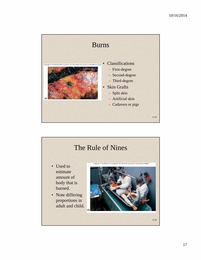
Burns
• Classifications– First-degree
– Second-degree
– Third-degree
• Skin Grafts– Split skin
– Artificial skin
– Cadavers or pigs
5-34
The Rule of Nines
• Used to estimate amount of body that is burned.
• Note differing proportions in adult and child.

10/16/2014
18
5-35
Aging Effects
• Skin more easily damaged because epidermis thins and amount of collagen decreases
• Skin infections more likely• Wrinkling occurs due to decrease in elastic fibers• Skin becomes drier • Decrease in blood supply causes poor ability to
regulate body temperature• Functioning melanocytes decrease or increase; age
spots• Sunlight ages skin more rapidly