Chapter 4 - INFLIBNETshodhganga.inflibnet.ac.in/bitstream/10603/12899/9/09_chapter 4.pdf ·...
22
91 Chapter 4 Novel decavanadate cluster complexes [H 2 V 10 O 28 ][LH] 4 ·nH 2 O (L= Imidazole, n=2 or 2-methylimidazole, n= 0): Preparation, characterization and genotoxic studies
Transcript of Chapter 4 - INFLIBNETshodhganga.inflibnet.ac.in/bitstream/10603/12899/9/09_chapter 4.pdf ·...
91
Chapter 4
Novel decavanadate cluster complexes [H2V10O28][LH]4nH2O (L=
Imidazole n=2 or 2-methylimidazole n= 0) Preparation
characterization and genotoxic studies
92
INTRODUCTION
The recent developments in crystal engineering have opened up new dimensions
in inorganic metallo-organic and coordination chemistry researches [1-3] Complexes
with supramolecular assemblies have received attentions owing to their potential
applications as models for several metallo-enzymes and as useful optical as well as
electronic materials [4-7] Several polymetallic anionic clusters have been found to
display a strong tendency to form robust ion-pairs with appropriate or selective counter
cations This tendency has often helped attaining the minimum requirement(s) for the
formation of stable crystal motifs [8] The structural diversity of polymetallic anionic
hosts requires tailored made cationic guests to generate high affinity and selectivity
between the target ion and the receptor henceforth plays a crucial role in guest-host
interactions and self assemblies [9-13] The polyoxovanadate anions eg the dimer
(H2V2O72- HV2O7
3- or V2O74-) the tetramer (V4O12
4-) the pentamer (V5O155-) and the
decamer (H2V10O284- HV10O28
5- or V10O286-) [1415] are known to possess different
geometric features in aqueous solutions but exhibit nearly identical biological activities
[1617] However decavanadate anions have attracted researchers owing to their uses in
crystallization of proteins and in many other biological mechanisms [1617]
It has been emphasized that only big size counter cation as guests or protonated
organic ligands like glycylglycine (Gly-Gly) glycylhistidine (Gly-His) or even
biimidazole derivatives can help to form stable complexes incorporating the big anionic
decavanadate moieties [HnV10O28](6ndashn)ndash (n = 1ndash4) through strong ion-pair formation The
presence of proton in such anionic units have crucial role in supporting the crystal
93
packing via additional extensive H-boning to generate 2D supramolecular assemblies
[18] However to our knowledge stable complexes of the [H2V10O28]4ndash anion with small
moieties like imidazole or even their substituted derivatives which are small in size have
not been exploited in this regard In this chapter isolation of stable crystals and structural
characterization of the novel complexes [H2V10O28]ndash[LH]+ are discussed The genotoxic
properties of the compounds have also been examined
94
EXPERIMENTAL
Materials and method
VOSO45H2O (sd-fine) Imidazole (Merck) 2-methylimidazole (Aldrich) and
adipic acidiminodiacetic acid (Aldrich) were used as received while commercial solvents
were distilled before use The complexes [H2V10O28](ImzH)42 H2O (1) and
[H2V10O28](2-MeImzH)4 (2) were prepared using VOSO45H2O Imidazole (Imz)2-
methylimidazole (2-MeImz) and adipic acid (H2ada) or iminodiacetic acid (H2ida) in
111 stoichiometric ratio as outlined below
Instrumentation
The elemental (C H and N) analyses were obtained from Micro-Analytical
Laboratory of Central Drug Research Institute (CDRI) Lucknow India FT-IR spectra of
compounds were recorded as KBr discs on Perkin Elmer Model spectrum GX
spectrophotometer51V NMR measurements were performed on a BrukerAM-400MHz
spectrometer at 1052 MHz equipped with a 5 mm multinuclear inverse probe The
spectrum was acquired at 25 C using 05 ml of samples dispersed in 10 D2O under the
following conditions 90 pulse angle spectral width 454 Hz acquisition time 0086 s
number of transients between 12 000 and 25 000 and a relaxation delay of 001 s using
VOCl3 as an external reference X-band EPR spectrum was recorded at room temperature
on a Varian E-112 spectrometer operating at 91 GHz DART-MS analyses were
performed on a JEOL-AccuTOF JMS-T100LC Mass spectrometer having a DART
(direct analysis in real time) source
95
Crystallographic data collection and structure analysis
A single crystal of compound (2) was mounted on a glass fibre and all
measurements were performed on BRUKER SMART APEX CCD diffractometer with
graphite monochromator using Mo-kα (λ = 071069Aring) radiation at 293 K The structure
was solved by direct method using the program SHELXS-97 [19] and subsequent Fourier
difference techniques and refined anisotropically by fullmatrix least-squares on F2 using
SHELXL-97 [20]
Synthesis of the complexes [H2V10O28](ImzH)42 H2O (1) and [H2V10O28](2-
MeImzH)4 (2)
Imz or 2-MeImz (6mmol) was added in small batches to an aqueous solution of
adipic acid (031 gm 3 mmol) or iminodiacetic acid (039 gm 3 mmol) with stirring to
form a homogenous mixture The stirring was continued for 1 h and the pH of solution
was slightly acidic (pH ~ 60) Vanadyl sulphate (075 gm 3 mmol) solution in 10 mL
H2O was added drop wise to the above mixture which produced light yellow (1) or
orange (2) colour solutions after overnight stirring at RT It was then refluxed for 18 h
giving a stable green colour solution which was left standing in air in a beaker at room
temperature Crystals of (1) and (2) were obtained after few days Crystals of the complex
(2) were suitable for single crystal X-ray studies
Complex (1) Yield 38 mp gt300degC IR (KBr disk cm-1) ν(C-H) 2958 2918 1448
736 ν(N-H) 3140-3230(br) ν(H2O) 3300-3500(br) ν(V-OH) 1668(s br) ν(V=O)
96
985(s) νasym(VndashOndashV) 826 796 νsym(VndashOndashV) 600535 Anal Calcd for V10O30N8H22C12
C 1136 H 173 N 883 Found C 1124 H 169 N 880
Complex (2) Yield 40 mp gt300degC IR (KBr disk cm-1) ν(C-H) 2916 2839 ν(N-H)
3150-3220(br) ν(Ar) 1623 1634 ν(V-OH) 1700(s br) ν(V=O) 965(s) νasym(VndashOndashV)
834 798 745 νsym(VndashOndashV) 605 560 535 Anal Calcd for V10O28N8H28C13 C 1490
H 219 N 869 Found C 1499 H 335 N 871
Cyclic voltammetric studies
Cyclic voltammetry (CV) was performed on EGampG PAR 273
PotentiostatGalvanostat equipped with an IBM PS2 computer with EGampG M270
software for data acquisition The three-electrode cell configuration comprised of a
platinum sphere a platinum plate and Ag(s)AgNO3 were used as working auxiliary and
reference electrodes respectively The supporting electrolyte used was [nBu4N]ClO4
Platinum sphere electrode was sonicated for two minutes in dilute nitric acid dilute
hydrazine hydrate and then in double-distilled water to remove the impurities The
solutions were deoxygenated by bubbling research grade nitrogen and an atmosphere of
nitrogen was maintained over the solution during measurements
Comet assay (single cell gel electrophoresis)
The complexes (1) and (2) were incubated with lymphocyte at 37 degC for 1 h
Comet assay was then performed under alkaline conditions essentially according to the
reported procedure [21] with slight modifications Fully frosted microscopic slides pre-
97
coated with 10 normal melting agarose were used at about 50 ordmC (dissolved in Ca2+
and Mg2+ free PBS) Around 10000 cells were mixed with 80 microl of 10 low melting
point agarose to form a cell suspension and pipetted over the first layer and covered
immediately by a cover slip The slides were placed on a flat tray and kept on ice for 10
min to solidify the agarose The cover slips were removed and a third layer of 05 low
melting point agarose (75 microl) was pipetted Cover slips were placed over it and it was
allowed to solidify on ice for 5 minutes The cover slips were removed and the slides
were immersed in cold lysis solution containing 25 M NaCl 100 mM EDTA and 10 mM
Tris pH 10 1 Triton X-100 was added before use for a minimum of 1 h at 4 ordmC After
lysis DNA was allowed to unwind for 30 minutes in alkaline electrophoretic solution
consisting of 300 mM NaOH and 1mM EDTA pH gt13 Electrophoresis was performed
at 4 ordmC in field strength of 07 Vcm and 300 mA current The slides were then
neutralized with cold 04 M Tris pH 75 stained with 75 ml ethidium bromide (20
mgml) and covered with a cover slip They were then placed in a humidified chamber to
prevent drying of the gel and analyzed the same day Slides were scored using an image
analysis system (Komet 55 Kinetic Imaging Liverpool UK) attached to an Olympus
(CX41) fluorescent microscope (Olympus Optical Co Tokyo Japan) and a COHU 4910-
integrated CC camera (equipped with 510ndash560 nm excitation and 590 nm barrier filters)
(COHU San Diego CA USA) Images from 50 cells (25 from each replicate slide) were
analyzed The parameter taken to assess lymphocytes DNA damage was tail length
(migration of DNA from the nucleus micrometer)
98
RESULTS amp DISCUSSION
In the present work two new complexes (1 and 2) were synthesized Scheme 1
illustrates the way the target compounds were prepared The structures of the compounds
were elucidated by elemental analyses FT-IR 51VNMR EPR and DART-MS techniques
Imz or 2-MeImz
H2ada H2idaRT pH = 6
Homogenous clear solution
VOSO45H2ORefluxed
[H2V10O28][ImzH]42H2O (1) [H2V10O28][2-MeImzH]4 (2)
Scheme 1
99
FT-IR Spectra
IR spectra of (1) and (2) exhibited well defined absorption frequencies arising
from the characteristic ring ν(C=N) and ν(C=C) stretching vibrations [22] of protonated
imidazolium moiety along with those characteristic of the [H2V10O28]4ndash polyhedron [14-
16] The intense band at 985 (1) or 965 cmndash1 (2) can be assigned to the stretching mode
of vibration of the terminal V=O bonds The bridging νsym- and νasym-(V-O-V) stretching
vibrations were observed at the appropriate positions [14-16] The EPR spectra at RT for
(1) and (2) were silent indicating that in the present [H2V10O28]4ndash moiety all the
vanadium ions are in pentavalent oxidation state (d0 configuration) 51V NMR of (1) and
(2) exhibited peaks at -418 -515 and -421 -510 ppm respectively characteristic of
decavanadate anions [23]
DART-MS Spectra
The DART (direct analysis in real time) mass studies of [H2V10O28](ImzH)42H2O
(1) and [H2V10O28](2-MeImzH)4 (2) were performed to investigate the possible
fragmentation pattern and the stability of the species thus formed The MS data of (1) and
(2) strongly support the dissociation of the molecular units The counter cations appear as
the intense peak at mz = 94 (100) in (1) and mz = 83 (98) in (2) The fragmentation
pattern of [H2V10O28]( ImzH)42H2O (1) is shown in Fig 1S(a) where the unstable
[H2V10O28]4- further splits into the lower vanadium oxides ie into one unit of [V2O2]2+ at
mz = 134 (36) 7 units of [VO3+H] mz = 100 (23) and one unit of [V3O5]2+ at mz =
233 (43) However the resultant anionic species for complex(2) shown in Fig 1S(b)
100
gets fragmented (being unstable in nature in absence of the counter cation) forming
[H2VO2+3H] (mz = 89 48) [V2O4-H] (165 15) [H2VO2+4H] (mz = 90 3)
[V2O5] (mz = 182 25) and [V4O10-3H] (mz = 361 38) The DART MS
fragmentation spectra of complexes (1) and (2) show that the polyoxovanadate moiety
[H2V10O28]4ndash is stable in presence of strongly interacting counter [ImzH]+[2-
MeImzH]+cations otherwise it splits into various vanadium oxide constituents differing in
the oxidation states
Crystal structure of [H2V10O28](2-MeImzH)4 (2)
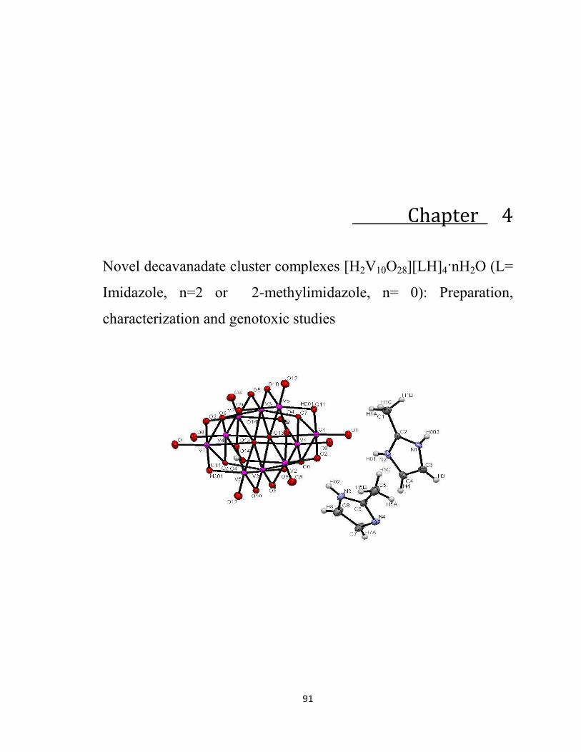
The ORTEP view and packing diagram for (2) are shown in Figs 1 and 2
respectively The structure shows that the decavanadate anion [H2V10O28]4ndash unit is
supported through the strong ion-pair formation with the counter cation ie the
protonated species of the ligand moiety [2-MeImzH]+ It is evident from Fig 1(a) that
[H2V10O28]4ndash moiety has an inversion centre on the mid point of O(2) and O(2A) with D2h
symmetry and consists of ten VO6 octahedra packed with sharing edges
101
Fig 1(a) ORTEP view of complex (2) (out of four only two 2-MeImzH+ units are shown for symmetrical representation)
The structural parameters are comparable to that of the reported decavanadate
anionic moieties [111415] The important X-ray crystallographic data which include
selected bond lengths and bond angles etc are provided in Tables 1 and 2 Figs 1(a)
and (b) indicate that each decavanadate anion [H2V10O28]4ndash is surrounded by four units of
the counter cation [2-MeImzH]+ The protonation sites are consistent with the location of
the Fourier difference maps and consideration based on the standard bond valance
summations [11] given by sumS = sum(d1791)51ndash where sumS is the sums of VO bond
valances for bridging oxygen atom and d is V-O distances in Aring The magnitude of sumS
helps ascertaining the nature of the bond order between V and O in the decavanadate unit
A value of sumS in 15-20 range reflects V=O while less that 15 is indicative of VndashOH
bond The observed magnitude of sumS = 11 for complex (2) confirms the presence of two
VndashOH bonds in [H2V10O28]4ndash
102
Fig 1(b) Packing diagram of complex (2)
The complex adopts a well organized two dimensional (2D) sheet structure (Fig
2) involving a strong H-bonding from the protonation site (ie pyrrolic NH+) It may be
concluded from the crystallographic studies that H-bonding plays an important role
alongwith the effective ion-pair interaction for the stability of the present complexes
[H2V10O28][LH]4 (1 amp 2)
103
Fig 2 2D sheet structure of complex (2) formed via extensive H-bonding (shown by
dotted lines)
104
Table 1 Crystallographic data and refinement parameters for (2)
CCDC deposition No 783596 empirical formula C16H28N8O28V10 fw 128986 crystal system monoclinic space group P2 (1) n a Aring 9370(2) b Aring 17279 (4) c Aring 12250(3) α deg 9000 β deg 97654(4) γ deg 9000 v Aring3 19658(8) Z 2 T 293(2) k Ө range for data collection 236 to 2833 Dc g cm-3 2179 Crystal size 026-032 F(000) 1268 Absorption coefficient (mm-1) 2367 Μ(Mo-Kα) Cm-1 071073 measurement device type lsquoCCD area detectorrsquo index ranges
-10 lt h lt 11 -21lt k lt 19 -7 lt l lt 15
reflections θ range 236 to 2833 reflections σ I net I 00639 reflections collected 4033 reflections independent 3416 (Rint=00727) datarestraintsparameters 40331300 goodness of fit on F2 1054 final R indices [Igt2σ (I)] R1=00442 wR2=01111 R indices all data R2=00547wR2= 01181 reflections threshold expression gt2σ (I) audit creation method SHELXL-97 structure refinement SHELXL-97
(Sheldrick 1997)
105
Table 2 Selected bond distances (Aring) of (2) V1-O1 1592 (3) V4-O14 2218(2) V1-O11 1791(2) O11-V1 1791(2) V1-O2 1888(2) O4-V4 1979(2) V1-O7 1939(2) O4-H001 0807(19) V1-O6 2025(2) O13-V2 1875(2) V1-O14 2298(2) O7-V3 1900(2) V1-V4 30079(10) O14-V3 2144(2) V1-V2 30864(9) O14-V1 2298(2) V1-V3 30965(10) O14-V2 2334(2) V1-V5 31061(10) N1-C2 1336(5) V3-O10 1680(2) N1-C3 1381(5) V3-O5 1702(2) N1-H002 075(5) V3-O7 1900(2) N3-C6 1326(5) V3-O6 1965(3) N3-C8 1379(5) V3-O14 2081(2) N3-H02 084(4) V3-O14 2144(2) C2-N2 1327(5) V3-V2 30521(10) C2-C1 1478(5) V3-V5 30817(10) N4-C6 1335(5) V3-V4 30848(9) N4-C7 1376(5) V3-V1 30965(10) N2-C4 1375(5) V2-O3 1595(2) N2-H01 071(5) V2-O2 1767(3) C4-C3 1351(6) V2-O13 1875(2) C4-H4 09300 V2-O5 1970(2) C6-C5 1482(5) V2-O4 2050(3) C1-H1A 09600 V2-O14 2334(2) C1-H1B 09600 V2-V5 30894(11) C1-H1C 09600 V5-O12 1599(2) C7-C8 1345(6) V5-O13 1783(2) C7-H7A 074(4) V5-O11 1897(2) C3-H3 09300 V5-O9 1934(3) V4-O14 2218(2) V5-O10 2069(2) O11-V1 1791(2) V5-O14 2305(2) O4-V4 1979(2) V5-V4 30758(11) O4-H001 0807(19) V5-V2 30894(11) O13-V2 1875(2) V5-V1 31061(9) O7-V3 1900(2) V4-O8 1592(3) O14-V3 2144(2) V4-O9 1775(2) O14-V1 2298(2) V4-O6 1918(2) O14-V2 2334(2) V4-O4 1979(2) N1-C2 1336(5) V4-O7 2039(2) N1-C3 1381(5)
106
Table 3 Selected bond angles (o) of (2) O1-V1-O11
10483(13)
V2-V1-V5
5985(2)
O1-V1-O2 10051(12) V3-V1-V5 5958(2) O11-V1-O2 9381(11) O10-V3-O5 10781(12) O1-V1-O7 10230(12) O10-V3-O7 9962(11) O11-V1-O7 9362(11) O5-V3-O7 9690(11) O2-V1-O7 15329(11) O10-V3-O6 9691(11) O1-V1-O6 9958(12) O5-V3-O6 9444(11) O11-V1-O6 15528(11) O7-V3-O6 15600(10) O2-V1-O6 8541(10) O10-V3-O14 8744(10) O7-V1-O6 7727(10) O5-V3-O14 16453(11) O1-V1-O14 17422(11) O7-V3-O14 8266(10) O11-V1-O14 8090(10) O6-V3-O14 8083(9) O2-V1-O14 7959(10) O10-V3-O14 16582(10) O7-V1-O14 7632(9) O5-V3-O14 8622(10) O6-V1-O14 7465(9) O7-V3-O14 8026(10) O1-V1-V4 9058(9) O6-V3-O14 7947(9) O11-V1-V4 13568(8) O14-V3-O14 7846(10) O2-V1-V4 12439(8) O10-V3-V2 14406(8) O7-V1-V4 4214(7) O5-V3-V2 3669(8) O6-V1-V4 3898(7) O7-V3-V2 9237(7) O14-V1-V4 8468(6) O6-V3-V2 8421(7) O1-V1-V2 13171(10) O14-V3-V2 12786(7) O11-V1-V2 8428(8) O14-V3-V2 4970(6) O2-V1-V2 3122(8) O10-V3-V5 3908(8) O7-V1-V2 12472(8) O5-V3-V5 14689(8) O6-V1-V2 8237(7) O7-V3-V5 9112(7) O14-V1-V2 4872(6) O6-V3-V5 9083(7) V4-V1-V2 11587(3) O14-V3-V5 4840(6) O1-V1-V3 13745(9) O14-V3-V5 12686(6) O11-V1-V3 7845(8) V2-V3-V5 17442(3) O2-V1-V3 12181(8) O10-V3-V4 8370(8) O7-V1-V3 3582(7) O5-V3-V4 13128(9) O6-V1-V3 8101(7) O7-V3-V4 12849(7) O14-V1-V3 4223(6) O6-V3-V4 3686(7) V4-V1-V3 6319(2) O14-V3-V4 4595(6) V2-V1-V3 9074(2) O14-V3-V4 8536(6) O1-V1-V5 13813(10) V2-V3-V4 11461(3) O11-V1-V5 3371(8) V5-V3-V4 5984(2)
107
Cyclic voltammetric (CV) studies
The electrochemical redox properties of the complexes (2) have been studied
using cyclic voltammetry in the potential range +10 to ndash10 V recorded at 015 Vsndash1
scan rate with reference to AgAgCl electrode at room temperature in the presence of
tetrabuylammonium perchlorate as supporting electrolyte
Fig 3 Cyclic voltammogram of complex (2)
Cathodic and anodic waves in the voltammogram (Fig 3) which contained the
medium intensity cathodic peak (Epc) at +03 V and an intense anodic peak (Ep
a) at minus008
V are reversibly coupled with the formation of a quasi reversible redox couple involving
108
1endash redox process [24-26] This results in a redox couple with half wave potential (Eordm12) =
minus019 V peak separation (∆E = EPc minus Ep
a) = 038 V and peak current ratio (IPcIP
a) = 18
This is consistent with the formation of a quasi-reversible redox couple VVIV as stable
entity in solution
Effect of complexes (1) and (2) on lymphocyte DNA breakage (comet assay)
Fig 4(a) and (b) show that for complexes (1) and (2) at a concentration of 50
microM the tail formation was 7 microMeter and 75 microMeter respectively The tail formation
[Fig 4(a)] and the apparent tail length [Fig 4(b)] relative to the control is the index of
DNA damage or in other words a measure of the toxic effect by the compounds It is
clear from Figs 4(a) and 4(b) that the tail length formation as well as the tail lengths at a
concentration 50 microM of the present complexes [ie 7 microMeter (1) amp 75 microMeter (2)] were
not remarkable in comparison to the control Thus it can be concluded from comet assay
that (1) and (2) can be easily exploited for the industrial applications as they have no
toxic effects on DNA
Fig 4(a) Comet assay pictures for complexes (1) and (2)
109
Fig 4(b) Tail length diagram for complexes (1) and (2)
110
CONCLUSION
Two novel polyoxovanadate clusters [H2V10O28](ImzH)42H2O (1) and
[H2V10O28](2-MeImzH)4 (2) were synthesized and characterized from spectral methods
X-ray crystallographic studies support strong cation-anion (ion-pair) interactions in
[H2V10O28]4ndash[LH]4+
and presence of H-bonding in a well organized manner to result in a
2D stacked supramolecular architecture The EPR spectral studies confirmed that the
decavanadate anion contains V5+ ions 51V NMR of the complexes exhibited peaks
characteristic of the decavanadate anions IR spectra indicated V=O V-OH V-O-V
bonds in the anionic and the ring C=N amp C=C bonds in the cationic species CV studies
of (2) indicated the formation of a quasi-reversible redox couple in aqueous solution
Genotoxic studies confirmed that the compounds are non toxic in nature and can be
exploited for industrial purposes
111
References
[1] J M Lehn Supramolecular Chemistry Concepts and Perspectives VCH Weinheim
Germany 1995
[2] M Eddaudi J Kim N Rosi D Vodak J Wechter M OrsquoKeefe O M Yagachi Science
295 (2002) 469
[3] K Kobayachi Y Yamada M Yamanaka Y Sei K Yamaguchi J Am Chem Soc 126
(2004) 13896
[4] M D Hollings Worth Science 295 (2002) 2410
[5] D Maspoch D Ruiz-Molina J Veciana J Mater Chem 14 (2004) 2713
[6] S Kitagawa R Kitaura S Noro Angew Chem Int Edit 43 (2004) 2334
[7] C L Bowes G A Ozin Adv Mater 8 (1996) 13
[8] G R Desiraju Angew Chem Int Ed 46 (2007) 8342
[9] A Bianchi K Bowman-James E Garcia-Espaned Supramolecular Chemistry of Anions
Wiley VCH New York 1997
[10] S Roelens R Tortini J Am Chem Soc 120 (1998) 12443
[11] D E Gross F P Schmidtchen W Antonius P A Gale V M Lynch J L Sessler Chem-
Eur J 14 (2008) 7822
[12] S Pappalarado V Villari S Slovak Y Cohen G Gattuso A Notti A Pappalarado I
Pisagatti M F Parisi Chem-Eur J 14 (2007) 8164
[13] Z A Siddiqi M Shahid S Kumar M Khalid S Noor J Organomet Chem 694 (2009)
3768
[14] D C Crans Comments Inorg Chem 16 (1994) 1
[15] D C Crans K Sudhakar TJ Zamborelli Biochemistry 31 (1992) 6812
[16] D C Crans Polyoxometalates From Platonic Solids to Anti-Retroviral Activity A Muller
M T Eds Pope Kluwer Academic Publishers Dordrecht The Netherlands 49 (1993) 399
[17] P Csermely A Martonosi G C Levy A J Ejchart Biochem J 230 (1985) 807
[18] Hitoshi Kumagai Masashi Arishima Susumu Kitagawa Satoshi Kawata Sumio Kaizaki
Inorg Chem 41 (2002) 1989
[19] (a) SAINT+ 602 ed Bruker AXS Madison WI 1999
(b) GM Sheldrick SADABS Empirical Absorption Correction Program University
of Goumlttingen Goumlttingen Germany 1997
112
(c) XPREP 51 ed Siemens Industrial Automation Inc Madison WI 1995
[20](a) A Altomare MC Burla M Camalli GL Cascarano C Giacovazzo A
Guagliardi AGG Moliterni G Polidori RJ Spagna Appl Crystallogr 32
(1999) 115
(b) GM Sheldrick SHELXL-97 Program for Crystal Structure Refinement
University of Goumlttingen Goumlttingen Germany 1997
[21] NP Singh MT McCoy RR Tice EL Schneider A simple technique for quantitation of
low levels of DNA damage in individual cells Exp Cell Res 175 (1988) 184ndash191
[22] K Nakamoto in Infrared and Raman Spectra of Inorganic and Coordination
Compounds Wiley-Interscience New York Publication 1886 pp 191
[23] R S Drago Physical Methods for Chemists Sannders College Publishing New York
1992
[24] ZA Siddiqi M Shahid M Khalid S Noor S Kumar Spectrochim Acta Part A
74 (2009) 391
[25] ZA Siddiqi M Shahid M Khalid S Noor S Kumar Spectrochim Acta Part A
75 (2010) 61
[26] ZA SiddiqiVJ Mathew Polyhedron 13 (1994) 799

92
INTRODUCTION
The recent developments in crystal engineering have opened up new dimensions
in inorganic metallo-organic and coordination chemistry researches [1-3] Complexes
with supramolecular assemblies have received attentions owing to their potential
applications as models for several metallo-enzymes and as useful optical as well as
electronic materials [4-7] Several polymetallic anionic clusters have been found to
display a strong tendency to form robust ion-pairs with appropriate or selective counter
cations This tendency has often helped attaining the minimum requirement(s) for the
formation of stable crystal motifs [8] The structural diversity of polymetallic anionic
hosts requires tailored made cationic guests to generate high affinity and selectivity
between the target ion and the receptor henceforth plays a crucial role in guest-host
interactions and self assemblies [9-13] The polyoxovanadate anions eg the dimer
(H2V2O72- HV2O7
3- or V2O74-) the tetramer (V4O12
4-) the pentamer (V5O155-) and the
decamer (H2V10O284- HV10O28
5- or V10O286-) [1415] are known to possess different
geometric features in aqueous solutions but exhibit nearly identical biological activities
[1617] However decavanadate anions have attracted researchers owing to their uses in
crystallization of proteins and in many other biological mechanisms [1617]
It has been emphasized that only big size counter cation as guests or protonated
organic ligands like glycylglycine (Gly-Gly) glycylhistidine (Gly-His) or even
biimidazole derivatives can help to form stable complexes incorporating the big anionic
decavanadate moieties [HnV10O28](6ndashn)ndash (n = 1ndash4) through strong ion-pair formation The
presence of proton in such anionic units have crucial role in supporting the crystal
93
packing via additional extensive H-boning to generate 2D supramolecular assemblies
[18] However to our knowledge stable complexes of the [H2V10O28]4ndash anion with small
moieties like imidazole or even their substituted derivatives which are small in size have
not been exploited in this regard In this chapter isolation of stable crystals and structural
characterization of the novel complexes [H2V10O28]ndash[LH]+ are discussed The genotoxic
properties of the compounds have also been examined
94
EXPERIMENTAL
Materials and method
VOSO45H2O (sd-fine) Imidazole (Merck) 2-methylimidazole (Aldrich) and
adipic acidiminodiacetic acid (Aldrich) were used as received while commercial solvents
were distilled before use The complexes [H2V10O28](ImzH)42 H2O (1) and
[H2V10O28](2-MeImzH)4 (2) were prepared using VOSO45H2O Imidazole (Imz)2-
methylimidazole (2-MeImz) and adipic acid (H2ada) or iminodiacetic acid (H2ida) in
111 stoichiometric ratio as outlined below
Instrumentation
The elemental (C H and N) analyses were obtained from Micro-Analytical
Laboratory of Central Drug Research Institute (CDRI) Lucknow India FT-IR spectra of
compounds were recorded as KBr discs on Perkin Elmer Model spectrum GX
spectrophotometer51V NMR measurements were performed on a BrukerAM-400MHz
spectrometer at 1052 MHz equipped with a 5 mm multinuclear inverse probe The
spectrum was acquired at 25 C using 05 ml of samples dispersed in 10 D2O under the
following conditions 90 pulse angle spectral width 454 Hz acquisition time 0086 s
number of transients between 12 000 and 25 000 and a relaxation delay of 001 s using
VOCl3 as an external reference X-band EPR spectrum was recorded at room temperature
on a Varian E-112 spectrometer operating at 91 GHz DART-MS analyses were
performed on a JEOL-AccuTOF JMS-T100LC Mass spectrometer having a DART
(direct analysis in real time) source
95
Crystallographic data collection and structure analysis
A single crystal of compound (2) was mounted on a glass fibre and all
measurements were performed on BRUKER SMART APEX CCD diffractometer with
graphite monochromator using Mo-kα (λ = 071069Aring) radiation at 293 K The structure
was solved by direct method using the program SHELXS-97 [19] and subsequent Fourier
difference techniques and refined anisotropically by fullmatrix least-squares on F2 using
SHELXL-97 [20]
Synthesis of the complexes [H2V10O28](ImzH)42 H2O (1) and [H2V10O28](2-
MeImzH)4 (2)
Imz or 2-MeImz (6mmol) was added in small batches to an aqueous solution of
adipic acid (031 gm 3 mmol) or iminodiacetic acid (039 gm 3 mmol) with stirring to
form a homogenous mixture The stirring was continued for 1 h and the pH of solution
was slightly acidic (pH ~ 60) Vanadyl sulphate (075 gm 3 mmol) solution in 10 mL
H2O was added drop wise to the above mixture which produced light yellow (1) or
orange (2) colour solutions after overnight stirring at RT It was then refluxed for 18 h
giving a stable green colour solution which was left standing in air in a beaker at room
temperature Crystals of (1) and (2) were obtained after few days Crystals of the complex
(2) were suitable for single crystal X-ray studies
Complex (1) Yield 38 mp gt300degC IR (KBr disk cm-1) ν(C-H) 2958 2918 1448
736 ν(N-H) 3140-3230(br) ν(H2O) 3300-3500(br) ν(V-OH) 1668(s br) ν(V=O)
96
985(s) νasym(VndashOndashV) 826 796 νsym(VndashOndashV) 600535 Anal Calcd for V10O30N8H22C12
C 1136 H 173 N 883 Found C 1124 H 169 N 880
Complex (2) Yield 40 mp gt300degC IR (KBr disk cm-1) ν(C-H) 2916 2839 ν(N-H)
3150-3220(br) ν(Ar) 1623 1634 ν(V-OH) 1700(s br) ν(V=O) 965(s) νasym(VndashOndashV)
834 798 745 νsym(VndashOndashV) 605 560 535 Anal Calcd for V10O28N8H28C13 C 1490
H 219 N 869 Found C 1499 H 335 N 871
Cyclic voltammetric studies
Cyclic voltammetry (CV) was performed on EGampG PAR 273
PotentiostatGalvanostat equipped with an IBM PS2 computer with EGampG M270
software for data acquisition The three-electrode cell configuration comprised of a
platinum sphere a platinum plate and Ag(s)AgNO3 were used as working auxiliary and
reference electrodes respectively The supporting electrolyte used was [nBu4N]ClO4
Platinum sphere electrode was sonicated for two minutes in dilute nitric acid dilute
hydrazine hydrate and then in double-distilled water to remove the impurities The
solutions were deoxygenated by bubbling research grade nitrogen and an atmosphere of
nitrogen was maintained over the solution during measurements
Comet assay (single cell gel electrophoresis)
The complexes (1) and (2) were incubated with lymphocyte at 37 degC for 1 h
Comet assay was then performed under alkaline conditions essentially according to the
reported procedure [21] with slight modifications Fully frosted microscopic slides pre-
97
coated with 10 normal melting agarose were used at about 50 ordmC (dissolved in Ca2+
and Mg2+ free PBS) Around 10000 cells were mixed with 80 microl of 10 low melting
point agarose to form a cell suspension and pipetted over the first layer and covered
immediately by a cover slip The slides were placed on a flat tray and kept on ice for 10
min to solidify the agarose The cover slips were removed and a third layer of 05 low
melting point agarose (75 microl) was pipetted Cover slips were placed over it and it was
allowed to solidify on ice for 5 minutes The cover slips were removed and the slides
were immersed in cold lysis solution containing 25 M NaCl 100 mM EDTA and 10 mM
Tris pH 10 1 Triton X-100 was added before use for a minimum of 1 h at 4 ordmC After
lysis DNA was allowed to unwind for 30 minutes in alkaline electrophoretic solution
consisting of 300 mM NaOH and 1mM EDTA pH gt13 Electrophoresis was performed
at 4 ordmC in field strength of 07 Vcm and 300 mA current The slides were then
neutralized with cold 04 M Tris pH 75 stained with 75 ml ethidium bromide (20
mgml) and covered with a cover slip They were then placed in a humidified chamber to
prevent drying of the gel and analyzed the same day Slides were scored using an image
analysis system (Komet 55 Kinetic Imaging Liverpool UK) attached to an Olympus
(CX41) fluorescent microscope (Olympus Optical Co Tokyo Japan) and a COHU 4910-
integrated CC camera (equipped with 510ndash560 nm excitation and 590 nm barrier filters)
(COHU San Diego CA USA) Images from 50 cells (25 from each replicate slide) were
analyzed The parameter taken to assess lymphocytes DNA damage was tail length
(migration of DNA from the nucleus micrometer)
98
RESULTS amp DISCUSSION
In the present work two new complexes (1 and 2) were synthesized Scheme 1
illustrates the way the target compounds were prepared The structures of the compounds
were elucidated by elemental analyses FT-IR 51VNMR EPR and DART-MS techniques
Imz or 2-MeImz
H2ada H2idaRT pH = 6
Homogenous clear solution
VOSO45H2ORefluxed
[H2V10O28][ImzH]42H2O (1) [H2V10O28][2-MeImzH]4 (2)
Scheme 1
99
FT-IR Spectra
IR spectra of (1) and (2) exhibited well defined absorption frequencies arising
from the characteristic ring ν(C=N) and ν(C=C) stretching vibrations [22] of protonated
imidazolium moiety along with those characteristic of the [H2V10O28]4ndash polyhedron [14-
16] The intense band at 985 (1) or 965 cmndash1 (2) can be assigned to the stretching mode
of vibration of the terminal V=O bonds The bridging νsym- and νasym-(V-O-V) stretching
vibrations were observed at the appropriate positions [14-16] The EPR spectra at RT for
(1) and (2) were silent indicating that in the present [H2V10O28]4ndash moiety all the
vanadium ions are in pentavalent oxidation state (d0 configuration) 51V NMR of (1) and
(2) exhibited peaks at -418 -515 and -421 -510 ppm respectively characteristic of
decavanadate anions [23]
DART-MS Spectra
The DART (direct analysis in real time) mass studies of [H2V10O28](ImzH)42H2O
(1) and [H2V10O28](2-MeImzH)4 (2) were performed to investigate the possible
fragmentation pattern and the stability of the species thus formed The MS data of (1) and
(2) strongly support the dissociation of the molecular units The counter cations appear as
the intense peak at mz = 94 (100) in (1) and mz = 83 (98) in (2) The fragmentation
pattern of [H2V10O28]( ImzH)42H2O (1) is shown in Fig 1S(a) where the unstable
[H2V10O28]4- further splits into the lower vanadium oxides ie into one unit of [V2O2]2+ at
mz = 134 (36) 7 units of [VO3+H] mz = 100 (23) and one unit of [V3O5]2+ at mz =
233 (43) However the resultant anionic species for complex(2) shown in Fig 1S(b)
100
gets fragmented (being unstable in nature in absence of the counter cation) forming
[H2VO2+3H] (mz = 89 48) [V2O4-H] (165 15) [H2VO2+4H] (mz = 90 3)
[V2O5] (mz = 182 25) and [V4O10-3H] (mz = 361 38) The DART MS
fragmentation spectra of complexes (1) and (2) show that the polyoxovanadate moiety
[H2V10O28]4ndash is stable in presence of strongly interacting counter [ImzH]+[2-
MeImzH]+cations otherwise it splits into various vanadium oxide constituents differing in
the oxidation states
Crystal structure of [H2V10O28](2-MeImzH)4 (2)
The ORTEP view and packing diagram for (2) are shown in Figs 1 and 2
respectively The structure shows that the decavanadate anion [H2V10O28]4ndash unit is
supported through the strong ion-pair formation with the counter cation ie the
protonated species of the ligand moiety [2-MeImzH]+ It is evident from Fig 1(a) that
[H2V10O28]4ndash moiety has an inversion centre on the mid point of O(2) and O(2A) with D2h
symmetry and consists of ten VO6 octahedra packed with sharing edges
101
Fig 1(a) ORTEP view of complex (2) (out of four only two 2-MeImzH+ units are shown for symmetrical representation)
The structural parameters are comparable to that of the reported decavanadate
anionic moieties [111415] The important X-ray crystallographic data which include
selected bond lengths and bond angles etc are provided in Tables 1 and 2 Figs 1(a)
and (b) indicate that each decavanadate anion [H2V10O28]4ndash is surrounded by four units of
the counter cation [2-MeImzH]+ The protonation sites are consistent with the location of
the Fourier difference maps and consideration based on the standard bond valance
summations [11] given by sumS = sum(d1791)51ndash where sumS is the sums of VO bond
valances for bridging oxygen atom and d is V-O distances in Aring The magnitude of sumS
helps ascertaining the nature of the bond order between V and O in the decavanadate unit
A value of sumS in 15-20 range reflects V=O while less that 15 is indicative of VndashOH
bond The observed magnitude of sumS = 11 for complex (2) confirms the presence of two
VndashOH bonds in [H2V10O28]4ndash
102
Fig 1(b) Packing diagram of complex (2)
The complex adopts a well organized two dimensional (2D) sheet structure (Fig
2) involving a strong H-bonding from the protonation site (ie pyrrolic NH+) It may be
concluded from the crystallographic studies that H-bonding plays an important role
alongwith the effective ion-pair interaction for the stability of the present complexes
[H2V10O28][LH]4 (1 amp 2)
103
Fig 2 2D sheet structure of complex (2) formed via extensive H-bonding (shown by
dotted lines)
104
Table 1 Crystallographic data and refinement parameters for (2)
CCDC deposition No 783596 empirical formula C16H28N8O28V10 fw 128986 crystal system monoclinic space group P2 (1) n a Aring 9370(2) b Aring 17279 (4) c Aring 12250(3) α deg 9000 β deg 97654(4) γ deg 9000 v Aring3 19658(8) Z 2 T 293(2) k Ө range for data collection 236 to 2833 Dc g cm-3 2179 Crystal size 026-032 F(000) 1268 Absorption coefficient (mm-1) 2367 Μ(Mo-Kα) Cm-1 071073 measurement device type lsquoCCD area detectorrsquo index ranges
-10 lt h lt 11 -21lt k lt 19 -7 lt l lt 15
reflections θ range 236 to 2833 reflections σ I net I 00639 reflections collected 4033 reflections independent 3416 (Rint=00727) datarestraintsparameters 40331300 goodness of fit on F2 1054 final R indices [Igt2σ (I)] R1=00442 wR2=01111 R indices all data R2=00547wR2= 01181 reflections threshold expression gt2σ (I) audit creation method SHELXL-97 structure refinement SHELXL-97
(Sheldrick 1997)
105
Table 2 Selected bond distances (Aring) of (2) V1-O1 1592 (3) V4-O14 2218(2) V1-O11 1791(2) O11-V1 1791(2) V1-O2 1888(2) O4-V4 1979(2) V1-O7 1939(2) O4-H001 0807(19) V1-O6 2025(2) O13-V2 1875(2) V1-O14 2298(2) O7-V3 1900(2) V1-V4 30079(10) O14-V3 2144(2) V1-V2 30864(9) O14-V1 2298(2) V1-V3 30965(10) O14-V2 2334(2) V1-V5 31061(10) N1-C2 1336(5) V3-O10 1680(2) N1-C3 1381(5) V3-O5 1702(2) N1-H002 075(5) V3-O7 1900(2) N3-C6 1326(5) V3-O6 1965(3) N3-C8 1379(5) V3-O14 2081(2) N3-H02 084(4) V3-O14 2144(2) C2-N2 1327(5) V3-V2 30521(10) C2-C1 1478(5) V3-V5 30817(10) N4-C6 1335(5) V3-V4 30848(9) N4-C7 1376(5) V3-V1 30965(10) N2-C4 1375(5) V2-O3 1595(2) N2-H01 071(5) V2-O2 1767(3) C4-C3 1351(6) V2-O13 1875(2) C4-H4 09300 V2-O5 1970(2) C6-C5 1482(5) V2-O4 2050(3) C1-H1A 09600 V2-O14 2334(2) C1-H1B 09600 V2-V5 30894(11) C1-H1C 09600 V5-O12 1599(2) C7-C8 1345(6) V5-O13 1783(2) C7-H7A 074(4) V5-O11 1897(2) C3-H3 09300 V5-O9 1934(3) V4-O14 2218(2) V5-O10 2069(2) O11-V1 1791(2) V5-O14 2305(2) O4-V4 1979(2) V5-V4 30758(11) O4-H001 0807(19) V5-V2 30894(11) O13-V2 1875(2) V5-V1 31061(9) O7-V3 1900(2) V4-O8 1592(3) O14-V3 2144(2) V4-O9 1775(2) O14-V1 2298(2) V4-O6 1918(2) O14-V2 2334(2) V4-O4 1979(2) N1-C2 1336(5) V4-O7 2039(2) N1-C3 1381(5)
106
Table 3 Selected bond angles (o) of (2) O1-V1-O11
10483(13)
V2-V1-V5
5985(2)
O1-V1-O2 10051(12) V3-V1-V5 5958(2) O11-V1-O2 9381(11) O10-V3-O5 10781(12) O1-V1-O7 10230(12) O10-V3-O7 9962(11) O11-V1-O7 9362(11) O5-V3-O7 9690(11) O2-V1-O7 15329(11) O10-V3-O6 9691(11) O1-V1-O6 9958(12) O5-V3-O6 9444(11) O11-V1-O6 15528(11) O7-V3-O6 15600(10) O2-V1-O6 8541(10) O10-V3-O14 8744(10) O7-V1-O6 7727(10) O5-V3-O14 16453(11) O1-V1-O14 17422(11) O7-V3-O14 8266(10) O11-V1-O14 8090(10) O6-V3-O14 8083(9) O2-V1-O14 7959(10) O10-V3-O14 16582(10) O7-V1-O14 7632(9) O5-V3-O14 8622(10) O6-V1-O14 7465(9) O7-V3-O14 8026(10) O1-V1-V4 9058(9) O6-V3-O14 7947(9) O11-V1-V4 13568(8) O14-V3-O14 7846(10) O2-V1-V4 12439(8) O10-V3-V2 14406(8) O7-V1-V4 4214(7) O5-V3-V2 3669(8) O6-V1-V4 3898(7) O7-V3-V2 9237(7) O14-V1-V4 8468(6) O6-V3-V2 8421(7) O1-V1-V2 13171(10) O14-V3-V2 12786(7) O11-V1-V2 8428(8) O14-V3-V2 4970(6) O2-V1-V2 3122(8) O10-V3-V5 3908(8) O7-V1-V2 12472(8) O5-V3-V5 14689(8) O6-V1-V2 8237(7) O7-V3-V5 9112(7) O14-V1-V2 4872(6) O6-V3-V5 9083(7) V4-V1-V2 11587(3) O14-V3-V5 4840(6) O1-V1-V3 13745(9) O14-V3-V5 12686(6) O11-V1-V3 7845(8) V2-V3-V5 17442(3) O2-V1-V3 12181(8) O10-V3-V4 8370(8) O7-V1-V3 3582(7) O5-V3-V4 13128(9) O6-V1-V3 8101(7) O7-V3-V4 12849(7) O14-V1-V3 4223(6) O6-V3-V4 3686(7) V4-V1-V3 6319(2) O14-V3-V4 4595(6) V2-V1-V3 9074(2) O14-V3-V4 8536(6) O1-V1-V5 13813(10) V2-V3-V4 11461(3) O11-V1-V5 3371(8) V5-V3-V4 5984(2)
107
Cyclic voltammetric (CV) studies
The electrochemical redox properties of the complexes (2) have been studied
using cyclic voltammetry in the potential range +10 to ndash10 V recorded at 015 Vsndash1
scan rate with reference to AgAgCl electrode at room temperature in the presence of
tetrabuylammonium perchlorate as supporting electrolyte
Fig 3 Cyclic voltammogram of complex (2)
Cathodic and anodic waves in the voltammogram (Fig 3) which contained the
medium intensity cathodic peak (Epc) at +03 V and an intense anodic peak (Ep
a) at minus008
V are reversibly coupled with the formation of a quasi reversible redox couple involving
108
1endash redox process [24-26] This results in a redox couple with half wave potential (Eordm12) =
minus019 V peak separation (∆E = EPc minus Ep
a) = 038 V and peak current ratio (IPcIP
a) = 18
This is consistent with the formation of a quasi-reversible redox couple VVIV as stable
entity in solution
Effect of complexes (1) and (2) on lymphocyte DNA breakage (comet assay)
Fig 4(a) and (b) show that for complexes (1) and (2) at a concentration of 50
microM the tail formation was 7 microMeter and 75 microMeter respectively The tail formation
[Fig 4(a)] and the apparent tail length [Fig 4(b)] relative to the control is the index of
DNA damage or in other words a measure of the toxic effect by the compounds It is
clear from Figs 4(a) and 4(b) that the tail length formation as well as the tail lengths at a
concentration 50 microM of the present complexes [ie 7 microMeter (1) amp 75 microMeter (2)] were
not remarkable in comparison to the control Thus it can be concluded from comet assay
that (1) and (2) can be easily exploited for the industrial applications as they have no
toxic effects on DNA
Fig 4(a) Comet assay pictures for complexes (1) and (2)
109
Fig 4(b) Tail length diagram for complexes (1) and (2)
110
CONCLUSION
Two novel polyoxovanadate clusters [H2V10O28](ImzH)42H2O (1) and
[H2V10O28](2-MeImzH)4 (2) were synthesized and characterized from spectral methods
X-ray crystallographic studies support strong cation-anion (ion-pair) interactions in
[H2V10O28]4ndash[LH]4+
and presence of H-bonding in a well organized manner to result in a
2D stacked supramolecular architecture The EPR spectral studies confirmed that the
decavanadate anion contains V5+ ions 51V NMR of the complexes exhibited peaks
characteristic of the decavanadate anions IR spectra indicated V=O V-OH V-O-V
bonds in the anionic and the ring C=N amp C=C bonds in the cationic species CV studies
of (2) indicated the formation of a quasi-reversible redox couple in aqueous solution
Genotoxic studies confirmed that the compounds are non toxic in nature and can be
exploited for industrial purposes
111
References
[1] J M Lehn Supramolecular Chemistry Concepts and Perspectives VCH Weinheim
Germany 1995
[2] M Eddaudi J Kim N Rosi D Vodak J Wechter M OrsquoKeefe O M Yagachi Science
295 (2002) 469
[3] K Kobayachi Y Yamada M Yamanaka Y Sei K Yamaguchi J Am Chem Soc 126
(2004) 13896
[4] M D Hollings Worth Science 295 (2002) 2410
[5] D Maspoch D Ruiz-Molina J Veciana J Mater Chem 14 (2004) 2713
[6] S Kitagawa R Kitaura S Noro Angew Chem Int Edit 43 (2004) 2334
[7] C L Bowes G A Ozin Adv Mater 8 (1996) 13
[8] G R Desiraju Angew Chem Int Ed 46 (2007) 8342
[9] A Bianchi K Bowman-James E Garcia-Espaned Supramolecular Chemistry of Anions
Wiley VCH New York 1997
[10] S Roelens R Tortini J Am Chem Soc 120 (1998) 12443
[11] D E Gross F P Schmidtchen W Antonius P A Gale V M Lynch J L Sessler Chem-
Eur J 14 (2008) 7822
[12] S Pappalarado V Villari S Slovak Y Cohen G Gattuso A Notti A Pappalarado I
Pisagatti M F Parisi Chem-Eur J 14 (2007) 8164
[13] Z A Siddiqi M Shahid S Kumar M Khalid S Noor J Organomet Chem 694 (2009)
3768
[14] D C Crans Comments Inorg Chem 16 (1994) 1
[15] D C Crans K Sudhakar TJ Zamborelli Biochemistry 31 (1992) 6812
[16] D C Crans Polyoxometalates From Platonic Solids to Anti-Retroviral Activity A Muller
M T Eds Pope Kluwer Academic Publishers Dordrecht The Netherlands 49 (1993) 399
[17] P Csermely A Martonosi G C Levy A J Ejchart Biochem J 230 (1985) 807
[18] Hitoshi Kumagai Masashi Arishima Susumu Kitagawa Satoshi Kawata Sumio Kaizaki
Inorg Chem 41 (2002) 1989
[19] (a) SAINT+ 602 ed Bruker AXS Madison WI 1999
(b) GM Sheldrick SADABS Empirical Absorption Correction Program University
of Goumlttingen Goumlttingen Germany 1997
112
(c) XPREP 51 ed Siemens Industrial Automation Inc Madison WI 1995
[20](a) A Altomare MC Burla M Camalli GL Cascarano C Giacovazzo A
Guagliardi AGG Moliterni G Polidori RJ Spagna Appl Crystallogr 32
(1999) 115
(b) GM Sheldrick SHELXL-97 Program for Crystal Structure Refinement
University of Goumlttingen Goumlttingen Germany 1997
[21] NP Singh MT McCoy RR Tice EL Schneider A simple technique for quantitation of
low levels of DNA damage in individual cells Exp Cell Res 175 (1988) 184ndash191
[22] K Nakamoto in Infrared and Raman Spectra of Inorganic and Coordination
Compounds Wiley-Interscience New York Publication 1886 pp 191
[23] R S Drago Physical Methods for Chemists Sannders College Publishing New York
1992
[24] ZA Siddiqi M Shahid M Khalid S Noor S Kumar Spectrochim Acta Part A
74 (2009) 391
[25] ZA Siddiqi M Shahid M Khalid S Noor S Kumar Spectrochim Acta Part A
75 (2010) 61
[26] ZA SiddiqiVJ Mathew Polyhedron 13 (1994) 799