Chapter 1 Cells and Viruses · 2017-08-29 · 1.1 Cell Structure All cells contain cytoplasm, cell...
13
Chapter 1 Cells and Viruses Cells are the smallest structural component of all known living organisms capable of self-maintenance and reproduction. Although cells vary greatly in their appearance or size, their structure is basically similar. Even the plant and animal cells show a significant degree of similarity in their overall organization. There are two types of cells: eukaryotic and prokaryotic. The main dif- ference between them is the method of genetic material storage: in eukaryotic cells — in an isolated nucleus, in prokaryotic cells — directly in the cytoplasm (there is no nucleus). Prokaryotic cells are usually independent (unicellular), while eukaryotic cells are often found in multicellular organisms. Eukaryotic organisms are organisms composed of eukaryotic cells. We clas- sify here: animals, plants, fungi, and protists (including organisms that do not fit into the remaining groups). A new classification of prokaryotic organisms distinguishes archaea (formerly known as archaebacteria) and bacteria (for- merly eubacteria). Both are single cell organisms. Archaea live in extreme environments. They are divided into three main groups on the basis of liv- ing environment: extreme thermophiles (live in high-temperature environ- ments, rich in sulfur compounds and low pH, such as hot springs and geysers), extreme halophiles (live in environments with high concentrations of NaCl, such as the Dead Sea), and methanogens (live in anaerobic environments such as swamps, produce methane and do not tolerate oxygen). 1.1 Cell Structure All cells contain cytoplasm, cell membrane, and DNA (the carrier of genetic information). The cytoplasm is the liquid filling of the cell, with high density, holding all the cell’s internal sub-structures (called the organelles), except for the nucleus. It contains also insoluble substances (so called cytoplasmic in- clusions) and free ribosomes. A cell membrane surrounds the cytoplasm and W. Widlak: Molecular Biology, LNBI 8248, pp. 1–13, 2013. c Springer-Verlag Berlin Heidelberg 2013
Transcript of Chapter 1 Cells and Viruses · 2017-08-29 · 1.1 Cell Structure All cells contain cytoplasm, cell...

Chapter 1Cells and Viruses
Cells are the smallest structural component of all known living organismscapable of self-maintenance and reproduction. Although cells vary greatlyin their appearance or size, their structure is basically similar. Even theplant and animal cells show a significant degree of similarity in their overallorganization.
There are two types of cells: eukaryotic and prokaryotic. The main dif-ference between them is the method of genetic material storage: in eukaryoticcells — in an isolated nucleus, in prokaryotic cells — directly in the cytoplasm(there is no nucleus). Prokaryotic cells are usually independent (unicellular),while eukaryotic cells are often found in multicellular organisms.
Eukaryotic organisms are organisms composed of eukaryotic cells. We clas-sify here: animals, plants, fungi, and protists (including organisms that do notfit into the remaining groups). A new classification of prokaryotic organismsdistinguishes archaea (formerly known as archaebacteria) and bacteria (for-merly eubacteria). Both are single cell organisms. Archaea live in extremeenvironments. They are divided into three main groups on the basis of liv-ing environment: extreme thermophiles (live in high-temperature environ-ments, rich in sulfur compounds and low pH, such as hot springs and geysers),extreme halophiles (live in environments with high concentrations of NaCl,such as the Dead Sea), and methanogens (live in anaerobic environmentssuch as swamps, produce methane and do not tolerate oxygen).
1.1 Cell Structure
All cells contain cytoplasm, cell membrane, and DNA (the carrier of geneticinformation). The cytoplasm is the liquid filling of the cell, with high density,holding all the cell’s internal sub-structures (called the organelles), except forthe nucleus. It contains also insoluble substances (so called cytoplasmic in-clusions) and free ribosomes. A cell membrane surrounds the cytoplasm and
W. Widłak: Molecular Biology, LNBI 8248, pp. 1–13, 2013.c© Springer-Verlag Berlin Heidelberg 2013

2 1 Cells and Viruses
isolates cell from the environment. The cell membrane is composed of thephospholipid bilayer, which contains proteins and carbohydrates. The phos-pholipid bilayer is also the basic component of other biological membranes.DNA (deoxyribonucleic acid) is the genetic material. Eukaryotic cells possessseveral pieces of DNA, located in organelles surrounded by a phospholipidmembrane (in the nucleus and mitochondria). Prokaryotic cells possess asingle DNA molecule, directly in the cytoplasm.
1.1.1 Prokaryotic Cell Structure
A prokaryotic cell is simpler and smaller than an eukaryotic cell. A typicalbacterial cell is 5μm long and 1μm wide. There are also smaller bacteria(0.5μm in diameter), or bigger. A prokaryotic cell contains DNA, cytoplasm,plasma membrane, and also some of the following components: cell wall, ca-psule, slime, flagellum and fimbriae/pili (Fig 1.1). It does not contain organ-elles. Prokaryotes are always single-cell organisms; however most are capableof forming stable aggregate communities. When such communities are en-cased in a stabilizing polymer matrix (“slime”), they may be called “biofilms”.Bacterial biofilms may be 100 times more resistant to antibiotics than free-living unicells and may be nearly impossible to remove from surfaces oncethey have colonized them.
Fig. 1.1 Generalized structure of the prokaryotic cell
A bacterial cell wall contains a unique structure called peptidoglycan. Ar-chaea do not have peptidoglycan, which means that they are resistant tomany antibiotics interacting with the cell wall. Due to differences in cell wallstructure and the effect of staining by the Gram dye, bacteria can be di-vided into two groups: Gram-negative and Gram-positive. The cell wall ofGram-negative bacteria consists of three layers: the periplasmic space, whichis an open area on the outside of the plasma membrane, then a thin layerof peptidoglycan, and an outer membrane surrounding the peptidoglycan.

1.1 Cell Structure 3
In the cell wall of Gram-positive bacteria, there is no periplasmic space andouter membrane, while the peptidoglycan layer is thicker. As a result, theyare more sensitive to the lysozyme (an enzyme that is naturally present e.g.in saliva or tears and is able to hydrolyze bacteria cell wall), and penicillin(antibiotic derived from fungi).
Capsule and slime form hydrophilic surrounding of the cell wall in most ba-cteria. Capsule sticks more closely to the wall than slime. Flagellum is a tail-like projection that protrudes from the cell body. It facilitates the movementof mobile bacteria. Fimbriae and pili are structures similar to short hairs.They attach cells to other cells, which is important when infecting otherorganisms.
Spores are small, haploid and unicellular structures used for reproduction.They can be found in plants, algae, fungi, protozoa and bacteria. Bacteriaspores have thick wall, which allows them to survive in wide range of tem-peratures, humidity and other unfavorable conditions, which kills vegetativeforms exhibiting normal life activity.
All information essential for the bacteria survival (in the environmenttypical for it) are saved in the chromosome (bacterial nucleoid). Bacterialchromosome is usually large, circular piece of DNA, but sometimes linearchromosomes and several copies of them occur. Bacteria may have also plas-mids. These are (usually) circular DNA molecules that are replicated inde-pendently of the chromosome. Plasmids often contain genes that could bevery useful, for example include information how to use a compound presentin environment. If this compound is in the environment, the bacteria gainan obvious advantage over other bacteria, which do not have the ability todecompose that compound. Plasmids can be different in sizes, from tiny tomegaplasmids exceeding the size of the genomes of some bacteria. Plasmidsare natural vectors, by which continuous exchange of genetic informationamong bacteria (sometimes even completely unrelated) occurs. For exam-ple, many antibiotic resistance genes reside on plasmids, facilitating theirtransfer. Plasmids serve as important tools in bioengineering, where they arecommonly used to multiply or express particular genes.
1.1.2 Eukaryotic Cell Structure
Eukaryotic cells contain organelles, which are defined as membrane-enclosedstructures (such as nucleus, mitochondria, chloroplasts, endoplasmic retic-ulum, Golgi apparatus, lysosomes, vacuoles or peroxisomes). Animal cell issurrounded only by the plasma membrane, while the plant cell has an ad-ditional layer called the cell wall, which is formed from cellulose and otherpolymers (Fig. 1.2). Typical size of eukaryotic cells varies from 5 to 50 μm.
The nucleus is the largest structure in the eukaryotic cell. It is not part ofthe cytoplasm, which can be defined as everything that is surrounded by a

4 1 Cells and Viruses
(a)
(b)
Fig. 1.2 A typical eukaryotic cell illustration: (a) animal; (b) plant
cell membrane, excluding the nucleus. The term “cytoplasm” is however oftenused in the narrower sense, meaning only the cytosol. Cytosol is the cytop-lasm after exclusion of all organelles. It is a liquid, water colloid containingproteins (suspended or dissolved), lipids, fatty acids, free amino acids andminerals (e.g. calcium, magnesium, sodium). Complex network of proteinfibrils (microfilaments) and microtubules, forming the cytoskeleton (whichallows the cell to maintain its shape and size), is also an important componentof the cytoplasm.
1.2 Biological Membranes and Their Lipid Component
All biomembranes (cell membrane and membranes of all intracellular or-ganelles) are highly selective permeable barriers, setting out the boundaries
1.2 Biological Membranes and Their Lipid Component 5
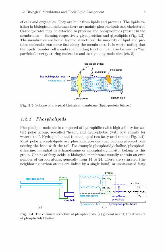
of cells and organelles. They are built from lipids and proteins. The lipids ex-isting in biological membranes there are mainly phospholipids and cholesterol.Carbohydrates may be attached to proteins and phospholipids present in themembranes — forming respectively glycoproteins and glycolipids (Fig. 1.3).The membranes are liquid layered structures: the majority of lipid and pro-teins molecules can move fast along the membranes. It is worth noting thatthe lipids, besides cell membrane building function, can also be used as “fuelparticles”, energy storing molecules and as signaling molecules (ch. 8).
Fig. 1.3 Scheme of a typical biological membrane (lipid-protein bilayer)
1.2.1 Phospholipids
Phospholipid molecule is composed of hydrophilic (with high affinity for wa-ter) polar group, so-called “head”, and hydrophobic (with low affinity forwater) “tail”. Hydrophobic tail is made up of two fatty acid chains (Fig. 1.4).Most polar phospholipids are phosphoglycerides that contain glycerol con-necting the head with the tail. For example phosphatidylcholine, phosphati-dylserine, phosphatidylethanolamine or phosphatidylinositol belong to thisgroup. Chains of fatty acids in biological membranes usually contain an evennumber of carbon atoms, generally from 14 to 24. There are saturated (theneighboring carbon atoms are linked by a single bond) or unsaturated fatty
(a) (b)
Fig. 1.4 The chemical structure of phospholipids: (a) general model, (b) structureof phosphatidylcholine

6 1 Cells and Viruses
acids (when some of the neighboring carbon atoms are connected by a doublebond). The chain length and degree of saturation of fatty acids in lipids hassignificant influence on the fluidity of biological membranes.
When phospholipid molecules are placed in water, the hydrophilic headsare oriented facing the water and the hydrophobic tails avoid water. As result,the structure called the bilayer sheet, liposome, or micelle may be formed(Fig. 1.5).
Fig. 1.5 The structures formed by phospholipids in aqueous solutions. The fig-ure presents a section through the liposome and micelle and a fragment of thebilayer sheet. Altered from: http://upload.wikimedia.org/wikipedia/commons/c/c6/Phospholipids_aqueous_solution_structures.svg
1.2.2 Cholesterol and Steroids
Cholesterol does not exist in most prokaryotic cells, whereas it is a commoncomponent of mammalian cell membranes. It fulfills a critical role in the reg-ulation of membrane fluidity. Also synthesis of steroid hormones is based on
Fig. 1.6 The structure of cholesterol and other important steroids. They are char-acterized by the presence of four hydrocarbon rings, marked here as A, B, C andD. Due to the presence of the hydroxyl group (-OH) cholesterol has amphipathicproperties (it has hydrophobic and hydrophilic parts).

1.3 Nucleus 7
cholesterol (Fig. 1.6; ch. 8.1). Accumulation of cholesterol in an artery wallplays a key role in atherosclerosis — disorder, which can lead to heart attackor stroke.
1.3 Nucleus
The nucleus consists of a nuclear envelope (constructed from two membranes;Fig. 1.7), the nucleolus and nucleoplasm. The outer nuclear membrane is con-tinuous with the membrane of the rough endoplasmic reticulum (RER). Thespace between the membranes (perinuclear space) is continuous with theRER lumen. With the inner face of the nuclear envelope the nuclear laminais associated, which is built from fibrous proteins called lamins. The nuclearlamina binds inner nuclear membrane with genetic material. The geneticmaterial formed in the chromatin/chromosomes is located mainly in nucleo-plasm. However, the parts of various chromosomes, which contain groups ofgenes encoding rRNA (ch. 3.3.3), could be concentrated in one region formingthe nucleolus. Its main role is the production of rRNA.
Fig. 1.7 Scheme of a nuclear envelope which is built from two layers of doublelipid membrane. In the nucleus of mammals there are about 4,000 nuclear canals(pores) present, each composed of more than 100 various proteins.
1.3.1 Chromosomes and Karyotype
Chromosome is condensed structure, which is formed from a single DNAmolecule and associated proteins. In cells which are not dividing at the mo-ment, chromosomes are not visible in light microscope, because DNA togetherwith accompanying proteins (i.e. chromatin) is loosely spreaded across thenucleus. During cell division, in the stage of metaphase (see ch. 7), chromatincondenses and becomes visible in the form of chromosomes.
Each chromosome has the short and long arm (designated as p, from the“petit” and q, from the “queue”). The arms are separated by narrow region

8 1 Cells and Viruses
called the centromere (Fig. 1.8). The chromosomes of the same shape and size,containing similar genetic information (i.e. genes) are called homologous chro-mosomes. In diploid cells, one of them comes from the mother and the otherfrom the father. During cell division, at metaphase, replicated chromosomescontain two sister chromatids linked with centromere. Sister chromatids areidentical copies of the chromosome: they contain the same genes and thesame alleles (i.e. versions of genes in a particular location — locus — onthe homologous chromosome). However, homologous chromosomes containthe same genes, but alleles can vary. During cell division pairs of chromo-somes are separated into two daughter cells.
Fig. 1.8 Homologous chromosomes during mitotic cell division
Chromosomes are visible under the microscope only after staining. Then,they look like strings with dark and light transverse stripes. A set of chromo-somes present in the cell during metaphase is called a karyotype. Humangerm cells (egg or sperm) contain one set of chromosomes (haploid cell, 1n),that is 23 pieces: 22 autosomes and the X or Y. Somatic cells (remaining cellsof the body) normally contain two homologous sets of chromosomes, i.e. 46chromosomes (diploid, 2n). One set comes from mother, the other — fromfather. In other species the number of chromosomes varies from 1 to 1260.
Human somatic cells contain two chromosomes that determine sex: XX infemales and XY in males. In germ cells there is only one sex chromosome: infemale it is always X chromosome, in male — X or Y.

1.4 Cytoplasmic Organelles 9
1.4 Cytoplasmic Organelles
Organelles can be defined as intracellular, bounded by membrane, protein-lipid structures (the cell nucleus is also an organellum). In cytoplasm there arepresent mitochondria, chloroplasts, endoplasmic reticulum, Golgi apparatus,peroxisomes, lysosomes, vacuoles, and glyoxysomes.
1.4.1 Mitochondria
Eukaryotic cells have a lot of mitochondria — they can be up to a quarter ofthe cytoplasm volume. They are elongated organelles with a diameter rangingfrom 0.5 to 10 μm (the size corresponds to a bacterial cell). Mitochondria arecomposed from two membranes: outer and inner, which is strongly folded.Mitochondria have their own DNA (defined as the mtDNA) coding for pro-teins and RNA. However the majority of mitochondrial proteins are encodedby nuclear DNA.
Fig. 1.9 Chemical structure of ATP (adenosine triphosphate)
The main task of mitochondria is the production of ATP (adenosinetriphosphate), which provides source of energy for most cellular processes(Fig. 1.9). Energy is stored in phosphate bonds and can be released duringthe hydrolysis of ATP to ADP (adenosine diphosphate) and AMP (adenosinemonophosphate).
In animal cells, the catabolism (degradation of glucose and fatty acids)is the main source of energy for ATP synthesis. The production of ATP ta-kes place in the inner mitochondrial membrane. Energy is transferred fromelectron transport chain to the ATP synthase by movements of protons acrossthis membrane. As a result of disturbances in electron transport, the free ra-dicals (and other reactive oxygen species) can be generated, which can leadto damage of cell structures and to cell senescence.
10 1 Cells and Viruses
1.4.2 Chloroplasts
Cloroplasts, like mitochondria, are composed from inner and outer mem-brane. Chloroplasts also have their own DNA, but most of their proteins areencoded by nuclear DNA. The inner membrane of the chloroplast forms thy-lakoids, that contain chlorophyll. It absorbs light necessary for the light re-action of photosynthesis. In this stage, thanks to the energy absorbed fromlight quanta, oxygen molecule and hydrogen ions are formed from two wa-ter molecules. This results in forming of the concentration gradient in thethylakoid membrane. The flow of ions through the membrane is connectedwith the synthesis of ATP. Stroma is the liquid component of the chloro-plast, in which, using the energy from ATP, CO2 is assimilated and synthesisof saccharides occurs (dark reactions of photosynthesis).
1.4.3 Endoplasmic Reticulum and Golgi Complex
Endoplasmic reticulum (ER) forms an irregular network of cisternae, vesiclesand tubules inside the cell isolated from the cytosol by biological membranes.It exists in two forms: smooth and rough. The main role of the rough endoplas-mic reticulum is to process the newly synthesized proteins, so it is associatedwith ribosomes (ch. 6.2.1). Smooth endoplasmic reticulum is involved in thesynthesis and metabolism of lipids. It appears in highest quantities in livercells (hepatocytes).
After the initial processing of proteins in rough endoplasmic reticulum,they are closed in transport vesicles and directed to other membranous struc-tures — the Golgi apparatus (ch. 6.2.2). These structures are the main placeof sorting and modifications of proteins and lipids. Here, glycoproteins areproduced and they are transported further to their destinations. Golgi appa-ratus is also a place of synthesis of polysaccharides and mucopolysaccharides.
1.4.4 Peroxisomes, Lysosomes, Vacuoles, andGlyoxysomes
Peroxisomes contain enzymes, which allow degradation of amino acids andfatty acids. In these reactions the hydrogen peroxide is formed, which is ha-rmful to cells. It is converted to water and oxygen by an enzyme calledcatalase.
The main function of lysosomes is degradation of unnecessary macromole-cules. Macromolecules are hydrolyzed by the respective enzymes: nucleic acids(DNA and RNA) by nucleases, proteins by proteases, and lipids by lipa-ses. Lysosomes are present only in animal cells. In plant cells their functionsare performed by the vacuoles.

1.5 Viruses, Bacteriophages, and Virophages 11
Vacuoles store water, ions, saccharides, amino acids and other small mo-lecules. They can also store unnecessary cell products, which can undergo de-gradation here. Vacuoles usually fill about 30% of the cell volume, but canincrease its volume up to 90%.
Glyoxysomes are found mainly in plant seeds. Their main function is toconvert fatty acids into acetyl-CoA molecules, which in glyoxylate cycle areconverted into monosaccharides. This mechanism is important in the use offat reserves of oilseeds. It occurs during germination and when it is necessaryto launch a non-carbohydrate energy reserves in plants. Glyoxysomes andperoxisomes are also called microbodies.
1.5 Viruses, Bacteriophages, and Virophages
Viruses are macromolecular complexes built from proteins and nucleic acids.Viruses are not able to replicate by themselves — so they can not be treatedas living organisms. Replication of viruses is possible only within other cells,using enzymes from infected cells. Because of that viruses are intracellularparasites. Most viruses have a diameter between 10 and 300 nm (but disc-overed in 2013 Pandoraviruses have a size approaching 1μm). The moleculeof the virus (virion) consists of one or more molecules of DNA or RNA (asa carrier of genetic information of the virus) and a protein shell called acapsid. Some viruses have an additional lipid envelope which surrounds thecapsid. Most viruses are pathogens — after penetration into living cells theycause the diseases. Various viruses specifically attack different types of cells(e.g. the HIV virus attacks only the immune cells, mainly T lymphocytes).When a virus encounters “his” cell, it attaches to the cell membrane usingspecific receptors (proteins on the surface of host cells, which normally haveother functions). Then, it can inject its genetic material into the cell or wholevirus is absorbed entirely by the cell. Inside the cell, based on the informationcontained in the genetic material of the virus, there are created new virusmolecules. They can remain in the cell for a long latent phase, or leave afterthe cell destruction. It sometimes happens, that the virus remains in the cellin form of genetic material integrated with the host genome.
Viruses can also infect bacteria. Such viruses are called bacteriophages(or commonly phages). Bacteriophages can be used for treating bacterialinfections as an alternative to antibiotics. In biotechnology and genetic en-gineering, phages and other viruses are used to transfer genetic informationbetween cells (as so-called “vectors”).
There are also viruses that infect other viruses. The first known, calledSputnik, was discovered in 2008. It is a subviral agent that reproducesin amoeba cells which are already infected by a certain helper virus.

12 1 Cells and Viruses
Sputnik uses the helper virus’s machinery for reproduction and inhibits repli-cation of the helper virus. Viruses that depend on co-infection of the hostcell by helper viruses are known as satellite viruses. They act as a para-site of helper viruses. In analogy to the term bacteriophage they were calledvirophages.
The risk associated with viral infections may be very different. Many viralinfections do not cause diseases, for example, most people are infected withcytomegalovirus (CMV), however, do not show symptoms of the disease.There are also viruses that often lead to death of the organism, such as coro-navirus which cause Severe Acute Respiratory Syndrome (SARS) or the HIVvirus causing AIDS.
1.5.1 Classification of Viruses
The classification of viruses could be based on the type of genetic materialheld by the virus and on strategy of genetic material replication (the Bal-timore classification). Besides double-stranded DNA, the carrier of geneticinformation in prokaryotic and eukaryotic cells, also single-stranded DNA orRNA can be a genetic material in viruses. Viruses were divided into sevenclasses (ds — double strand, ss — single strand):
I. dsDNA viruses — contain double-stranded DNA;II. ssDNA viruses — contain single-stranded DNA;
III. dsRNA viruses — contain double-stranded RNA;IV. ssRNA(+) viruses — contain single-stranded RNA, (+) sense;V. ssRNA (-) viruses — contain single-stranded RNA, (-) sense;
VI. RNA viruses that use virally encoded reverse transcriptase toproduce DNA from the initial virion RNA genome;
VII. DNA viruses that use virally encoded reverse transcriptase.
The last two classes of viruses are known as retroviruses. Rewriting of thegenetic information from RNA to DNA is an essential part of their replica-tion cycle. This reaction is catalyzed by reverse transcriptase — an enzymeencoded by the virus genome (ch. 5.9).
References
1. Adams, M. (ed.): Subcellular Compartments. In: Miko, I. (ed.) Cell Biology. Na-ture Education (2011), http://www.nature.com/scitable/topic/subcellular-compartments-14122679
2. Coté, G., De Tullio, M. (eds.): Cell Origins and Metabolism. In: Miko, I. (ed.)Cell Biology. Nature Education (2011), http://www.nature.com/scitable/topic/cell-origins-and-metabolism-14122694
3. Davidson, M.W.: Introduction to Cell and Virus Structure. In: Molecular Ex-pressions Website. The Florida State University (2000),http://micro.magnet.fsu.edu/cells/index.html

References 13
4. La Scola, B., Desnues, C., Pagnier, I., et al.: The virophage as a unique parasiteof the giant mimivirus. Nature 455, 100–104 (2008)
5. Liang, B.: Construction of the Cell Membrane. Wisc-Online.com (2001),http://www.wisc-online.com/objects/ViewObject.aspx?ID=AP1101
6. Lodish, H., Berk, A., Zipursky, S.L., et al.: The Dynamic Cell. In: MolecularCell Biology, 4th edn., W.H. Freeman, New York (2000)
7. Mc Grath, S., van Sinderen, D. (eds.): Bacteriophage: Genetics and MolecularBiology, 1st edn. Caister Academic Press, Norfolk (2007)
8. O’Connor, C.M., Adams, J.U.: Essentials of Cell Biology. NPG Education, Cam-bridge (2010)
9. Ogata, H., Claverie, J.M.: Microbiology. How to Infect a Mimivirus. Science 321,1305–1306 (2008)