CFTR-deficient pigs display alterations of bone ...
11
HAL Id: hal-02406264 https://hal.univ-reims.fr/hal-02406264 Submitted on 23 Oct 2020 HAL is a multi-disciplinary open access archive for the deposit and dissemination of sci- entific research documents, whether they are pub- lished or not. The documents may come from teaching and research institutions in France or abroad, or from public or private research centers. L’archive ouverte pluridisciplinaire HAL, est destinée au dépôt et à la diffusion de documents scientifiques de niveau recherche, publiés ou non, émanant des établissements d’enseignement et de recherche français ou étrangers, des laboratoires publics ou privés. Distributed under a Creative Commons Attribution - NonCommercial - NoDerivatives| 4.0 International License CFTR-deficient pigs display alterations of bone microarchitecture and composition at birth Julien Braux, Marie-Laure Jourdain, Christine Guillaume, Valérie Untereiner, Olivier Piot, Andrea Baehr, Nikolai Klymiuk, Nathalie Winter, Mustapha Berri, Dominique Buzoni-Gatel, et al. To cite this version: Julien Braux, Marie-Laure Jourdain, Christine Guillaume, Valérie Untereiner, Olivier Piot, et al.. CFTR-deficient pigs display alterations of bone microarchitecture and composition at birth. Journal of Cystic Fibrosis, Elsevier, 2020, 19 (3), pp.466-475. 10.1016/j.jcf.2019.10.023. hal-02406264
Transcript of CFTR-deficient pigs display alterations of bone ...

HAL Id: hal-02406264https://hal.univ-reims.fr/hal-02406264
Submitted on 23 Oct 2020
HAL is a multi-disciplinary open accessarchive for the deposit and dissemination of sci-entific research documents, whether they are pub-lished or not. The documents may come fromteaching and research institutions in France orabroad, or from public or private research centers.
L’archive ouverte pluridisciplinaire HAL, estdestinée au dépôt et à la diffusion de documentsscientifiques de niveau recherche, publiés ou non,émanant des établissements d’enseignement et derecherche français ou étrangers, des laboratoirespublics ou privés.
Distributed under a Creative Commons Attribution - NonCommercial - NoDerivatives| 4.0International License
CFTR-deficient pigs display alterations of bonemicroarchitecture and composition at birth
Julien Braux, Marie-Laure Jourdain, Christine Guillaume, Valérie Untereiner,Olivier Piot, Andrea Baehr, Nikolai Klymiuk, Nathalie Winter, Mustapha
Berri, Dominique Buzoni-Gatel, et al.
To cite this version:Julien Braux, Marie-Laure Jourdain, Christine Guillaume, Valérie Untereiner, Olivier Piot, et al..CFTR-deficient pigs display alterations of bone microarchitecture and composition at birth. Journalof Cystic Fibrosis, Elsevier, 2020, 19 (3), pp.466-475. �10.1016/j.jcf.2019.10.023�. �hal-02406264�

Journal of Cystic Fibrosis 19 (2020) 466–475
Contents lists available at ScienceDirect
Journal of Cystic Fibrosis
journal homepage: www.elsevier.com/locate/jcf
Original Article
CFTR-deficient pigs display alterations of bone microarchitecture and
composition at birth
Julien Braux
a , Marie-Laure Jourdain
a , Christine Guillaume
a , Valérie Untereiner b , Olivier Piot c , Andrea Baehr d , Nikolai Klymiuk
d , Nathalie Winter e , Mustapha Berri e , Dominique Buzoni-Gatel e , Ignaccio Caballero
e , Antoine Guillon
f , Mustapha Si-Tahar f , Jacky Jacquot a , ∗, Frédéric Velard
a , ∗
a Université de Reims Champagne Ardenne, BIOS EA 4691, Biomatériaux et Inflammation en site osseux, SFR CAP-Santé (FED 4231), 1, Avenue du Maréchal
Juin, 51097 Reims, France b Université de Reims Champagne Ardenne (URCA), PICT Platform, Reims, 1, Avenue du Maréchal Juin, 51097 Reims, France c Université de Reims Champagne-Ardenne, BioSpecT (Translational BioSpectroscopy) EA 7506, 1, Avenue du Maréchal Juin, 51097 Reims, France d Institute of Molecular Animal Breeding and Biotechnology, Gene Center, Ludwig-Maximilians-Universitat Munchen, Hackerstrasse 27, 85764,
Oberschleissheim, Germany e INRA, UMR1282 ISP, Centre de recherches INRA Val de Loire, 37380, Nouzilly, France f Inserm, Centre d’Etude des Pathologies Respiratoires, UMR1100/EA6305, 10 Boulevard Tonnellé, 37032, Tours, France
a r t i c l e i n f o
Article history:
Received 17 April 2019
Revised 7 October 2019
Accepted 22 October 2019
Available online 29 November 2019
Keywords:
Cystic fibrosis
Bone disease
Pigs
Cftr
Cortical bone
Trabecular bone
Femur
a b s t r a c t
Background: The lack of cystic fibrosis transmembrane conductance regulator (CFTR) function causes cys-
tic fibrosis (CF), predisposing to severe lung disease, reduced growth and osteopenia. Both reduced bone
content and strength are increasingly recognized in infants with CF before the onset of significant lung
disease, suggesting a developmental origin and a possible role in bone disease pathogenesis. The role
of CFTR in bone metabolism is unclear and studies on humans are not feasible. Deletion of CFTR in pigs
( CFTR −/ − pigs) displays at birth severe malformations similar to humans in the intestine, respiratory tract,
pancreas, liver, and male reproductive tract.
Methods: We compared bone parameters of CFTR −/ − male and female pigs with those of their wild-type
(WT) littermates at birth. Morphological and microstructural properties of femoral cortical and trabecular
bone were evaluated using micro-computed tomography ( μCT), and their chemical compositions were
examined using Raman microspectroscopy.
Results: The integrity of the CFTR −/ − bone was altered due to changes in its microstructure and chemical
composition in both sexes. Low cortical thickness and high cortical porosity were found in CFTR −/- pigs
compared to sex-matched WT littermates. Moreover, an increased chemical composition heterogeneity
associated with higher carbonate/phosphate ratio and higher mineral crystallinity was found in CFTR −/ −
trabecular bone, but not in CFTR −/ − cortical bone.
Conclusions: The loss of CFTR directly alters the bone composition and metabolism of newborn pigs.
Based on these findings, we speculate that bone defects in patients with CF could be a primary, rather
than a secondary consequence of inflammation and infection.
© 2019 The Author(s). Published by Elsevier B.V. on behalf of European Cystic Fibrosis Society.
This is an open access article under the CC BY-NC-ND license.
( http://creativecommons.org/licenses/by-nc-nd/4.0/ )
a
W
t
d
1. Introduction
Cystic fibrosis (CF) disease, which is caused by mutations of
the CF transmembrane conductance regulator ( CFTR) gene, is char-
∗ Corresponding authors.
E-mail addresses: [email protected] (J. Jacquot), frederic.velard@univ-
reims.fr (F. Velard).
p
w
v
i
https://doi.org/10.1016/j.jcf.2019.10.023
1569-1993/© 2019 The Author(s). Published by Elsevier B.V. on behalf of European Cystic
( http://creativecommons.org/licenses/by-nc-nd/4.0/ )
cterized by multiorgan deficiencies that begin early in life [1,2] .
ith the increasing life expectancy due to improvements in the
reatment of the disease’s pulmonary and gastrointestinal disor-
ers, other CF complications and comorbidities have become more
revalent, such as diabetes and CF-related bone disease (CFBD)
ith 55–65% of affected patients being older than 45 [3–5] . Indi-
iduals with CF have low bone mineral density (BMD) [6–8] and
ncreased fracture rate as early as adolescence, [9] which leads to
Fibrosis Society. This is an open access article under the CC BY-NC-ND license.

J. Braux, M.-L. Jourdain and C. Guillaume et al. / Journal of Cystic Fibrosis 19 (2020) 466–475 467
e
t
f
p
t
i
p
s
c
u
l
fl
C
i
[
e
[
d
t
s
s
f
b
I
o
k
i
c
f
[
i
r
c
t
r
p
m
n
m
p
d
m
C
t
t
b
a
t
t
i
a
2
2
l
T
m
p
b
v
C
a
i
b
C
e
2
o
C
t
c
l
v
e
t
w
o
s
u
w
i
t
d
c
o
t
T
u
(
b
s
d
(
u
d
c
t
(
n
l
(
v
a
t
i
T
t
T
i
o
n
μ
2
c
xcessive kyphosis, thus weakening the thoracic skeletal architec-
ure and resulting in an accelerated decline in lung function, inef-
ective cough and airway clearance [10–12] .
The identification of bone composition and microarchitecture in
atients with CF is a major determinant of bone density and frac-
ure risk. Well done cross-sectional and longitudinal studies have
ndicated diminished bone accrual during the skeletal formative
eriod of late childhood and adolescence [11,13,14] . A recent report
howed a 9-fold higher fracture rate in young German CF adults
ompared to the age-matched reference population [15] . It is still
nknown if the smaller stature in CF patients is a direct cause of
oss of CFTR or a manifestation of nutritional deficiencies and in-
ammation [16] . Lower BMD gains were observed in infants with
F as early as the age of 6 with normal nutritional status, suggest-
ng that CFBD may be due to a primary defect in bone metabolism
17] .
CFTR protein was first discovered as being expressed during
arly development and was then identified in human bone cells
18] , so CFTR deficiency may impact fetal tissues during in utero
evelopment. Cortical bone mass, comprising 85% of the skele-
on, and bone microarchitecture are major determinants of bone
trength and fracture risk in humans [18] . Skeletal homeosta-
is is maintained throughout life by the balance between bone-
orming osteoblasts (which derive from mesenchymal cells) and
one-resorptive osteoclasts (which have hematopoietic origin) [19] .
n patients with CF, we suggested a direct genetic component
f CFBD with an abnormal elevation in receptor activator of NF-
B ligand (RANK-L) in osteoblasts bearing the F508del mutation
n Cftr [20] . We recently reported that the CFTR potentiator iva-
aftor improved BMD in G551D-bearing CF patients and argued
or a link between CFTR activity and the function of osteoblasts
7] and osteoclasts precursors [21] . Several studies made on an-
mal models (rats, mice and pigs) with CF shortly after birth
evealed abnormalities in bone development [24–27] and tra-
heal cartilage with early airflow obstruction [27–31] , suggesting
he critical role of CFTR dysfunction. Several other species (fer-
et [32] , zebrafish [33] ) were used for this investigation on CF
athology.
The anatomy and physiology of pigs are similar to that of hu-
ans [22] . The CFTR −/ − pig model develops both intestinal (meco-
ium ileus, microcolon) and lung pathology, lesions in smooth
uscle and cartilage rings resembling alterations observed in CF
atients [23] . This emphasizes that CF pigs mimic the human con-
ition to overcome several of the limitations found in CF mouse
odels. We recently targeted the porcine CFTR gene and generated
FTR −/ − pigs which also display at birth severe malformations in
he intestine, respiratory tract, pancreas, liver, and male reproduc-
ive tract [ 24 , 25 ]. These phenotypic abnormalities closely resemble
oth the human CF pathology as well as alterations observed in
previously published CF pig model generated by a different gene
argeting strategy [26] . This new model provided us with an oppor-
unity to ask whether CFTR −/ − pigs (male and female) also man-
fest a defective bone microarchitecture and chemical composition
t birth.
. Methods
.1. Animals
All experiments were conducted in accordance with the guide-
ines of the Institutional Animal Care and Use Committee at INRA.
he protocol was approved by the “Comité d’Ethique en Expéri-
entation Animale Val de Loire” (n ° 0 0 028.01). CFTR + / − pigs were
roduced by replacing the exon 1 of the CFTR gene by a STOP
ox and a neo cassette using homologous recombination by BAC
ectors as previously described [25] . Single male and female
FTR + / − transgenic pigs were moved to INRA, Nouzilly (France)
nd mated to generate wild-type (WT), CFTR + / − and CFTR −/ − an-
mals. Piglets were euthanized with an i.v. overdose of pento-
arbital (Dolethal, Vétoquinol, France). A total of 17 WT and 23
FTR −/ − newborn piglets from 8 different litters were used in our
xperiments.
.2. μCT evaluation
This study was conducted following the quantitative analysis
f bone by micro-computed tomography recommendations from
ampbell and Sophocleus [27] . We examined the pigs’ femoral cor-
ical and trabecular bones less than 24 h after birth using mi-
ro computed tomography ( μCT, Skyscan 1076) scans with the fol-
owing settings: tube voltage, 80 kV; tube current, 0.125 mA; and
oxel size, 17,9 × 17,9 × 17,9 μm
3 . Samples were isolated, fixed in
thanol (70% in Dulbecco’s Phosphate Buffer Saline, Gibco), washed
hen stored in same ethanol solution at 4 °C. For scanning, samples
ere cleaned from surrounding soft tissue, rehydrated through an
vernight storage in saline medium, then scanned directly in that
olution. Three-dimensional (3D) images were rebuilt and analysed
sing respectively the NRecon GPU version and CTAn (Bruker) soft-
are programs. Bone mineral densities values were obtained us-
ng a standard regression line generated by converting the attenua-
ion coefficient from scans of hydroxyapatite standards with known
ensities to mineral density (0.25 and 0.75 g.cm
−3 ). After 3D re-
onstruction, bone volumes were segmented using a global thresh-
ld of 0.373 g/cm
3 which was set using the mean density leading
o the segment bone retrieved in scans after manual segmentation.
he whole femoral bone was first studied to avoid bias due to the
se of regions of interest. Common bone parameters were assessed
Femoral Length, Tissue volume (TV), Bone volume (BV), Percent
one volume (BV/TV), Tissue surface (TS), Bone surface (BS), Bone
urface density (BS/TV), and Bone Mineral Density (BMD).
Then a 3.4-mm-wide region of interest centered on the mid-
le of the femur was analysed to complete the cortical bone study
Fig. 1 A, B). In cortical regions, we analysed the cortical total vol-
me (Ct.TV), the cortical bone volume (Ct.BV), the bone volume
ensity (Ct.BV/Ct.TV), the cortical mineral density (Ct.BMD), the
ortical tissue surface (Ct.TS), the cortical bone surface (Ct.BS),
he cortical bone surface density (Ct.TS/TV), the cortical thickness
Ct.Th), the cortical radius (Ct.Rd), and the ratio of cortical thick-
ess / bone radius. The porosity of bones was assessed by calcu-
ating the porosity (defined as a connected assemblage of empty
black) voxels that is fully surrounded on all sides by solid (white)
oxels) expressed as pores number, volume, surface and percent-
ge of the volume of pores within the bone. X-rays 3D rebuilds
hat highlighed porosities were calculated on the samples present-
ng median values in terms of volume of pores.
For the trabecular bone, Tb.BV, Tb.TV, Tb.BV/Tb.TV, Tb.TS, Tb.BS,
b.BS/Tb.TS trabecular number (Tb.N), trabecular thickness (Tb.Th),
rabecular separation (Tb.Sp), trabecular pattern factor (Tb.Pf), and
b.BMD were calculated while analysing a 2-mm-wide region of
nterest located 1 mm under the growth plate ( Fig. 1 A, C). A total
f 23 newborn CFTR −/- piglets (14 males and 9 females) and 17
ewborn WT piglets (8 males and 9 females) were subjected to
CT scan.
.3. Raman imaging spectroscopy
The composition and distribution of organic and inorganic
omponents of the cortical and trabecular bones were evaluated

468 J. Braux, M.-L. Jourdain and C. Guillaume et al. / Journal of Cystic Fibrosis 19 (2020) 466–475
Fig. 1. Femoral bone regions of interest are: A ) whole femoral bone, B ) cortical bone was analyzed in a ROI centered on the femoral midshaft defined as the midpoint of the
femoral bone, C ) trabecular bone was analyzed at the distal femoral metaphysis, starting 1 mm away from the growth plate.
e
u
s
i
i
r
d
i
c
o
w
s
i
1
I
C
a
c
r
l
1
t
a
p
r
a
a
2
i
i
w
R
w
s
using Raman imaging spectroscopy. Raman provides quantitative
information on the changes in the mineral and matrix composi-
tion as well as the nature and quantities of mineral constituents.
Raman spectroscopy was chosen over infrared spectroscopy in this
study as it has several inherent advantages such as minimal sam-
ple preparation, application to biological samples and potential in
vivo implementation [28] .
Raman images were recorded using a LabRam Aramis spectrom-
eter (Horiba Scientific, Villeneuve d’Ascq, France) equipped with an
Olympus microscope (model BX41) and a 100 × long-working dis-
tance objective (Olympus, NA 0.9) for sample excitation and scat-
tered light collection. The laser used for excitation was a diode
emitting a radiation at 785 nm, near infrared excitation source,
delivering around 13 mW to the sample. The Raman signal was
dispersed using a 1200 lines/mm grating. For each bone sample
(4 CFTR −/- and 3 WT piglets) subjected to Raman spectroscopy,
twenty images were recorded in both cortical and trabecular areas
of femurs. For each acquisition, the total exposure time was 60 s
using 2 accumulations of 30 s. For any region of each sample, 20
spectra were combined to obtain an average spectrum. These were
then averaged for all of the bones of each genotype to obtain an
overall average. The spectra wavenumbers were detected on the
fingerprint range (60 0–180 0 cm
−1 ). Before analysis, raw Raman
spectra need to be pre-processed. This was done with a home-
made software using the Matlab environment (The MathWorks,
Inc., Natick, Massachusetts). All spectra were first corrected for
instrument response, and smoothed with a Savistsky-Golay func-
tion (second order polynomial, 7-point window length) to reduce
noise. Afterwards, all spectra were baseline corrected with a poly-
nomial function (order 2) and were vector-normalized in order to
make the spectra comparable. An unsupervised processing method,
principal component analysis (PCA), was performed on the pre-
processed data to explore the spectral variability of the datasets.
This method allows spectral data reduction replacing original and
correlated variables by synthetic and uncorrelated variables called
principal components (PCs). These PCs contain the total informa-
tion and are arranged to explain the highest to lowest variance of
the dataset. The results are presented using the scores of the most
xplained PCs. PC loadings are also useful to highlight the molec-
lar origins of the spectral variability specific to a dataset. In this
tudy, the mean spectrum of each group was subtracted from the
ndividual spectra of the same group in order to remove redundant
nformation, and PCA score plots were performed in the spectral
ange 110 0–80 0 cm
−1 .
Bone is composed of ~65 wt% mineral-non-stoichiometric hy-
roxyapatite (Ca 10 (PO 4 ) 6 (OH) 2 ), 10 wt% water, and 25 wt% organ-
cs. Organics mainly include type I collagen (~22.5 wt%), non-
ollagenous proteins (2.5–3.75 wt%), and lipids (1–10 wt%) [29] . In
ur Raman study, the intensities of the following vibration peaks
ere measured: phosphate υ1 at ~958 cm-1 (PO 4 3 − symmetric
tretching), carbonate υ1 at ~1071 cm
−1 (CO 3 2 − symmetric stretch-
ng), CH 2 wagging at ~1450 cm
−1 (C
–H bending), amide I around
667 cm-1 (mainly C
= O stretching of the peptide bonds), amide
II at ~1243 cm
−1 (in -phase combination of the N
–H bending and
–N stretching). The width of phosphate υ1 at ~958 cm
−1 was
lso measured for both cortical and trabecular bones. Three bone
ompositional parameters were examined: i/ the mineral-to-matrix
atio estimated using the integrated intensities under the base-
ined peak from 915 to 1215 cm
−1 over the amide I area (1596–
720 cm
−1 ), ii/ the carbonate-to-phosphate ratio calculated from
he ratio of integrated intensities 850–895 cm
−1 /915–1215 cm
−1 ,
nd iii/ the mineral crystallinity determined by the inverse of the
hosphate 958 cm
−1 bandwidth. It was reported by a number of
esearchers that the bandwidth of the PO 4 3 −υ1 peak decreases
s the mineral crystallinity improves (increase in the crystal size
nd/or atomic ordering) [30] .
.4. Statistical analyses
The effect of genotype was statistically assessed. Morpholog-
cal measures of CFTR −/ − and WT bones were compared us-
ng the Wilcoxon signed rank test (paired samples) where pigs
ere compared to their siblings. Measurements obtained from the
aman analysis were evaluated using analysis of variance (one-
ay nested ANOVA). P -values smaller than 0.05 were considered
ignificant.

J. Braux, M.-L. Jourdain and C. Guillaume et al. / Journal of Cystic Fibrosis 19 (2020) 466–475 469
Table 1
Piglet characteristics and macrostructural indexes.
n
Body Weight
(kg - mean ± SD)
Femur Length
(mm - mean ± SD)
Femur Volume
(cm3 - mean ± SD)
Femur Bone Volume
(cm3 - mean ± SD)
% Bone Volume
(mean ± SD)
Whole Bone
WT
Male 8 1.098 ± 0.186 40.56 ± 2.38 1.94 ± 0.40 1.03 ± 0.29 53.32 ± 6.04
Female 9 1.182 ± 0.293 41.23 ± 2.91 ∗ 2.11 ± 0.46# 1.15 ± 0.29§ 54.34 ± 4.90 μ
CFTR −/ −Male 14 1.185 ± 0.339 40.52 ± 4.34 1.92 ± 0.48 0.98 ± 0.22 51.35 ± 7.24
Female 9 1.170 ± 0.281 39.50 ± 2.10 ∗ 1.86 ± 0.51# 0.95 ± 0.34§ 50.49 ± 6.41 μ
Wilcoxon signed rank tests; ∗ , #, §, μ means statistically different (respectively p = 0,029, p = 0,040, p = 0,010 and p = 0,048).
3
3
W
(
m
W
v
p
4
t
v
F
t
b
d
s
l
3
e
t
1
c
p
n
. Results
.1. Femoral bone volume was reduced in CFTR KO piglets
At birth, the whole-body weight of 23 CFTR −/ − and 17
T newborn pigs was similar whatever the sex and genotype
Table 1 ). To determine whether CFTR played a role in bone
ass, we first compared femur length and femoral volume of
T and CFTR −/ − pigs ( Table 1 ). Both femur length and femoral
olume were significantly reduced in CFTR −/ − female pigs com-
ared to their respective WT littermates (39.50 mm ± 2.10 vs
1.23 mm ± 2.91 and 1.86 ± 0.51 cm
3 vs 2.11 ± 0.46 cm
3 , respec-
ively).
CFTR −/ − female pigs had significantly reduced femoral bone
olume in comparison to WT female pigs (reduced 17%, p < 0.05).
emoral bone volume was also reduced in male CFTR −/ − pigs, but
iFig. 2. Representative 3-D images of femoral cortical bone ( A ), cortical min
his did not reach statistical significance. Since the percentage of
one volume (femur bone volume / femur volume) was still re-
uced in CFTR -/- pigs, the reduction of femoral bone volume ob-
erved in CFTR -/- pigs could not be explained only by the shorter
ength and/or circumference of the femur ( Table 1 ).
.2. Cortical bone mass was reduced and intracortical porosity
nhanced in CFTR KO piglets
Comparisons of cortical bone representative 3-D images be-
ween WT and CFTR −/ − pigs were obtained as shown in the Figs.
A, B and depicted in Fig. 2 A. The cortical mineral density and
ortical thickness/radius were lower in CFTR −/- male and female
igs compared to sex-matched WT littermates ( Fig. 2 B, C). We
ext assessed whether the loss of CFTR affected the cortical poros-
ty, via μCT-based analyses. Views of representative 3D images of
eral density (B ), and cortical thickness ( C ) of WT and CFTR −/ − pigs.
470 J. Braux, M.-L. Jourdain and C. Guillaume et al. / Journal of Cystic Fibrosis 19 (2020) 466–475
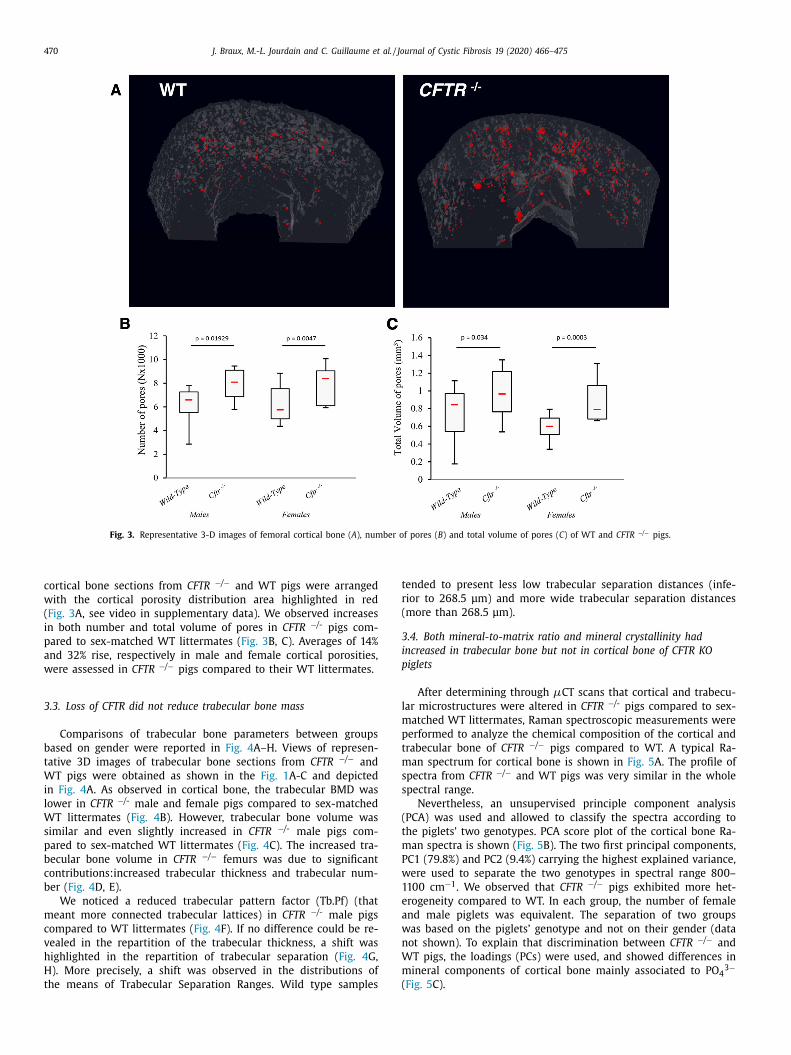
Fig. 3. Representative 3-D images of femoral cortical bone ( A ), number of pores ( B ) and total volume of pores ( C ) of WT and CFTR −/ − pigs.
t
r
(
3
i
p
l
m
p
t
m
s
s
(
t
m
P
w
1
e
a
w
n
W
m
(
cortical bone sections from CFTR −/ − and WT pigs were arranged
with the cortical porosity distribution area highlighted in red
( Fig. 3 A, see video in supplementary data). We observed increases
in both number and total volume of pores in CFTR −/- pigs com-
pared to sex-matched WT littermates ( Fig. 3 B, C). Averages of 14%
and 32% rise, respectively in male and female cortical porosities,
were assessed in CFTR −/ − pigs compared to their WT littermates.
3.3. Loss of CFTR did not reduce trabecular bone mass
Comparisons of trabecular bone parameters between groups
based on gender were reported in Fig. 4 A–H. Views of represen-
tative 3D images of trabecular bone sections from CFTR −/ − and
WT pigs were obtained as shown in the Fig. 1 A-C and depicted
in Fig. 4 A. As observed in cortical bone, the trabecular BMD was
lower in CFTR −/- male and female pigs compared to sex-matched
WT littermates ( Fig. 4 B). However, trabecular bone volume was
similar and even slightly increased in CFTR −/- male pigs com-
pared to sex-matched WT littermates ( Fig. 4 C). The increased tra-
becular bone volume in CFTR −/ − femurs was due to significant
contributions:increased trabecular thickness and trabecular num-
ber ( Fig. 4 D, E).
We noticed a reduced trabecular pattern factor (Tb.Pf) (that
meant more connected trabecular lattices) in CFTR −/- male pigs
compared to WT littermates ( Fig. 4 F). If no difference could be re-
vealed in the repartition of the trabecular thickness, a shift was
highlighted in the repartition of trabecular separation ( Fig. 4 G,
H). More precisely, a shift was observed in the distributions of
the means of Trabecular Separation Ranges. Wild type samples
ended to present less low trabecular separation distances (infe-
ior to 268.5 μm) and more wide trabecular separation distances
more than 268.5 μm).
.4. Both mineral-to-matrix ratio and mineral crystallinity had
ncreased in trabecular bone but not in cortical bone of CFTR KO
iglets
After determining through μCT scans that cortical and trabecu-
ar microstructures were altered in CFTR −/- pigs compared to sex-
atched WT littermates, Raman spectroscopic measurements were
erformed to analyze the chemical composition of the cortical and
rabecular bone of CFTR −/ − pigs compared to WT. A typical Ra-
an spectrum for cortical bone is shown in Fig. 5 A. The profile of
pectra from CFTR −/ − and WT pigs was very similar in the whole
pectral range.
Nevertheless, an unsupervised principle component analysis
PCA) was used and allowed to classify the spectra according to
he piglets’ two genotypes. PCA score plot of the cortical bone Ra-
an spectra is shown ( Fig. 5 B). The two first principal components,
C1 (79.8%) and PC2 (9.4%) carrying the highest explained variance,
ere used to separate the two genotypes in spectral range 800–
100 cm
−1 . We observed that CFTR −/ − pigs exhibited more het-
rogeneity compared to WT. In each group, the number of female
nd male piglets was equivalent. The separation of two groups
as based on the piglets’ genotype and not on their gender (data
ot shown). To explain that discrimination between CFTR −/ − and
T pigs, the loadings (PCs) were used, and showed differences in
ineral components of cortical bone mainly associated to PO 4 3 −
Fig. 5 C).

J. Braux, M.-L. Jourdain and C. Guillaume et al. / Journal of Cystic Fibrosis 19 (2020) 466–475 471
Fig. 4. Representative 3-D images of male femoral trabecular bone (A), trabecular mineral density (B), percent trabecular volume (C), trabecular thickness (D), trabecular
number (E), trabecular pattern factor (F), and repartition of the mean percentages of each trabecular thickness ranges and separations retrieved in samples (G, H) of WT and
CFTR -/- pigs.

472 J. Braux, M.-L. Jourdain and C. Guillaume et al. / Journal of Cystic Fibrosis 19 (2020) 466–475
Fig. 5. Typical Raman spectrum of cortical bone from the WT (red line) and CFTR −/ − (blue line) pigs. Major bone mineral and matrix band positions and associated spectral
regions are marked ( A ). PCA score plot (PC1 vs PC2) with WT (red points) and CFTR −/ − (blue points) pigs ( B ). Loadings of PC1 (orange curve) and PC2 (brown curve) ( C) .
PCA and loadings were calculated in the spectral range 80 0–110 0 cm-1. Each symbol/color couple represents an independent animal. (For interpretation of the references to
color in this figure legend, the reader is referred to the web version of this article.)
p
a
(
d
u
p
A typical Raman spectrum for trabecular bone is shown in
Fig. 6 A. The spectral profiles between CFTR −/ − and WT pigs mainly
showed differences in the regions of amide III and CO 3 2 − bands.
PCA score plots were performed on spectra extracted from trabec-
ular bone in two genotypes. The first two principal components
PC1 and PC2 respectively carried 55.3% and 18.7% of total variance
( Fig. 6 B). The score plots showed a separation of CFTR −/ − and WT
igs according to the PC1 in the spectral range 80 0–110 0 cm
−1 ,
s well as differences in mineral components associated to CO 3 2 −
type-B carbonate) and phosphate groups (PO 4 3 −) ( Fig. 6 C).
In addition to this multivariate processing by PCA, spectral
ata were submitted to ratiometric analyses. The mean val-
es in the phosphate υ1/amide I ratio, type -B carbonate/ phos-
hate υ1ratio, and 1/PO 4 3 − were calculated in both cortical and

J. Braux, M.-L. Jourdain and C. Guillaume et al. / Journal of Cystic Fibrosis 19 (2020) 466–475 473
Fig. 6. Typical Raman spectrum of trabecular bone from the WT (red line) and CFTR −/ − (blue line) pigs. Major bone mineral and matrix band positions and associated
spectral regions are marked ( A ). PCA score plot (PC1 vs PC2) with WT (red points)
and CFTR −/ − (blue points) pigs ( B ). Loadings of PC1 (orange curve) and PC2 (brown
curve) ( C) . PCA and loadings were calculated in the spectral range 80 0–110 0 cm
−1 .
Each symbol/color couple represents an independent animal.
t
t
m
p
4
b
p
t
d
p
p
a
s
p
d
a
p
i
s
p
O
c
t
a
b
c
d
b
b
b
a
B
i
t
3
t
d
i
l
i
a
i
C
p
t
i
a
a
(
f
t
i
i
r
rabecular bones for each genotype ( Table 2 ). A marked increase in
he mineral/matrix ratio, the type-B carbonate substitution and the
ineral crystallinity were observed in trabecular bone of CFTR −/ −
igs compared to WT, but not found in cortical bone.
Table 2
The Phosphate υ1/Amide I Ratio, Type -B Carbonate/P
and Cortical Bone in CFTR -/- and WT Pigs.
Trabecular bone
Genotype WT CFTR -/- WT
υ1PO 4 3- /amide I 26.57 ± 8.33 ∗ 0.22 ±
(CO 3 2- / υ1PO 4
3- ) 21.40 ± 9.33 ∗ 0.20 ±1/ υ1PO 4
3- (x 10 + 3 ) 0.957 ± 0.336 ∗ 0.625 ±
Values are mean ± SEM. n = 4 ∗ significantly different to WT ( p < 0.01).
. Discussion
Cortical bone composition and density are key determinants of
one strength and fracture risk in humans [18] . Recent studies re-
orted an elevated fracture rate in CF patients [ 5 , 12 , 31 ] in rela-
ion with macroscopic bone architecture [32] , and others described
eficits in trabecular and cortical bone as well as strength in pre-
ubertal children, adolescents and adults with CF [ 8 , 33–35 ]. Peo-
le with CF have several risk factors to develop low bone mass
nd density including chronic lung inflammation, pancreatic in-
ufficiency, malnutrition, delayed puberty, glucocorticoid use, and
ossibly CFTR dysfunction itself [ 3 , 11 ]. Therefore, it is necessary to
iscriminate between the clinical disease’s multiple contributions
nd the intrinsic defect due to CFTR deficiency. At birth, lungs of CF
igs lack inflammation, but with time, they spontaneously develop
nfection, inflammation and remodeling [36] . To address the con-
equence of CFTR deficiency on bone microarchitecture, we com-
ared bone parameters of CFTR −/ − pigs to WT littermates at birth.
ur data prove that CFTR is fundamental to maintain the correct
hemical composition and microarchitecture of bone.
Several hypotheses emerged from our study regarding the func-
ion of CFTR in the bone metabolism. Our data demonstrate that
defective bone health is readily apparent in CFTR −/ − pigs at
irth. The length and bone volume of their femurs were reduced
ompared to WT pigs in females. Interestingly, Rogan et al. also
emonstrated that CF piglets had reduced humeral length and
one mineral content [37] . In our study, we evidenced an altered
one microarchitecture in CFTR −/ − pigs. Our μCT analysis of the
one architecture and mineral density reveals that CFTR deficiency
lters cortical bone structure and reduces cortical and trabecular
MD. Low cortical thickness and high cortical porosity were found
n CFTR −/- pigs. In contrast, trabecular bone volume was not al-
ered in these pigs. In our study, we evaluated averages of 14% and
2% rise in porosity of CFTR −/- male and female pigs compared
o sex-matched WT littermates. Elevated porosity is well known to
iminish bone resistance against fracture by increasing the veloc-
ty of crack propagation [ 38 , 39 ]. The identification of mechanisms
eading to enhanced porosity is likely to improve our understand-
ng of the pathogenesis of CFBD.
Prior investigations have revealed that bone cortical thinning
nd increased porosity occur when the rate of bone remodel-
ng is modified [40] . A possible altered bone remodeling of the
FTR −/- pigs is suggested by changes in the bones’ chemical com-
osition. Our Raman analysis of the CFTR −/ − femurs indicated
hat, compared with WT pigs’, their cortical bone was different
n mineral components associated to CO 3 2 − (type-B carbonate),
nd their trabecular bone was different in mineral components
ssociated to CO 3 2 − (type-B carbonate) and phosphate groups
PO 4 3 −). In addition, at the trabecular level, carbonate substitutes
or phosphate ions in bone (type-B carbonate) demonstrated that
he phosphate environment of the mineral was distorted with an
ncreased mineral/matrix ratio and elevated mineral crystallinity
n CFTR −/ − trabecular bone, all indicative of disrupted bone
emodeling.
hosphate υ1ratio and 1 υ/PO 4 3- in Trabecular
Cortical bone
CFTR -/- WT
0.04 0.044 ± 0.009 0.061 ± 0.045
0.01 0.205 ± 0.031 0.205 ± 0.031
0.208 0.600 ± 0.295 0.608 ± 0.283

474 J. Braux, M.-L. Jourdain and C. Guillaume et al. / Journal of Cystic Fibrosis 19 (2020) 466–475
A
t
2
i
S
f
R
Our study supplements the multiple studies on CF mice and
rats, which have identified reductions in cortical bone mass at-
tributed to the loss of CFTR [41–44] . Our laboratory [45] and others
[ 46 , 47 ] previously issued studies suggesting evidence of direct de-
pendence on bone growth due to CFTR , we indeed reported that
interventions to improve CFTR function in the F508del- CFTR mouse
[48] and in CF patients bearing the G551D-mutation in CFTR re-
sulted in enhanced bone density [7] .
4.1. Limitations
Our data bears advantages and limitations. First, we used an an-
imal model that was described to recapitulate human CF. Second,
we hypothesized that a pig’s bone was more similar to that of a
human’s than a mouse’s [50] . Third, we were capable of eliminat-
ing secondary consequences of the disease by studying newborn
pigs. As we did not address the role of bone CFTR , we were not
able to exclude the possibility that CFTR gene in bone cells con-
tributed to the phenotypes we observed.
We do not yet have a mechanism identified for the low cor-
tical bone thickness and enhanced cortical porosity in CFTR −/ −
pigs at birth, but we suspect the phenotype may be dependent
upon the altered osteoblastic activity and differentiation. Genetic
studies confirmed that activating the canonical WNT signaling in-
creased bone formation in mice by promoting osteoblast differ-
entiation and indirectly inhibiting bone resorption, thus reduc-
ing osteoclastogenesis mainly by osteoprotegerin (OPG) regulation
[ 19 , 49 ]. CFTR was shown to positively regulate WNT/ β-catenin
pathway in multiple cell types [ 51 , 52 ]. A decrease in canonical
WNT signaling was reported in CFTR −/ − rat osteoblasts [46] , as
well as a decreased WNT/ β-catenin in F508del- CFTR mice sig-
naling mediated reduced osteoblast differentiation and function
[53] . A direct effect of canonical WNT signaling of osteoblasts on
osteoclastogenesis was proposed, as mice lacking WNT16 in os-
teoblasts were characterized by a reduced cortical thickness and
high cortical bone porosity while their trabecular bone remained
unaffected [54] .
In F508del- CFTR human osteoblasts, we recently reported an
overexpression of receptor activator of nuclear factor kappa-B lig-
and (RANKL) and high membranous RANKL localization coupled
with a reduced OPG production which could be reversed with
CFTR activators [20] . That situation may reduce bone formation and
worsen bone resorption through the activation of osteoclasts lead-
ing to low cortical bone mass in people with CF. Further molecular
studies are needed to determine which of the signaling pathways
may have contributed to the low cortical bone mass and high bone
cortical porosity
5. Conclusion
We provide evidence that loss of CFTR alters bone chemical
composition and microarchitecture in pigs at birth. This might in-
dicate that bone defects in people with cystic fibrosis are likely
primary, regardless of significant clinical confounders such as in-
flammation and infection from the start of life. The lower cortical
bone mass in our study’s CFTR −/ − pigs and present in children and
adolescents with CF [8] suggests that the use of CFTR potentiators
and correctors might benefit people with CF bone disease as far as
their skeletal health is concerned.
Declaration of Competing Interest
All authors report no conflicts of interest in this work.
cknowledgments
This study was supported in part by the French Associa-
ion Vaincre la Mucoviscidose, grants RF20130500925 and RF
0170501938 . The authors thank Dr. Marjorie Cantener for correct-
ng the manuscript.
upplementary materials
Supplementary material associated with this article can be
ound, in the online version, at doi: 10.1016/j.jcf.2019.10.023 .
eferences
[1] Riordan JR , Rommens JM , Kerem B , et al. Identification of the cystic fibrosisgene: cloning and characterization of complementary DNA [published erratum
appears in Science 1989 sep 29;245(4925):1437]. Science 1989;245:1066–73 .
[2] Stoltz DA , Meyerholz DK , Welsh MJ . Origins of cystic fibrosis lung disease. NEngl J Med 2015;372:351–62 .
[3] Jacquot J , Delion M , Gangloff S , et al. Bone disease in cystic fibrosis: newpathogenic insights opening novel therapies. Osteoporos Int 2016;27:1401–12 .
[4] Plant BJ , Goss CH , Plant WD , et al. Management of comorbidities in older pa-tients with cystic fibrosis. Lancet Respir Med 2013;1:164–74 .
[5] Putman MS , Milliren CE , Derrico N , et al. Compromised bone microarchitec-
ture and estimated bone strength in young adults with cystic fibrosis. J ClinEndocrinol Metab 2014;99:3399–407 .
[6] Aris RM , Merkel PA , Bachrach LK , et al. Guide to bone health and disease incystic fibrosis. J Clin Endocrinol Metab 2005;90:1888–96 .
[7] Sermet-Gaudelus I , Delion M , Durieu I , et al. Bone demineralization is im-proved by ivacaftor in patients with cystic fibrosis carrying the p.Gly551Asp
mutation. J Cyst Fibros 2016;15:e67–9 . [8] Kelly A , Schall J , Stallings VA , et al. Trabecular and cortical bone deficits are
present in children and adolescents with cystic fibrosis. Bone 2016;90:7–14 .
[9] Buntain HM , Greer RM , Schluter PJ , et al. Bone mineral density in Australianchildren, adolescents and adults with cystic fibrosis: a controlled cross sec-
tional study. Thorax 2004;59:149–55 . [10] Paccou J , Zeboulon N , Combescure C , et al. The prevalence of osteoporosis, os-
teopenia, and fractures among adults with cystic fibrosis: a systematic litera-ture review with meta-analysis. Calcif Tissue Int 2010;86:1–7 .
[11] Sermet-Gaudelus I , Castanet M , Retsch-Bogart G , et al. Update on cystic fi-
brosis-related bone disease: a special focus on children. Paediatr Respir Rev2009;10:134–42 .
[12] Aris RM , Renner JB , Winders AD , et al. Increased rate of fractures and severekyphosis: sequelae of living into adulthood with cystic fibrosis. Ann Intern
Med 1998;128:186–93 . [13] Buntain HM , Schluter PJ , Bell SC , et al. Controlled longitudinal study of
bone mass accrual in children and adolescents with cystic fibrosis. Thorax
2006;61:146–54 . [14] Sparks AA , McGee SJ , Boone CE , et al. ’Old’ bones in young bodies: the tale of
cystic fibrosis. Curr Opin Endocrinol Diabetes Obes 2009;16:407–14 . [15] Stahl M , Holfelder C , Kneppo C , et al. Multiple prevalent fractures in relation
to macroscopic bone architecture in patients with cystic fibrosis. J Cyst Fibros2016 .
[16] Bournez M , Bellis G , Huet F . Growth during puberty in cystic fibrosis: a retro-
spective evaluation of a French cohort. Arch Dis Child 2012;97:714–20 . [17] Sermet-Gaudelus I , Souberbielle JC , Ruiz JC , et al. Low bone mineral density in
young children with cystic fibrosis. Am J Respir Crit Care Med 2007;175:951–7 .[18] Zebaze RM , Ghasem-Zadeh A , Bohte A , et al. Intracortical remodelling and
porosity in the distal radius and post-mortem femurs of women: a cross-sec-tional study. Lancet 2010;375:1729–36 .
[19] Baron R , Kneissel M . WNT signaling in bone homeostasis and disease: from
human mutations to treatments. Nat Med 2013;19:179–92 . [20] Delion M , Braux J , Jourdain ML , et al. Overexpression of RANKL in osteoblasts:
a possible mechanism of susceptibility to bone disease in cystic fibrosis. JPathol 2016;240:50–60 .
[21] Velard F , Jourdain ML , Abdallah D , et al. Overexpression of RANK and M-CSFRin Monocytes of G551D-Bearing patients with cystic fibrosis. Am J Respir Crit
Care Med 2018;198:968–70 .
[22] Meyerholz DK . Lessons learned from the cystic fibrosis pig. Theriogenology2016;86:427–32 .
[23] Meyerholz DK , Stoltz DA , Namati E , et al. Loss of cystic fibrosis transmem-brane conductance regulator function produces abnormalities in tracheal de-
velopment in neonatal pigs and young children. Am J Respir Crit Care Med2010;182:1251–61 .
[24] Guillon A , Chevaleyre C , Barc C , et al. Computed tomography (CT) scanningfacilitates early identification of neonatal cystic fibrosis piglets. PLoS ONE
2015;10:e0143459 .
[25] Klymiuk N , Mundhenk L , Kraehe K , et al. Sequential targeting of CFTR byBAC vectors generates a novel pig model of cystic fibrosis. J Mol Med (Berl)
2012;90:597–608 . [26] Rogers CS , Stoltz DA , Meyerholz DK , et al. Disruption of the CFTR gene pro-
duces a model of cystic fibrosis in newborn pigs. Science 2008;321:1837–41 .

J. Braux, M.-L. Jourdain and C. Guillaume et al. / Journal of Cystic Fibrosis 19 (2020) 466–475 475
[
[
[
[
[
[
[
[
[
[
[
[
[
[
[
[
[
[
[
[
[27] Campbell GM , Sophocleous A . Quantitative analysis of bone and soft tissueby micro-computed tomography: applications to ex vivo and in vivo studies.
Bonekey Rep 2014;3:564 . 28] Mandair GS , Morris MD . Contributions of Raman spectroscopy to the under-
standing of bone strength. Bonekey Rep 2015;4:620 . 29] Goldberg M , Boskey AL . Lipids and biomineralizations. Prog Histochem Cy-
tochem 1996;31:1–187 . 30] Akkus O , Polyakova-Akkus A , Adar F , et al. Aging of microstructural compart-
ments in human compact bone. J Bone Miner Res 2003;18:1012–19 .
[31] Haworth CS , Freemont AJ , Webb AK , et al. Hip fracture and bone histomor-phometry in a young adult with cystic fibrosis. Eur Respir J 1999;14:478–9 .
32] Stahl M , Holfelder C , Kneppo C , et al. Multiple prevalent fractures in relationto macroscopic bone architecture in patients with cystic fibrosis. J Cyst Fibros
2018;17:114–20 . [33] O’Brien CE , Com G , Fowlkes J , et al. Peripheral quantitative computed tomog-
raphy detects differences at the radius in prepubertal children with cystic fi-
brosis compared to healthy controls. PLoS ONE 2018;13:e0191013 . 34] Kelly A , Schall JI , Stallings VA , et al. Deficits in bone mineral content in chil-
dren and adolescents with cystic fibrosis are related to height deficits. J ClinDensitom 2008;11:581–9 .
[35] Nishiyama KK , Agarwal S , Kepley A , et al. Adults with cystic fibrosis havedeficits in bone structure and strength at the distal tibia despite similar size
and measuring standard and relative sites. Bone 2018;107:181–7 .
36] Stoltz DA , Meyerholz DK , Pezzulo AA , et al. Cystic fibrosis pigs develop lungdisease and exhibit defective bacterial eradication at birth. Sci Transl Med
2010;2:29ra31 . [37] Rogan MP , Reznikov LR , Pezzulo AA , et al. Pigs and humans with cystic fibrosis
have reduced insulin-like growth factor 1 (IGF1) levels at birth. Proc Natl AcadSci U S A 2010;107:20571–5 .
38] Carriero A , Doube M , Vogt M , et al. Altered lacunar and vascular porosity in
osteogenesis imperfecta mouse bone as revealed by synchrotron tomographycontributes to bone fragility. Bone 2014;61:116–24 .
39] Carriero A , Zimmermann EA , Paluszny A , et al. How tough is brittle bone?Investigating osteogenesis imperfecta in mouse bone. J Bone Miner Res
2014;29:1392–401 . 40] Shigdel R , Osima M , Ahmed LA , et al. Bone turnover markers are associated
with higher cortical porosity, thinner cortices, and larger size of the proximal
femur and non-vertebral fractures. Bone 2015;81:1–6 .
[41] Dif F , Marty C , Baudoin C , et al. Severe osteopenia in CFTR-null mice. Bone2004;35:595–603 .
42] Le Henaff C , Gimenez A , Hay E , et al. The F508del mutation in cystic fibro-sis transmembrane conductance regulator gene impacts bone formation. Am J
Pathol 2012;180:2068–75 . 43] Paradis J , Wilke M , Haston CK . Osteopenia in Cftr-deltaF508 mice. J Cyst Fibros
2011;9:239–45 . 44] Rosenberg LA , Schluchter MD , Parlow AF , et al. Mouse as a model of growth
retardation in cystic fibrosis. Pediatr Res 2006;59:191–5 .
45] Velard F , Delion M , Lemaire F , et al. Cystic fibrosis bone disease: is the CFTRcorrector C18 an option for therapy? Eur Respir J 2015;45:845–8 .
46] Stalvey MS , Clines KL , Havasi V , et al. Osteoblast cftr inactivation reduces dif-ferentiation and osteoprotegerin expression in a mouse model of cystic fibro-
sis-related bone disease. PLoS ONE 2013;8:e80098 . [47] Stalvey MS , Havasi V , Tuggle KL , et al. Reduced bone length, growth plate
thickness, bone content, and IGF-I as a model for poor growth in the CFTR-d-
eficient rat. PLoS ONE 2017;12:e0188497 . 48] Le Henaff C , Hay E , Velard F , et al. Enhanced F508del-CFTR channel ac-
tivity ameliorates bone pathology in murine cystic fibrosis. Am J Pathol2014;184:1132–41 .
49] Maupin KA , Droscha CJ , Williams BO . A comprehensive overview of skeletalphenotypes associated with alterations in Wnt/beta-catenin signaling in hu-
mans and mice. Bone Res 2013;1:27–71 .
50] Croker SL , Reed W , Donlon D . Comparative cortical bone thickness betweenthe long bones of humans and five common non-human mammal taxa. Foren-
sic Sci Int 2016;260:e1–104.e17 104 . [51] Pankow S , Bamberger C , Calzolari D , et al. F508 CFTR interactome remodelling
promotes rescue of cystic fibrosis. Nature 2015;528:510–16 . 52] Sun H , Wang Y , Zhang J , et al. CFTR mutation enhances dishevelled degrada-
tion and results in impairment of Wnt-dependent hematopoiesis. Cell Death
Dis 2018;9:275 . 53] Le Henaff C , Mansouri R , Modrowski D , et al. Increased NF-kappaB activity and
decreased Wnt/beta-Catenin signaling mediate reduced osteoblast differentia-tion and function in Deltaf508 cystic fibrosis transmembrane conductance reg-
ulator (CFTR) mice. J Biol Chem 2015;290:18009–17 . 54] Moverare-Skrtic S , Henning P , Liu X , et al. Osteoblast-derived WNT16 re-
presses osteoclastogenesis and prevents cortical bone fragility fractures. Nat
Med 2014;20:1279–88 .