Cervical Nerves in Impingement Regions - echoprocedures.nl · Besides the mechanical compression we...
31
Cervical Nerves in Impingement Regions lower cervical nerves scanned with ultrasound At Voorhorst/EchoProcedures www.echoprocedures.nl 1
Transcript of Cervical Nerves in Impingement Regions - echoprocedures.nl · Besides the mechanical compression we...

Cervical Nerves in Impingement Regions
lower cervical nerves scanned with
ultrasound
At VoorhorstEchoProcedures
wwwechoproceduresnl
1
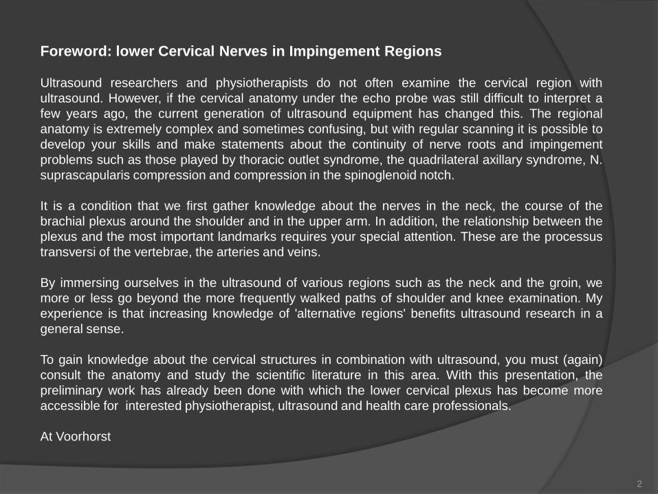
Foreword lower Cervical Nerves in Impingement Regions
Ultrasound researchers and physiotherapists do not often examine the cervical region with
ultrasound However if the cervical anatomy under the echo probe was still difficult to interpret a
few years ago the current generation of ultrasound equipment has changed this The regional
anatomy is extremely complex and sometimes confusing but with regular scanning it is possible to
develop your skills and make statements about the continuity of nerve roots and impingement
problems such as those played by thoracic outlet syndrome the quadrilateral axillary syndrome N
suprascapularis compression and compression in the spinoglenoid notch
It is a condition that we first gather knowledge about the nerves in the neck the course of the
brachial plexus around the shoulder and in the upper arm In addition the relationship between the
plexus and the most important landmarks requires your special attention These are the processus
transversi of the vertebrae the arteries and veins
By immersing ourselves in the ultrasound of various regions such as the neck and the groin we
more or less go beyond the more frequently walked paths of shoulder and knee examination My
experience is that increasing knowledge of alternative regions benefits ultrasound research in a
general sense
To gain knowledge about the cervical structures in combination with ultrasound you must (again)
consult the anatomy and study the scientific literature in this area With this presentation the
preliminary work has already been done with which the lower cervical plexus has become more
accessible for interested physiotherapist ultrasound and health care professionals
At Voorhorst
2
Proloque
We will focus on the normal anatomy and its presentation with ultrasound equipment Regular anatomy has many variants that is a fact No blood vessel pattern is the same on the left or right side no nerve runs in the exact same spot There are also more differences between individuals than agreements when we observe closely This argues for laying nerve blocks always under ultrasound guidance because the various nerve branches can be localized very accurately
Although no constricted nerves are shown at this location it is good to realize that impingement problems have many causes and are likely to be under-diagnosed because of the varied complaints presentations
A nerve that is trapped - or has been seated - shows a fusiform thickening sometimes the characteristic honeycomb pattern has disappeared or become blurred on your echo plate because of edema that has accumulated between the nerve fibers The nerves can be compressed but the accompanying arteries and veins can also be involved in clamping with all its consequences
Known causes of mechanical compression are the scalene anticuss syndrome a neck rib form variations of the clavicle and the first rib bone fractures (clavicle first rib humerus) connective tissue strings enlarged processus transversi of C7 ganglion cysts or a lung tumor (Pancoast tumor)
Besides the mechanical compression we know inflammation of the plexus brachialis by unknown cause as it is the case with neuralgic amyotrophy The exact cause of this disorder is unknown but is thought to be a mistake of the immune system
3
Disclaimer
This presentation is intended for educational purposes The compiler refers to persons or
institutions if literal texts are used or if texts can be traced to certain individuals In addition to
the right to quote the image quote right is used Never are images from elsewhere intended as
embellishments of the whole but always as elements necessary for the presentation to
maintain or increase its educational value If despite the above someone feels essential
shortcomings please contact the compiler so that images or texts can be adapted or removed if
necessary
4
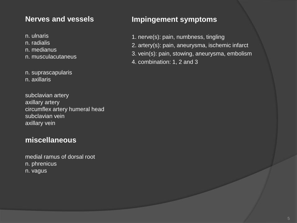
Nerves and vessels
n ulnaris
n radialis
n medianus
n musculacutaneus
n suprascapularis
n axillaris
subclavian artery
axillary artery
circumflex artery humeral head
subclavian vein
axillary vein
miscellaneous
medial ramus of dorsal root
n phrenicus
n vagus
Impingement symptoms
1 nerve(s) pain numbness tingling
2 artery(s) pain aneurysma ischemic infarct
3 vein(s) pain stowing aneurysma embolism
4 combination 1 2 and 3
5
1 nerve root C5
2 nerve root C6
3 nerve root C7
4 nerve root C8
5 nerve root T1
6 subclavian artery
7 subclavian vein
8 footprint m scalenus anterior
C5
C7
EchoProceduresVoorhorst
C6 1
6
7
8
2
3
4 5
plexus brachialis (C5-C8-T1) and vessels
Schematic
6
Anatomie
interscalene triangel m scalenus anterior + m scalenus medius + first rib
left and right Anatomy of the Human Body 1918 Henry Gray (1825ndash1861) (green arrows and numbers EchoProcedures)
1
2
3
5
4 6
1 plexus brachialis
2 subclavian artery
3 subclavian vein
4 m scalenus anterior
5 m scalenus medius
6 clavicula 7
1 m sternocleidomastoideus (sternal part)
2 m sternocleidomastodeus (clavicular part)
3 m scalenus anterior
4 m scalenus medius
5 v jugularis externa
6 m levator scapulae
7 m splenius capitis
8 m trapezius
1 2 3 4
7
8
6
5
EchoproceduresVoorhorst
plexus brachialis cervical region
In vivo
8
Impingement regions
scalene triangel
supraclavicular
subclavicular
infrascapular
pectoralis minor
upper arm
suprascapular notch
spinoglenoid notch
quadrilateral space
D
F
A
B
C
E
G
EchoProceduresVoorhorst
C
D
Anatomy of the Human Body 1918
Henry Gray (1825ndash1861)
7
G
E F
A
EchoProceduresVoorhorst
B
9
Bony landmarks
for ultrasound identification nerve root situated between anterior and posterior tuberculum
of transvers process
1 nerve root
2 medial ramus
3 anterior tuberculum
4 posterior tuberculum
Anatomy of the Human Body 1918 Henry Gray (1825ndash1861)
(Yellow EchoProcedures )
2
1 1
2
3
4
10
1 m scalenus anterior
2 m scalenus medius
3 tuberculum processus posterior
4 tuberculum processus posterior
5 nerve root C5
6 nerve root C6
7 m sternocleidomastoideus
8 jugular vein
9 common carotic artery
10 processus tranversus
11 pedicle
12 m longus colli
EchoProceduresVoorhorst
12
Ultrasound landmarks
brachial nerve root identification in the paravertebral area
probe
c6
A
11
1 nerve root C5 superior trunk
2 nerve root C6 middle trunk
3 nerve root C7 inferior trunk
4 m scaleneus anterior
5 m sternocleidomastoideus
6 m scalenus medius
prevertebral fascia
EchoProceduresVoorhorst EchoProceduresVoorhorst
paravertebral plexus brachialis level C7
Anatomy of the Human
Body 1918 Henry
Gray (1825ndash1861)
Probe
Ultrasound landmarks A
12
EchoProceduresVoorhorst
anterior
supraclavicular artery and brachial plexus in close relation with ribs and pleura
1 supraclavicular artery 2 upper part cervical trunk 3 middle part
cervical trunk 4 lower part cervical trunk 5 first rib 6 second rib 7
pleura 8 intercostal muscles 8 intercostal muscles 9 lung
probe
Ultrasound landmarks B
1
2
3
4
5
6
7
7
8
9
EchoProceduresVoorhorst
13
lsquoThe incidence of arterial compression in patients diagnosed with neurogenic
thoracic outlet syndrome is not accurately known In a consecutive series of
148 patients diagnosed with neurogenic thoracic outlet syndrome a standard
objective method of Doppler ultrasound examination was implemented with
standard manoeuvres for the arm involved [hellip] It was was found that 75
patients (51) had definite arterial occlusion The potential for even venous
compression was examined in this type of patient and 11 patients (8) were
found to have vascular compression of the subclavian vein when performing
the manoeuvresrsquo
Arterial compression
The vascular component in neurogenic-arterial thoracic outlet syndrome J Ernesto Molina MD PhD and
Jonathan DrsquoCunha MD PhD
14
2 1 plexus brachialis
2 subclavian artery
3 subclavian vein
4 m pectoralis minor
5 axillary vein
6 axillary artery
plexus brachialis on chest and upper arm
4
4
C7
2 3
1
EchoProceduresVoorhorst
5
6
4
3
2
1
Schematic
15
B
120 deg elevation + abduction humerus
A
neutral position humerus
normal axillary artery just distal the clavicula neutral and elevated humerus
EchoProceduresVoorhorst EchoProceduresVoorhorst
EchoProceduresVoorhorst
probe
B
Ultrasound landmarks B
16
axillary artery and plexus brachialis under pectoralis minor
EchoProceduresVoorhorst
pma pectoralis major
pm pectoralis minor
A axillary artery
EchoProceduresVoorhorst
probe
Ultrasound landmarks C
17
axillary artery under pectoralis minor
neutral humerus position
axillary artery under pectoralis minor
humerus120 deg elevated
2
no compression at this level 1 pectoralis minor
2 axillary artery
A Shoulder in
neutral position
B Shoulder 120 deg
elevated
EchoProceduresVoorhorst
lateral lateral
1
1
2
EchoProceduresVoorhorst
A B
B
EchoProceduresVoorhorst
probe
Ultrasound landmarks C
18
axillary nerves and vessels axillary nerves and axillary artery
situated just under the m pectoralis
minor
1 n medialis
2 n radialis (running at the posterior side of the upper arm)
3 n ulnaris
4 n musculocutaneus
Cunninghams Manual of
Practical Anatomy 11th ed
1942
1 2
3
4
Anatomy of the Human Body 1918
Henry Gray (1825ndash1861)
m pectoralis minor
Anatomie
19
In vivo
plexus brachialis on chest and upper arm region
1
2 1
2 3
4
4
4 EchoProceduresVoorhorst
Cunninghams Manual of Practical
Anatomy 11th ed 1942
1
1 coracoid proces
2 brachial vein
3 brachial plexus and artery
4 m pectoralis minor
Anatomy of the Human Body 1918 Henry
Gray (1825ndash1861)
20
Anatomy of the Human Body 1918 Henry
Gray (1825ndash1861)
probe position1
2
1 3
4
5
4
6
7
EchoProceduresVoorhorst
plexus and vessels in upper arm (position 1)
1 n musculacutaneus
2 n medianus
3 n ulnaris
4 n radialis
5 axillary artery
6 humerus
7 m biceps
8 m coracobrachialis
9 m triceps
10 axillary vein
Ultrasound landmarks D
21
plexus and vessels in upper arm (position 2)
1 n musculocutaneus
2 m biceps
3 m coracobrachialis
4 humerus
5 axillary artery
6 axillary vein
Anatomy of the Human Body 1918 Henry
Gray (1825ndash1861)
probe placement position 2
EchoProceduresVoorhorst
Ultrasound landmarks D
22
Inside the suprascapular notch n
suprascapularis (C5) Motor
innervation m supraspinatus + m
infraspinatus Sensory innervation
AC-joint + glenohumeral joint
Anatomy of the Human Body 1918 Henry
GrayGray (1821861)
lateral
1
2
2
3
4
EchoProceduresVoorhorst
1 supraclavicular notch 2 supraspinatus 3 tendon
supraspinatus 4 trapezius Anatomy of the Human
Body 1918 Henry Gray (1825ndash
1861)
probe
E Ultrasound landmarks
scan both statically and dynamically
- during passive humeral abduction -
to neutralize compression of the SSP 23
spinoglenoid notch
1
2 3
4
5
6
EchoProceduresVoorhorst
nerve and artery are situated
inside the spinoglenoid notch
Anatomy of the Human Body 1918 Henry
Gray (1825ndash1861)
1 spinoglenoid notch
2 labrum glenoidale
3 humerus
4 articular cartilage
5 m deltoideus
6 infraspinatus
probe
Greyacutes anatomy
F Ultrasound landmarks
scan both statically and dynamically -
during passive humeral exoration - to
neutralize compression of the ISP
24
Motor innervation n axillaris (C5
C6) m teres minor + m deltoideus
Sensory innervation skin above
deltoid muscle
4
1 axillary nerve
2 circumflex axillary artery
3 m teres minor
4 humerus
1 circumflex axillary artery
2 axillary nerve
3 m deltoideus
4 m teres minor
green quadrilateral space
1 2
3
EchoProceduresVoorhorst
4 Anatomy of the Human Body 1918 Henry
Gray (1825ndash1861) (yellow and green
EchoProcedures)
1 2
3
4
4
G Ultrasound landmarks
25
probe
Anatomy of the human
body Henry Gray
Miscellaneous
medial ramus of dorsal root
n phrenicus
n vagus
26
1 nerve root 2 medial ramus innervating facet joints 3 anterior tuberculum 4 posterior tuberculum
Anatomy of the Human Body 1918 Henry Gray (1825ndash1861)
(Yellow EchoProcedures)
1 2
1 2
3 4
Bony landmarks
articular pilllar for ultrasound identification medial ramus
27
medial ramus innervating facet joints
EchoProceduresVoorhorst
probe
longitudinal articular pilar under the open
arrow the medial ramus and artery
1 3 inferior articular process
2 4 superior articular process
EchoProceduresVoorhorst
1 2
3 4
posterior
1 nerve root
2 medial ramus
3 articular pilar(s)
4artery
3
3
distal
EchoProceduresVoorhorst
4
Landmarks
28
n phrenicus C3C4 C5
VJ jugular vein
ACI common carotic artery
n vagus cranial nerve X
1 carotic artery
2 n vagus
3 jugular vein
4 m sternocleidomastoideus
5 thyroid
medial
Anatomy of the
Human Body 1918
Henry Gray (1825ndash
1861)
1
2
1 n phrenicus
2 n vagus
lateral medial
EchoProceduresVoorhorst EchoProceduresVoorhorst
Landmarks
29
Suggested Readings 1
Martinoli Brachial Plexus normal anatomy and scanning technique lecture 27-06-2018 Amsterdam
The cervical plexus anatomy and ultrasound guided blocks
httpwwwapicareonlinecomthe-cervical-plexus-anatomy-and-ultrasound-guided-blocks
Risk of Encountering Dorsal Scapular and Long Thoracic Nerves during Ultrasound-guided Interscalene Brachial Plexus
Block with Nerve Stimulator Korean J Pain 2016 July Vol 29 No 3 179-184 pISSN 2005-9159 eISSN 2093-0569
httpdxdoiorg103344kjp2016293179
Anatomy of the Human Body 1918 Henry Gray (1825ndash1861)
High frequency ultrasound of the cervical spine and brachial plexus - essentials
fileCUsersEigenaaarDownloadsECR2014_C-1692pdf
Ultrasound-Guided Interscalene Brachial Plexus Block
httpswwwnysoracomultrasound-guided-interscalene-brachial-plexus-block
Guided Intervention in Cervical Spine J Korean Orthop Assoc 2015 Apr50(2)77-92 KoreanPublished online April 30 2015
httpsdoiorg104055jkoa201550277
Researchgate
httpswwwresearchgatenetfigureNormal-brachial-plexus-A-Transverse-ultrasound-image
Spinal accesory nerve
httpswwwresearchgatenetfigureSpinal-accessory-nerve-A-Schematic-drawing-shows-the-pertinent-anatomy-of-the
spinal_fig14_47756593_sg=mRNj0a1gnwJGsZ7NsEzVPpIcRzb
eW9_xspNYHgYvIoDHTjAJEFtyxqUCcuZ2U357BboOun7Mdux3U1iWPNB4A
30
Suggested Readings 2
Quadrilateral Space Syndrome Diagnosis and Clinical Management
Patrick T Hangge12 Ilana Breen23 Hassan Albadawi2 M Grace Knuttinen2 Sailendra G Naidu2 andRahmi Oklu2
httpswwwncbinlmnihgovpmcarticlesPMC5920460
Dynamic ultrasonography of the shoulder
httpswwwe-ultrasonographyorgjournalviewphpnumber=206
Sonographic evaluation of the axillary artery during simulated overhead throwing
positionStapleton C Herrington LC and George K positionStapleton C Herrington LC and George K
httpusirsalfordacuk23061stapleton_et_al_2008_PTiSpdfpublic
Ultrasonographic Diagnosis of Thoracic Outlet Syndrome Secondary to Brachial Plexus Piercing Variation
fileCUsersEigenaaarDownloadsdiagnostics-07-00040pdf
The vascular component in neurogenic-arterial thoracic outlet syndromeJ Ernesto Molina MD PhD1 and Jonathan DrsquoCunha
MD PhD2
httpswwwncbinlmnihgovpmcarticlesPMC2728415
Ultrasound-guided interventional procedures for cervical pain
httpwwwasociacionandaluzadeldoloreswp-contentuploads201702US-cervicalpdf
httpswwwnysoracomultrasound-guided-supraclavicular-brachial-plexus-block-2
Ultrasound-Guided Supraclavicular Brachial Plexus Block
31
Ultrasound-Guided Superficial Cervical Plexus Block
httpswwwnysoracomultrasound-guided-superficial-cervical-plexus-block
Foreword lower Cervical Nerves in Impingement Regions
Ultrasound researchers and physiotherapists do not often examine the cervical region with
ultrasound However if the cervical anatomy under the echo probe was still difficult to interpret a
few years ago the current generation of ultrasound equipment has changed this The regional
anatomy is extremely complex and sometimes confusing but with regular scanning it is possible to
develop your skills and make statements about the continuity of nerve roots and impingement
problems such as those played by thoracic outlet syndrome the quadrilateral axillary syndrome N
suprascapularis compression and compression in the spinoglenoid notch
It is a condition that we first gather knowledge about the nerves in the neck the course of the
brachial plexus around the shoulder and in the upper arm In addition the relationship between the
plexus and the most important landmarks requires your special attention These are the processus
transversi of the vertebrae the arteries and veins
By immersing ourselves in the ultrasound of various regions such as the neck and the groin we
more or less go beyond the more frequently walked paths of shoulder and knee examination My
experience is that increasing knowledge of alternative regions benefits ultrasound research in a
general sense
To gain knowledge about the cervical structures in combination with ultrasound you must (again)
consult the anatomy and study the scientific literature in this area With this presentation the
preliminary work has already been done with which the lower cervical plexus has become more
accessible for interested physiotherapist ultrasound and health care professionals
At Voorhorst
2
Proloque
We will focus on the normal anatomy and its presentation with ultrasound equipment Regular anatomy has many variants that is a fact No blood vessel pattern is the same on the left or right side no nerve runs in the exact same spot There are also more differences between individuals than agreements when we observe closely This argues for laying nerve blocks always under ultrasound guidance because the various nerve branches can be localized very accurately
Although no constricted nerves are shown at this location it is good to realize that impingement problems have many causes and are likely to be under-diagnosed because of the varied complaints presentations
A nerve that is trapped - or has been seated - shows a fusiform thickening sometimes the characteristic honeycomb pattern has disappeared or become blurred on your echo plate because of edema that has accumulated between the nerve fibers The nerves can be compressed but the accompanying arteries and veins can also be involved in clamping with all its consequences
Known causes of mechanical compression are the scalene anticuss syndrome a neck rib form variations of the clavicle and the first rib bone fractures (clavicle first rib humerus) connective tissue strings enlarged processus transversi of C7 ganglion cysts or a lung tumor (Pancoast tumor)
Besides the mechanical compression we know inflammation of the plexus brachialis by unknown cause as it is the case with neuralgic amyotrophy The exact cause of this disorder is unknown but is thought to be a mistake of the immune system
3
Disclaimer
This presentation is intended for educational purposes The compiler refers to persons or
institutions if literal texts are used or if texts can be traced to certain individuals In addition to
the right to quote the image quote right is used Never are images from elsewhere intended as
embellishments of the whole but always as elements necessary for the presentation to
maintain or increase its educational value If despite the above someone feels essential
shortcomings please contact the compiler so that images or texts can be adapted or removed if
necessary
4
Nerves and vessels
n ulnaris
n radialis
n medianus
n musculacutaneus
n suprascapularis
n axillaris
subclavian artery
axillary artery
circumflex artery humeral head
subclavian vein
axillary vein
miscellaneous
medial ramus of dorsal root
n phrenicus
n vagus
Impingement symptoms
1 nerve(s) pain numbness tingling
2 artery(s) pain aneurysma ischemic infarct
3 vein(s) pain stowing aneurysma embolism
4 combination 1 2 and 3
5
1 nerve root C5
2 nerve root C6
3 nerve root C7
4 nerve root C8
5 nerve root T1
6 subclavian artery
7 subclavian vein
8 footprint m scalenus anterior
C5
C7
EchoProceduresVoorhorst
C6 1
6
7
8
2
3
4 5
plexus brachialis (C5-C8-T1) and vessels
Schematic
6
Anatomie
interscalene triangel m scalenus anterior + m scalenus medius + first rib
left and right Anatomy of the Human Body 1918 Henry Gray (1825ndash1861) (green arrows and numbers EchoProcedures)
1
2
3
5
4 6
1 plexus brachialis
2 subclavian artery
3 subclavian vein
4 m scalenus anterior
5 m scalenus medius
6 clavicula 7
1 m sternocleidomastoideus (sternal part)
2 m sternocleidomastodeus (clavicular part)
3 m scalenus anterior
4 m scalenus medius
5 v jugularis externa
6 m levator scapulae
7 m splenius capitis
8 m trapezius
1 2 3 4
7
8
6
5
EchoproceduresVoorhorst
plexus brachialis cervical region
In vivo
8
Impingement regions
scalene triangel
supraclavicular
subclavicular
infrascapular
pectoralis minor
upper arm
suprascapular notch
spinoglenoid notch
quadrilateral space
D
F
A
B
C
E
G
EchoProceduresVoorhorst
C
D
Anatomy of the Human Body 1918
Henry Gray (1825ndash1861)
7
G
E F
A
EchoProceduresVoorhorst
B
9
Bony landmarks
for ultrasound identification nerve root situated between anterior and posterior tuberculum
of transvers process
1 nerve root
2 medial ramus
3 anterior tuberculum
4 posterior tuberculum
Anatomy of the Human Body 1918 Henry Gray (1825ndash1861)
(Yellow EchoProcedures )
2
1 1
2
3
4
10
1 m scalenus anterior
2 m scalenus medius
3 tuberculum processus posterior
4 tuberculum processus posterior
5 nerve root C5
6 nerve root C6
7 m sternocleidomastoideus
8 jugular vein
9 common carotic artery
10 processus tranversus
11 pedicle
12 m longus colli
EchoProceduresVoorhorst
12
Ultrasound landmarks
brachial nerve root identification in the paravertebral area
probe
c6
A
11
1 nerve root C5 superior trunk
2 nerve root C6 middle trunk
3 nerve root C7 inferior trunk
4 m scaleneus anterior
5 m sternocleidomastoideus
6 m scalenus medius
prevertebral fascia
EchoProceduresVoorhorst EchoProceduresVoorhorst
paravertebral plexus brachialis level C7
Anatomy of the Human
Body 1918 Henry
Gray (1825ndash1861)
Probe
Ultrasound landmarks A
12
EchoProceduresVoorhorst
anterior
supraclavicular artery and brachial plexus in close relation with ribs and pleura
1 supraclavicular artery 2 upper part cervical trunk 3 middle part
cervical trunk 4 lower part cervical trunk 5 first rib 6 second rib 7
pleura 8 intercostal muscles 8 intercostal muscles 9 lung
probe
Ultrasound landmarks B
1
2
3
4
5
6
7
7
8
9
EchoProceduresVoorhorst
13
lsquoThe incidence of arterial compression in patients diagnosed with neurogenic
thoracic outlet syndrome is not accurately known In a consecutive series of
148 patients diagnosed with neurogenic thoracic outlet syndrome a standard
objective method of Doppler ultrasound examination was implemented with
standard manoeuvres for the arm involved [hellip] It was was found that 75
patients (51) had definite arterial occlusion The potential for even venous
compression was examined in this type of patient and 11 patients (8) were
found to have vascular compression of the subclavian vein when performing
the manoeuvresrsquo
Arterial compression
The vascular component in neurogenic-arterial thoracic outlet syndrome J Ernesto Molina MD PhD and
Jonathan DrsquoCunha MD PhD
14
2 1 plexus brachialis
2 subclavian artery
3 subclavian vein
4 m pectoralis minor
5 axillary vein
6 axillary artery
plexus brachialis on chest and upper arm
4
4
C7
2 3
1
EchoProceduresVoorhorst
5
6
4
3
2
1
Schematic
15
B
120 deg elevation + abduction humerus
A
neutral position humerus
normal axillary artery just distal the clavicula neutral and elevated humerus
EchoProceduresVoorhorst EchoProceduresVoorhorst
EchoProceduresVoorhorst
probe
B
Ultrasound landmarks B
16
axillary artery and plexus brachialis under pectoralis minor
EchoProceduresVoorhorst
pma pectoralis major
pm pectoralis minor
A axillary artery
EchoProceduresVoorhorst
probe
Ultrasound landmarks C
17
axillary artery under pectoralis minor
neutral humerus position
axillary artery under pectoralis minor
humerus120 deg elevated
2
no compression at this level 1 pectoralis minor
2 axillary artery
A Shoulder in
neutral position
B Shoulder 120 deg
elevated
EchoProceduresVoorhorst
lateral lateral
1
1
2
EchoProceduresVoorhorst
A B
B
EchoProceduresVoorhorst
probe
Ultrasound landmarks C
18
axillary nerves and vessels axillary nerves and axillary artery
situated just under the m pectoralis
minor
1 n medialis
2 n radialis (running at the posterior side of the upper arm)
3 n ulnaris
4 n musculocutaneus
Cunninghams Manual of
Practical Anatomy 11th ed
1942
1 2
3
4
Anatomy of the Human Body 1918
Henry Gray (1825ndash1861)
m pectoralis minor
Anatomie
19
In vivo
plexus brachialis on chest and upper arm region
1
2 1
2 3
4
4
4 EchoProceduresVoorhorst
Cunninghams Manual of Practical
Anatomy 11th ed 1942
1
1 coracoid proces
2 brachial vein
3 brachial plexus and artery
4 m pectoralis minor
Anatomy of the Human Body 1918 Henry
Gray (1825ndash1861)
20
Anatomy of the Human Body 1918 Henry
Gray (1825ndash1861)
probe position1
2
1 3
4
5
4
6
7
EchoProceduresVoorhorst
plexus and vessels in upper arm (position 1)
1 n musculacutaneus
2 n medianus
3 n ulnaris
4 n radialis
5 axillary artery
6 humerus
7 m biceps
8 m coracobrachialis
9 m triceps
10 axillary vein
Ultrasound landmarks D
21
plexus and vessels in upper arm (position 2)
1 n musculocutaneus
2 m biceps
3 m coracobrachialis
4 humerus
5 axillary artery
6 axillary vein
Anatomy of the Human Body 1918 Henry
Gray (1825ndash1861)
probe placement position 2
EchoProceduresVoorhorst
Ultrasound landmarks D
22
Inside the suprascapular notch n
suprascapularis (C5) Motor
innervation m supraspinatus + m
infraspinatus Sensory innervation
AC-joint + glenohumeral joint
Anatomy of the Human Body 1918 Henry
GrayGray (1821861)
lateral
1
2
2
3
4
EchoProceduresVoorhorst
1 supraclavicular notch 2 supraspinatus 3 tendon
supraspinatus 4 trapezius Anatomy of the Human
Body 1918 Henry Gray (1825ndash
1861)
probe
E Ultrasound landmarks
scan both statically and dynamically
- during passive humeral abduction -
to neutralize compression of the SSP 23
spinoglenoid notch
1
2 3
4
5
6
EchoProceduresVoorhorst
nerve and artery are situated
inside the spinoglenoid notch
Anatomy of the Human Body 1918 Henry
Gray (1825ndash1861)
1 spinoglenoid notch
2 labrum glenoidale
3 humerus
4 articular cartilage
5 m deltoideus
6 infraspinatus
probe
Greyacutes anatomy
F Ultrasound landmarks
scan both statically and dynamically -
during passive humeral exoration - to
neutralize compression of the ISP
24
Motor innervation n axillaris (C5
C6) m teres minor + m deltoideus
Sensory innervation skin above
deltoid muscle
4
1 axillary nerve
2 circumflex axillary artery
3 m teres minor
4 humerus
1 circumflex axillary artery
2 axillary nerve
3 m deltoideus
4 m teres minor
green quadrilateral space
1 2
3
EchoProceduresVoorhorst
4 Anatomy of the Human Body 1918 Henry
Gray (1825ndash1861) (yellow and green
EchoProcedures)
1 2
3
4
4
G Ultrasound landmarks
25
probe
Anatomy of the human
body Henry Gray
Miscellaneous
medial ramus of dorsal root
n phrenicus
n vagus
26
1 nerve root 2 medial ramus innervating facet joints 3 anterior tuberculum 4 posterior tuberculum
Anatomy of the Human Body 1918 Henry Gray (1825ndash1861)
(Yellow EchoProcedures)
1 2
1 2
3 4
Bony landmarks
articular pilllar for ultrasound identification medial ramus
27
medial ramus innervating facet joints
EchoProceduresVoorhorst
probe
longitudinal articular pilar under the open
arrow the medial ramus and artery
1 3 inferior articular process
2 4 superior articular process
EchoProceduresVoorhorst
1 2
3 4
posterior
1 nerve root
2 medial ramus
3 articular pilar(s)
4artery
3
3
distal
EchoProceduresVoorhorst
4
Landmarks
28
n phrenicus C3C4 C5
VJ jugular vein
ACI common carotic artery
n vagus cranial nerve X
1 carotic artery
2 n vagus
3 jugular vein
4 m sternocleidomastoideus
5 thyroid
medial
Anatomy of the
Human Body 1918
Henry Gray (1825ndash
1861)
1
2
1 n phrenicus
2 n vagus
lateral medial
EchoProceduresVoorhorst EchoProceduresVoorhorst
Landmarks
29
Suggested Readings 1
Martinoli Brachial Plexus normal anatomy and scanning technique lecture 27-06-2018 Amsterdam
The cervical plexus anatomy and ultrasound guided blocks
httpwwwapicareonlinecomthe-cervical-plexus-anatomy-and-ultrasound-guided-blocks
Risk of Encountering Dorsal Scapular and Long Thoracic Nerves during Ultrasound-guided Interscalene Brachial Plexus
Block with Nerve Stimulator Korean J Pain 2016 July Vol 29 No 3 179-184 pISSN 2005-9159 eISSN 2093-0569
httpdxdoiorg103344kjp2016293179
Anatomy of the Human Body 1918 Henry Gray (1825ndash1861)
High frequency ultrasound of the cervical spine and brachial plexus - essentials
fileCUsersEigenaaarDownloadsECR2014_C-1692pdf
Ultrasound-Guided Interscalene Brachial Plexus Block
httpswwwnysoracomultrasound-guided-interscalene-brachial-plexus-block
Guided Intervention in Cervical Spine J Korean Orthop Assoc 2015 Apr50(2)77-92 KoreanPublished online April 30 2015
httpsdoiorg104055jkoa201550277
Researchgate
httpswwwresearchgatenetfigureNormal-brachial-plexus-A-Transverse-ultrasound-image
Spinal accesory nerve
httpswwwresearchgatenetfigureSpinal-accessory-nerve-A-Schematic-drawing-shows-the-pertinent-anatomy-of-the
spinal_fig14_47756593_sg=mRNj0a1gnwJGsZ7NsEzVPpIcRzb
eW9_xspNYHgYvIoDHTjAJEFtyxqUCcuZ2U357BboOun7Mdux3U1iWPNB4A
30
Suggested Readings 2
Quadrilateral Space Syndrome Diagnosis and Clinical Management
Patrick T Hangge12 Ilana Breen23 Hassan Albadawi2 M Grace Knuttinen2 Sailendra G Naidu2 andRahmi Oklu2
httpswwwncbinlmnihgovpmcarticlesPMC5920460
Dynamic ultrasonography of the shoulder
httpswwwe-ultrasonographyorgjournalviewphpnumber=206
Sonographic evaluation of the axillary artery during simulated overhead throwing
positionStapleton C Herrington LC and George K positionStapleton C Herrington LC and George K
httpusirsalfordacuk23061stapleton_et_al_2008_PTiSpdfpublic
Ultrasonographic Diagnosis of Thoracic Outlet Syndrome Secondary to Brachial Plexus Piercing Variation
fileCUsersEigenaaarDownloadsdiagnostics-07-00040pdf
The vascular component in neurogenic-arterial thoracic outlet syndromeJ Ernesto Molina MD PhD1 and Jonathan DrsquoCunha
MD PhD2
httpswwwncbinlmnihgovpmcarticlesPMC2728415
Ultrasound-guided interventional procedures for cervical pain
httpwwwasociacionandaluzadeldoloreswp-contentuploads201702US-cervicalpdf
httpswwwnysoracomultrasound-guided-supraclavicular-brachial-plexus-block-2
Ultrasound-Guided Supraclavicular Brachial Plexus Block
31
Ultrasound-Guided Superficial Cervical Plexus Block
httpswwwnysoracomultrasound-guided-superficial-cervical-plexus-block

Anatomie
interscalene triangel m scalenus anterior + m scalenus medius + first rib
left and right Anatomy of the Human Body 1918 Henry Gray (1825ndash1861) (green arrows and numbers EchoProcedures)
1
2
3
5
4 6
1 plexus brachialis
2 subclavian artery
3 subclavian vein
4 m scalenus anterior
5 m scalenus medius
6 clavicula 7
1 m sternocleidomastoideus (sternal part)
2 m sternocleidomastodeus (clavicular part)
3 m scalenus anterior
4 m scalenus medius
5 v jugularis externa
6 m levator scapulae
7 m splenius capitis
8 m trapezius
1 2 3 4
7
8
6
5
EchoproceduresVoorhorst
plexus brachialis cervical region
In vivo
8
Impingement regions
scalene triangel
supraclavicular
subclavicular
infrascapular
pectoralis minor
upper arm
suprascapular notch
spinoglenoid notch
quadrilateral space
D
F
A
B
C
E
G
EchoProceduresVoorhorst
C
D
Anatomy of the Human Body 1918
Henry Gray (1825ndash1861)
7
G
E F
A
EchoProceduresVoorhorst
B
9
Bony landmarks
for ultrasound identification nerve root situated between anterior and posterior tuberculum
of transvers process
1 nerve root
2 medial ramus
3 anterior tuberculum
4 posterior tuberculum
Anatomy of the Human Body 1918 Henry Gray (1825ndash1861)
(Yellow EchoProcedures )
2
1 1
2
3
4
10
1 m scalenus anterior
2 m scalenus medius
3 tuberculum processus posterior
4 tuberculum processus posterior
5 nerve root C5
6 nerve root C6
7 m sternocleidomastoideus
8 jugular vein
9 common carotic artery
10 processus tranversus
11 pedicle
12 m longus colli
EchoProceduresVoorhorst
12
Ultrasound landmarks
brachial nerve root identification in the paravertebral area
probe
c6
A
11
1 nerve root C5 superior trunk
2 nerve root C6 middle trunk
3 nerve root C7 inferior trunk
4 m scaleneus anterior
5 m sternocleidomastoideus
6 m scalenus medius
prevertebral fascia
EchoProceduresVoorhorst EchoProceduresVoorhorst
paravertebral plexus brachialis level C7
Anatomy of the Human
Body 1918 Henry
Gray (1825ndash1861)
Probe
Ultrasound landmarks A
12
EchoProceduresVoorhorst
anterior
supraclavicular artery and brachial plexus in close relation with ribs and pleura
1 supraclavicular artery 2 upper part cervical trunk 3 middle part
cervical trunk 4 lower part cervical trunk 5 first rib 6 second rib 7
pleura 8 intercostal muscles 8 intercostal muscles 9 lung
probe
Ultrasound landmarks B
1
2
3
4
5
6
7
7
8
9
EchoProceduresVoorhorst
13
lsquoThe incidence of arterial compression in patients diagnosed with neurogenic
thoracic outlet syndrome is not accurately known In a consecutive series of
148 patients diagnosed with neurogenic thoracic outlet syndrome a standard
objective method of Doppler ultrasound examination was implemented with
standard manoeuvres for the arm involved [hellip] It was was found that 75
patients (51) had definite arterial occlusion The potential for even venous
compression was examined in this type of patient and 11 patients (8) were
found to have vascular compression of the subclavian vein when performing
the manoeuvresrsquo
Arterial compression
The vascular component in neurogenic-arterial thoracic outlet syndrome J Ernesto Molina MD PhD and
Jonathan DrsquoCunha MD PhD
14
2 1 plexus brachialis
2 subclavian artery
3 subclavian vein
4 m pectoralis minor
5 axillary vein
6 axillary artery
plexus brachialis on chest and upper arm
4
4
C7
2 3
1
EchoProceduresVoorhorst
5
6
4
3
2
1
Schematic
15
B
120 deg elevation + abduction humerus
A
neutral position humerus
normal axillary artery just distal the clavicula neutral and elevated humerus
EchoProceduresVoorhorst EchoProceduresVoorhorst
EchoProceduresVoorhorst
probe
B
Ultrasound landmarks B
16
axillary artery and plexus brachialis under pectoralis minor
EchoProceduresVoorhorst
pma pectoralis major
pm pectoralis minor
A axillary artery
EchoProceduresVoorhorst
probe
Ultrasound landmarks C
17
axillary artery under pectoralis minor
neutral humerus position
axillary artery under pectoralis minor
humerus120 deg elevated
2
no compression at this level 1 pectoralis minor
2 axillary artery
A Shoulder in
neutral position
B Shoulder 120 deg
elevated
EchoProceduresVoorhorst
lateral lateral
1
1
2
EchoProceduresVoorhorst
A B
B
EchoProceduresVoorhorst
probe
Ultrasound landmarks C
18
axillary nerves and vessels axillary nerves and axillary artery
situated just under the m pectoralis
minor
1 n medialis
2 n radialis (running at the posterior side of the upper arm)
3 n ulnaris
4 n musculocutaneus
Cunninghams Manual of
Practical Anatomy 11th ed
1942
1 2
3
4
Anatomy of the Human Body 1918
Henry Gray (1825ndash1861)
m pectoralis minor
Anatomie
19
In vivo
plexus brachialis on chest and upper arm region
1
2 1
2 3
4
4
4 EchoProceduresVoorhorst
Cunninghams Manual of Practical
Anatomy 11th ed 1942
1
1 coracoid proces
2 brachial vein
3 brachial plexus and artery
4 m pectoralis minor
Anatomy of the Human Body 1918 Henry
Gray (1825ndash1861)
20
Anatomy of the Human Body 1918 Henry
Gray (1825ndash1861)
probe position1
2
1 3
4
5
4
6
7
EchoProceduresVoorhorst
plexus and vessels in upper arm (position 1)
1 n musculacutaneus
2 n medianus
3 n ulnaris
4 n radialis
5 axillary artery
6 humerus
7 m biceps
8 m coracobrachialis
9 m triceps
10 axillary vein
Ultrasound landmarks D
21
plexus and vessels in upper arm (position 2)
1 n musculocutaneus
2 m biceps
3 m coracobrachialis
4 humerus
5 axillary artery
6 axillary vein
Anatomy of the Human Body 1918 Henry
Gray (1825ndash1861)
probe placement position 2
EchoProceduresVoorhorst
Ultrasound landmarks D
22
Inside the suprascapular notch n
suprascapularis (C5) Motor
innervation m supraspinatus + m
infraspinatus Sensory innervation
AC-joint + glenohumeral joint
Anatomy of the Human Body 1918 Henry
GrayGray (1821861)
lateral
1
2
2
3
4
EchoProceduresVoorhorst
1 supraclavicular notch 2 supraspinatus 3 tendon
supraspinatus 4 trapezius Anatomy of the Human
Body 1918 Henry Gray (1825ndash
1861)
probe
E Ultrasound landmarks
scan both statically and dynamically
- during passive humeral abduction -
to neutralize compression of the SSP 23
spinoglenoid notch
1
2 3
4
5
6
EchoProceduresVoorhorst
nerve and artery are situated
inside the spinoglenoid notch
Anatomy of the Human Body 1918 Henry
Gray (1825ndash1861)
1 spinoglenoid notch
2 labrum glenoidale
3 humerus
4 articular cartilage
5 m deltoideus
6 infraspinatus
probe
Greyacutes anatomy
F Ultrasound landmarks
scan both statically and dynamically -
during passive humeral exoration - to
neutralize compression of the ISP
24
Motor innervation n axillaris (C5
C6) m teres minor + m deltoideus
Sensory innervation skin above
deltoid muscle
4
1 axillary nerve
2 circumflex axillary artery
3 m teres minor
4 humerus
1 circumflex axillary artery
2 axillary nerve
3 m deltoideus
4 m teres minor
green quadrilateral space
1 2
3
EchoProceduresVoorhorst
4 Anatomy of the Human Body 1918 Henry
Gray (1825ndash1861) (yellow and green
EchoProcedures)
1 2
3
4
4
G Ultrasound landmarks
25
probe
Anatomy of the human
body Henry Gray
Miscellaneous
medial ramus of dorsal root
n phrenicus
n vagus
26
1 nerve root 2 medial ramus innervating facet joints 3 anterior tuberculum 4 posterior tuberculum
Anatomy of the Human Body 1918 Henry Gray (1825ndash1861)
(Yellow EchoProcedures)
1 2
1 2
3 4
Bony landmarks
articular pilllar for ultrasound identification medial ramus
27
medial ramus innervating facet joints
EchoProceduresVoorhorst
probe
longitudinal articular pilar under the open
arrow the medial ramus and artery
1 3 inferior articular process
2 4 superior articular process
EchoProceduresVoorhorst
1 2
3 4
posterior
1 nerve root
2 medial ramus
3 articular pilar(s)
4artery
3
3
distal
EchoProceduresVoorhorst
4
Landmarks
28
n phrenicus C3C4 C5
VJ jugular vein
ACI common carotic artery
n vagus cranial nerve X
1 carotic artery
2 n vagus
3 jugular vein
4 m sternocleidomastoideus
5 thyroid
medial
Anatomy of the
Human Body 1918
Henry Gray (1825ndash
1861)
1
2
1 n phrenicus
2 n vagus
lateral medial
EchoProceduresVoorhorst EchoProceduresVoorhorst
Landmarks
29
Suggested Readings 1
Martinoli Brachial Plexus normal anatomy and scanning technique lecture 27-06-2018 Amsterdam
The cervical plexus anatomy and ultrasound guided blocks
httpwwwapicareonlinecomthe-cervical-plexus-anatomy-and-ultrasound-guided-blocks
Risk of Encountering Dorsal Scapular and Long Thoracic Nerves during Ultrasound-guided Interscalene Brachial Plexus
Block with Nerve Stimulator Korean J Pain 2016 July Vol 29 No 3 179-184 pISSN 2005-9159 eISSN 2093-0569
httpdxdoiorg103344kjp2016293179
Anatomy of the Human Body 1918 Henry Gray (1825ndash1861)
High frequency ultrasound of the cervical spine and brachial plexus - essentials
fileCUsersEigenaaarDownloadsECR2014_C-1692pdf
Ultrasound-Guided Interscalene Brachial Plexus Block
httpswwwnysoracomultrasound-guided-interscalene-brachial-plexus-block
Guided Intervention in Cervical Spine J Korean Orthop Assoc 2015 Apr50(2)77-92 KoreanPublished online April 30 2015
httpsdoiorg104055jkoa201550277
Researchgate
httpswwwresearchgatenetfigureNormal-brachial-plexus-A-Transverse-ultrasound-image
Spinal accesory nerve
httpswwwresearchgatenetfigureSpinal-accessory-nerve-A-Schematic-drawing-shows-the-pertinent-anatomy-of-the
spinal_fig14_47756593_sg=mRNj0a1gnwJGsZ7NsEzVPpIcRzb
eW9_xspNYHgYvIoDHTjAJEFtyxqUCcuZ2U357BboOun7Mdux3U1iWPNB4A
30
Suggested Readings 2
Quadrilateral Space Syndrome Diagnosis and Clinical Management
Patrick T Hangge12 Ilana Breen23 Hassan Albadawi2 M Grace Knuttinen2 Sailendra G Naidu2 andRahmi Oklu2
httpswwwncbinlmnihgovpmcarticlesPMC5920460
Dynamic ultrasonography of the shoulder
httpswwwe-ultrasonographyorgjournalviewphpnumber=206
Sonographic evaluation of the axillary artery during simulated overhead throwing
positionStapleton C Herrington LC and George K positionStapleton C Herrington LC and George K
httpusirsalfordacuk23061stapleton_et_al_2008_PTiSpdfpublic
Ultrasonographic Diagnosis of Thoracic Outlet Syndrome Secondary to Brachial Plexus Piercing Variation
fileCUsersEigenaaarDownloadsdiagnostics-07-00040pdf
The vascular component in neurogenic-arterial thoracic outlet syndromeJ Ernesto Molina MD PhD1 and Jonathan DrsquoCunha
MD PhD2
httpswwwncbinlmnihgovpmcarticlesPMC2728415
Ultrasound-guided interventional procedures for cervical pain
httpwwwasociacionandaluzadeldoloreswp-contentuploads201702US-cervicalpdf
httpswwwnysoracomultrasound-guided-supraclavicular-brachial-plexus-block-2
Ultrasound-Guided Supraclavicular Brachial Plexus Block
31
Ultrasound-Guided Superficial Cervical Plexus Block
httpswwwnysoracomultrasound-guided-superficial-cervical-plexus-block

Bony landmarks
for ultrasound identification nerve root situated between anterior and posterior tuberculum
of transvers process
1 nerve root
2 medial ramus
3 anterior tuberculum
4 posterior tuberculum
Anatomy of the Human Body 1918 Henry Gray (1825ndash1861)
(Yellow EchoProcedures )
2
1 1
2
3
4
10
1 m scalenus anterior
2 m scalenus medius
3 tuberculum processus posterior
4 tuberculum processus posterior
5 nerve root C5
6 nerve root C6
7 m sternocleidomastoideus
8 jugular vein
9 common carotic artery
10 processus tranversus
11 pedicle
12 m longus colli
EchoProceduresVoorhorst
12
Ultrasound landmarks
brachial nerve root identification in the paravertebral area
probe
c6
A
11
1 nerve root C5 superior trunk
2 nerve root C6 middle trunk
3 nerve root C7 inferior trunk
4 m scaleneus anterior
5 m sternocleidomastoideus
6 m scalenus medius
prevertebral fascia
EchoProceduresVoorhorst EchoProceduresVoorhorst
paravertebral plexus brachialis level C7
Anatomy of the Human
Body 1918 Henry
Gray (1825ndash1861)
Probe
Ultrasound landmarks A
12
EchoProceduresVoorhorst
anterior
supraclavicular artery and brachial plexus in close relation with ribs and pleura
1 supraclavicular artery 2 upper part cervical trunk 3 middle part
cervical trunk 4 lower part cervical trunk 5 first rib 6 second rib 7
pleura 8 intercostal muscles 8 intercostal muscles 9 lung
probe
Ultrasound landmarks B
1
2
3
4
5
6
7
7
8
9
EchoProceduresVoorhorst
13
lsquoThe incidence of arterial compression in patients diagnosed with neurogenic
thoracic outlet syndrome is not accurately known In a consecutive series of
148 patients diagnosed with neurogenic thoracic outlet syndrome a standard
objective method of Doppler ultrasound examination was implemented with
standard manoeuvres for the arm involved [hellip] It was was found that 75
patients (51) had definite arterial occlusion The potential for even venous
compression was examined in this type of patient and 11 patients (8) were
found to have vascular compression of the subclavian vein when performing
the manoeuvresrsquo
Arterial compression
The vascular component in neurogenic-arterial thoracic outlet syndrome J Ernesto Molina MD PhD and
Jonathan DrsquoCunha MD PhD
14
2 1 plexus brachialis
2 subclavian artery
3 subclavian vein
4 m pectoralis minor
5 axillary vein
6 axillary artery
plexus brachialis on chest and upper arm
4
4
C7
2 3
1
EchoProceduresVoorhorst
5
6
4
3
2
1
Schematic
15
B
120 deg elevation + abduction humerus
A
neutral position humerus
normal axillary artery just distal the clavicula neutral and elevated humerus
EchoProceduresVoorhorst EchoProceduresVoorhorst
EchoProceduresVoorhorst
probe
B
Ultrasound landmarks B
16
axillary artery and plexus brachialis under pectoralis minor
EchoProceduresVoorhorst
pma pectoralis major
pm pectoralis minor
A axillary artery
EchoProceduresVoorhorst
probe
Ultrasound landmarks C
17
axillary artery under pectoralis minor
neutral humerus position
axillary artery under pectoralis minor
humerus120 deg elevated
2
no compression at this level 1 pectoralis minor
2 axillary artery
A Shoulder in
neutral position
B Shoulder 120 deg
elevated
EchoProceduresVoorhorst
lateral lateral
1
1
2
EchoProceduresVoorhorst
A B
B
EchoProceduresVoorhorst
probe
Ultrasound landmarks C
18
axillary nerves and vessels axillary nerves and axillary artery
situated just under the m pectoralis
minor
1 n medialis
2 n radialis (running at the posterior side of the upper arm)
3 n ulnaris
4 n musculocutaneus
Cunninghams Manual of
Practical Anatomy 11th ed
1942
1 2
3
4
Anatomy of the Human Body 1918
Henry Gray (1825ndash1861)
m pectoralis minor
Anatomie
19
In vivo
plexus brachialis on chest and upper arm region
1
2 1
2 3
4
4
4 EchoProceduresVoorhorst
Cunninghams Manual of Practical
Anatomy 11th ed 1942
1
1 coracoid proces
2 brachial vein
3 brachial plexus and artery
4 m pectoralis minor
Anatomy of the Human Body 1918 Henry
Gray (1825ndash1861)
20
Anatomy of the Human Body 1918 Henry
Gray (1825ndash1861)
probe position1
2
1 3
4
5
4
6
7
EchoProceduresVoorhorst
plexus and vessels in upper arm (position 1)
1 n musculacutaneus
2 n medianus
3 n ulnaris
4 n radialis
5 axillary artery
6 humerus
7 m biceps
8 m coracobrachialis
9 m triceps
10 axillary vein
Ultrasound landmarks D
21
plexus and vessels in upper arm (position 2)
1 n musculocutaneus
2 m biceps
3 m coracobrachialis
4 humerus
5 axillary artery
6 axillary vein
Anatomy of the Human Body 1918 Henry
Gray (1825ndash1861)
probe placement position 2
EchoProceduresVoorhorst
Ultrasound landmarks D
22
Inside the suprascapular notch n
suprascapularis (C5) Motor
innervation m supraspinatus + m
infraspinatus Sensory innervation
AC-joint + glenohumeral joint
Anatomy of the Human Body 1918 Henry
GrayGray (1821861)
lateral
1
2
2
3
4
EchoProceduresVoorhorst
1 supraclavicular notch 2 supraspinatus 3 tendon
supraspinatus 4 trapezius Anatomy of the Human
Body 1918 Henry Gray (1825ndash
1861)
probe
E Ultrasound landmarks
scan both statically and dynamically
- during passive humeral abduction -
to neutralize compression of the SSP 23
spinoglenoid notch
1
2 3
4
5
6
EchoProceduresVoorhorst
nerve and artery are situated
inside the spinoglenoid notch
Anatomy of the Human Body 1918 Henry
Gray (1825ndash1861)
1 spinoglenoid notch
2 labrum glenoidale
3 humerus
4 articular cartilage
5 m deltoideus
6 infraspinatus
probe
Greyacutes anatomy
F Ultrasound landmarks
scan both statically and dynamically -
during passive humeral exoration - to
neutralize compression of the ISP
24
Motor innervation n axillaris (C5
C6) m teres minor + m deltoideus
Sensory innervation skin above
deltoid muscle
4
1 axillary nerve
2 circumflex axillary artery
3 m teres minor
4 humerus
1 circumflex axillary artery
2 axillary nerve
3 m deltoideus
4 m teres minor
green quadrilateral space
1 2
3
EchoProceduresVoorhorst
4 Anatomy of the Human Body 1918 Henry
Gray (1825ndash1861) (yellow and green
EchoProcedures)
1 2
3
4
4
G Ultrasound landmarks
25
probe
Anatomy of the human
body Henry Gray
Miscellaneous
medial ramus of dorsal root
n phrenicus
n vagus
26
1 nerve root 2 medial ramus innervating facet joints 3 anterior tuberculum 4 posterior tuberculum
Anatomy of the Human Body 1918 Henry Gray (1825ndash1861)
(Yellow EchoProcedures)
1 2
1 2
3 4
Bony landmarks
articular pilllar for ultrasound identification medial ramus
27
medial ramus innervating facet joints
EchoProceduresVoorhorst
probe
longitudinal articular pilar under the open
arrow the medial ramus and artery
1 3 inferior articular process
2 4 superior articular process
EchoProceduresVoorhorst
1 2
3 4
posterior
1 nerve root
2 medial ramus
3 articular pilar(s)
4artery
3
3
distal
EchoProceduresVoorhorst
4
Landmarks
28
n phrenicus C3C4 C5
VJ jugular vein
ACI common carotic artery
n vagus cranial nerve X
1 carotic artery
2 n vagus
3 jugular vein
4 m sternocleidomastoideus
5 thyroid
medial
Anatomy of the
Human Body 1918
Henry Gray (1825ndash
1861)
1
2
1 n phrenicus
2 n vagus
lateral medial
EchoProceduresVoorhorst EchoProceduresVoorhorst
Landmarks
29
Suggested Readings 1
Martinoli Brachial Plexus normal anatomy and scanning technique lecture 27-06-2018 Amsterdam
The cervical plexus anatomy and ultrasound guided blocks
httpwwwapicareonlinecomthe-cervical-plexus-anatomy-and-ultrasound-guided-blocks
Risk of Encountering Dorsal Scapular and Long Thoracic Nerves during Ultrasound-guided Interscalene Brachial Plexus
Block with Nerve Stimulator Korean J Pain 2016 July Vol 29 No 3 179-184 pISSN 2005-9159 eISSN 2093-0569
httpdxdoiorg103344kjp2016293179
Anatomy of the Human Body 1918 Henry Gray (1825ndash1861)
High frequency ultrasound of the cervical spine and brachial plexus - essentials
fileCUsersEigenaaarDownloadsECR2014_C-1692pdf
Ultrasound-Guided Interscalene Brachial Plexus Block
httpswwwnysoracomultrasound-guided-interscalene-brachial-plexus-block
Guided Intervention in Cervical Spine J Korean Orthop Assoc 2015 Apr50(2)77-92 KoreanPublished online April 30 2015
httpsdoiorg104055jkoa201550277
Researchgate
httpswwwresearchgatenetfigureNormal-brachial-plexus-A-Transverse-ultrasound-image
Spinal accesory nerve
httpswwwresearchgatenetfigureSpinal-accessory-nerve-A-Schematic-drawing-shows-the-pertinent-anatomy-of-the
spinal_fig14_47756593_sg=mRNj0a1gnwJGsZ7NsEzVPpIcRzb
eW9_xspNYHgYvIoDHTjAJEFtyxqUCcuZ2U357BboOun7Mdux3U1iWPNB4A
30
Suggested Readings 2
Quadrilateral Space Syndrome Diagnosis and Clinical Management
Patrick T Hangge12 Ilana Breen23 Hassan Albadawi2 M Grace Knuttinen2 Sailendra G Naidu2 andRahmi Oklu2
httpswwwncbinlmnihgovpmcarticlesPMC5920460
Dynamic ultrasonography of the shoulder
httpswwwe-ultrasonographyorgjournalviewphpnumber=206
Sonographic evaluation of the axillary artery during simulated overhead throwing
positionStapleton C Herrington LC and George K positionStapleton C Herrington LC and George K
httpusirsalfordacuk23061stapleton_et_al_2008_PTiSpdfpublic
Ultrasonographic Diagnosis of Thoracic Outlet Syndrome Secondary to Brachial Plexus Piercing Variation
fileCUsersEigenaaarDownloadsdiagnostics-07-00040pdf
The vascular component in neurogenic-arterial thoracic outlet syndromeJ Ernesto Molina MD PhD1 and Jonathan DrsquoCunha
MD PhD2
httpswwwncbinlmnihgovpmcarticlesPMC2728415
Ultrasound-guided interventional procedures for cervical pain
httpwwwasociacionandaluzadeldoloreswp-contentuploads201702US-cervicalpdf
httpswwwnysoracomultrasound-guided-supraclavicular-brachial-plexus-block-2
Ultrasound-Guided Supraclavicular Brachial Plexus Block
31
Ultrasound-Guided Superficial Cervical Plexus Block
httpswwwnysoracomultrasound-guided-superficial-cervical-plexus-block
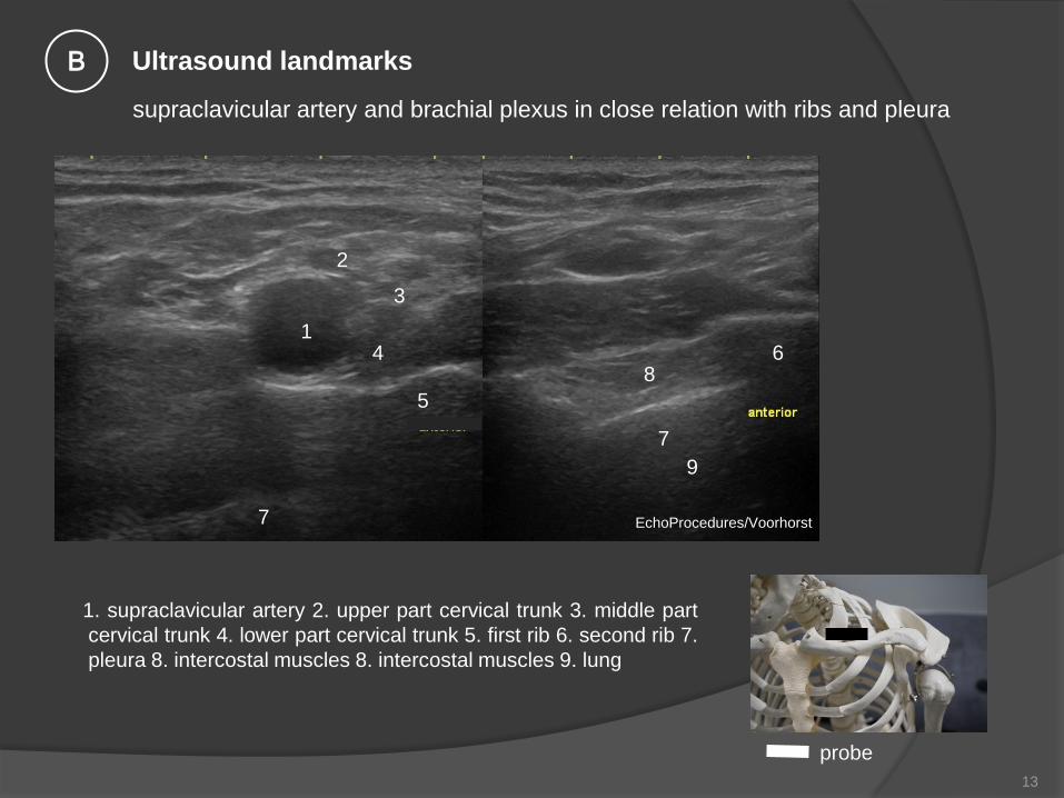
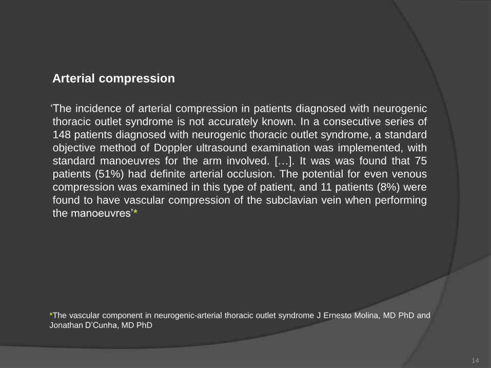

B
120 deg elevation + abduction humerus
A
neutral position humerus
normal axillary artery just distal the clavicula neutral and elevated humerus
EchoProceduresVoorhorst EchoProceduresVoorhorst
EchoProceduresVoorhorst
probe
B
Ultrasound landmarks B
16
axillary artery and plexus brachialis under pectoralis minor
EchoProceduresVoorhorst
pma pectoralis major
pm pectoralis minor
A axillary artery
EchoProceduresVoorhorst
probe
Ultrasound landmarks C
17
axillary artery under pectoralis minor
neutral humerus position
axillary artery under pectoralis minor
humerus120 deg elevated
2
no compression at this level 1 pectoralis minor
2 axillary artery
A Shoulder in
neutral position
B Shoulder 120 deg
elevated
EchoProceduresVoorhorst
lateral lateral
1
1
2
EchoProceduresVoorhorst
A B
B
EchoProceduresVoorhorst
probe
Ultrasound landmarks C
18
axillary nerves and vessels axillary nerves and axillary artery
situated just under the m pectoralis
minor
1 n medialis
2 n radialis (running at the posterior side of the upper arm)
3 n ulnaris
4 n musculocutaneus
Cunninghams Manual of
Practical Anatomy 11th ed
1942
1 2
3
4
Anatomy of the Human Body 1918
Henry Gray (1825ndash1861)
m pectoralis minor
Anatomie
19
In vivo
plexus brachialis on chest and upper arm region
1
2 1
2 3
4
4
4 EchoProceduresVoorhorst
Cunninghams Manual of Practical
Anatomy 11th ed 1942
1
1 coracoid proces
2 brachial vein
3 brachial plexus and artery
4 m pectoralis minor
Anatomy of the Human Body 1918 Henry
Gray (1825ndash1861)
20
Anatomy of the Human Body 1918 Henry
Gray (1825ndash1861)
probe position1
2
1 3
4
5
4
6
7
EchoProceduresVoorhorst
plexus and vessels in upper arm (position 1)
1 n musculacutaneus
2 n medianus
3 n ulnaris
4 n radialis
5 axillary artery
6 humerus
7 m biceps
8 m coracobrachialis
9 m triceps
10 axillary vein
Ultrasound landmarks D
21
plexus and vessels in upper arm (position 2)
1 n musculocutaneus
2 m biceps
3 m coracobrachialis
4 humerus
5 axillary artery
6 axillary vein
Anatomy of the Human Body 1918 Henry
Gray (1825ndash1861)
probe placement position 2
EchoProceduresVoorhorst
Ultrasound landmarks D
22
Inside the suprascapular notch n
suprascapularis (C5) Motor
innervation m supraspinatus + m
infraspinatus Sensory innervation
AC-joint + glenohumeral joint
Anatomy of the Human Body 1918 Henry
GrayGray (1821861)
lateral
1
2
2
3
4
EchoProceduresVoorhorst
1 supraclavicular notch 2 supraspinatus 3 tendon
supraspinatus 4 trapezius Anatomy of the Human
Body 1918 Henry Gray (1825ndash
1861)
probe
E Ultrasound landmarks
scan both statically and dynamically
- during passive humeral abduction -
to neutralize compression of the SSP 23
spinoglenoid notch
1
2 3
4
5
6
EchoProceduresVoorhorst
nerve and artery are situated
inside the spinoglenoid notch
Anatomy of the Human Body 1918 Henry
Gray (1825ndash1861)
1 spinoglenoid notch
2 labrum glenoidale
3 humerus
4 articular cartilage
5 m deltoideus
6 infraspinatus
probe
Greyacutes anatomy
F Ultrasound landmarks
scan both statically and dynamically -
during passive humeral exoration - to
neutralize compression of the ISP
24
Motor innervation n axillaris (C5
C6) m teres minor + m deltoideus
Sensory innervation skin above
deltoid muscle
4
1 axillary nerve
2 circumflex axillary artery
3 m teres minor
4 humerus
1 circumflex axillary artery
2 axillary nerve
3 m deltoideus
4 m teres minor
green quadrilateral space
1 2
3
EchoProceduresVoorhorst
4 Anatomy of the Human Body 1918 Henry
Gray (1825ndash1861) (yellow and green
EchoProcedures)
1 2
3
4
4
G Ultrasound landmarks
25
probe
Anatomy of the human
body Henry Gray
Miscellaneous
medial ramus of dorsal root
n phrenicus
n vagus
26
1 nerve root 2 medial ramus innervating facet joints 3 anterior tuberculum 4 posterior tuberculum
Anatomy of the Human Body 1918 Henry Gray (1825ndash1861)
(Yellow EchoProcedures)
1 2
1 2
3 4
Bony landmarks
articular pilllar for ultrasound identification medial ramus
27
medial ramus innervating facet joints
EchoProceduresVoorhorst
probe
longitudinal articular pilar under the open
arrow the medial ramus and artery
1 3 inferior articular process
2 4 superior articular process
EchoProceduresVoorhorst
1 2
3 4
posterior
1 nerve root
2 medial ramus
3 articular pilar(s)
4artery
3
3
distal
EchoProceduresVoorhorst
4
Landmarks
28
n phrenicus C3C4 C5
VJ jugular vein
ACI common carotic artery
n vagus cranial nerve X
1 carotic artery
2 n vagus
3 jugular vein
4 m sternocleidomastoideus
5 thyroid
medial
Anatomy of the
Human Body 1918
Henry Gray (1825ndash
1861)
1
2
1 n phrenicus
2 n vagus
lateral medial
EchoProceduresVoorhorst EchoProceduresVoorhorst
Landmarks
29
Suggested Readings 1
Martinoli Brachial Plexus normal anatomy and scanning technique lecture 27-06-2018 Amsterdam
The cervical plexus anatomy and ultrasound guided blocks
httpwwwapicareonlinecomthe-cervical-plexus-anatomy-and-ultrasound-guided-blocks
Risk of Encountering Dorsal Scapular and Long Thoracic Nerves during Ultrasound-guided Interscalene Brachial Plexus
Block with Nerve Stimulator Korean J Pain 2016 July Vol 29 No 3 179-184 pISSN 2005-9159 eISSN 2093-0569
httpdxdoiorg103344kjp2016293179
Anatomy of the Human Body 1918 Henry Gray (1825ndash1861)
High frequency ultrasound of the cervical spine and brachial plexus - essentials
fileCUsersEigenaaarDownloadsECR2014_C-1692pdf
Ultrasound-Guided Interscalene Brachial Plexus Block
httpswwwnysoracomultrasound-guided-interscalene-brachial-plexus-block
Guided Intervention in Cervical Spine J Korean Orthop Assoc 2015 Apr50(2)77-92 KoreanPublished online April 30 2015
httpsdoiorg104055jkoa201550277
Researchgate
httpswwwresearchgatenetfigureNormal-brachial-plexus-A-Transverse-ultrasound-image
Spinal accesory nerve
httpswwwresearchgatenetfigureSpinal-accessory-nerve-A-Schematic-drawing-shows-the-pertinent-anatomy-of-the
spinal_fig14_47756593_sg=mRNj0a1gnwJGsZ7NsEzVPpIcRzb
eW9_xspNYHgYvIoDHTjAJEFtyxqUCcuZ2U357BboOun7Mdux3U1iWPNB4A
30
Suggested Readings 2
Quadrilateral Space Syndrome Diagnosis and Clinical Management
Patrick T Hangge12 Ilana Breen23 Hassan Albadawi2 M Grace Knuttinen2 Sailendra G Naidu2 andRahmi Oklu2
httpswwwncbinlmnihgovpmcarticlesPMC5920460
Dynamic ultrasonography of the shoulder
httpswwwe-ultrasonographyorgjournalviewphpnumber=206
Sonographic evaluation of the axillary artery during simulated overhead throwing
positionStapleton C Herrington LC and George K positionStapleton C Herrington LC and George K
httpusirsalfordacuk23061stapleton_et_al_2008_PTiSpdfpublic
Ultrasonographic Diagnosis of Thoracic Outlet Syndrome Secondary to Brachial Plexus Piercing Variation
fileCUsersEigenaaarDownloadsdiagnostics-07-00040pdf
The vascular component in neurogenic-arterial thoracic outlet syndromeJ Ernesto Molina MD PhD1 and Jonathan DrsquoCunha
MD PhD2
httpswwwncbinlmnihgovpmcarticlesPMC2728415
Ultrasound-guided interventional procedures for cervical pain
httpwwwasociacionandaluzadeldoloreswp-contentuploads201702US-cervicalpdf
httpswwwnysoracomultrasound-guided-supraclavicular-brachial-plexus-block-2
Ultrasound-Guided Supraclavicular Brachial Plexus Block
31
Ultrasound-Guided Superficial Cervical Plexus Block
httpswwwnysoracomultrasound-guided-superficial-cervical-plexus-block

axillary artery under pectoralis minor
neutral humerus position
axillary artery under pectoralis minor
humerus120 deg elevated
2
no compression at this level 1 pectoralis minor
2 axillary artery
A Shoulder in
neutral position
B Shoulder 120 deg
elevated
EchoProceduresVoorhorst
lateral lateral
1
1
2
EchoProceduresVoorhorst
A B
B
EchoProceduresVoorhorst
probe
Ultrasound landmarks C
18
axillary nerves and vessels axillary nerves and axillary artery
situated just under the m pectoralis
minor
1 n medialis
2 n radialis (running at the posterior side of the upper arm)
3 n ulnaris
4 n musculocutaneus
Cunninghams Manual of
Practical Anatomy 11th ed
1942
1 2
3
4
Anatomy of the Human Body 1918
Henry Gray (1825ndash1861)
m pectoralis minor
Anatomie
19
In vivo
plexus brachialis on chest and upper arm region
1
2 1
2 3
4
4
4 EchoProceduresVoorhorst
Cunninghams Manual of Practical
Anatomy 11th ed 1942
1
1 coracoid proces
2 brachial vein
3 brachial plexus and artery
4 m pectoralis minor
Anatomy of the Human Body 1918 Henry
Gray (1825ndash1861)
20
Anatomy of the Human Body 1918 Henry
Gray (1825ndash1861)
probe position1
2
1 3
4
5
4
6
7
EchoProceduresVoorhorst
plexus and vessels in upper arm (position 1)
1 n musculacutaneus
2 n medianus
3 n ulnaris
4 n radialis
5 axillary artery
6 humerus
7 m biceps
8 m coracobrachialis
9 m triceps
10 axillary vein
Ultrasound landmarks D
21
plexus and vessels in upper arm (position 2)
1 n musculocutaneus
2 m biceps
3 m coracobrachialis
4 humerus
5 axillary artery
6 axillary vein
Anatomy of the Human Body 1918 Henry
Gray (1825ndash1861)
probe placement position 2
EchoProceduresVoorhorst
Ultrasound landmarks D
22
Inside the suprascapular notch n
suprascapularis (C5) Motor
innervation m supraspinatus + m
infraspinatus Sensory innervation
AC-joint + glenohumeral joint
Anatomy of the Human Body 1918 Henry
GrayGray (1821861)
lateral
1
2
2
3
4
EchoProceduresVoorhorst
1 supraclavicular notch 2 supraspinatus 3 tendon
supraspinatus 4 trapezius Anatomy of the Human
Body 1918 Henry Gray (1825ndash
1861)
probe
E Ultrasound landmarks
scan both statically and dynamically
- during passive humeral abduction -
to neutralize compression of the SSP 23
spinoglenoid notch
1
2 3
4
5
6
EchoProceduresVoorhorst
nerve and artery are situated
inside the spinoglenoid notch
Anatomy of the Human Body 1918 Henry
Gray (1825ndash1861)
1 spinoglenoid notch
2 labrum glenoidale
3 humerus
4 articular cartilage
5 m deltoideus
6 infraspinatus
probe
Greyacutes anatomy
F Ultrasound landmarks
scan both statically and dynamically -
during passive humeral exoration - to
neutralize compression of the ISP
24
Motor innervation n axillaris (C5
C6) m teres minor + m deltoideus
Sensory innervation skin above
deltoid muscle
4
1 axillary nerve
2 circumflex axillary artery
3 m teres minor
4 humerus
1 circumflex axillary artery
2 axillary nerve
3 m deltoideus
4 m teres minor
green quadrilateral space
1 2
3
EchoProceduresVoorhorst
4 Anatomy of the Human Body 1918 Henry
Gray (1825ndash1861) (yellow and green
EchoProcedures)
1 2
3
4
4
G Ultrasound landmarks
25
probe
Anatomy of the human
body Henry Gray
Miscellaneous
medial ramus of dorsal root
n phrenicus
n vagus
26
1 nerve root 2 medial ramus innervating facet joints 3 anterior tuberculum 4 posterior tuberculum
Anatomy of the Human Body 1918 Henry Gray (1825ndash1861)
(Yellow EchoProcedures)
1 2
1 2
3 4
Bony landmarks
articular pilllar for ultrasound identification medial ramus
27
medial ramus innervating facet joints
EchoProceduresVoorhorst
probe
longitudinal articular pilar under the open
arrow the medial ramus and artery
1 3 inferior articular process
2 4 superior articular process
EchoProceduresVoorhorst
1 2
3 4
posterior
1 nerve root
2 medial ramus
3 articular pilar(s)
4artery
3
3
distal
EchoProceduresVoorhorst
4
Landmarks
28
n phrenicus C3C4 C5
VJ jugular vein
ACI common carotic artery
n vagus cranial nerve X
1 carotic artery
2 n vagus
3 jugular vein
4 m sternocleidomastoideus
5 thyroid
medial
Anatomy of the
Human Body 1918
Henry Gray (1825ndash
1861)
1
2
1 n phrenicus
2 n vagus
lateral medial
EchoProceduresVoorhorst EchoProceduresVoorhorst
Landmarks
29
Suggested Readings 1
Martinoli Brachial Plexus normal anatomy and scanning technique lecture 27-06-2018 Amsterdam
The cervical plexus anatomy and ultrasound guided blocks
httpwwwapicareonlinecomthe-cervical-plexus-anatomy-and-ultrasound-guided-blocks
Risk of Encountering Dorsal Scapular and Long Thoracic Nerves during Ultrasound-guided Interscalene Brachial Plexus
Block with Nerve Stimulator Korean J Pain 2016 July Vol 29 No 3 179-184 pISSN 2005-9159 eISSN 2093-0569
httpdxdoiorg103344kjp2016293179
Anatomy of the Human Body 1918 Henry Gray (1825ndash1861)
High frequency ultrasound of the cervical spine and brachial plexus - essentials
fileCUsersEigenaaarDownloadsECR2014_C-1692pdf
Ultrasound-Guided Interscalene Brachial Plexus Block
httpswwwnysoracomultrasound-guided-interscalene-brachial-plexus-block
Guided Intervention in Cervical Spine J Korean Orthop Assoc 2015 Apr50(2)77-92 KoreanPublished online April 30 2015
httpsdoiorg104055jkoa201550277
Researchgate
httpswwwresearchgatenetfigureNormal-brachial-plexus-A-Transverse-ultrasound-image
Spinal accesory nerve
httpswwwresearchgatenetfigureSpinal-accessory-nerve-A-Schematic-drawing-shows-the-pertinent-anatomy-of-the
spinal_fig14_47756593_sg=mRNj0a1gnwJGsZ7NsEzVPpIcRzb
eW9_xspNYHgYvIoDHTjAJEFtyxqUCcuZ2U357BboOun7Mdux3U1iWPNB4A
30
Suggested Readings 2
Quadrilateral Space Syndrome Diagnosis and Clinical Management
Patrick T Hangge12 Ilana Breen23 Hassan Albadawi2 M Grace Knuttinen2 Sailendra G Naidu2 andRahmi Oklu2
httpswwwncbinlmnihgovpmcarticlesPMC5920460
Dynamic ultrasonography of the shoulder
httpswwwe-ultrasonographyorgjournalviewphpnumber=206
Sonographic evaluation of the axillary artery during simulated overhead throwing
positionStapleton C Herrington LC and George K positionStapleton C Herrington LC and George K
httpusirsalfordacuk23061stapleton_et_al_2008_PTiSpdfpublic
Ultrasonographic Diagnosis of Thoracic Outlet Syndrome Secondary to Brachial Plexus Piercing Variation
fileCUsersEigenaaarDownloadsdiagnostics-07-00040pdf
The vascular component in neurogenic-arterial thoracic outlet syndromeJ Ernesto Molina MD PhD1 and Jonathan DrsquoCunha
MD PhD2
httpswwwncbinlmnihgovpmcarticlesPMC2728415
Ultrasound-guided interventional procedures for cervical pain
httpwwwasociacionandaluzadeldoloreswp-contentuploads201702US-cervicalpdf
httpswwwnysoracomultrasound-guided-supraclavicular-brachial-plexus-block-2
Ultrasound-Guided Supraclavicular Brachial Plexus Block
31
Ultrasound-Guided Superficial Cervical Plexus Block
httpswwwnysoracomultrasound-guided-superficial-cervical-plexus-block
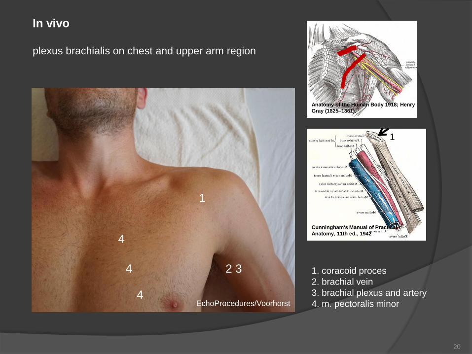

Anatomy of the Human Body 1918 Henry
Gray (1825ndash1861)
probe position1
2
1 3
4
5
4
6
7
EchoProceduresVoorhorst
plexus and vessels in upper arm (position 1)
1 n musculacutaneus
2 n medianus
3 n ulnaris
4 n radialis
5 axillary artery
6 humerus
7 m biceps
8 m coracobrachialis
9 m triceps
10 axillary vein
Ultrasound landmarks D
21
plexus and vessels in upper arm (position 2)
1 n musculocutaneus
2 m biceps
3 m coracobrachialis
4 humerus
5 axillary artery
6 axillary vein
Anatomy of the Human Body 1918 Henry
Gray (1825ndash1861)
probe placement position 2
EchoProceduresVoorhorst
Ultrasound landmarks D
22
Inside the suprascapular notch n
suprascapularis (C5) Motor
innervation m supraspinatus + m
infraspinatus Sensory innervation
AC-joint + glenohumeral joint
Anatomy of the Human Body 1918 Henry
GrayGray (1821861)
lateral
1
2
2
3
4
EchoProceduresVoorhorst
1 supraclavicular notch 2 supraspinatus 3 tendon
supraspinatus 4 trapezius Anatomy of the Human
Body 1918 Henry Gray (1825ndash
1861)
probe
E Ultrasound landmarks
scan both statically and dynamically
- during passive humeral abduction -
to neutralize compression of the SSP 23
spinoglenoid notch
1
2 3
4
5
6
EchoProceduresVoorhorst
nerve and artery are situated
inside the spinoglenoid notch
Anatomy of the Human Body 1918 Henry
Gray (1825ndash1861)
1 spinoglenoid notch
2 labrum glenoidale
3 humerus
4 articular cartilage
5 m deltoideus
6 infraspinatus
probe
Greyacutes anatomy
F Ultrasound landmarks
scan both statically and dynamically -
during passive humeral exoration - to
neutralize compression of the ISP
24
Motor innervation n axillaris (C5
C6) m teres minor + m deltoideus
Sensory innervation skin above
deltoid muscle
4
1 axillary nerve
2 circumflex axillary artery
3 m teres minor
4 humerus
1 circumflex axillary artery
2 axillary nerve
3 m deltoideus
4 m teres minor
green quadrilateral space
1 2
3
EchoProceduresVoorhorst
4 Anatomy of the Human Body 1918 Henry
Gray (1825ndash1861) (yellow and green
EchoProcedures)
1 2
3
4
4
G Ultrasound landmarks
25
probe
Anatomy of the human
body Henry Gray
Miscellaneous
medial ramus of dorsal root
n phrenicus
n vagus
26
1 nerve root 2 medial ramus innervating facet joints 3 anterior tuberculum 4 posterior tuberculum
Anatomy of the Human Body 1918 Henry Gray (1825ndash1861)
(Yellow EchoProcedures)
1 2
1 2
3 4
Bony landmarks
articular pilllar for ultrasound identification medial ramus
27
medial ramus innervating facet joints
EchoProceduresVoorhorst
probe
longitudinal articular pilar under the open
arrow the medial ramus and artery
1 3 inferior articular process
2 4 superior articular process
EchoProceduresVoorhorst
1 2
3 4
posterior
1 nerve root
2 medial ramus
3 articular pilar(s)
4artery
3
3
distal
EchoProceduresVoorhorst
4
Landmarks
28
n phrenicus C3C4 C5
VJ jugular vein
ACI common carotic artery
n vagus cranial nerve X
1 carotic artery
2 n vagus
3 jugular vein
4 m sternocleidomastoideus
5 thyroid
medial
Anatomy of the
Human Body 1918
Henry Gray (1825ndash
1861)
1
2
1 n phrenicus
2 n vagus
lateral medial
EchoProceduresVoorhorst EchoProceduresVoorhorst
Landmarks
29
Suggested Readings 1
Martinoli Brachial Plexus normal anatomy and scanning technique lecture 27-06-2018 Amsterdam
The cervical plexus anatomy and ultrasound guided blocks
httpwwwapicareonlinecomthe-cervical-plexus-anatomy-and-ultrasound-guided-blocks
Risk of Encountering Dorsal Scapular and Long Thoracic Nerves during Ultrasound-guided Interscalene Brachial Plexus
Block with Nerve Stimulator Korean J Pain 2016 July Vol 29 No 3 179-184 pISSN 2005-9159 eISSN 2093-0569
httpdxdoiorg103344kjp2016293179
Anatomy of the Human Body 1918 Henry Gray (1825ndash1861)
High frequency ultrasound of the cervical spine and brachial plexus - essentials
fileCUsersEigenaaarDownloadsECR2014_C-1692pdf
Ultrasound-Guided Interscalene Brachial Plexus Block
httpswwwnysoracomultrasound-guided-interscalene-brachial-plexus-block
Guided Intervention in Cervical Spine J Korean Orthop Assoc 2015 Apr50(2)77-92 KoreanPublished online April 30 2015
httpsdoiorg104055jkoa201550277
Researchgate
httpswwwresearchgatenetfigureNormal-brachial-plexus-A-Transverse-ultrasound-image
Spinal accesory nerve
httpswwwresearchgatenetfigureSpinal-accessory-nerve-A-Schematic-drawing-shows-the-pertinent-anatomy-of-the
spinal_fig14_47756593_sg=mRNj0a1gnwJGsZ7NsEzVPpIcRzb
eW9_xspNYHgYvIoDHTjAJEFtyxqUCcuZ2U357BboOun7Mdux3U1iWPNB4A
30
Suggested Readings 2
Quadrilateral Space Syndrome Diagnosis and Clinical Management
Patrick T Hangge12 Ilana Breen23 Hassan Albadawi2 M Grace Knuttinen2 Sailendra G Naidu2 andRahmi Oklu2
httpswwwncbinlmnihgovpmcarticlesPMC5920460
Dynamic ultrasonography of the shoulder
httpswwwe-ultrasonographyorgjournalviewphpnumber=206
Sonographic evaluation of the axillary artery during simulated overhead throwing
positionStapleton C Herrington LC and George K positionStapleton C Herrington LC and George K
httpusirsalfordacuk23061stapleton_et_al_2008_PTiSpdfpublic
Ultrasonographic Diagnosis of Thoracic Outlet Syndrome Secondary to Brachial Plexus Piercing Variation
fileCUsersEigenaaarDownloadsdiagnostics-07-00040pdf
The vascular component in neurogenic-arterial thoracic outlet syndromeJ Ernesto Molina MD PhD1 and Jonathan DrsquoCunha
MD PhD2
httpswwwncbinlmnihgovpmcarticlesPMC2728415
Ultrasound-guided interventional procedures for cervical pain
httpwwwasociacionandaluzadeldoloreswp-contentuploads201702US-cervicalpdf
httpswwwnysoracomultrasound-guided-supraclavicular-brachial-plexus-block-2
Ultrasound-Guided Supraclavicular Brachial Plexus Block
31
Ultrasound-Guided Superficial Cervical Plexus Block
httpswwwnysoracomultrasound-guided-superficial-cervical-plexus-block
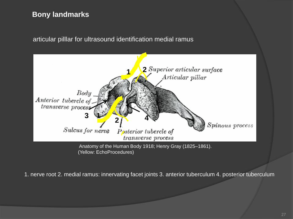