Cecilia Teodora Satta Aika Yamaguchi and Brian S. Leander
20
MORPHOLOGY AND MOLECULAR PHYLOGENY OF A NEW MARINE, SAND-DWELLING DINOFLAGELLATE GENUS, PACHENA (DINOPHYCEAE), WITH DESCRIPTIONS OF THREE NEW SPECIES 1 Mona Hoppenrath 2 Senckenberg am Meer, German Centre for Marine Biodiversity Research (DZMB), S€ udstrand 44, Wilhelmshaven D – 26382, Germany Albert Re ~ n e Departament de Biologia Marina i Oceanografia, Institut de Ci encies del Mar (CSIC), Pg. Mar ıtim de la Barceloneta, 37-49, Barcelona, Catalonia 08003, Spain Cecilia Teodora Satta Dipartimento di Architettura, Design e Urbanistica, University of Sassari, Via Piandanna 4, Sassari 07100, Italy Agenzia Ricerca per l’Agricoltura (AGRIS), Loc Bonassai, Olmedo, Sassari 07100, Italy Aika Yamaguchi Faculty of Science, Hokkaido University, Sapporo 060-0810, Japan and Brian S. Leander The Departments of Botany and Zoology, University of British Columbia, 6270 University Boulevard, Vancouver BC V6T 1Z4, Canada Marine benthic dinoflagellates are interesting not only because some epiphytic genera can cause harmful algal blooms but also for understanding dinoflagellate evolution and diversification. Our understanding of their biodiversity is far from complete, and many thecate genera have unusual tabulation patterns that are difficult to relate to the diverse known phytoplankton taxa. A new sand- dwelling genus, Pachena gen. nov., is described based on morphological and DNA sequence data. Three species were discovered in distant locations and are circumscribed, namely, P. leibnizii sp. nov. from Canada, P. abriliae sp. nov. from Spain, and P. meriddae sp. nov. from Italy. All species are tiny (about 9–23 lm long) and heterotrophic. Species are characterized by their tabulation (APC 4 0 3a 6 00 5c 5s 5 000 2 0000 ), an apical hook covering the apical pore, an ascending cingulum, and a sulcus with central list. The first anterior intercalary plate is uniquely “sandwiched” between two plates. The species share these features and differ in the relative sizes and arrangements of their plates, especially on the epitheca. The ornamentation of thecal plates is species-specific. The new molecular phylogenies based on SSU and LSU rDNA sequences contribute to understanding the evolution of the planktonic relatives of Pachena, the Thoracosphaeraceae. Key index words: benthic; distribution; morphology; Peridiniales; protists; taxonomy; Thoracosphaer- aceae Abbreviations : AICc, corrected Akaike information criterion; APC, apical pore complex; (B)PP, (Baye- sian) posterior probability; BS, bootstrap; DIC, differential interference contrast; HMDS, hexam- ethyldisilazane; ML, maximum likelihood; Po, apical pore plate; 1 0 , first apical plate; 2 0 , second apical plate; 3 0 , third apical plate; 4 0 , forth apical plate; 1a, first intercalary plate; 2a, second intercalary plate; 3a, third intercalary plate; 1 00 , first precingular plate; 2 00 , second precingular plate; 3 00 , third precingular plate; 4 00 , fourth precingular plate; 5 00 , fifth precingular plate; 6 00 , sixth precingular plate; c1, first cingular plate; c2, second cingular plate; c3, third cingular plate; c4, fourth cingular plate; c5, fifth cingular plate; Sa, ante- rior sulcal plate; Sd, right sulcal plate; Ss, left sulcal plate; Sp, posterior sulcal plate; Sm, median sulcal plate; 1 000 , first postcingular plate; 2 000 , second postcin- gular plate; 3 000 , third postcingular plate; 4 000 , fourth postcingular plate; 5 000 , fifth postcingular plate; 1 0000 , first antapical plate; 2 0000 , second antapical plate 1 Received 11 December 2019. Accepted 14 February 2020. First Published Online 6 March 2020. Published Online 2 April 2020, Wiley Online Library (wileyonlinelibrary.com). 2 Author for correspondence: e-mail: mhoppenrath@sencken berg.de. Editorial Responsibility: C. Lane (Associate Editor) 798 J. Phycol. 56, 798–817 (2020) © 2020 The Authors. Journal of Phycology published by Wiley Periodicals, Inc. on behalf of Phycological Society of America This is an open access article under the terms of the Creative Commons Attribution License, which permits use, distribution and reproduction in any medium, provided the original work is properly cited. DOI: 10.1111/jpy.12984
Transcript of Cecilia Teodora Satta Aika Yamaguchi and Brian S. Leander
MORPHOLOGY AND MOLECULAR PHYLOGENY OF A NEW MARINE, SAND-DWELLINGDINOFLAGELLATE GENUS, PACHENA (DINOPHYCEAE), WITH DESCRIPTIONS OF THREE
NEW SPECIES1
Mona Hoppenrath2
Senckenberg am Meer, German Centre for Marine Biodiversity Research (DZMB), S€udstrand 44, Wilhelmshaven D – 26382,
Germany
Albert Re~n�e
Departament de Biologia Marina i Oceanografia, Institut de Ci�encies del Mar (CSIC), Pg. Mar�ıtim de la Barceloneta, 37-49,
Barcelona, Catalonia 08003, Spain
Cecilia Teodora Satta
Dipartimento di Architettura, Design e Urbanistica, University of Sassari, Via Piandanna 4, Sassari 07100, Italy
Agenzia Ricerca per l’Agricoltura (AGRIS), Loc Bonassai, Olmedo, Sassari 07100, Italy
Aika Yamaguchi
Faculty of Science, Hokkaido University, Sapporo 060-0810, Japan
and Brian S. Leander
The Departments of Botany and Zoology, University of British Columbia, 6270 University Boulevard, Vancouver BC V6T 1Z4,
Canada
Marine benthic dinoflagellates are interesting notonly because some epiphytic genera can causeharmful algal blooms but also for understandingdinoflagellate evolution and diversification. Ourunderstanding of their biodiversity is far fromcomplete, and many thecate genera have unusualtabulation patterns that are difficult to relate to thediverse known phytoplankton taxa. A new sand-dwelling genus, Pachena gen. nov., is described basedon morphological and DNA sequence data. Threespecies were discovered in distant locations and arecircumscribed, namely, P. leibnizii sp. nov. fromCanada, P. abriliae sp. nov. from Spain, and P.meriddae sp. nov. from Italy. All species are tiny(about 9–23 lm long) and heterotrophic. Species arecharacterized by their tabulation (APC 40 3a 600 5c 5s5000 20000), an apical hook covering the apical pore, anascending cingulum, and a sulcus with central list.The first anterior intercalary plate is uniquely“sandwiched” between two plates. The species sharethese features and differ in the relative sizes andarrangements of their plates, especially on theepitheca. The ornamentation of thecal plates isspecies-specific. The new molecular phylogenies
based on SSU and LSU rDNA sequences contributeto understanding the evolution of the planktonicrelatives of Pachena, the Thoracosphaeraceae.
Key index words: benthic; distribution; morphology;Peridiniales; protists; taxonomy; Thoracosphaer-aceae
Abbreviations: AICc, corrected Akaike informationcriterion; APC, apical pore complex; (B)PP, (Baye-sian) posterior probability; BS, bootstrap; DIC,differential interference contrast; HMDS, hexam-ethyldisilazane; ML, maximum likelihood; Po, apicalpore plate; 10, first apical plate; 20, second apical plate;30, third apical plate; 40, forth apical plate; 1a, firstintercalary plate; 2a, second intercalary plate; 3a, thirdintercalary plate; 100, first precingular plate; 200, secondprecingular plate; 300, third precingular plate; 400,fourth precingular plate; 500, fifth precingular plate;600, sixth precingular plate; c1, first cingular plate; c2,second cingular plate; c3, third cingular plate; c4,fourth cingular plate; c5, fifth cingular plate; Sa, ante-rior sulcal plate; Sd, right sulcal plate; Ss, left sulcalplate; Sp, posterior sulcal plate; Sm, median sulcalplate; 1000, first postcingular plate; 2000, second postcin-gular plate; 3000, third postcingular plate; 4000, fourthpostcingular plate; 5000, fifth postcingular plate; 10000,first antapical plate; 20000, second antapical plate
1Received 11 December 2019. Accepted 14 February 2020. FirstPublished Online 6 March 2020. Published Online 2 April 2020,Wiley Online Library (wileyonlinelibrary.com).
2Author for correspondence: e-mail: [email protected].
Editorial Responsibility: C. Lane (Associate Editor)
798
J. Phycol. 56, 798–817 (2020)© 2020 The Authors. Journal of Phycology published by Wiley Periodicals, Inc. on behalf of Phycological Society of AmericaThis is an open access article under the terms of the Creative Commons Attribution License, which permits use,distribution and reproduction in any medium, provided the original work is properly cited.DOI: 10.1111/jpy.12984

The first studies on sand-dwelling dinoflagellateswere conducted in the early twentieth century(Kofoid and Swezy 1921, Herdman 1922, 1924a,b,Balech 1956), even though they were not studiedcomprehensively until the 2000s (Hoppenrath2000a, Murray 2003, Tamura 2005, Mohammad-Noor et al. 2007, Al-Yamani and Saburova 2010).The studies showed that the species composition isdistinct from planktonic communities and the spe-cies diversity was largely unexplored (Hoppenrathet al. 2014). Epiphytic species have received moreattention from the scientific community, mainlybecause many of them are toxin producers and aretoxic to humans (Berdalet et al. 2017). Still there isundiscovered biodiversity among benthic, especiallysand-dwelling, dinoflagellates with new taxondescriptions nearly every year, including new gen-era: Vulcanodinium (N�ezan and Chom�erat 2011),Moestrupia (Hansen and Daugbjerg 2011), Ankistro-dinium (Hoppenrath et al. 2012), Testudodinium(Horiguchi et al. 2012), Bispinodinium (Yamadaet al. 2013), Ailadinium (Saburova and Chom�erat2014), Madanidinium (Chom�erat and Bilien 2014),Aduncodinium (Kang et al. 2015), Fukuyoa (G�omezet al. 2015), Pellucidodinium (Onuma et al. 2015),Laciniporus (Saburova and Chom�erat 2019), andPsammodinium (Re~n�e and Hoppenrath 2019).
Benthic, sand-dwelling species seem to have mor-phological adaptations reflecting their life in theinterstitial habitat, such as smooth (i.e., without strik-ing extensions like wings, spines, or horns) and flat-tened cell shapes (Hoppenrath et al. 2014). Severaltaxa cover their apical pore with thecal extensions(e.g., Rhinodinium has a large apical hook; Murrayet al. 2006); some Amphidiniopsis species and Herdma-nia have a small hook (Hoppenrath 2000b, Murrayand Patterson 2002, Toriumi et al. 2002, Yamaguchiet al. 2011, Re~n�e et al. 2020); Apicoporus has finger-like projections (Sparmann et al. 2008); Laciniporushas a small flap-shaped projection (Saburova andChom�erat 2019); and Sinophysis has parallel uprightprojections (Hoppenrath 2000c, Chom�erat 2016).Many thecate, benthic dinoflagellate taxa have unu-sual tabulation patterns that are difficult to relate tothe known tabulations in planktonic taxa (Hoppen-rath et al. 2014). For example, Madanidinium has noapical pore (Chom�erat and Bilien 2014); Plagiodiniumhas no precingular or no apical plate series (Faustand Balech 1993, Wakeman et al. 2018), dependingon interpretation; Thecadinium sensu stricto and Pseu-dothecadinium have only incomplete precingular plateseries and other special plate arrangements (Efimovaet al. 2019, Selina et al. 2019); Pseudadenoides is theonly known genus with a complete posterior inter-calary plate series (Hoppenrath et al. 2003, 2017).
A species diversity survey of marine sandy sedi-ments in British Columbia, Canada, revealed speciesrichness including new taxa (Hoppenrath and Lean-der 2007, 2008, Sparmann et al. 2008, Hoppenrathet al. 2014, 2017; M. Hoppenrath unpub. data).
Studies on benthic dinoflagellates from the Mediter-ranean Sea have mainly focused on epiphytic toxicspecies (Vila et al. 2001, Aligizaki and Nikolaidis2006, Aligizaki et al. 2009, Penna et al. 2012),whereas sand-dwelling dinoflagellates have beenpoorly studied and information is scarce (Re~n�eet al. 2020). Here, a new genus is described that wasfirst discovered on the western shoreline of Vancou-ver Island, Canada and further species wererecorded in Spanish and Italian Mediterranean Seasamples.
METHODS
Sampling, cell extractions, and microscopy. Sand samples fromCanada were collected with a spoon during low tide atPachena Beach (48°47034.6″ N, 125°07019.0″ W), Vancou-ver Island, British Columbia, in May and June 2005, Apriland June 2006, and May and June 2007. The sand sam-ples were transported directly to the laboratory, anddinoflagellates were separated from the sand by extractionthrough a fine filter (mesh size 45 lm) using melting sea-water ice (Uhlig 1964). Cells of the new taxon wereobserved directly with a Leica DMIL inverted microscope(Wetzlar, Germany) and isolated by micropipetting forthe preparations described below. For differential interfer-ence contrast (DIC) light microscopy, pipetted cells wereviewed with a Zeiss Axioplan 2 imaging microscope (Carl-Zeiss, Oberkochen, Germany) connected to a LeicaDC500 color digital camera.
Mediterranean sediment samples from the Catalan Coastwere obtained at Castelldefels Beach (41°15037.0″ N;1°55048.8″ E) during spring and summer months from 2015to 2017. Sediment samples from Sardinian beaches wereobtained at Platamona Beach (40°49027.1″ N; 8°31036.4″ E)and La Speranza Beach (40°29043.1″ N; 8°22012.1″ E) duringsummer months in 2015 and 2018. Surface samples weretaken by snorkeling at a depth of approximately 1.5–2 m withplastic bottles. The distance to the shore depended on theunderwater slope of each beach. The sediments were kept atroom temperature, in the dark, and immediately taken to thelaboratory. Once there, cells were extracted from the sedi-ment using the melting seawater-ice method (Uhlig 1964).Subsamples were fixed with Lugol’s iodine or formaldehyde(2%) and preserved in the dark at 4°C. Alive and fixed sam-ples from the Catalan coast were observed under a phase-con-trast Leica DM-IRB inverted microscope (Leica Microsystems,Wetzlar, Germany) connected to a ProgRes C10 (JenoptikLaser, Optik Systeme GmbH, Jena, Germany) digital camera.Cell measurements were conducted using the ProgRes Cap-turePro software (Jenoptik Laser; Optik Systeme GmbH). Livesamples from the Sardinian coast were observed under a Zeiss100 inverted microscope (Carl Zeiss, Oberkochen, Germany),equipped with DIC. Digital photos were taken using a ZeissAxiocam (Carl Zeiss). Cell measurements were obtained fromLM and SEM images using the ImageJ software (1.47v; W.Rasband, USA).
For scanning electron microscopy observations (1) theCanadian mixed-extraction samples were fixed overnight withacidic Lugol’s solution. Cells were transferred onto a 5 lmpolycarbonate membrane filter (Corning Separations Div.,Acton, MA, USA), washed with distilled water, dehydratedwith a graded series of ethanol, rinsed twice in hexamethyld-isilazane (HMDS), and oven-dried at 65°C. Filters weremounted on stubs, sputter-coated with gold, and viewedunder a Hitachi S4700 scanning electron microscope; (2) thefixed Mediterranean subsamples were filtered into a 3.0–
DESCRIPTION OF PACHENA GEN. NOV. 799
5.0 lm polycarbonate filter, and washed in seawater and dis-tilled water for 15 min. A subsequent dehydration was carriedout in a 25, 50, 75, 90, 96, and 100% ethanol series for ca.10 min. The final step of 100% ethanol was repeated twice.The filters were critical-point dried or rinsed twice in HMDSand dried for 5 min at 60°C. The dried filters were thenmounted on stubs, sputter coated with gold-palladium. Cata-lan samples were examined with a HITACHI S-3500N scan-ning electron microscope (Hitachi High Technologies Corp.,Tokyo, Japan) at the Servei de Microscopia Electr�onica (ICM-CSIC) in Spain and a Tescan VEGA3 microscope (Elektro-nen-Optik-Service GmbH, Dortmund, Germany) in Germany.Sardinian samples were examined with a Tescan VEGA3microscope (Elektronen-Optik-Service GmbH) in Germany.
Polymerase chain reactions and phylogenetic analyses. Canadianspecimens isolated from a raw sample were washed with fil-tered (eukaryote-free) seawater and 100 cells deposited in35 lL distilled water in a 1.5 mL Eppendorf tube (Dia-MedLab Supplies Inc., Mississauga, ON, Canada) heated at 65°Cand afterwards stored in a freezer. DNA amplification wascarried out using 10 lL of this cell preparation and puRe-Taq Ready-To-Go PCR Beads (Amersham Biosciences, Piscat-away, NJ, USA). The protocol using universal eukaryoticprimers (PF1-R4) was described in Hoppenrath and Leander(2007, 2010). PCR products corresponding to the expectedsize were gel isolated and cloned into the pCR2.1 using theTOPO TA cloning kit (Invitrogen, Carlsbad, CA, USA). Oneclone from Pachena leibnizii sp. nov. was sequenced with ABIbig-dye reaction mix (Applied Biosystems, Foster City, CA,USA) using the vector primers and internal primers orientedin both directions (See Table 1 for GenBank accession num-bers).
Around 40 specimens from the Catalan Coast were iso-lated from a raw sample from May 2017, washed with filteredand autoclaved seawater, and transferred to a 1.5 mL Eppen-dorf tube containing 200 lL of seawater. Genomic DNA wasextracted using a DNeasy Blood & Tissue kit (Qiagen, Hil-den, Germany) following the manufacturer instructions. Twomicroliters of this extract was subjected to a first 25 lL PCRusing EK-82F – 28S-1611R primers with an amplification mix-ture containing 2 lL of 10X buffer (TaKaRa Bio), 1.5 mMMgCl2, 1 U of TaKaRa Taq DNA polymerase (TaKaRa Bio),0.2 mM of each dNTP, and 0.4 mM of each primer. PCRamplification conditions were as follows: initial denaturationfor 3 min at 95°C, followed by 6 cycles of 15 s at 95°C, 30 sat 58–53°C, decreasing 1°C each cycle, and 2 min at 72°C,and 34 additional cycles at annealing temperature of 52°C,followed by a final extension step for 5 min at 72°C. Theresulting product was used as template for semi-nested PCRsto amplify the SSU and LSU rDNA regions independently,using primers EK-82F and EK-1520R, and 28S-1F – 28S-1611R, respectively. Each amplification reaction contained1 lL of template, 2.5 lL of 10X buffer (Invitrogen, ThermoFisher Scientific Corp.) containing 15 mM MgCl2, 1.25 U ofPlatinum Taq DNA polymerase (Invitrogen, Thermo Fisher
Scientific Corp.), 0.2 mM of each dNTP, and 0.4 mM of eachprimer. PCR conditions were as follows: initial denaturationfor 2 min at 94°C, 35 cycles of 15 s at 94°C, 30 s at 55°C,and 1 min at 72°C, followed by a final extension step for5 min at 72°C. A second semi-nested, using 1 lL of the previ-ous PCR product as template was performed for each regionusing primers DIN-464F and EK-1520R, and 28S-1F and 28S-803R, respectively. Four lL of PCR products were elec-trophoresed in an agarose gel and visualized under UV illu-mination. Purification and Sanger sequencing was carriedout by external services (Genewiz, Takeley, UK) using bothforward and reverse primers. See Table 1 for GenBank acces-sion numbers.
Several specimens from Platamona Beach, Italy were isolatedfrom samples collected in June 2015 and 2018 and one speci-men from La Speranza Beach, Italy, was isolated from a samplecollected in June 2018. Isolated cells were washed in severaldrops of filtered seawater and transferred to a 0.2 mL PCRtubes containing 5 lL of lysis buffer (400 ng � lL�1 ProteinaseK and 0.005% SDS). Tubes were subjected to freezing at -20°Cfor 10 min, heating at 60°C for 30 min, and then at 95°C for10 min to facilitate cell lysis. Resulting lysates were directly usedas a template for amplification of SSU and LSU rDNA frag-ments.
The lysate from two specimens of Platamona (sample June2015) was amplified for the LSU rDNA using D1R and D2Cprimers. The 50 lL PCR mixture contained 5 lL of 10X buf-fer, 1.5 lL of MgCl2, 0.25 lL of Hot start Taq DNA poly-merase and 1 lL of dNTP 0.2 mM each (Qiagen), and0.4 mM of each primer. PCR amplification conditions were asfollows: initial denaturation for 5 min at 95°C, 40 cycles of 20s at 95°C, 30 s at 55°C, and 1 min at 72°C, followed by a finalextension step for 10 min at 72°C.
The lysate of one specimen from Platamona and onefrom La Speranza (samples June 2018) were used as tem-plate for amplification of SSU rDNA fragment using EukAand EukB primers. Each 50 lL PCR amplification reactioncontained 5 lL of 10X buffer, 1.5 lL of MgCl2, and0.25 lL of Hot start Taq DNA polymerase (Biotechrabbit),1 lL of dNTP 0.2 mM each (Qiagen), and 0.4 mM of eachprimer. PCR conditions were as follows: initial denaturationfor 5 min at 94°C, 34 cycles of 1 m at 94°C, 1:30 m at55°C, and 2 min at 72°C, followed by a final extension stepfor 7 min at 72°C. The resulting product was used as tem-plate for nested PCRs using primers Dino18SF1 and 18S-comR1. Each PCR amplification reaction contained 5 lL of10X buffer, 1.5 lL of MgCl2, and 0.25 lL of Hot start TaqDNA polymerase (Biotechrabbit), 1 lL of dNTP 0.2 mMeach (Qiagen), 0.4 mM of each primer, and 1 lL of tem-plate. PCR conditions were as follows: initial denaturationfor 5 min at 94°C, 29 cycles of 45 s at 94°C, 1 m at 55°C,and 3 min at 72°C, followed by a final extension step for10 min at 72°C. All information regarding the primers usedduring the amplification protocols can be found inTable S1 in the Supporting Information.
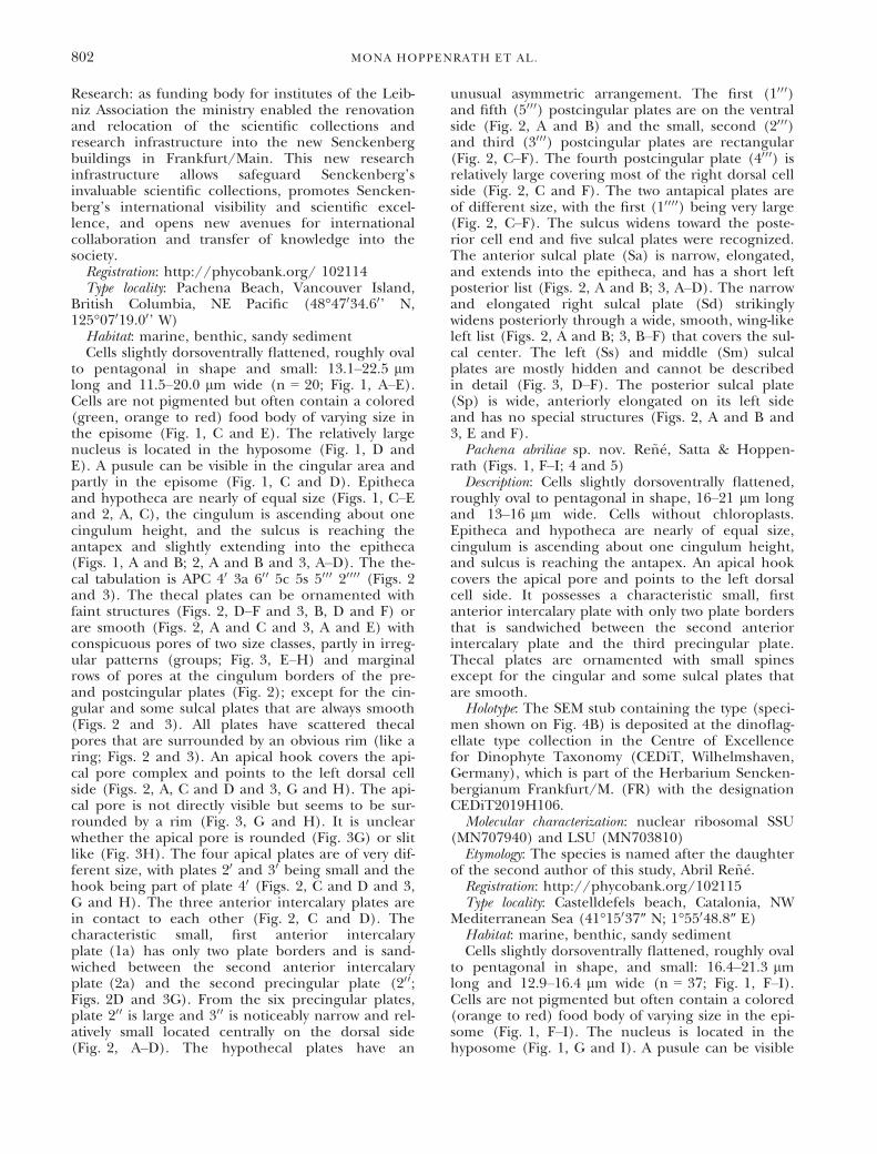
TABLE 1. List of rDNA sequences obtained in this study, including their location and date of isolation, length in basepairs (bp), and GenBank accession number.
Species Origin Year of isolation rDNA region Length (bp) Acc. no. Isolate
Pachena leibnizii Pachena Beach (Canada) 2007 SSU 1804 MN707939 Vancouver IslandPachena abriliae Castelldefels (Catalonia) 2017 SSU 1140 MN707940 CastelldefelsPachena abriliae Castelldefels (Catalonia) 2017 LSU 689 MN703810 CastelldefelsPachena abriliae La Speranza (Sardinia) 2018 SSU 1571 MN707941 Speranza6Pachena meriddae Platamona (Sardinia) 2018 SSU 1552 MN707942 Platamona19Pachena sp. Platamona (Sardinia) 2015 LSU 656 MN703811 Platamona28
800 MONA HOPPENRATH ET AL.

Four lL of all PCR products were electrophoresed in anagarose gel and visualized under UV illumination. Purifica-tion and Sanger sequencing was carried out by external ser-vices (Macrogen Europe, Amsterdam, Netherlands in 2015and Genoscreen, Lille, France in 2018) using both forwardand reverse primers. See Table 1 for GenBank accessionnumbers.
The SSU and LSU rDNA sequences were aligned usingMUSCLE (Edgar 2004) and viewed in Mesquite v3.11 (Mad-dison and Maddison 2015). Highly variable regions were elim-inated using Gblocks (Castresana 2000, Talavera andCastresana 2007). The final alignments of the SSU and LSUrDNA dataset consisted of 48 with 1768 and 49 taxa with1167 sites, respectively. The best-fit model for each datasetwas chosen by ModelFinder in IQ-TREE under AICc (Trifino-poulos et al. 2016, Kalyaanamoorthy et al. 2017). Maximum-likelihood (ML) analyses were run with IQ-TREE usingTIM2 + F + R4 and GTR+F+R5, as the model of evolutionfor the SSU and LSU rDNA, respectively. Bootstrap analyseswere run for each dataset with 1000 replicates to evaluate sta-tistical reliability. MrBayes v3.2.5 was used to perform Baye-sian analyses (Ronquist and Huelsenbeck 2003) with the GTR+ I + G and four Monte-Carlo–Markov chains starting from arandom tree. For the SSU dataset a total of 2,500,000 andthe first 6,250 trees were discarded; for the LSU rDNA data-set 1,000,000 generations were calculated and the first 2,500trees in each run were discarded. Trees were sampled every100 generations in both analyses. Posterior probabilities (PP)correspond to the frequency at which a given node was foundin the post-burn-in trees.
RESULTS
Pachena gen. nov. Hoppenrath, Satta & Re~n�eDescription: Thecate, heterotrophic dinoflagellate
without stigma. Thecal tabulation: APC 40 3a 600 5c5s 5000 20000. Dorsoventrally flattened cells; with apicalhook covering the apical pore, pointing to the leftdorsal cell side; ascending cingulum; sulcus
reaching the antapex. First anterior intercalary plate“sandwiched” between two plates.Etymology: The genus is named after the sampling
area of its first discovery, “Pachena Beach” nearBamfield, Vancouver Island, British Columbia,Canada.Registration: http://phycobank.org/ 102113Type: Pachena leibnizii sp. nov. HoppenrathPachena leibnizii sp. nov. Hoppenrath (Figs. 1, A–
E, 2 and 3)Description: Cells slightly dorsoventrally flattened,
roughly oval to pentagonal in shape, 13–23 lmlong, and 12–20 lm wide. Cells without chloroplas-ts. Epitheca and hypotheca are nearly of equal size,cingulum is ascending about one cingulum height,and sulcus is reaching the antapex. An apical hookcovers the apical pore and points to the left dorsalcell side. It possesses a characteristic small, firstanterior intercalary plate with only two plate bordersthat is sandwiched between the second anteriorintercalary plate and the second precingular plate.Thecal plates are smooth or faintly ornamentedexcept for the cingular and some sulcal plates thatare always smooth.Holotype: The SEM stub containing the type (speci-
men shown in Fig. 2A) is deposited at the dinoflag-ellate type collection in the Centre of Excellencefor Dinophyte Taxonomy (CEDiT, Wilhelmshaven,Germany), which is part of the Herbarium Sencken-bergianum Frankfurt/M. (FR) with the designationCEDiT2019H105.Molecular characterization: nuclear ribosomal SSU
(MN707939)Etymology: The species is named in honor of the
German Federal Ministry of Education and
FIG. 1. Light micrographs of the investigated Pachena gen. nov. species. (A–E) Pachena leibnizii sp. nov. from Canada. (F–I) Pachenaabriliae sp. nov. from Catalonia, Spain. J, K) Pachena meriddae sp. nov. from Sardinia, Italy. The ascending cingulum (c) and the sulcus(s)reaching the posterior cell end can be recognized (A, B, F). All cells contain a colored food body (fb). The nucleus (n) is located in thehyposome and sometimes the pusule (p) is visible. Note the apical hook (arrow) and posterior spines (arrowheads) in the Mediterraneanspecies. Scale bars = 10 lm. [Color figure can be viewed at wileyonlinelibrary.com]
DESCRIPTION OF PACHENA GEN. NOV. 801
Research: as funding body for institutes of the Leib-niz Association the ministry enabled the renovationand relocation of the scientific collections andresearch infrastructure into the new Senckenbergbuildings in Frankfurt/Main. This new researchinfrastructure allows safeguard Senckenberg’sinvaluable scientific collections, promotes Sencken-berg’s international visibility and scientific excel-lence, and opens new avenues for internationalcollaboration and transfer of knowledge into thesociety.
Registration: http://phycobank.org/ 102114Type locality: Pachena Beach, Vancouver Island,
British Columbia, NE Pacific (48°47034.60’ N,125°07019.00’ W)
Habitat: marine, benthic, sandy sedimentCells slightly dorsoventrally flattened, roughly oval
to pentagonal in shape and small: 13.1–22.5 lmlong and 11.5–20.0 lm wide (n = 20; Fig. 1, A–E).Cells are not pigmented but often contain a colored(green, orange to red) food body of varying size inthe episome (Fig. 1, C and E). The relatively largenucleus is located in the hyposome (Fig. 1, D andE). A pusule can be visible in the cingular area andpartly in the episome (Fig. 1, C and D). Epithecaand hypotheca are nearly of equal size (Figs. 1, C–Eand 2, A, C), the cingulum is ascending about onecingulum height, and the sulcus is reaching theantapex and slightly extending into the epitheca(Figs. 1, A and B; 2, A and B and 3, A–D). The the-cal tabulation is APC 40 3a 600 5c 5s 5000 20000 (Figs. 2and 3). The thecal plates can be ornamented withfaint structures (Figs. 2, D–F and 3, B, D and F) orare smooth (Figs. 2, A and C and 3, A and E) withconspicuous pores of two size classes, partly in irreg-ular patterns (groups; Fig. 3, E–H) and marginalrows of pores at the cingulum borders of the pre-and postcingular plates (Fig. 2); except for the cin-gular and some sulcal plates that are always smooth(Figs. 2 and 3). All plates have scattered thecalpores that are surrounded by an obvious rim (like aring; Figs. 2 and 3). An apical hook covers the api-cal pore complex and points to the left dorsal cellside (Figs. 2, A, C and D and 3, G and H). The api-cal pore is not directly visible but seems to be sur-rounded by a rim (Fig. 3, G and H). It is unclearwhether the apical pore is rounded (Fig. 3G) or slitlike (Fig. 3H). The four apical plates are of very dif-ferent size, with plates 20 and 30 being small and thehook being part of plate 40 (Figs. 2, C and D and 3,G and H). The three anterior intercalary plates arein contact to each other (Fig. 2, C and D). Thecharacteristic small, first anterior intercalaryplate (1a) has only two plate borders and is sand-wiched between the second anterior intercalaryplate (2a) and the second precingular plate (200;Figs. 2D and 3G). From the six precingular plates,plate 200 is large and 300 is noticeably narrow and rel-atively small located centrally on the dorsal side(Fig. 2, A–D). The hypothecal plates have an
unusual asymmetric arrangement. The first (1000)and fifth (5000) postcingular plates are on the ventralside (Fig. 2, A and B) and the small, second (2000)and third (3000) postcingular plates are rectangular(Fig. 2, C–F). The fourth postcingular plate (4000) isrelatively large covering most of the right dorsal cellside (Fig. 2, C and F). The two antapical plates areof different size, with the first (10000) being very large(Fig. 2, C–F). The sulcus widens toward the poste-rior cell end and five sulcal plates were recognized.The anterior sulcal plate (Sa) is narrow, elongated,and extends into the epitheca, and has a short leftposterior list (Figs. 2, A and B; 3, A–D). The narrowand elongated right sulcal plate (Sd) strikinglywidens posteriorly through a wide, smooth, wing-likeleft list (Figs. 2, A and B; 3, B–F) that covers the sul-cal center. The left (Ss) and middle (Sm) sulcalplates are mostly hidden and cannot be describedin detail (Fig. 3, D–F). The posterior sulcal plate(Sp) is wide, anteriorly elongated on its left sideand has no special structures (Figs. 2, A and B and3, E and F).Pachena abriliae sp. nov. Re~n�e, Satta & Hoppen-
rath (Figs. 1, F–I; 4 and 5)Description: Cells slightly dorsoventrally flattened,
roughly oval to pentagonal in shape, 16–21 lm longand 13–16 lm wide. Cells without chloroplasts.Epitheca and hypotheca are nearly of equal size,cingulum is ascending about one cingulum height,and sulcus is reaching the antapex. An apical hookcovers the apical pore and points to the left dorsalcell side. It possesses a characteristic small, firstanterior intercalary plate with only two plate bordersthat is sandwiched between the second anteriorintercalary plate and the third precingular plate.Thecal plates are ornamented with small spinesexcept for the cingular and some sulcal plates thatare smooth.Holotype: The SEM stub containing the type (speci-
men shown on Fig. 4B) is deposited at the dinoflag-ellate type collection in the Centre of Excellencefor Dinophyte Taxonomy (CEDiT, Wilhelmshaven,Germany), which is part of the Herbarium Sencken-bergianum Frankfurt/M. (FR) with the designationCEDiT2019H106.Molecular characterization: nuclear ribosomal SSU
(MN707940) and LSU (MN703810)Etymology: The species is named after the daughter
of the second author of this study, Abril Re~n�e.Registration: http://phycobank.org/102115Type locality: Castelldefels beach, Catalonia, NW
Mediterranean Sea (41°15037″ N; 1°55048.8″ E)Habitat: marine, benthic, sandy sedimentCells slightly dorsoventrally flattened, roughly oval
to pentagonal in shape, and small: 16.4–21.3 lmlong and 12.9–16.4 lm wide (n = 37; Fig. 1, F–I).Cells are not pigmented but often contain a colored(orange to red) food body of varying size in the epi-some (Fig. 1, F–I). The nucleus is located in thehyposome (Fig. 1, G and I). A pusule can be visible
802 MONA HOPPENRATH ET AL.

in the hyposome (Fig. 1, F and H). Epitheca andhypotheca are nearly of equal size (Figs. 1, G–I and4, A–F), the cingulum is ascending about one cingu-lum height, and the sulcus is reaching the antapex(Figs. 1F and 4, A and B). The thecal tabulation isAPC 40 3a 600 5c 5s 5000 20000 (Figs. 4 and 5). The the-cal plates are ornamented with small spines exceptfor the cingular and some sulcal plates that aresmooth (Figs. 4, 5). In some cells cingular plates
can have a faint bar-like ornamentation (Fig. 5, Fand H). All plates have scattered thecal pores thatare surrounded by a narrow rim (like a ring; Figs. 4,A–D and 5, A and H). An apical hook covers theapical pore complex and points to the left dorsalcell side (Fig. 4, A–F). The apical pore is notdirectly visible but seems to be surrounded by araised rim (Fig. 5, B, D and E). The apical poreplate possesses a row of thecal pores at its margin
FIG. 2. Scanning electron micrographs showing the tabulation of the type species Pachena leibnizii sp. nov. from British Columbia,Canada. (A, B) Ventral views. (C, D) Dorsal views. (E, F) Dorsal hypotheca and cingulum. The apical hook is marked by an arrowhead. 10-40, apical plates; 1a-3a, anterior intercalary plates; 1″-6″, precingular plates; 1‴-5‴, postcingular plates; 1″″-2″″, antapical plates; c1-c5, cingu-lar plates; Sa, anterior sulcal plate; Sd, right sulcal plate; Ss, left sulcal plate; Sp, posterior sulcal plate; Scale bars = 10 lm (A–D), 5 lm(E, F).
DESCRIPTION OF PACHENA GEN. NOV. 803
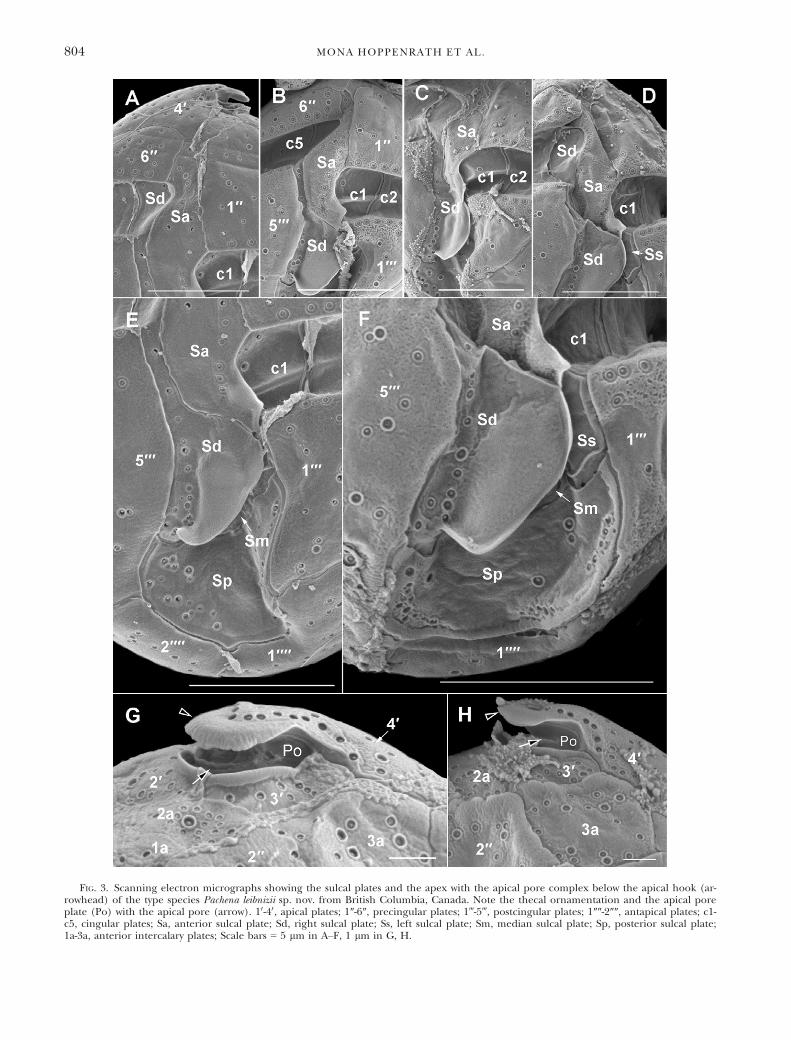
FIG. 3. Scanning electron micrographs showing the sulcal plates and the apex with the apical pore complex below the apical hook (ar-rowhead) of the type species Pachena leibnizii sp. nov. from British Columbia, Canada. Note the thecal ornamentation and the apical poreplate (Po) with the apical pore (arrow). 10-40, apical plates; 1″-6″, precingular plates; 1‴-5‴, postcingular plates; 1″″-2″″, antapical plates; c1-c5, cingular plates; Sa, anterior sulcal plate; Sd, right sulcal plate; Ss, left sulcal plate; Sm, median sulcal plate; Sp, posterior sulcal plate;1a-3a, anterior intercalary plates; Scale bars = 5 lm in A–F, 1 lm in G, H.
804 MONA HOPPENRATH ET AL.

FIG. 4. Scanning electron micrographs showing the tabulation of the new species Pachena abriliae sp. nov. from Catalonia, Spain. (A, B)Ventral views. (C–E) Dorsal views. (F) Left lateral view. (G, H) Right lateral view, mainly of the hypotheca. The apical hook is marked byan arrowhead. 10-40, apical plates; 1a-3a, anterior intercalary plates; 1″-6″, precingular plates, 1‴-5‴, postcingular plates; 1″″-2″″, antapicalplates; c1-c5, cingular plates; Sa, anterior sulcal plate; Sd, right sulcal plate; Sp, posterior sulcal plate; Scale bars = 10 lm.
DESCRIPTION OF PACHENA GEN. NOV. 805

(Fig. 5A). The four apical plates are of very differ-ent size, with plate 30 being the smallest and thehook being part of plate 40 (Figs. 4, A and B and 5,A–D). The three anterior intercalary plates are incontact to each other and cover a large part of thedorsal epitheca (Figs. 4, C–E; 5, A, D and E). Thecharacteristic small, first anterior intercalaryplate (1a) has only two plate borders and is sand-wiched between the second anterior intercalaryplate (2a) and the third precingular plate (300;Fig. 5, A, C–E). From the six precingular plates,
plate 300 is noticeably elongated (Figs. 4, A-F and 5,A–E). The hypothecal plates have an unusual asym-metric arrangement. The first (1000) and fifth (5000)postcingular plates are on the ventral side (Figs. 4,A and B and 5, F and H) and the small, second(2000) and third (3000) postcingular plates are rectan-gular (Fig. 4, C–F). The two antapical plates are ofdifferent size, with the first (10000) being very largeand covering most of the dorsal hypotheca (Figs. 4,C–H and 5, F and H). The sulcus widens toward theposterior cell end and five sulcal plates were
FIG. 5. Scanning electron micrographs showing the tabulation of the new species Pachena abriliae sp. nov. from Catalonia, Spain. (A)Apical view of the epitheca. (B) Ventral to left lateral view of the epitheca. (C) Left lateral view of the epitheca. (D, E) Dorsal view of theepitheca. (F) Antapical to ventral view. (G) Detail of the sulcus. (H) Hypotheca with cingulum in ventral view showing all sulcal plates. 10-40, apical plates; 1a-3a, anterior intercalary plates; 1″-6″, precingular plates; 1‴-5‴, postcingular plates; 1″″-2″″, antapical plates; c1-c5, cingu-lar plates; Sa, anterior sulcal plate; Sd, right sulcal plate; Ss, left sulcal plate; Sm, median sulcal plate; Sp, posterior sulcal plate; Scalebars = 5 lm.
806 MONA HOPPENRATH ET AL.

recognized (Figs. 4, A, B, G and H and 5, G andH). The anterior sulcal plate (Sa) is narrow andelongated, with a spiny right margin and a short leftposterior list (Figs. 4, A and B and 5, G and H).The narrow and elongated right sulcal plate (Sd)strikingly widens posteriorly through a wide, ser-rated, wing-like left list (Figs. 4, A and B, 5, G andH) that covers the sulcal center. The left (Ss) andmiddle (Sm) sulcal plates are mostly hidden andcannot be described in detail (Fig. 5, G and H).The posterior sulcal plate (Sp) is visible mainlybetween the two antapical plates and has no specialstructures (Figs. 4, A, B, G and H and 5, F–H).
Pachena meriddae sp. nov. Satta, Re~n�e & Hoppen-rath (Figs. 1, J and K, 6 and 7)
Description: Cells slightly dorsoventrally flattened,pentagonal, with triangular epitheca and trapezoidhypotheca, 9–13 lm long and 6–10 lm wide. Cellswithout chloroplasts. Epitheca and hypotheca arenearly of equal size, cingulum is ascending aboutone cingulum height, and sulcus is reaching theantapex. An apical hook covers the apical pore andpoints to the left dorsal cell side. Epithecal platesare ornamented with ridges and some spines,hypothecal plates with spines, cingular and sulcalplates are smooth.
Holotype: The SEM stub containing the type (speci-men shown on Fig. 6B) is deposited at the dinoflag-ellate type collection in the Centre of Excellencefor Dinophyte Taxonomy (CEDiT, Wilhelmshaven,Germany), which is part of the Herbarium Sencken-bergianum Frankfurt/M. (FR) with the designationCEDiT2019H107.
Molecular characterization: nuclear ribosomal SSU(MN707942)
Etymology: The species is named to honor Mr. Mar-cello Meridda, for his tireless support in taking fieldsamples.
Registration: http://phycobank.org/102116Type locality: Platamona Beach, Sardinia, Mediter-
ranean Sea (40°49027.1″ N; 8°31036.4″ E)Habitat: marine, benthic, sandy sedimentCells slightly dorsoventrally flattened, with trian-
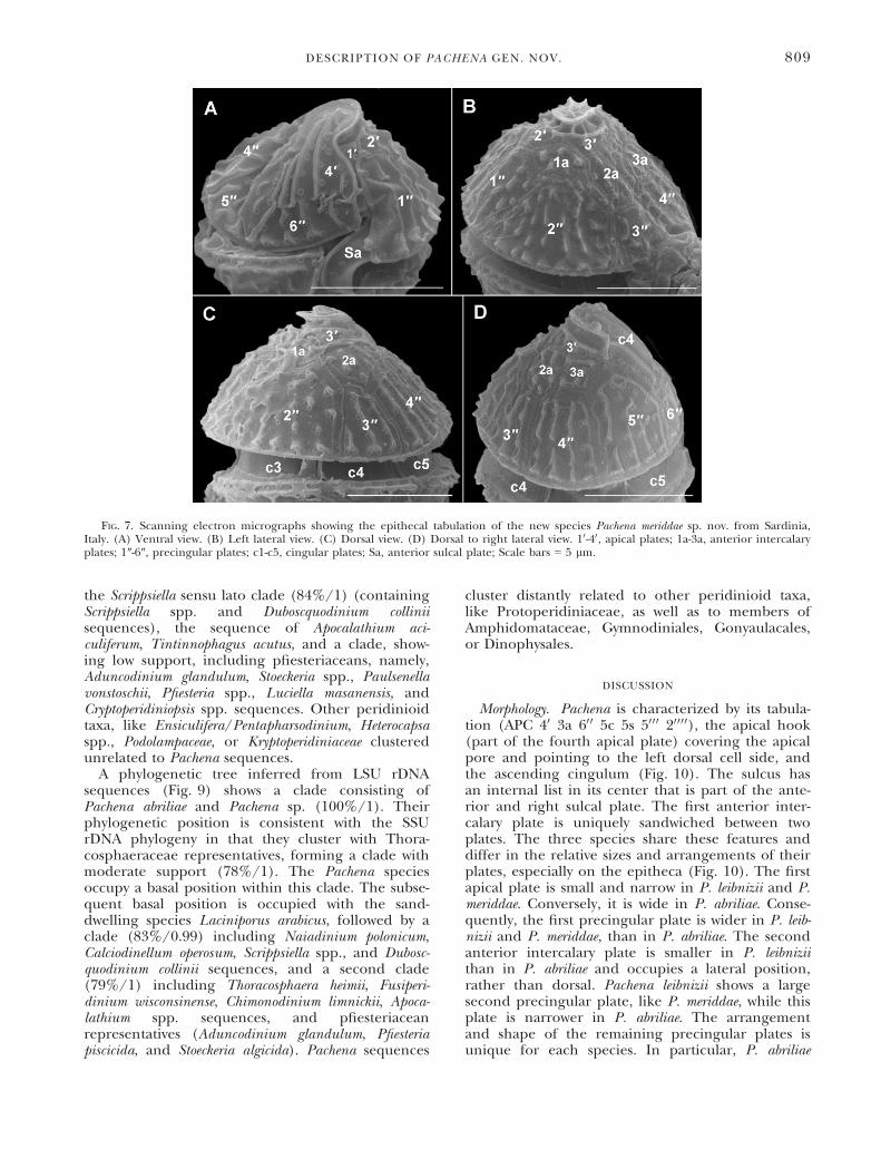
gular epitheca and trapezoid hypotheca, pentagonalin shape and small: 9.4–13.2 lm long and 6.3–10.2 lm wide (n = 20; Figs. 1, J and K and 6, A–F).Cells are not pigmented but often contain a colored(orange to red) food body of varying size in the epi-some (Fig. 1, J and K). The nucleus is located inthe hyposome (Fig. 1J). A pusule can be visible cen-trally (Fig. 1K). Epitheca and hypotheca are nearlyof equal size (Figs. 1, J and K and 6, A–F), the cin-gulum is ascending about one cingulum height(Fig. 6, A and B). The sulcus characteristicallyextends the end of the transverse furrow anteriorlyand is reaching the antapex (Fig. 7, A–C). The the-cal tabulation is APC 40 3a 600 5c 3 + s 5000 20000(Figs. 6 and 7). The epithecal plates are orna-mented with ridges and some spines, the hypothecalplates with spines, and cingular and sulcal plates are
smooth (Figs. 6 and 7). All plates have scatteredthecal pores that are surrounded by a narrow rim(like a ring; Figs. 6, G and H and 7). An apicalhook covers the apical pore complex and points tothe left dorsal cell side (Fig. 6). The apical pore isnot directly visible. The four apical plates are of verydifferent size, with a narrow plate 10, small plates 20and 30, and the hook being part of plate 40 (Fig. 7).The three anterior intercalary plates are in contactto each other (Fig. 7, B–D) but their shapes couldnot reliably be determined. Further observations arenecessary to clarify whether plate 1a is triangular(Fig. 7C) or pentagonal (Fig. 7B) and not sand-wiched between two plates. Plate 2a is in contactwith plates 30, 1a, 200, 300, 300, and 3a (Fig. 7, B–D)but the outline needs verification. It is uncertainwhether plate 3a is in contact with plate 30 (Fig. 7D)or not (Fig. 7B). To understand whether the ante-rior intercalary plates are very variable in this spe-cies much more cell observations are needed. Fromthe six precingular plates, plate 200 is relatively largeand 300 is noticeably smaller and narrower andlocated centrally on the dorsal side (Figs. 6, E, Fand H and 7C). The hypothecal plates have an unu-sual asymmetric arrangement. The postcingularplates 1000 and 5000 are on the ventral side (Fig. 6, A–C) and the small plates 2000 and 3000 are rectangular(Fig. 6, D and E). The two antapical plates are ofdifferent size, with plate 10000 being very large andcovering most of the dorsal hypotheca (Fig. 6, D–F). The sulcus widens toward the posterior cell endand only three sulcal plates were recognized. Theanterior sulcal plate (Sa) is narrow and elongated,with a smooth ridge on the right margin and ashort left posterior list (Fig. 6, A–C). The narrowand elongated right sulcal plate (Sd) opticallyextends the end of the transverse furrow anteriorlyand it widens posteriorly through a wide, smoothleft list that is posteriorly elongated and tapered(Fig. 6, B and C). A left (Ss) and middle (Sm) sul-cal plate were not recognized. The posterior sulcalplate (Sp) is visible mainly between the two antapi-cal plates (Fig. 6, A and B).Phylogenetic relationships. SSU rDNA sequences
were obtained for the three species described. Thesequences corresponding to Pachena leibnizii show a97.5% and a 98.1% pairwise similarity with thosefrom P. abriliae (Castelldefels) and P. meriddae,respectively, and the sequences of these two latterspecies show a 96.2% pairwise similarity betweenthem. Both sequences corresponding to P. abriliaewere 99.8% similar. A phylogenetic tree inferredfrom SSU rDNA sequences (Fig. 8) showed that allof the Pachena sequences form a clade (94% BS / 1BPP), where P. leibnizii and P. meriddae (100%/1)are more closely related to each other than to P.abriliae (100%/1). The Pachena clade clusters withThoracosphaeraceae (Peridiniales) representatives,even though the clade shows low statistical support(63%/-). The Thoracosphaeraceae cluster includes
DESCRIPTION OF PACHENA GEN. NOV. 807

FIG. 6. Scanning electron micrographs showing the tabulation of the new species Pachena meriddae sp. nov. from Sardinia, Italy. (A–C)Ventral views. (D, E) Dorsal views. (F) Left lateral view. (G) Right lateral view. (H) Dorsal view. The apical hook is marked by an arrow-head. 10-40, apical plates; 1a-3a, anterior intercalary plates; 1″-6″, precingular plates; 1‴-5‴, postcingular plates; 1″″-2″″, antapical plates; c1-c5, cingular plates; Sa, anterior sulcal plate; Sd, right sulcal plate; Sp, posterior sulcal plate; Scale bars = 5 lm.
808 MONA HOPPENRATH ET AL.
the Scrippsiella sensu lato clade (84%/1) (containingScrippsiella spp. and Duboscquodinium colliniisequences), the sequence of Apocalathium aci-culiferum, Tintinnophagus acutus, and a clade, show-ing low support, including pfiesteriaceans, namely,Aduncodinium glandulum, Stoeckeria spp., Paulsenellavonstoschii, Pfiesteria spp., Luciella masanensis, andCryptoperidiniopsis spp. sequences. Other peridinioidtaxa, like Ensiculifera/Pentapharsodinium, Heterocapsaspp., Podolampaceae, or Kryptoperidiniaceae clusteredunrelated to Pachena sequences.
A phylogenetic tree inferred from LSU rDNAsequences (Fig. 9) shows a clade consisting ofPachena abriliae and Pachena sp. (100%/1). Theirphylogenetic position is consistent with the SSUrDNA phylogeny in that they cluster with Thora-cosphaeraceae representatives, forming a clade withmoderate support (78%/1). The Pachena speciesoccupy a basal position within this clade. The subse-quent basal position is occupied with the sand-dwelling species Laciniporus arabicus, followed by aclade (83%/0.99) including Naiadinium polonicum,Calciodinellum operosum, Scrippsiella spp., and Dubosc-quodinium collinii sequences, and a second clade(79%/1) including Thoracosphaera heimii, Fusiperi-dinium wisconsinense, Chimonodinium limnickii, Apoca-lathium spp. sequences, and pfiesteriaceanrepresentatives (Aduncodinium glandulum, Pfiesteriapiscicida, and Stoeckeria algicida). Pachena sequences
cluster distantly related to other peridinioid taxa,like Protoperidiniaceae, as well as to members ofAmphidomataceae, Gymnodiniales, Gonyaulacales,or Dinophysales.
DISCUSSION
Morphology. Pachena is characterized by its tabula-tion (APC 40 3a 600 5c 5s 5000 20000), the apical hook(part of the fourth apical plate) covering the apicalpore and pointing to the left dorsal cell side, andthe ascending cingulum (Fig. 10). The sulcus hasan internal list in its center that is part of the ante-rior and right sulcal plate. The first anterior inter-calary plate is uniquely sandwiched between twoplates. The three species share these features anddiffer in the relative sizes and arrangements of theirplates, especially on the epitheca (Fig. 10). The firstapical plate is small and narrow in P. leibnizii and P.meriddae. Conversely, it is wide in P. abriliae. Conse-quently, the first precingular plate is wider in P. leib-nizii and P. meriddae, than in P. abriliae. The secondanterior intercalary plate is smaller in P. leibniziithan in P. abriliae and occupies a lateral position,rather than dorsal. Pachena leibnizii shows a largesecond precingular plate, like P. meriddae, while thisplate is narrower in P. abriliae. The arrangementand shape of the remaining precingular plates isunique for each species. In particular, P. abriliae
FIG. 7. Scanning electron micrographs showing the epithecal tabulation of the new species Pachena meriddae sp. nov. from Sardinia,Italy. (A) Ventral view. (B) Left lateral view. (C) Dorsal view. (D) Dorsal to right lateral view. 10-40, apical plates; 1a-3a, anterior intercalaryplates; 1″-6″, precingular plates; c1-c5, cingular plates; Sa, anterior sulcal plate; Scale bars = 5 lm.
DESCRIPTION OF PACHENA GEN. NOV. 809

FIG. 8. Maximum likelihood phylogenetic tree inferred from SSU rDNA sequences, including representative Thoracosphaeraceaesequences and a selection of other dinoflagellate taxa. The sequence of the Perkinsea Perkinsus atlanticus was used as outgroup. Sequencesfrom this study are in bold and highlighted by the gray box. The other clades are marked with vertical lines on the right. The branchesleading to the fast-evolving taxa are indicated by dashed and shortened by one quarter. The scale bar represents inferred evolutionary dis-tance in changes/site. Bootstrap (BS) values and Bayesian posterior probabilities (BPP) are provided at each node (% BS/BPP), onlyshowing those > 50% and > 0.7, respectively.
810 MONA HOPPENRATH ET AL.

FIG. 9. Maximum likelihood phylogenetic tree inferred from LSU rDNA sequences, including a representation of Thoracosphaeraceaesequences as well as sequences belonging to main dinoflagellate orders and families. The sequence of the Apicomplexa Neospora caninumwas used as outgroup. Sequences from this study are in bold and highlighted by the gray box. The other clades are marked with verticallines on the right. The branches leading to the fast-evolving taxa are indicated by dashed and shortened by half. The scale bar representsinferred evolutionary distance in changes/site. Bootstrap (BS) values and Bayesian posterior probabilities (BPP) are provided at each node(% BS/BPP), only showing those >50% and >0.7, respectively.
DESCRIPTION OF PACHENA GEN. NOV. 811
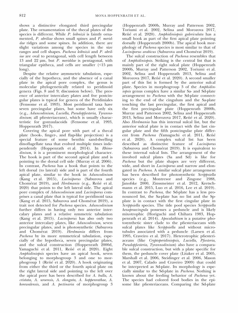
shows a distinctive elongated third precingularplate. The ornamentation of the thecal plates of thespecies is different. While P. leibnizii is faintly orna-mented, P. abriliae shows small spines and P. merid-dae ridges and some spines. In addition, there areslight variations among the species in the sizeranges and cell shapes. Pachena leibnizii and P. abril-iae are oval to pentagonal, with cell length between13 and 22 lm, but P. meriddae is pentagonal, withtriangular epitheca, and cells are smaller (<15 lmlong).
Despite the relative asymmetric tabulation, espe-cially of the hypotheca, and the absence of a canalplate in the apical pore complex, the genus ismolecular phylogenetically related to peridinoidgenera (Figs. 8 and 9; discussion below). The pres-ence of anterior intercalary plates and five postcin-gular plates is typical for genera of the Peridiniales(Fensome et al. 1993). Most peridinoid taxa haveseven precingular plates, but some have only six(e.g., Aduncodinium, Luciella, Pseudopfiesteria, Tyranno-dinium; all pfiesteriaceans), which is usually charac-teristic for gonyaulacoids (Fensome et al. 1993,Hoppenrath 2017).
Covering the apical pore with part of a thecalplate (hook-, finger-, and flap-like projection) is aspecial feature of some benthic sand-dwellingdinoflagellate taxa that evolved multiple times inde-pendently (Hoppenrath et al. 2014). In Rhino-dinium, it is a prominent morphological character.The hook is part of the second apical plate and ispointing to the dorsal cell side (Murray et al. 2006).In contrast, Pachena has a hook that points to theleft dorsal (to lateral) side and is part of the fourthapical plate, similar to the hook in Aduncodinium(Kang et al. 2015), Laciniporus (Saburova andChom�erat 2019), and Herdmania (e.g., Re~n�e et al.2020) that points to the left lateral side. The apicalpore complex of Aduncodinium and Laciniporus com-prises a canal plate that is typical for peridinoid taxa(Kang et al. 2015, Saburova and Chom�erat 2019), atrait not detected for Pachena species. Aduncodiniumfurther differs in having only two anterior inter-calary plates and a relative symmetric tabulation(Kang et al. 2015). Laciniporus has also only twoanterior intercalary plates, like Aduncodinium, sevenprecingular plates, and is photosynthetic (Saburovaand Chom�erat 2019). Herdmania differs fromPachena by its more symmetrical tabulation, espe-cially of the hypotheca, seven precingular plates,and the sulcal construction (Hoppenrath 2000d,Yamaguchi et al. 2011, Re~n�e et al. 2020). EightAmphidiniopsis species have an apical hook, sevenbelonging to morphogroup 3 and one to mor-phogroup 1 (Re~n�e et al. 2020). A hook originatingfrom either the third or the fourth apical plate onthe right lateral side and pointing to the left overthe apical pore has been described for A. bulla, A.cristata, A. uroensis, A. elongata, A. hoppenrathae, A.korewalensis, and A. pectinaria of morphogroup 3
(Hoppenrath 2000b, Murray and Patterson 2002,Toriumi et al. 2002, Selina and Morozova 2017,Re~n�e et al. 2020). Amphidiniopsis galericulata has asmall hook as part of the first apical plate pointingdorsally (Hoppenrath 2000b). The apical hook mor-phology of Pachena species is most similar to that ofLaciniporus arabicus (Saburova and Chom�erat 2019).The sulcal construction of Pachena resembles that
of Amphidiniopsis. Striking is the central list that ismainly part of the right sulcal plate (Hoppenrath2000b, Murray and Patterson 2002, Toriumi et al.2002, Selina and Hoppenrath 2013, Selina andMorozova 2017, Re~n�e et al. 2020). A second smallerpart of this list is formed by the anterior sulcalplate. Species in morphogroup 3 of the Amphidin-iopsis genus complex have a similar Sa- and Sd-platearrangement to Pachena with the Sd-plate connect-ing to the end of the cingulum and the Sa-platetouching the last precingular, the first apical andthe first precingular plate (Hoppenrath 2000b,Murray and Patterson 2002, Selina and Hoppenrath2013, Selina and Morozova 2017, Re~n�e et al. 2020).Also Herdmania has this internal sulcal list, but theanterior sulcal plate is in contact with the last cin-gular plate and the fifth postcingular plate differ-ent from Pachena (Yamaguchi et al. 2011, Re~n�eet al. 2020). A complex sulcal list has beendescribed as distinctive feature of Laciniporus(Saburova and Chom�erat 2019). It is equivalent tothese internal sulcal lists. The arrangement of theinvolved sulcal plates (Sa and Sd) is like forPachena but the plate shapes are very different,wide, and short in Laciniporus and narrow and elon-gated in Pachena. A similar sulcal plate arrangementhas been described for photosynthetic Scrippsiellaspecies (e.g., Montresor and Zingone 1988,Janofske 2000, Zinssmeister et al. 2012, Kretsch-mann et al. 2015, Luo et al. 2016, Lee et al. 2019).In contrast to Pachena, the Sd-plate has a less pro-nounced list, the Sa-plate has no list and the Sp-plate is in contact with the first cingular plate inScrippsiella species. The tide pool species Scrippsiellahexapraecingula possesses a peduncle and is likelymixotrophic (Horiguchi and Chihara 1983, Hop-penrath et al. 2014). Apocalathium is a putative pho-tosynthetic sister clade to the pfiesteriaceans withsulcal plates like Scrippsiella and without micro-tubules associated with a peduncle (Larsen et al.1995, Craveiro et al. 2017). Heterotrophic pfiesteri-aceans (like Cryptoperidiniopsis, Luciella, Pfiesteria,Pseudopfiesteria, Tyrannodinium) also have a compara-ble sulcal construction, but with a plate specific forthem, the peduncle cover plate (Litaker et al. 2005,Marshall et al. 2006, Steidinger et al. 2006, Masonet al. 2007, Calado and Craveiro 2009) that couldbe interpreted as Sd-plate. Its morphology is espe-cially similar to the Sd-plate in Pachena. Nothing isknown about the feeding behavior of Pachena yet.The species had colored food bodies in the epi-some like pfiesteriaceans. Comparing the Sd-plate
812 MONA HOPPENRATH ET AL.

morphology to the peduncle cover plate, it can behypothesized that Pachena is a benthic pedunclefeeder, like the related Aduncodinium (Kang et al.2015). A similar food body in the epi- or hyposomeis characteristic for Cabra, Rhinodinium, and Roscof-fia, benthic heterotrophic genera related to thefamily Podolampadaceae (Hoppenrath et al. 2014).Their feeding mode has not yet been documented.Planktonic Podolampadaceae are pallium feeders(Sch€utt 1895, Carbonell-Moore 2004).
The ascending cingulum is a character known fromAmphidiniopsis, Herdmania, and some Protoperidiniumsensu stricto species (subgenus and section Protoperi-dinium), taxa from the order Peridiniales (Balech1974, Faust 2002, Yamaguchi et al. 2011, Re~n�e et al.2020).Three anterior intercalary plates can occur in the
Peridiniales. The major Protoperidinium species (i.e.,Protoperidinium sensu stricto, including the sectionsProtoperidinium, Avellana, Conica, Excentrica, and
FIG. 10. Comparative tabulation drawings of the three Pachena gen. nov. species. (A–C) Pachena leibnizii sp. nov. (D–F) Pachena abriliaesp. nov. (G, H) Pachena meriddae sp. nov. (A, D, G) Ventral cell side. (B, E, H) Dorsal cell side. (C, F) Epitheca. 10-40, apical plates; 1a-3a,anterior intercalary plates; 1″-6″, precingular plates; 1‴-5‴, postcingular plates; 1″″-2″″, antapical plates; c1-c5, cingular plates; Sa, anteriorsulcal plate; Sd, right sulcal plate; Ss, left sulcal plate; Sm, median sulcal plate; Sp, posterior sulcal plate.
DESCRIPTION OF PACHENA GEN. NOV. 813

Divergentia) are known to have two or three anteriorintercalary plates (Yamaguchi and Horiguchi 2005,Yamaguchi et al. 2006). Among these sections Avel-lana and Excentrica have two anterior intercalaryplates (Yamaguchi et al. 2006). These sections arenot monophyletic in the Protoperidinium s.s. clade(Yamaguchi et al. 2006). It is assumed that thereduction of the number of anterior intercalaryplates from three to two occurred in different lin-eages (Yamaguchi et al. 2006). Archaeperidinium hasonly two anterior intercalary plates, but the closelyrelated Amphidiniopsis species (except morphogroup2) and Herdmania have three anterior intercalaryplates (Yamaguchi et al. 2011, 2016, Re~n�e et al.2020). Scrippsiella species have three anterior inter-calary plates and similar sulcal plates, see above (e.g.,Janofske 2000, Zinssmeister et al. 2012, Kretschmannet al. 2015). All Heterocapsa species, except for thetype, have three anterior intercalary plates and arephotosynthetic and characterized by specific bodyscales (Iwataki 2008, Tillmann et al. 2017). All thesegenera differ in the special arrangement of the threeanterior intercalary plates and the shape of the 1aplate, plus possessing seven precingular plates.Nearly all species of Azadinium (family Amphidomat-aceae) possess three anterior intercalary plates (e.g.,Tillmann et al. 2018, and references therein). Thisgenus has four apical and six precingular plates likePachena but Azadinium is distinct from it by the APC,the sulcal construction, a descending cingulum withsix plates, and six postcingular plates (e.g., Tillmannet al. 2014, 2018).Phylogenetic considerations. SSU rDNA sequences
clearly separated the species in accordance with themorpho-species concept. Pachena leibnizii and P.meriddae formed a sister clade to P. abriliae, in agree-ment with their morphological similarities, mostlyregarding the epithecal plate pattern.
Sequences from Pachena occupied a basal positionwithin the Thoracosphaeraceae, even though thisclade showed low statistical support. However, andas observed for the phylogenetically closely relatedsand-dwelling species Laciniporus arabicus (Saburovaand Chom�erat 2019), its phylogenetic positionbased on SSU rDNA sequences was unstable andvaried depending on the taxon sampling used forphylogenetic inference. This situation is not unusualfor benthic sand-dwelling species, which commonlyrepresent diverging lineages clustering unrelated toother known (mostly planktonic) representatives,e.g., Madanidinium adanidinium loirii (Chom�erat andBilien 2014), Apicoporus spp. (Sparmann et al.2008), or Plagiodinium belizeanum (Wakeman et al.2018).
LSU rDNA sequences could only be obtained forPachena abriliae and an unidentified Pachena speci-men. However, their phylogenetic placement at thebase of the Thoracosphaeraceae was more robustand in agreement with the position obtained in thephylogenetic analysis of SSU rDNA sequences. The
molecular phylogenetic data suggest that planktonicThoracosphaeraceae evolved from benthic taxa likePachena and Laciniporus.The Thoracosphaeraceae includes heterotrophic,
autotrophic, and parasitic representatives, from mar-ine and freshwater environments, and some showingnoncalcareous or calcareous cysts (Elbr€achter et al.2008, Gottschling and Soehner 2013, Saburova andChom�erat 2019). Pachena could be the most basaltaxon of the Thoracosphaeraceae with a hetero-trophic nutrition and possible peduncle feeding,even though we could not verify the peduncle pres-ence or prey source. Furthermore, the inability toestablish cultures impeded the observation of cyst-production for Pachena representatives, unknown sofar for heterotrophic sand-dwelling species. Thesand-dwelling Laciniporus arabicus also occupies abasal position within the clade, and in this case, it isautotrophic and produces non-calcareous cysts.Diversity and biogeography. In this study, cells of a
Pachena species were observed in a benthic samplefrom the German Bight, but they could not be iden-tified to species (M. Hoppenrath, unpub. data).Also, in the Mediterranean Sea, cells were recordedthat likely belong to one or two further undescribedspecies (M. Hoppenrath, A. Re~n�e, C.T. Satta,unpub. data). Additional observations and data areneeded for their delimitations. Selina (2016) docu-mented a sand-dwelling species from Peter theGreat Bay, Sea of Japan, and identified it as cf. Herd-mania. Judged from the cell shape, the apical hook,cingulum path, and food body location (Selina2016, p. 465, fig. 4, k and l) it could be a Pachenaspecies. Thus, the genus shows a wide distribution,and several species probably coexist at the samelocation. However, the overall distribution of thedifferent species remains to be determined.
We thank Wolf-Henning Kusber for his help finding the cor-rect name endings. This work was supported by a postdoc-toral research salary to MH and AY from the Assembling theTree of Life grant (NSF #EF-0629624) and operating funds toBSL from the National Science and Engineering ResearchCouncil of Canada (NSERC 2019-03986); AR thanks R. Galli-sai (ICM-CSIC) and T. Sl�amov�a (Univ. Prague, Czech Repub-lic) for their help during samplings and samples processing.AR was funded by a MECD grant “Estancia de Movilidad enel extranjero Jos�e Castillejo” (CAS17/00237), a “SenckenbergTaxonomy Grant 2017″, and a DAAD “Research Stays forUniversity Academics and Scientists 2018” Grant (91644317).CTS thanks Prof. Antonella Lugli�e for the continuous andimportant economic and scientific support. CTS was fundedby a DAAD Grant within the “Research Stays for UniversityAcademics and Scientists 2014” Grant (A/14/01530).
AUTHOR CONTRIBUTIONS
M.H. conceptualization, sampling, microscopy,molecular work, original draft preparation, writing, andediting; A.R. sampling, microscopy, molecular work,phylogenetic analyses, and writing; C.T.S. sampling,microscopy, and molecular work; A.Y. phylogenetic
814 MONA HOPPENRATH ET AL.

analyses and data discussion; B.S.L. funding acquisition(infrastructure and salary support), review, and editing.All authors read and approved the final manuscript.Aligizaki, K. & Nikolaidis, G. 2006. The presence of the poten-
tially toxic genera Ostreopsis and Coolia (Dinophyceae) in thenorth Aegean Sea, Greece. Harmful Algae 5:717–30.
Aligizaki, K., Nikolaidis, G., Katikou, P., Baxevanis, A. D. & Abat-zopoulos, T. J. 2009. Potentially toxic epiphytic Prorocentrum(Dinophyceae) species in Greek coastal waters. Harmful Algae8:299–311.
Al-Yamani, F. Y. & Saburova, M. A. 2010. Illustrated Guide on theFlagellates of Kuwait’s Intertidal Soft Sediments. Kuwait Institutefor Scientific Research, Safat, Kuwait, 197 pp.
Balech, E. 1956. �Etude des dinoflagell�es du sable de Roscoff. Rev.Algol. 2:29–52.
Balech, E. 1974. El genero Protoperidinium Bergh, 1881 (PeridiniumEhrenberg, 1831, partim). Hydrobiolog�ıa 4:1–79.
Berdalet, E., Tester, P. A., Chinain, M., Fraga, S., Lem�ee, R.,Litaker, W., Penna, A., Usup, G., Vila, M. & Zingone, A.2017. Harmful algal blooms in benthic systems: Recent pro-gress and future research. Oceanography 30:36–45.
Calado, A. J. & Craveiro, S. C. 2009. Description of Tyrannodiniumgen. nov., a freshwater dinoflagellate closely related to themarine Pfiesteria-like species. J. Phycol. 45:1195–205.
Carbonell-Moore, M. C. 2004. On the taxonomical position of Les-sardia Saldarriaga et Taylor within the family Podolam-padaceae Lindemann (Dinophyceae). Phycol. Res. 52:340–5.
Castresana, J. 2000. Selection of conserved blocks from multiplealignments for their use in phylogenetic analysis. Mol. Biol.Evol. 17:540–52.
Chom�erat, N. 2016. Studies on the benthic genus Sinophysis(Dinophysales, Dinophyceae): I. a taxonomic investigationfrom Martinique Island, including two new species and eluci-dation of the epithecal plate pattern. Phycologia 55:445–61.
Chom�erat, N. & Bilien, G. 2014. Madanidinium loirii gen. et sp.nov. (Dinophyceae), a new marine benthic dinoflagellatefrom Martinique Island, Eastern Caribbean. Eur. J. Phycol.49:165–78.
Craveiro, S. C., Daugbjerg, N., Moestrup, Ø. & Calado, A. J. 2017.Studies on Peridinium aciculiferum and Peridinium malmogiense(= Scrippsiella hangoei): comparison with Chimonodinium lom-nickii and description of Apocalathium gen. nov. (Dino-phyceae). Phycologia 56:21–35.
Edgar, R. C. 2004. MUSCLE: multiple sequence alignment withhigh accuracy and high throughput. Nucleic Acids Res.35:1792–7.
Efimova, K. V., Selina, M. S. & Hoppenrath, M. 2019. New mor-phological data and molecular phylogeny of the benthicdinoflagellate Pseudothecadinium campbellii (Dinophyceae,Gonyaulacales). Europ. J. Protistol. 71:125638.
Elbr€achter, M., Gottschling, M., Hildebrand-Habel, T., Keupp, H.,Kohring, R., Lewis, J., Meier, K. J. S. et al. 2008. Establishingan agenda for calcareous dinoflagellate research (Thora-cosphaeraceae, Dinophyceae) including a nomenclatural syn-opsis of generic names. Taxon 57:1289–303.
Faust, M. A. 2002. Protoperidinium belizeanum sp. nov. (Dino-phyceae) from Manatee Cay, Belize, Central America. J. Phy-col. 38:390–4.
Faust, M. A. & Balech, E. 1993. A further SEM study of marinebenthic dinoflagellates from a mangrove island, Twin Cays,Belize, including Plagiodinium belizeanum gen. et sp. nov. J.Phycol. 29:826–32.
Fensome, R. A., Taylor, F. J. R., Norris, G., Sarjeant, W. A. S.,Wharton, D. I. & Williams, G. L. 1993. A classification of liv-ing and fossil dinoflagellates. Micropaleontol. Spec. Pub. 7:1–351.
G�omez, F., Qiu, D., Lopes, R. M. & Lin, S. 2015. Fukuyoa paulensisgen. et sp. nov., a new genus for the globular species of thedinoflagellate Gambierdiscus (Dinophyceae). PLoS ONE 10:e0119676.
Gottschling, M. & Soehner, S. 2013. An updated list of genericnames in the Thoracosphaeraceae. Microorganisms 1:122–36.
Hansen, G. & Daugbjerg, N. 2011. Moestrupia oblonga gen. &comb. nov. (syn.: Gyrodinium oblongum), a new marinedinoflagellate genus characterized by light and electronmicroscopy, photosynthetic pigments and LSU rDNAsequence. Phycologia 50:583–99.
Herdman, E. C. 1922. Notes on dinoflagellates and other organ-isms causing discolouration of sant at Port Erin II. Proc.Trans. Liverpool Biol. Soc. 36:15–30.
Herdman, E. C. 1924a. Notes on dinoflagellates and other organ-isms causing discolouration of sant at Port Erin III. Proc.Trans. Liverpool Biol. Soc. 38:58–63.
Herdman, E. C. 1924b. Notes on dinoflagellates and other organ-isms causing discolouration of sant at Port Erin IV. Proc.Trans. Liverpool Biol. Soc. 38:75–84.
Hoppenrath, M. 2000a. Taxonomische und €okologische Untersuchun-gen von Flagellaten mariner Sande, dissertation. University ofHamburg, Germany, 311 pp.
Hoppenrath, M. 2000b. Morphology and taxonomy of six marinesand-dwelling Amphidiniopsis species (Dinophyceae, Peri-diniales), four of them new, from the German Bight, NorthSea. Phycologia 39:482–97.
Hoppenrath, M. 2000c. Morphology and taxonomy of Sinoph-ysis (Dinophyceae, Dinophysiales) including two new marinesand-dwelling species from the North German Wadden Sea.Eur. J. Phycol. 35:153–62.
Hoppenrath, M. 2000d. An emended description of Herdmanialitoralis Dodge (Dinophyceae) including the plate formula.Nova Hedwigia 71:481–9.
Hoppenrath, M. 2017. Dinoflagellate taxonomy – a review andproposal of a revised classification. Mar. Biodiv. 47:381–403.
Hoppenrath, M. & Leander, B. S. 2007. Morphology and phy-logeny of the pseudocolonial dinoflagellates Polykrikoslebourae and Polykrikos herdmanae n. sp. Protist 158:209–27.
Hoppenrath, M. & Leander, B. S. 2008. Morphology and molecularphylogeny of a new marine sand-dwelling Prorocen-trum species, P. tsawwassenense sp. nov. (Dinophyceae, Proro-centrales) from British Columbia, Canada. J. Phycol. 44:451–66.
Hoppenrath, M. & Leander, B. S. 2010. Dinoflagellates phylogenyas inferred from heat shock protein 90 and ribosomal genesequences. PLoS ONE 5:e13220.
Hoppenrath, M., Murray, S., Chom�erat, N. & Horiguchi, T. 2014.Marine benthic dinoflagellates - unveiling their worldwidebiodiversity. Phycol. Res. 62:322.
Hoppenrath, M., Murray, S., Sparmann, S. & Leander, B. S. 2012. Mor-phology and molecular phylogeny of Ankistrodinium gen. nov.(Dinophyceae), a new genus of marine sand-dwelling dinoflagel-lates formerly classified within Amphidinium. J. Phycol. 48:1143–52.
Hoppenrath, M., Schweikert, M. & Elbr€achter, M. 2003. Morpho-logical reinvestigation and characterisation of the marine,sand-dwelling dinoflagellate Adenoides eludens (Dinophyceae).Eur. J. Phycol. 38:385–94.
Hoppenrath, M., Yubuki, N., Stern, R. & Leander, B. S. 2017.Ultrastructure and molecular phylogenetic position of a newmarine sand-dwelling dinoflagellate from British Columbia,Canada: Pseudadenoides polypyrenoides sp. nov. (Dinophyceae).Eur. J. Phycol. 52:208–24.
Horiguchi, T. & Chihara, M. 1983. Scrippsiella hexapraecingula sp.nov. (Dinophyceae), a tide pool dinoflagellate from thenorthwest Pacific. Bot. Mag. Tokyo 96:351–8.
Horiguchi, T., Tamura, M., Katsumata, K. & Yamaguchi, A. 2012.Testudodinium gen. nov. (Dinophyceae), a new genus of sand-dwelling dinoflagellates formerly classified in the genusAmphidinium. Phycol. Res. 60:137–49.
Iwataki, M. 2008. Taxonomy and identification of the armoreddinoflagellate genus Heterocapsa (Peridiniales, Dinophyceae).Plankton Benthos Res. 3:135–42.
Janofske, D. 2000. Scrippsiella trochoidea and Scrippsiella regalis, nov.comb. (Peridiniales, Dinophyceae): a comparison. J. Phycol.36:178–89.
Kalyaanamoorthy, S., Minh, B. Q., Wong, T. K. F., von Haeseler,A. & Jermiin, L. S. 2017. ModelFinder: fast model selectionfor accurate phylogenetic estimates. Nat. Methods 14:587–9.
DESCRIPTION OF PACHENA GEN. NOV. 815

Kang, N. S., Jeong, H. J., Moestrup, Ø., Jang, T. Y., Lee, S. Y. &Lee, M. J. 2015. Aduncodinium gen. nov. and A. glandulacomb. nov. (Dinophyceae, Pfiesteriaceae), from coastalwaters off Korea: Morphology and molecular characteriza-tion. Harmful Algae 41:25–37.
Kofoid, C. A. & Swezy, O. 1921. The Free-living Unarmored Dinoflag-ellata. Memoirs of the University of California, Vol 5. Univer-sity of California Press, Berkeley, CA, USA, 563 pp.
Kretschmann, J., Elbr€achter, M., Zinssmeister, C., Soehner, S.,Kirsch, M., Kusber, W. H. & Gottschling, M. 2015. Taxo-nomic clarification of the dinophyte Peridinium acuminatumEhrenb., � Scrippsiella acuminata, comb. nov. (Thora-cosphaeraceae, Peridiniales). Phytotaxa 220:239–56.
Larsen, J., Kuosa, H., Ik€avalko, J., Kivi, K. & H€allfors, S. 1995. Aredescription of Scrippsiella hangoei (Schiller) comb. nov. – a‘red tide’ dinoflagellate from the northern Baltic. Phycologia34:135–44.
Lee, S. Y., Jeong, H. J., Kim, S. J., Lee, K. H. & Jang, S. H. 2019.Scrippsiella masanensis sp. nov. (Thoracosphaerales, Dino-phyceae), a photosynthetic dinoflagellate from the coastalwaters of southern Korea. Phycologia 58:287–99.
Litaker, R. W., Steidinger, K. A., Mason, P. L., Landsberg, J. H.,Shields, J. D., Reece, K. S., Haas, L. W. et al. 2005. Thereclassification of Pfiesteria shumwayae: Pseudopfiesteria, gen.nov. J. Phycol. 41:643–51.
Luo, Z., Mertens, K. N., Bagheri, S., Aydin, H., Takano, Y., Mat-suoka, K., Mccarthy, F. M. G. & Gu, H. 2016. Cyst-theca rela-tionship and phylogenetic positions of Scrippsiella plana sp.nov. and S. spinifera (Peridiniales, Dinophyceae). Eur. J. Phy-col. 51:188–202.
Maddison, W. P. & Maddison, D. R. 2015. Mesquite: a modularsystem for evolutionary analysis. Version 3.04. https://mesquiteproject.org
Marshall, H. G., Hargraves, P. E., Burkholder, J. M., Parrow, M.W., Elbr€achter, M., Allen, E. H., Knowlton, V. M. et al. 2006.Taxonomy of Pfiesteria (Dinophyceae). Harmful Algae 5:481–96.
Mason, P. L., Litaker, R. W., Jeong, H. J., Ha, J. H., Reece, K. S.,Stokes, N. A., Park, J. Y. et al. 2007. Description of a newgenus of Pfiesteria-like dinoflagellate, Luciella gen. nov. (Dino-phyceae), including two new species: Luciella masanensis sp.nov. and Luciella atlantis sp. nov. J. Phycol. 43:799–810.
Mohammad-Noor, N., Daugbjerg, N., Moestrup, Ø. & Anton, A.2007. Marine epibenthic dinoflagellates from Malaysia – astudy of live cultures and preserved samples based on lightand scanning electron microscopy. Nordic J. Bot. 24:629–90.
Montresor, M. & Zingone, A. 1988. Scrippsiella precaria sp. nov.(Dinophyceae), a marine dinoflagellate from the Gulf ofNaples. Phycologia 27:387–94.
Murray, S. 2003. Diversity and phylogenetics of sand-dwellingdinoflagellates from Southern Australia. PhD Thesis. Univer-sity of Sydney, Sydney, Australia, 202 pp.
Murray, S., Hoppenrath, M., Preisfeld, A., Larsen, J., Yoshimatsu,S., Toriumi, S. & Patterson, D. J. 2006. Phylogenetics of Rhin-odinium broomeense gen. et sp. nov., a peridinoid, sand-dwell-ing dinoflagellate (Dinophyceae). J. Phycol. 42:934–42.
Murray, S. & Patterson, D. J. 2002. Amphidiniopsis korewalensis sp.nov., a new heterotrophic benthic dinoflagellate. Phycologia41:382–8.
N�ezan, E. & Chom�erat, N. 2011. Vulcanodinium rugosum gen. etsp. nov. (Dinophyceae), un nouveau dinoflagell�e marin de lacote m�editerran�eenne franc�aise. Cryptogam. Algol. 32:3–18.
Onuma, R., Watanabe, K. & Horiguchi, T. 2015. Pellucidodiniumpsammophilum gen. & sp. nov. and Nusuttodinium desymbiontumsp. nov. (Dinophyceae), two novel heterotrophs closelyrelated to kleptochloroplastidic dinoflagellates. Phycologia54:192–209.
Penna, A., Fraga, S., Battocchi, C., Casabianca, S., Riob�o, P., Gia-cobbe, M., Totti, C. et al. 2012. Genetic diversity of thegenus Ostreopsis Schmidt: phylogeographical considerationsand molecular methodology applications for field detectionin the Mediterranean Sea. Cryptogam. Algol. 33:153–63.
Re~n�e, A. & Hoppenrath, M. 2019. Psammodinium inclinatum gen.nov. et comb. nov. (=Thecadinium inclinatum Balech) is theclosest relative to the toxic dinoflagellate genera Gambierdis-cus and Fukuyoa. Harmful Algae 84:161–71.
Re~n�e, A., Satta, C. T., L�opez-Garc�ıa, P. & Hoppenrath, M. 2020.Re-evaluation of Amphidiniopsis (Dinophyceae) mor-phogroups based on phylogenetic relationships, and descrip-tion of three new sand-dwelling species from the NWMediterranean. J. Phycol. 56:68–84.
Ronquist, F. & Huelsenbeck, J. P. 2003. MrBayes 3: Bayesian phyloge-netic inference under mixed models. Bioinformatics 19:1572–4.
Saburova, M. & Chom�erat, N. 2014. Ailadinium reticulatum gen. et sp.nov. (Dinophyceae), a new thecate, marine, sand-dwellingdinoflagellate from the northern Red Sea. J. Phycol. 50:1120–36.
Saburova, M. & Chom�erat, N. 2019. Laciniporus arabicus gen. etsp. nov. (Dinophyceae, Peridiniales), a new thecate, marine,sand-dwelling dinoflagellate from the northern IndianOcean (Arabian Sea). J. Phycol. 55:84–103.
Sch€utt, F. 1895. Die peridineen der plankton expedition. Ergebn.Plankton-Exped. Humboldt Stiftung 4:1–170.
Selina, M. S. 2016. Seasonal and long-term dynamics of sand-dwelling dinoflagellates (Dinoflagellata) in Peter the GreatBay, Sea of Japan. Russ. J. Mar. Biol. 42:458–70.
Selina, M. S., Efimova, K. V. & Hoppenrath, M. 2019. Delimitation ofthe genus Thecadinium (Dinophyceae) sensu stricto with two spe-cies Thecadinium kofoidii and T. pseudokofoidii sp. nov. using mor-phological and molecular data. Phycologia 58:36–50.
Selina, M. & Hoppenrath, M. 2013. Morphology and taxonomy ofseven marine sand-dwelling Amphidiniopsis species (Peri-diniales, Dinophyceae), including two new species, A. kono-valovae sp. nov. and A. striata sp. nov., from the Sea of Japan,Russia. Mar. Biodiv. 43:87–104.
Selina, M. & Morozova, T. V. 2017. Morphology and taxonomy ofthree new marine sand-dwelling Amphidiniopsis species (Peri-diniales, Dinophyceae) from the Sea of Japan, Russia. Phycolo-gia 56:1–13.
Sparmann, S. F., Leander, B. S. & Hoppenrath, M. 2008. Compara-tive morphology and molecular phylogeny of taxa of the newmarine benthic dinoflagellate genus Apicoporus, classified for-merly within Amphidinium sensu lato. Protist 159:383–99.
Steidinger, K. A., Landsberg, J. H., Mason, P. L., Vogelbein, W.K., Tester, P. A. & Litaker, R. W. 2006. Cryptoperidiniopsis bro-dyi gen. et sp. nov., (Dinophyceae), a small lightly armoreddinoflagellate in the Pfiesteriaceae. J. Phycol. 42:951–61.
Talavera, G. & Castresana, J. 2007. Improvement of phylogeniesafter removing divergent and ambiguously aligned blocksfrom protein sequence alignments. Syst. Biol. 56:564–77.
Tamura, M. 2005. A systematic study of benthic marine dinoflag-ellates. PhD thesis, Graduate School of Science, Division ofBiological Sciences, Hokkaido University, Japan, 117 pp.
Tillmann, U., Edvardsen, B., Krock, B., Smith, K. F., Paterson, R.F. & Voß, D. 2018. Diversity, distribution, and azaspirazids ofAmphidomoataceae (Dinophyceae) along the Norwegiancoast. Harmful Algae 80:15–34.
Tillmann, U., Gottschling, M., N�ezan, E., Krock, B. & Bilien, G.2014. Morphological and molecular characterization of threenew Azadinium species (Amphidomataceae, Dinophyceae)from the Irminger Sea. Protist 165:417–44.
Tillmann, U., Hoppenrath, M., Gottschling, M., Kusber, W. H. &Elbr€achter, M. 2017. Plate pattern clarification of the marinedinophyte Heterocapsa triquetra sensu Stein (Dinophyceae)collected at the Kiel Fjord (Germany). J. Phycol. 53:1305–24.
Toriumi, S., Yoshimatsu, S. & Dodge, J. D. 2002. Amphidiniopsisuroensis sp. nov. and Amphidiniopsis pectinaria sp. nov. (Dino-phyceae): Two new benthic dinoflagellates from Japan. Phy-col. Res. 50:115–24.
Trifinopoulos, J., Nguyen, L. T., von Haeseler, A. & Minh, B. Q.2016. W-IQ-TREE: a fast online phylogenetic tool for maxi-mum likelihood analysis. Nucl. Acids. Res. 44:W232–5.
Uhlig, G. 1964. Eine einfache Methode zur Extraktion der vagi-len, mesopsammalen Microfauna. Helgol. Wiss. Meeresunters.11:178–85.
816 MONA HOPPENRATH ET AL.

Vila, M., Garc�es, E. & Mas�o, M. 2001. Potentially toxic epiphyticdinoflagellate assemblages on macroalgae in the NWMediterranean. Aquat. Microb. Ecol. 26:51–60.
Wakeman, K., Hoppenrath, M., Yamaguchi, A., Gavelis, G. S.,Leander, B. S. & Nozaki, H. 2018. Ultrastructure of the mar-ine benthic dinoflagellate Plagiodinium belizeanum (Dino-phyceae) from the southeast Pacific island of Okinawa,Japan. Phycologia 57:209–22.
Yamada, N., Terada, R., Tanaka, A. & Horiguchi, T. 2013. Bispino-dinium angelaceum gen. et sp. nov. (Dinophyceae), a newsand-dwelling dinoflagellate from the seafloor off MageshimaIsland, Japan. J. Phycol. 49:555–69.
Yamaguchi, A., Hoppenrath, M., Pospelova, V., Horiguchi, T. &Leander, B. S. 2011. Molecular phylogeny of the marinesand-dwelling dinoflagellate Herdmania litoralis and anemended description of the closely related planktonic genusArchaeperidinium J€orgensen. Eur. J. Phycol. 46:98–112.
Yamaguchi, A. & Horiguchi, T. 2005. Molecular phylogeneticstudy of the heterotrophic dinoflagellate genus Protoperi-dinium (Dinophyceae) inferred from small subunit rRNAgene sequences. Phycol. Res. 53:30–42.
Yamaguchi, A., Kawamura, H. &Horiguchi, T. 2006. A further phylo-genetic study of the heterotrophic dinoflagellate genus, Pro-
toperidinium (Dinophyceae) based on SSU and LSU ribosomalRNA gene sequences. Phycol. Res. 54:317–29.
Yamaguchi, A., Yoshimatsu, S., Hoppenrath, M., Wakemand, K. C.& Kawai, H. 2016. Molecular phylogeny of the benthicdinoflagellate genus Amphidiniopsis and its relationships withthe Family Protoperidiniaceae. Protist 167:568–83.
Zinssmeister, C., Soehner, S., Kirsch, M., Facher, E., Meier, K. J.S., Keupp, H. & Gottschling, M. 2012. Same but different:two novel bicarinate species of extant calcareous dinophytes(Thoracosphaeraceae, Peridiniales) from the MediterraneanSea. J. Phycol. 48:1107–18.
Supporting Information
Additional Supporting Information may befound in the online version of this article at thepublisher’s web site:
Table S1. Primers used during the amplifica-tion protocols.
DESCRIPTION OF PACHENA GEN. NOV. 817