CaseSeriesofPost-ThrombolysisPatients ...downloads.hindawi.com/archive/2011/254569.pdf · benefit...
5
Hindawi Publishing Corporation Cardiovascular Psychiatry and Neurology Volume 2011, Article ID 254569, 4 pages doi:10.1155/2011/254569 Case Report Case Series of Post-Thrombolysis Patients Undergoing Hemicraniectomy for Malignant Anterior Circulation Ischaemic Stroke A. Williams, 1 M. Sittampalam, 2 N. Barua, 3 and A. Mohd Nor 2 1 Department of Neurosurgery, Derriford Hospital, Plymouth, PL6 8DH, UK 2 Department of Neurology, Derriford Hospital, Plymouth, PL6 8DH, UK 3 Department of Neurosurgery, Frenchay Hospital, Bristol, BS16 1LE, UK Correspondence should be addressed to A. Williams, [email protected] Received 30 September 2010; Revised 31 October 2010; Accepted 13 February 2011 Academic Editor: Gjumrakch Aliev Copyright © 2011 A. Williams et al. This is an open access article distributed under the Creative Commons Attribution License, which permits unrestricted use, distribution, and reproduction in any medium, provided the original work is properly cited. While ischaemic stroke remains a leading cause of death and disability, there have been recent advancements in treatment modalities including thrombolysis and decompressive hemicraniectomy. A retrospective review of patients treated in our NHS teaching hospital, in Plymouth (UK), over a 2 year period identified 17 thrombolysed patients, of whom two had undergone subsequent decompressive hemicraniectomy. These were non-dominant hemisphere strokes in young patients, aged 51 and 57. Initial NIHSS scores were 16 and 17, and they received thrombolysis at 2 hrs 42 min and 5 hrs 10 min post onset of symptoms respectively. CT imaging demonstrated cerebral swelling with significant midline shift in both cases, and decompressive hemicraniectomy was undertaken at 29 hrs 8 min and 27 hrs 30 min post-thrombolysis. We found no significant intra-operative complications attributable to prior use of thrombolytics. Both patients have had acceptable psychological and physical outcomes, with Barthel Index scores of 40 and 25, and MMSE scores of 29/30 and 27/30. We conclude that the use of thrombolytic therapy does not contra-indicate subsequent decompressive hemicraniectomy in well selected patients with non-dominant hemisphere strokes. More research in this field is required to elucidate factors which would facilitate recognition of stroke patients who will benefit most from aggressive medical and neurosurgical intervention. 1. Introduction The acute treatment for ischaemic stroke, a leading cause of death and permanent disability in the Western world, underwent profound changes after the randomised double- blind NINDS study of intravenous thrombolysis in 1995 [1]. Despite its benefits, thrombolysis has remained an underused intervention perhaps due to its short window of efficacy, although this is being reconsidered [2], and delayed presentations to hospital are being addressed with multiple public health campaigns. Its benefits are, however, clearly documented. ECASS II [3], a multicentred European trial of 800 patients, using Alteplase (0.9 mg/kg) intravenously (IV), combined with strict CT and blood pressure inclusion criteria, supported the use of Alteplase within 3 hours of symptom onset. The potential benefits were counterbalanced by a 2- to 5-times increase in symptomatic intracranial haemorrhage. The NINDS study [1] suggested that intrac- erebral haemorrhage occurred in 5.8% of patients receiving tPA, which was associated with a 45% fatality rate. In parallel with recent studies of thrombolysis, there has been an increase in interest in decompressive hemi- craniectomy for anterior circulation ischaemic strokes since its initial description in 1935 [4]. Large cerebral infarcts are often associated with significant cerebral oedema, which may be significant enough to cause herniation and death. Hem- icraniectomy was devised to counteract this postinfarction complication. This is most commonly a phenomenon of the second to fifth days after ictus [5], and half of deaths within the first month of young patients with ischaemic stroke can be attributed to space-occupying malignant infarction [6]. Again, this treatment is not without its controversies, the most predominant of which being that while it prevents death, it may promote life but with complete dependence.
Transcript of CaseSeriesofPost-ThrombolysisPatients ...downloads.hindawi.com/archive/2011/254569.pdf · benefit...

Hindawi Publishing CorporationCardiovascular Psychiatry and NeurologyVolume 2011, Article ID 254569, 4 pagesdoi:10.1155/2011/254569
Case Report
Case Series of Post-Thrombolysis PatientsUndergoing Hemicraniectomy for Malignant AnteriorCirculation Ischaemic Stroke
A. Williams,1 M. Sittampalam,2 N. Barua,3 and A. Mohd Nor2
1 Department of Neurosurgery, Derriford Hospital, Plymouth, PL6 8DH, UK2 Department of Neurology, Derriford Hospital, Plymouth, PL6 8DH, UK3 Department of Neurosurgery, Frenchay Hospital, Bristol, BS16 1LE, UK
Correspondence should be addressed to A. Williams, [email protected]
Received 30 September 2010; Revised 31 October 2010; Accepted 13 February 2011
Academic Editor: Gjumrakch Aliev
Copyright © 2011 A. Williams et al. This is an open access article distributed under the Creative Commons Attribution License,which permits unrestricted use, distribution, and reproduction in any medium, provided the original work is properly cited.
While ischaemic stroke remains a leading cause of death and disability, there have been recent advancements in treatmentmodalities including thrombolysis and decompressive hemicraniectomy. A retrospective review of patients treated in our NHSteaching hospital, in Plymouth (UK), over a 2 year period identified 17 thrombolysed patients, of whom two had undergonesubsequent decompressive hemicraniectomy. These were non-dominant hemisphere strokes in young patients, aged 51 and57. Initial NIHSS scores were 16 and 17, and they received thrombolysis at 2 hrs 42 min and 5 hrs 10 min post onset ofsymptoms respectively. CT imaging demonstrated cerebral swelling with significant midline shift in both cases, and decompressivehemicraniectomy was undertaken at 29 hrs 8 min and 27 hrs 30 min post-thrombolysis. We found no significant intra-operativecomplications attributable to prior use of thrombolytics. Both patients have had acceptable psychological and physical outcomes,with Barthel Index scores of 40 and 25, and MMSE scores of 29/30 and 27/30. We conclude that the use of thrombolytic therapydoes not contra-indicate subsequent decompressive hemicraniectomy in well selected patients with non-dominant hemispherestrokes. More research in this field is required to elucidate factors which would facilitate recognition of stroke patients who willbenefit most from aggressive medical and neurosurgical intervention.
1. Introduction
The acute treatment for ischaemic stroke, a leading causeof death and permanent disability in the Western world,underwent profound changes after the randomised double-blind NINDS study of intravenous thrombolysis in 1995[1]. Despite its benefits, thrombolysis has remained anunderused intervention perhaps due to its short window ofefficacy, although this is being reconsidered [2], and delayedpresentations to hospital are being addressed with multiplepublic health campaigns. Its benefits are, however, clearlydocumented. ECASS II [3], a multicentred European trialof 800 patients, using Alteplase (0.9 mg/kg) intravenously(IV), combined with strict CT and blood pressure inclusioncriteria, supported the use of Alteplase within 3 hours ofsymptom onset. The potential benefits were counterbalancedby a 2- to 5-times increase in symptomatic intracranial
haemorrhage. The NINDS study [1] suggested that intrac-erebral haemorrhage occurred in 5.8% of patients receivingtPA, which was associated with a 45% fatality rate.
In parallel with recent studies of thrombolysis, therehas been an increase in interest in decompressive hemi-craniectomy for anterior circulation ischaemic strokes sinceits initial description in 1935 [4]. Large cerebral infarcts areoften associated with significant cerebral oedema, which maybe significant enough to cause herniation and death. Hem-icraniectomy was devised to counteract this postinfarctioncomplication. This is most commonly a phenomenon of thesecond to fifth days after ictus [5], and half of deaths withinthe first month of young patients with ischaemic stroke canbe attributed to space-occupying malignant infarction [6].Again, this treatment is not without its controversies, themost predominant of which being that while it preventsdeath, it may promote life but with complete dependence.

2 Cardiovascular Psychiatry and Neurology
12/8/08 11:23 12/8/08 19:05 13/8/08 15:24 26/8/08 15:45
Figure 1
Recently, pooled data from three European multicentered,randomised, controlled trials (DECIMAL, DESTINY, andHAMLET) has suggested mortality and poor functionaloutcome can be improved by undertaking decompressivehemicraniectomy without increasing the numbers of com-pletely dependent patients [7].
Combining these two interventions offers further oppor-tunities to improve patient outcomes, but also raises furtherquestions. Most predominant is the fear that precedent use ofthrombolysis could predispose patients to excess risk of bothintraoperative and postoperative haemorrhage. We sought toinvestigate this possibility in this retrospective analysis.
2. Methods
We examined clinical and surgical outcomes when decom-pressive hemicraniectomy was undertaken following intra-venous thrombolysis in the same patients, and specificallywhen hemicraniectomy was undertaken for space-occupyingcerebral oedema rather than post-thrombolysis haemor-rhage. We analysed data from a cohort of patients treated inDerriford Hospital over a 30-month period from 13/05/2006to 29/01/2009. Patients were identified by searching thehospital clinical coding database, using procedure codes forthrombolysis. The patients excluded either were thrombol-ysed for other reasons (e.g., myocardial infarction) or didnot subsequently undergo hemicraniectomy. A total of 17patients received intravenous thrombolysis between thesedates, and two of these patients subsequently underwentdecompressive hemicraniectomy. We undertook a detailedcase notes study with interest focused on intraoperative andperioperative issues regarding the recent use of thrombolysisand patients’ long-term outcome.
Case 1 (SK (Figure 1)). A 51-year-old right-handed femalepresented to the emergency department at 10:25 on 12/08/08,with a sudden onset left hemiplegia, occurring at 09:18, andinvolving the upper motor neurone of the VIIth cranial nerveand her upper and lower limbs, and a left-sided hemineglect.On admission her GCS was 15. Her past medical historyincluded Hepatitis A in 1967. She had no medical allergies,and, aside from the oral contraceptive pill, she was on nomedications. There was no contributory family history, andshe was a life-long nonsmoker.
A noncontrast CT brain scan at 11:23 demonstrated char-acteristic grey and white matter changes in the right middlecerebral artery (MCA) territory and density within the right
MCA, indicating acute infarct. There was no evidence ofacute haemorrhage or hydrocephalus. Her admission bloodprofile was unremarkable (Hb 14.4, PLT 235, PT 12.8, APTT23.2).
She was diagnosed with a right MCA ischaemic stroke,and, following appropriate consent, recombinant tissueplasminogen activator (rTPA), Alteplase was administered at12:00. She was given a 5.4 mg bolus over 2 min, and thenan infusion of 48.6 mg over the subsequent hour. A totalof 54 mg rTPA was administered intravenously. Her bloodpressure remained stable throughout.
Her neurological status remained unchanged until13/08/08 when her GCS dropped to 9 (E3V1M5), and aCT brain scan (at 15:24) demonstrated an extensive rightanterior cerebral artery (ACA) and MCA territory infarction.The associated mass effect resulted in 10 mm of midline shiftto the left. There was radiological evidence of right uncalherniation. No intracranial haemorrhage was demonstrable.
After failed medical control of ICP, she underwentan emergency decompressive hemicraniectomy at 18:15 on13/08/08. A standard operative protocol was used: a traumaflap was fashioned and frontotemporoparietal craniotomywas performed, and the dura was incised in a stellate pattern.The dura was left open, and an artificial dural patch (Dura-Guard, Synovis, St. Paul, USA) was laid over the defect. Nosubgaleal drain was sited. Routine bipolar diathermy wassufficient for haemostasis. This was an uncomplicated proce-dure. Her postoperative haemoglobin was 11.7 (preoperativewas 14.4). No intraoperative blood component transfusionswere administered, though, one day later, she received a 2-unit packed red cell transfusion.
Thereafter, she was managed on the intensive therapyunit (ITU) for medical optimisation of her intracranial pres-sure (ICP). Clexane 40 mg s/c, Aspirin 75 mg, and Clopido-grel 75 mg were commenced within a week. A routine post-operative scan on 26/08/08 was satisfactory. The evolution ofradiological changes on CT scanning are shown in Figure 1.
She was transferred for physical and neurological rehabil-itation three weeks after surgery. At her most recent outpa-tient review (6 months after surgery), her MMSE was 29/30.She was able to move from lying to sitting independently, totransfer from bed to chair and chair to car with the assistanceof one, to stand with supervision of one, and to walk 20metres with hand-rail assistance. She lives in a nursinghome, with no difficulties with swallowing or meeting hernutritional needs, and she is continent. She had requiredantidepressant medication during rehabilitation, though thisis no longer the case. She has a Barthel Index Score of 40.
Case 2 (DH (Figure 2)). A 57-year-old right-handed malepresented with sudden onset left hemiplegia at 13:30 on the09/01/09 involving the upper motor neurone of the VIIthcranial nerve, and his left upper and lower limbs, but witha GCS of 15. His past medical history included right-sidedsciatica and a childhood hypospadias repair. He had nomedical allergies, nor used regular medications. He was anonsmoker with an unremarkable family history.
A noncontrast CT brain scan at 17:03 demonstratedclear evidence of an acute, nonhaemorrhagic infarct in the
Cardiovascular Psychiatry and Neurology 3
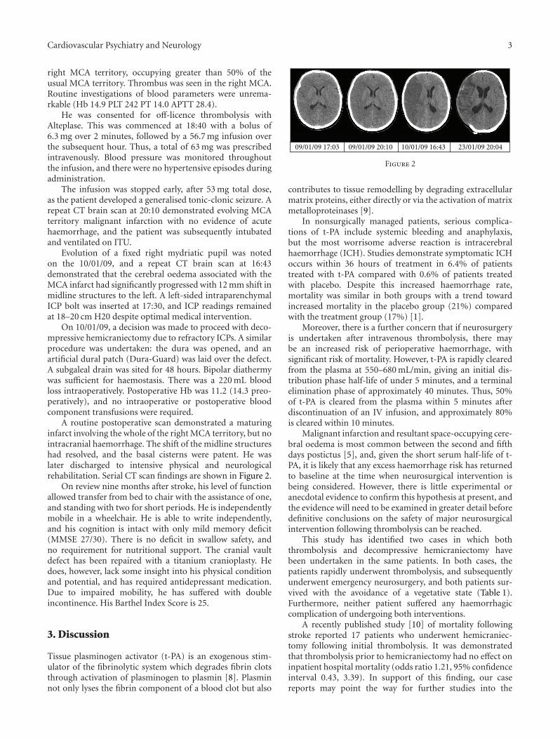
right MCA territory, occupying greater than 50% of theusual MCA territory. Thrombus was seen in the right MCA.Routine investigations of blood parameters were unrema-rkable (Hb 14.9 PLT 242 PT 14.0 APTT 28.4).
He was consented for off-licence thrombolysis withAlteplase. This was commenced at 18:40 with a bolus of6.3 mg over 2 minutes, followed by a 56.7 mg infusion overthe subsequent hour. Thus, a total of 63 mg was prescribedintravenously. Blood pressure was monitored throughoutthe infusion, and there were no hypertensive episodes duringadministration.
The infusion was stopped early, after 53 mg total dose,as the patient developed a generalised tonic-clonic seizure. Arepeat CT brain scan at 20:10 demonstrated evolving MCAterritory malignant infarction with no evidence of acutehaemorrhage, and the patient was subsequently intubatedand ventilated on ITU.
Evolution of a fixed right mydriatic pupil was notedon the 10/01/09, and a repeat CT brain scan at 16:43demonstrated that the cerebral oedema associated with theMCA infarct had significantly progressed with 12 mm shift inmidline structures to the left. A left-sided intraparenchymalICP bolt was inserted at 17:30, and ICP readings remainedat 18–20 cm H20 despite optimal medical intervention.
On 10/01/09, a decision was made to proceed with deco-mpressive hemicraniectomy due to refractory ICPs. A similarprocedure was undertaken: the dura was opened, and anartificial dural patch (Dura-Guard) was laid over the defect.A subgaleal drain was sited for 48 hours. Bipolar diathermywas sufficient for haemostasis. There was a 220 mL bloodloss intraoperatively. Postoperative Hb was 11.2 (14.3 preo-peratively), and no intraoperative or postoperative bloodcomponent transfusions were required.
A routine postoperative scan demonstrated a maturinginfarct involving the whole of the right MCA territory, but nointracranial haemorrhage. The shift of the midline structureshad resolved, and the basal cisterns were patent. He waslater discharged to intensive physical and neurologicalrehabilitation. Serial CT scan findings are shown in Figure 2.
On review nine months after stroke, his level of functionallowed transfer from bed to chair with the assistance of one,and standing with two for short periods. He is independentlymobile in a wheelchair. He is able to write independently,and his cognition is intact with only mild memory deficit(MMSE 27/30). There is no deficit in swallow safety, andno requirement for nutritional support. The cranial vaultdefect has been repaired with a titanium cranioplasty. Hedoes, however, lack some insight into his physical conditionand potential, and has required antidepressant medication.Due to impaired mobility, he has suffered with doubleincontinence. His Barthel Index Score is 25.
3. Discussion
Tissue plasminogen activator (t-PA) is an exogenous stim-ulator of the fibrinolytic system which degrades fibrin clotsthrough activation of plasminogen to plasmin [8]. Plasminnot only lyses the fibrin component of a blood clot but also
09/01/09 17:03 09/01/09 20:10 10/01/09 16:43 23/01/09 20:04
Figure 2
contributes to tissue remodelling by degrading extracellularmatrix proteins, either directly or via the activation of matrixmetalloproteinases [9].
In nonsurgically managed patients, serious complica-tions of t-PA include systemic bleeding and anaphylaxis,but the most worrisome adverse reaction is intracerebralhaemorrhage (ICH). Studies demonstrate symptomatic ICHoccurs within 36 hours of treatment in 6.4% of patientstreated with t-PA compared with 0.6% of patients treatedwith placebo. Despite this increased haemorrhage rate,mortality was similar in both groups with a trend towardincreased mortality in the placebo group (21%) comparedwith the treatment group (17%) [1].
Moreover, there is a further concern that if neurosurgeryis undertaken after intravenous thrombolysis, there maybe an increased risk of perioperative haemorrhage, withsignificant risk of mortality. However, t-PA is rapidly clearedfrom the plasma at 550–680 mL/min, giving an initial dis-tribution phase half-life of under 5 minutes, and a terminalelimination phase of approximately 40 minutes. Thus, 50%of t-PA is cleared from the plasma within 5 minutes afterdiscontinuation of an IV infusion, and approximately 80%is cleared within 10 minutes.
Malignant infarction and resultant space-occupying cere-bral oedema is most common between the second and fifthdays postictus [5], and, given the short serum half-life of t-PA, it is likely that any excess haemorrhage risk has returnedto baseline at the time when neurosurgical intervention isbeing considered. However, there is little experimental oranecdotal evidence to confirm this hypothesis at present, andthe evidence will need to be examined in greater detail beforedefinitive conclusions on the safety of major neurosurgicalintervention following thrombolysis can be reached.
This study has identified two cases in which boththrombolysis and decompressive hemicraniectomy havebeen undertaken in the same patients. In both cases, thepatients rapidly underwent thrombolysis, and subsequentlyunderwent emergency neurosurgery, and both patients sur-vived with the avoidance of a vegetative state (Table 1).Furthermore, neither patient suffered any haemorrhagiccomplication of undergoing both interventions.
A recently published study [10] of mortality followingstroke reported 17 patients who underwent hemicraniec-tomy following initial thrombolysis. It was demonstratedthat thrombolysis prior to hemicraniectomy had no effect oninpatient hospital mortality (odds ratio 1.21, 95% confidenceinterval 0.43, 3.39). In support of this finding, our casereports may point the way for further studies into the

4 Cardiovascular Psychiatry and Neurology
Table 1
Patient 1 Patient 2
Age 51 57
Time from onset of symptoms toarrival to hospital
1 hr 7 min 2 hrs 8 min
NIHSS scores on admission 16 17
Time from onset of symptoms tothrombolysis
2 hrs 42 min 5 hrs 10 min
Time from onset of symptoms tooperation
32 hrs 57 min 32 hrs 40 min
Time between thrombolysis andoperation
29 hrs 8 min 27 hrs 30 min
Barthel index scores 40 25
Mini-mental state examination 29/30 27/30
role of neurosurgical intervention following intravenousthrombolysis. While it is impossible to make any definitiveconclusions from a case series of two patients, we foundno evidence that hemicraniectomy is associated with excesshaemorrhage risk in patients receiving prior thrombolysis.More research into these treatments is needed to furtherelucidate the potential for both modalities to be used inappropriate patients.
Despite these successful cases, our findings clearly havecaveats. These are both young patients, and selection for suchaggressive interventions is appropriately biased in favour ofthe younger patient in whom the outcomes after stroke aredemonstrably better. Both were nondominant hemispherestokes, and clearly remained significantly disabled. There-fore, it would be unwise to extrapolate any findings todominant hemisphere strokes.
4. Conclusions
Intracerebral haemorrhage is a potentially fatal complicationof intravenous thrombolysis. However, there is very littleevidence to support or refute the hypothesis that priorthrombolysis results in excess haemorrhage risk for patientswho suffer space-occupying cerebral oedema and are selectedfor subsequent decompressive hemicraniectomy. Decom-pressive hemicraniectomy following thrombolysis representsan aggressive treatment paradigm, and the decision toproceed with such intervention requires a multidisciplinaryteam approach and a frank and open discussion withpatients and/or their relatives. Whilst decompressive hem-icraniectomy may preserve both life and function in well-selected patients, the potential risks of surgery, including life-threatening haemorrhage, must be discussed with patientsand/or relatives in order to obtain a fully informed consent.This case series supports the hypothesis that decompressivehemicraniectomy following intravenous thrombolysis is notassociated with excess haemorrhage risk. However, definitiveguidelines must be based on larger prospective clinicalstudies.
References
[1] J. R. Marler, “Tissue plasminogen activator for acute ischemicstroke,” The New England Journal of Medicine, vol. 333, no. 24,pp. 1581–1587, 1995.
[2] A. Stemer and P. Lyden, “Evolution of the thrombolytic treat-ment window for acute ischemic stroke,” Current Neurologyand Neuroscience Reports, vol. 10, no. 1, pp. 29–33, 2010.
[3] D. Jenkinson, “ECASS-II: intravenous alteplase in acuteischaemic stroke. European Co-operative Acute Stroke Study-II,” The Lancet, vol. 353, no. 9146, pp. 67–68, 1999.
[4] T. Greco, “Post-traumatic thrombosis of the carotid artery,”Archivio Italiano di Chirurgia, vol. 29, pp. 757–784, 1935.
[5] J. I. Frank, “Large hemispheric infarction, deterioration, andintracranial pressure,” Neurology, vol. 45, no. 7, pp. 1286–1290, 1995.
[6] J. Biller, H. P. Adams, A. Bruno, B. B. Love, and E. E. Marsh,“Mortality in acute cerebral infarction in young adults—a ten-year experience,” Angiology, vol. 42, no. 3, pp. 224–230, 1991.
[7] K. Vahedi, J. Hofmeijer, E. Juettler et al., “Early decompressivesurgery in malignant infarction of the middle cerebral artery:a pooled analysis of three randomised controlled trials,” TheLancet Neurology, vol. 6, no. 3, pp. 215–222, 2007.
[8] H. R. Lijnen and D. Collen, “Tissue-type plasminogen activa-tor,” Annales de Biologie Clinique, vol. 45, no. 2, pp. 198–201,1987.
[9] H. R. Lijnen, “Plasmin and matrix metalloproteinases invascular remodeling,” Thrombosis and Haemostasis, vol. 86,no. 1, pp. 324–333, 2001.
[10] A. Alshekhlee, C. Horn, R. Jung, A. Alawi, and S. Cruz-Flores, “In-hospital mortality in acute ischemic stroke treatedwith hemicraniectomy in US hospitals,” Journal of Stroke andCerebrovascular Diseases. In press.

Submit your manuscripts athttp://www.hindawi.com
Stem CellsInternational
Hindawi Publishing Corporationhttp://www.hindawi.com Volume 2014
Hindawi Publishing Corporationhttp://www.hindawi.com Volume 2014
MEDIATORSINFLAMMATION
of
Hindawi Publishing Corporationhttp://www.hindawi.com Volume 2014
Behavioural Neurology
EndocrinologyInternational Journal of
Hindawi Publishing Corporationhttp://www.hindawi.com Volume 2014
Hindawi Publishing Corporationhttp://www.hindawi.com Volume 2014
Disease Markers
Hindawi Publishing Corporationhttp://www.hindawi.com Volume 2014
BioMed Research International
OncologyJournal of
Hindawi Publishing Corporationhttp://www.hindawi.com Volume 2014
Hindawi Publishing Corporationhttp://www.hindawi.com Volume 2014
Oxidative Medicine and Cellular Longevity
Hindawi Publishing Corporationhttp://www.hindawi.com Volume 2014
PPAR Research
The Scientific World JournalHindawi Publishing Corporation http://www.hindawi.com Volume 2014
Immunology ResearchHindawi Publishing Corporationhttp://www.hindawi.com Volume 2014
Journal of
ObesityJournal of
Hindawi Publishing Corporationhttp://www.hindawi.com Volume 2014
Hindawi Publishing Corporationhttp://www.hindawi.com Volume 2014
Computational and Mathematical Methods in Medicine
OphthalmologyJournal of
Hindawi Publishing Corporationhttp://www.hindawi.com Volume 2014
Diabetes ResearchJournal of
Hindawi Publishing Corporationhttp://www.hindawi.com Volume 2014
Hindawi Publishing Corporationhttp://www.hindawi.com Volume 2014
Research and TreatmentAIDS
Hindawi Publishing Corporationhttp://www.hindawi.com Volume 2014
Gastroenterology Research and Practice
Hindawi Publishing Corporationhttp://www.hindawi.com Volume 2014
Parkinson’s Disease
Evidence-Based Complementary and Alternative Medicine
Volume 2014Hindawi Publishing Corporationhttp://www.hindawi.com