CASE STUDY OF PORPHYRIA CUTANEATARDA IN …
20
CASE STUDY OF PORPHYRIA CUTANEATARDA IN COMBINATION WITH HEMOCHROMATOSIS IN A COLLEGIATE MALE ATHLETE APRIL WALNOFER, OMS II CARA CONRAD, OMS II DR. JOHN BAILEY, DO MICHELLE BOYD, ATC
Transcript of CASE STUDY OF PORPHYRIA CUTANEATARDA IN …

CASE STUDY OF PORPHYRIA CUTANEA TARDA IN
COMBINATION WITH HEMOCHROMATOSIS IN A
COLLEGIATE MALE ATHLETE
APRIL WALNOFER, OMS II
CARA CONRAD, OMS II
DR. JOHN BAILEY, DO
MICHELLE BOYD, ATC

INTRODUCTION- PORPHYRIA CUTANEA TARDA
PCT- most common form of porphyria
Characterized as chronic skin blistering and skin friability on
sun-exposed areas, especially dorsum of the hand
Abnormal heme synthesis; Inhibition of uroporphyrinogen
decarboxylase by ~80%
• Mutation of enzyme is only present in a minority of patients
Clinical disease precipitated by at least two known risk
factors Genetic factors Acquired factors
Genetic hemochromatosis Alcohol consumption
Uroporphyrinogen decarboxylase mutation Tobacco use
Estrogen use (females)
Hepatitis C infection
HIV infection

INTRODUCTION- PORPHYRIA CUTANEA TARDA
Decreased UROD function
Accumulation of
uroporphyrinogen III in skin
Increased photosensitivity
to UV-A light
Generation of reactive
oxygen species
Oxidative skin damage

INTRODUCTION- HEMOCHROMATOSIS
Excessive iron accumulation
Common clinical presentation: cirrhosis, lethargy, diabetes mellitus, skin pigmentation; typically presents after age 40
Autosomal recessive disorder
HFE gene mutation on chromosome 6
C282Y mutation and/or H63D mutation
Disruption of iron sensing via HFE protein
Increased intestinal Fe absorption
Iron accumulation in organs- liver, pancreas, skin, heart, pituitary, joints
Complications: restrictive cardiomyopathy, dilated cardiomyopathy, hypogonadism, arthropathy, and hepatocellular carcinoma

PATIENT PRESENTATION AND CHIEF COMPLAINT
23 year old collegiate male quarterback
presented with blistering skin lesions on
his hands.
No significant medical history and was
returning to football practice after working
in construction over the summer.

HISTORY OF PRESENT ILLNESS
Lesions became larger, more tender and fragile. The athlete’s hands were
so tender he could not catch ball under center.
Athlete’s symptoms progressed to include lesions on both arms and face.
Dark colored scarring appeared on his hands.
Denies systemic symptoms.

HISTORY
Past surgical history- treatment of a fracture
Past medical history- Cellulitis of right elbow treated with doxycycline 6
weeks before initial workup
Social history- User of smokeless tobacco products; 7-8 alcoholic drinks
twice a week
Family history- Significant for cancer and hypertension
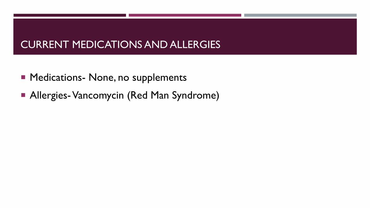
CURRENT MEDICATIONS AND ALLERGIES
Medications- None, no supplements
Allergies-Vancomycin (Red Man Syndrome)

PHYSICAL EXAM
Vital signs: BP- 128/81, HR- 80 bpm, Temp- 98.8˚F, RR- 18 bpm, BMI 26.7
Heart, lungs, abdomen and neurologic exam were unremarkable.
No jaundice was noted on the skin examination. Multiple lesions on both
hands, arms and face. Lesions were in various stages of healing along with
areas of hyperpigmentation.

LABS
▪ Initial tests were ordered:
• Allergy testing
• Lesion biopsy
▪ Follow up tests:
• Comprehensive metabolic panel & CBC with differential
• Heavy metal panel
• HCV, HSV, HIV antibody tests
• Iron studies (Serum Iron, Iron Saturation, Transferrin,)
• Uroporphyrins
• Fibroscan
• Genetic testing
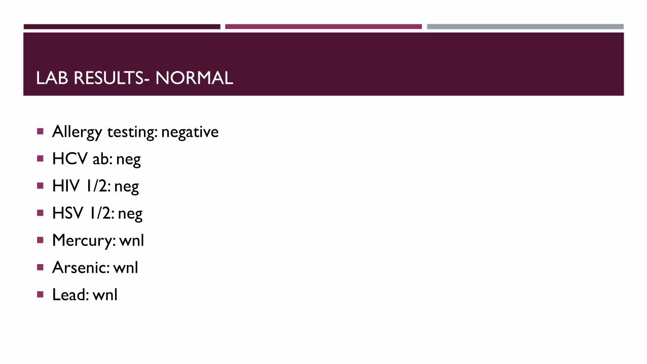
LAB RESULTS- NORMAL
Allergy testing: negative
HCV ab: neg
HIV 1/2: neg
HSV 1/2: neg
Mercury: wnl
Arsenic: wnl
Lead: wnl

LAB RESULTS- ABNORMAL
Lesion biopsy: subepidermal blister with rare dyskeratotic keratinocytes, mild lymphocytic infiltrate and red blood cell extravasation
Liver enzymes:
• AST: 64 U/L (normal 0-40 U/L)
• ALT: 158 U/L (0-44 U/L)
Iron study:
• Transferrin: 963 mg/dL (200-450 mg/dL)
• Iron (serum): 204 mcg/dL (38-169 mcg/dL)
• Iron (transferrin) saturation: 92% (15-55%)
Uroporphyrins: 1167 nmol/mmol creatinine (<2 nmol/mmol creatinine)
Fibroscan: 8.5 kPa (2-7 kPa)
Genetic testing: homozygous C282Y HFE mutation

DIFFERENTIAL DIAGNOSIS
Allergic Reaction to Chromium
Hemochromatosis
Porphyria Cutanea Tarda

WORKING DIAGNOSIS
Porphyria Cutanea Tarda in combination with Hereditary Hemochromatosis
Iron overload has been documented to accelerate the inactivation of
UROD by affecting the quantity and/or activity level
Genetic factors Acquired factors
Genetic hemochromatosis Alcohol consumption
Uroporphyrinogen
decarboxylase mutation
Tobacco use
Estrogen use
(females)
HCV infection
HIV infection
Three ideas how this may occur
1. Iron can catalyze formation of reactive oxygen species
and therefore enhance oxidation of uroporphyrinogen
to uroporphyrin
2. Iron can indirectly inhibit UROD actibity by enhancing
nonporphyrin products that directly inhibit the enzyme
3. Iron can induce aminolevulinic acid synthase, increasing
production of uroporphyrinogen

TREATMENT OPTIONS/PLAN
Unsuccessful treatment: allergy shots and steroid injections, minimal
improvement
Successful treatment: phlebotomy every week (remove a total of 30 units
of blood) with labs every 2 months, elimination of tobacco and alcohol
use
OUTCOMES- IMPACT ON PLAY
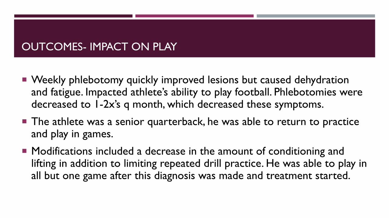
Weekly phlebotomy quickly improved lesions but caused dehydration and fatigue. Impacted athlete’s ability to play football. Phlebotomies were decreased to 1-2x’s q month, which decreased these symptoms.
The athlete was a senior quarterback, he was able to return to practice and play in games.
Modifications included a decrease in the amount of conditioning and lifting in addition to limiting repeated drill practice. He was able to play in all but one game after this diagnosis was made and treatment started.

OUTCOMES- FOLLOW UP APPOINTMENTS
Test Results
AST 47 U/L
ALT 90 U/L
Transferrin
Iron (serum) 47 mcg/dL
Iron saturation 17%
Ferritin 57 ng/mL
Test Results
AST 26 U/L
ALT 36 U/L
Transferrin 251 mcg/dL
Iron (serum) 49 mcg/dL
Iron saturation 20%
Ferritin 50 ng/mL
3 month check up 6 month check up
12 phlebotomies

UPDATES OF PATIENT
Family members tested for HFE genotype and iron status- sister
was also diagnosed with hereditary hemochromatosis, no
symptoms
Patient currently has no restrictions. No longer uses tobacco
nor drinks alcohol. He is a regular blood donor giving blood
q56 days (minimum of 3x’s/year)

CONCLUSIONS
Genetic factors Acquired factors
Genetic hemochromatosis Alcohol consumption
Uroporphyrinogen decarboxylase mutation Tobacco use
Estrogen use (females)
Hepatitis C infection
HIV infection
REFERENCES
Bissell, D. M., Anderson, K. E., & Bonkovsky, H. L. (2017). Porphyria. New England Journal of Medicine, 377(9), 862-872. doi:10.1056/nejmra1608634
Bovenschen, H., & Vissers, W. (2009, June 17). Primary hemochromatosis presented by porphyria cutanea tarda: A case report. Cases Journal, 2(7246). doi:10.4076/1757-1626-2-7246
de Geus, H. R., & Dees, A. (2006). Sporadic porphyria cutanea tarda due to haemochromatosis. The Netherlands journal of medicine, 64(8), 307–309.
Edwards, M. V., Ray, J. M., & Bacon, B. R. (2019). Sporadic Porphyria CutaneaTarda as the Initial Manifestation of Hereditary Hemochromatosis. ACG Case Reports Journal, 6(11). doi:10.14309/crj.0000000000000247
Fernandes, A., Preza, G. C., Phung, Y., Domenico, I. D., Kaplan, J., Ganz, T., & Nemeth, E. (2009). The molecular basis of hepcidin-resistanthereditary hemochromatosis. Blood, 114(2), 437-443. doi:10.1182/blood-2008-03-146134
Roberts, A. G., Whatley, S. D., Morgan, R. R., Worwood, M., & Elder, G. H. (1997). Increased frequency of the haemochromatosis Cys282Tyrmutation in sporadic porphyria cutanea tarda. The Lancet, 349(9048), 321-323. doi:10.1016/s0140-6736(96)09436-6
Salgia, R. J., & Brown, K. (2015, February 01). Diagnosis and Management of Hereditary Hemochromatosis. Clinics in Liver Disease, 19(1), 187-198. doi:10.1016/j.cld.2014.09.011