Case Studies in HIV and Kidney Disease - · PDF fileCase Studies in HIV and Kidney Disease...
28
Case Studies in HIV and Kidney Disease Samir K. Gupta, MD, MS Division of Infectious Diseases Indiana University School of Medicine Indianapolis, IN
-
Upload
truongminh -
Category
Documents
-
view
216 -
download
1
Transcript of Case Studies in HIV and Kidney Disease - · PDF fileCase Studies in HIV and Kidney Disease...

Case Studies in HIV and Kidney Disease
Samir K. Gupta, MD, MS Division of Infectious Diseases
Indiana University School of Medicine Indianapolis, IN
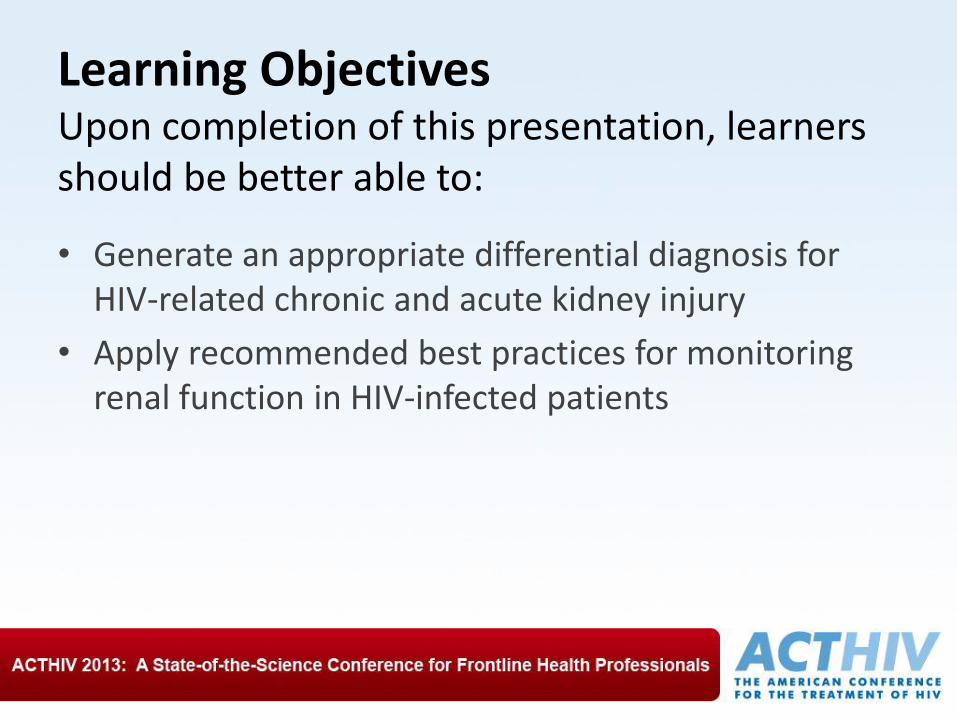
Learning Objectives Upon completion of this presentation, learners should be better able to:
• Generate an appropriate differential diagnosis for
HIV-related chronic and acute kidney injury
• Apply recommended best practices for monitoring renal function in HIV-infected patients
Faculty and Planning Committee Disclosures Please consult your program book.
There will be no off-label/investigational uses discussed in this presentation.
Off-Label Disclosure

Case #1 • A 64 year-old, African-American man presents to the ER with
diffuse muscle pains, fever, oral thrush, odynophagia, vomiting, and diarrhea. He is admitted for rehydration and treatment for his Candida esophagitis. He is found to be HIV-positive with AIDS (CD4 43 cells/µL; HIV viral load 150,000 c/mL).
• His creatinine on admission was 6.9 mg/dL, which has improved to (but stayed at) 4.4 mg/dL after receipt of IV fluids. He’s feeling much better, has defervesced, and is able to eat and drink again after receiving fluconazole.
• Serum glucose is 73 mg/dL. Hepatitis B and C serologies are negative. His UA shows 3+ protein, 0 glucose, 0 WBC, 12 RBC. Renal ultrasonography shows bilaterally enlarged, hyperechoic kidneys. His blood pressure is 106/66 mmHg.

Case #1 What is the most likely etiology of his persistent renal insufficiency?
A. Sepsis
B. Amyloidosis
C. HIV-associated nephropathy
D. Fluconazole toxicity
E. Acute HCV infection

Case #1 What is the most likely etiology of his persistent renal insufficiency?
A. Sepsis
B. Amyloidosis
C. HIV-associated nephropathy
D. Fluconazole toxicity
E. Acute HCV infection

HIV-Associated Nephropathy (HIVAN)
• First described in 1984 – Case series from New York and Miami in black IDU
– Subsequently described in other groups of African descent (Europe, Caribbean, Africa)
• Typical clinical presentation – Heavy proteinuria (nephrotic range)
– Rapid progression to end stage renal disease if left untreated
– Patients usually present during advanced HIV/AIDS stage (CD4 counts < 200/L)
– Histology shows a focal segmental glomerulosclerosis (collapsing variant) with reticuloendothelial inclusions on electron microscopy
– Ultrasonography typically show bilaterally enlarged, hyperechoic kidneys
Rao, NEJM, 2004. Laradi, JASN, 1998.
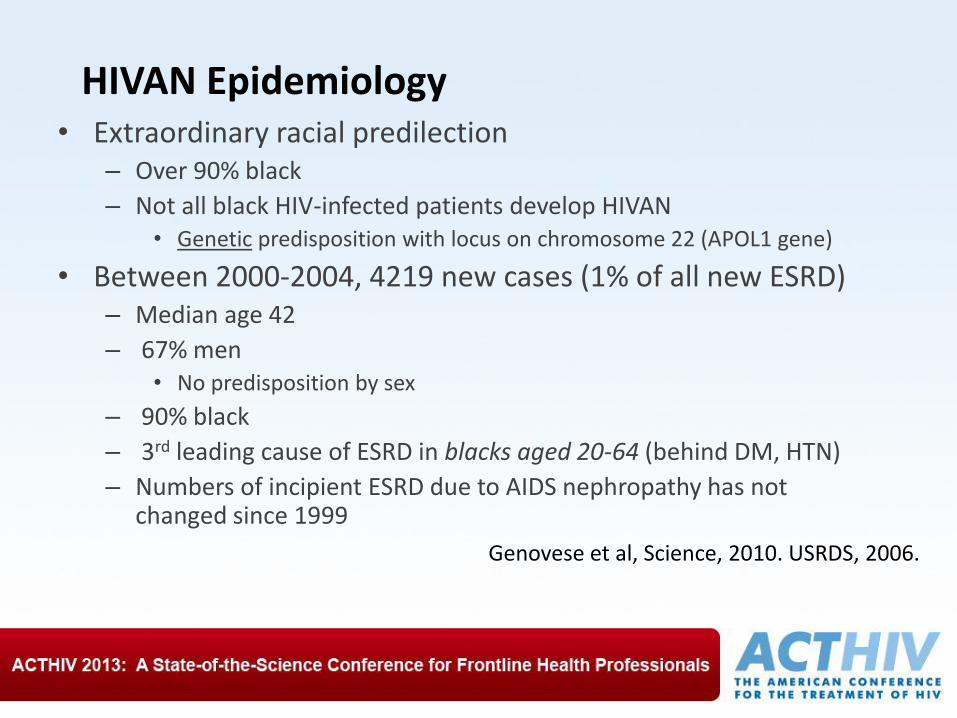
HIVAN Epidemiology • Extraordinary racial predilection
– Over 90% black
– Not all black HIV-infected patients develop HIVAN • Genetic predisposition with locus on chromosome 22 (APOL1 gene)
• Between 2000-2004, 4219 new cases (1% of all new ESRD) – Median age 42
– 67% men • No predisposition by sex
– 90% black
– 3rd leading cause of ESRD in blacks aged 20-64 (behind DM, HTN)
– Numbers of incipient ESRD due to AIDS nephropathy has not changed since 1999
Genovese et al, Science, 2010. USRDS, 2006.

HIVAN Epidemiology
• Estimated prevalence of 0.93% in ART-untreated Africans in England
• Incidence appears to be decreasing with availability of ART
• Undetectable viral load suggests CKD is not due to HIVAN (more often HTN, DM, HCV)
Lucas, AIDS, 2004. Post, Clin Infect Dis, 2008. Atta, Clin Infect Dis, 2007.

Changes in eGFR with ART in the ACTG/ALLRT Cohort
Kalayjian et al, AIDS, 2008. Race was not a predictor of
improvement – all improved!!

HIVAN
69.8% (30) a Membranoproliferative glomerulonephritis
45.9% (17) a 10.8% (4)
4.7% (2) Diabetes mellitus
5.4% (2)
14.0% (6) Hypertension
5.4% (2)
4.7% (2) Amyloid
5.4% (2)
2.3% (1) Chronic focal glomerulonephritis
2.7% (1)
Focal segmental glomerulosclerosis
5.4% (2)
Membranous glomerulopathy
2.7% (1)
Nonspecific
2.7% (1)
No tissue obtained
8.1% (3)
Mesangial glomerulonephritis
5.4% (2)
Heroin abuse
2.3% (1) Nephrotoxic drugs
2.3% (1) Total
37
43 HIVAN is HIV-associated nephropathy
a P = 0.03 (HIVAN vs. all others)
Spectrum of Renal Diseases in HIV Etiology Biopsy No biopsy
Szczech et al, Kidney Int, 2003.

Case #2 • A 47 year-old, HIV-infected Hispanic woman has been receiving
tenofovir/emtricitabine/atazanavir/ritonavir for 2 years with excellent results (CD4 565 cells/µL; viral load <20 c/mL). She also has chronic active HCV infection, but her provider is waiting to start HCV treatment until newer oral therapies become available. Her co-morbidities include type II diabetes and chronic low back pain, for which she frequently takes ibuprofen.
• She presents to the ER with edema, nausea, and vomiting. She is afebrile. Her creatinine is 4.6 mg/dL; it was 1.3 mg/dL when last checked 6 months ago.
• Her Hgb A1C is 8.9%. UA shows 3+ protein, 2+ glucose, trace ketones, 0 WBC, 3 RBC, neg leukocyte esterase. Her last UA, obtained 2 years ago at treatment initiation, was completely normal. Renal ultrasonography now shows bilaterally small kidneys without hydronephrosis.

Case #2 What is the least likely etiology of her worsening kidney disease?
A. Antiretroviral-induced toxicity
B. HIVAN
C. Diabetic nephropathy
D. NSAID nephropathy
E. HCV nephropathy

Case #2 What is the least likely etiology of her worsening kidney disease?
A. Antiretroviral-induced toxicity
B. HIVAN
C. Diabetic nephropathy
D. NSAID nephropathy
E. HCV nephropathy

Measurement of Renal Function • Glomerular filtration rate (GFR)
– Best measure for overall renal function – Renal clearance of a marker from plasma – Volume of plasma that can be cleared of that
marker per unit of time • Serum creatinine is most frequently used
marker of renal function – Endogenously produced – Dependent on muscle mass – Mostly filtered by glomerulus but also secreted through proximal
tubule • Serum Cystatin C
– Endogenously produced by all nucleated cells – Not dependent on muscle mass (more stable concentrations in
circulation) – Freely filtered by glomerulus without tubular secretion

Issues with Creatinine • Creatinine is secreted through
proximal tubule • Tubular secretion of creatinine is
inhibited by cimetidine, trimethoprim, dapsone, cobicistat, dolutegravir
• Tubular secretion is proportionally more important when glomerular filtration is reduced
• Now standardized assay, so laboratory variation is minimized
• Large intrapersonal variation (7-20%) • Minimum detectable change up to
0.2mg/dL • Serum creatinine may not increase
until 50% of true GFR is reduced
Krop, Arch Int Med, 1999. Coresh, AJKD, 2002. Levey, Ann Int Med, 1999. Elion, AIDS, 2011. van Lunzen, Lancet Infect Dis, 2012.
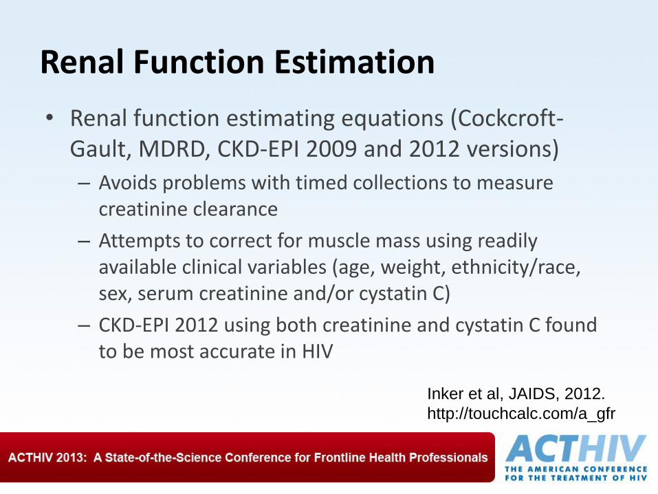
Renal Function Estimation
• Renal function estimating equations (Cockcroft-Gault, MDRD, CKD-EPI 2009 and 2012 versions)
– Avoids problems with timed collections to measure creatinine clearance
– Attempts to correct for muscle mass using readily available clinical variables (age, weight, ethnicity/race, sex, serum creatinine and/or cystatin C)
– CKD-EPI 2012 using both creatinine and cystatin C found to be most accurate in HIV
Inker et al, JAIDS, 2012.
http://touchcalc.com/a_gfr

Normal Renal Function • Serum creatinine in NHANES III Men Women Age Healthy All Healthy All
20-39 1.14 1.09(1.33) 0.92 0.87(1.08)
40-59 1.17 1.11(1.38) 0.95 0.90(1.14)
• Best mean estimates for GFR are 130mL/min/1.73m2 and
120mL/min/1.73m2 in healthy young men and women, respectively
• Wide variation (age, race, sex, diet) • Key point is complications (death, CV disease, ESRD, anemia,
hyperPTH/calcium dysregulation) increase as a continuum with increasing serum creatinine – even with mild increases and especially when GFR falls below 60mL/min/1.73m2
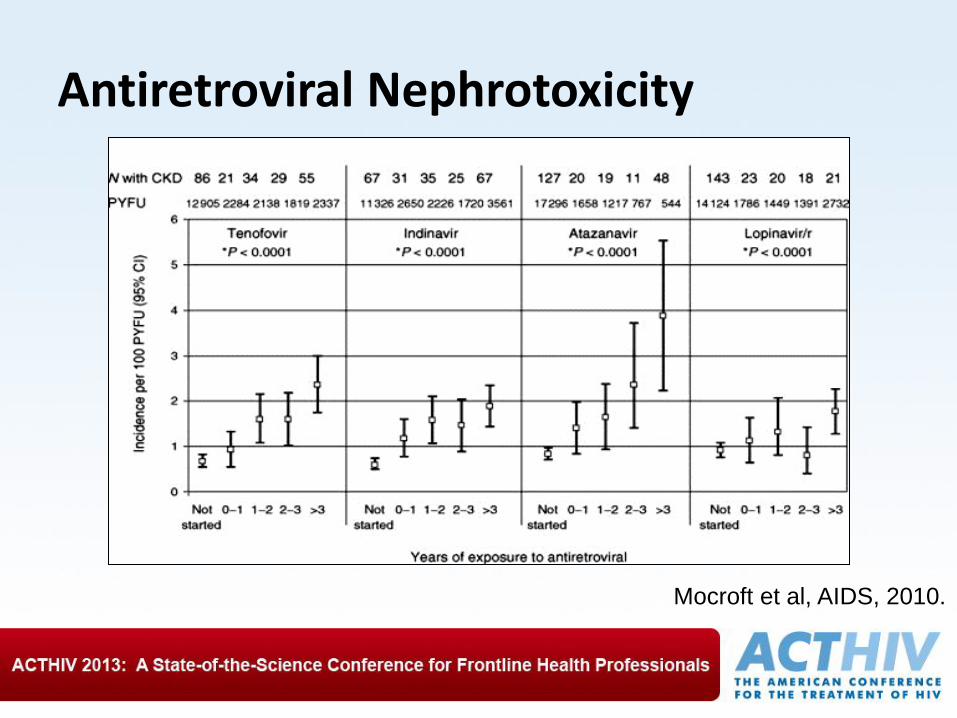
Antiretroviral Nephrotoxicity
Mocroft et al, AIDS, 2010.

Incidence of acute renal failure (ARF), measured from date of first attendance, in 1752 patients who initiated HIV care during the study period.
Roe J et al. Clin Infect Dis. 2008;47:242-249
© 2008 by the Infectious Diseases Society of America

Demographic and laboratory characteristics of patients with acute renal failure (ARF) and patients without renal failure.
Roe J et al. Clin Infect Dis. 2008;47:242-249
© 2008 by the Infectious Diseases Society of America

Demographic and laboratory characteristics of patients with acute renal failure (ARF) and patients without renal failure.
Roe J et al. Clin Infect Dis. 2008;47:242-249
© 2008 by the Infectious Diseases Society of America

Demographic and laboratory characteristics of patients with acute renal failure (ARF) and patients without renal failure.
Roe J et al. Clin Infect Dis. 2008;47:242-249
© 2008 by the Infectious Diseases Society of America

Demographic and laboratory characteristics of patients with acute renal failure (ARF) and patients without renal failure.
Roe J et al. Clin Infect Dis. 2008;47:242-249
© 2008 by the Infectious Diseases Society of America

Demographic and laboratory characteristics of patients with acute renal failure (ARF) and patients without renal failure.
Roe J et al. Clin Infect Dis. 2008;47:242-249
© 2008 by the Infectious Diseases Society of America
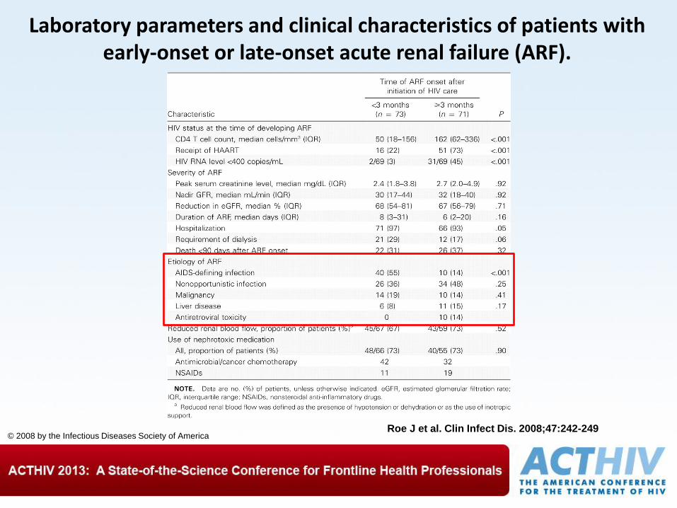
Laboratory parameters and clinical characteristics of patients with early-onset or late-onset acute renal failure (ARF).
Roe J et al. Clin Infect Dis. 2008;47:242-249 © 2008 by the Infectious Diseases Society of America
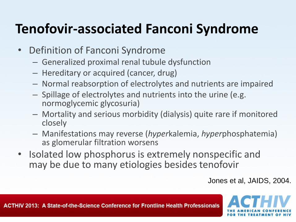
Tenofovir-associated Fanconi Syndrome
• Definition of Fanconi Syndrome – Generalized proximal renal tubule dysfunction – Hereditary or acquired (cancer, drug) – Normal reabsorption of electrolytes and nutrients are impaired – Spillage of electrolytes and nutrients into the urine (e.g.
normoglycemic glycosuria) – Mortality and serious morbidity (dialysis) quite rare if monitored
closely – Manifestations may reverse (hyperkalemia, hyperphosphatemia)
as glomerular filtration worsens
• Isolated low phosphorus is extremely nonspecific and may be due to many etiologies besides tenofovir
Jones et al, JAIDS, 2004.
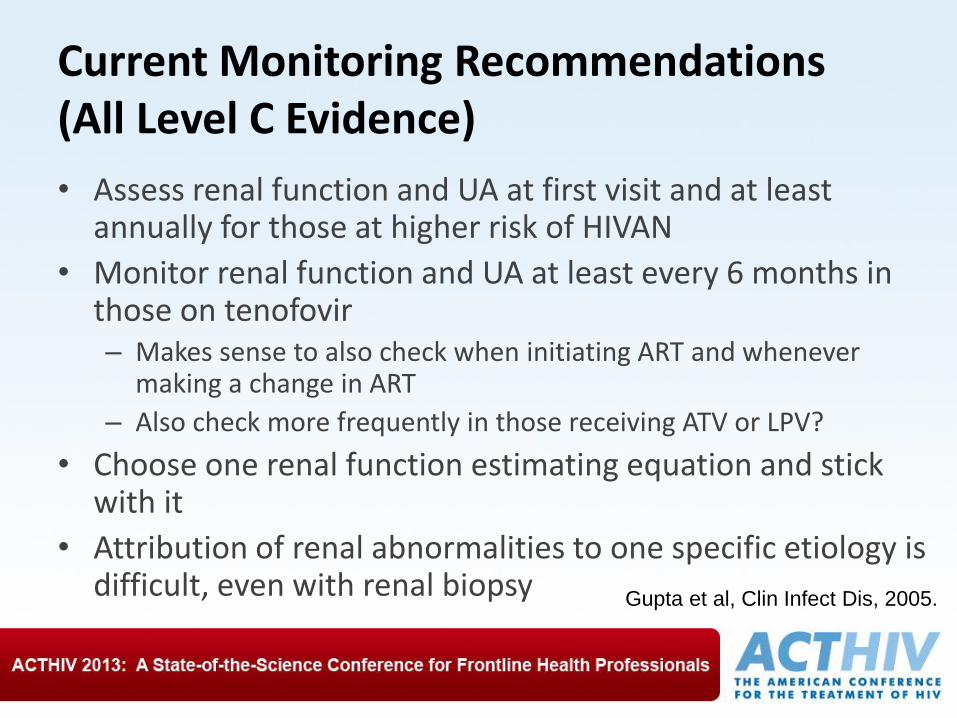
Current Monitoring Recommendations (All Level C Evidence)
• Assess renal function and UA at first visit and at least annually for those at higher risk of HIVAN
• Monitor renal function and UA at least every 6 months in those on tenofovir – Makes sense to also check when initiating ART and whenever
making a change in ART
– Also check more frequently in those receiving ATV or LPV?
• Choose one renal function estimating equation and stick with it
• Attribution of renal abnormalities to one specific etiology is difficult, even with renal biopsy
Gupta et al, Clin Infect Dis, 2005.
![Kidney Disease in HIV PatientsKidney Disease in HIV Patients › assets › 2387 › Wyatt 2010 [Compatibility Mode]… · Slide #15 HIVAN Pathogenesis: Mouse Model z“Tg26” HIV-1](https://static.fdocuments.in/doc/165x107/5f0b7ee67e708231d430cda7/kidney-disease-in-hiv-patientskidney-disease-in-hiv-a-assets-a-2387-a-wyatt.jpg)