Case Report Video Capsule Retention in …downloads.hindawi.com/journals/crigm/2013/607142.pdfVideo...
5
Hindawi Publishing Corporation Case Reports in Gastrointestinal Medicine Volume 2013, Article ID 607142, 4 pages http://dx.doi.org/10.1155/2013/607142 Case Report Video Capsule Retention in Inflammatory Bowel Disease: An Unusual Presentation and Discussion of Retrieval Methods R. Goel, 1 J. Hardman, 1 M. Gulati, 2 and J. O’Donohue 1 1 Department of Gastroenterology & General Medicine, University Hospital Lewisham, Lewisham High Street, London SE13 6LH, UK 2 Department of Radiology, Queen Elizabeth Hospital, Stadium Road, Woolwich, London SE18 4QH, UK Correspondence should be addressed to R. Goel; [email protected] Received 15 January 2013; Accepted 7 February 2013 Academic Editors: H. Akiho, M. Iizuka, S. Karoui, and F. H. Mourad Copyright © 2013 R. Goel et al. is is an open access article distributed under the Creative Commons Attribution License, which permits unrestricted use, distribution, and reproduction in any medium, provided the original work is properly cited. Inflammatory Bowel Disease (IBD) is characterized by chronic inflammation in the gastrointestinal (GI) tract. Video capsule endoscopy (VCE) is widely used to investigate the small bowel, and capsule retention is the most serious potential complication. Endoscopic and surgical management has been reported, but in the absence of bowel obstruction, there is little consensus as to which should be employed. In this case report, we describe a patient who was investigated with VCE for weight loss and anaemia. He had previously undergone colectomy with ileoanal pouch formation for ulcerative colitis (UC). Capsule retention occurred at an ileal stricture and he was subsequently diagnosed with Crohn’s disease (CD). We describe his medical management and successful capsule retrieval using endoscopic methods. is case also highlights the importance of screening for intestinal strictures in an atypical presentation of UC following colectomy. 1. Introduction IBD is a chronic inflammatory condition comprising UC and CD. e incidence of UC varies greatly between 0.5 and 24.5/100,000 persons, while that of CD varies between 0.1 and 16/100,000 persons worldwide [1]. e precise aetiol- ogy remains unknown but is thought to be a combination of genetic, immunologic, and environmental factors. e diagnosis is made on the clinical history, laboratory inves- tigations, and endoscopic and histologic appearances. UC is characterized by confluent mucosal inflammation which is confined to the colon. In contrast, CD is characterized by patchy transmural inflammation which can affect any part of the GI tract, though it commonly involves the ileocaecal region. Enteric fistulas and stricturing are pathological fea- tures unique to CD which distinguish it from UC. Video capsule endoscopy (VCE) is used for the inves- tigation of small bowel lesions. Its main indication is for investigation of anaemia due to occult GI bleeding. Following fasting, the patient swallows a small capsule (consisting of a camera, a light source, and a wireless circuit for the transmission of signals). As the capsule moves through the GI tract, images are transmitted to an external data recorder. ese data are transferred to a computer for interpretation. e capsule is then passed in the patient’s stool [2]. We report a case of VCE retention and recovery in a patient with IBD without resorting to surgical intervention. 2. Case Report A 36-year-old man presented to the Gastroenterology Outpa- tients Department with a four-month history of weight loss and iron-deficiency anaemia. He denied abdominal pain and described no change in bowel habit or rectal bleeding. He had previously been diagnosed with UC approximately 20 years ago and had undergone a colectomy with ileostomy two years later. 15 years ago, he had an ileoanal pouch formed. Oesophagoduodenogastroscopy (OGD) and flexible pou- choscopy were unremarkable. VCE was performed; how- ever the capsule was not passed (Figure 1). Pouchoscopy/ retrograde ileoscopy with a paediatric colonoscope showed a normal pouch with scattered aphthous ulcers in the ileum (Figure 2). Two strictures were identified, the more signif- icant 100 cm proximal to the pouch, which could not be traversed by the scope. Small bowel barium follow-through
Transcript of Case Report Video Capsule Retention in …downloads.hindawi.com/journals/crigm/2013/607142.pdfVideo...

Hindawi Publishing CorporationCase Reports in Gastrointestinal MedicineVolume 2013, Article ID 607142, 4 pageshttp://dx.doi.org/10.1155/2013/607142
Case ReportVideo Capsule Retention in Inflammatory Bowel Disease:An Unusual Presentation and Discussion of Retrieval Methods
R. Goel,1 J. Hardman,1 M. Gulati,2 and J. O’Donohue1
1 Department of Gastroenterology & General Medicine, University Hospital Lewisham, Lewisham High Street, London SE13 6LH, UK2Department of Radiology, Queen Elizabeth Hospital, Stadium Road, Woolwich, London SE18 4QH, UK
Correspondence should be addressed to R. Goel; [email protected]
Received 15 January 2013; Accepted 7 February 2013
Academic Editors: H. Akiho, M. Iizuka, S. Karoui, and F. H. Mourad
Copyright © 2013 R. Goel et al. This is an open access article distributed under the Creative Commons Attribution License, whichpermits unrestricted use, distribution, and reproduction in any medium, provided the original work is properly cited.
Inflammatory Bowel Disease (IBD) is characterized by chronic inflammation in the gastrointestinal (GI) tract. Video capsuleendoscopy (VCE) is widely used to investigate the small bowel, and capsule retention is the most serious potential complication.Endoscopic and surgical management has been reported, but in the absence of bowel obstruction, there is little consensus as towhich should be employed. In this case report, we describe a patient who was investigated with VCE for weight loss and anaemia.He had previously undergone colectomywith ileoanal pouch formation for ulcerative colitis (UC). Capsule retention occurred at anileal stricture and he was subsequently diagnosed with Crohn’s disease (CD). We describe his medical management and successfulcapsule retrieval using endoscopic methods. This case also highlights the importance of screening for intestinal strictures in anatypical presentation of UC following colectomy.
1. Introduction
IBD is a chronic inflammatory condition comprising UCand CD. The incidence of UC varies greatly between 0.5 and24.5/100,000 persons, while that of CD varies between 0.1and 16/100,000 persons worldwide [1]. The precise aetiol-ogy remains unknown but is thought to be a combinationof genetic, immunologic, and environmental factors. Thediagnosis is made on the clinical history, laboratory inves-tigations, and endoscopic and histologic appearances. UC ischaracterized by confluent mucosal inflammation which isconfined to the colon. In contrast, CD is characterized bypatchy transmural inflammation which can affect any partof the GI tract, though it commonly involves the ileocaecalregion. Enteric fistulas and stricturing are pathological fea-tures unique to CD which distinguish it from UC.
Video capsule endoscopy (VCE) is used for the inves-tigation of small bowel lesions. Its main indication is forinvestigation of anaemia due to occult GI bleeding. Followingfasting, the patient swallows a small capsule (consistingof a camera, a light source, and a wireless circuit for thetransmission of signals). As the capsule moves through theGI tract, images are transmitted to an external data recorder.
These data are transferred to a computer for interpretation.The capsule is then passed in the patient’s stool [2].
We report a case of VCE retention and recovery in apatient with IBD without resorting to surgical intervention.
2. Case Report
A 36-year-oldman presented to theGastroenterologyOutpa-tients Department with a four-month history of weight lossand iron-deficiency anaemia. He denied abdominal pain anddescribed no change in bowel habit or rectal bleeding. He hadpreviously been diagnosed with UC approximately 20 yearsago and had undergone a colectomywith ileostomy two yearslater. 15 years ago, he had an ileoanal pouch formed.
Oesophagoduodenogastroscopy (OGD) andflexible pou-choscopy were unremarkable. VCE was performed; how-ever the capsule was not passed (Figure 1). Pouchoscopy/retrograde ileoscopy with a paediatric colonoscope showeda normal pouch with scattered aphthous ulcers in the ileum(Figure 2). Two strictures were identified, the more signif-icant 100 cm proximal to the pouch, which could not betraversed by the scope. Small bowel barium follow-through

2 Case Reports in Gastrointestinal Medicine
Figure 1: Plain abdominal X-ray showing retained capsule.
Aphthous ulcer
(a)
Aphthous ulcer
(b)Stricture 90 cms
(c)
Stricture 80 cms
(d)
Figure 2: Pouchoscopy/ileoscopy showing aphthous ulcer and nontraversable stricture proximal to ileoanal reservoir.
Case Reports in Gastrointestinal Medicine 3
confirmed the endoscopy findings and showed the capsulewas impacted but freely mobile proximal to this stricture.
The capsule was retained proximal to an ileal stricture.The findings of small bowel ulceration led to a diagnosisof small bowel CD being made. There was no history ofnonsteroidal anti-inflammatory drug (NSAID) consump-tion, vasculitis, or connective tissue disease.
A course of budesonide was given to resolve the inflam-matory component of the stricture and the patient wasfollowed up for two months with serial abdominal radio-graphs, but the capsule did not pass. At repeat ileoscopy, thedominant ileal stricture could not be traversed by a paediatriccolonoscope even after gentle balloon dilatation. An attemptto insert a biodegradable stent was unsuccessful.
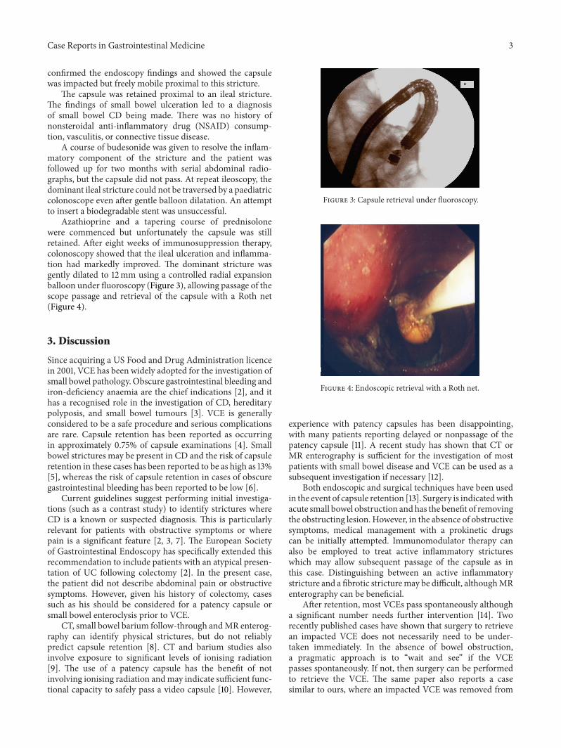
Azathioprine and a tapering course of prednisolonewere commenced but unfortunately the capsule was stillretained. After eight weeks of immunosuppression therapy,colonoscopy showed that the ileal ulceration and inflamma-tion had markedly improved. The dominant stricture wasgently dilated to 12mm using a controlled radial expansionballoon under fluoroscopy (Figure 3), allowing passage of thescope passage and retrieval of the capsule with a Roth net(Figure 4).
3. Discussion
Since acquiring a US Food and Drug Administration licencein 2001, VCE has been widely adopted for the investigation ofsmall bowel pathology. Obscure gastrointestinal bleeding andiron-deficiency anaemia are the chief indications [2], and ithas a recognised role in the investigation of CD, hereditarypolyposis, and small bowel tumours [3]. VCE is generallyconsidered to be a safe procedure and serious complicationsare rare. Capsule retention has been reported as occurringin approximately 0.75% of capsule examinations [4]. Smallbowel strictures may be present in CD and the risk of capsuleretention in these cases has been reported to be as high as 13%[5], whereas the risk of capsule retention in cases of obscuregastrointestinal bleeding has been reported to be low [6].
Current guidelines suggest performing initial investiga-tions (such as a contrast study) to identify strictures whereCD is a known or suspected diagnosis. This is particularlyrelevant for patients with obstructive symptoms or wherepain is a significant feature [2, 3, 7]. The European Societyof Gastrointestinal Endoscopy has specifically extended thisrecommendation to include patients with an atypical presen-tation of UC following colectomy [2]. In the present case,the patient did not describe abdominal pain or obstructivesymptoms. However, given his history of colectomy, casessuch as his should be considered for a patency capsule orsmall bowel enteroclysis prior to VCE.
CT, small bowel barium follow-through andMR enterog-raphy can identify physical strictures, but do not reliablypredict capsule retention [8]. CT and barium studies alsoinvolve exposure to significant levels of ionising radiation[9]. The use of a patency capsule has the benefit of notinvolving ionising radiation andmay indicate sufficient func-tional capacity to safely pass a video capsule [10]. However,
Figure 3: Capsule retrieval under fluoroscopy.
Figure 4: Endoscopic retrieval with a Roth net.
experience with patency capsules has been disappointing,with many patients reporting delayed or nonpassage of thepatency capsule [11]. A recent study has shown that CT orMR enterography is sufficient for the investigation of mostpatients with small bowel disease and VCE can be used as asubsequent investigation if necessary [12].
Both endoscopic and surgical techniques have been usedin the event of capsule retention [13]. Surgery is indicatedwithacute small bowel obstruction andhas the benefit of removingthe obstructing lesion. However, in the absence of obstructivesymptoms, medical management with a prokinetic drugscan be initially attempted. Immunomodulator therapy canalso be employed to treat active inflammatory strictureswhich may allow subsequent passage of the capsule as inthis case. Distinguishing between an active inflammatorystricture and a fibrotic stricturemay be difficult, althoughMRenterography can be beneficial.
After retention, most VCEs pass spontaneously althougha significant number needs further intervention [14]. Tworecently published cases have shown that surgery to retrievean impacted VCE does not necessarily need to be under-taken immediately. In the absence of bowel obstruction,a pragmatic approach is to “wait and see” if the VCEpasses spontaneously. If not, then surgery can be performedto retrieve the VCE. The same paper also reports a casesimilar to ours, where an impacted VCE was removed from

4 Case Reports in Gastrointestinal Medicine
the jejunum of a known CD patient [15]. Ultimately, there isno consensus for management of capsule retention and thechoice of retrieval technique should therefore be consideredon an individual case basis.
Conflict of Interests
The authors declare that they have no conflict of interests.
Consent
Consent was obtained from the patient.
References
[1] P. L. Lakatos, “Recent trends in the epidemiology of inflamma-tory bowel diseases: up or down?” World Journal of Gastroen-terology, vol. 12, no. 38, pp. 6102–6108, 2006.
[2] National Institute for Health and Clinical Excellence, WirelessCapsule Endoscopy For Investigation of the Small Bowel, 2004.
[3] S. D. Ladas, K. Triantafyllou, C. Spada et al., “European Soci-ety of Gastrointestinal Endoscopy (ESGE): recommendations(2009) on clinical use of video capsule endoscopy to investigatesmall-bowel, esophageal and colonic diseases,” Endoscopy, vol.42, no. 3, pp. 220–227, 2010.
[4] J. S. Barkin and S. Friedman, “Wireless capsule endoscopyrequiring surgical intervention. The world’s experience,” TheAmerican Journal of Gastroenterology, vol. 97, pp. A-83–A-89,2002.
[5] A. S. Cheifetz, A. A. Kornbluth, P. Legnani et al., “The risk ofretention of the capsule endoscope in patients with known orsuspected Crohn’s disease,” American Journal of Gastroenterol-ogy, vol. 101, no. 10, pp. 2218–2222, 2006.
[6] D. M. Sears, A. Avots-Avotins, K. Culp, and M. W. Gavin,“Frequency and clinical outcome of capsule retention duringcapsule endoscopy for GI bleeding of obscure origin,”Gastroin-testinal Endoscopy, vol. 60, no. 5, pp. 822–827, 2004.
[7] R. Sidhu, D. S. Sanders, A. J. Morris, and M. E. McAlindon,“Guidelines on small bowel enteroscopy and capsule endoscopyin adults,” Gut, vol. 57, no. 1, pp. 125–136, 2008.
[8] E. Rondonotti, J. M. Herrerias, M. Pennazio, A. Caunedo,M. Mascarenhas-Saraiva, and R. de Franchis, “Complications,limitations, and failures of capsule endoscopy: a review of 733cases,” Gastrointestinal Endoscopy, vol. 62, no. 5, pp. 712–716,2005.
[9] J. M. Herrerias, J. A. Leighton, G. Costamagna et al.,“Agile patency system eliminates risk of capsule retention inpatients with known intestinal strictures who undergo capsuleendoscopy,” Gastrointestinal Endoscopy, vol. 67, no. 6, pp. 902–909, 2008.
[10] M. Boysen and M. Ritter, “Obstruction from capsuleendoscopy,” Western Journal of Emergency Medicine, vol.11, pp. 71–73, 2010.
[11] M. Delvaux, B. Soussan, V. Laurent, E. Lerebours, and G. Gay,“Clinical evaluation of the use of the M2A patency capsulesystem before a capsule endoscopy procedure, in patients withknown or suspected intestinal stenosis,” Endoscopy, vol. 37, no.9, pp. 801–807, 2005.
[12] F. T. Fork,N.Karlsson, S. Kadhem, andB.Ohlsson, “Small bowelenteroclysis with magnetic resonance imaging and computed
tomography in patients with failed and uncertain passage of apatency capsule,” BMC Medical Imaging, vol. 12, no. 1, article 3,2012.
[13] D. Cave, P. Legnani, R. de Franchis, and B. S. Lewis, “ICCEconsensus for capsule retention,” Endoscopy, vol. 37, no. 10, pp.1065–1067, 2005.
[14] J. H. Cheon, Y. S. Kim, I. S. Lee et al., “Can we predictspontaneous capsule passage after retention? A nationwidestudy to evaluate the incidence and clinical outcomes of capsuleretention,” Endoscopy, vol. 39, no. 12, pp. 1046–1052, 2007.
[15] G. Hauser, D. Stimac, V. Giljaca, and B. M. Sincic, “Videocapsule retention—endoscopic or surgical problem?” Clinicsand Research in Hepatology and Gastroenterology, vol. 36, no.6, pp. e135–e136, 2012.

Submit your manuscripts athttp://www.hindawi.com
Stem CellsInternational
Hindawi Publishing Corporationhttp://www.hindawi.com Volume 2014
Hindawi Publishing Corporationhttp://www.hindawi.com Volume 2014
MEDIATORSINFLAMMATION
of
Hindawi Publishing Corporationhttp://www.hindawi.com Volume 2014
Behavioural Neurology
EndocrinologyInternational Journal of
Hindawi Publishing Corporationhttp://www.hindawi.com Volume 2014
Hindawi Publishing Corporationhttp://www.hindawi.com Volume 2014
Disease Markers
Hindawi Publishing Corporationhttp://www.hindawi.com Volume 2014
BioMed Research International
OncologyJournal of
Hindawi Publishing Corporationhttp://www.hindawi.com Volume 2014
Hindawi Publishing Corporationhttp://www.hindawi.com Volume 2014
Oxidative Medicine and Cellular Longevity
Hindawi Publishing Corporationhttp://www.hindawi.com Volume 2014
PPAR Research
The Scientific World JournalHindawi Publishing Corporation http://www.hindawi.com Volume 2014
Immunology ResearchHindawi Publishing Corporationhttp://www.hindawi.com Volume 2014
Journal of
ObesityJournal of
Hindawi Publishing Corporationhttp://www.hindawi.com Volume 2014
Hindawi Publishing Corporationhttp://www.hindawi.com Volume 2014
Computational and Mathematical Methods in Medicine
OphthalmologyJournal of
Hindawi Publishing Corporationhttp://www.hindawi.com Volume 2014
Diabetes ResearchJournal of
Hindawi Publishing Corporationhttp://www.hindawi.com Volume 2014
Hindawi Publishing Corporationhttp://www.hindawi.com Volume 2014
Research and TreatmentAIDS
Hindawi Publishing Corporationhttp://www.hindawi.com Volume 2014
Gastroenterology Research and Practice
Hindawi Publishing Corporationhttp://www.hindawi.com Volume 2014
Parkinson’s Disease
Evidence-Based Complementary and Alternative Medicine
Volume 2014Hindawi Publishing Corporationhttp://www.hindawi.com