CASE REPORT Intramuscular sinusoidal haemangioma with ...
4
CASE REPORT Intramuscular sinusoidal haemangioma with secondary Masson’s phenomenon Paremala Konda, Radhika M Bavle, Soumya Makarla, Sudhakara Muniswamappa Department of Oral Pathology, Krishnadevaraya College of Dental Sciences, Bangalore, Karnataka, India Correspondence to Dr Soumya Makarla, [email protected] Accepted 5 December 2015 To cite: Konda P, Bavle RM, Makarla S, et al. BMJ Case Rep Published online: [ please include Day Month Year] doi:10.1136/ bcr-2013-201457 SUMMARY Intramuscular haemangiomas (IMHs) are rare benign vascular neoplasms that account for approximately 0.8% of all haemangiomas. The histology of IMHs can reveal cavernous dilated spaces. We report an interesting case of haemangioma in the deep skeletal muscle of the right labial mucosa in a young man involving the orbicularis oris muscle which showed additional features of sinusoidal arrangement with a secondary Masson’s phenomenon. BACKGROUND Haemangiomas are common soft tissue tumours which account for about 7% of all benign tumours. They occur commonly during infancy and child- hood. 1 Intramuscular location of these lesions is rare and constitutes <1% of all haemangiomas. 2 Approximately 15% of intramuscular haemangiomas (IMHs) occur in the head and neck area. The mas- seter and trapezius muscles are most commonly affected (60%) followed by the periorbital, sterno- mastoid, temporalis, geniohyoid and medial ptery- goid muscles. 3 Due to their rarity, deep location, intriguing aetiopathogenesis, variable clinical presen- tation and differences in treatment modality, IMHs require special attention. Sinusoidal haemangiomas, although a distinct subset of cavernous haemangi- omas, are rare. Their clinicopathological character- istics are important and need to be recognised to avoid any diagnostic pitfalls. 4 Masson’s tumour, or intravascular papillary endothelial hyperplasia, repre- sents an unusual endothelial proliferation which can occur primarily in a venous channel or secondarily in a vascular anomaly. 5 It is an important entity which needs to be distinguished from malignancies such as angiosarcoma to avoid radical surgery. CASE PRESENTATION A male student in his early 20s reported to the out- patient department of our hospital with a chief complaint of a solitary painless swelling on the lower right side of the face that had gradually been increasing in size over a period of 4 years. Extraoral examination revealed an ill-defined swel- ling near the angle of the mouth on the right side measuring approximately 2.0×3.0 cm and extend- ing 1 cm above the lower border of the mandible. The swelling was firm to palpate. The overlying skin appeared normal with no rise in the local tem- perature ( figure 1A). Intraoral examination revealed a solitary ill- circumscribed swelling in the right labial mucosa measuring approximately 2.0 cm×2.5 cm in diam- eter, extending in relation to 43 to 44 region, with mild obliteration of the labial vestibule. There was no tenderness on palpation or any signs of discharge. The overlying mucosa appeared stretched but normal. The mucosa over the swelling was slightly blanched with one area showing a slight bluish hue ( figure 1B). There was no associated cervical lymph- adenopathy. The patient’s periodontal status was good with a full complement of permanent teeth. The lesion was surgically excised under local anaesthesia and submitted with a provisional diag- nosis of mucocele for histopathological examin- ation. On macroscopic examination the excised lesion was roughly oval in shape, brown in colour, measuring 1.8×1.0 cm with an irregular surface and firm consistency ( figure 2). Figure 1 (A) Clinical photograph showing a solitary swelling on the right side below the angle of the mouth. (B) Clinical photograph showing a solitary ill-circumscribed swelling in the left buccal mucosa obliterating the buccal vestibule with an area exhibiting a bluish hue. Konda P, et al. BMJ Case Rep 2016. doi:10.1136/bcr-2013-201457 1 Unusual presentation of more common disease/injury on 30 November 2021 by guest. Protected by copyright. http://casereports.bmj.com/ BMJ Case Reports: first published as 10.1136/bcr-2013-201457 on 4 January 2016. Downloaded from
Transcript of CASE REPORT Intramuscular sinusoidal haemangioma with ...
CASE REPORT
Intramuscular sinusoidal haemangioma withsecondary Masson’s phenomenonParemala Konda, Radhika M Bavle, Soumya Makarla, Sudhakara Muniswamappa
Department of Oral Pathology,Krishnadevaraya College ofDental Sciences, Bangalore,Karnataka, India
Correspondence toDr Soumya Makarla,[email protected]
Accepted 5 December 2015
To cite: Konda P,Bavle RM, Makarla S, et al.BMJ Case Rep Publishedonline: [please include DayMonth Year] doi:10.1136/bcr-2013-201457
SUMMARYIntramuscular haemangiomas (IMHs) are rare benignvascular neoplasms that account for approximately 0.8% ofall haemangiomas. The histology of IMHs can revealcavernous dilated spaces. We report an interesting case ofhaemangioma in the deep skeletal muscle of the rightlabial mucosa in a young man involving the orbicularis orismuscle which showed additional features of sinusoidalarrangement with a secondary Masson’s phenomenon.
BACKGROUNDHaemangiomas are common soft tissue tumourswhich account for about 7% of all benign tumours.They occur commonly during infancy and child-hood.1 Intramuscular location of these lesions is rareand constitutes <1% of all haemangiomas.2
Approximately 15% of intramuscular haemangiomas(IMHs) occur in the head and neck area. The mas-seter and trapezius muscles are most commonlyaffected (60%) followed by the periorbital, sterno-mastoid, temporalis, geniohyoid and medial ptery-goid muscles.3 Due to their rarity, deep location,intriguing aetiopathogenesis, variable clinical presen-tation and differences in treatment modality, IMHsrequire special attention. Sinusoidal haemangiomas,although a distinct subset of cavernous haemangi-omas, are rare. Their clinicopathological character-istics are important and need to be recognised toavoid any diagnostic pitfalls.4 Masson’s tumour, orintravascular papillary endothelial hyperplasia, repre-sents an unusual endothelial proliferation which canoccur primarily in a venous channel or secondarily ina vascular anomaly.5 It is an important entity which
needs to be distinguished from malignancies such asangiosarcoma to avoid radical surgery.
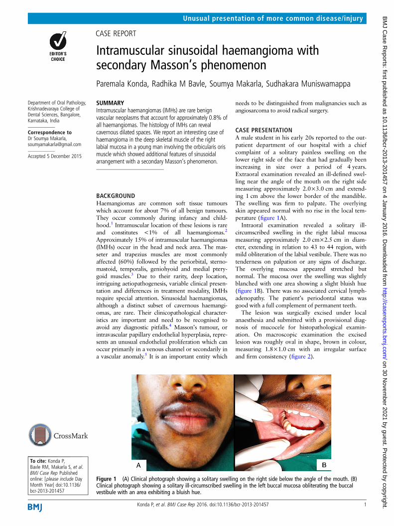
CASE PRESENTATIONA male student in his early 20s reported to the out-patient department of our hospital with a chiefcomplaint of a solitary painless swelling on thelower right side of the face that had gradually beenincreasing in size over a period of 4 years.Extraoral examination revealed an ill-defined swel-ling near the angle of the mouth on the right sidemeasuring approximately 2.0×3.0 cm and extend-ing 1 cm above the lower border of the mandible.The swelling was firm to palpate. The overlyingskin appeared normal with no rise in the local tem-perature (figure 1A).Intraoral examination revealed a solitary ill-
circumscribed swelling in the right labial mucosameasuring approximately 2.0 cm×2.5 cm in diam-eter, extending in relation to 43 to 44 region, withmild obliteration of the labial vestibule. There was notenderness on palpation or any signs of discharge.The overlying mucosa appeared stretched butnormal. The mucosa over the swelling was slightlyblanched with one area showing a slight bluish hue(figure 1B). There was no associated cervical lymph-adenopathy. The patient’s periodontal status wasgood with a full complement of permanent teeth.The lesion was surgically excised under local
anaesthesia and submitted with a provisional diag-nosis of mucocele for histopathological examin-ation. On macroscopic examination the excisedlesion was roughly oval in shape, brown in colour,measuring 1.8×1.0 cm with an irregular surfaceand firm consistency (figure 2).
Figure 1 (A) Clinical photograph showing a solitary swelling on the right side below the angle of the mouth. (B)Clinical photograph showing a solitary ill-circumscribed swelling in the left buccal mucosa obliterating the buccalvestibule with an area exhibiting a bluish hue.
Konda P, et al. BMJ Case Rep 2016. doi:10.1136/bcr-2013-201457 1
Unusual presentation of more common disease/injury on 30 N
ovember 2021 by guest. P
rotected by copyright.http://casereports.bm
j.com/
BM
J Case R
eports: first published as 10.1136/bcr-2013-201457 on 4 January 2016. Dow
nloaded from

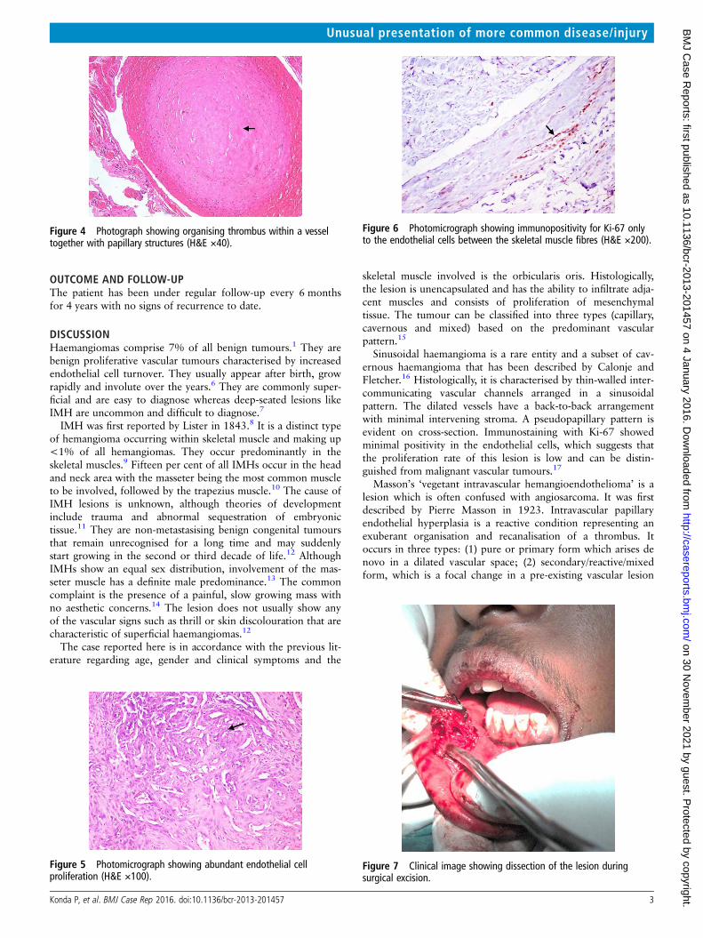
Histopathological examination of the formalin-fixed, pro-cessed and H&E-stained tissue sections revealed connectivetissue stroma containing numerous closely packed enlargedendothelial-lined blood vessels interspersed between the skeletalmuscle fibres (figure 3A, B). Vascular spaces were thin-walled,variable in size, dilated with a back-to-back arrangement in asinusoidal fashion (figure 3C). Few blood vessels were engorgedwith red blood cells. The endothelial cells were bland with noevidence of pleomorphism or mitosis (figure 3D). Intravascularorganising thrombi were evident. Papillary projections into thevascular lumen were present with evidence of Masson’s phe-nomenon (figure 4). Other areas showed abundant endothelialcell proliferations (figure 5). Few skeletal muscle fibres exhibitedareas of degeneration.
Immunohistochemical staining with Ki-67 showed positivityin the nuclei of the endothelial cells lining the blood vessels
between the skeletal muscle fibres (figure 6), indicating anincrease in their mitotic activity.
Based on the above histopathological features, a final diagno-sis of intramuscular sinusoidal haemangioma with secondaryMasson’s phenomenon was made.
INVESTIGATIONSA routine complete haemogram including clotting and bleedingtime was done prior to the surgery. Diagnosis preoperatively isnot straightforward and this condition is mostly recognised onlyduring surgical exploration or on histopathological examination.
DIFFERENTIAL DIAGNOSISIMH can be differentiated from vascular malformations by com-plete or incomplete lining around the endothelial cells bysmooth muscle cells and fibres forming a complete or partiallymatured wall which is lacking in haemangiomas.
Angiosarcomas can be identified by necrosis, haemorrhage,mitotic figures, nuclear and cellular pleomorphism, all of whichare lacking in haemangiomas. Intramuscular lipomas, myxomasand granular cell tumours can be identified by the presence ofadipose tissue, myxomatous tissue and granular cell elements inthe tissue.
TREATMENTA planned incision was made under local anaesthesia. Thelesion ruptured while the dissection was being done. Separationof the lesion along with the muscle bands was carried out. Thebleeding was arrested with electrocautery and the surgical sitewas closed with silk sutures (figure 7).
Figure 2 Macroscopic examination of the excised lesion showing anirregular surface of firm consistency and brown in colour.
Figure 3 (A) Photomicrograph showing endothelial-lined capillaries engorged with red blood cells between the skeletal muscle bundles (H&E×100). (B) Photomicrograph showing plump endothelial-lined capillaries between the skeletal muscle bundles (H&E ×200). (C) Photomicrographshowing thin-walled dilated vascular spaces with a back-to-back arrangement in a sinusoidal fashion along with a large papillary projection (H&E×100). (D) Photomicrograph showing bland endothelial cells lining a vascular space with no evidence of pleomorphism (H&E ×100).
2 Konda P, et al. BMJ Case Rep 2016. doi:10.1136/bcr-2013-201457
Unusual presentation of more common disease/injury on 30 N
ovember 2021 by guest. P
rotected by copyright.http://casereports.bm
j.com/
BM
J Case R
eports: first published as 10.1136/bcr-2013-201457 on 4 January 2016. Dow
nloaded from
OUTCOME AND FOLLOW-UPThe patient has been under regular follow-up every 6 monthsfor 4 years with no signs of recurrence to date.
DISCUSSIONHaemangiomas comprise 7% of all benign tumours.1 They arebenign proliferative vascular tumours characterised by increasedendothelial cell turnover. They usually appear after birth, growrapidly and involute over the years.6 They are commonly super-ficial and are easy to diagnose whereas deep-seated lesions likeIMH are uncommon and difficult to diagnose.7
IMH was first reported by Lister in 1843.8 It is a distinct typeof hemangioma occurring within skeletal muscle and making up<1% of all hemangiomas. They occur predominantly in theskeletal muscles.9 Fifteen per cent of all IMHs occur in the headand neck area with the masseter being the most common muscleto be involved, followed by the trapezius muscle.10 The cause ofIMH lesions is unknown, although theories of developmentinclude trauma and abnormal sequestration of embryonictissue.11 They are non-metastasising benign congenital tumoursthat remain unrecognised for a long time and may suddenlystart growing in the second or third decade of life.12 AlthoughIMHs show an equal sex distribution, involvement of the mas-seter muscle has a definite male predominance.13 The commoncomplaint is the presence of a painful, slow growing mass withno aesthetic concerns.14 The lesion does not usually show anyof the vascular signs such as thrill or skin discolouration that arecharacteristic of superficial haemangiomas.12
The case reported here is in accordance with the previous lit-erature regarding age, gender and clinical symptoms and the
skeletal muscle involved is the orbicularis oris. Histologically,the lesion is unencapsulated and has the ability to infiltrate adja-cent muscles and consists of proliferation of mesenchymaltissue. The tumour can be classified into three types (capillary,cavernous and mixed) based on the predominant vascularpattern.15
Sinusoidal haemangioma is a rare entity and a subset of cav-ernous haemangioma that has been described by Calonje andFletcher.16 Histologically, it is characterised by thin-walled inter-communicating vascular channels arranged in a sinusoidalpattern. The dilated vessels have a back-to-back arrangementwith minimal intervening stroma. A pseudopapillary pattern isevident on cross-section. Immunostaining with Ki-67 showedminimal positivity in the endothelial cells, which suggests thatthe proliferation rate of this lesion is low and can be distin-guished from malignant vascular tumours.17
Masson’s ‘vegetant intravascular hemangioendothelioma’ is alesion which is often confused with angiosarcoma. It was firstdescribed by Pierre Masson in 1923. Intravascular papillaryendothelial hyperplasia is a reactive condition representing anexuberant organisation and recanalisation of a thrombus. Itoccurs in three types: (1) pure or primary form which arises denovo in a dilated vascular space; (2) secondary/reactive/mixedform, which is a focal change in a pre-existing vascular lesion
Figure 4 Photograph showing organising thrombus within a vesseltogether with papillary structures (H&E ×40).
Figure 5 Photomicrograph showing abundant endothelial cellproliferation (H&E ×100).
Figure 6 Photomicrograph showing immunopositivity for Ki-67 onlyto the endothelial cells between the skeletal muscle fibres (H&E ×200).
Figure 7 Clinical image showing dissection of the lesion duringsurgical excision.
Konda P, et al. BMJ Case Rep 2016. doi:10.1136/bcr-2013-201457 3
Unusual presentation of more common disease/injury on 30 N
ovember 2021 by guest. P
rotected by copyright.http://casereports.bm
j.com/
BM
J Case R
eports: first published as 10.1136/bcr-2013-201457 on 4 January 2016. Dow
nloaded from
(haemangioma, vascular malformation, pyogenic granuloma);and (3) extravascular rare form, which is due to organisation ofhaematoma.18
The histogenesis of this lesion is obscure, so it represents alesion of reactive nature rather than a true neoplasm.19
It consists of a mass of anastomosing vascular channels with avariable degree of intraluminal papillary projections. The stromaconsists of hyalinised eosinophilic non-collagenous materialwhich is non-refractile on polarisation.5
Our case is a secondary/reactive form which formed in a pre-existing haemangioma as a focal change. There are no reportedcases of malignant transformation or metastasis of IMH. Therecurrence rate, which is about 50%, is not related to the histo-logical variant or anatomical location of the lesion.11
Our patient had an intramuscular variant of haemangiomainvolving the orbicularis oris which exhibited a sinusoidal patternon histological examination. The lesion showed thrombus forma-tion, which was resolving, with secondary Masson’s phenomenon.
Learning points
▸ Intramuscular haemangioma may start in childhood andshould be considered in the differential diagnosis of anyisolated muscle enlargement. The treatment of choice shouldbe individualised according to the patient’s age, symptoms,cosmetic, functional or neurological deficits, depth ofinvasion and vascular structure of the tumour.
▸ Sinusoidal haemangiomas are rare and generally requirea multidisciplinary approach. Wide excision of the tumouris surgically recommended to prevent relapse andrecurrence.
▸ Masson’s phenomenon is clinically important as it presents as amass which may be histologically mistaken for angiosarcoma.Thus, the pathologist should be aware of this lesion to avoid anincorrect diagnosis and aggressive treatment.
Contributors All the authors contributed equally to the manuscript.
Competing interests None declared.
Patient consent Obtained.
Provenance and peer review Not commissioned; externally peer reviewed.
REFERENCES1 Weiss SW, Goldbum JR. Benign tumors and tumor-like lesions of blood vessels. In:
Enzinger and Weiss’s Soft Tissue Tumors. 5th edn. St Louis: Mosby, 2008:636.2 Bucci T, De Glullo F, Insabato L, et al. Cavernous hemangioma of the temporalis
muscle: case report and review of literature. Acta Otorhinolaryngol 2008;28:83–6.3 Nurliza I, Kenali MS, Sani A. Intramuscular haemangioma in the head and neck.
Med J Malaysia 2007;62:409–10.4 Song BH, Youn SH, Park EJ, et al. A case of sinusoidal hemangioma with lipoma.
Ann Dermatol 2011;23:s250–3.5 David EE, Rosalie E, Bernett LJ Jr, et al. Lever’s histopathology of the skin. 9th edn.
Lippincott Williams and Williams, 2005:1015.6 Gamper TJ, Morgan RF. Vascular anomalies: hemangiomas. Plast Reconstr Surg
2002;110:572–85.7 Engelstad BL, Gilula LA, Kyriaskos M. Ossifed skeletal muscle hemangioma:
radiologic and pathologic features. Skeletal Radiol 1980;5:35–40.8 Liston R. Case of erectile tumour in the popliteal space. Med Chir Trans
1843;26:120–32.9 Watson WL, McCarty WD. Blood and lymph vessel tumors: a report of 1056 cases.
Surg Gynecol Obstet 1940;71:571–88.10 Clemis JD, Briggs DR, Changus GW. Intramuscular hemangiomas in the head and
neck. Can J Otolaryngol 1975;4:339–46.11 Gregory KI, Gerald JB, Allen LS. Intramuscular hemangioma of the mentalis muscle.
Oral Surg Oral Med Oral Pathol 1985;60:476–81.12 Wolf GT, Daniel F, Krause CJ, et al. Intramuscular hemangiomas of the head and
neck. Laryngoscope 1985;95:210–3.13 Hoehn JG, Farrow GM, Devine KD. Invasive hemangiomas of the head and neck.
Am J Surg 1970;120:495–8.14 Odabasi AO, Metin KK, Mutlu C, et al. Intramuscular hemangioma of the masseter
muscle. Eur Arch Otorhinolaryngol 1999;256:366–9.15 Allen PW, Enzinger FM. Hemangioma of skeletal muscle: an analysis of 89 cases.
Cancer 1972:29;8–22.16 Calonje E, Fletcher CD. Sinusoidal hemangioma. A distinctive benign vascular
neoplasm within the group cavernous hemangiomas. Am J Surg Pathol1991;15:1130–5.
17 Kim EH, Kim YC, Lee SW. A case of polypoid hemangioma. Korean J Dermatol2006;44:1016–17.
18 Pablo MD, Salil S, Meena C, et al. Masson’s tumor: differential diagnosis of necklump in children. Int J Pediatr Otolaryngol Extra 2006;1:196–9.
19 Kuo TT, Sayers CP, Rosai Juan. Masson’s “vegetant intravascularhemangioendothelioma”. A lesion often mistaken for angiosarcoma. Cancer1976;38:1227–36.
Copyright 2015 BMJ Publishing Group. All rights reserved. For permission to reuse any of this content visithttp://group.bmj.com/group/rights-licensing/permissions.BMJ Case Report Fellows may re-use this article for personal use and teaching without any further permission.
Become a Fellow of BMJ Case Reports today and you can:▸ Submit as many cases as you like▸ Enjoy fast sympathetic peer review and rapid publication of accepted articles▸ Access all the published articles▸ Re-use any of the published material for personal use and teaching without further permission
For information on Institutional Fellowships contact [email protected]
Visit casereports.bmj.com for more articles like this and to become a Fellow
4 Konda P, et al. BMJ Case Rep 2016. doi:10.1136/bcr-2013-201457
Unusual presentation of more common disease/injury on 30 N
ovember 2021 by guest. P
rotected by copyright.http://casereports.bm
j.com/
BM
J Case R
eports: first published as 10.1136/bcr-2013-201457 on 4 January 2016. Dow
nloaded from