Case of Idiopathic Hypereosinophilic Syndrome with Articular … · though she had anorexia. On...
5
207 Received:January 29, 2018, Revised:March 5, 2018, Accepted:March 5, 2018 Corresponding to:Yun Jong Lee http://orcid.org/0000-0001-7615-8611 Department of Internal Medicine, Seoul National University Bundang Hospital, 82 Gumi-ro 173beon-gil, Bundang-gu, Seongnam 13620, Korea. E-mail:[email protected] Copyright ⓒ 2018 by The Korean College of Rheumatology. All rights reserved. This is a Open Access article, which permits unrestricted non-commerical use, distribution, and reproduction in any medium, provided the original work is properly cited. Case Report pISSN: 2093-940X, eISSN: 2233-4718 Journal of Rheumatic Diseases Vol. 25, No. 3, July, 2018 https://doi.org/10.4078/jrd.2018.25.3.207 Case of Idiopathic Hypereosinophilic Syndrome with Articular Involvement Ji Hyoun Kim 1 , You-Jung Ha 1 , Eun Ha Kang 1 , Yeong Wook Song 2,3 , Yun Jong Lee 1,4 1 Department of Internal Medicine, Seoul National University Bundang Hospital, Seongnam, 2 Department of Internal Medicine, Seoul National University Hospital, 3 WCU Department of Molecular Medicine and Biopharmaceutical Sciences, Medical Research Institute, Seoul National University College of Medicine, 4 Department of Internal Medicine, Seoul National University College of Medicine, Seoul, Korea Idiopathic hypereosinophilic syndrome (IHES) is a rare disease that is characterized by otherwise unexplained persistent eosi- nophilia and organ damage caused by eosinophilic infiltration. Its manifestations are highly variable but clinically apparent ar- thritis is uncommonly observed. Although Korean cases of severe eosinophilia in patients with rheumatoid arthritis (RA) or IHES concurrent with RA have been published, there are no reports of IHES with joint involvement. This paper reports a case of IHES presenting with persistent peripheral eosinophilia, fever, skin rash, multiple lymphadenopathy, and polyarthritis, including the distal interphalangeal joints of the hands. (J Rheum Dis 2018;25:207-211) Key Words. Hypereosinophilic syndrome, Arthritis, Finger joint, Cyclosporine INTRODUCTION Hypereosinophilic syndrome (HES) is a rare disorder characterized by peripheral eosinophilia (>1,500/mm 3 on at least two occasions or for ≥6 months) and end-or- gan damage secondary to the eosinophilia [1]. The clin- ical features of HES are dependent on the organ system involved with tissue eosinophilia and HES commonly af- fects hematologic, cutaneous, cardiovascular, pulmonary, and neurologic systems. Concerning musculoskeletal in- volvement, myalgia and arthralgia can occur with HES [1,2]. Moreover, idiopathic HES (IHES) cases with joint involvement have been rarely described in English-writ- ten literature [3-6]. However, similar clinical features have been also reported when HES developed in patients with long-standing or concurrent rheumatoid arthritis (RA) [7-9]. Although Korean cases of severe eosinophilia in patients with RA or IHES concurrent with RA have been pub- lished [8-10], IHES with joint involvement has not been reported yet. Herein, we report a case of IHES with poly- arthritis involving the distal interphalangeal (DIP) joints of the hands. The study was approved by the Institutional Review Board of the Seoul National University Bundang Hospital (IRB no. B-1801-447-701). CASE REPORT A 48-year old woman was admitted to Seoul National University Bundang Hospital for evaluation of recurrent febrile episodes, pruritic exfoliative skin rash, and polyarthralgia. She had been healthy until 9 months ago when she was diagnosed with adhesive capsulitis in both shoulders. After 1 month, she developed pain and swel- ling in both 2nd and the left 4th hand DIP joints and noted pruritic erythematous lesions on the trunk. Laboratory examinations at another hospital showed an increased eosinophil counts (843/mm 3 ) and raised erythrocyte sed- imentation rate (ESR, 98 mm/hr). Triamcinolone in- tra-articular injections relieved her symptoms for 1
-
Upload
truongquynh -
Category
Documents
-
view
215 -
download
0
Transcript of Case of Idiopathic Hypereosinophilic Syndrome with Articular … · though she had anorexia. On...

207
Received:January 29, 2018, Revised:March 5, 2018, Accepted:March 5, 2018
Corresponding to:Yun Jong Lee http://orcid.org/0000-0001-7615-8611Department of Internal Medicine, Seoul National University Bundang Hospital, 82 Gumi-ro 173beon-gil, Bundang-gu, Seongnam 13620, Korea. E-mail:[email protected]
Copyright ⓒ 2018 by The Korean College of Rheumatology. All rights reserved.This is a Open Access article, which permits unrestricted non-commerical use, distribution, and reproduction in any medium, provided the original work is properly cited.
Case ReportpISSN: 2093-940X, eISSN: 2233-4718Journal of Rheumatic Diseases Vol. 25, No. 3, July, 2018https://doi.org/10.4078/jrd.2018.25.3.207
Case of Idiopathic Hypereosinophilic Syndrome with Articular Involvement
Ji Hyoun Kim1, You-Jung Ha1, Eun Ha Kang1, Yeong Wook Song2,3, Yun Jong Lee1,4
1Department of Internal Medicine, Seoul National University Bundang Hospital, Seongnam, 2Department of Internal Medicine, Seoul National University Hospital, 3WCU Department of Molecular Medicine and Biopharmaceutical Sciences, Medical Research Institute, Seoul National University College of Medicine, 4Department of Internal Medicine, Seoul National University College of Medicine, Seoul, Korea
Idiopathic hypereosinophilic syndrome (IHES) is a rare disease that is characterized by otherwise unexplained persistent eosi-nophilia and organ damage caused by eosinophilic infiltration. Its manifestations are highly variable but clinically apparent ar-thritis is uncommonly observed. Although Korean cases of severe eosinophilia in patients with rheumatoid arthritis (RA) or IHES concurrent with RA have been published, there are no reports of IHES with joint involvement. This paper reports a case of IHES presenting with persistent peripheral eosinophilia, fever, skin rash, multiple lymphadenopathy, and polyarthritis, including the distal interphalangeal joints of the hands. (J Rheum Dis 2018;25:207-211)
Key Words. Hypereosinophilic syndrome, Arthritis, Finger joint, Cyclosporine
INTRODUCTION
Hypereosinophilic syndrome (HES) is a rare disorder characterized by peripheral eosinophilia (>1,500/mm3 on at least two occasions or for ≥6 months) and end-or-gan damage secondary to the eosinophilia [1]. The clin-ical features of HES are dependent on the organ system involved with tissue eosinophilia and HES commonly af-fects hematologic, cutaneous, cardiovascular, pulmonary, and neurologic systems. Concerning musculoskeletal in-volvement, myalgia and arthralgia can occur with HES [1,2]. Moreover, idiopathic HES (IHES) cases with joint involvement have been rarely described in English-writ-ten literature [3-6]. However, similar clinical features have been also reported when HES developed in patients with long-standing or concurrent rheumatoid arthritis (RA) [7-9].Although Korean cases of severe eosinophilia in patients
with RA or IHES concurrent with RA have been pub-lished [8-10], IHES with joint involvement has not been
reported yet. Herein, we report a case of IHES with poly-arthritis involving the distal interphalangeal (DIP) joints of the hands. The study was approved by the Institutional Review Board of the Seoul National University Bundang Hospital (IRB no. B-1801-447-701).
CASE REPORT
A 48-year old woman was admitted to Seoul National University Bundang Hospital for evaluation of recurrent febrile episodes, pruritic exfoliative skin rash, and polyarthralgia. She had been healthy until 9 months ago when she was diagnosed with adhesive capsulitis in both shoulders. After 1 month, she developed pain and swel-ling in both 2nd and the left 4th hand DIP joints and noted pruritic erythematous lesions on the trunk. Laboratory examinations at another hospital showed an increased eosinophil counts (843/mm3) and raised erythrocyte sed-imentation rate (ESR, 98 mm/hr). Triamcinolone in-tra-articular injections relieved her symptoms for 1
Ji Hyoun Kim et al.
208 J Rheum Dis Vol. 25, No. 3, July, 2018
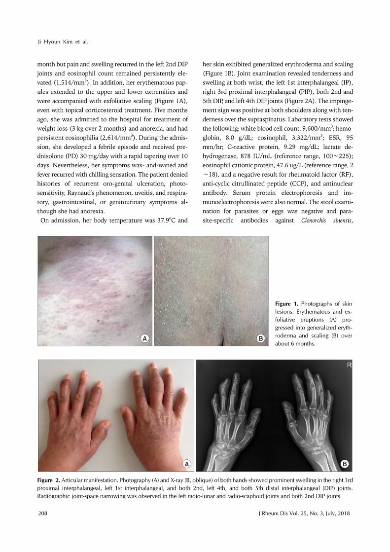
Figure 1. Photographs of skin lesions. Erythematous and ex-foliative eruptions (A) pro-gressed into generalized eryth-roderma and scaling (B) over about 6 months.
Figure 2. Articular manifestation. Photography (A) and X-ray (B, oblique) of both hands showed prominent swelling in the right 3rdproximal interphalangeal, left 1st interphalangeal, and both 2nd, left 4th, and both 5th distal interphalangeal (DIP) joints. Radiographic joint-space narrowing was observed in the left radio-lunar and radio-scaphoid joints and both 2nd DIP joints.
month but pain and swelling recurred in the left 2nd DIP joints and eosinophil count remained persistently ele-vated (1,514/mm3). In addition, her erythematous pap-ules extended to the upper and lower extremities and were accompanied with exfoliative scaling (Figure 1A), even with topical corticosteroid treatment. Five months ago, she was admitted to the hospital for treatment of weight loss (3 kg over 2 months) and anorexia, and had persistent eosinophilia (2,614/mm3). During the admis-sion, she developed a febrile episode and received pre-dnisolone (PD) 30 mg/day with a rapid tapering over 10 days. Nevertheless, her symptoms wax- and-waned and fever recurred with chilling sensation. The patient denied histories of recurrent oro-genital ulceration, photo-sensitivity, Raynaud's phenomenon, uveitis, and respira-tory, gastrointestinal, or genitourinary symptoms al-though she had anorexia.On admission, her body temperature was 37.9oC and
her skin exhibited generalized erythroderma and scaling (Figure 1B). Joint examination revealed tenderness and swelling at both wrist, the left 1st interphalangeal (IP), right 3rd proximal interphalangeal (PIP), both 2nd and 5th DIP, and left 4th DIP joints (Figure 2A). The impinge-ment sign was positive at both shoulders along with ten-derness over the supraspinatus. Laboratory tests showed the following: white blood cell count, 9,600/mm3; hemo-globin, 8.0 g/dL; eosinophil, 3,322/mm3; ESR, 95 mm/hr; C-reactive protein, 9.29 mg/dL; lactate de-hydrogenase, 878 IU/mL (reference range, 100∼225); eosinophil cationic protein, 47.6 ug/L (reference range, 2∼18), and a negative result for rheumatoid factor (RF), anti-cyclic citrullinated peptide (CCP), and antinuclear antibody. Serum protein electrophoresis and im-munoelectrophoresis were also normal. The stool exami-nation for parasites or eggs was negative and para-site-specific antibodies against Clonorchis sinensis,

Idiopathic Hypereosinophilic Syndrome with Arthritis
www.jrd.or.kr 209
Figure 3. Synovial biopsy showing edema, synovial cell hy-perplasia, and infiltration of lymphoplasma cells and eosino-phils (H&E, ×400).
Paragonimus westermani, Cysticercus, and Sparganum were all negative. Computed tomography (CT) showed spleno-megaly and multiple lymphadenopathies in the neck, chest and abdominal cavity. The enlarged lymph nodes were hypermetabolic in the positron emission tomog-raphy/CT. Joint X-ray showed soft tissue swelling in PIP and DIP joints and joint space narrowing was observed in both 2nd DIP and the left wrist joints (Figure 2B). Skin bi-opsy showed a spongiotic dermatitis with perivascular lymphocytic infiltration with eosinophils. The inguinal lymph node and bone marrow biopsy revealed eosino-philic infiltration. There was no evidence of major gene deletion or rearrangement for lymphoproliferative dis-orders including fibroblast growth factor receptor-1 and platelet-derived growth factor receptor beta gene re-arrangement. Synovial tissue was obtained from the right 3rd PIP joint and histopathology examination also showed chronic active inflammation with eosinophilic in-filtration (Figure 3).She was finally diagnosed with IHES involving skin,
lymph node, bone marrow and peripheral joints including the hand DIP joints. Following the initiation of PD 20 mg/day (about 0.5 mg/kg/day) and cyclosporine 100 mg/day, her fever and arthralgia disappeared rapidly over 1 to 2 days, and the blood eosinophil count was normal-ized over 5 days. The patient was followed up with gradu-al tapering of the PD to 10 mg every other day. The re-currence of symptom or the development of radiographic progression was not observed for 6 months.
DISCUSSION
The current case was diagnosed as having IHES based on persistent eosinophilia (>1,500/mm3 for >6 months) without an identifiable etiology and eosinophilic infiltra-tion in the target organs [1]. Clinical manifestations of HES depend on the organ system involved, and the skin, heart, lungs, and central or peripheral nervous system are involved in >50% of patients. Other clinical features in-clude fever, hepatosplenomegaly, gastroenteritis, and co-agulation disorder [1,2]. Therefore, fever, chronic pruritic skin rash with eosinophilic infiltration, and multiple eosi-nophilic lymphadenopathy in this case were considered attributed to the IHES. In patients presenting with arthritis/arthralgia and ele-
vated eosinophil counts, the differential diagnosis in-cludes a variety of conditions including RA with eosino-philia, eosinophilic granulomatosis with polyangiitis, and eosinophilic arthritis [11]. Eosinophilia accompanying RA is sometimes found [10] and a recent study reported that eosinophilia in early RA could be a marker of higher disease activity and poor therapeutic response [12]. Another possibility would include RA concurrent with HES. Choi et al. [8] reported a case of seronegative RA concomitant with IHES and Park et al. [9] described a case of IHES preceding seropositive RA. However, joint mani-festation in our case was not compatible with that of typi-cal RA because of the negative RF and anti-CCP, eosino-philic synovial infiltration, and multiple DIP joint involvement. Additionally, our patient had no patho-logical evidence of vasculitis and her clinical features were not compatible with eosinophilic arthritis. Eosinophilic arthritis shows a lack of systemic symptoms and normal laboratory findings except for eosinophilia and many patients with eosinophilic arthritis respond to antiparasitic drugs [11].Even though joint involvement is not common in pa-
tients with HES, arthritis/arthralgia has been described in previous literatures [1,2,4]. Through a search of PubMed and Google Scholar, we identified 3 English- written reports describing the joint involvement of HES patients in detail [3,5,6]. It took 2∼8 years before estab-lishing the diagnosis of IHES. All the cases showed a bi-lateral involvement of the wrist or hand joints and a pre-dominant involvement of the upper extremities, despite negative RF. Interestingly, subcutaneous nodules over the joints were observed in 3 cases, and on pathological ex-
Ji Hyoun Kim et al.
210 J Rheum Dis Vol. 25, No. 3, July, 2018
amination, the nodules showed eosinophilic infiltration. Joint X-rays exhibited no erosion or bony abnormality in 2 cases but one had destructive lesions in the meta-carpophalngeal joint and 1st IP joints [6]. Therefore, some of their clinical features overlapped with those of RA. In this context, the case of reported by Choi et al. [8] might have articular involvement contributed by IHES not by seronegative RA; her synovial pathology revealed eosinophilic infiltration that is not a feature of RA synovitis. Our patients had a unique involvement of the hand DIP joint, but other articular manifestations were similar to those described in previous reports. Based on this, we finally concluded that her polyarthritis was sec-ondary to IHES. Generally, Th2 cytokines are considered anti-inflam-
matory in nature, and an eosinophil response could sup-press inflammatory arthritis in experimental arthritis models [13]. It has often been reported that articular manifestations in HES include arthralgia or non-erosive arthritis [1,2,4]. However, cases by Anders and Schatten-kirchner [6] as well as ours cases revealed radiographic joint damage. Our patients showed mild joint-space nar-rowing, but the case of Anders and Schattenkirchner [6] revealed more destructive change; it might be a result of the duration of arthritis (8 months versus 2 years). Additionally, eosinophils can produce a variety of proin-flammatory mediators including non-canonical factors of osteoclast differentiation and matrix-degrading metal-loproteinases [14]. Therefore, although joint damage is not a main feature in patients with HES, it could induce joint inflammation and tissue destruction.Concerning the treatment of HES, corticosteroids have
been one of the most widely utilized agents and second- line agents include hydroxyurea, interferon-α, tyrosine kinase inhibitors, and monoclonal antibodies to inter-leukin-5 [1]. The response to corticosteroids is known to be dramatic and rapid in most patients but about 75% re-quire subsequent chronic maintenance therapy with corticosteroids. Because vital organs were not affected in the current case, we started a moderate dose of PD (0.5 mg/kg/day) and added cyclosporine as a steroid-sparing agent. Because eosinophilic granulopoiesis is thought to be under the control of T lymphocytes in HES, cyclo-sporine could be a reasonable therapeutic option. In addi-tion, cyclosporine is the most commonly used anti-rheu-matic drug for the treatment of IHES [15] and was re-ported to be successfully used in a patient with IHES and RA [7].
SUMMARY
This is the first Korean case of IHES affecting the hand and wrist joints including the DIP joints. Our literature review shows that HES uncommonly affects the joints and can result in significant joint tissue damage in some patients. Because its articular manifestations could re-semble those of RA, a synovial biopsy may be useful for a differential diagnosis between RA and HES-associated arthritis.
CONFLICT OF INTEREST
No potential conflict of interest relevant to this article was reported.
REFERENCES
1. Curtis C, Ogbogu P. Hypereosinophilic Syndrome. Clin Rev Allergy Immunol 2016;50:240-51.
2. Weller PF, Bubley GJ. The idiopathic hypereosinophilic syndrome. Blood 1994;83:2759-79.
3. Brogadir SP, Goldwein MI, Schumacher HR. A hyper-eosinophilic syndrome mimicking rheumatoid arthritis. Am J Med 1980;69:799-802.
4. Spry CJ, Davies J, Tai PC, Olsen EG, Oakley CM, Goodwin JF. Clinical features of fifteen patients with the hyper-eosinophilic syndrome. Q J Med 1983;52:1-22.
5. Martín-Santos JM, Mulero J, Andréu JL, de Villa LF, Bernaldo-de Quirós L, Noguera E. Arthritis in idiopathic hy-pereosinophilic syndrome. Arthritis Rheum 1988;31: 120-5.
6. Anders HJ, Schattenkirchner M. Destructive joint lesions and bursitis in idiopathic hypereosinophilic syndrome. Rheumatology (Oxford) 1999;38:185-6.
7. Chaudhuri K, Dubey S, Zaphiropoulos G. Idiopathic hyper-eosinophilic syndrome in a patient with long-standing rheu-matoid arthritis: a case report. Rheumatology (Oxford) 2002;41:349-50.
8. Choi JH, Jung JW, Song HJ, Song KE, Choi JH, Suh YJ, et al. A case of seronegative rheumatoid arthritis with idiopathic hypereosinophilic syndrome. J Korean Rheum Assoc 2003; 10:200-5.
9. Park JH, Lee WS, Park SJ, Yoo WH. Hypereosinophilic syn-drome associated with the onset of rheumatoid arthritis: a case report. J Rheum Dis 2017;24:165-8.
10. Sohn CI, Kim MK, Lee KC, Jung SS, Lee IH, Bae SC, et al. A case of rheumatoid arthritis accompanied by severe eosinophilia. J Korean Rheum Assoc 1994;1:98-102.
11. Tay C. Eosinophilic arthritis. Rheumatology (Oxford) 1999;38:1188-94.
12. Guellec D, Milin M, Cornec D, Tobon GJ, Marhadour T, Jousse-Joulin S, et al. Eosinophilia predicts poor clinical outcomes in recent-onset arthritis: results from the ESPOIR cohort. RMD Open 2015;1:e000070.

Idiopathic Hypereosinophilic Syndrome with Arthritis
www.jrd.or.kr 211
13. Chen Z, Andreev D, Oeser K, Krljanac B, Hueber A, Kleyer A, et al. Th2 and eosinophil responses suppress inflam-matory arthritis. Nat Commun 2016;7:11596.
14. Kita H. Eosinophils: multifaceted biological properties and roles in health and disease. Immunol Rev 2011;242:161-77.
15. Ogbogu PU, Bochner BS, Butterfield JH, Gleich GJ, Huss-Marp J, Kahn JE, et al. Hypereosinophilic syndrome: a multicenter, retrospective analysis of clinical characteristics and response to therapy. J Allergy Clin Immunol 2009; 124:1319-25.e3.