CASE 42 INGUINAL HERNIA - Elsevier...
12
64 Abdomen and trunk CASE 42 INSTRUCTION ‘Examine this gentleman’s groin.’ APPROACH Expose the patient from umbilicus to knees. VITAL POINTS TOP TIP 1 Should the hernia be examined with the patient lying down or standing up? The answer is that it doesn’t matter. It is generally considered to be easier to define the anatomy with the patient supine, and if the hernia can be detected with the patient on the couch, then examine them there. If no lump can be felt, or if no couch is available, then stand the patient up first. TOP TIP 2 Don’t forget that the same opening instruction may apply to the examination of the scrotum, and it may be a scrotal lump rather than a hernia and if you cannot see a visible swelling. Proceed by asking the patient where the lump is. TOP TIP 3 An inguinal hernia arises above and medial to the pubic tubercle. By contrast, a femoral hernia arises below and lateral to it (see Fig. 22). TOP TIP 4 Be completely conversant with the anatomy of the inguinal region and the location of surface anatomy (Fig. 22 and Fig. 23). • Midinguinal point = point halfway along a line joining the ASIS and the midline = location of femoral artery CASE 42 INGUINAL HERNIA *** Figure 22 Surface anatomy of groin examination for herniae. Inguinal hernia Femoral hernia Deep inguinal ring Inferior epigastric artery Midinguinal point Midpoint of the inguinal ligament Midline
Transcript of CASE 42 INGUINAL HERNIA - Elsevier...

64 Abdomen and trunk CASE 42
INSTRUCTION
‘ Examine this gentleman ’ s groin. ’
APPROACH
Expose the patient from umbilicus to knees.
VITAL POINTS
TOP TIP 1
Should the hernia be examined with the patient lying down or standing up? The answer is that it doesn ’ t matter. It is generally considered to be easier to defi ne the anatomy with the patient supine, and if the hernia can be detected with the patient on the couch, then examine them there. If no lump can be felt, or if no couch is available, then stand the patient up fi rst.
TOP TIP 2
Don ’ t forget that the same opening instruction may apply to the examination of the scrotum, and it may be a scrotal lump rather than a hernia and if you cannot see a visible swelling. Proceed by asking the patient where the lump is.
TOP TIP 3
An inguinal hernia arises above and medial to the pubic tubercle. By contrast, a femoral hernia arises below and lateral to it (see Fig. 22 ) .
TOP TIP 4
Be completely conversant with the anatomy of the inguinal region and the location of surface anatomy ( Fig. 22 and Fig. 23 ) .
• Midinguinal point = point halfway along a line joining the ASIS and the midline = location of femoral artery
CASE 42 INGUINAL HERNIA * * *
Figure 22 Surface anatomy of groin examination for herniae.
Inguinal
hernia
Femoral hernia
Deep
inguinal
ring
Inferior
epigastric
artery
Midinguinal
point
Midpoint of
the inguinal
ligamentMidline

CASE 42 Abdomen and trunk 65
• Midpoint of the inguinal ligament = halfway along inguinal ligament (i.e. between the pubic tubercle and ASIS) = deep inguinal ring.
Accordingly, the femoral pulse is located medial to the deep inguinal ring.
The objectives of the examination
are to:
1. Confi rm that the lump is a hernia 2. Differentiate an inguinal from a femoral hernia 3. If it is an inguinal hernia, establish whether it
is direct or indirect.
Inspect
• Look at the groin for old surgical scars (is the hernia recurrent?) – this requires very careful inspection to identify old, small scars that are frequently hidden in groin creases and/or by hair
• If the hernia is obvious, then begin to examine it
• If it cannot be seen then ask the patient to cough and observe for the appearance of a lump
• If it is still not obvious, ask: ‘ Have you noticed a lump in your groin? ’ and get the patient to demonstrate it
• Fulfi l objective (1) above and confi rm that the lump is a hernia by: • Asking the patient to cough, while
observing for an expansile cough impulse • Getting the patient to reduce the hernia if
possible. If the patient cannot achieve this, attempt to reduce the lump yourself
by applying gentle pressure in the direction of the inguinal canal. If you cannot reduce the lump it is either (1) not a hernia or (2) incarcerated (irreducible).
Palpate
• Begin by defi ning the anatomy (see Figs. 22 and 23 )
• Palpate the pubic tubercle and the anterior superior iliac spine. The former can reliably be located by palpating in the midline downwards from the umbilicus until the symphysis pubis is reached ( Fig. 24 ) . Then move laterally to identify the tubercle
• Place one fi nger on each to demonstrate that the inguinal ligament runs between the two ( Fig. 25 )
• Ask the patient to cough for the second time and demonstrate that the hernia arises above this line, medial to the pubic tubercle (see Fig. 22 ) . This fulfi ls objective (2) (see above) and confi rms that the hernia is inguinal and not femoral
• If it wasn ’ t obvious on inspection alone, demonstrate that the lump has an expansile cough impulse: place one hand over the lump and ask the patient to cough
• It is only possible to fulfi l objective (3) above, if the hernia is reducible. With the hernia fully reduced, try to control the hernia at the deep inguinal ring. Redefi ne the inguinal ligament again and place one fi nger 1 cm craniad to the midpoint of the inguinal ligament (halfway along the inguinal ligament) ( Fig. 26 ) . Ask the patient to cough for the third and fi nal time. If the hernia is controlled by your fi nger at the deep inguinal ring it is an INDIRECT inguinal hernia. If the hernia is not controlled
Figure 23 Anatomy of the inguinal ligament.
Midpoint of inguinal ligament
External iliac artery
Mid-inguinal point
Anterior superioriliac spine
Inguinal ligament
Pubic tubercle
Pubicsymphysis
Femoral artery

66 Abdomen and trunk CASE 42
Completion
Complete your examination by asking to examine the scrotum for incidental scrotal lumps and to examine the contralateral groin for herniae.
QUESTIONS
(a) What is the difference between a direct
and an indirect inguinal hernia?
Indirect inguinal herniae:
• Are the remnants of a patent processus vaginalis
• Arise from the abdominal cavity lateral to the inferior epigastric vessels at operation, passing obliquely through the deep inguinal ring and travelling through the inguinal canal with the spermatic cord
• May continue through the superfi cial inguinal ring into the scrotum
Direct inguinal herniae:
• Are the result of a weak posterior wall to the inguinal canal
• Arise medial to the inferior epigastric vessels at operation
• This weakness causes the abdominal contents to bulge through the wall into the inguinal canal but the hernia is not within the spermatic cord.
(b) What are the contents of the
spermatic cord?
and reappears, then it is a DIRECT inguinal hernia. Remember that the accuracy of clinical examination in distinguishing a direct from an indirect hernia is low (e.g. 56% of direct herniae were wrongly classifi ed by consultant surgeons as indirect on clinical examination in one such study, see Further Reading )
• Other aspects of the lump (as for any lump) may be defi ned at this stage, e.g. skin changes
• Decide whether the lump is confi ned to the inguinal region or descends into the scrotum.
Figure 24 Palpation of the symphysis pubis.
Figure 25 Identifi cation of the inguinal ligament using bony landmarks.
Figure 26 Attempting to control the reduced hernia at the deep ring.
Three arteries:
• Artery to vas deferens (from inferior vesicular artery)
• Testicular artery (from aorta) • Cremasteric artery (from inferior epigastric
artery) Three nerves:
• Ilioinguinal nerve (L1) on the front of the cord • Nerve to cremaster (from genitofemoral
nerve) • Autonomic nerves (sympathetic fi bres from
T10) Three other structures:
• Vas deferens • Pampiniform plexus of veins (drains right
testis into inferior vena cava and left testis into renal vein)
• Lymphatics (drain the testis to the para-aortic lymph nodes).

CASE 42 Abdomen and trunk 67
• Bruising – occurs in 30% • Pain – often very severe and patients should
be discharged with adequate analgesia; chronic groin pain persists in 5% of patients
• Haematoma – 10% • Infection – 1% • Ischaemic orchitis – 0.5% (caused by
thrombosis of the pampiniform plexus draining the testis) • Previous vasectomy is a predisposing
cause • Dissection beyond (more medial than) the
pubic tubercle is one operative risk and so this practice should be avoided
• Recurrence – should be < 0.5% • Normally due to inadequate ring and
posterior wall closure • Occasionally due to over-tight sutures.
FURTHER READING
Cameron AE : Accuracy of clinical diagnosis of direct and indirect inguinal hernia . Br J Surg 81 ( 2 ): 250 , 1994 .
Liem MS , van Vroonhoven TJ : Laparoscopic inguinal hernia repair . Br J Surg 83 ( 9 ): 1197 – 1204 , 1996 .
McGreevy JM : Groin hernia and surgical truth (editorial) . Am J Surg 176 ( 4 ): 301 – 304 , 1998 .
(c) What would you tell patients about their
recovery from inguinal hernia repair?
• Early mobilization is important • They should keep the area clean and wash
carefully, especially after the clips/sutures have been removed
• They are able to bathe immediately • They may need to be off work for 6 weeks if
their job involves heavy lifting • They should avoid prolonged coughing
(control chronic obstructive pulmonary disease preoperatively)
• They should take laxatives if they get constipated postoperatively.
ADVANCED QUESTIONS
(a) How would you perform a hernia repair?
Be prepared to discuss a method of hernia repair you have learned. The main points to remember are:
• Testicular damage should be mentioned as a specifi c risk-factor
• The operation can be performed under local or general anaesthetic and often as a day case
• The Royal College of Surgeons of England has recommended the Lichtenstein mesh repair and the Shouldice repair
• Laparoscopic repair is increasingly recognised as having a role in the repair of bilateral and/or recurrent herniae.
(b) What are the complications of inguinal
hernia repair?
Complications should be divided into immediate (fi rst 24 h), early (within the fi rst month) and late (later than the fi rst month) and further into general for any procedure and specifi c for this procedure.
Specifi c complications to mention:
• Urinary retention
Differential diagnosis of a lump in the groin
Use the acronym L-SHAPE
• L ymph node/ L ipoma of the cord
• S apheno-varix/ S kin lesions (sebaceous cyt/lipoma, etc.)
• H ernia – inguinal/femoral
• A neurysmal dilatation of the femoral artery
• P soas abscess/bursa
• E ctopic/undescended testis.

68 Abdomen and trunk CASE 43
of infusions/catheters/drains, etc. and then second, just prior to inspecting the abdomen itself, inspect more closely for scars, caput medusae, etc.
VITAL POINTS
Inspect for peripheral stigmata of
abdominal disease
Hands
• Take the patient ’ s right hand and look for signs of chronic liver disease (see Case 46 ) and infl ammatory bowel disease (see Case 50 )
• Look for digital clubbing (see Case 28 ) by inspecting the angle between the nail bed and fold ( Fig. 27 ) and by performing Schamroth ’ s test ( Fig. 28 ) ; a small diamond-shaped ‘ window ’ is normally apparent between the nail beds when the distal phalanges are opposed. If this window is obliterated, the test is positive and clubbing is present. Clubbing is confi rmed by the following: • Fluctuation and softening of the nail bed
(increased ballotability) • Loss of the normal < 165 ° angle between
the nailbed and the fold • Increased convexity of the nail fold • Thickening of the fi nger distally
(resembling a drumstick) • Shiny aspect and striation of the nail and
skin
INSTRUCTION
‘ Examine this patient ’ s abdominal system/abdomen. ’
APPROACH
TOP TIP 1
Should i begin with the hands?
Listen carefully to the examiner ’ s instructions. If you are told to examine the ABDOMINAL SYSTEM, then begin with the hands and conduct a complete examination of the entire patient. By contrast, if you are told to examine the ABDOMEN, begin with the abdominal examination itself (commence at ‘ Expose the patient ’ s abdomen ’ below); do not annoy your examiner by asking whether you should begin with the hands!
TOP TIP 2
Exposure of the patient.
The exposure of the abdomen is very important and opinions differ as to how much of the abdomen needs to be exposed (and at what point) during the clinical examination. Clearly, if there is an inguinal hernia or a scrotal lump, this will be missed if the external genitalia are not exposed. Equally, a patient who has an epigastric hernia should not have his or her dignity compromised.
You are not expected to say, ‘ I would expose the patient from nipple to knees ’ ; this is both inappropriate to say in front of a patient and misses the point – that the degree of exposure depends on the case and how much the examiner expects of you.
We would advocate beginning by positioning the patient fl at on the bed, but keeping the groin covered until later on, starting your examination looking for peripheral stigmata of gastrointestinal disease.
TOP TIP 3
Inspect and then inspect again.
Assuming that you have been instructed to undertake an examination of the entire abdominal system, you should inspect the patient TWICE : fi rst, before you begin noting the general condition of the patient, the presence
CASE 43 ABDOMINAL EXAMINATION – GENERAL
APPROACH * * *
Figure 27 Examining for digital clubbing by inspecting the angle between the nail bed and fold.

CASE 43 Abdomen and trunk 69
Mouth
• Look in the mouth (and smell) for oral manifestations of chronic gastrointestinal disease, e.g. hepatic foetor, pallor of the mucous membranes (see Case 72 for full description of mouth signs in abdominal disease).
Neck
• Palpate the neck for supraclavicular lymphadenopathy – Virchow ’ s node (also known as Troisier ’ s sign) is found in the supraclavicular fossa between the sternal and clavicular heads of the sternocleidomastoid muscle.
Trunk
• Inspect the rest of the arms and upper chest wall for spider naevi, which are in the distribution of drainage of the superior vena cava (see Case 41 )
• Briefl y inspect the back, by asking the patient to roll towards and away from you.
Expose the patient ’ s abdomen
• If it is a man, ask him to remove his shirt • If it is a woman, keep the bra on but expose
the chest otherwise • Expose down to the symphysis pubis,
keeping the genitalia covered.
Stand back from the couch
Stand at the end or to the side of the couch, and inspect the abdomen thoroughly, looking for:
• Distension • Scars (see Case 62 ) • Visible pulsation • Ask the patient to take a deep breath in and
hold it, seeing the transmitted pulsation of an abdominal aortic aneurysm
• Ask the patient to cough or lift their head off the bed, demonstrating any herniae – especially important if scars are present.
Palpate
Kneel down next to the patient ’ s right side and ask if they have any pain anywhere, before beginning to palpate the abdomen. Look at the patient ’ s face the whole time when attempting to elicit any tenderness:
• Gastrointestinal disease associated with digital clubbing includes infl ammatory bowel disease, malabsorption and cirrhosis, especially primary biliary cirrhosis
• Look for koilonychia (spoon-shaped nails in iron-defi ciency anaemia) and pallor of the palmar creases in anaemia
• Check for a liver fl ap with both of the patient ’ s hands (you can ask the patient to put his hands out in front of him and cock his wrists back as if he is stopping traffi c) ( Fig. 29 ) ; in practice, this is a late sign of hepatic encephalopathy and these patients will not be in examinations.
Eyes
• Look at the conjunctivae for pallor (anaemia can only be confi rmed following haemoglobin measurement) and at the sclerae for jaundice (see Case 14 ) .
Figure 28 Schamroth ’ s test to establish the presence of digital clubbing.
Figure 29 Examining for evidence of a liver fl ap by extending the outstretched wrists.

70 Abdomen and trunk CASE 43
Finish your examination here
Make sure to cover the patient back up and ensure they are comfortable.
Completion
Say that you would like to:
• Review the observation chart (temperature, blood pressure, pulse and respiratory rate)
• Examine the lower limbs for peripheral oedema
• Examine the external genitalia and groin (if not already done)
• Perform a digital rectal examination • Dipstick the urine.
TOP TIP 4
The term ‘ per rectal ’ is incorrect; ‘ digital rectal examination ’ is more appropriate.
Figure 30 Auscultation of the abdomen.
• Begin furthest away from you and palpate the nine regions of the abdomen with the four fi ngers of your right hand held together, fi rst lightly, testing particularly for tenderness
• When you arrive in the epigastric region, pause for pulsation , testing for an abdominal aortic aneurysm (see Case 115 )
• Continue with deep palpation in the same nine regions, feeling for any masses – often this is done using two hands interlocked above each other
• Palpate the liver (see Case 46 ) and spleen (see Case 49 ) in turn
• Attempt to ballot the kidneys (see Case 61 ).
Percuss
• If there is hepatosplenomegaly the spleen and liver should be percussed
• If there is any abdominal distension you should percuss for ascites (see Case 57 ).
Auscultate ( Fig. 30 )
• Over the liver for a bruit • In the left iliac fossa for bowel sounds • Over the abdominal aorta and iliac vessels
for bruits.
Continue
Continue if you have not found any abnormality by exposing the external genitalia and examining the scrotum (see Case 51 ) and inguinal canal for herniae (see Case 44 ). Palpate the femoral pulses and check for femoral herniae.
C. E. Troisier (1844 – 1919) . Professor of Pathology, Paris. Also described haemachromatosis (Troisier syndrome).
R. L. K. Virchow (1821 – 1902) . Professor of Pathology, Wurzberg and Berlin. Also described Virchow space (perivascular space of Virchow – Robin), Virchow cell (lepra cell) and the Virchow triad of thrombogenesis.
L. Schamroth (1924 – 1988) . Professor of Cardiology, Baragwanath Hospital, South Africa. A leading authority on electrocardiography and cardiac arrhythmias and originally demonstrated fi nger clubbing on himself!
CASE 44 Abdomen and trunk 71
• Examine the neck for Virchow ’ s node (see Case 43 ).
Inspect
• The abdomen may be distended with ascites • There may be distended veins around the
umbilicus if there is portal hypertension (caput medusae, see Case 128 ).
TOP TIP
When examining the eyes for jaundice or pallor, only look into one eye (the sign will be bilateral if present). It is easier to use your left thumb to lower the patient ’ s lower eyelid and ask them to look towards the ceiling in order to get the best opportunity to inspect the sclera and conjunctiva.
If you suspect a jaundiced sclera but you are unsure, continue to examine the patient ’ s soft palate with a pen torch. Bilirubin is avidly taken up by tissues that are rich in elastin, and therefore the soft palate is a sensitive indicator of the presence of jaundice.
Palpate
• Palpate the abdomen as previously (see Case 43 )
• Palpate carefully in the right upper quadrant identifying any tenderness or masses, remembering Courvoisier ’ s law: in the presence of jaundice if a mass (the gall bladder) is palpable in the right upper quadrant, the cause is unlikely to be due to gallstones.
Finish your examination here
Completion
Say that you would like to:
• Complete the abdominal examination (see Case 43 ) by checking the hernial orifi ces, examining the external genitalia, performing a digital rectal examination and examining the lower limbs for peripheral oedema
CASE 44 SURGICAL JAUNDICE * * *
INSTRUCTION 1
‘ Ask this patient some questions about her jaundice. ’
APPROACH
Direct your questions to fi nding out whether the cause of her jaundice is likely to be pre-hepatic, hepatic or post-hepatic. Remember that in the surgical short case, post-hepatic is the most likely cause and this is the easiest to diagnose from the history:
• Have you noticed any change in the colour of your urine?
• Have you noticed any change in the colour of your stools?
• Have you noticed yourself feeling itchy? If the patient has noticed pale stools and dark urine, then explore possible causes:
• Weight-loss, change in bowel habit, loss of appetite and back pain are associated with primary and secondary intra-abdominal malignancies
• Younger age, previous biliary colic or episodic right upper quadrant pains may indicate gallstones.
Continue by asking some questions differentiating the other types of jaundice, and possibly to identify any risk-factors for hepatic jaundice, such as foreign travel (hepatitis A), blood transfusion (hepatitis B and C), sore throat (Epstein – Barr virus), alcohol intake, and use of certain drugs such as the oral contraceptive pill and phenothiazines.
INSTRUCTION 2
‘ Examine this lady ’ s abdominal system. ’
APPROACH
Expose the patient as previously (Case 43) and begin by examining the hands.
VITAL POINTS
• Look for signs of chronic liver disease (see Case 46 ).
• Confi rm the presence of jaundice by looking at the sclera (see Top Tip)

72 Abdomen and trunk CASE 44
Table 14 Causes of jaundice and effects on liver function tests
Pre-hepatic Hepatic Post-hepatic
Major causes Haemolysis Hereditary, e.g.
Gilbert ’ s syndrome
Hepatitis Decompensated chronic liver disease Drugs
Gall stones Carcinoma head of pancreas Lymph nodes
Bili type Unconjugated Conjugated ConjugatedBili increase + + + + + / + + + + + + ALT + / + + + + / + + + + / + + ALP − / + + / + + + + / + + +
Bili, bilirubin; ALT, alanine aminotransferase (similar rises with aspartate aminotransferase, AST); ALP, alkaline phosphatase.
• Check the temperature to see whether the potential obstruction has been complicated by infection
• Dipstick the urine for raised levels of bilirubin.
QUESTIONS
(a) How can jaundice be classifi ed?
Jaundice is yellow discolouration of the skin and mucous membranes caused by the accumulation of bile pigments. The causes can be classifi ed ( Table 14 ) into:
• Pre-hepatic • Hepatic • Post-hepatic.
(b) What level does the serum bilirubin need
to rise to before jaundice can be detected on
clinical examination?
Normal bilirubin is < 17 mmol/L and it usually has to reach at least three times this before the sclera is discoloured (i.e. > 50 mmol/L). Very high levels of bilirubin are usually associated with hepatic jaundice.
(c) Assuming this lady has obstructive
jaundice, how should she be investigated?
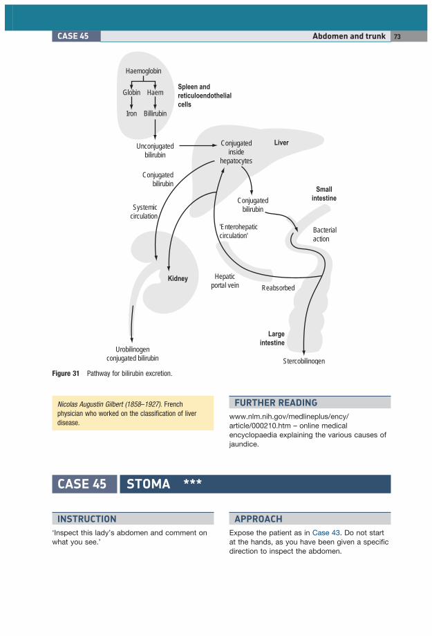
Urine should be tested for raised bilirubin ( Fig. 31 ) .
Blood tests:
• Full blood count: evidence of anaemia in GI malignancies or associated infection
• Renal function: any evidence of hepatorenal syndrome
• Liver function tests: see below • Clotting: functional assessment of hepatic
impairment. Radiological investigations:
• Ultrasound will show: • Presence of underlying liver disease • Degree of dilatation of the common bile
duct ( > 8 mm is abnormal) • Presence of gall stones • Presence of lymphadenopathy or a
pancreatic mass • CT scan • Endoscopic retrograde
cholangiopancreatography (ERCP) • Magnetic resonance
cholangiopancreatography (MRCP).
(d) How might the liver function tests help in
distinguishing the types of jaundice?
This is particularly important in OSCEs, where you may be asked to interpret liver function test results (see Table 14 ) .
ADVANCED QUESTIONS
(a) What are the causes of postoperative
jaundice?
• Pre-hepatic jaundice can occur due to haemolysis, especially following a transfusion
• Hepatic jaundice can result from the use of halogenated anaesthetics, sepsis or intra- or postoperative hypotension
• Post-hepatic jaundice can occur due to biliary injury (such as in laparoscopic cholecystectomy).
CASE 45 Abdomen and trunk 73
FURTHER READING
www.nlm.nih.gov/medlineplus/ency/article/000210.htm – online medical encyclopaedia explaining the various causes of jaundice .
Figure 31 Pathway for bilirubin excretion.
Haemoglobin
HaemGlobin
Iron Billirubin
Spleen and
reticuloendothelial
cells
Liver
Small
intestine
Large
intestine
Kidney
Conjugatedinside
hepatocytes
Conjugatedbilirubin
Conjugatedbilirubin
Unconjugatedbilirubin
Hepaticportal vein
'Enterohepaticcirculation'
Bacterialaction
Systemiccirculation
Stercobilinogen
Urobilinogenconjugated bilirubin
Reabsorbed
Nicolas Augustin Gilbert (1858 – 1927) . French physician who worked on the classifi cation of liver disease.
CASE 45 STOMA * * *
INSTRUCTION
‘ Inspect this lady ’ s abdomen and comment on what you see. ’
APPROACH
Expose the patient as in Case 43. Do not start at the hands, as you have been given a specifi c direction to inspect the abdomen.
74 Abdomen and trunk CASE 45
Finish your examination here
Completion
Say that you would like to:
• Continue to examine the rest of the abdomen to look for reasons why the stoma may have been formed in the fi rst place.
QUESTIONS
(a) What are the indications for forming a
stoma?
• Feeding, e.g. feeding gastrostomy/jejunostomy • Lavage, e.g. appendicostomy • Decompression – bypass of an obstructing
bowel lesion distal to the stoma • Diversion
1. Protection of a distal bowel anastomosis – Previously contaminated peritoneal
cavity – Anatomical considerations – ileorectal
anastomosis 2. Urinary diversion following cystectomy
• Exteriorization • Perforated or contaminated bowel, e.g.
distal abscess/fi stula • Permanent stoma, e.g. abdomino-perineal
resection of rectum.
(b) How would you prepare a patient who is
going for surgery which will involve forming a
stoma?
• Psychosocial and physical preparation • Explanation of indications and
complications • This includes seeing a Clinical Nurse
Specialist in Stoma Care preoperatively, who would normally mark the site
• Marking of the stoma site – with the patient standing up as he or she must be able to see the stoma • The stoma must be within the rectus
abdominus muscle • Away from scars or skin creases • Away from bony points or waistline of
clothes • At a site that is easily accessible to
the patient, e.g. not under a large fold of fat.
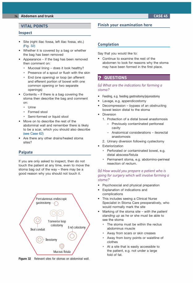
Figure 32 Relevant sites for stomas on abdominal wall.
Tranverse loopcolostomy
Ileal conduit
Ileostomy
Percutaneous endoscopicgastrostomy
End colostomy
Mucous fistula
VITAL POINTS
Inspect
• Site (right iliac fossa, left iliac fossa, etc.) ( Fig. 32 )
• Whether it is covered by a bag or whether the bag has been removed
• Appearance – if the bag has been removed then comment on: • Mucosal lining – does it look healthy? • Presence of a spout or fl ush with the skin • End (one opening) or loop (an afferent
and efferent portion of bowel with one common opening or two separate openings)
• Contents – if there is a bag covering the stoma then describe the bag and comment on: • Urine • Formed stool • Semi-formed or liquid stool
• Move on to describe the rest of the abdominal wall and remember there is likely to be a scar, which you should also describe (see Case 62 )
• Are there any other drains/healed stoma sites?
Palpate
If you are only asked to inspect, then do not touch the patient at any time, even to move the stoma bag out of the way – there may be a good reason why you should not touch it.

CASE 46 Abdomen and trunk 75
(d) How can you tell the difference between
an ileostomy and a colostomy?
See Table 15 .
(e) How would you rehabilitate a patient
following the placement of a stoma?
• Diet should be normal • Bag should be changed once or twice a day
(needs to be emptied more frequently than this if it is urine or fl uid faeces)
• Ileostomies should have the base plate under the bag changed every 5 days and the bag changed daily
• Psychological and psychosexual support.
FURTHER READING
Cheung MT : Complications of an abdominal stoma: an analysis of 322 stomas . Aust N Z J Surg 65 ( 11 ): 808 – 811 , 1995 .
Shellito PC : Complications of abdominal stoma surgery . Dis Colon Rectum 41 ( 12 ): 1562 – 1572 , 1998 .
www.colostomyassociation.org.uk – British Colostomy Association website – help and information for patients who are just about to have a colostomy .
(c) What are the complications of forming a
stoma?
Complications can always be divided into ‘ specifi c to the procedure ’ vs ‘ general to any surgical procedure ’ , and also into immediate ( < 24 h), early ( < 1 month) and late ( > 1 month).
Specifi c complications:
• Ischaemia/gangrene • Haemorrhage • Retraction • Prolapse/intussusception • Parastomal hernia • Stenosis • Skin excoriation General complications – related to underlying disease:
• Stoma diarrhoea – related to water and electrolyte imbalances, hypokalaemia being the commonest and most important consequence
• Nutritional disorders • Stones – both gall stones and renal stones
increase in frequency following an ileostomy • Psychosexual • Residual disease, e.g. Crohn ’ s and
parastomal fi stula.
Table 15
Ileostomy Colostomy
Site Right iliac fossa Left iliac fossaSurface Spout (contents are erosive and
can damage local skin)Flush with skin
Contents Watery – small bowel content Formed faecesExamples of permanent stomas Post panproctocolectomy Abdominoperineal resection of rectumExamples of temporary stomas Loop ileostomy over low
anastomosis of anterior resectionHartmann ’ s procedure (end colostomy)
CASE 46 HEPATOMEGALY * * *
INSTRUCTION
‘ Examine this gentleman ’ s abdominal system. ’
APPROACH
Expose the patient as per Case 43 and begin by examining the hands.
VITAL POINTS
Inspect
Look for the peripheral stigmata of chronic liver disease. In the hands:
• Digital clubbing – feature associated with cirrhosis of the liver, especially of primary biliary cirrhosis (see also Cases 28 and 43 )